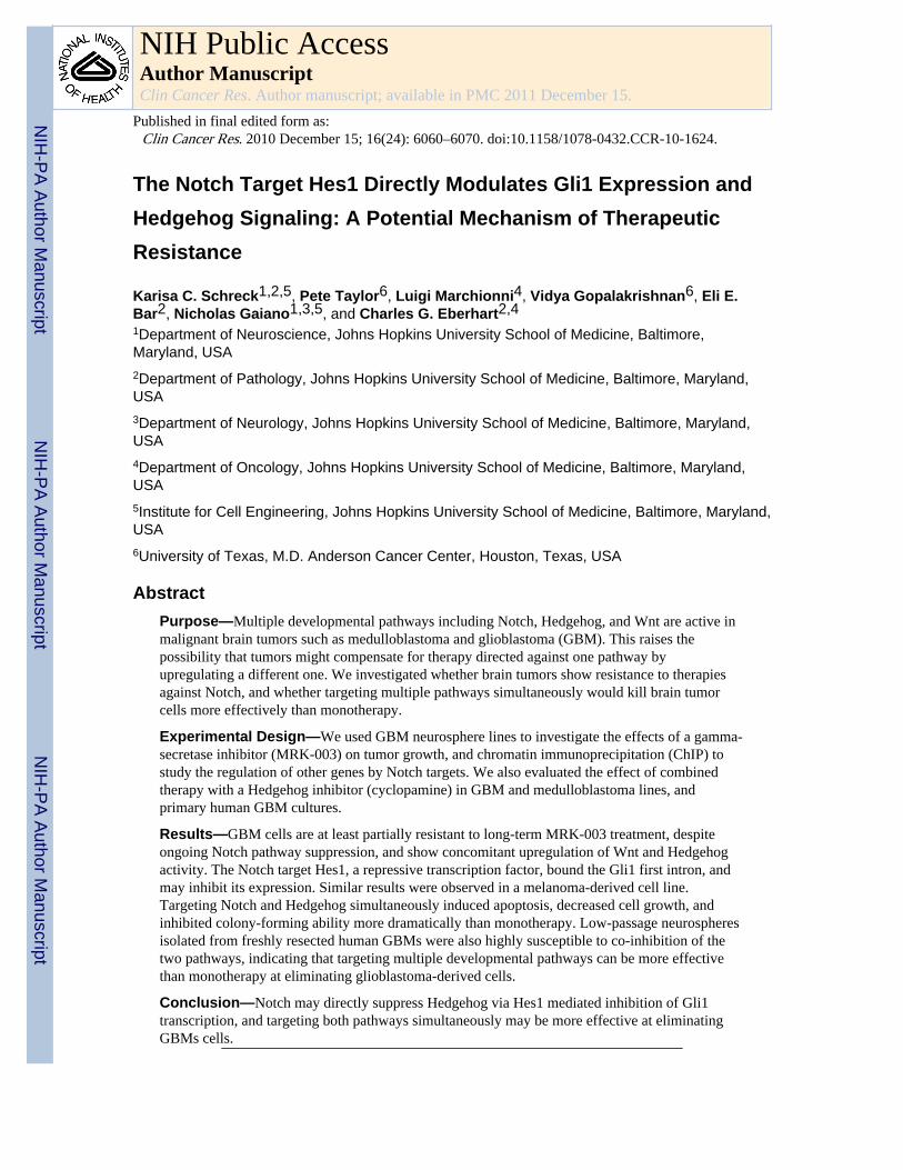
The Notch Target Hes1 Directly Modulates Gli1 Expression and Hedgehog Signaling: A Potential Mechanism of Therapeutic Resistance Karisa C. Schreck 1,2,5 , Pete Taylor 6 , Luigi Marchionni 4 , Vidya Gopalakrishnan 6 , Eli E. Bar 2 , Nicholas Gaiano 1,3,5 , and Charles G. Eberhart 2,4 1 Department of Neuroscience, Johns Hopkins University School of Medicine, Baltimore, Maryland, USA 2 Department of Pathology, Johns Hopkins University School of Medicine, Baltimore, Maryland, USA 3 Department of Neurology, Johns Hopkins University School of Medicine, Baltimore, Maryland, USA 4 Department of Oncology, Johns Hopkins University School of Medicine, Baltimore, Maryland, USA 5 Institute for Cell Engineering, Johns Hopkins University School of Medicine, Baltimore, Maryland, USA 6 University of Texas, M.D. Anderson Cancer Center, Houston, Texas, USA Abstract Purpose—Multiple developmental pathways including Notch, Hedgehog, and Wnt are active in malignant brain tumors such as medulloblastoma and glioblastoma (GBM). This raises the possibility that tumors might compensate for therapy directed against one pathway by upregulating a different one. We investigated whether brain tumors show resistance to therapies against Notch, and whether targeting multiple pathways simultaneously would kill brain tumor cells more effectively than monotherapy. Experimental Design—We used GBM neurosphere lines to investigate the effects of a gamma- secretase inhibitor (MRK-003) on tumor growth, and chromatin immunoprecipitation (ChIP) to study the regulation of other genes by Notch targets. We also evaluated the effect of combined therapy with a Hedgehog inhibitor (cyclopamine) in GBM and medulloblastoma lines, and primary human GBM cultures. Results—GBM cells are at least partially resistant to long-term MRK-003 treatment, despite ongoing Notch pathway suppression, and show concomitant upregulation of Wnt and Hedgehog activity. The Notch target Hes1, a repressive transcription factor, bound the Gli1 first intron, and may inhibit its expression. Similar results were observed in a melanoma-derived cell line. Targeting Notch and Hedgehog simultaneously induced apoptosis, decreased cell growth, and inhibited colony-forming ability more dramatically than monotherapy. Low-passage neurospheres isolated from freshly resected human GBMs were also highly susceptible to co-inhibition of the two pathways, indicating that targeting multiple developmental pathways can be more effective than monotherapy at eliminating glioblastoma-derived cells. Conclusion—Notch may directly suppress Hedgehog via Hes1 mediated inhibition of Gli1 transcription, and targeting both pathways simultaneously may be more effective at eliminating GBMs cells. NIH Public Access Author Manuscript Clin Cancer Res. Author manuscript; available in PMC 2011 December 15. Published in final edited form as: Clin Cancer Res. 2010 December 15; 16(24): 6060–6070. doi:10.1158/1078-0432.CCR-10-1624. NIH-PA Author Manuscript NIH-PA Author Manuscript NIH-PA Author Manuscript

Welcome message from author
This document is posted to help you gain knowledge. Please leave a comment to let me know what you think about it! Share it to your friends and learn new things together.
Transcript
The Notch Target Hes1 Directly Modulates Gli1 Expression andHedgehog Signaling: A Potential Mechanism of TherapeuticResistance
Karisa C. Schreck1,2,5, Pete Taylor6, Luigi Marchionni4, Vidya Gopalakrishnan6, Eli E.Bar2, Nicholas Gaiano1,3,5, and Charles G. Eberhart2,41Department of Neuroscience, Johns Hopkins University School of Medicine, Baltimore,Maryland, USA2Department of Pathology, Johns Hopkins University School of Medicine, Baltimore, Maryland,USA3Department of Neurology, Johns Hopkins University School of Medicine, Baltimore, Maryland,USA4Department of Oncology, Johns Hopkins University School of Medicine, Baltimore, Maryland,USA5Institute for Cell Engineering, Johns Hopkins University School of Medicine, Baltimore, Maryland,USA6University of Texas, M.D. Anderson Cancer Center, Houston, Texas, USA
AbstractPurpose—Multiple developmental pathways including Notch, Hedgehog, and Wnt are active inmalignant brain tumors such as medulloblastoma and glioblastoma (GBM). This raises thepossibility that tumors might compensate for therapy directed against one pathway byupregulating a different one. We investigated whether brain tumors show resistance to therapiesagainst Notch, and whether targeting multiple pathways simultaneously would kill brain tumorcells more effectively than monotherapy.
Experimental Design—We used GBM neurosphere lines to investigate the effects of a gamma-secretase inhibitor (MRK-003) on tumor growth, and chromatin immunoprecipitation (ChIP) tostudy the regulation of other genes by Notch targets. We also evaluated the effect of combinedtherapy with a Hedgehog inhibitor (cyclopamine) in GBM and medulloblastoma lines, andprimary human GBM cultures.
Results—GBM cells are at least partially resistant to long-term MRK-003 treatment, despiteongoing Notch pathway suppression, and show concomitant upregulation of Wnt and Hedgehogactivity. The Notch target Hes1, a repressive transcription factor, bound the Gli1 first intron, andmay inhibit its expression. Similar results were observed in a melanoma-derived cell line.Targeting Notch and Hedgehog simultaneously induced apoptosis, decreased cell growth, andinhibited colony-forming ability more dramatically than monotherapy. Low-passage neurospheresisolated from freshly resected human GBMs were also highly susceptible to co-inhibition of thetwo pathways, indicating that targeting multiple developmental pathways can be more effectivethan monotherapy at eliminating glioblastoma-derived cells.
Conclusion—Notch may directly suppress Hedgehog via Hes1 mediated inhibition of Gli1transcription, and targeting both pathways simultaneously may be more effective at eliminatingGBMs cells.
NIH Public AccessAuthor ManuscriptClin Cancer Res. Author manuscript; available in PMC 2011 December 15.
Published in final edited form as:Clin Cancer Res. 2010 December 15; 16(24): 6060–6070. doi:10.1158/1078-0432.CCR-10-1624.
NIH
-PA Author Manuscript
NIH
-PA Author Manuscript
NIH
-PA Author Manuscript

IntroductionGlioblastoma (GBM) is the most common malignant primary central nervous system tumorin adults and is characterized by resistance to chemo- and radiotherapy (1). Prognosisremains very poor, with most patients surviving less than two years (2) despite recentadvances in surgery and chemotherapy. It has become clear that GBMs are a diverse groupof tumors, with different subtypes activating distinct sets of oncogenes and signalingpathways (3). Because of this, no single therapy is likely to be effective against all GBMs,and a number of pharmacologic agents with activity against specific targets such as EGFR,Akt, Hedgehog, mTOR, PI3K, PDGFR, Raf, TGF-β are being developed (4). However, eventhe use of targeted therapies can be limited by the emergence of resistant tumor cells, andresistance to EGFR inhibitors (5) and Hedgehog inhibitors (6) has already been documented.
An important developmental pathway required in at least a subset of GBMs is Notch.Aberrant Notch signaling was implicated in the initiation of T-cell lymphoblastic leukemiain the early 1990s (7), and has since been demonstrated in many different hematopoietic andepithelial tumors (8-10). Upregulation of Notch pathway components has been demonstratedin GBM (11-13) as well as the malignant embryonal tumor medulloblastoma (14,15), andNotch pathway inhibition has emerged as a potential therapy for malignant brain tumors.The four Notch receptors (Notch 1-4) bind ligands (Jagged and Delta) expressed on adjacentcells, permitting cleavage of Notch via ADAM metalloprotease and then gamma-secretase(16). The released intracellular domain of Notch (ICD) translocates to the nucleus, where itbinds CBF-1/RBP-J and promotes transcription of the Hes/Hey genes which help maintain aprogenitor-like state by repressing transcription of pro-differentiation genes duringdevelopment (17,18).
Many different techniques for Notch blockade have been attempted, including gamma-secretase inhibitors (GSI) (19), siRNA (12), monoclonal antibodies (20-22), and smallinhibitory molecules directly affecting the transcriptional complex (23). siRNA and GSIshave been tested in the context of malignant brain tumors (12,13,19,24) with promisingresults in vitro and in xenograft models. Over twenty Phase I/II clinical trials investigatingthe efficacy of GSIs in tumors are actively recruiting or awaiting activation(www.clinicaltrials.gov), but it is uncertain whether inhibition of Notch signaling alone willbe sufficient to prevent tumor growth as cancer adaptation is well-documented. We assessedthe effects of Notch inhibition on malignant brain tumor cells and the potential emergence oftherapeutic resistance. Some GBM neurosphere lines that survived long-term Notchinhibition upregulated Wnt and Hedgehog, with the latter effect due potentially to Hes1binding and inhibiting Gli1 at the transcriptional level. We found that inhibiting Notch andHedgehog simultaneously dramatically decreased growth of neurosphere cultures andprimary human GBM cells, suggesting this regulatory mechanism may contribute toresistance.
Materials and MethodsCell Culture
DAOY, PFSK, U87, 22RV1, H157, KMS12, L428, Mel10, Reh, TOV-112D, and U937were maintained in the recommended media with 10% fetal bovine serum (FBS) unlessotherwise specified. HSR-GBM1 and HSR-GBM2 were maintained as neurosphere culturesin serum-free neurosphere media (25). Cell line identity was verified using SNP analysis.For all assays, cells were counted using GUAVA Viacount reagent as per manufacturer’sinstructions (#4000-0040, Millipore, Billerica, MA) and equal numbers of viable cells wereused for all experiments.
Schreck et al. Page 2
Clin Cancer Res. Author manuscript; available in PMC 2011 December 15.
NIH
-PA Author Manuscript
NIH
-PA Author Manuscript
NIH
-PA Author Manuscript
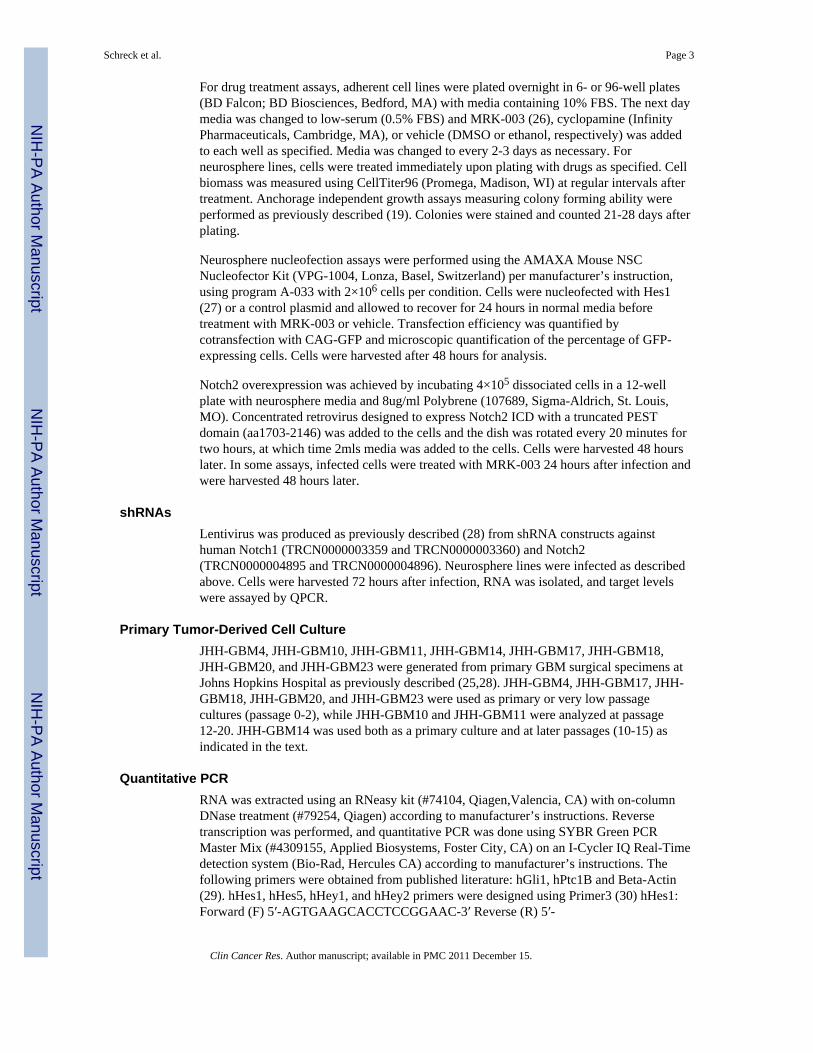
For drug treatment assays, adherent cell lines were plated overnight in 6- or 96-well plates(BD Falcon; BD Biosciences, Bedford, MA) with media containing 10% FBS. The next daymedia was changed to low-serum (0.5% FBS) and MRK-003 (26), cyclopamine (InfinityPharmaceuticals, Cambridge, MA), or vehicle (DMSO or ethanol, respectively) was addedto each well as specified. Media was changed to every 2-3 days as necessary. Forneurosphere lines, cells were treated immediately upon plating with drugs as specified. Cellbiomass was measured using CellTiter96 (Promega, Madison, WI) at regular intervals aftertreatment. Anchorage independent growth assays measuring colony forming ability wereperformed as previously described (19). Colonies were stained and counted 21-28 days afterplating.
Neurosphere nucleofection assays were performed using the AMAXA Mouse NSCNucleofector Kit (VPG-1004, Lonza, Basel, Switzerland) per manufacturer’s instruction,using program A-033 with 2×10⁶ cells per condition. Cells were nucleofected with Hes1(27) or a control plasmid and allowed to recover for 24 hours in normal media beforetreatment with MRK-003 or vehicle. Transfection efficiency was quantified bycotransfection with CAG-GFP and microscopic quantification of the percentage of GFP-expressing cells. Cells were harvested after 48 hours for analysis.
Notch2 overexpression was achieved by incubating 4×10⁵ dissociated cells in a 12-wellplate with neurosphere media and 8ug/ml Polybrene (107689, Sigma-Aldrich, St. Louis,MO). Concentrated retrovirus designed to express Notch2 ICD with a truncated PESTdomain (aa1703-2146) was added to the cells and the dish was rotated every 20 minutes fortwo hours, at which time 2mls media was added to the cells. Cells were harvested 48 hourslater. In some assays, infected cells were treated with MRK-003 24 hours after infection andwere harvested 48 hours later.
shRNAsLentivirus was produced as previously described (28) from shRNA constructs againsthuman Notch1 (TRCN0000003359 and TRCN0000003360) and Notch2(TRCN0000004895 and TRCN0000004896). Neurosphere lines were infected as describedabove. Cells were harvested 72 hours after infection, RNA was isolated, and target levelswere assayed by QPCR.
Primary Tumor-Derived Cell CultureJHH-GBM4, JHH-GBM10, JHH-GBM11, JHH-GBM14, JHH-GBM17, JHH-GBM18,JHH-GBM20, and JHH-GBM23 were generated from primary GBM surgical specimens atJohns Hopkins Hospital as previously described (25,28). JHH-GBM4, JHH-GBM17, JHH-GBM18, JHH-GBM20, and JHH-GBM23 were used as primary or very low passagecultures (passage 0-2), while JHH-GBM10 and JHH-GBM11 were analyzed at passage12-20. JHH-GBM14 was used both as a primary culture and at later passages (10-15) asindicated in the text.
Quantitative PCRRNA was extracted using an RNeasy kit (#74104, Qiagen,Valencia, CA) with on-columnDNase treatment (#79254, Qiagen) according to manufacturer’s instructions. Reversetranscription was performed, and quantitative PCR was done using SYBR Green PCRMaster Mix (#4309155, Applied Biosystems, Foster City, CA) on an I-Cycler IQ Real-Timedetection system (Bio-Rad, Hercules CA) according to manufacturer’s instructions. Thefollowing primers were obtained from published literature: hGli1, hPtc1B and Beta-Actin(29). hHes1, hHes5, hHey1, and hHey2 primers were designed using Primer3 (30) hHes1:Forward (F) 5′-AGTGAAGCACCTCCGGAAC-3′ Reverse (R) 5′-
Schreck et al. Page 3
Clin Cancer Res. Author manuscript; available in PMC 2011 December 15.
NIH
-PA Author Manuscript
NIH
-PA Author Manuscript
NIH
-PA Author Manuscript

TCACCTCGTTCATGCACTC-3′ hHes5: Forward (F) 5′-CCGGTGGTGGAGAAGATG-3′Reverse (R) 5′-TAGTCCTGGTGCAGGCTCTT-3′ hAxin2 primers were a generous giftfrom Brian Simons: Forward (F) 5′- CTGGTGGCTGGTGCAAAGAC-3′ Reverse (R) 5′-CGAGTGTGAGGTCCACGGAA-3′. The standard curve technique was used to determineexpression levels and values were normalized to beta actin.
Protein AnalysisProtein was extracted from cell pellets using RIPA buffer (R0278, Sigma-Aldrich) and 30ugwas run on each lane of a NuPage 4-12% Bis-Tris gel (NP0321, Invitrogen) according tomanufacturer’s instructions. The antibodies used were as follows: rabbit anti-Hes1 (1:400,AVIVA Systems Biology, San Diego CA, ARP32372; 1:1000, Toray Industries, Tebiro,Kamakura, Japan), rabbit monoclonal anti-Cleaved Notch1 (Cell Signaling, Danvers, MA,#2421 and #4147), and mouse monoclonal anti-GAPDH (1:50,000, Research DiagnosticsInc., Flanders NJ, RDI-TRK564-6C5).
Chromatin Immunoprecipitation (ChIP)ChIP was performed using two different techniques and antibodies. For the Magna ChIP kit(17-610, Millipore), cells were grown in the appropriate media, harvested during log-phasegrowth, crosslinked using 1% formaldehyde, and processed according to the manufacturer’sinstructions. The positive control was anti-Acetyl Histone H3, negative control was rabbitIgG, and Rabbit anti-Hes1 was used for pulldown (AVIVA Systems Biology). Non-quantitative PCR was performed using the primers in Supplemental Table 1. GAPDH andHes1 primer sets were used as negative and positive controls, respectively, as Hes1 haspreviously been shown to bind its own promoter (31, 32). Some samples were run usingquantitative PCR as well.
The other technique used for ChIP has been previously described (33). Briefly, cells weregrown in the appropriate media, harvested during log-phase growth, and crosslinked withformaldehyde. 5 μg of Hes1 antibody (AB5702, Millipore) or 5 μg control (rabbit IgG) wasadded to the sample and incubated for 12 hrs at 4°C. After washing, crosslinking wasreversed and quantitative PCR was performed as described above using the primers in Table1. p63 and p27Kip primer sets were used as negative and positive controls, respectively (34).Linear amplification of each primer set used for qPCR was verified by a standard curve.qPCR calculations were done as previously described (35). Briefly, the average of the cyclethreshold values (CTs) was calculated for each input, sample, and control. The input CT wassubtracted from the corresponding sample and control CTs. The following formula was thenapplied: power (1.9, negative ln (subtracted value)). This value was used for furthercalculations. Each sample and control was normalized by dividing both numbers by thehighest value so that each ChIP experiment was scaled from 0 to 1 and outliers wereremoved. For sample minus control values, a negative number was replaced with a zero.
Gene Expression AnalysisGene expression was measured using Agilent’s 44K whole human genome microarrays atthe Johns Hopkins Oncology Microarray Core, with labeling, hybridization, and detectionperformed according to manufacturer’s instructions (Agilent Technologies, Santa Clara,CA). Differential gene expression, gene set enrichment analyses, and Analysis of FunctionalAnnotation (AFA) were performed as previously described (36,37), using statisticalpackages from the R/Bioconductor project (38,39). Gene annotation for the microarray usedin this study was obtained from the corresponding R-Bioconductor metadata packages. Rawexpression data along with MIAME required information is located in the GEO database(40).
Schreck et al. Page 4
Clin Cancer Res. Author manuscript; available in PMC 2011 December 15.
NIH
-PA Author Manuscript
NIH
-PA Author Manuscript
NIH
-PA Author Manuscript
ResultsGBM Neurosphere Cultures Survive Long-term Notch Inhibition
To confirm previous reports (24,41) and examine Notch suppression in the newly derivedGBM neurosphere lines JHH-GBM10 and JHH-GBM14, we treated dissociatedneurospheres with MRK-003 and evaluated expression of the Notch pathway targets Hes1and Hes5. We also looked at an adherent, serum-cultured GBM-derived line, U87. Weobserved that MRK-003 treatment significantly decreased expression of Hes1 in all lines,with inhibition ranging from 32% (JHH-GBM10) to 57% (JHH-GBM14; Fig. 1A).Significant reductions in the expression of Hes5 ranging from 87% (HSR-GBM1) to 95%(JHH-GBM14; Fig. 1A) were also noted in three GBM neurosphere lines. Interestingly, U87and HSR-GBM2 did not express Hes5 at sufficient levels for quantitation, indicating someheterogeneity in Notch target levels between tumors. MRK-003 treatment also inhibitedHes1 expression by 40% in the medulloblastoma cell line DAOY (Supplemental Fig. 1A). Inaddition to downregulation of Hes1 transcripts, Hes1 protein levels were suppressed byMRK-003 treatment in HSR-GBM1 (Fig. 1B) and DAOY (Supplemental Fig. 1B), with70-80% inhibition following 2 μM doses (P<0.0001). Cleaved Notch1 levels also decreaseddramatically with MRK-003 treatment, dropping to almost undetectable levels (Fig. 1B) asmeasured by two different antibodies. We confirmed dose-dependent inhibition of HSR-GBM1 and DAOY cell growth following application of MRK-003 in relatively short termsix-to-nine day growth assays (Fig. 1C and Supplemental Fig. 1C).
Interestingly, although neurosphere lines HSR-GBM1 and HSR-GBM2 showed a short-termresponse to Notch inhibition, they continued to grow even after three or more passages withcontinuous MRK-003 treatment (Fig. 1D and data not shown). These experiments wereperformed three or more times with similar results, indicating the capacity for long-termgrowth was a reproducible phenomenon. Notch signaling was still quite pronounced at theend of long-term MRK-003 treatment as demonstrated by very low Hes1 and Hes5 levels(Fig. 1D), suggesting that ongoing growth was not due to reactivation of the Notch pathway.Higher concentrations of MRK-003 (2-10 μM) did result in complete cessation of cellgrowth over the course of 10-14 days, indicating that at sufficiently high drug levels cells donot easily develop resistance.
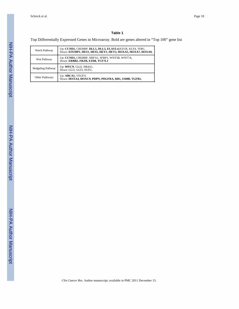
Up-regulation of Hedgehog and Wnt Signaling Following Notch InhibitionGiven the ability of a subset of malignant brain tumor cells to survive long term MRK-003treatment, we performed a microarray assay on four GBM-derived neurosphere lines withtwo different doses of MRK-003 to identify Notch targets and determine if other pathwaysmight be upregulated to compensate for the loss of Notch activity (HSR-GBM1, N=3 eachfor two doses; HSR-GBM2, N=3; JHH-GBM10, N=3; and JHH-GBM14, N=2). Asexpected, canonical Notch targets including Hes1, Hes5, and Hey1 were suppressed 2.7-28fold following MRK-003 treatment. A list of the top differentially regulated genes seen incommon across all lines is included in Supplemental Table 2. We performed a gene setenrichment analysis (GSEA) comparing our data against literature-cited, manually curatedpathway lists with genes up- and down-regulated by pathway activation and found that, asexpected, Notch was significantly changed (P = 0.013). We found that two otherdevelopmental and proliferative pathways were significantly altered: Wnt (P = 0.00013) andHedgehog (P = 0.0027; Table 1). Closer analysis of specific Hedgehog and Wnt targetsindicated that there was an overall trend towards upregulation of canonical targets andpathway components in the presence of MRK-003.
Since Wnt and Hedgehog play an important role in stem cell maintenance and proliferationduring normal brain development and can be dysregulated in tumors, we hypothesized that
Schreck et al. Page 5
Clin Cancer Res. Author manuscript; available in PMC 2011 December 15.
NIH
-PA Author Manuscript
NIH
-PA Author Manuscript
NIH
-PA Author Manuscript
upregulation of these pathways might compensate for MRK-003 induced loss of Notchsignaling. Using QPCR to validate our microarray data, we found that the Hedgehog targetsPatched1B and Gli1 were both significantly upregulated by up to two-fold in neurospherelines following MRK-003 treatment (2-5 μM) (Fig. 2A). The Wnt signaling target Axin2also showed significant upregulation (Fig. 2A). In contrast, medulloblastoma cell lineDAOY showed a 40% decrease in Ptc1B mRNA and no significant change in Gli1 or Axin2(Supplemental Fig. 1D).
As GSIs are able to cleave proteins other than Notch, we used shRNA to confirm the effectof Notch inhibition on Hedgehog signaling. Infection of HSR-GBM1 neurospheres withlentivirus encoding shRNA targeting either Notch1 or Notch2 resulted in significantreductions in receptor mRNA levels (data not shown). However, inhibiting either Notch1 orNotch2 alone resulted in an overall increase in Notch signaling as evidenced by upregulationof the Notch targets Hes1 and Hes5, suggesting compensation by other Notch receptors(data not shown). We therefore simultaneously used shRNAs against both Notch1 andNotch2, and saw a decrease in mRNA encoding the two Notch receptors of approximately30-50%, along with a decrease in Notch targets by a similar amount (Fig. 2A). Inhibition ofNotch signaling using shNotch1 and shNotch2 induced Gli1 expression by approximatelytwo-fold (Fig. 2A; P = 0.02).
Hes1 Regulates Hedgehog Signaling via Direct Binding to Gli1As previous work has suggested the potential for crosstalk between the Notch and Hedgehogsignaling cascades (14), we focused on the upregulation of Hedgehog signaling in responseto MRK-003. Increased levels of Notch pathway components are seen in Hedgehog-drivenmedulloblastoma models (15,42) and it was initially suggested that these tumors may bedependent on Notch signaling for survival (15). More recent studies however, indicate thatHedgehog-driven medulloblastomas can grow in the absence of canonical Notch activity(43,44). Additionally, several groups have demonstrated that Hedgehog pathwaycomponents Gli1 and Gli2 are able to positively regulate Hes1 independently of Notch(45,46). However, when we treated several GBM neurosphere lines with the Hedgehoginhibitor cyclopamine we did not observe decreases in Hes1 or other Notch pathway targets,suggesting that Hedgehog does not play a significant role regulating Notch targets inmalignant gliomas (Supplemental Fig. 2A).
We investigated the possibility of direct Gli1 regulation by Hes1, as potential Hes1 bindingsites were previously identified in the first Gli1 intron using in silico analysis (47). Hes1negatively regulates transcription of targets, thus loss of Hes1 following Notch blockadewould be predicted to relieve repression of Gli1 expression and activate the Hedgehogcascade (48). To investigate whether this mechanism occurs in brain tumors, we usedchromatin immunoprecipitation (ChIP) to examine whether Hes1 bound to the five N-boxespresent in the Gli1 first intron (Fig. 2B). In HSR-GBM1, we found using quantitative PCRthat Hes1 interacted with all five N-boxes as strongly as with a site in the p27 promoterpreviously shown to be bound by Hes1 as a positive control (34). We also used anindependent set of PCR primers and an alternate ChIP protocol to confirm binding to thefive N-boxes in a different neurosphere line, JHH-GBM10 (Supplemental Fig. 2B). Incontrast to the GBM lines, Hes1 did not bind N-box 1, 4, or 5 in DAOY cells, and showedonly moderate affinity for N-boxes 2 and 3, demonstrating the degree of binding to these N-boxes is context dependent (Supplemental Fig. 2C). This is consistent with our expressiondata demonstrating that Hedgehog pathway targets Gli1 and Ptch1B increase in response toNotch pathway inhibition in HSR-GBM1, but show no change in response to MRK-003 inDAOY. Finally, we evaluated three primary human GBM cultures (passage 0), and foundthat Hes1 bound the Gli1 first intron Nboxes in one of these (Fig. 2C), suggesting that ourfinding can be expanded to include some primary GBMs.
Schreck et al. Page 6
Clin Cancer Res. Author manuscript; available in PMC 2011 December 15.
NIH
-PA Author Manuscript
NIH
-PA Author Manuscript
NIH
-PA Author Manuscript
When we treated neurosphere lines with MRK-003, Hes1 binding to the Gli1 first intron wasreduced, suggesting decreased ability to repress Gli1 expression (Fig. 2B and SupplementalFig. 2C). Hes1 also bound less to its own promoter at a site of auto-regulation previouslydocumented (31,32), indicating that Hes1 binding may be globally decreased. Thesefindings suggest a direct negative regulatory relationship between Notch and Hedgehogsignaling in some GBMs, whereby cells can upregulate Hedgehog signaling in response topharmacological Notch pathway inhibition.
To help confirm the presence of such a regulatory mechanism, we looked at the effect ofNotch pathway activation on Gli1 and other Hedgehog pathway targets. We infected HSR-GBM1 with retrovirus expressing activated Notch2 (NICD2) or control retrovirus andevaluated Notch and Hedgehog pathway activity. Consistent with our model, we found thatNotch signaling targets were upregulated (Supplemental Fig. 2D) while the Hedgehogtargets Gli1 and Ptc1B were significantly downregulated by 50% and 30%, respectively(P=0.007 and 0.002; Fig. 2D). Experiments using retrovirus encoding activated Notch1 orNotch3 gave very similar results (data not shown). Next, we sought to determine if the effectof Notch overexpression could be mediated through Hes1. After nucleofecting HSR-GBM1with Hes1 or a control plasmid, we found a significant 30% reduction (P=0.02) of Gli1 inresponse to Hes1 overexpression (Fig 2D). The modest decreased we observed may be dueto limited transfection efficiency (30-50%), as it is difficult to introduce plasmids intoneurosphere lines. We sought to further confirm that the upregulation in Gli1 withMRK-003 treatment was due to Notch pathway inhibition rather than other effects of theGSI by treating cells with MRK-003 one day after infecting them with control or NICD2-expressing retrovirus. While Gli1 levels increased in response to MRK-003 with the controlvirus, they remained at baseline when NICD2 was present (Fig. 2D), suggesting that Notch2activity is able to rescue the inhibition-effect of MRK-003. Taken together, our data suggestthat increased expression of Hedgehog targets following Notch blockade is due at least inpart to the loss of Hes1 binding at the Gli1 locus (Fig. 2D).
Hedgehog Inhibition by Notch Is Seen in Other CancersRegulation of Gli1 expression by Hes1 has not been previously reported, and we wanted todetermine whether other cancer types demonstrated a similar feedback mechanism. Wescreened eight cell lines derived from prostate, lung, ovarian, skin, and hematopoetic cellcancers (22RV1, H157, KMS12, L428, Mel10, Reh, TOV-112D, U937) and found thatNotch signaling was inhibited by MRK-003 in two. TOV-112D, an ovarian-derived line,showed a 60% decrease in Hes1 expression upon treatment with 2μM MRK-003 (P=0.0002)and Mel10, a melanoma-derived line, showed a more moderate decrease in Hes1 expressionof 20% (P=0.002). Interestingly, only Mel10 showed an upregulation in Gli1 and Ptc1B withMRK-003 treatment (P=0.031 and P=0.05, respectively; Fig. 3A). Axin2 remainedunchanged in both cell lines following MRK-003 introduction. Chromatinimmunoprecipitation showed that Hes1 consistently (N=3) bound the Gli1 N-boxes 2 and 3in Mel10 (Fig. 3B), along with other N-boxes to a varying degree (data not shown).
Co-inhibition of Notch and Hedgehog Decreases Growth and ClonogenicityWe next evaluated whether inhibiting Hedgehog signaling in addition to Notch wouldaugment anti-tumor effects in GBM neurosphere lines. A Hedgehog antagonist(cyclopamine) and MRK-003 were administered to HSR-GBM1 alone or together in orderto detect potential interactions. We observed that cell growth over eleven days decreasedslightly with MRK-003 or cyclopamine monotherapy as compared to vehicle, but decreasedby approximately 90% in the presence of both drugs (Fig. 3C). Despite the absence of Hes1binding to the Gli1 intron in DAOY, similar results were seen with co-treatment(Supplemental Fig. 3A). Because the main effect seemed to be accumulation of dead cells,
Schreck et al. Page 7
Clin Cancer Res. Author manuscript; available in PMC 2011 December 15.
NIH
-PA Author Manuscript
NIH
-PA Author Manuscript
NIH
-PA Author Manuscript
rather than elongation of processes suggesting differentiation, we performed an Annexin Vassay to evaluate the percentage of apoptotic cells. Doing so, we found that the percentageof apoptotic cells doubled (13%) with co-treatment as compared to either drug alone (7%;Fig. 3D).
We next investigated whether co-treatment would decrease the number of clonogenic cellsusing an anchorage independent growth assay. HSR-GBM1, HSR-GBM2, and U87 weredissociated to single-cells, plated in soft agar, and treated continuously with vehicle,monotherapy, or co-treatment. Co-treatment was significantly more effective thanmonotherapy for the latter two lines, with up to 90% reduction in colony number ascompared to cyclopamine alone, and up to 50% reduction as compared to MRK-003 alone(Fig. 4A-C). For HSR-GBM1, the difference between MRK-003 alone and MRK-003 pluscyclopamine was more modest and not statistically significant (P=0.077), although this trendwas repeatable across independent experiments (N=3).
To assess our ability to specifically deplete clonogenic cells, we examined if pre-treatmentwould lead to a more pronounced reduction in colony formation upon co-treatment.Neurosphere cultures were grown with treatment for seven days in suspension, and thenwashed and equal numbers of viable cells plated in soft agar. Despite no further drugtreatment, the number of colonies in the co-treatment cohort was reduced by 90% ascompared with vehicle or cyclopamine, and by 50% as compared with MRK-003 alone (Fig.4B). This shows that colony-forming cells are more sensitive to combined drug treatmentand are eliminated from the population somewhat selectively. We also assessed expressionof CD133, a putative tumor stem-cell marker, via FACS in HSR-GBM1 and DAOY treatedfor two days with vehicle, monotherapy, or both drugs. However, in these two lines co-treatment did not decrease the percentage of CD133-positive cells to a greater degree thanmonotherapy (data not shown). This may reflect the imperfect ability of CD133 alone toprospectively identify stem-like clonogenic cells, a fact highlighted in several recent reports(49).
Primary Human-Derived GBMs Respond Strongly to Co-Inhibition of Notch and HedgehogAs a final test for the efficacy of combined inhibition, we treated several freshly resectedprimary human GBMs cultured in neurosphere media. First, we investigated whetherMRK-003 was able to inhibit Notch signaling in primary GBM cultures and found that bothHes1 and Hes5 were decreased in primary GBM-derived low-passage neurosphere cultures(Supplemental Fig. 3B), and that baseline expression of Notch and Hedgehog signaling wassimilar to that of GBM neurosphere cultures. Next, we looked at the ability of co-treatmentto prevent colony formation. We dissociated the tumors, plated them in serum-freeneurosphere media, and treated the primary cultures. While some variation was observed,most likely due to the heterogeneous nature of clinical samples, overall we saw a dramaticdecrease in the number and size of spheres forming over a period of 2-4 weeks in the co-treatment cohort as compared with either monotherapy or vehicle (Fig. 5A). Similar resultswere seen in primary passages from three GBMs (JHH-GBM17, JHH-GBM18, and JHH-GBM20) and in passage two cells from JHH-GBM14. A significant decrease of 50-80% inthe number of colonies formed was seen in the lines with sufficient material for multiplereplicates (Fig. 5B). The fourth line, JHH-GBM20, showed results similar to JHH-GBM14,but the assay was only performed once due to limited starting material. We also observed adecrease in the average size of tumor neurospheres formed in co-treated cultures ascompared to vehicle or monotherapy, (P<0.05), suggesting a limited proliferative capacity inremaining cells (Fig. 5C).
Schreck et al. Page 8
Clin Cancer Res. Author manuscript; available in PMC 2011 December 15.
NIH
-PA Author Manuscript
NIH
-PA Author Manuscript
NIH
-PA Author Manuscript
DiscussionGBMs are highly resistant to current treatments such as chemotherapy and radiation, and adeeper understanding of GBM biology is necessary for effective tumor elimination. In lightof this, large-scale efforts characterizing genetic alterations in GBM have demonstrated thatGBM subtypes exist and suggested that multiple pathways could be targeted to treat tumorsmore effectively (3,50,51). We investigated whether resistance could emerge in response totreatment with a Notch inhibitor in vitro, and found a significant number of cells were ableto grow in the presence of long-term MRK-003 treatment. We found Hedgehog signalingwas up-regulated in response to Notch inhibition and that direct interaction between Hes1and Gli1 occurs in GBM neurosphere lines and primary GBM samples. This interactioncould account for the increase in Hedgehog signaling during Notch suppression and is apotential mechanism of resistance. We demonstrated that targeting both Notch andHedgehog simultaneously increases apoptosis and inhibits colony-forming ability moredramatically than either monotherapy. Moreover, freshly dissected human GBMs are alsohighly susceptible to co-inhibition. These findings indicate that targeting both pathways ismore effective than monotherapy at eliminating GBM cells in some neurospheres in vitro,and that suggest that co-treatment should be considered in patients after additionalpreclinical in vivo studies are performed.
The finding that Hes1 binds to the Gli1 locus and may regulate its expression at atranscriptional level is both novel and unexpected. The fact that only a subset of cell lines,primary GBM specimens, and cancer types exhibit this relationship suggests other factors,such as binding partners, heterochromatin structure, or methylation, may also be important.We investigated whether inverse expression levels between Hes1 and Gli1 were seen inhuman GBMs by analyzing public databases (www.rembrant.org) and previously publisheddata sets (52), but found no significant relationship between the two (data not shown). Thismay be due to the fact that this regulatory relationship is only present in a subset of humanGBMs. The molecular mechanism and functional role of this feedback mechanism in cancerrequires further investigation, as does the question of whether this regulatory mechanismoccurs in normal development.
In summary, co-treatment with Hedgehog and Notch inhibitors clearly increases cell deathand decreases colony forming ability in vitro, suggesting the potential as a combinatorialchemotherapy agent. In vivo studies will be required to evaluate whether or not this is afeasible treatment in mouse xenografts prior to any clinical trials. A study evaluatingHedgehog and Notch co-inhibition in advanced breast cancer is currently underway(NCT01071564) and may pave the road for determining the safety of co-treatment. Our datasupports the increasing awareness of cell signaling complexity in tumors, and the potentialfor adaptation and evasion following individual pathway blockade. Further evaluation of themolecular links between the Notch, Hedgehog and Wnt pathways seems indicated, andsuccessful tumor elimination may require broader approaches targeting several pathways.
Statement of Translational Relevance
The emergence of therapeutic resistance is a significant concern when targeting manysignaling pathways and tumor types. We found that in vitro Notch pathway blockade inglioblastoma (GBM) cells using a gamma-secretase inhibitor (GSI) led to increasedactivity in two other pathways important for neural development—Wnt and Hedgehog.The Notch target Hes1, a transcriptional repressor, can directly bind the first Gli1 intron,suggesting a mechanism by which Notch can inhibit Hedgehog activity. Inhibition ofboth Notch and Hedgehog in vitro dramatically decreased the growth of GBM cell lines,as well as low-passage neurospheres derived from primary human tumors. These findings
Schreck et al. Page 9
Clin Cancer Res. Author manuscript; available in PMC 2011 December 15.
NIH
-PA Author Manuscript
NIH
-PA Author Manuscript
NIH
-PA Author Manuscript
demonstrate that Notch-targeted therapeutics can lead to alterations in otherdevelopmental signaling cascades which promote tumor survival, and suggest thatcombined treatment with Hedgehog pathway inhibitors may be able to increase theefficacy of GSIs in some cancer patients.
Supplementary MaterialRefer to Web version on PubMed Central for supplementary material.
AcknowledgmentsWe would like to thank the following for reagents: Ryoichiro Kageyama and Tetso Sudo (Hes1 antibody),Madeleine Carreau (Hes1 plasmid), Angelo Vescovi (HSR-GBM1 and HSR-GBM2), Craig Peacock and ZhenhuaHuang (adherent cell lines), the JHU Genetic Core (shRNAs). Thanks to Naheed Gul and Michael Coonfield fortechnical assistance. We would like to acknowledge grant support to Dr. Eberhart from NIH/NS55089 and theBrain Tumor Funders Collaborative, and to Dr. Golpakrishnan from the American Society Grant RSG-090273-01-DDC and the Brain Tumor Spore Grant 5P50CA127001-02. Additional thanks to Merck and its scientists forproviding MRK-003.
References1. Louis, DN.; International Agency for Research on Cancer. WHO classification of tumours of the
central nervous system. 4 th ed.. International Agency for Research on Cancer; Lyon: 2007.2. Adamson C, Kanu OO, Mehta AI, et al. Glioblastoma multiforme: a review of where we have been
and where we are going. Expert Opin Investig Drugs. 2009; 18:1061–83.3. Phillips HS, Kharbanda S, Chen R, et al. Molecular subclasses of high-grade glioma predict
prognosis, delineate a pattern of disease progression, and resemble stages in neurogenesis. CancerCell. 2006; 9:157–73. [PubMed: 16530701]
4. Kanu OO, Mehta A, Di C, et al. Glioblastoma multiforme: a review of therapeutic targets. ExpertOpin Ther Targets. 2009; 13:701–18. [PubMed: 19409033]
5. Huang TT, Sarkaria SM, Cloughesy TF, Mischel PS. Targeted therapy for malignant gliomapatients: lessons learned and the road ahead. Neurotherapeutics. 2009; 6:500–12. [PubMed:19560740]
6. Yauch RL, Dijkgraaf GJ, Alicke B, et al. Smoothened mutation confers resistance to a Hedgehogpathway inhibitor in medulloblastoma. Science. 2009; 326:572–4. [PubMed: 19726788]
7. Ellisen LW, Bird J, West DC, et al. TAN-1, the human homolog of the Drosophila notch gene, isbroken by chromosomal translocations in T lymphoblastic neoplasms. Cell. 1991; 66:649–61.[PubMed: 1831692]
8. Bolos V, Grego-Bessa J, de la Pompa JL. Notch signaling in development and cancer. Endocr Rev.2007; 28:339–63. [PubMed: 17409286]
9. Aster JC, Pear WS, Blacklow SC. Notch signaling in leukemia. Annu Rev Pathol. 2008; 3:587–613.[PubMed: 18039126]
10. Koch U, Radtke F. Notch and cancer: a double-edged sword. Cell Mol Life Sci. 2007; 64:2746–62.[PubMed: 17687513]
11. Ignatova TN, Kukekov VG, Laywell ED, Suslov ON, Vrionis FD, Steindler DA. Human corticalglial tumors contain neural stem-like cells expressing astroglial and neuronal markers in vitro.Glia. 2002; 39:193–206. [PubMed: 12203386]
12. Purow BW, Haque RM, Noel MW, et al. Expression of Notch-1 and its ligands, Delta-like-1 andJagged-1, is critical for glioma cell survival and proliferation. Cancer Res. 2005; 65:2353–63.[PubMed: 15781650]
13. Kanamori M, Kawaguchi T, Nigro JM, et al. Contribution of Notch signaling activation to humanglioblastoma multiforme. J Neurosurg. 2007; 106:417–27. [PubMed: 17367064]
Schreck et al. Page 10
Clin Cancer Res. Author manuscript; available in PMC 2011 December 15.
NIH
-PA Author Manuscript
NIH
-PA Author Manuscript
NIH
-PA Author Manuscript
14. Solecki DJ, Liu XL, Tomoda T, Fang Y, Hatten ME. Activated Notch2 signaling inhibitsdifferentiation of cerebellar granule neuron precursors by maintaining proliferation. Neuron. 2001;31:557–68. [PubMed: 11545715]
15. Hallahan AR, Pritchard JI, Hansen S, et al. The SmoA1 mouse model reveals that notch signalingis critical for the growth and survival of sonic hedgehog-induced medulloblastomas. Cancer Res.2004; 64:7794–800. [PubMed: 15520185]
16. Louvi A, Artavanis-Tsakonas S. Notch signalling in vertebrate neural development. Nat RevNeurosci. 2006; 7:93–102. [PubMed: 16429119]
17. Kopan R, Ilagan MX. The canonical Notch signaling pathway: unfolding the activationmechanism. Cell. 2009; 137:216–33. [PubMed: 19379690]
18. Iso T, Kedes L, Hamamori Y. HES and HERP families: multiple effectors of the Notch signalingpathway. J Cell Physiol. 2003; 194:237–55. [PubMed: 12548545]
19. Fan X, Matsui W, Khaki L, et al. Notch pathway inhibition depletes stem-like cells and blocksengraftment in embryonal brain tumors. Cancer Res. 2006; 66:7445–52. [PubMed: 16885340]
20. Li K, Li Y, Wu W, et al. Modulation of Notch signaling by antibodies specific for the extracellularnegative regulatory region of NOTCH3. J Biol Chem. 2008; 283:8046–54. [PubMed: 18182388]
21. Hoey T, Yen WC, Axelrod F, et al. DLL4 blockade inhibits tumor growth and reduces tumor-initiating cell frequency. Cell Stem Cell. 2009; 5:168–77. [PubMed: 19664991]
22. Wu Y, Cain-Hom C, Choy L, et al. Therapeutic antibody targeting of individual Notch receptors.Nature. 464:1052–7. [PubMed: 20393564]
23. Moellering RE, Cornejo M, Davis TN, et al. Direct inhibition of the NOTCH transcription factorcomplex. Nature. 2009; 462:182–8. [PubMed: 19907488]
24. Fan X, Khaki L, Zhu TS, et al. Notch Pathway Blockade Depletes CD133-Positive GlioblastomaCells and Inhibits Growth of Tumor Neurospheres and Xenografts. Stem Cells. 2009
25. Galli R, Binda E, Orfanelli U, et al. Isolation and characterization of tumorigenic, stem-like neuralprecursors from human glioblastoma. Cancer Res. 2004; 64:7011–21. [PubMed: 15466194]
26. Lewis HD, Leveridge M, Strack PR, et al. Apoptosis in T cell acute lymphoblastic leukemia cellsafter cell cycle arrest induced by pharmacological inhibition of notch signaling. Chem Biol. 2007;14:209–19. [PubMed: 17317574]
27. Tremblay CS, Huang FF, Habi O, et al. HES1 is a novel interactor of the Fanconi anemia corecomplex. Blood. 2008; 112:2062–70. [PubMed: 18550849]
28. Bar EE, Lin A, Mahairaki V, Matsui W, Eberhart CG. Hypoxia increases the expression of stem-cell markers and promotes clonogenicity in glioblastoma neurospheres. Am J Pathol. 177:1491–502. [PubMed: 20671264]
29. Bar EE, Chaudhry A, Lin A, et al. Cyclopamine-mediated hedgehog pathway inhibition depletesstem-like cancer cells in glioblastoma. Stem Cells. 2007; 25:2524–33. [PubMed: 17628016]
30. Rosen, SaSHJ. Primer3 on the WWW for general users and for biologist programmers. HumanaPress; Totowa, NJ: 2000.
31. Takebayashi K, Sasai Y, Sakai Y, Watanabe T, Nakanishi S, Kageyama R. Structure, chromosomallocus, and promoter analysis of the gene encoding the mouse helix-loop-helix factor HES-1.Negative autoregulation through the multiple N box elements. J Biol Chem. 1994; 269:5150–6.[PubMed: 7906273]
32. Hirata H, Yoshiura S, Ohtsuka T, et al. Oscillatory expression of the bHLH factor Hes1 regulatedby a negative feedback loop. Science. 2002; 298:840–3. [PubMed: 12399594]
33. Aguilera DG, Das CM, Sinnappah-Kang ND, et al. Reactivation of death receptor 4 (DR4)expression sensitizes medulloblastoma cell lines to TRAIL. J Neurooncol. 2009; 93:303–18.[PubMed: 19148581]
34. Murata K, Hattori M, Hirai N, et al. Hes1 directly controls cell proliferation through thetranscriptional repression of p27Kip1. Mol Cell Biol. 2005; 25:4262–71. [PubMed: 15870295]
35. Aparicio O, Geisberg JV, Struhl K. Chromatin immunoprecipitation for determining theassociation of proteins with specific genomic sequences in vivo. Curr Protoc Cell Biol. 2004Chapter 17:Unit 17 7.
Schreck et al. Page 11
Clin Cancer Res. Author manuscript; available in PMC 2011 December 15.
NIH
-PA Author Manuscript
NIH
-PA Author Manuscript
NIH
-PA Author Manuscript
36. Daniel VC, Marchionni L, Hierman JS, et al. A primary xenograft model of small-cell lung cancerreveals irreversible changes in gene expression imposed by culture in vitro. Cancer Res. 2009;69:3364–73. [PubMed: 19351829]
37. Schaeffer EM, Marchionni L, Huang Z, et al. Androgen-induced programs for prostate epithelialgrowth and invasion arise in embryogenesis and are reactivated in cancer. Oncogene. 2008;27:7180–91. [PubMed: 18794802]
38. Gentleman RC, Carey VJ, Bates DM, et al. Bioconductor: open software development forcomputational biology and bioinformatics. Genome Biol. 2004; 5:R80. [PubMed: 15461798]
39. Ihaka R, Gentleman R. R: A language for data analysis and graphics. Journal of Computational andGraphical Statistics. 1996; 5:299–314.
40. Wheeler DL, Barrett T, Benson DA, et al. Database resources of the National Center forBiotechnology Information. Nucleic Acids Res. 2008; 36:D13–D21. [PubMed: 18045790]
41. Wang J, Wakeman TP, Lathia JD, et al. Notch promotes radioresistance of glioma stem cells. StemCells. 28:17–28. [PubMed: 19921751]
42. Dakubo GD, Mazerolle CJ, Wallace VA. Expression of Notch and Wnt pathway components andactivation of Notch signaling in medulloblastomas from heterozygous patched mice. J Neurooncol.2006; 79:221–7. [PubMed: 16598417]
43. Julian E, Dave RK, Robson JP, Hallahan AR, Wainwright BJ. Canonical Notch signaling is notrequired for the growth of Hedgehog pathway-induced medulloblastoma. Oncogene.
44. Hatton BA, Villavicencio EH, Pritchard J, et al. Notch signaling is not essential in sonic hedgehog-activated medulloblastoma. Oncogene.
45. Wall DS, Mears AJ, McNeill B, et al. Progenitor cell proliferation in the retina is dependent onNotch-independent Sonic hedgehog/Hes1 activity. J Cell Biol. 2009; 184:101–12. [PubMed:19124651]
46. Ingram WJ, McCue KI, Tran TH, Hallahan AR, Wainwright BJ. Sonic Hedgehog regulates Hes1through a novel mechanism that is independent of canonical Notch pathway signalling. Oncogene.2008; 27:1489–500. [PubMed: 17873912]
47. Katoh Y, Katoh M. Integrative genomic analyses on GLI1: positive regulation of GLI1 byHedgehog-GLI, TGFbeta-Smads, and RTK-PI3K-AKT signals, and negative regulation of GLI1by Notch-CSL-HES/HEY, and GPCR-Gs-PKA signals. Int J Oncol. 2009; 35:187–92. [PubMed:19513567]
48. Katoh Y, Katoh M. Hedgehog signaling pathway and gastrointestinal stem cell signaling network(review). Int J Mol Med. 2006; 18:1019–23. [PubMed: 17089004]
49. Chen R, Nishimura MC, Bumbaca SM, et al. A hierarchy of self-renewing tumor-initiating celltypes in glioblastoma. Cancer Cell. 17:362–75. [PubMed: 20385361]
50. Parsons DW, Jones S, Zhang X, et al. An integrated genomic analysis of human glioblastomamultiforme. Science. 2008; 321:1807–12. [PubMed: 18772396]
51. Comprehensive genomic characterization defines human glioblastoma genes and core pathways.Nature. 2008; 455:1061–8. [PubMed: 18772890]
52. Verhaak RG, Hoadley KA, Purdom E, et al. Integrated genomic analysis identifies clinicallyrelevant subtypes of glioblastoma characterized by abnormalities in PDGFRA, IDH1, EGFR, andNF1. Cancer Cell. 17:98–110. [PubMed: 20129251]
Schreck et al. Page 12
Clin Cancer Res. Author manuscript; available in PMC 2011 December 15.
NIH
-PA Author Manuscript
NIH
-PA Author Manuscript
NIH
-PA Author Manuscript
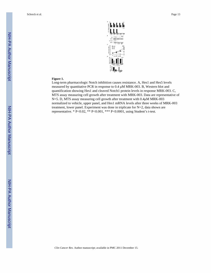
Figure 1.Long-term pharmacologic Notch inhibition causes resistance. A, Hes1 and Hes5 levelsmeasured by quantitative PCR in response to 0.4 μM MRK-003. B, Western blot andquantification showing Hes1 and cleaved Notch1 protein levels in response MRK-003. C,MTS assay measuring cell growth after treatment with MRK-003. Data are representative ofN=5. D, MTS assay measuring cell growth after treatment with 0.4μM MRK-003normalized to vehicle, upper panel, and Hes1 mRNA levels after three weeks of MRK-003treatment, lower panel. Experiment was done in triplicate for N=2, data shown arerepresentative. * P<0.02, ** P<0.001, *** P<0.0001, using Student’s t-test.
Schreck et al. Page 13
Clin Cancer Res. Author manuscript; available in PMC 2011 December 15.
NIH
-PA Author Manuscript
NIH
-PA Author Manuscript
NIH
-PA Author Manuscript
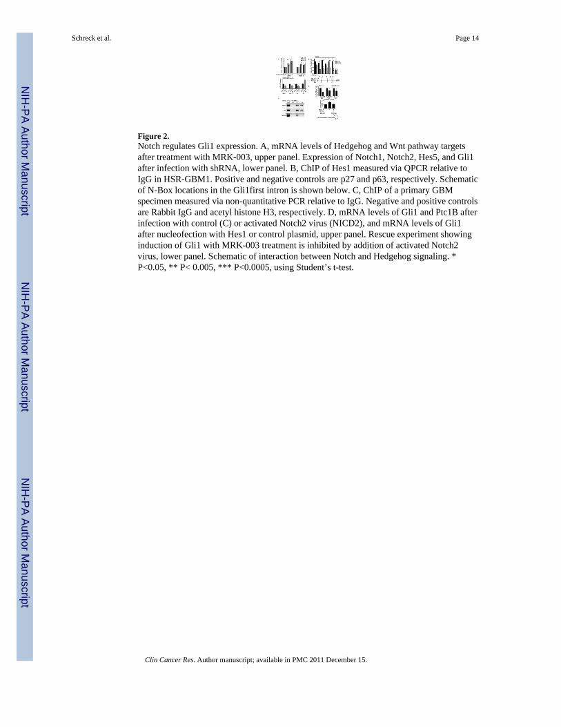
Figure 2.Notch regulates Gli1 expression. A, mRNA levels of Hedgehog and Wnt pathway targetsafter treatment with MRK-003, upper panel. Expression of Notch1, Notch2, Hes5, and Gli1after infection with shRNA, lower panel. B, ChIP of Hes1 measured via QPCR relative toIgG in HSR-GBM1. Positive and negative controls are p27 and p63, respectively. Schematicof N-Box locations in the Gli1first intron is shown below. C, ChIP of a primary GBMspecimen measured via non-quantitative PCR relative to IgG. Negative and positive controlsare Rabbit IgG and acetyl histone H3, respectively. D, mRNA levels of Gli1 and Ptc1B afterinfection with control (C) or activated Notch2 virus (NICD2), and mRNA levels of Gli1after nucleofection with Hes1 or control plasmid, upper panel. Rescue experiment showinginduction of Gli1 with MRK-003 treatment is inhibited by addition of activated Notch2virus, lower panel. Schematic of interaction between Notch and Hedgehog signaling. *P<0.05, ** P< 0.005, *** P<0.0005, using Student’s t-test.
Schreck et al. Page 14
Clin Cancer Res. Author manuscript; available in PMC 2011 December 15.
NIH
-PA Author Manuscript
NIH
-PA Author Manuscript
NIH
-PA Author Manuscript
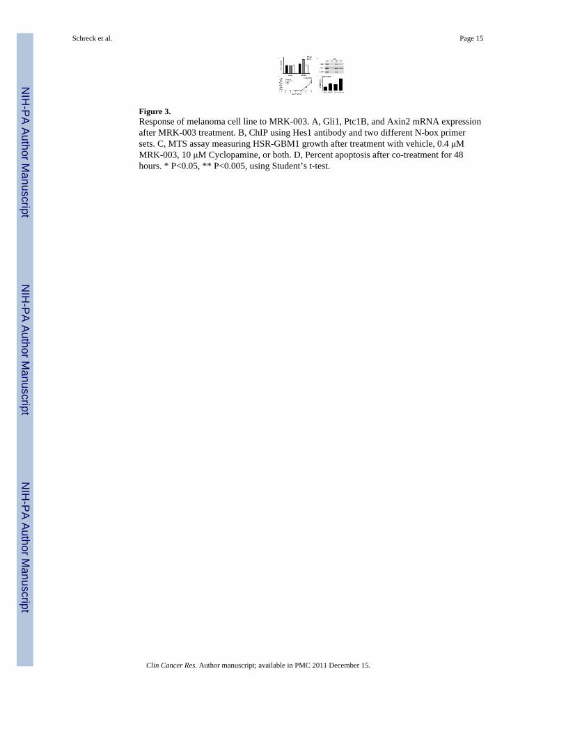
Figure 3.Response of melanoma cell line to MRK-003. A, Gli1, Ptc1B, and Axin2 mRNA expressionafter MRK-003 treatment. B, ChIP using Hes1 antibody and two different N-box primersets. C, MTS assay measuring HSR-GBM1 growth after treatment with vehicle, 0.4 μMMRK-003, 10 μM Cyclopamine, or both. D, Percent apoptosis after co-treatment for 48hours. * P<0.05, ** P<0.005, using Student’s t-test.
Schreck et al. Page 15
Clin Cancer Res. Author manuscript; available in PMC 2011 December 15.
NIH
-PA Author Manuscript
NIH
-PA Author Manuscript
NIH
-PA Author Manuscript
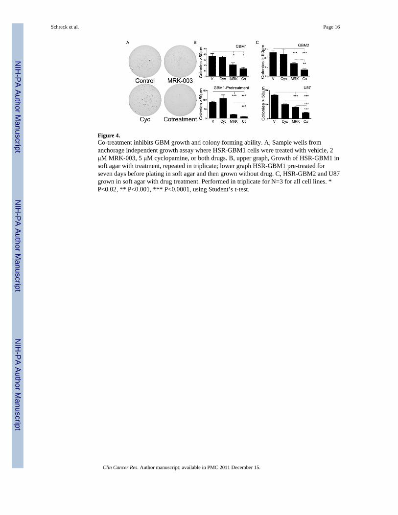
Figure 4.Co-treatment inhibits GBM growth and colony forming ability. A, Sample wells fromanchorage independent growth assay where HSR-GBM1 cells were treated with vehicle, 2μM MRK-003, 5 μM cyclopamine, or both drugs. B, upper graph, Growth of HSR-GBM1 insoft agar with treatment, repeated in triplicate; lower graph HSR-GBM1 pre-treated forseven days before plating in soft agar and then grown without drug. C, HSR-GBM2 and U87grown in soft agar with drug treatment. Performed in triplicate for N=3 for all cell lines. *P<0.02, ** P<0.001, *** P<0.0001, using Student’s t-test.
Schreck et al. Page 16
Clin Cancer Res. Author manuscript; available in PMC 2011 December 15.
NIH
-PA Author Manuscript
NIH
-PA Author Manuscript
NIH
-PA Author Manuscript
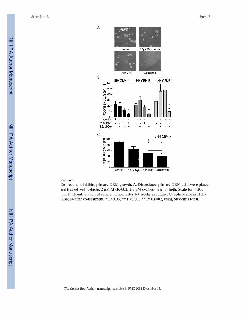
Figure 5.Co-treatment inhibits primary GBM growth. A, Dissociated primary GBM cells were platedand treated with vehicle, 2 μM MRK-003, 2.5 μM cyclopamine, or both. Scale bar = 300μm. B, Quantification of sphere number after 1-4 weeks in culture. C, Sphere size in JHH-GBM14 after co-treatment. * P<0.05, ** P<0.002 ** P<0.0002, using Student’s t-test.
Schreck et al. Page 17
Clin Cancer Res. Author manuscript; available in PMC 2011 December 15.
NIH
-PA Author Manuscript
NIH
-PA Author Manuscript
NIH
-PA Author Manuscript
NIH
-PA Author Manuscript
NIH
-PA Author Manuscript
NIH
-PA Author Manuscript
Schreck et al. Page 18
Table 1
Top Differentially Expressed Genes in Microarray. Bold are genes altered in “Top 100” gene list
Notch Pathway Up: CCND1, CREBBP, DLL1, DLL3, ELAVL4,IGF1R, KLF4, TFRC,Down: EFEMP1, HES1, HES5, HEY1, HEY2, HOXA5, HOXA7, HOXA9,
Wnt Pathway Up: CCND1, CREBBP, NRFA1, SFRP1, WNT5B, WNT7A,Down: ERBB2, FRZB, FZD8, TCF7L1
Hedgehog Pathway Up: MYCN, GLI2, NR4A1,Down: GLI1, GLI3, SUFU,
Other Pathways Up: ABCA1, VEGFADown: HOXA4, HOXC9, PDPN, PDGFRA, RB1, S100B, TGFB1,
Clin Cancer Res. Author manuscript; available in PMC 2011 December 15.
Related Documents