4871 Introduction Many biological processes involve polypeptide translocation across phospholipid membranes. Among them are export to the extracellular milieu (Blobel, 1995), transport in and out of the nucleus (Weis, 2003), and import into mitochondria (Endo et al., 2003; Gordon et al., 2000) and peroxisomes (Holroyd and Erdmann, 2001). These processes use specific transporters and frequently involve transmembrane pores and channels. The proteins translocated usually possess appropriate signal sequences and, in the case of classical protein secretion, this is a hydrophobic N-terminal sequence that allows the protein to enter the ER-Golgi pathway (Blobel, 2000). However, several extracellular proteins lack signal sequences, and their export proceeds through endoplasmic reticulum (ER)-Golgi-independent non-classical routes (Table 1) (reviewed by Nickel, 2003). Currently, we know little about the export mechanisms of most of these proteins, the only two common features being the absence of a signal sequence in the protein and the insensitivity of the pathway to brefeldin A [a drug that specifically inhibits ER-to-Golgi transport (Misumi et al., 1986)]. Nevertheless, what is apparent is that non- classical export is not a single pathway but instead comprises several transport mechanisms. Here, we concentrate on two biologically important and functionally related proteins, fibroblast growth factor 1 (FGF1) and interleukin (IL)-1α, and their non-classical export pathways. Structure and function of FGF1 and IL-1α FGF1 and FGF2 are prototypical members of the FGF family (Szebenyi and Fallon, 1999). FGFs have a wide variety of biological activities. During embryogenesis, these growth factors regulate mesodermal induction, neurulation, and the formation of the circulatory and skeletal systems (Friesel and Maciag, 1999). Subsequently, they play a crucial role in angiogenesis, tissue regeneration, inflammation and the formation of some tumors (Friesel and Maciag, 1999). Their biological effects are mediated through activation of four transmembrane phosphotyrosine kinase receptors (FGFR1-4), with the participation of cell-surface heparan sulfate proteoglycans (HSPGs), and consequently require release of the polypeptide (Friesel and Maciag, 1999). Most members of the family therefore possess classical signal sequences but FGF1 and FGF2 are devoid of such sequences and thus are released by novel secretion mechanisms (Coulier et al., 1997; Friesel and Maciag, 1999). The existence of FGF1- and FGF2-specific secretion pathways might represent a protective mechanism developed in the course of evolution and might be related to their high mitogenic potential and widespread expression. Indeed, a recombinant derivative of FGF1 that has an attached N- terminal signal sequence is a potent oncoprotein (Forough et al., 1993). Significantly, the FGFs of Caenorhabditis elegans and Drosophila have signal sequences (Coulier et al., 1997). Apparently, strictly programmed mosaic development of these organisms can rely on the regulation of FGF availability solely at the level of its expression. The more complicated and less hierarchical developmental strategies of chordates probably required the evolution of signal-peptide-less FGFs, whose accessibility might be more flexibly regulated post- translationally. The IL-1 family (Dinarello, 1996; Stylianou and Saklatvala, 1998), of which IL-1α and IL-1β are prototypical members, numbers at least ten proteins. Nine of these, including IL-1α and IL-1β, do not have signal sequences despite acting through transmembrane receptors and thus requiring export (Dinarello, 1998; Stylianou and Saklatvala, 1998). These proteins are potent pro-inflammatory cytokines (Dinarello, 1996), inducing 4871 Non-classical protein release independent of the ER-Golgi pathway has been reported for an increasing number of proteins lacking an N-terminal signal sequence. The export of FGF1 and IL-1α, two pro-angiogenic polypeptides, provides two such examples. In both cases, export is based on the Cu 2+ -dependent formation of multiprotein complexes containing the S100A13 protein and might involve translocation of the protein across the membrane as a ‘molten globule’. FGF1 and IL-1α are involved in pathological processes such as restenosis and tumor formation. Inhibition of their export by Cu 2+ chelators is thus an effective strategy for treatment of several diseases. Key words: Fibroblast growth factor 1, FGF1, Interleukin 1α, IL-1α, Release, Non-classical, Copper, Synaptotagmin 1, S100A13 Summary The non-classical export routes: FGF1 and IL-1α point the way Igor Prudovsky 1 , Anna Mandinova 1 , Raffaella Soldi 1 , Cinzia Bagala 1 , Irene Graziani 1 , Matteo Landriscina 2 , Francesca Tarantini 3 , Maria Duarte 1 , Stephen Bellum 1 , Holly Doherty 1 and Thomas Maciag 1, * 1 Center for Molecular Medicine, Maine Medical Center Research Institute, 81 Research Drive, Scarborough, Maine 04074, USA 2 Department of Oncology, Catholic University of Rome, School of Medicine, Rome 00168, Italy 3 Department of Geriatric Medicine, University of Florence, School of Medicine, Florence 50139, Italy *Author for correspondence (e-mail: [email protected]) Journal of Cell Science 116, 4871-4881 © 2003 The Company of Biologists Ltd doi:10.1242/jcs.00872 Commentary

Welcome message from author
This document is posted to help you gain knowledge. Please leave a comment to let me know what you think about it! Share it to your friends and learn new things together.
Transcript
4871
IntroductionMany biological processes involve polypeptide translocationacross phospholipid membranes. Among them are export to theextracellular milieu (Blobel, 1995), transport in and out of thenucleus (Weis, 2003), and import into mitochondria (Endo etal., 2003; Gordon et al., 2000) and peroxisomes (Holroyd andErdmann, 2001). These processes use specific transporters andfrequently involve transmembrane pores and channels. Theproteins translocated usually possess appropriate signalsequences and, in the case of classical protein secretion, this isa hydrophobic N-terminal sequence that allows the protein toenter the ER-Golgi pathway (Blobel, 2000).
However, several extracellular proteins lack signalsequences, and their export proceeds through endoplasmicreticulum (ER)-Golgi-independent non-classical routes (Table1) (reviewed by Nickel, 2003). Currently, we know little aboutthe export mechanisms of most of these proteins, the only twocommon features being the absence of a signal sequence in theprotein and the insensitivity of the pathway to brefeldin A [adrug that specifically inhibits ER-to-Golgi transport (Misumiet al., 1986)]. Nevertheless, what is apparent is that non-classical export is not a single pathway but instead comprisesseveral transport mechanisms. Here, we concentrate on twobiologically important and functionally related proteins,fibroblast growth factor 1 (FGF1) and interleukin (IL)-1α, andtheir non-classical export pathways.
Structure and function of FGF1 and IL-1 αFGF1 and FGF2 are prototypical members of the FGF family(Szebenyi and Fallon, 1999). FGFs have a wide variety ofbiological activities. During embryogenesis, these growthfactors regulate mesodermal induction, neurulation, and theformation of the circulatory and skeletal systems (Friesel and
Maciag, 1999). Subsequently, they play a crucial role inangiogenesis, tissue regeneration, inflammation and theformation of some tumors (Friesel and Maciag, 1999). Theirbiological effects are mediated through activation of fourtransmembrane phosphotyrosine kinase receptors (FGFR1-4),with the participation of cell-surface heparan sulfateproteoglycans (HSPGs), and consequently require release ofthe polypeptide (Friesel and Maciag, 1999). Most members ofthe family therefore possess classical signal sequences butFGF1 and FGF2 are devoid of such sequences and thus arereleased by novel secretion mechanisms (Coulier et al., 1997;Friesel and Maciag, 1999).
The existence of FGF1- and FGF2-specific secretionpathways might represent a protective mechanism developedin the course of evolution and might be related to their highmitogenic potential and widespread expression. Indeed, arecombinant derivative of FGF1 that has an attached N-terminal signal sequence is a potent oncoprotein (Forough etal., 1993). Significantly, the FGFs of Caenorhabditis elegansand Drosophila have signal sequences (Coulier et al., 1997).Apparently, strictly programmed mosaic development of theseorganisms can rely on the regulation of FGF availability solelyat the level of its expression. The more complicated and lesshierarchical developmental strategies of chordates probablyrequired the evolution of signal-peptide-less FGFs, whoseaccessibility might be more flexibly regulated post-translationally.
The IL-1 family (Dinarello, 1996; Stylianou and Saklatvala,1998), of which IL-1α and IL-1β are prototypical members,numbers at least ten proteins. Nine of these, including IL-1αand IL-1β, do not have signal sequences despite acting throughtransmembrane receptors and thus requiring export (Dinarello,1998; Stylianou and Saklatvala, 1998). These proteins arepotent pro-inflammatory cytokines (Dinarello, 1996), inducing
4871
Non-classical protein release independent of the ER-Golgipathway has been reported for an increasing number ofproteins lacking an N-terminal signal sequence. The exportof FGF1 and IL-1α, two pro-angiogenic polypeptides,provides two such examples. In both cases, export isbased on the Cu2+-dependent formation of multiproteincomplexes containing the S100A13 protein and mightinvolve translocation of the protein across the membrane
as a ‘molten globule’. FGF1 and IL-1α are involved inpathological processes such as restenosis and tumorformation. Inhibition of their export by Cu 2+ chelators isthus an effective strategy for treatment of several diseases.
Key words: Fibroblast growth factor 1, FGF1, Interleukin 1α, IL-1α,Release, Non-classical, Copper, Synaptotagmin 1, S100A13
Summary
The non-classical export routes: FGF1 and IL-1 α pointthe wayIgor Prudovsky 1, Anna Mandinova 1, Raffaella Soldi 1, Cinzia Bagala 1, Irene Graziani 1, Matteo Landriscina 2,Francesca Tarantini 3, Maria Duarte 1, Stephen Bellum 1, Holly Doherty 1 and Thomas Maciag 1,*1Center for Molecular Medicine, Maine Medical Center Research Institute, 81 Research Drive, Scarborough, Maine 04074, USA2Department of Oncology, Catholic University of Rome, School of Medicine, Rome 00168, Italy3Department of Geriatric Medicine, University of Florence, School of Medicine, Florence 50139, Italy*Author for correspondence (e-mail: [email protected])
Journal of Cell Science 116, 4871-4881 © 2003 The Company of Biologists Ltddoi:10.1242/jcs.00872
Commentary

4872
biosynthesis of a variety of inflammation-related molecules,such as tumor necrosis factor (TNF), transforming growthfactor (TGF)-β, granulocyte colony-stimulating factor (G-CSF), macrophage colony-stimulating factor (M-CSF),cyclooxygenase 2, endothelin-1, phospholipase A2, andinducible nitric oxide synthase (Dinarello, 1996). Unlike theFGFs, IL-1s are synthesized as higher molecular weightprecursor (p) proteins. pIL-1α is cleaved by calpain or calpain-like proteases to form mature (m) IL-1α, and pIL-1β is cleavedby the IL-1-converting enzyme to form mIL-1β (Dinarello,1996; Stylianou and Saklatvala, 1998).
Comparison of the crystal structures of FGF1, FGF2, IL-1αand IL-1β (Graves et al., 1990; Venkataraman et al., 1999; Zhuet al., 1991) reveals that they have very similar folds in spiteof their very low sequence similarity (Fig. 1). These proteinscontain β-barrel structures that are often found intransmembrane proteins, including bacterial pore-formingproteins (Chen and Funk, 2001; Heuck et al., 2000; Montoyaand Gouaux, 2003) and are crucial for membrane insertion ofsome proteins (Heuck et al., 2000). This provided the firstindication that similarities might exist between the releasemechanisms of the IL-1 and FGF prototypes. It was especiallyinteresting to compare the release of IL-1α and FGF1 since
these two proteins have antagonistic effects upon theproliferation and migration of endothelial cells (Maier et al.,1990; Friesel and Maciag, 1999).
Cell stress induces FGF1 and IL-1 α releaseUnder normal conditions, cells expressing FGF1 and IL-1α donot release these proteins. However, several stresses, such asheat shock (Jackson et al., 1992), hypoxia (Mouta Carreira etal., 2001), cultivation under low serum conditions (Shin et al.,1996) and cell treatment with low-density lipoproteins (LDLs)(Ananyeva et al., 1997), induce release of FGF1 from NIH3T3 cells. Heat shock induces the export of mIL-1α fromhuman promonocytic leukemia cells and activated peripheralmononuclear cells (Tarantini et al., 2001; Mandinova et al.,2003). The two latter cell types also exhibit heat-shock-induced export of pIL-1α (Mandinova et al., 2003).Interestingly, similarly to FGF2 (Shi et al., 1997), pIL-1α isnot secreted from stressed NIH 3T3 cells (Tarantini et al.,2001). The retention of pIL-1α is most likely because of thenuclear localization sequence (Wessendorf et al., 1993) in itscleavable N-terminal precursor domain. Although pIL-1αis cleaved in monocytes/macrophages (Dinarello, 1992;
Journal of Cell Science 116 (24)
Table 1. Proteins exported through non-classical pathwaysSignal Export sensitivity
Protein Reference peptide to brefeldin A Release characteristics
Secretory transglutaminase Aumuller et al., 1999 – Insensitive Constitutive, through membrane blebbingThioredoxin Rubartelli et al.,1992; – Insensitive Induced by antigen-specific T cells, intracellular vesicles
Rubartelli et al., 1995; not involvedAngelini et al., 2002
Galectins Hughes, 1999; Sato et al., – Insensitive Constitutive, through membrane blebbing1993; Lindstedt et al., 1993
Il-1α Tarantini et al., 2001; – Insensitive Stress-induced, Cu2+-dependent, in complex with S100A13Mandinova et al., 2003
Il-1β Rubartelli et al., 1990; – Insensitive Stress-induced, ABC-transporter-dependent, through the Andrei et al., 1999 endolysosomal pathway
FGF1 Jackson et al., 1992; – Insensitive Stress-induced, Cu2+-dependent, in complex with S100A13 Tarantini et al., 1998; and p40 Syt1LaVallee et al., 1998; Mouta Carreira et al., 1998; Landriscina et al., 2001a; Landriscina et al., 2001b
FGF2 Florkiewicz et al., 1995; Mignatti et al., 1992; – Insensitive Constitutive, Na+/K+ ATPase-dependentEngling et al., 2002
Sphingosine kinase Ancellin et al., 2002 – Insensitive Constitutive, inhibited by cytochalasinAnnexin I Chapman et al., 2003 – Not tested Glucocorticoid-induced, ABC-transporter-dependentAnnexin II Peterson et al., 2003 – Not tested Thrombin-induced, in complex with p11p40 Synaptotagmin 1 LaVallee et al., 1998; – Insensitive Constitutive
Tarantini et al., 1998S100A13 Landriscina et al., 2001a; – Insensitive Constitutive
Landriscina et al., 2001bHIV Tat Chang et al., 1997 – Insensitive ConstitutiveHerpes VP 22 protein Elliott and O’Hare, 1997 – Insensitive ConstitutiveFoamy virus Bet protein Lecellier et al., 2002 – Insensitive ConstitutiveEngrailed 2 Joliot et al., 1998; Maizel – Insensitive Attenuated by the CK2-dependent phosphorylation
et al., 1999; Maizel et al., 2002
HMGB1 Gardella et al., 2002; – Insensitive Stress-induced, through an endolysosomal pathwayPassalacqua et al., 1997; Passalacqua et al., 1998; Sparatore et al., 1996
LeishmaniaHASPB protein Denny et al., 2000 – Insensitive Constitutive, acylation-dependent

4873FGF1 and IL-1α release
Dinarello, 1996), the cleavage does not occur in NIH 3T3 cells(Tarantini et al., 2001), perhaps because of high levels of thecalpain inhibitor calpastatin (Goll et al., 2003) in the cytosolof these cells. In macrophage-like cells, the control of pIL-1αnuclear localization is less stringent since these cells displaypIL-1α both in the nucleus and in the cytoplasm (Beuscher etal., 1988; Kobayashi et al., 1990).
Significantly, co-expression of pIL-1α and FGF1 in NIH3T3 cells inhibits the stress-induced release of FGF1 (Tarantiniet al., 2001). It appears that the release pathways used by FGF1and IL-1α interact. Thus, pIL-1α could bind some importantprotein(s) shared by these pathways and sequester it in thenucleus. Indeed, FGF1 and IL-1α release pathways shareseveral similarities, including the delayed character of export,which becomes detectable only after 90 minutes of stress(Jackson et al., 1992; Tarantini et al., 2001). This delaypresumably reflects the need for stress-induced synthesis ofproteins that participate in the export of IL-1α and FGF1because both IL-1α release and FGF1 release are sensitive toinhibition of transcription and translation (Jackson et al., 1992;Tarantini et al., 2001).
FGF2 and IL-1β also exhibit non-classical release. However,their export mechanisms appear to be quite different. UnlikeFGF1 and IL-1α, FGF2 is exported constitutively (Florkiewiczet al., 1995; Mignatti et al., 1992). The release of FGF2 ishighly sensitive to the inhibitors of Na+/K+ ATPase (Dahl etal., 2000; Florkiewicz et al., 1998), whereas the export of FGF1is refractory to these compounds (F.T., I.P. and T.M.,unpublished).
The release of IL-1β is induced by lipopolysaccharides(Andrei et al., 1999; Rubartelli et al., 1990) but not by heatshock (A.M. and T.M., unpublished). In addition, unlike theexport of FGF1 and IL-1α (Jackson et al., 1992) (F.T., I.P. andT.M., unpublished), IL-1β release is sensitive to methylamine(Rubartelli et al., 1990), an inhibitor of exocytosis, and tosulfonylurea glybenclamide, an inhibitor of the mammalianATP-binding cassette (ABC) translocator ABC1 (Andrei et al.,1999). Another notable difference is that, unlike FGF1 and IL-1α, which are distributed homogeneously in the cytoplasm(Prudovsky et al., 2002), IL-1β is contained within cytoplasmic
vesicles expressing lysosomal but not ER-Golgi markers(Andrei et al., 1999). Export of IL-1β thus appears to be basedon its intracellular translocation into lysosome-like vesiclesand the subsequent exocytotic fusion of these vesicles with thecell membrane (Andrei et al., 1999). It will be interesting todetermine whether the constitutive release of FGF2 is alsosensitive to these pharmacological agents. Thus, in spite of thevery similar 3D structures of FGF1 and IL-1 prototypes, FGF2and IL-1β appear to be secreted through pathways differentfrom IL-1α and FGF1.
The stress-mediated intracellular transport of FGF1and IL-1 αThe inhibition of FGF1 and IL-1α release by 2-deoxyglucose(Jackson et al., 1992; Tarantini et al., 2001) demonstrates thatthese pathways are dependent on ATP. In addition, an intactactin cytoskeleton is important, since release of FGF1 and IL-1α is sensitive to agents that attenuate actin stress fibers, suchas latrunculin and amlexanox (Landriscina et al., 2000; MoutaCarreira et al., 1998; Tarantini et al., 2001). By contrast,microtubule inhibitors, such as nocodazole, fail to inhibit FGF1release (F.T., I.P. and T.M., unpublished). Real-time confocalstudies of cells transfected with an FGF1-GFP chimera havedemonstrated stress-induced migration of cytosolic FGF1 tothe cell membrane 60 minutes after heat shock and thistranslocation can be completely inhibited by amlexanox(Prudovsky et al., 2002). Likewise, heat shock also inducestranslocation of an IL-1α-RFP chimera from the cytosol to thecell membrane (Mandinova et al., 2003). Although the actincytoskeleton transports different types of cytoplasmicmembrane vesicle (Rogers and Gelfand, 2000), at least at thelevel of fluorescence microscopy, neither FGF1 nor IL-1αappears to be present in vesicular structures under normalconditions or during heat shock (Prudovsky et al., 2002).
The stress-induced formation of multiprotein FGF1and IL-1 α release complexesFGF1 is released during stress as a covalent cysteine-linked
Fig. 1.Three-dimensional representation of the β-barrel structures of human mIL-1α (Graves et al., 1990) and human FGF1 (Lozano et al.,2000). β-sheet domains are indicated in yellow and are depicted as rotating counter clockwise around the open centers of the structures. Thestructures were downloaded from the Protein Data Bank of the NCBI (http://www.rcsb.org/pdb/).

4874
homodimer (Jackson et al., 1992). The evolutionarilyconserved Cys30 residue is crucial for its stress-mediatedrelease (Tarantini et al., 1995). Interestingly, the FGF1homodimer exhibits a low heparin affinity (compared with themonomer), as well as low mitogenic activity in vitro (Englekaand Maciag, 1992). Dimer formation might therefore be a wayof storing and possibly transporting FGF1 in an inactive form.However, low heparin affinity makes dimeric FGF1 potentiallymore susceptible to proteolysis, since heparin and HSPGsprotect FGF family members from proteases (Friesel andMaciag, 1999; Rosengart et al., 1988). The balance betweenthe monomeric and dimeric forms of FGF1 in the extracellularcompartment might be regulated by stress-inducedextracellular reducing and oxidizing agents. For example, thereducing agent thioredoxin is a signal-peptide-less proteinreleased through a non-classical pathway (Rubartelli et al.,1992; Rubartelli et al., 1995), and Cu2+ ions are potentialextracellular oxidizing agents that could convert reducedmonomeric FGF1 to the dimeric form. Indeed, in a cell-freesystem, Cu2+ efficiently induces FGF1 dimerization (Englekaand Maciag, 1992). However, the role of intracellular Cu2+ inthe release of FGF1 and IL-1α is even more significant (seebelow).
IL-1α release does not appear to depend on covalentdimerization. mIL-1α is exported as a monomeric, biologicallyactive cytokine (Mandinova et al., 2003; Tarantini et al., 2001).Moreover, it has no evolutionarily conserved equivalent ofCys30 in FGF1 (Furutani et al., 1986; Lomedico et al., 1984),and a cysteine-free IL-1α mutant is released normally inresponse to cellular stress (A.M., I.P. and T.M., unpublished).
Both mIL-1α and FGF1 are exported as components ofmultiprotein release complexes that, at least in the case ofFGF1, assemble near the inner surface of the plasma membrane(Prudovsky et al., 2002). The first evidence for such complexesresulted from HPLC analysis of high-molecular-weight FGF1-containing fractions from bovine and ovine brains (Maciag etal., 1982; Mouta Carreira et al., 1998). Brain-derived FGF1 isassociated with at least four other polypeptides, which includeS100A13 and the p40 form of synaptotagmin 1 (Syt1) (Burgesset al., 1985; Mouta Carreira et al., 1998). S100A13 belongs tothe S100 family of polypeptides, which are small acidicproteins that have two Ca2+-binding EF-hand domains(Heizmann et al., 2002). The biological functions of mostS100s are not defined but, significantly, the intracellulardistributions and/or expression levels of some family membersare modified in response to cellular stress (Breen et al., 1999;Du et al., 2002; Duarte et al., 1999; Hoyaux et al., 2000; Hsiehet al., 2002; Kucharczak et al., 2001; Lam et al., 2001;Mandinova et al., 1998; Migheli et al., 1999; Zhang et al.,2002). A specific structural characteristic of S100A13 is thepresence of a C-terminal domain rich in basic residues (Wickiet al., 1996). The expression of S100A13 in NIH 3T3 cells isdetectable by RT-PCR analysis (Landriscina et al., 2001a).
All S100 proteins lack classical signal sequences, but at leastsome of them are released into the extracellular compartment(Heizmann and Cox, 1998). S100A13 transfected into NIH3T3 cells is constitutively released (Landriscina et al., 2001a);however, when it is co-expressed with either FGF1 or mIL-1α,its release becomes stress-dependent (Landriscina et al., 2001a;Mandinova et al., 2003). This observation in conjunction withexperiments using a dominant-negative S100A13 deletion
mutant lacking the basic C-terminal domain demonstrated thatS100A13 is an indispensable part of the multiprotein FGF1release complex (Landriscina et al., 2001a). Similarexperiments provided evidence that S100A13 expression isalso critical for IL-1α release (Mandinova et al., 2003).Interestingly, although a cysteine-free FGF1 mutant is notreleased in response to stress (Tarantini et al., 1995), its co-expression with S100A13 results in the stress-induced exportof both proteins (Landriscina et al., 2001a). It appears thatoverexpression of S100A13 induces the non-covalentdimerization of cysteine-free FGF1.
The p40 Syt1 component of the brain-derived FGF1-containing multiprotein complex represents the extravesicularportion of the transmembrane p65 Syt1 protein. Syt1participates in the docking of a variety of secretory vesicles,including synaptic vesicles, at the cell membrane prior to theirsubsequent exocytosis (Sudhoff and Rizo, 1996). Similarly toother members of the synaptotagmin protein family, Syt1displays two Ca2+-binding C2 domains in its extravesicularportion (Marqueze et al., 2000). p40 Syt1 is believed to beproduced by proteolytic cleavage of p65 near itstransmembrane domain (Marqueze et al., 2000; Sudhoff andRizo, 1996). In contrast to p65 Syt1, which displays a classicalN-terminal signal peptide in its primary structure and localizesprimarily to the ER-Golgi apparatus, cytoplasmic vesicles andcell membrane, signal-peptide-less p40 Syt1 displays a diffusecytosolic distribution (C.B., I.P. and T.M., unpublished).Interestingly, like S100A13, p40 Syt1 is also constitutivelyreleased from cells under normal cell culture conditions(LaVallee et al., 1998). Experiments using either an antisensestrategy or the expression of a dominant-negative p65 Syt1mutant, as well as immunoblot analysis of the exported FGF1complex at non-reducing low denaturation conditions forelectrophoresis, demonstrated that, similarly to S100A13, p40Syt1 is a crucial component of the FGF1 release complex(LaVallee et al., 1998; Tarantini et al., 1998).
Surprisingly, unlike S100A13, p40 Syt1 is dispensable forIL-1α release (Tarantini et al., 2001). However, it isconceivable that IL-1α uses another member of thesynaptotagmin family or other C2-domain-containingpolypeptides, for example calpain, the intracellular proteaseresponsible for pIL-1α cleavage, which contains a C2 domain(Goll et al., 2003) and associates with annexin II (Barnes andGomes, 2002). Interestingly, although expression of FGF1 inthe presence of S100A13 inhibits the constitutive release ofS100A13, it does not affect release of p40 Syt1 (LaVallee etal., 1998).
Annexin II might also be a part of the FGF1 and IL-1αrelease complexes. This protein exhibits inducible flippingfrom the inner to the outer surface of the cell membrane(Peterson, 2003), where it functions as a receptor forplasminogen and plasminogen activators (Hajjar et al., 1994;Hajjar et al., 1998). Studies using amlexanox affinitychromatography were able to resolve annexin II in a non-covalent complex with S100A13 (Oyama et al., 1997), and wehave recently demonstrated the presence of annexin II in thebrain-derived FGF1-containing multiprotein complex (R.S.,I.P. and T.M., unpublished). Since annexin II formsheterotetramers with S100A10 (p11) (Kim and Hajjar, 2002),its participation in the multiprotein complexes might rely uponinteractions with S100A13. Furthermore, since annexin II
Journal of Cell Science 116 (24)
4875FGF1 and IL-1α release
associates with the inner surface of the plasma membrane (Gollet al., 2003) and the assembly of the FGF1 multiproteincomplex also occurs near the inner surface of the plasmamembrane (Prudovsky et al., 2002), it is possible that annexinII serves as the site of assembly for the non-classical export ofthese multiprotein complexes. However, more experiments areneeded to verify its role in FGF1 and IL-1α release.
The role of Cu 2+ in FGF1 and IL-1 α exportHow do the FGF1 and IL-1α release complexes assemble?Association of the members of these multiprotein aggregatesmight involve Cu2+. FGF1, IL-1α, S100A13 and p40 Syt1are Cu2+-binding proteins (Engleka and Maciag, 1992;Landriscina et al., 2001b; Mandinova et al., 2003). Also, Cu2+
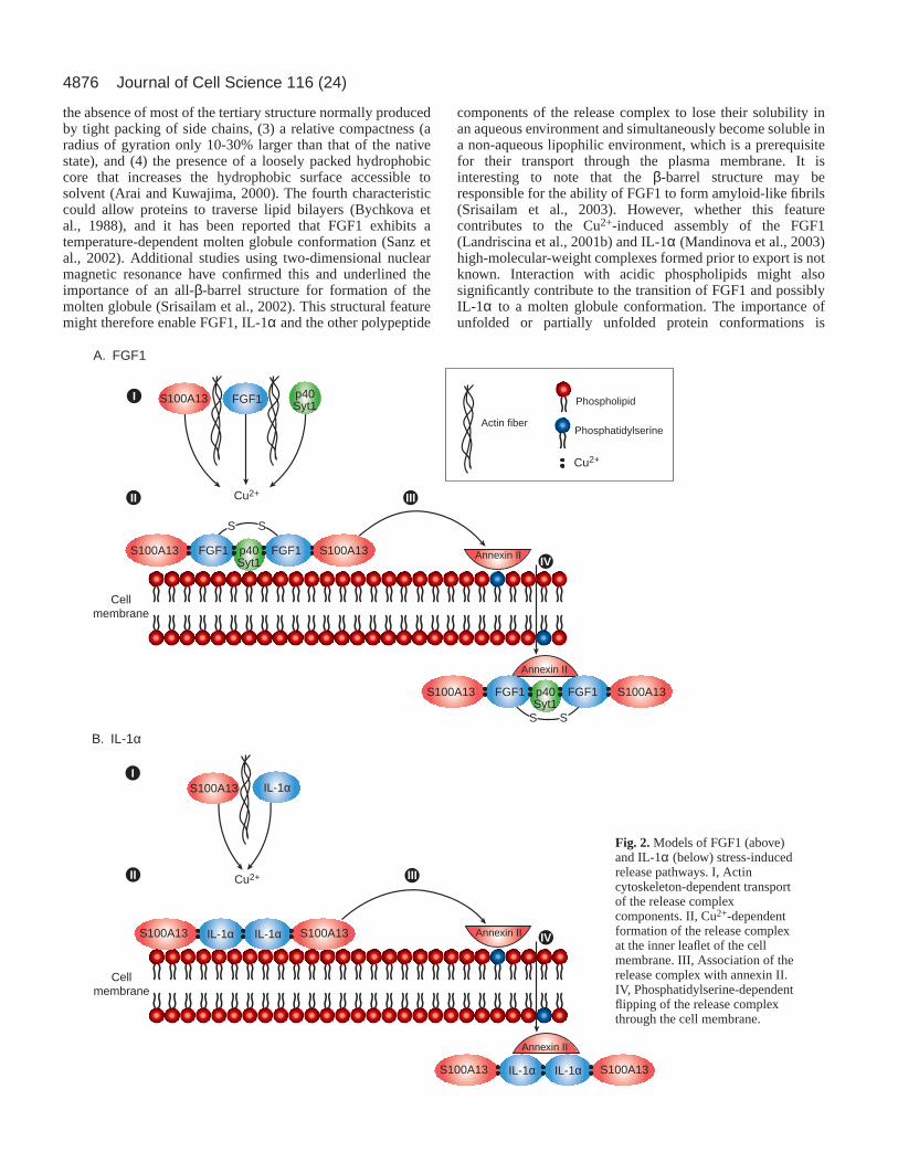
specifically induces formation of FGF1 but not FGF2homodimers even though two of the three Cys residues presentin FGF1 are conserved in FGF2 (Engleka and Maciag, 1992).In addition, several studies have demonstrated angiogenic andpro-inflammatory effects of Cu2+ (Brewer, 2001; Gullino,1983; Hannan and McAuslan, 1982; Raju et al., 1982; Zoli etal., 1998), indicating that Cu2+ might participate in thenon-classical release of angiogenic and pro-inflammatorypolypeptides. The role of Cu2+ in mediating the release ofFGF1 and IL-1α export has been examined in some detail, andindeed we and others have demonstrated in a cell-free systemthat Cu2+ is able to induce the formation of a complexcontaining p40Syt1, FGF1 and S100A13 at a molar ratioof 1:2:2, respectively, as well as the formation of aheterotetrameric (2:2) IL-1α-S100A13 complex (Landriscinaet al., 2001b; Mandinova et al., 1998). The depletion ofintracellular free Cu2+ through continuous application of aspecific chelator, tetrathiomolybdate (TTM), can attenuate thestress-induced release of IL-1α and FGF1, as well as ofS100A13 when co-expressed with IL-1α or FGF1 (Landriscinaet al., 2001b; Mandinova et al., 2003). These data indicate thatthe stress-induced Cu2+-dependent assembly of IL-1α andFGF1 multiprotein release complexes is indeed a prerequisitefor the non-classical export of these proteins in vitro (Fig. 2).
The plasma membrane as a platform for theassembly of release complexesConsiderable experimental evidence indicates that the Cu2+-dependent formation of IL-1α and FGF1 multiprotein releasecomplexes occurs at the inner leaflet of the cell membrane.Indeed, TTM treatment does not prevent the stress-inducedmigration of FGF1 to the cell membrane (Prudovsky et al.,2002), and thus complex formation (including formation of theFGF1 homodimer) does not appear to be important for theintracellular transport of FGF1 to the cell periphery. Moreover,dominant-negative mutants of S100A13 and p40 Syt1 that areknown to inhibit FGF1 release are transported to the cellmembrane in response to heat shock, and they do not preventthe stress-induced translocation of FGF1 to the periphery(Prudovsky et al., 2002). Apparently, the members of the FGF1multiprotein complex follow independent stress-inducedpathways to the cell periphery. Interestingly, FGF1, IL-1α, p40Syt1 and members of the S100 family can all bind acidicphospholipids in a cell-free system (Heizmann et al., 1998;Marqueze et al., 2000; Mandinova et al., 2003; Tarantini et al.,
1995). Furthermore, mutational analyses have revealed specificacidic phospholipid-binding domains in FGF1 (Tarantini et al.,1995) and Syt1 (Fernandez et al., 2001). Interestingly, a few ofthese acidic phospholipids are asymmetrically distributedbetween the leaflets of the plasma membrane (Pomorski et al.,2001) and thus, under normal conditions, acidic phospholipidssuch as phosphatidylserine localize preferentially to the innerleaflet. However, in response to a variety of different stresses,including heat shock, phosphatidylserine flips to the outerleaflet (Sims and Wiedmer, 2001).
Phosphatidylserine could drive the transmembranetranslocation of the IL-1α and FGF1 release complexes sinceimmunofluorescence data suggest that the inner side of the cellmembrane is a platform for the assembly of IL-1α and FGF1release complexes after the participant proteins reach themembrane through heat-shock-induced, actin-dependenttransport. Cu2+ ions needed for the assembly of releasecomplexes might be provided by transmembrane Cu2+
transporters (Finney and O’Halloran, 2003), and it isnoteworthy that the recently characterized human Cu2+
transporter 1 (hCtr1) is activated by cellular stress (Lee et al.,2002). Since free Cu2+ is virtually absent from the cytosol(Rae et al., 1999), the inner leaflet of the cell membrane is themost likely locale for the function of transient Cu2+ ions in theassembly of the FGF1 and IL-1α multiprotein complexes.However, it is unclear whether the Cu2+ ions involved inthe formation of these complexes are released into theextracellular compartment with the exported polypeptidesor whether they are recycled back to their intracellulartransporters.
Potential role of detergent-like properties and themolten globule state of proteins in facilitating non-classical protein exportThe key moment in non-classical export is translocation acrossthe cell membrane. The mechanism might involve localdestabilization of the phospholipid bilayer at the inner surfaceof the plasma membrane, which would allow the protein toinsert into the membrane and eventually exit the cell. Severalproteins including bactericidal peptides (Wiese et al., 2003)and viral fusion proteins (Dutch et al., 2000) have detergent-like properties that destabilize and permeabilize phospholipidbilayers. It has been observed that FGF1 has similar properties,demonstrating that it can induce temperature-dependentpermeabilization of phosphatidylserine/phosphatidylglycerolliposomes (Mach and Middaugh, 1995). IL-1α also behavessimilarly (Oku et al., 1995; Mandinova et al., 2003). Thephospholipid-binding and detergent-like activities of theseproteins indicate that the inner leaflet of the cell membranecould contain sites that recognize the multiprotein complexesdestined for release. These sites could contain specific acidicphospholipid ‘signatures’ that determine both the compositionof assembled protein aggregates and the export mechanism.
Translocation of a protein across the bilayer might requireconformational changes that increase its hydrophobicity.Proteins can achieve this by adopting a ‘molten globule’conformation (Ptitsyn, 1995). This is a partially unfoldedintermediate conformation assumed during denaturation andrenaturation (Arai and Kuwajima, 2000; Ptitsyn, 1995). It ischaracterized by (1) the presence of secondary structure, (2)
4876
the absence of most of the tertiary structure normally producedby tight packing of side chains, (3) a relative compactness (aradius of gyration only 10-30% larger than that of the nativestate), and (4) the presence of a loosely packed hydrophobiccore that increases the hydrophobic surface accessible tosolvent (Arai and Kuwajima, 2000). The fourth characteristiccould allow proteins to traverse lipid bilayers (Bychkova etal., 1988), and it has been reported that FGF1 exhibits atemperature-dependent molten globule conformation (Sanz etal., 2002). Additional studies using two-dimensional nuclearmagnetic resonance have confirmed this and underlined theimportance of an all-β-barrel structure for formation of themolten globule (Srisailam et al., 2002). This structural featuremight therefore enable FGF1, IL-1α and the other polypeptide
components of the release complex to lose their solubility inan aqueous environment and simultaneously become soluble ina non-aqueous lipophilic environment, which is a prerequisitefor their transport through the plasma membrane. It isinteresting to note that the β-barrel structure may beresponsible for the ability of FGF1 to form amyloid-like fibrils(Srisailam et al., 2003). However, whether this featurecontributes to the Cu2+-induced assembly of the FGF1(Landriscina et al., 2001b) and IL-1α (Mandinova et al., 2003)high-molecular-weight complexes formed prior to export is notknown. Interaction with acidic phospholipids might alsosignificantly contribute to the transition of FGF1 and possiblyIL-1α to a molten globule conformation. The importance ofunfolded or partially unfolded protein conformations is
Journal of Cell Science 116 (24)
S S
S S
Annexin II
Annexin II
Cellmembrane
Annexin II
Annexin II
Cellmembrane
Syt1S100A13 FGF1 p40 FGF1 S100A13
Syt1S100A13 FGF1 p40 FGF1 S100A13
S100A13 S100A13IL-1α IL-1α
Cu2+
S100A13
III
S100A13
Cu2+
Phospholipid
PhosphatidylserineActin fiber
Cu2+
S100A13 S100A13IL-1α IL-1α
III
IV
IV
II
II
I
I
A. FGF1
B. IL-1α
IL-1α
FGF1Syt1p40
Fig. 2.Models of FGF1 (above)and IL-1α (below) stress-inducedrelease pathways. I, Actincytoskeleton-dependent transportof the release complexcomponents. II, Cu2+-dependentformation of the release complexat the inner leaflet of the cellmembrane. III, Association of therelease complex with annexin II.IV, Phosphatidylserine-dependentflipping of the release complexthrough the cell membrane.

4877FGF1 and IL-1α release
stressed by results demonstrating that, upon overexpression ofrhodanese and GFP, excess unfolded proteins are evacuatedfrom the cells through non-classical export pathways (Sloan etal., 1994; Tanudji et al., 2002).
Whereas there is no direct biophysical evidence for theability of either S100A13 or p40 Syt1 to assume the moltenglobule conformation, both proteins are known to beconstitutively released independently of FGF1 or IL-1α(Landriscina et al., 2001a; LaVallee et al., 1998) and interactwith acidic phospholipids. They could therefore play a role aschaperones that stabilize FGF1 and possibly IL-1α in a moltenglobule conformation. Indeed, observing the interactionbetween a bacterial pilin and its chaperone, Knight and co-authors (Zavialov et al., 2003) have recently demonstrated thatchaperones can maintain polypeptides in a partially folded,high-energy state. Previously, cytosolic chaperones, such asmembers of the Hsp70 family, had been shown to maintainthe mitochondrial pre-proteins in a translocation-competentconformation, which is crucial for their post-translationalimport into mitochondria (Gordon et al., 2000). It is possiblethat S100A13 performs a chaperone-like service needed formembrane translocation since its overexpression alleviates therequirements of IL-1α and FGF1 export for new transcriptionand translation (Landriscina et al., 2001b; Mandinova et al.,2003). Indeed, some other members of the S100 family havechaperone activity (Heizmann et al., 2002), and S100A10, alsoknown as p11, serves as a chaperone for the hepatitis virus Bpolymerase and is needed for its nuclear translocation (Choi etal., 2003). Also, acting as a chaperone, it appears to be crucialfor insertion of annexin II into the plasma membrane, as wellas its thrombin-induced flipping to the outer surface of theplasma membrane (Peterson et al., 2003).
The pathological significance of non-classical FGF1and IL-1 α exportThe elucidation of the mechanisms responsible for the non-classical export of FGF1 and IL-1α has required the use of invitro methods of analysis and, as a result, it has been difficultto determine the role of these mechanisms in vivo and/or inpathological processes. However, clinical studies pioneered byG. Brewer and S. Merajver (Brewer et al., 2000; Cox et al.,2001) in which the Cu2+ chelator TTM was used to managethe progress of stage IV tumors in humans have providedinsight into the potential role of TTM as an angiogenicinhibitor capable of attenuating mammary gland tumorformation in the Her transgenic mouse (Pan et al., 2002). Sincethese studies suggested that TTM can repress thetranscriptional activation of NF-κB, and NF-κB liesdownstream of IL-1 receptor signaling (Baldwin, 1996), itappeared possible that TTM functions as a repressor of non-classical IL-1α export. Both FGF1 and IL-1 play a pro-angiogenic role in vivo (Friesel and Maciag, 1999; Voronov etal., 2003) although, in vitro, FGF1 stimulates proliferation andmigration of endothelial cells (Maciag et al., 1979; McMahonet al., 1997), whereas IL-1α inhibits both of these activities(Maier et al., 1990). It appears that the regulation ofangiogenesis and inflammation involves a coordination of non-classical FGF1 and IL-1α release. Indeed, IL-1α stimulates theinfiltration of tissues with macrophages (Dinarello, 1996),which present an abundant source of the FGF prototypes (Sano
et. al., 1990; Brogi et al., 1993). The absence of extracellularIL-1α in a tumor setting would limit the recruitment of FGF1-laden mononuclear cells (Sano et al., 1990; Sano et al., 1992)to tumor sites exhibiting an anoxic and/or hypoxicmicroenvironment. Thus, in the absence of mononuclear cellinfiltration, FGF1 would not be delivered to the tumorenvironment and, even if FGF1 was available within the tumormicrovasculature itself, TTM would also repress its export.
Interestingly, a similar mechanism has been proposed toexplain the response to injury in large vessels as a result ofcatheter-mediated clinical management of atheroscleroticarteries (Mandinov et al., 2003). Since the infiltration ofmononuclear cells into the injured area in response to therelease of IL-1α could result in the generation of an FGF1-richmicroenvironment, and FGF1 is a potent mitogen for thevascular smooth muscle cells (Winkles et al., 1987), its exportinto the extracellular compartment could be responsible for theonset of restenosis. Indeed, the long-term administration ofTTM significantly suppresses restenosis induced by catheterinjury in the rat carotid artery (Mandinov et al., 2003). Thearterial walls of TTM-treated rats display a strong attenuationof neointimal growth, impaired vasa vasorum formation,little, if any, macrophage/monocyte infiltration and, mostimportantly, very low levels of FGF1 and IL-1α expressionwhen compared with injured arteries from control animals.Thus, the inhibition of restenosis by TTM could be due to theability of the Cu2+ chelator to repress the stress-induced releaseof pro-inflammatory IL-1α, which would prevent infiltration ofmononuclear cells known to be a source of pro-angiogenic andpro-restenotic FGF1 in the wall of the damaged vessel. Thesedata also suggest that the repression of non-classical FGF1 andIL-1α export by Cu2+ chelation might ultimately be useful forthe clinical management of pro-inflammatory angiogenesis inhumans.
These data corroborate the preclinical and clinical reports onthe ability of TTM to inhibit solid tumor growth (Brewer et al.,2000; Cox et al., 2001), which depends on the availability ofpro-angiogenic polypeptides (Folkman, 2002). Thus, thepotential significance of the role of Cu2+ as a mediator of thenon-classical export of FGF1 and IL-1α could provide analternative approach for the clinical management of otherpathological conditions dependent on pro-inflammatoryangiogenesis, such as rheumatoid arthritis (Maini and Taylor,2000). Indeed, studies have demonstrated that Zn2+/Cu2+
chelation can repress the onset of Alzheimer’s disease in theβ-amyloid transgenic mouse (Cherny et al., 2001). Because theβ-amyloid gene is known to be regulated by IL-1α in humanendothelial cells (Goldgaber et al., 1989), it is likely that therepression of Alzheimer’s disease is due, at least in part, to theabsence of extracellular IL-1α. The recent report (Voronov etal., 2003) that IL-1α- and IL-1β-null mice cannot sustain anactive angiogenic environment to support tumor growth isconsistent with the premise that the function of these signal-peptide-less polypeptides is crucial for the regulation of pro-inflammatory angiogenic responses in vivo.
Future directionsSeveral important questions related to non-classicalpolypeptide release remain to be answered. Are there otheralternative pathways for non-classical export of FGF1 and IL-

4878
1α and, if so, how are they regulated? Is the molten globule acommon feature of such mechanisms? What are the transportermolecules responsible for actin-dependent translocation of theproteins from the cytosol to the cell membranes? How is thistranslocation induced by cellular stress? Which phospholipidsor which groups of phospholipids interact with specific proteinmembers of the release complexes? Do phospholipidsignatures permanently exist in the inner leaflet of the cellmembrane or are they arranged in response to cellular stress?How does the Cu2+- and phospholipid-dependent formation ofmultiprotein release complexes induce their subsequenttranslocation across the cell membrane? What is the source ofenergy used for stress-induced transmembrane translocation?Solving these problems will result in a better understanding ofthe non-classical protein release and eventually in an improvedability to regulate both inflammation and angiogenesis.
We thank Norma Albrecht for expert administrative assistance. Thiswork was supported in part by NIH grants RR15555, HL35627 andHL32348 to T.M.
ReferencesAnanyeva, N. M., Tijurmin, A. V., Berliner, J. A., Chisolm, G. M., Liau,
G., Winkles, J. A. and Haudenschild, C. C.(1997). Oxidized LDLmediates the release of fibroblast growth factor-1. Arterioscler. Thromb.Vasc. Biol.17, 445-453.
Ancellin, N., Colmont, C., Su, J., Li, Q., Mittereder, N., Chae, S. S.,Stefansson, S., Liau, G. and Hla, T. (2002). Extracellular export ofsphingosine kinase-1 enzyme. Sphingosine 1-phosphate generation and theinduction of angiogenic vascular maturation. J. Biol. Chem.277, 6667-6675.
Andrei, C., Dazzi, C., Lotti, L., Torrisi, M. R., Chimini, G. and Rubartelli,A. (1999). The secretory route of the leaderless protein interleukin 1betainvolves exocytosis of endolysosome-related vesicles. Mol. Biol. Cell 10,1463-1475.
Angelini, G., Gardella, S., Ardy, M., Ciriolo, M. R., Filomeni, G., diTrapani, G., Clarke, F., Sitia, R. and Rubartelli, A. (2002). Antigen-presenting dendritic cells provide the reducing extracellularmicroenvironment required for T lymphocyte activation. Proc. Natl. Acad.Sci. USA99, 1491-1496.
Arai, M. and Kuwajima, K. (2000). Role of the molten globule state inprotein folding. Adv. Protein Chem.53, 209-282.
Aumuller, G., Wilhelm, B. and Seitz, J.(1999). Apocrine secretion – fact orartifact? Anat. Anz.181, 437-446.
Baldwin, A. S., Jr (1996). The NF-kappa B and I kappa B proteins: newdiscoveries and insights. Annu. Rev. Immunol.14, 649-683.
Barnes, J. A. and Gomes, A. V.(2002). Proteolytic signals in the primarystructure of annexins. Mol. Cell. Biochem.231, 1-7.
Beuscher, H. U., Nickells, M. W. and Colten, H. R.(1988). The precursorof interleukin-1 alpha is phosphorylated at residue serine 90. J. Biol. Chem.263, 4023-4028.
Blobel, G. (1995). Unidirectional and bidirectional protein traffic acrossmembranes. Cold Spring Harb. Symp. Quant. Biol.60, 1-10.
Blobel, G. (2000). Protein targeting (Nobel lecture). Chembiochem1, 86-102.Breen, E. C., Fu, Z. and Normand, H.(1999). Calcyclin gene expression is
increased by mechanical strain in fibroblasts and lung. Am. J. Respir. CellMol. Biol. 21, 746-752.
Brewer, G. J. (2001). Copper control as an antiangiogenic anticancertherapy: lessons from treating Wilson’s disease. Exp. Biol. Med.226, 665-673.
Brewer, G. J., Dick, R. D., Grover, D. K., LeClaire, V., Tseng, M., Wicha,M., Pienta, K., Redman, B. G., Jahan, T., Sondak, V. K. et al. (2000).Treatment of metastatic cancer with tetrathiomolybdate, an anticopper,antiangiogenic agent: Phase I study. Clin. Cancer Res.6, 1-10.
Brogi, E., Winkles, J. A.., Underwood, R., Clinton, S. K., Alberts, G. F.and Libby, P. (1993). Distinct patterns of expression of fibroblast growthfactors and their receptors in human atheroma and nonatheroscleroticarteries. Association of acidic FGF with plaque microvessels andmacrophages. J. Clin. Invest.92, 2408-2418.
Burgess, W. H., Mehlman, T., Friesel, R., Johnson, W. V. and Maciag, T.
(1985). Multiple forms of endothelial cell growth factor. Rapid isolation andbiological and chemical characterization. J. Biol. Chem.260, 11389-11392.
Bychkova, V. E., Pain, R. H. and Ptitsyn, O. B.(1988). The ‘molten globule’state is involved in the translocation of proteins across membranes? FEBSLett. 238, 231-234.
Chang, H. C., Samaniego, F., Nair, B. C., Buonaguro, L. and Ensoli, B.(1997). HIV-1 Tat protein exits from cells via a leaderless secretory pathwayand binds to extracellular matrix-associated heparan sulfate proteoglycansthrough its basic region. AIDS11, 1421-1431.
Chapman, L. P., Epton, M. J., Buckingham, J. C., Morris, J. F. andChristian, H. C. (2003). Evidence for a role of the adenosine 5′-triphosphate-binding cassette transporter A1 in the externalization ofannexin I from pituitary folliculo-stellate cells. Endocrinology144, 1062-1073.
Chen, X. S. and Funk, C. D.(2001). The N-terminal ‘beta-barrel’ domain of5-lipoxygenase is essential for nuclear membrane translocation. J. Biol.Chem.276, 811-818.
Cherny, R. A., Atwood, C. S., Xilinas, M. E., Gray, D. N., Jones, W. D.,McLean, C. A., Barnham, K. J., Volitakis, I., Fraser, F. W., Kim, Y. etal. (2001). Treatment with a copper-zinc chelator markedly and rapidlyinhibits beta-amyloid accumulation in Alzheimer’s disease transgenic mice.Neuron30, 665-676.
Choi, J., Chang, J. S., Song, M. S., Ahn, B. Y., Park, Y., Lim, D. S. andHan, Y. S. (2003). Association of hepatitis B virus polymerase withpromyelocytic leukemia nuclear bodies mediated by the S100 family proteinp11. Biochem. Biophys. Res. Commun.305, 1049-1056.
Coulier, F., Pontarotti, P., Roubin, R., Hartung, H., Goldfarb, M. andBirnbaum, D. (1997). Of worms and men: an evolutionary perspective onthe fibroblast growth factor (FGF) and FGF receptor families. J. Mol. Evol.44, 43-56.
Cox, C., Teknos, T. N., Barrios, M., Brewer, G. J., Dick, R. D. andMerajver, S. D. (2001). The role of copper suppression as an antiangiogenicstrategy in head and neck squamous cell carcinoma. Laryngoscope111, 696-701.
Dahl, J. P., Binda, A., Canfield, V. A. and Levenson, R.(2000). Participationof Na,K-ATPase in FGF-2 secretion: rescue of ouabain-inhibitable FGF-2secretion by ouabain-resistant Na,K-ATPase alpha subunits. Biochemistry39, 14877-14883.
Denny, P. W., Gokool, S., Russell, D. G., Field, M. C. and Smith, D. F.(2000). Acylation-dependent protein export in Leishmania. J. Biol. Chem.275, 11017-11025.
Dinarello, C. A. (1992). The biology of interleukin-1.Chem. Immunol. 51, 1-32.
Dinarello, C. A. (1996). Biologic basis for interleukin-1 in disease. Blood87,2095-2147.
Dinarello, C. A. (1998). Interleukin-1, interleukin-1 receptors and interleukin-1 receptor antagonist. Int. Rev. Immunol.16, 457-499.
Du, X. J., Cole, T. J., Tenis, N., Gao, X. M., Kontgen, F., Kemp, B. E. andHeierhorst, J. (2002). Impaired cardiac contractility response tohemodynamic stress in S100A1-deficient mice. Mol. Cell. Biol. 22, 2821-2829.
Duarte, W. R., Mikuni-Takagaki, Y., Kawase, T., Limura, T., Oida, S.,Ohya, K., Takenaga, K., Ishikawa, L. and Kasugai, S.(1999). Effects ofmechanical stress on the mRNA expression of S100A4 and cytoskeletalcomponents by periodontal ligament cells. J. Med. Dent. Sci.46, 117-122.
Dutch, R. E., Jardetzky, T. S. and Lamb, R. A.(2000). Virus membranefusion proteins: biological machines that undergo a metamorphosis. Biosci.Rep.20, 597-612.
Elliott, G. and O’Hare, P. (1997). Intercellular trafficking and proteindelivery by a herpesvirus structural protein. Cell 88, 223-233.
Endo, T., Yamamoto, H. and Esaki, M.(2003). Functional cooperation andseparation of translocators in protein import into mitochondria, the double-membrane bounded organelles. J. Cell Sci.116, 3259-3267.
Engleka, K. A. and Maciag, T. (1992). Inactivation of human fibroblastgrowth factor-1 (FGF-1) activity by interaction with copper ions involvesFGF-1 dimer formation induced by copper-catalyzed oxidation. J. Biol.Chem.267, 11307-11315.
Engling, A., Backhaus, R., Stegmayer, C., Zehe, C., Seelenmeyer, C.,Kehlenbach, A., Schwappach, B., Wegehingel, S. and Nickel, W.(2002).Biosynthetic FGF-2 is targeted to non-lipid raft microdomains followingtranslocation to the extracellular surface of CHO cells. J. Cell Sci.115,3619-3631.
Fernandez, I., Arac, D., Ubach, J., Gerber, S. H., Shin, O., Gao, Y.,Anderson, R. G., Sudhof, T. C. and Rizo, J.(2001). Three-dimensional
Journal of Cell Science 116 (24)
4879FGF1 and IL-1α release
structure of the synaptotagmin 1 C2B-domain: synaptotagmin 1 as aphospholipid binding machine. Neuron32, 1057-1069.
Finney, L. A. and O’Halloran, T. V. (2003). Transition metal speciation inthe cell: insights from the chemistry of metal ion receptors. Science300,931-936.
Florkiewicz, R. Z., Anchin, J. and Baird, A. (1998). The inhibition offibroblast growth factor-2 export by cardenolides implies a novel functionfor the catalytic subunit of Na+,K+-ATPase. J. Biol. Chem.273, 544-551.
Florkiewicz, R. Z., Majack, R. A., Buechler, R. D. and Florkiewicz, E.(1995). Quantitative export of FGF-2 occurs through an alternative, energy-dependent, non-ER/Golgi pathway. J. Cell Physiol.162, 388-399.
Folkman, J. (2002). Role of angiogenesis in tumor growth and metastasis.Semin. Oncol.29, 15-18.
Forough, R., Zhan, X., MacPhee, M., Friedman, S., Engleka, K. A., Sayers,T., Wiltrout, R. H. and Maciag, T. (1993). Differential transformingabilities of non-secreted and secreted forms of human fibroblast growthfactor-1. J. Biol. Chem.268, 2960-2968.
Friesel, R. and Maciag, T.(1999). Fibroblast growth factor prototype releaseand fibroblast growth factor receptor signaling. Thromb. Haemost. 82, 748-754.
Furutani, Y., Notake, M., Fukui, T., Ohue, M., Nomura, H., Yamada, M.and Nakamura, S.(1986). Complete nucleotide sequence of the gene forhuman interleukin 1 alpha. Nucleic Acids Res.14, 3167-3179.
Gardella, S., Andrei, C., Ferrera, D., Lotti, L. V., Torrisi, M. R., Bianchi,M. E. and Rubartelli, A. (2002). The nuclear protein HMGB1 is secretedby monocytes via a non-classical, vesicle-mediated secretory pathway.EMBO Rep.3, 995-1001.
Goldgaber, D., Harris, H. W., Hla, T., Maciag, T., Donnelly, R. J.,Jacobsen, J. S., Vitek, M. P. and Gajdusek, D. C.(1989). Interleukin 1regulates synthesis of amyloid beta-protein precursor mRNA in humanendothelial cells. Proc. Natl. Acad. Sci. USA86, 7606-7610.
Goll, D. E., Thompson, V. F., Li, H., Wei, W. and Cong, J.(2003). TheCalpain System. Physiol. Rev.83, 731-801.
Gordon, D., Dancis, A. and Pain, D. (2000). Mechanisms of mitochodrialprotein import. Essays Biochem.36, 61-73.
Graves, B. J., Hatada, M. H., Hendrickson, W. A., Miller, J. K., Madison,V. S. and Satow, Y.(1990). Structure of interleukin 1 alpha at 2.7-Aresolution. Biochemistry29, 2679-2684.
Gullino, P. M. (1983). Angiogenesis and neoplastic growth. Prog. Clin. Biol.Res.132C, 101-107.
Hajjar, K. A., Jacovina, A. T. and Chacko, J. (1994). An endothelial cellreceptor for plasminogen/tissue plasminogen activator. I. Identity withannexin II. J. Biol. Chem.269, 21191-21197.
Hajjar, K. A., Mauri, L., Jacovina, A. T., Zhong, F., Mirza, U. A., Padovan,J. C. and Chait, B. T.(1998). Tissue plasminogen activator binding to theannexin II tail domain. Direct modulation by homocysteine. J. Biol. Chem.273, 9987-9993.
Hannan, G. N. and McAuslan, B. R.(1982). Modulation of synthesis ofspecific proteins in endothelial cells by copper, cadmium, and disulfiram: anearly response to an angiogenic inducer of cell migration. J. Cell. Physiol.111, 207-212.
Heizmann, C. W. and Cox, J. A.(1998). New perspectives on S100 proteins:a multi-functional Ca(2+)-, Zn(2+)- and Cu(2+)-binding protein family.Biometals11, 383-397.
Heizmann, C. W., Fritz, G. and Schafer, B. W.(2002). S100 proteins:structure, functions and pathology. Front. Biosci.7, d1356-d1368.
Heuck, A. P., Hotze, E. M., Tweten, R. K. and Johnson, A. E.(2000).Mechanism of membrane insertion of a multimeric beta-barrel protein:perfringolysin O creates a pore using ordered and coupled conformationalchanges. Mol. Cell 6, 1233-1242.
Holroyd, C. and Erdmann, R. (2001). Protein translocation machineries ofperoxisomes. FEBS Lett.501, 6-10.
Hoyaux, D., Decaestecker, C., Heizmann, C. W., Vogl, T., Schafer, B. W.,Salmon, I., Kiss, R. and Pochet, R.(2000). S100 proteins in Corporaamylacea from normal human brain. Brain Res.867, 280-288.
Hsieh, H. L., Schafer, B. W., Cox, J. A. and Heizmann, C. W.(2002).S100A13 and S100A6 exhibit distinct translocation pathways in endothelialcells. J. Cell Sci. 115, 3149-3158.
Hughes, R. C. (1999). Secretion of the galectin family of mammaliancarbohydrate-binding proteins. Biochim. Biophys. Acta1473, 172-185.
Jackson, A., Friedman, S., Zhan, X., Engleka, K. A., Forough, R. andMaciag, T. (1992). Heat shock induces the release of fibroblast growthfactor 1 from NIH 3T3 cells. Proc. Natl. Acad. Sci. USA89, 10691-10695.
Joliot, A., Maizel, A., Rosenberg, D., Trembleau, A., Dupas, S., Volovitch,
M. and Prochiantz, A. (1998). Identification of a signal sequence necessaryfor the unconventional secretion of Engrailed homeoprotein. Curr. Biol. 8,856-863.
Kim, J. and Hajjar, K. A. (2002). Annexin II: a plasminogen-plasminogenactivator co-receptor. Front. Biosci.7, d341-d348.
Kobayashi, Y., Oppenheim, J. J. and Matsushima, K.(1990). Calcium-dependent binding of phosphorylated human pre interleukin 1 alpha tophospholipids. J. Biochem.107, 666-670.
Kucharczak, J., Pannequin, J., Camby, I., Decaestecker, C., Kiss, R. andMartinez, J. (2001). Gastrin induces over-expression of genes involved inhuman U373 glioblastoma cell migration. Oncogene20, 7021-7028.
Lam, A. G., Koppal, T., Akama, K. T., Guo, L., Craft, J. M., Samy, B.,Schavocky, J. P., Watterson, D. M. and van Eldik, L. J.(2001).Mechanism of glial activation by S100B: involvement of the transcriptionfactor NFkappaB. Neurobiol. Aging22, 765-772.
Landriscina, M., Prudovsky, I., Carreira, C. M., Soldi, R., Tarantini, F.and Maciag, T. (2000). Amlexanox reversibly inhibits cell migration andproliferation and induces the Src-dependent disassembly of actin stressfibers in vitro. J. Biol. Chem.275, 32753-32762.
Landriscina, M., Soldi, R., Bagala, C., Micucci, I., Bellum, S., Tarantini,F., Prudovsky, I. and Maciag, T. (2001a). S100A13 participates in therelease of fibroblast growth factor 1 in response to heat shock in vitro. J.Biol. Chem.276, 22544-22552.
Landriscina, M., Bagala, C., Mandinova, A., Soldi, R., Micucci, I., Bellum,S., Prudovsky, I. and Maciag, T.(2001b). Copper induces the assembly ofa multiprotein aggregate implicated in the release of fibroblast growth factor1 in response to stress. J. Biol. Chem.276, 25549-25557.
LaVallee, T. M., Tarantini, F., Gamble, S., Carreira, C. M., Jackson, A.and Maciag, T. (1998). Synaptotagmin-1 is required for fibroblast growthfactor-1 release. J. Biol. Chem.273, 22217-22123.
Lecellier, C. H., Vermeulen, W., Bachelerie, F., Giron, M. L. and Saib, A.(2002). Intra- and intercellular trafficking of the foamy virus auxiliary betprotein. J. Virol. 76, 3388-3394.
Lee, J., Pena, M. M., Nose, Y. and Thiele, D. J.(2002). Biochemicalcharacterization of the human copper transporter Ctr1. J. Biol. Chem.277,4380-4387.
Lindstedt, R., Apodaca, G., Barondes, S. H., Mostov, K. E. and Leffler, H.(1993). Apical secretion of a cytosolic protein by Madin-Darby caninekidney cells. Evidence for polarized release of an endogenous lectin by anonclassical secretory pathway. J. Biol. Chem.268, 11750-11757.
Lomedico, P. T., Gubler, U., Hellmann, C. P., Dukovich, M., Giri, J. G.,Pan, Y. C., Collier, K., Semionow, R., Chua, A. O. and Mizel, S. B.(1984). Cloning and expression of murine interleukin-1 cDNA inEscherichia coli. Nature312, 458-462.
Lozano, R. M., Pineda-Lucena, A., Gonzalez, C., Angeles Jimenez, M.,Cuevas, P., Redondo-Horcajo, M., Sanz, J. M., Rico, M. and Gimenez-Gallego, G.(2000). 1HNMR structural characterization of a nonmitogenic,vasodilatory, ischemia-protector and neuromodulatory acidic fibroblastgrowth factor. Biochemistry39, 4982-4993.
Mach, H. and Middaugh, C. R. (1995). Interaction of partially structuredstates of acidic fibroblast growth factor with phospholipid membranes.Biochemistry34, 9913-9920.
Maciag, T., Cerundolo, J., Ilsley, S., Kelley, P. R. and Forand, R.(1979).An endothelial cell growth factor from bovine hypothalamus: identificationand partial characterization. Proc. Natl. Acad. Sci. USA76, 5674-5678.
Maciag, T., Hoover, G. A. and Weinstein, R.(1982). High and low molecularweight forms of endothelial cell growth factor. J. Biol. Chem.257, 5333-5336.
Maier, J. A., Voulalas, P., Roeder, D. and Maciag, T.(1990). Extension ofthe life-span of human endothelial cells by an interleukin-1 alpha antisenseoligomer. Science249, 1570-1574.
Maini, R. N. and Taylor, P. C. (2000). Anti-cytokine therapy for rheumatoidarthritis. Annu. Rev. Med.51, 207-229.
Maizel, A., Bensaude, O., Prochiantz, A. and Joliot, A.(1999). A shortregion of its homeodomain is necessary for engrailed nuclear export andsecretion. Development126, 3183-3190.
Maizel, A., Tassetto, M., Filhol, O., Cochet, C., Prochiantz, A. and Joliot,A. (2002). Engrailed homeoprotein secretion is a regulated process.Development129, 3545-3553.
Mandinov, L., Mandinova, A., Kyurkchiev, S., Kyurkchiev, D., Kehayov,I., Kolev, V., Soldi, R., Bagala, C., de Muinck, E. D., Lindner, V. et al.(2003). Copper chelation represses the vascular response to injury. Proc.Natl. Acad. Sci. USA100, 6700-6705.
Mandinova, A., Atar, D., Schafer, B. W., Spiess, M., Aebi, U. and

4880
Heizmann, C. W. (1998). Distinct subcellular localization of calciumbinding S100 proteins in human smooth muscle cells and their relocation inresponse to rises in intracellular calcium. J. Cell Sci.111, 2043-2054.
Mandinova, A., Soldi, R., Graziani, I., Bagala, C., Bellum, S., Landriscina,M., Tarantini, F., Prudovsky, I. and Maciag, T.(2003). S100A13 mediatesthe copper-dependent stress-induced release of IL-1alpha from both humanU937 and murine NIH 3T3 cells. J. Cell Sci.116, 2687-2696.
Marqueze, B., Berton, F. and Seagar, M.(2000). Synaptotagmins inmembrane traffic: which vesicles do the tagmins tag? Biochimie82, 409-420.
McMahon, G. A., Garfinkel, S., Prudovsky, I., Hu, X. and Maciag, T.(1997). Intracellular precursor interleukin (IL)-1alpha, but not mature IL-1alpha, is able to regulate human endothelial cell migration in vitro. J. Biol.Chem.272, 28202-28205.
Migheli, A., Cordera, S., Bendotti, C., Atzori, C., Piva, R. and Schiffer, D.(1999). S-100beta protein is upregulated in astrocytes and motor neurons inthe spinal cord of patients with amyotrophic lateral sclerosis. Neurosci. Lett.261, 25-28.
Mignatti, P., Morimoto, T. and Rifkin, D. B. (1992). Basic fibroblast growthfactor, a protein devoid of secretory signal sequence, is released by cells viaa pathway independent of the endoplasmic reticulum-Golgi complex. J. CellPhysiol. 151, 81-93.
Misumi, Y., Miki, K., Takatsuki, A., Tamura, G. and Ikehara, Y. (1986).Novel blockade by brefeldin A of intracellular transport of secretory proteinsin cultured rat hepatocytes. J. Biol. Chem.261, 11398-11403.
Montoya, M. and Gouaux, E.(2003). Beta-barrel membrane protein foldingand structure viewed through the lens of alpha-hemolysin. Biochim.Biophys. Acta1609, 19-27.
Mouta Carreira, C., LaVallee, T., Tarantini, F., Jackson, A., Lathrop, J.T., Hampton, B., Burgess, W. H. and Maciag, T.(1998). S100A13 isinvolved in the regulation of fibroblast growth factor-1 and p40synaptotagmin-1 release in vitro. J. Biol. Chem. 273, 22224-22231.
Mouta Carreira, C. M., Nasser, S. M., di Tomaso, E., Padera, T. P.,Boucher, Y., Tomarev, S. I. and Jain, R. K.(2001). LYVE-1 is notrestricted to the lymph vessels: expression in normal liver blood sinusoidsand down-regulation in human liver cancer and cirrhosis. Cancer Res.61,8079-8084.
Nickel, W. (2003). The mystery of nonclassical protein secretion. A currentview on cargo proteins and potential export routes. Eur. J. Biochem.270,2109-2119.
Oku, N., Saito, N., Okada, S., Watanabe, N. and Kobayashi, Y.(1995).Permeability change of liposomal membrane induced by interleukin-1 alpha.J. Biochem. Tokyo118, 832-835.
Oyama, Y., Shishibori, T., Yamashita, K., Naya, T., Nakagiri, S., Maeta,H. and Kobayashi, R.(1997). Two distinct anti-allergic drugs, amlexanoxand cromolyn, bind to the same kinds of calcium binding proteins, exceptcalmodulin, in bovine lung extract. Biochem. Biophys. Res. Commun.240,341-347.
Pan, Q., Kleer, C. G., van Golen, K. L., Irani, J., Bottema, K. M., Bias, C.,de Carvalho, M., Mesri, E. A., Robins, D. M., Dick, R. D. et al. (2002).Copper deficiency induced by tetrathiomolybdate suppresses tumor growthand angiogenesis. Cancer Res. 62, 4854-4859.
Passalacqua, M., Zicca, A., Sparatore, B., Patrone, M., Melloni, E. andPontremoli, S. (1997). Secretion and binding of HMG1 protein to theexternal surface of the membrane are required for murine erythroleukemiacell differentiation. FEBS Lett.400, 275-279.
Passalacqua, M., Patrone, M., Picotti, G. B., del Rio, M., Sparatore, B.,Melloni, E. and Pontremoli, S.(1998). Stimulated astrocytes release high-mobility group 1 protein, an inducer of LAN-5 neuroblastoma celldifferentiation. Neuroscience82, 1021-1028.
Peterson, E., Sutherland, M., Nesheim, M., Pryzdial, E.(2003). Thrombininduces endothelial cell-surface exposure of the plasminogen receptorannexin 2. J. Cell Sci.116, 2399-2408.
Pomorski, T., Hrafnsdottir, S., Devaux, P., van Meer, G. (2001). Lipiddistribution and transport across cellular membranes. Semin. Cell Dev. Biol,12, 139-148.
Prudovsky, I., Bagala, C., Tarantini, F., Mandinova, A., Soldi, R., Bellum,S. and Maciag, T.(2002). The intracellular translocation of the componentsof the fibroblast growth factor 1 release complex precedes their assemblyprior to export. J. Cell Biol.158, 201-208.
Ptitsyn, O. B.(1995). Molten globule and protein folding. Adv. Protein Chem.47, 83-229.
Rae, T. D., Schmidt, P. J., Pufahl, R. A., Culotta, V. C. and O’Halloran, T.V. (1999). Undetectable intracellular free copper: the requirement of acopper chaperone for superoxide dismutase. Science284, 805-808.
Raju, K. S., Alessandri, G., Ziche, M. and Gullino, P. M. (1982).Ceruloplasmin, copper ions, and angiogenesis. J. Natl. Cancer Inst.69,1183-1188.
Rogers, S. L. and Gelfand, V. I.(2000). Membrane trafficking, organelletransport, and the cytoskeleton. Curr. Opin. Cell Biol.12, 57-62.
Rosengart, T. K., Johnson, W. V., Friesel, R., Clark, R. and Maciag,T. (1988). Heparin protects heparin-binding growth factor-I fromproteolytic inactivation in vitro. Biochem. Biophys. Res. Commun.152,432-440.
Rubartelli, A., Cozzolino, F., Talio, M. and Sitia, R. (1990). A novelsecretory pathway for interleukin-1 beta, a protein lacking a signal sequence.EMBO J. 9, 1503-1510.
Rubartelli, A., Bajetto, A., Allavena, G., Wollman, E. and Sitia, R.(1992).Secretion of thioredoxin by normal and neoplastic cells through a leaderlesssecretory pathway. J. Biol. Chem.267, 24161-24164.
Rubartelli, A., Bonifaci, N. and Sitia, R. (1995). High rates of thioredoxinsecretion correlate with growth arrest in hepatoma cells. Cancer Res.55,675-680.
Sano, H., Forough, R., Maier, J. A., Case, J. P., Jackson, A., Engleka, K.,Maciag, T. and Wilder, R. L. (1990). Detection of high levels of heparinbinding growth factor-1 (acidic fibroblast growth factor) in inflammatoryarthritic joints. J. Cell Biol.110, 1417-1426.
Sano, H., Hla, T., Maier, J. A., Crofford, L. J., Case, J. P., Maciag, T. andWilder, R. L. (1992). In vivo cyclooxygenase expression in synovial tissuesof patients with rheumatoid arthritis and osteoarthritis and rats with adjuvantand streptococcal cell wall arthritis. J. Clin. Invest.89, 97-108.
Sanz, J. M., Jimenez, M. A. and Gimenez-Gallego, G.(2002). Hints ofnonhierarchical folding of acidic fibroblast growth factor. Biochemistry41,1923-1933.
Sato, S., Burdett, I. and Hughes, R. C.(1993). Secretion of the baby hamsterkidney 30-kDa galactose-binding lectin from polarized and nonpolarizedcells: a pathway independent of the endoplasmic reticulum-Golgi complex.Exp. Cell. Res.207, 8-18.
Shi, J., Friedman, S. and Maciag, T.(1997). A carboxyl-terminal domain infibroblast growth factor (FGF)-2 inhibits FGF-1 release in response to heatshock in vitro. J. Biol. Chem.272, 1142-1147.
Shin, J. T., Opalenik, S. R., Wehby, J. N., Mahesh, V. K., Jackson, A.,Tarantini, F., Maciag, T. and Thompson, J. A.(1996). Serum-starvationinduces the extracellular appearance of FGF-1. Biochim. Biophys. Acta1312, 27-38.
Sims, P. J. and Wiedmer, T.(2001). Unraveling the mysteries of phospholipidscrambling. Thromb. Haemost.86, 266-275.
Sloan, I. S., Horowitz, P. M. and Chirgwin, J. M.(1994). Rapid secretionby a nonclassical pathway of overexpressed mammalian mitochondrialrhodanese. J. Biol. Chem.269, 27625-27630.
Sparatore, B., Passalacqua, M., Patrone, M., Melloni, E. and Pontremoli,S.(1996). Extracellular high-mobility group 1 protein is essential for murineerythroleukaemia cell differentiation. Biochem. J.320, 253-256.
Srisailam, S., Wang, H. M., Kumar, T. K., Rajalingam, D., Sivaraja, V.,Sheu, H. S., Chang, Y. C. and Yu, C.(2002). Amyloid-like fibril formationin an all beta-barrel protein involves the formation of partially structuredintermediate(s). J. Biol. Chem.277, 19027-19036.
Srisailam, S., Kumar, T. K., Rajalingam, D., Kathir, K. M., Sheu, H. S.,Jan, F. J., Chao, P. C. and Yu, C.(2003). Amyloid-like fibril formation inan all beta-barrel protein. Partially structured intermediate state(s) is aprecursor for fibril formation. J. Biol. Chem.278, 17701-17709.
Stylianou, E. and Saklatvala, J.(1998). Interleukin-1. Int. J. Biochem. CellBiol. 30, 1075-1079.
Sudhoff, T. C. and Rizo, J.(1996). Synaptotagmins: C2-domain proteins thatregulate membrane traffic. Neuron17, 379-388.
Szebenyi, G. and Fallon, J. F.(1999). Fibroblast growth factors asmultifunctional signaling factors. Int. Rev. Cytol.185, 45-106.
Tanudji, M., Hevi, S. and Chuck, S. L. (2002). Improperly golded greenfluorescent protein is secreted via a non-classical pathway. J. Cell Sci.115,3849-3857.
Tarantini, F., Gamble, S., Jackson, A. and Maciag, T.(1995). The cysteineresidue responsible for the release of fibroblast growth factor-1 residues ina domain independent of the domain for phosphatidylserine binding. J. Biol.Chem.270, 29039-29042.
Tarantini, F., LaVallee, T., Jackson, A., Gamble, S., Carreira, C. M.,Garfinkel, S., Burgess, W. H. and Maciag, T.(1998). The extravesiculardomain of synaptotagmin-1 is released with the latent fibroblast growthfactor-1 homodimer in response to heat shock. J. Biol. Chem.273, 22209-22216.
Journal of Cell Science 116 (24)

4881FGF1 and IL-1α release
Tarantini, F., Micucci, I., Bellum, S., Landriscina, M., Garfinkel, S.,Prudovsky, I. and Maciag, T. (2001). The precursor but not the matureform of IL1alpha blocks the release of FGF1 in response to heat shock. J.Biol. Chem.276, 5147-5151.
Venkataraman, G., Raman, R., Sasisekharan, V. and Sasisekharan, R.(1999). Molecular characteristics of fibroblast growth factor-fibroblastgrowth factor receptor-heparin-like glycosaminoglycan complex. Proc.Natl. Acad. Sci. USA96, 3658-3663.
Voronov, E., Shouval, D. S., Krelin, Y., Cagnano, E., Benharroch, D.,Iwakura, Y., Dinarello, C. A. and Apte, R. N.(2003). IL-1 is required fortumor invasiveness and angiogenesis. Proc. Natl. Acad. Sci. USA100, 2645-2650.
Weis, K. (2003). Regulating access to the genome: nucleocytoplasmictransport throughout the cell cycle. Cell 112, 441-451.
Wessendorf, J. H., Garfinkel, S., Zhan, X., Brown, S. and Maciag, T.(1993). Identification of a nuclear localization sequence within the structureof the human interleukin-1 alpha precursor. J. Biol. Chem.268, 22100-22104.
Wicki, R., Schafer, B. W., Erne, P. and Heizmann, C. W.(1996).Characterization of the human and mouse cDNAs coding for S100A13, anew member of the S100 protein family. Biochem. Biophys. Res. Commun.227, 594-599.
Wiese, A., Gutsmann, T. and Seydel, U.(2003). Towards antibacterialstrategies: studies on the mechanisms of interaction between antibacterialpeptides and model membranes. J. Endotoxin Res.9, 67-84.
Winkles, J. A., Friesel, R., Burgess, W. H., Howk, R., Mehlman, T.,Weinstein, R. and Maciag, T.(1987). Human vascular smooth muscle cellsboth express and respond to heparin-binding growth factor I (endothelialcell growth factor). Proc. Natl. Acad. Sci. USA84, 7124-7128.
Zavialov, A. V., Berglund, J., Pudney, A. F., Fooks, L. J., Ibrahim, T. M.,MacIntyre, S. and Knight, S. D. (2003). Structure and biogenesis of thecapsular F1 antigen from Yersinia pestis. Preserved folding energy drivesfiber formation. Cell 113, 587-596.
Zhang, T., Woods, T. L. and Elder, J. T.(2002). Differential responses ofS100A2 to oxidative stress and increased intracellular calcium in normal,immortalized, and malignant human keratinocytes. J. Invest. Dermatol.119,1196-1201.
Zhu, X., Komiya, H., Chirino, A., Faham, S., Fox, G. M., Arakawa, T.,Hsu, B. T. and Rees, D. C.(1991). Three-dimensional structures of acidicand basic fibroblast growth factors. Science251, 90-93.
Zoli, A., Altomonte, L., Caricchio, R., Galossi, A., Mirone, L., Ruffini, M.P. and Magaro, M. (1998). Serum zinc and copper in active rheumatoidarthritis: correlation with interleukin 1 beta and tumour necrosis factoralpha. Clin. Rheumatol.17, 378-382.
Related Documents