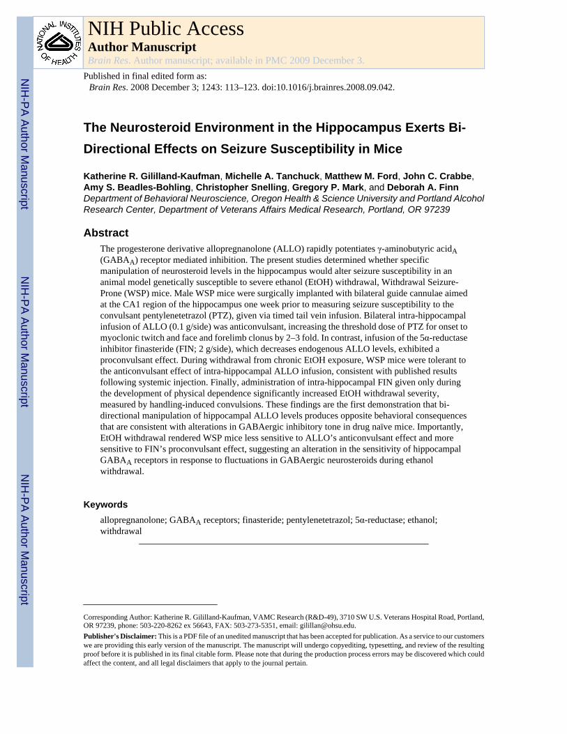
The Neurosteroid Environment in the Hippocampus Exerts Bi- Directional Effects on Seizure Susceptibility in Mice Katherine R. Gililland-Kaufman, Michelle A. Tanchuck, Matthew M. Ford, John C. Crabbe, Amy S. Beadles-Bohling, Christopher Snelling, Gregory P. Mark, and Deborah A. Finn Department of Behavioral Neuroscience, Oregon Health & Science University and Portland Alcohol Research Center, Department of Veterans Affairs Medical Research, Portland, OR 97239 Abstract The progesterone derivative allopregnanolone (ALLO) rapidly potentiates γ-aminobutyric acid A (GABA A ) receptor mediated inhibition. The present studies determined whether specific manipulation of neurosteroid levels in the hippocampus would alter seizure susceptibility in an animal model genetically susceptible to severe ethanol (EtOH) withdrawal, Withdrawal Seizure- Prone (WSP) mice. Male WSP mice were surgically implanted with bilateral guide cannulae aimed at the CA1 region of the hippocampus one week prior to measuring seizure susceptibility to the convulsant pentylenetetrazol (PTZ), given via timed tail vein infusion. Bilateral intra-hippocampal infusion of ALLO (0.1 g/side) was anticonvulsant, increasing the threshold dose of PTZ for onset to myoclonic twitch and face and forelimb clonus by 2–3 fold. In contrast, infusion of the 5α-reductase inhibitor finasteride (FIN; 2 g/side), which decreases endogenous ALLO levels, exhibited a proconvulsant effect. During withdrawal from chronic EtOH exposure, WSP mice were tolerant to the anticonvulsant effect of intra-hippocampal ALLO infusion, consistent with published results following systemic injection. Finally, administration of intra-hippocampal FIN given only during the development of physical dependence significantly increased EtOH withdrawal severity, measured by handling-induced convulsions. These findings are the first demonstration that bi- directional manipulation of hippocampal ALLO levels produces opposite behavioral consequences that are consistent with alterations in GABAergic inhibitory tone in drug naïve mice. Importantly, EtOH withdrawal rendered WSP mice less sensitive to ALLO’s anticonvulsant effect and more sensitive to FIN’s proconvulsant effect, suggesting an alteration in the sensitivity of hippocampal GABA A receptors in response to fluctuations in GABAergic neurosteroids during ethanol withdrawal. Keywords allopregnanolone; GABA A receptors; finasteride; pentylenetetrazol; 5α-reductase; ethanol; withdrawal Corresponding Author: Katherine R. Gililland-Kaufman, VAMC Research (R&D-49), 3710 SW U.S. Veterans Hospital Road, Portland, OR 97239, phone: 503-220-8262 ex 56643, FAX: 503-273-5351, email: [email protected]. Publisher's Disclaimer: This is a PDF file of an unedited manuscript that has been accepted for publication. As a service to our customers we are providing this early version of the manuscript. The manuscript will undergo copyediting, typesetting, and review of the resulting proof before it is published in its final citable form. Please note that during the production process errors may be discovered which could affect the content, and all legal disclaimers that apply to the journal pertain. NIH Public Access Author Manuscript Brain Res. Author manuscript; available in PMC 2009 December 3. Published in final edited form as: Brain Res. 2008 December 3; 1243: 113–123. doi:10.1016/j.brainres.2008.09.042. NIH-PA Author Manuscript NIH-PA Author Manuscript NIH-PA Author Manuscript

Welcome message from author
This document is posted to help you gain knowledge. Please leave a comment to let me know what you think about it! Share it to your friends and learn new things together.
Transcript
The Neurosteroid Environment in the Hippocampus Exerts Bi-Directional Effects on Seizure Susceptibility in Mice
Katherine R. Gililland-Kaufman, Michelle A. Tanchuck, Matthew M. Ford, John C. Crabbe,Amy S. Beadles-Bohling, Christopher Snelling, Gregory P. Mark, and Deborah A. FinnDepartment of Behavioral Neuroscience, Oregon Health & Science University and Portland AlcoholResearch Center, Department of Veterans Affairs Medical Research, Portland, OR 97239
AbstractThe progesterone derivative allopregnanolone (ALLO) rapidly potentiates γ-aminobutyric acidA(GABAA) receptor mediated inhibition. The present studies determined whether specificmanipulation of neurosteroid levels in the hippocampus would alter seizure susceptibility in ananimal model genetically susceptible to severe ethanol (EtOH) withdrawal, Withdrawal Seizure-Prone (WSP) mice. Male WSP mice were surgically implanted with bilateral guide cannulae aimedat the CA1 region of the hippocampus one week prior to measuring seizure susceptibility to theconvulsant pentylenetetrazol (PTZ), given via timed tail vein infusion. Bilateral intra-hippocampalinfusion of ALLO (0.1 g/side) was anticonvulsant, increasing the threshold dose of PTZ for onset tomyoclonic twitch and face and forelimb clonus by 2–3 fold. In contrast, infusion of the 5α-reductaseinhibitor finasteride (FIN; 2 g/side), which decreases endogenous ALLO levels, exhibited aproconvulsant effect. During withdrawal from chronic EtOH exposure, WSP mice were tolerant tothe anticonvulsant effect of intra-hippocampal ALLO infusion, consistent with published resultsfollowing systemic injection. Finally, administration of intra-hippocampal FIN given only duringthe development of physical dependence significantly increased EtOH withdrawal severity,measured by handling-induced convulsions. These findings are the first demonstration that bi-directional manipulation of hippocampal ALLO levels produces opposite behavioral consequencesthat are consistent with alterations in GABAergic inhibitory tone in drug naïve mice. Importantly,EtOH withdrawal rendered WSP mice less sensitive to ALLO’s anticonvulsant effect and moresensitive to FIN’s proconvulsant effect, suggesting an alteration in the sensitivity of hippocampalGABAA receptors in response to fluctuations in GABAergic neurosteroids during ethanolwithdrawal.
Keywordsallopregnanolone; GABAA receptors; finasteride; pentylenetetrazol; 5α-reductase; ethanol;withdrawal
Corresponding Author: Katherine R. Gililland-Kaufman, VAMC Research (R&D-49), 3710 SW U.S. Veterans Hospital Road, Portland,OR 97239, phone: 503-220-8262 ex 56643, FAX: 503-273-5351, email: [email protected]'s Disclaimer: This is a PDF file of an unedited manuscript that has been accepted for publication. As a service to our customerswe are providing this early version of the manuscript. The manuscript will undergo copyediting, typesetting, and review of the resultingproof before it is published in its final citable form. Please note that during the production process errors may be discovered which couldaffect the content, and all legal disclaimers that apply to the journal pertain.
NIH Public AccessAuthor ManuscriptBrain Res. Author manuscript; available in PMC 2009 December 3.
Published in final edited form as:Brain Res. 2008 December 3; 1243: 113–123. doi:10.1016/j.brainres.2008.09.042.
NIH
-PA Author Manuscript
NIH
-PA Author Manuscript
NIH
-PA Author Manuscript
1. IntroductionOne well-documented nongenomic effect of steroid hormones and their derivatives is the rapidpotentiation of γ-aminobutyric acidA (GABAA) receptor function. The progesterone derivativeallopregnanolone (ALLO) is the most potent endogenous positive modulator of GABAAreceptors yet identified and fluctuations in endogenous levels in vivo occur within the rangeof concentrations that potentiate GABAergic inhibition in vitro (Belelli and Lambert, 2005).Clinical and preclinical research show that ALLO fluctuations can contribute to diversedisorders such as premenstrual and postpartum dysphoric disorder (Follesa et al., 2004),catamenial epilepsy (Herzog and Frye, 2003), depression (Uzunova et al., 1998) and ethanol(EtOH) withdrawal (Romeo et al., 1996). The inverse relationship between endogenous ALLOlevels and anxiety, depression, and seizure susceptibility are consistent with ALLO’spharmacological profile, since exogenous administration produces anxiolytic, anticonvulsantand antidepressant effects (Gasior et al., 1999; Hirani et al., 2002). This inverse relationshipwas reported in cohorts of male subjects with depression (Uzunova et al., 1998) or in the earlyphase of alcoholic withdrawal (Romeo et al., 1996). Treatment with drugs which restoredALLO levels to those found in control subjects significantly reduced the symptoms of anxietyand depression in both the depressed and alcoholic subjects (Romeo et al., 2000; Ströhle et al.,1999). Thus, endogenous ALLO levels may be important in maintaining normal GABAergicbrain function.
Recent studies have demonstrated that withdrawal from chronic EtOH exposure significantlydecreased plasma ALLO levels in seizure prone mouse genotypes (Finn et al., 2004a) andhippocampal ALLO levels in rats (Cagetti et al., 2004). These decreases in endogenous ALLOlevels corresponded to increased anxiety and seizure susceptibility and decreased expressionof the biosynthetic enzyme 5α-reductase (Cagetti et al., 2003; Cagetti et al., 2004; Finn et al.,2004a), suggesting that there might be an inverse relationship between GABAergicneurosteroid levels and behavioral changes in excitability during EtOH withdrawal. Given thisevidence, it was hypothesized that a sustained reduction in ALLO during EtOH withdrawal tolevels below the physiologically relevant range would decrease GABAergic inhibition invivo and contribute to the manifestation of a more severe withdrawal syndrome.
Results in one genetic animal model of EtOH withdrawal severity, namely the WithdrawalSeizure Prone (WSP) and Resistant (WSR) selected lines, are consistent with the idea thatgenetic differences in EtOH withdrawal severity are due, in part, to differences in themodulatory effects of GABAergic neurosteroids. The WSP and WSR selected lines were bredin duplicate for their severity of (WSP) or resistance to (WSR) handling-induced convulsions(HICs) following chronic EtOH exposure (Crabbe et al., 1985). EtOH withdrawal convulsionsare at least 10-fold higher in the WSP than in the WSR line following an equivalent exposureto 72 hr EtOH vapor (Crabbe et al., 1985). Notably, there is a persistent decrease in endogenousALLO levels as well as decreased sensitivity to ALLO during EtOH withdrawal in WSP, butnot in WSR mice (Beckley et al., 2008; Finn et al., 2006; Finn et al., 2004a). Tolerance to theanticonvulsant effect of ALLO during EtOH withdrawal in WSP mice corresponded to adecrease in functional sensitivity of GABAA receptors to ALLO. Thus, the WSP line is anideal animal model to examine critical brain regions important for ALLO’s modulatory effectson brain function during EtOH-naive and EtOH withdrawal conditions.
Investigations into the neuroanatomical substrates influenced by withdrawal from chronicEtOH exposure determined that c-fos expression was significantly increased in thehippocampus during withdrawal from chronic EtOH exposure (Dave et al., 1990; Morgan etal., 1992), and that co-administration of the convulsant pentylenetetrazol (PTZ) during EtOHwithdrawal produced a further increase in c-fos expression (Putzke et al., 1996). Relevant toGABAergic neurosteroids, several studies documented that the brain sites where ALLO shows
Gililland-Kaufman et al. Page 2
Brain Res. Author manuscript; available in PMC 2009 December 3.
NIH
-PA Author Manuscript
NIH
-PA Author Manuscript
NIH
-PA Author Manuscript
the highest potency for modulation of GABAA receptor function across brain regions are:hippocampus > cortex = amygdala (Finn and Gee, 1993; Gee et al., 1988). However, there isheterogeneity within the hippocampus since electrophysiological studies determined thatGABAA receptors in the CA1 region of the hippocampus were much more sensitive to thepositive modulatory effect of neurosteroids than receptors in the dentate gyrus (Belelli andHerd, 2003; Harney et al., 2003). Additionally, chronic EtOH withdrawal produced significantdecreases in the expression and activity of hippocampal 5α-reductase in rats (Cagetti et al.,2004) and in WSP mice (Finn et al., 2004a). Thus, the hippocampus is a brain region relevantfor seizure susceptibility during EtOH withdrawal, GABAA receptor sensitivity to ALLO, andchanges in the expression and activity of the 5α-reductase during EtOH withdrawal.
The goal of the present set of experiments was to examine the physiological significance ofmanipulation of GABAergic neurosteroid levels in the hippocampus of WSP mice that wereEtOH naïve or undergoing withdrawal. Separate studies determined whether: 1) manipulatingALLO levels in the hippocampus would produce bi-directional effects on seizure susceptibility,measured by the change in sensitivity to PTZ-induced convulsions (i.e., an increase inhippocampal ALLO with ALLO infusions would be anticonvulsant and conversely, a decreasein hippocampal ALLO with finasteride (FIN) infusion would be proconvulsant), 2) withdrawalfrom chronic EtOH exposure would produce tolerance to the anticonvulsant effect ofhippocampal ALLO infusion similar to results following systemic ALLO administration,measured by the change in sensitivity to PTZ-induced convulsions (Finn et al., 2006), and 3)intra-hippocampal infusions of FIN during the development of physical dependence wouldincrease chronic EtOH withdrawal severity, measured by HICs.
2. Results2.1 Intra-hippocampal infusion revealed an anticonvulsant effect of ALLO and aproconvulsant effect of FIN
Since the hippocampus is an integral component of the limbic seizure circuitry (Gale, 1988),the overall hypothesis of these studies was that enhancing the positive modulation ofhippocampal GABAA receptors via ALLO infusion would be anticonvulsant and that reducingthe positive modulation of hippocampal GABAA receptors via FIN injection and infusionwould be proconvulsant. The CA1 region of the hippocampus was targeted for drug delivery,based on the enhanced sensitivity of GABAA receptors in this region versus the dentate gyrus(Belelli and Herd, 2003; Harney et al., 2003). Seizure susceptibility to PTZ, measured by timedtail vein infusion (5 mg/ml, 0.5 ml/min), was the dependent measure in these studies. Byanalyzing the PTZ threshold dose for onset to the various convulsant endpoints (see Methodsfor details), we could determine whether a drug was having an anticonvulsant (i.e., an increasein PTZ seizure threshold) or proconvulsant (i.e., a decrease in PTZ seizure threshold) effect.
In the 1st study, bilateral intra-hippocampal ALLO (0.1 μg/side) or vehicle (VEH) was infusedapproximately 5 min prior to administration of PTZ. ALLO infusion produced ananticonvulsant effect, as indicated by the significant increase in the threshold dose for onset tomyoclonic twitch (MC twitch; sudden involuntary muscle jerk; Figure 1A; t(13)= 4.59, p=0.05), with a trend for an increase in face and forelimb clonus (FF clonus; rapid writhingmovements of the head and neck and forelimb clonus; Figure 1B; t(14)= 3.352, p= 0.08), whencompared with mice receiving an infusion of VEH. There was no effect of intra-hippocampalALLO administration on running bouncing clonus (RB clonus, whole body clonus, includingrunning and jumps), or tonic hindlimb extension (THE; extreme rigidity, with forelimbs andhindlimbs extended caudally; data not shown).
In the 2nd study, FIN pre-treatment (50 mg/kg) or VEH was administered systemicallyapproximately 24 hours prior to the bilateral infusion of FIN (2 μg/side) or VEH into the
Gililland-Kaufman et al. Page 3
Brain Res. Author manuscript; available in PMC 2009 December 3.
NIH
-PA Author Manuscript
NIH
-PA Author Manuscript
NIH
-PA Author Manuscript
hippocampus. The infusions occurred 3–4 hours prior to testing for seizure susceptibility toPTZ. As depicted in Figure 2, FIN pretreatment was proconvulsant, as indicated by thesignificant decrease in the threshold dose for onset to MC Twitch (Figure 2A; t(14)= −2.332,p= 0.035) and the trend for a decrease in FF Clonus (Figure 2B; t(14)= −1.958, p= 0.08) versusvalues in mice pretreated with VEH. There were no effects of intra-hippocampal FIN infusionon RB clonus or THE (data not shown). It is worth noting that in both studies, infusion ofALLO or FIN had minimal effects on susceptibility to the subsequently occurring brainstemseizure endpoints (RB Clonus and THE), suggesting that manipulation of the neurosteroidenvironment of the hippocampus was selectively affecting limbic seizure endpoints.
A 3rd study was conducted to address the issue of site specificity, with mice receiving a bilateralinfusion of ALLO (0.1 μg/side) or VEH at a site 1 mm dorsal to the CA1 region in the dorsalparietal cortex, approximately 5 minutes prior to administration of PTZ. Notably, thisanatomical control study determined that infusion of ALLO did not significantly alter PTZ-induced convulsions for any seizure endpoint, when compared with values in mice receivingVEH infusions [ts(8)< 1.872, ps> 0.10, data not shown]. The mean ± SEM threshold dose ofPTZ for onset to MC twitch was 25.85± 1.19 mg/kg (VEH, n= 4) and 29.21± 2.95 mg/kg(ALLO, n=6) and for onset to FF clonus was 34.65± 2.65 mg/kg (VEH) and 38.81± 4.35 mg/kg (ALLO).
Finally, a subset of mice from the first two studies (~75% of mice in each study) weremicroinjected with methylene blue (a dye with a similar molecular weight to that of ALLO)into the CA1 region of the hippocampus to histologically determine the accuracy and diffusionparameters of the intracerebral injections. As depicted in Figure 3, the microinjection ofmethylene blue did not diffuse beyond the boundaries of the injection target (CA1 region ofthe hippocampus), confirming that our microinjection studies were localized to the region ofinterest.
2.2 Intra-hippocampal infusion during EtOH withdrawal revealed tolerance to theanticonvulsant effect of ALLO
Based on the finding that WSP mice exhibited tolerance to the anticonvulsant effect of systemicALLO during EtOH withdrawal (Beckley et al., 2008; Finn et al., 2006), the goal of this studywas to determine whether WSP mice would exhibit tolerance to the anticonvulsant effect ofintra-hippocampal ALLO during EtOH withdrawal. Separate groups of WSP mice wereexposed to 72 hours of EtOH vapor or air and tested for sensitivity to PTZ, measured by tailvein infusion, during peak withdrawal (hours 5.5–8). PTZ was administered approximately 5min after the intra-CA1 infusion of ALLO (0.1 μg/side) or VEH.
Daily blood samples taken from a subset of mice during the 72 hours of EtOH vapor exposuredocumented that there was no significant difference in blood EtOH concentration (BEC) duringthe development of physical dependence in mice that would subsequently receive ALLO versusVEH infusions during EtOH withdrawal [t(25)= 0.004, p>0.05, data not shown]. The mean ±SEM BEC upon the initiation of withdrawal after 72 hours of vapor exposure was 1.59± 0.17and 1.59± 0.10 mg/ml in the mice receiving infusions of ALLO and VEH, respectively.
Analysis of the PTZ threshold dose with a two by two ANOVA [treatment (air or EtOH) bydrug (ALLO or VEH)] revealed that there was a significant main effect of treatment on all fourseizure endpoints [Fs(1,39)> 14.143, ps< 0.001], with EtOH exposed mice requiring lowerPTZ doses to reach each endpoint (Figure 4A–D). There also was a significant main effect ofALLO infusion on all four endpoints [Fs(1,39)> 13.262, ps< 0.001] and a significant interactionbetween treatment and ALLO infusion [Fs (1,39)> 8.461, p< 0.006]. Due to these interactions,subsequent analyses confirmed that the anticonvulsant effect of ALLO was markedly andsignificantly reduced during EtOH withdrawal. In the air-exposed mice, ALLO infusion
Gililland-Kaufman et al. Page 4
Brain Res. Author manuscript; available in PMC 2009 December 3.
NIH
-PA Author Manuscript
NIH
-PA Author Manuscript
NIH
-PA Author Manuscript
significantly increased the PTZ threshold dose for onset to all four convulsion endpoints [ts(10)> 5.118, ps< 0.001]. In contrast, infusion of ALLO during EtOH withdrawal did notsignificantly increase the PTZ threshold dose for onset to any seizure endpoint. Additionalanalyses in VEH treated animals indicate that EtOH exposure significantly decreased the PTZthreshold dose for onset to RB clonus and THE [ts(29)> 6.110, ps< 0.001], this finding suggeststhat the proconvulsant effect of EtOH withdrawal was evident primarily by the change in PTZsensitivity to the two brainstem convulsion endpoints.
2.3 Intra-hippocampal infusions revealed enhanced sensitivity to the pro-convulsant effectof FIN during withdrawal from chronic ethanol exposure
Based on FIN’s proconvulsant effect, the goal of this study was to determine whether intra-hippocampal FIN during the development of physical dependence would significantly increaseEtOH withdrawal severity. Because it was not known if this paradigm would affect seizuresusceptibility at peak withdrawal or if it would precipitate the onset of withdrawal, we chooseto measure hourly HICS as this would provide a better characterization of seizure susceptibilityacross the entire withdrawal profile. For this study, separate groups of air- and EtOH- exposedmice received a total of 4 daily infusions of FIN (2 μg/side) or VEH (administered 24 hr priorto, and each day of, chamber exposure to EtOH vapor or air).
Daily blood samples taken from a subset of mice during the 72 hours of EtOH vapor exposuredocumented that there was no significant difference in BEC during the development of physicaldependence in mice receiving FIN versus VEH infusions [t(24)= −1.143, p>0.05, data notshown]. The mean ± SEM BEC upon the initiation of withdrawal after 72 hours of vaporexposure was 1.44± 0.12 and 1.61± 0.08 mg/ml in the mice receiving infusions of FIN andVEH, respectively.
The hourly HIC measurements were compiled to create a withdrawal time course (see Figure5A). A multifactorial ANOVA revealed a main effect of FIN infusion [F(1,71)= 4.061, p=0.048 (FIN>VEH)], a main effect of time [F(17,1206)= 16.421, p< 0.001], a main effect ofEtOH exposure [F(1,71)= 87.38, p< 0.001 (EtOH>air)] and a significant time by EtOHexposure interaction [F(17,1207)= 8.45, p< 0.001].
As another index of withdrawal severity, the area under the withdrawal curve (AUC) wascalculated by the trapezoidal method and compared with a two way ANOVA (treatment bydrug). This analysis revealed significant main effects of EtOH exposure [F(1,66)= 64.42, p<0.001 (EtOH>air)], a main effect of FIN administration [F(1,66)= 4.352, p= 0.041 (FIN>VEH)]but no significant interaction on AUC [F(1,34)= 0.12, p= 0.73]. Due to our a priori hypothesesthat the proconvulsant effect of FIN would be enhanced during EtOH withdrawal, plannedcomparisons were conducted. FIN administration significantly increased AUC in EtOH-exposed mice [t(36)= 2.144, p= 0.039], while having no effect in air-exposed animals [t(30)=0.883, p= 0.384] (Figure 5B).
3. DiscussionThe present findings provide strong evidence to indicate that the neurosteroid environment inthe hippocampus has bi-directional effects on seizure susceptibility in EtOH naïve mice andduring EtOH withdrawal. Intra-hippocampal ALLO was anticonvulsant, whereas FIN wasproconvulsant, in EtOH naïve mice. During EtOH withdrawal, animals were tolerant to theanticonvulsant effect of ALLO infusion and more sensitive to the pro-convulsant effects ofinhibiting 5α-reductase. The present results indicate that GABAA receptor sensitivity in thehippocampus is a dynamic process and that alterations in local endogenous neurosteroid levelscan have behavioral consequences. Consistent with the idea that manipulation of local ALLOconcentration can produce physiologically relevant effects, Belelli and Herd (2003)
Gililland-Kaufman et al. Page 5
Brain Res. Author manuscript; available in PMC 2009 December 3.
NIH
-PA Author Manuscript
NIH
-PA Author Manuscript
NIH
-PA Author Manuscript
demonstrated that the use of inhibitors to block the oxidation of ALLO (thereby increasingALLO levels) unmasked a GABAergic inhibitory tone in hippocampal dentate gyrus neurons.The present findings represent one of the first demonstrations that bi-directional manipulationof hippocampal ALLO levels produces opposite behavioral consequences that are consistentwith alterations in GABAergic inhibition.
The anticonvulsant effect of bilateral infusion of ALLO into the CA1 region of thehippocampus was similar to that seen with systemic injection. The potent anticonvulsant effectof intro-hippocampal ALLO against forebrain (predominantly limbic) convulsant endpointswas documented by the 157% and 107% increases in the PTZ threshold dose for onset to MCTwitch and FF Clonus, respectively. This result is consistent with the involvement of thehippocampus in the limbic seizure circuit (Gale, 1988), and with the contribution of GABAAreceptors in the hippocampus to the anticonvulsant effect of ALLO (Rhodes and Frye,2005a). For comparative purposes, systemic administration of a 5 mg/kg dose of ALLOincreased the threshold dose of PTZ for onset to FF Clonus by 2-fold in air-exposed WSP mice(Finn et al., 2006). Taken in conjunction with the present findings, activation of GABAAreceptors in the hippocampus following ALLO infusion is sufficient to produce ananticonvulsant effect against limbic convulsion endpoints that is comparable to that seenfollowing systemic injection of ALLO.
Use of FIN to decrease endogenous ALLO levels had a proconvulsant effect in EtOH naïvemice, as evidenced by the 18% and 17 % decrease in the PTZ threshold dose for onset to MCTwitch and FF clonus respectively. This effect of FIN was selective for limbic convulsionendpoints, as RB Clonus and THE endpoints were not significantly affected. Recent work inprogesterone-primed female rats also found a potent proconvulsant effect of intra-hippocampalFIN (10 μg/side) against PTZ-induced convulsions (Rhodes and Frye, 2005a). Notably, theincreased seizure susceptibility following FIN infusion in the study by Rhodes and Frye(2005a) corresponded to a significant decrease in hippocampal ALLO levels. Taken inconjunction with the present finings, hippocampal ALLO levels can impact PTZ sensitivity.
The change in seizure susceptibility in naïve mice following infusion of FIN was lesspronounced than that seen with ALLO. One explanation for this difference could be the mannerin which the local neurosteroid environment was manipulated. That is, infusion of FIN woulddecrease basal endogenous ALLO concentrations whereas infusion of ALLO would increaseALLO levels to supra-physiological concentrations. Although ALLO levels were not measuredin the current studies, previous reports indicate that microinjection of a 0.1 μg ALLO doseincreased ALLO concentrations 3-fold over basal levels (Frye and Rhodes, 2006) and thatinfusions of FIN (10 μg) into the ventral tegmental area or hippocampus reduced ALLO levelsby approximately 60%–80% respectively (Frye and Vongher, 2001; Rhodes and Frye,2005a; 2005b).
Another consideration with regard to the FIN data is that inhibition of 5α-reductase could alterthe concentrations of other 5α-reduced steroids, some of which are GABAergic (e.g., thedeoxycorticosterone metabolite tetrahydrodeoxycorticosterone), or could shift local steroidmetabolism to favor the production of steroids with proconvulsant effects such ascorticosterone or estrogen Additionally, FIN administration may produce 3α-hydroxypregn-4en-20-on (3α-HP) by inducing the reduction of progesterone by 3α-hydroxysteroid dehydrogenase (Purdy et al., 1990). 3α-HP is a potent agonist at the GABAAreceptor, but is not found in high concentrations in rodents (Purdy et al., 1990). While all ofthese steroids must be taken into consideration, ALLO is the most potent GABAA receptoragonist and is found in physiological ranges in the blood and brain.
Gililland-Kaufman et al. Page 6
Brain Res. Author manuscript; available in PMC 2009 December 3.
NIH
-PA Author Manuscript
NIH
-PA Author Manuscript
NIH
-PA Author Manuscript
One concern with the interpretation of microinjection studies is whether a drug’s effect is dueto a specific action at the site of the microinjection or to drug diffusion (primarily, to sites distalto the injection target). The results of our anatomical control studies strongly suggested thatthe effect of ALLO following microinjection into the CA1 was not due to diffusion up theinjector track, but to a localized neurosteroid action at the injection target. Thus, theseanatomical and diffusion controls provide evidence that the effects of ALLO and FIN werelikely confined to the CA1 of the hippocampus.
EtOH withdrawal significantly increased sensitivity to PTZ in WSP-mice, consistent with anoverall proconvulsant effect of EtOH withdrawal. However, the change in PTZ sensitivity wasonly true for the later two convulsion endpoints, RB clonus and THE. This finding is consistentwith previous data -from our lab, whereby seizure prone genotypes (e.g. WSP and DBA/2J)do not exhibit an increased sensitivity to all PTZ convulsion endpoints during EtOH withdrawal(Finn and Crabbe, 1999; Finn et al., 2006; Finn et al., 2000). It is likely that the main effect ofEtOH withdrawal for all 4 seizure endpoints was due in part, to the concomitant decrease insensitivity to ALLO’s anticonvulsant effect during withdrawal.
Notably, EtOH withdrawal was accompanied by decreased sensitivity to the anticonvulsanteffect of intra-hippocampal ALLO administration in mice and increased sensitivity to theproconvulsant effects of intra-hippocampal FIN. Our reasoning for using two differentmeasures of sensitivity was based on the following. With regard to ALLO, we had previouslyshown that the anticonvulsant effect of ALLO against withdrawal-related HICS was verytransient (Finn et al., 1995). We subsequently determined that the assessment of ALLOsensitivity during EtOH withdrawal was much more quantitative when the examination waslimited to a single time point and that timed tail infusion of PTZ was a highly sensitive measureof sensitivity at a single time point of withdrawal (Finn et al., 2006). Further, we wanted todirectly compare the change in sensitivity to ALLO during EtOH withdrawal followingsystemic versus intra-hippocampal administration, since we had recently found that functionalsensitivity of GABAA receptors to ALLO was significantly reduced during EtOH withdrawal(Finn et al., 2006). Specifically, the potency of ALLO to potentiate GABA (10 M) stimulatedchloride flux was significantly reduced (rightward shift in the dose-response curve andsignificant increase in EC50) concomitant with a reduction in efficacy of ALLO in the 60–600nM concentration range during EtOH withdrawal. Consistent with this decrease in thefunctional sensitivity of GABAA receptors to ALLO during EtOH withdrawal, our data indicatethat the anticonvulsant effect of intra-hippocampal ALLO was significantly reduced duringEtOH withdrawal. These results provide further evidence for the critical involvement of thehippocampus in mediating tolerance to the anticonvulsant effect of ALLO during EtOHwithdrawal.
We chose a different tactic to assess sensitivity to FIN during EtOH withdrawal. While FINproduced a slight but significant proconvulsant effect in naïve WSP mice when PTZ thresholddose was the dependent measure, we reasoned that quantifying the effect of FIN on EtOHwithdrawal severity would be best achieved by an examination of the withdrawal time course(which had never been done). We also were asking a slightly different question, namely, doessuppression of endogenous GABAergic neurosteroids during the development of physicaldependence alter the expression of EtOH withdrawal? Interestingly, four daily infusions ofFIN produced a greater proconvulsant effect during EtOH withdrawal, suggesting thatdecreasing endogenous ALLO levels during the development of physical dependence produceda greater decrease in GABAergic inhibition and concomitant increase in withdrawal severity.This result contrasts with recent findings in our lab showing that FIN systemically administeredduring the development of physical dependence decreased withdrawal severity (Finn et al.,2004b; Gorin et al., 2005). However, systemically administered FIN significantly decreasedBEC during EtOH exposure and upon the initiation of withdrawal, suggesting that the decrease
Gililland-Kaufman et al. Page 7
Brain Res. Author manuscript; available in PMC 2009 December 3.
NIH
-PA Author Manuscript
NIH
-PA Author Manuscript
NIH
-PA Author Manuscript
in withdrawal severity in the earlier work was due to an indirect effect on EtOHpharmacokinetics. Notably, intra-hippocampal FIN did not alter BEC in the current study,suggesting that the increase in withdrawal severity was due to the manipulation of GABAergicneurosteroid tone in the hippocampus.
Decreased sensitivity to ALLO, which we have characterized as tolerance to the anticonvulsanteffect of ALLO, was only observed in one other seizure prone genotype during EtOHwithdrawal, the DBA/2J inbred mouse strain (Finn et al., 2000). In contrast, rats, C57BL/6Jand WSR mice exhibit increased sensitivity to the anticonvulsant effect of ALLO andalphaxalone during EtOH withdrawal (Beckley et al., 2008; Cagetti et al., 2004; Devaud et al.,1996; Finn et al., 2000). Collectively, data to date suggest that the plasticity of GABAAreceptors during EtOH withdrawal may differ between seizure prone and resistant genotypes,particularly with regard to ALLO sensitivity.
Evidence indicates that neurosteroid modulation of GABAA receptors can undergo dynamicchange. For example, one mechanism implicated in the timed release of oxytocin fromhypothalamic neurons involves fluctuations in ALLO levels and a concomitant decrease insensitivity of GABAA receptors to ALLO (Brussaard et al., 1997; Koksma et al., 2003). Thisdecreased GABAergic inhibition, due in part to a switch in GABAA receptor subunit expressionas well as to the activity of constitutive phosphatases and kinases (which would alter thephosphorylation state of GABAA receptors), allows for the timed release of oxytocin. Chronicintermittent EtOH exposure has been shown to alter the responsivity of synaptic andextrasynaptic GABAA receptors (Liang et al., 2004), which also exhibit differential sensitivityto GABAergic neurosteroids (Belelli and Lambert, 2005). Chronic intermittent EtOH exposurealso increased the localization of α4 subunits within GABAergic synapses, which woulddecrease the sensitivity to neurosteroids at synaptic GABAergic receptors (Liang et al.,2006). A number of mechanisms have been suggested to contribute to the changes inGABAA receptor sensitivity to different modulators during EtOH withdrawal, such asalterations in subunit expression and assembly of GABAA receptors, post-translationalmodifications, alterations in receptor trafficking or changes in the subcellular or synapticlocalization of receptors (Kumar et al., 2004). However, the specific mechanism(s) underlyingthe tolerance to ALLO during EtOH withdrawal in WSP mice remains to be determined.
In conclusion, the present findings are the first demonstration that bi-directional manipulationof hippocampal ALLO levels produces opposite behavioral consequences that are consistentwith alterations in GABAergic inhibition. These results have important implications forunderstanding neurosteroid plasticity as it pertains to disorders such as EtOH withdrawal.
4. Experimental Procedure4.1 General Procedures
4.1.1 Subjects—Two genetically independent WSP (WSP–1 and –2) and WSR (WSR–1and 2) lines have been bred from a genetically heterogeneous stock of known composition (i.e.,HS/Ibg) using within-family, bi-directional selection with replicate lines (Crabbe et al.,1985). Drug naïve adult male WSP mice from the first genetic replicate (WSP-1) were used inall studies. The mice were bred in the Veterinary Medical Unit at the Veterans Affairs MedicalCenter (Portland, OR). Mice were maintained in groups of 4 in individually ventilatedpolycarbonate cages (Thorens) under a 12:12 hr light/dark cycle (lights on at 0600) at 23 ± 1°C. All animals had free access to rodent chow (Labdiet 5001 rodent diet, PMI international)and tap water throughout the experiments. At the time of testing, mice were from selectedgeneration 26 (filial generations 95–100) and were approximately 100 days old. All procedureswere conducted in accordance with the Guide for the Care and Use of Laboratory Animals as
Gililland-Kaufman et al. Page 8
Brain Res. Author manuscript; available in PMC 2009 December 3.
NIH
-PA Author Manuscript
NIH
-PA Author Manuscript
NIH
-PA Author Manuscript
adopted and disseminated by the U.S. National Institutes of Health, and were approved by thelocal Institutional Animal Care and Use Committee.
4.1.2 Surgery and Microinjections—Mice were surgically implanted under isofluraneanesthesia with bilateral 26-gauge guide cannulae aimed at the dorsal CA1 region of thehippocampus (A –3.3 mm, L –2.7 mm, V –1.5 mm), using previously described procedures(Mark and Finn, 2002) a minimum of one-week prior to any behavioral manipulations.Anterior, lateral and ventral coordinates are referenced to bregma, midsaggital sinus andsurface of the level skull, respectively (Paxinos and Franklin, 2001). The guide cannulae werekept patent with 220 m o.d. stainless steel wire stylets. During drug injections, the microinjector(made of silica glass tubing; 75 m i.d., 150 m o.d.) extended 0.7 mm beyond the guide shaftso that the ventral position of the injection site was 2.2 mm. Thus, only the silica tubingpenetrated the tissue beyond the implanted guide shaft. A separate study utilized anatomicalcontrols that were surgically implanted with bilateral guide shafts aimed 1 mm dorsal to theCA1 (A –3.3 mm, L ± 2.7 mm, V −0.5 mm), so that the ventral position of the injection sitewas in the overlying cortex (−1.2 mm).
To conduct the microinjection, mice were gently restrained by hand, the stylets were removed,and the microinjectors were lowered to the target site. The distal ends of the injectors wereattached to 10 l Hamilton syringes mounted in syringe pumps (Razel Sci. Instr. Inc. Stanford,CN) calibrated to deliver fluid at a rate of 200 nl over 60 sec. The silica microinjectors remainedin place for an additional one-minute after injections. The extremely small lumen of the silicainjector permits reliable infusions of nanoliter volumes while preventing leakage out of theinjector before the pump was activated (Parada et al., 1993).
4.1.3 Drugs—Drugs for microinfusion (ALLO and FIN) were dissolved in a 20% w/v solutionof 2-hydroxypropyl-β-cyclodextrin (β-cyclodextrin) and delivered in a total volume of 200 nl/side. ALLO (0.1 g/side) was synthesized by and purchased from Dr. Robert Purdy (VAResearch Foundation, San Diego, CA), and FIN (2 g/side) was purchased from Steraloids(Newport, RI). Vehicle (VEH) infusions were the β-cyclodextrin solution delivered in a totalvolume of 200 nl/side. These doses were chosen to reliably decrease brain concentrations ofALLO (Rhodes and Frye, 2005b) or to increase brain levels of ALLO to physiologicallyrelevant concentrations (Frye and Rhodes, 2006).
PTZ (Sigma, St. Louis, MO) was dissolved in 0.9% saline (Baxter, Deerfield, IL) as a 5-mg/ml solution and administered via timed tail vein infusion (0.5 ml/min) into a lateral vein (Finnand Crabbe, 1999; Finn et al., 2006; Mark and Finn, 2002). The latency to onset of the fourconvulsion endpoints that characterize PTZ-induced convulsions was recorded in seconds andsubsequently converted to threshold dose (mg/kg). This method allowed for the observationand quantitative analysis of four different endpoints that characterize PTZ-induced convulsionsand reliably occur in progression: MC twitch (sudden involuntary muscle jerk), FF clonus(rapid writhing movements of the head and neck and forelimb clonus), RB clonus (whole bodyclonus, including running and jumps), and THE (extreme rigidity, with forelimbs andhindlimbs extended caudally). Upon exhibiting THE, the mice were immediately euthanized.
4.1.4 Ethanol Vapor Inhalation—WSP-1 mice were exposed to continuous EtOH vaporor air for 72 hrs in inhalation chambers, which is a well-documented method in our laboratoryto induce physical dependence (Finn and Crabbe, 1999; Finn et al., 2006). Briefly, on the firstday of the experiment, mice were divided into one of two groups: EtOH or control. Mice inthe EtOH groups were weighed and injected intraperitoneally (IP) with a priming dose of EtOH(1.5 g/kg; Pharmco Products, Brookfield, CT) and exposed to EtOH vapor (7 9 mg EtOH/literair) inside the inhalation chamber. Mice in the control groups were injected with an equivalentvolume of saline and exposed to air in a separate inhalation chamber. All mice were given daily
Gililland-Kaufman et al. Page 9
Brain Res. Author manuscript; available in PMC 2009 December 3.
NIH
-PA Author Manuscript
NIH
-PA Author Manuscript
NIH
-PA Author Manuscript
IP injections of pyrazole hydrochloride, an EtOH dehydrogenase inhibitor (68.1 mg/kg, ip), tostabilize blood EtOH concentrations in the EtOH-exposed mice and to control for anynonspecific effects of pyrazole injection in the air-exposed mice. At 24 and 48 hrs, all animalswere briefly removed from the chambers, weighed, injected with pyrazole and placed backinto their respective chamber. Tail blood samples (20 μl) were taken from a subset of the EtOH-exposed animals to monitor BEC. At 72 hrs, all animals were removed from the chambers, andtail blood samples were taken from the EtOH-exposed animals for subsequent analysis of BEC.Tails were nicked for the air-exposed groups, but no blood was taken. Then, mice were housedin polycarbonate cages with cob bedding and taken to a procedure room for behavioral testing.Depending on the study, mice were either tested for sensitivity to the anticonvulsant effect ofALLO during peak withdrawal (5.5–8 hrs), measured by seizure susceptibility to PTZ or weretested for withdrawal severity, measured by HICs, at select time points.
4.1.5 BEC determination—Blood samples were assessed for EtOH concentration by gaschromatography, using an Agilent 6890N gas chromatograph and a well documented methodin the laboratory of Dr. John Crabbe (Finn et al., 2006). Briefly, an Agilent 7683 autosamplerand injector were used to inject a 1 μl sample of the supernatant (from the processed bloodsample) onto a capillary column (DB-ALC2, J & W Scientific Inc., Folsom, CA). Sample peakarea was interpolated from a standard curve derived from 7 pairs of standards (0.25 4.0 mg/ml).
4.1.6 HIC scoring—HICs were scored according to a previously published scale (Crabbe etal., 1991). Briefly, the mouse was quickly observed in the home cage. If no spontaneousconvulsions were observed, a mild convulsion could be elicited by gently lifting the mouse bythe tail, observing it, and then turning it 180 degrees if necessary. The HIC scoring was on ascale from 0 to 7; a score of 0 indicated no convulsions, a score of 1–3 indicated tonic or clonicconvulsions obtained by a gentle turn, a score of 4–6 indicated convulsions elicited by onlylifting the mouse by the tail, and a score of 7 indicated a spontaneous convulsion observed inthe home cage. Scores ranging from 0–5 were observed.
4.1.7 Data Analysis—Analyses were conducted on data from animals in which cannulaeplacement was verified to be in the hippocampus (primarily the CA1 region), or 1 mm dorsal(anatomical controls). For the studies measuring sensitivity to PTZ-induced convulsions, eitherseparate t-tests or ANOVAs were used to examine effects of drug, treatment, or both factorson the PTZ threshold dose for onset to each of the 4 convulsion endpoints: MC twitch, FFclonus, RB clonus, and THE. For the studies measuring HICs, repeated measures ANOVAs(with time as a repeated factor) were used to analyze treatment and drug effects on hourlyHICs. Withdrawal severity was quantified by calculating the AUC for each animal, using thetrapezoidal method, as previously described (Crabbe et al., 1983; Metten and Crabbe, 2005).The AUC was analyzed for treatment and drug effects with ANOVA. In the case of a priorihypotheses, specific groups were analyzed with t-tests. When appropriate, Tukey’s post-hoctest was used. In all cases, significance was set at P ≤ 0.05.
4.2 The effect of intra-hippocampal ALLO or FIN on sensitivity to PTZThis experiment was carried out in three separate studies. In the first study, animals wereadministered PTZ approximately 5 min after a hippocampal infusion of ALLO (0.1 μg/side)or VEH. In the second study animals were administered PTZ approximately 3–4 hrs afterinfusion of FIN (2 μg/side) or VEH. In the FIN study, all mice were also were pretreated witha systemic injection of FIN (50 mg/kg) or VEH at 24 hr prior to the infusion of FIN, as thisschedule was found to significantly decrease endogenous ALLO levels in whole brain (Finn,unpublished). A subset of animals in the FIN study had previously been exposed to a 72 hourEtOH paradigm. These animals were equally distributed throughout the groups and had been
Gililland-Kaufman et al. Page 10
Brain Res. Author manuscript; available in PMC 2009 December 3.
NIH
-PA Author Manuscript
NIH
-PA Author Manuscript
NIH
-PA Author Manuscript
drug free for a period of 3 weeks. In the third study, animals received an infusion of ALLO(0.1 μg/side) or VEH at a site 1 mm dorsal to the hippocampus as an anatomical control. Theseanimals were also administered PTZ approximately 5 min after infusion. In all three studies,latencies to MC twitch, FF clonus, RB clonus, and THE were recorded in seconds andsubsequently converted to threshold dose in mg/kg. Upon exhibiting THE, the mice wereimmediately euthanized by cervical dislocation and brains were removed for verification ofcannulae placement using standard histological techniques. A subset of mice from the first twostudies (~75% of mice from each study) was used to measure the site specificity of themicroinjection. Following cervical dislocation, animals received bilateral infusions (200 nl/side) of a dye (methylene blue, Sigma, MW = 373.9) with a similar molecular weight to thatof ALLO (MW = 318.5) that was solubulized in the β-cyclodextrin vehicle solution. After dyeinfusion, all brains were removed and analyzed using standard histological techniques.
The advantage of timed tail vein infusion is that qualitatively distinct convulsive endpoints areproduced as a function of PTZ dose. As discussed in Gale (1988), there are two qualitativelydistinct convulsion components that are mediated by separate and independent anatomicalcircuits. MC twitch and FF clonus are associated with forebrain (limbic) neural circuits,whereas RB clonus and THE depend on hindbrain circuitry. Thus, interpretations of the presentstudies discuss MC twitch and FF clonus as similar types of convulsions and similarly groupresults for RB clonus and THE.
4.3 Sensitivity to the anticonvulsant effect of intra-hippocampal ALLO during ethanolwithdrawal
WSP-1 mice were exposed to continuous EtOH vapor or air for 72 hrs in inhalation chambers.Mice were tested for sensitivity to the anticonvulsant effect of intra-hippocampal ALLO duringpeak withdrawal (5.5–8 hrs), measured by seizure susceptibility to PTZ, similar to thatdescribed above. PTZ was administered approximately 5 min after hippocampal infusion ofALLO (0.1 μg/side) or VEH. Latencies to the 4 convulsion endpoints were recorded in secondsand subsequently converted to threshold dose in mg/kg. Upon exhibiting THE, the mice wereimmediately euthanized by cervical dislocation, and brains were removed for verification ofcannulae placement.
4.4 The effect of intra-hippocampal FIN during the development of physical dependence onethanol withdrawal severity
WSP-1 mice were exposed to continuous EtOH vapor or air for 72 hours in inhalationchambers. For this study, separate groups of air- and EtOH-exposed mice received a total of 4daily infusions of FIN (2 μg/side) or VEH (administered 24 hr prior to, and each day of, chamberexposure to EtOH vapor or air). Prior to the first FIN or VEH infusion, baseline HICs weremeasured. Upon removal from the inhalation chambers at 72 hrs, mice were scored for HICshourly for the initial 12 hrs and then at hrs 24, 26, 28 and 48. Mice were euthanized followingthe final HIC determination at 48 hrs, and brains were removed for verification of cannulaeplacement.
AcknowledgementsThis research was supported by USPHS grants AA12439 (DAF) and AA10760 and AA13519 (JCC) from the NationalInstitute on Alcohol Abuse and Alcoholism and the Department of Veterans Affairs (DAF, JCC).
ReferencesBeckley EH, Fretwell AM, Tanchuck MA, Gililland KR, Crabbe JC, Finn DA. Decreased anticonvulsant
efficacy of allopregnanolone during ethanol withdrawal in female Withdrawal Seizure-Prone vs.Withdrawal Seizure-Resistant mice Neuropharmacology 2008;54:365–374.
Gililland-Kaufman et al. Page 11
Brain Res. Author manuscript; available in PMC 2009 December 3.
NIH
-PA Author Manuscript
NIH
-PA Author Manuscript
NIH
-PA Author Manuscript
Belelli D, Herd MB. The contraceptive agent Provera enhances GABAA receptor- mediated inhibitoryneurotransmission in the rat hippocampus: evidence for endogenous neurosteroids? J Neurosci2003;23:10013–10020. [PubMed: 14602815]
Belelli D, Lambert JJ. Neurosteroids: endogenous regulators of the GABAA receptor. Nat Rev Neurosci2005;6:565–575. [PubMed: 15959466]
Brussaard AB, Kits KS, Baker RE, Willems WP, Leyting-Vermeulen JW, Voorn P, Smit AB, BicknellRJ, Herbison AE. Plasticity in fast synaptic inhibition of adult oxytocin neurons caused by switch inGABAA receptor subunit expression. Neuron 1997;19:1103–1114. [PubMed: 9390523]
Cagetti E, Liang J, Spigelman I, Olsen RW. Withdrawal from chronic intermittent ethanol treatmentchanges subunit composition, reduces synaptic function, and decreases behavioral responses topositive allosteric modulators of GABAA receptors. Mol Pharmacol 2003;63:53–64. [PubMed:12488536]
Cagetti E, Pinna G, Guidotti A, Baicy K, Olsen RW. Chronic intermittent ethanol (CIE) administrationin rats decreases levels of neurosteroids in hippocampus, accompanied by altered behavioral responsesto neurosteroids and memory function. Neuropharmacology 2004;46:570–579. [PubMed: 14975681]
Crabbe JC, Kosobud A, Young ER, Tam BR, McSwigan JD. Bidirectional selection for susceptibility toethanol withdrawal seizures in Mus musculus. Behav Genet 1985;15:521–536. [PubMed: 4096679]
Crabbe JC, Merrill CD, Belknap JK. Effects of convulsants on handling-induced convulsions in miceselected for ethanol withdrawal severity. Brain Res 1991;550:1–6. [PubMed: 1888987]
Crabbe JC, Young ER, Kosobud A. Genetic correlations with ethanol withdrawal severity. PharmacolBiochem Behav 1983;18(Suppl 1):541–547. [PubMed: 6685310]
Dave JR, Tabakoff B, Hoffman PL. Ethanol withdrawal seizures produce increased c-fos mRNA in mousebrain. Mol Pharmacol 1990;37:367–371. [PubMed: 2107390]
Devaud LL, Purdy RH, Finn DA, Morrow AL. Sensitization of γ-aminobutyric acidA receptors toneuroactive steroids in rats during ethanol withdrawal. J Pharmacol Exp Ther 1996;278:510–517.[PubMed: 8768698]
Finn DA, Crabbe JC. Chronic ethanol differentially alters susceptibility to chemically inducedconvulsions in Withdrawal Seizure-Prone and -Resistant mice. J Pharmacol Exp Ther 1999;288:782–790. [PubMed: 9918589]
Finn DA, Douglass AD, Beadles-Bohling AS, Tanchuck MA, Long SL, Crabbe JC. Selected linedifference in sensitivity to a GABAergic neurosteroid during ethanol withdrawal. Genes Brain Behav2006;5:53–63. [PubMed: 16436189]
Finn DA, Ford MM, Wiren KM, Roselli CE, Crabbe JC. The role of pregnane neurosteroids in ethanolwithdrawal: Behavioral genetic approaches. Pharmacol Ther 2004a;101:91–112. [PubMed:14761701]
Finn DA, Gallaher EJ, Crabbe JC. Differential change in neuroactive steroid sensitivity during ethanolwithdrawal. J Pharmacol Exp Ther 2000;292:394–405. [PubMed: 10604976]
Finn DA, Gee KW. The influence of estrus cycle on neurosteroid potency at the γ-aminobutyric acidAreceptor complex. J Pharmacol Exp Ther 1993;265:1374–1379. [PubMed: 8389864]
Finn DA, Long SL, Tanchuck MA, Crabbe JC. Interaction of chronic ethanol exposure and finasteride:sex and strain differences. Pharmacol Biochem Behav 2004b;78:435–443. [PubMed: 15251252]
Finn DA, Roberts AJ, Crabbe JC. Neuroactive steroid sensitivity in Withdrawal Seizure-Prone and -Resistant mice. Alcohol Clin Exp Res 1995;19:410–415. [PubMed: 7625575]
Follesa P, Biggio F, Caria S, Gorini G, Biggio G. Modulation of GABAA receptor gene expression byallopregnanolone and ethanol. Eur J Pharmacol 2004;500:413–425. [PubMed: 15464049]
Frye CA, Rhodes ME. Infusions of 5α-pregnan-3α-ol-20-one (3α,5α-THP) to the ventral tegmental area,but not the substantia nigra, enhance exploratory, anti-anxiety, social and sexual behaviours andconcomitantly increase 3α,5α-THP concentrations in the hippocampus, diencephalon and cortex ofovariectomised oestrogen-primed rats. J Neuroendocrinol 2006;18:960–975. [PubMed: 17076771]
Frye CA, Vongher JM. Ventral tegmental area infusions of inhibitors of the biosynthesis and metabolismof 3α,5α-THP attenuate lordosis of hormone-primed and behavioural oestrous rats and hamsters. JNeuroendocrinol 2001;13:1076–1086. [PubMed: 11722704]
Gale K. Progression and generalization of seizure discharge: Anatomical and neurochemical substrates.Epilepsia 1988;29(Suppl 2):S15–34. [PubMed: 2844521]
Gililland-Kaufman et al. Page 12
Brain Res. Author manuscript; available in PMC 2009 December 3.
NIH
-PA Author Manuscript
NIH
-PA Author Manuscript
NIH
-PA Author Manuscript
Gasior M, Carter RB, Witkin JM. Neuroactive steroids: potential therapeutic use in neurological andpsychiatric disorders. Trends Pharmacol Sci 1999;20:107–112. [PubMed: 10203866]
Gee KW, Bolger MB, Brinton RE, Coirini H, McEwen BS. Steroid modulation of the chloride ionophorein rat brain: structure-activity requirements, regional dependence and mechanism of action. JPharmacol Exp Ther 1988;246:803–812. [PubMed: 2841455]
Gorin RE, Crabbe JC, Tanchuck MA, Long SL, Finn DA. Effects of finasteride on chronic and acuteethanol withdrawal severity in the WSP and WSR selected lines. Alcohol Clin Exp Res 2005;29:939–948. [PubMed: 15976519]
Harney SC, Frenguelli BG, Lambert JJ. Phosphorylation influences neurosteroid modulation of synapticGABAA receptors in rat CA1 and dentate gyrus neurones. Neuropharmacology 2003;45:873–883.[PubMed: 14529725]
Herzog AG, Frye CA. Seizure exacerbation associated with inhibition of progesterone metabolism. AnnNeurol 2003;53:390–391. [PubMed: 12601707]
Hirani K, Khisti RT, Chopde CT. Behavioral action of ethanol in Porsolt's forced swim test: modulationby 3α-hydroxy-5α-pregnan-20-one. Neuropharmacology 2002;43:1339–1350. [PubMed: 12527484]
Koksma JJ, van Kesteren RE, Rosahl TW, Zwart R, Smit AB, Luddens H, Brussaard AB. Oxytocinregulates neurosteroid modulation of GABAA receptors in supraoptic nucleus around parturition. JNeurosci 2003;23:788–797. [PubMed: 12574407]
Kumar S, Fleming RL, Morrow AL. Ethanol regulation of γ-aminobutyric acidA receptors: genomic andnongenomic mechanisms. Pharmacol Ther 2004;101:211–226. [PubMed: 15031000]
Liang J, Cagetti E, Olsen RW, Spigelman I. Altered pharmacology of synaptic and extrasynapticGABAA receptors on CA1 hippocampal neurons is consistent with subunit changes in a model ofalcohol withdrawal and dependence. J Pharmacol Exp Ther 2004;310:1234–1245. [PubMed:15126642]
Liang J, Zhang N, Cagetti E, Houser CR, Olsen RW, Spigelman I. Chronic intermittent ethanol-inducedswitch of ethanol actions from extrasynaptic to synaptic hippocampal GABAA receptors. J Neurosci2006;26:1749–1758. [PubMed: 16467523]
Mark GP, Finn DA. The relationship between hippocampal acetylcholine release and cholinergicconvulsant sensitivity in Withdrawal Seizure-Prone and Withdrawal Seizure-Resistant selectedmouse lines. Alcohol Clin Exp Res 2002;26:1141–1152. [PubMed: 12198388]
Metten P, Crabbe JC. Alcohol withdrawal severity in inbred mouse (Mus musculus) strains. BehavNeurosci 2005;119:911–925. [PubMed: 16187819]
Morgan PF, Nadi NS, Karanian J, Linnoila M. Mapping rat brain structures activated during ethanolwithdrawal: role of glutamate and NMDA receptors. Eur J Pharmacol 1992;225:217–223. [PubMed:1355445]
Parada MA, Puig DP, Hoebel BG. A remote insertion technique for intracerebral microinjections in freelymoving animals. J Neurosci Methods 1993;50:237–241. [PubMed: 8107503]
Paxinos, G.; Franklin, K. The Mouse Brain in Stereotaxic Coordinates. 2. Academic Press; San Diego,CA: 2001.
Purdy RH, Morrow AL, Blinn JR, Paul SM. Synthesis, metabolism, and pharmacological activity of3α-hydroxy steroids which potentiate GABA-receptor-mediated chloride ion uptake in rat cerebralcortical synaptoneurosomes. J Med Chem 1990;33:1572–1581. [PubMed: 2160534]
Putzke J, Spanagel R, Tolle TR, Zieglgansberger W. The anti-craving drug acamprosate reduces c-fosexpression in rats undergoing ethanol withdrawal. Eur J Pharmacol 1996;317:39–48. [PubMed:8982717]
Rhodes ME, Frye CA. Actions at GABAA receptors in the hippocampus may mediate some antiseizureeffects of progestins. Epilepsy Behav 2005a;6:320–327. [PubMed: 15820338]
Rhodes ME, Frye CA. Attenuating 5α-pregnane-3α-ol-20-one formation in the hippocampus of femalerats increases pentylenetetrazole-induced seizures. Epilepsy Behav 2005b;6:140–146. [PubMed:15710296]
Romeo E, Brancati A, De Lorenzo A, Fucci P, Furnari C, Pompili E, Sasso GF, Spalletta G, Troisi A,Pasini A. Marked decrease of plasma neuroactive steroids during alcohol withdrawal. ClinNeuropharmacol 1996;19:366–369. [PubMed: 8829001]
Gililland-Kaufman et al. Page 13
Brain Res. Author manuscript; available in PMC 2009 December 3.
NIH
-PA Author Manuscript
NIH
-PA Author Manuscript
NIH
-PA Author Manuscript
Romeo E, Pompili E, di Michele F, Pace M, Rupprecht R, Bernardi G, Pasinib A. Effects of fluoxetine,indomethacine and placebo on 3α, 5α tetrahydroprogesterone (THP) plasma levels in uncomplicatedalcohol withdrawal. World J Biol Psychiatry 2000;1:101–104. [PubMed: 12607205]
Ströhle A, Romeo E, Hermann B, Pasini A, Spalletta G, di Michele F, Holsboer F, Rupprecht R.Concentrations of 3α-reduced neuroactive steroids and their precursors in plasma of patients withmajor depression and after clinical recovery. Biol Psychiatry 1999;45:274–277. [PubMed: 10023501]
Uzunova V, Sheline Y, Davis JM, Rasmusson A, Uzunov DP, Costa E, Guidotti A. Increase in thecerebrospinal fluid content of neurosteroids in patients with unipolar major depression who arereceiving fluoxetine or fluvoxamine. Proc Natl Acad Sci 1998;95:3239–3244. [PubMed: 9501247]
Gililland-Kaufman et al. Page 14
Brain Res. Author manuscript; available in PMC 2009 December 3.
NIH
-PA Author Manuscript
NIH
-PA Author Manuscript
NIH
-PA Author Manuscript
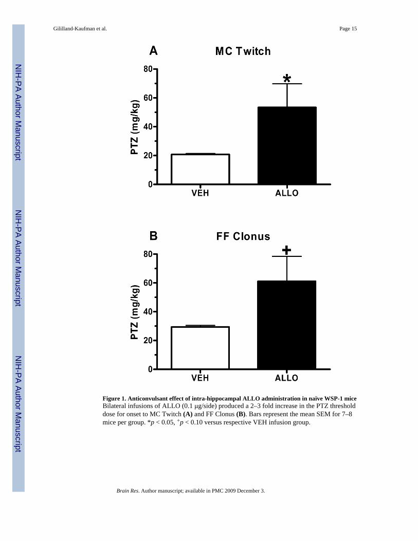
Figure 1. Anticonvulsant effect of intra-hippocampal ALLO administration in naïve WSP-1 miceBilateral infusions of ALLO (0.1 μg/side) produced a 2–3 fold increase in the PTZ thresholddose for onset to MC Twitch (A) and FF Clonus (B). Bars represent the mean SEM for 7–8mice per group. *p < 0.05, +p < 0.10 versus respective VEH infusion group.
Gililland-Kaufman et al. Page 15
Brain Res. Author manuscript; available in PMC 2009 December 3.
NIH
-PA Author Manuscript
NIH
-PA Author Manuscript
NIH
-PA Author Manuscript
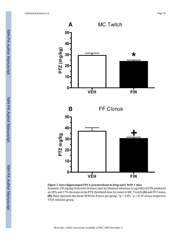
Figure 2. Intra-hippocampal FIN is proconvulsant in drug-naïve WSP-1 miceSystemic (50 mg/kg) followed 24 hours later by bilateral infusions (2 μg/side) of FIN producedan 18% and 17% decrease in the PTZ threshold dose for onset to MC Twitch (A) and FF Clonus(B). Bars represent the mean SEM for 8 mice per group. *p < 0.05, +p < 0.10 versus respectiveVEH infusion group.
Gililland-Kaufman et al. Page 16
Brain Res. Author manuscript; available in PMC 2009 December 3.
NIH
-PA Author Manuscript
NIH
-PA Author Manuscript
NIH
-PA Author Manuscript
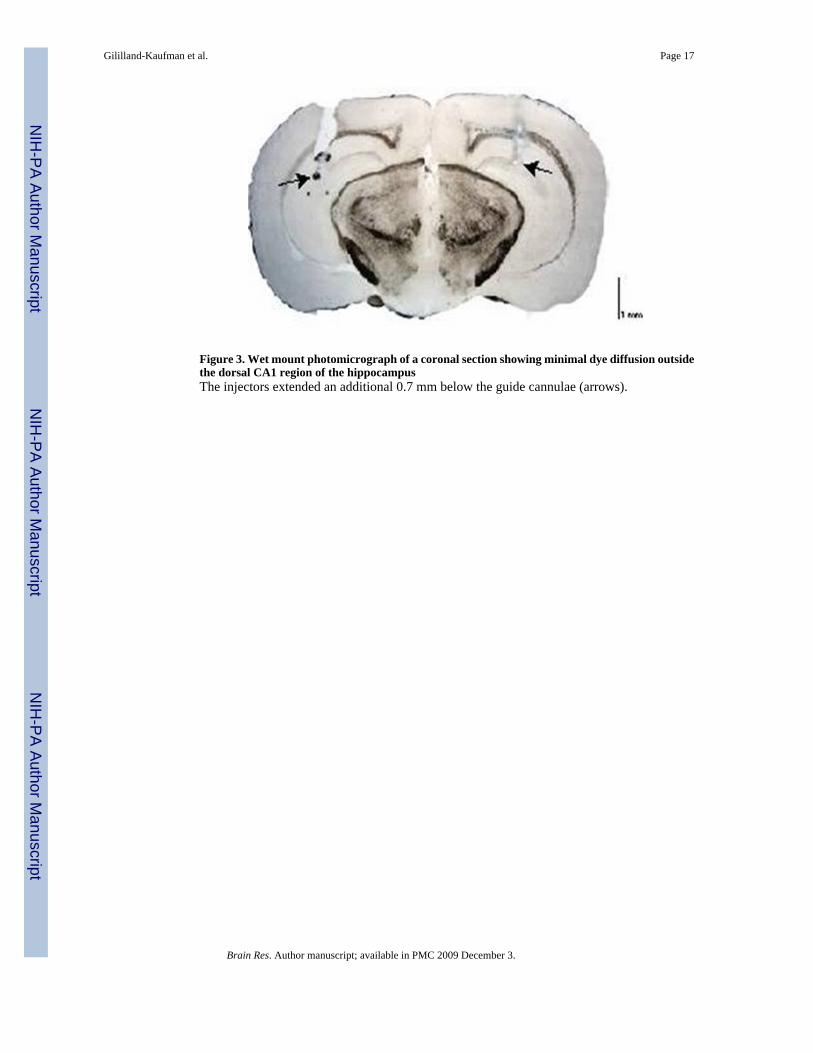
Figure 3. Wet mount photomicrograph of a coronal section showing minimal dye diffusion outsidethe dorsal CA1 region of the hippocampusThe injectors extended an additional 0.7 mm below the guide cannulae (arrows).
Gililland-Kaufman et al. Page 17
Brain Res. Author manuscript; available in PMC 2009 December 3.
NIH
-PA Author Manuscript
NIH
-PA Author Manuscript
NIH
-PA Author Manuscript
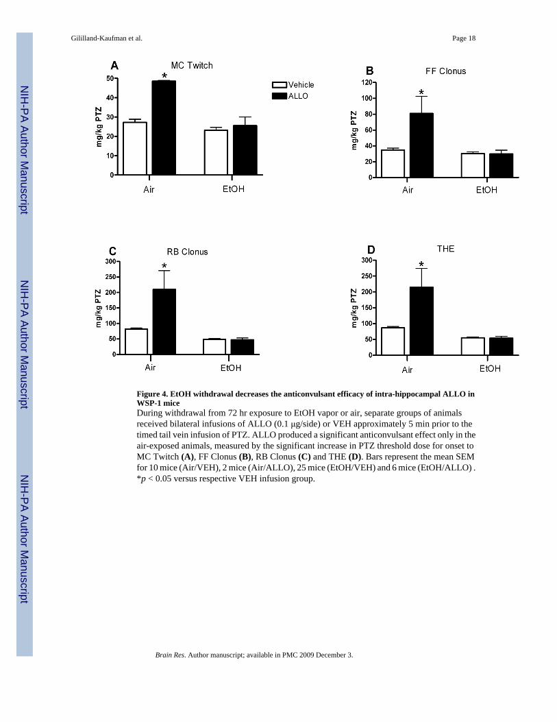
Figure 4. EtOH withdrawal decreases the anticonvulsant efficacy of intra-hippocampal ALLO inWSP-1 miceDuring withdrawal from 72 hr exposure to EtOH vapor or air, separate groups of animalsreceived bilateral infusions of ALLO (0.1 μg/side) or VEH approximately 5 min prior to thetimed tail vein infusion of PTZ. ALLO produced a significant anticonvulsant effect only in theair-exposed animals, measured by the significant increase in PTZ threshold dose for onset toMC Twitch (A), FF Clonus (B), RB Clonus (C) and THE (D). Bars represent the mean SEMfor 10 mice (Air/VEH), 2 mice (Air/ALLO), 25 mice (EtOH/VEH) and 6 mice (EtOH/ALLO) .*p < 0.05 versus respective VEH infusion group.
Gililland-Kaufman et al. Page 18
Brain Res. Author manuscript; available in PMC 2009 December 3.
NIH
-PA Author Manuscript
NIH
-PA Author Manuscript
NIH
-PA Author Manuscript
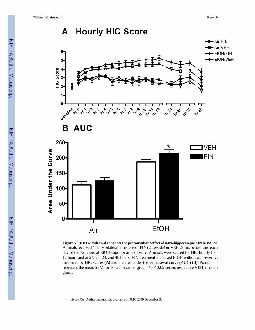
Figure 5. EtOH withdrawal enhances the proconvulsant effect of intra-hippocampal FIN in WSP-1Animals received 4 daily bilateral infusions of FIN (2 μg/side) or VEH 24 hrs before, and eachday of the 72 hours of EtOH vapor or air exposure. Animals were scored for HIC hourly for12 hours and at 24, 26, 28, and 48 hours. FIN treatment increased EtOH withdrawal severity,measured by HIC scores (A) and the area under the withdrawal curve (AUC) (B). Pointsrepresent the mean SEM for 16–20 mice per group. *p < 0.05 versus respective VEH infusiongroup.
Gililland-Kaufman et al. Page 19
Brain Res. Author manuscript; available in PMC 2009 December 3.
NIH
-PA Author Manuscript
NIH
-PA Author Manuscript
NIH
-PA Author Manuscript
Related Documents