Growth Factors, Cytokines, Cell Cycle Molecules The Neuronal Expression of MYC Causes a Neurodegenerative Phenotype in a Novel Transgenic Mouse Hyoung-gon Lee,* Gemma Casadesus, † Akihiko Nunomura, ‡ Xiongwei Zhu,* Rudy J. Castellani, § Sandy L. Richardson,* George Perry,* ¶ Dean W. Felsher, Robert B. Petersen,* † and Mark A. Smith* From the Departments of Pathology * and Neurosciences, † Case Western Reserve University, Cleveland, Ohio; the Interdisciplinary Graduate School of Medicine and Engineering, ‡ University of Yamanashi, Yamanashi, Japan; the Department of Pathology, § University of Maryland, Baltimore, Maryland; the College of Sciences, ¶ University of Texas at San Antonio, San Antonio, Texas; and the Department of Medicine and Pathology, Division of Oncology, Stanford University School of Medicine, Stanford, California Many different proteins associated with the cell cy- cle, including cyclins, cyclin-dependent kinases, and proto-oncogenes such as c-MYC (MYC), are in- creased in degenerating neurons. Consequently , an ectopic activation of the cell cycle machinery in neu- rons has emerged as a potential pathogenic mecha- nism of neuronal dysfunction and death in many neurodegenerative diseases , including Alzheimer’s disease. However , the exact role of cell cycle re-entry during disease pathogenesis is unclear , primarily be- cause of the lack of relevant research models to study the effects of cell cycle re-entry on mature neurons in vivo. To address this issue , we developed a new trans- genic mouse model in which forebrain neurons (CaMKII-MYC) can be induced to enter the cell cycle using the physiologically relevant proto-oncogene MYC to drive cell cycle re-entry. We show that such cell cycle re-entry results in neuronal cell death , gli- osis , and cognitive deficits. These findings provide compelling evidence that dysregulation of cell cycle re-entry results in neurodegeneration in vivo. Our current findings, coupled with those of previous reports, strengthen the hypothesis that neurode- generation in Alzheimer’s disease, similar to cellu- lar proliferation in cancer, is a disease that results from inappropriate cell cycle control. (Am J Pathol 2009, 174:891– 897; DOI: 10.2353/ajpath.2009.080583) Neurons in the normal brain are viewed as being quies- cent, not advancing past the G 0 period of the cell division cycle. However, in Alzheimer’s disease (AD), multiple lines of evidence suggest that neurons vulnerable to degeneration emerge from this postmitotic state—pheno- typically suggestive of cells that are cycling, rather than the normal terminally differentiated nondividing state. 1–5 Notably, cell cycle alterations are not limited to AD. For example, accumulation of hyperphosphorylated pRb (ppRb) and altered localization of E2F-1 also occurs in neurons in Parkinson’s disease and amyotrophic lateral sclerosis, sug- gesting that neurons re-enter the G 1 phase of the cell cycle. 6–8 The successful duplication of DNA, at least in AD, 9 –11 indicates that some neurons successfully complete S phase and precludes the possibility that the re-expression of various cell cycle markers observed is merely an epiphe- nomena caused by reduced proteosomal activity. 12 The causes or consequences of neuronal cell cycle are incompletely understood. Nonetheless, it is well known that the activation of cell cycle processes is part of the mechanism by which loss of trophic support during development leads to neuronal cell death, and there is Supported by the National Institutes of Health (grants AG031364, AG030096 and AG028679) and Alzheimer’s Association (NIRG-07-60164). H.L. and G.C. contributed equally to this study. Accepted for publication December 2, 2008. Supplemental material for this article can be found on http://ajp. amjpathol.org. M.A.S. is, or has in the past been, a paid consultant for, owns equity or stock options in, and/or receives grant funding from Neurotez, Neurop- harm, Edenland, Panacea Pharmaceuticals, and Voyager Pharmaceuti- cals. G.P. is a paid consultant for and/or owns equity or stock options in Takeda Pharmaceuticals, Voyager Pharmaceuticals, Panacea Pharma- ceuticals, and Neurotez Pharmaceuticals. Address reprint requests to Hyoung-gon Lee, Ph.D., or Mark A. Smith, Ph.D., Department of Pathology, Case Western Reserve University, 2103 Cornell Rd., Cleveland, OH 44106. E-mail: [email protected] and [email protected]. The American Journal of Pathology, Vol. 174, No. 3, March 2009 Copyright © American Society for Investigative Pathology DOI: 10.2353/ajpath.2009.080583 891

Welcome message from author
This document is posted to help you gain knowledge. Please leave a comment to let me know what you think about it! Share it to your friends and learn new things together.
Transcript
Growth Factors, Cytokines, Cell Cycle Molecules
The Neuronal Expression of MYC Causes aNeurodegenerative Phenotype in a NovelTransgenic Mouse
Hyoung-gon Lee,* Gemma Casadesus,†
Akihiko Nunomura,‡ Xiongwei Zhu,*Rudy J. Castellani,§ Sandy L. Richardson,*George Perry,*¶ Dean W. Felsher,�
Robert B. Petersen,*† and Mark A. Smith*From the Departments of Pathology * and Neurosciences,† Case
Western Reserve University, Cleveland, Ohio; the Interdisciplinary
Graduate School of Medicine and Engineering,‡ University of
Yamanashi, Yamanashi, Japan; the Department of Pathology,§
University of Maryland, Baltimore, Maryland; the College of
Sciences,¶ University of Texas at San Antonio, San Antonio,
Texas; and the Department of Medicine and Pathology,� Division
of Oncology, Stanford University School of Medicine,
Stanford, California
Many different proteins associated with the cell cy-cle , including cyclins , cyclin-dependent kinases ,and proto-oncogenes such as c-MYC (MYC), are in-creased in degenerating neurons. Consequently, anectopic activation of the cell cycle machinery in neu-rons has emerged as a potential pathogenic mecha-nism of neuronal dysfunction and death in manyneurodegenerative diseases, including Alzheimer’sdisease. However, the exact role of cell cycle re-entryduring disease pathogenesis is unclear, primarily be-cause of the lack of relevant research models to studythe effects of cell cycle re-entry on mature neurons invivo. To address this issue, we developed a new trans-genic mouse model in which forebrain neurons(CaMKII-MYC) can be induced to enter the cell cycleusing the physiologically relevant proto-oncogeneMYC to drive cell cycle re-entry. We show that suchcell cycle re-entry results in neuronal cell death, gli-osis, and cognitive deficits. These findings providecompelling evidence that dysregulation of cell cyclere-entry results in neurodegeneration in vivo. Ourcurrent findings , coupled with those of previousreports , strengthen the hypothesis that neurode-generation in Alzheimer’s disease , similar to cellu-lar proliferation in cancer , is a disease that results
from inappropriate cell cycle control. (Am J Pathol
2009, 174:891–897; DOI: 10.2353/ajpath.2009.080583)
Neurons in the normal brain are viewed as being quies-cent, not advancing past the G0 period of the cell divisioncycle. However, in Alzheimer’s disease (AD), multiplelines of evidence suggest that neurons vulnerable todegeneration emerge from this postmitotic state—pheno-typically suggestive of cells that are cycling, rather thanthe normal terminally differentiated nondividing state.1–5
Notably, cell cycle alterations are not limited to AD. Forexample, accumulation of hyperphosphorylated pRb (ppRb)and altered localization of E2F-1 also occurs in neurons inParkinson’s disease and amyotrophic lateral sclerosis, sug-gesting that neurons re-enter the G1 phase of the cellcycle.6–8 The successful duplication of DNA, at least inAD,9–11 indicates that some neurons successfully completeS phase and precludes the possibility that the re-expressionof various cell cycle markers observed is merely an epiphe-nomena caused by reduced proteosomal activity.12
The causes or consequences of neuronal cell cycleare incompletely understood. Nonetheless, it is wellknown that the activation of cell cycle processes is part ofthe mechanism by which loss of trophic support duringdevelopment leads to neuronal cell death, and there is
Supported by the National Institutes of Health (grants AG031364, AG030096and AG028679) and Alzheimer’s Association (NIRG-07-60164).
H.L. and G.C. contributed equally to this study.
Accepted for publication December 2, 2008.
Supplemental material for this article can be found on http://ajp.amjpathol.org.
M.A.S. is, or has in the past been, a paid consultant for, owns equity orstock options in, and/or receives grant funding from Neurotez, Neurop-harm, Edenland, Panacea Pharmaceuticals, and Voyager Pharmaceuti-cals. G.P. is a paid consultant for and/or owns equity or stock options inTakeda Pharmaceuticals, Voyager Pharmaceuticals, Panacea Pharma-ceuticals, and Neurotez Pharmaceuticals.
Address reprint requests to Hyoung-gon Lee, Ph.D., or Mark A. Smith,Ph.D., Department of Pathology, Case Western Reserve University, 2103Cornell Rd., Cleveland, OH 44106. E-mail: [email protected] [email protected].
The American Journal of Pathology, Vol. 174, No. 3, March 2009
Copyright © American Society for Investigative Pathology
DOI: 10.2353/ajpath.2009.080583
891
also evidence for a role for cell cycle regulators in neu-ronal death evoked by various stressors.13,14 In supportof the importance of cell cycle reactivation in mediatingcell death, when a powerful oncogene, SV40 T antigen, isexpressed specifically in maturing Purkinje cells or inforebrain neurons in transgenic mice, the cells replicatetheir DNA (ie, initiate cell cycle), but then subsequentlydegenerate and die.15
Unfortunately, studies performed to date in cell culturedo not faithfully reproduce age-related neurodegenera-tive diseases because the cell culture studies use em-bryo-derived primary neuronal cultures that do not accu-rately model adult neurons. Furthermore, in traditionaltransgenic mouse models,15 the expression of trans-genes during embryogenesis, which results in neurode-generation at an early age, is more akin to a develop-mental error than an age-dependent process associatedwith neurodegenerative disease. Although a recently de-veloped inducible SV40 T-antigen transgenic model16
addressed such developmental issues, SV40 T antigen isnot a physiologically relevant entity in any neurodegen-erative disease. As such, a faithful and physiologicallyrelevant model of neuronal cell cycle activation in matureneurons was lacking. To generate such a faithful model,in this study, we used the tetracycline-controlled trans-activator (tTA) system to generate bitransgenic mice(CaMKII-MYC) that can be induced to overexpress hu-man c-MYC (MYC) under the control of the CaMKII pro-moter that drives high transgene expression in forebrainneurons.17 MYC is well known for its oncogenic activityand overexpression of MYC is commonly associated withtumorigenesis. Indeed, previously we used the Tet sys-tem to generate mice that conditionally expressed a hu-man MYC transgene in hematopoietic cells and hepato-cytes18,19 and found that the sustained expression of theMYC transgene induced cell cycle progression and cul-minated in the formation of malignant T-cell lymphomas,acute myeloid leukemias, and hepatomas. Interestingly,and of physiological relevance to AD, phospho-MYC isincreased in dystrophic neurites and neurons with neu-rofibrillary tangles in AD and MYC is induced in degen-erating neurons in animal models of trauma and isch-emia.20 Therefore, MYC expression in transgenic mice inour model is not only a tool for inducing cell cycle re-entry inneurons, but MYC expression itself is pathophysiologicallyrelevant to AD and other neurodegenerative diseases.
Our findings reported in this study show that MYCinduction in CaMKII-MYC transgenic mice inducedneuronal-specific cell cycle re-entry, neurodegenera-tion, and, importantly, significant cognitive deficits. Thesefindings continue to strengthen our novel hypothesis thatneurodegeneration in AD, like cellular proliferation in can-cer, is a disease of inappropriate cell cycle control.
Materials and Methods
Animals
CaMKII-tTA17 and tet-o-MYC mice18 were mated to gen-erate CaMKII-MYC mice. Both CaMKII-tTA and tet-o-MYC
mice were established in the FVB strain. A doxycycline-containing diet (200 mg/kg; Bio-Serve, Frenchtown, NJ)was provided before weaning (4 to 6 weeks after birth),and, thereafter, MYC expression was induced by replac-ing the doxycycline diet with a regular diet for 5 weeks.After behavioral tests, the mice were sacrificed and thebrains were processed for immunocytochemistry andbiochemical analysis.
Immunocytochemistry
Immunocytochemistry was performed by the ABCmethod according to the manufacturer’s protocol (VectorLaboratories, Burlingame, CA). Formalin-fixed brainswere processed and embedded in paraffin. Six-�m-thickserial sections were cut, mounted onto slides, and rehy-drated according to standard protocols. All slides wererandomized and blinded with regard to genotype beforestaining and subsequent analysis. Briefly, slides wereimmersed in xylene, hydrated through graded ethanolsolutions, and endogenous peroxidase activity was elim-inated by incubation in 3% hydrogen peroxide for 30minutes. To reduce nonspecific binding, sections wereincubated for 30 minutes in 10% normal goat serum inTris-buffered saline (50 mmol/L Tris-HCl, 150 mmol/LNaCl, pH 7.6). After rinsing briefly with 1% normal goatserum in Tris-buffered saline, the sections were incu-bated overnight at 4°C with one of the following primaryantibody; anti-proliferating cell nuclear antigen (PCNA)mouse monoclonal antibody (Santa Cruz Biotechnology,Santa Cruz, CA), anti-Ki-67 rat monoclonal antibody(DAKO, Carpinteria, CA), anti-cyclin D1 rabbit monoclo-nal antibodies (Lab Vision, Fremont, CA), and anti-GFAPmouse monoclonal antibody (Chemicon, Billerica, MA).Antibodies were localized using 3–3�-diaminobenzidineas a chromogen (DAKO) after incubation with a second-ary antibody.
For double immunocytochemistry, the brain sectionswere incubated overnight at 4°C with anti-NeuN mousemonoclonal antibody (Millipore, Billerica, MA) or anti-MAP2 mouse monoclonal antibody (Millipore) in additionto anti-PCNA rabbit polyclonal antibody (Abcam, Cam-bridge, MA), anti-Ki-67 rat monoclonal antibody, or anti-BrdU rat monoclonal antibody (Abcam). Alexa Fluor 488-and 568-coupled secondary antibodies (Invitrogen,Carlsbad, CA) were used for detection. To exclude thepossibility of nonspecific reaction, all of the immunocyto-chemistry experiments contained at least one samplewithout a primary antibody. Images were acquiredthrough an AxioCam camera on an Axiovert 200M micro-scope (Zeiss, Thornwood, NY). Images were then ana-lyzed with the Axiovision software (Zeiss). Hematoxylinand eosin (H&E) staining and Nissl stains were also per-formed for routine histochemical and morphologicalanalyses.
BrdU Incorporation Analysis
DNA synthesis was directly examined by analysis ofBrdU incorporation. For this experiment, 50 mg/kg of
892 Lee et alAJP March 2009, Vol. 174, No. 3
the thymidine analog 5-bromo-2�-deoxyuridine (BrdU)(Sigma, St. Louis, MO) was intraperitoneally injected intoeach mouse. A day after injection, mice were anesthe-tized and perfused with phosphate-buffered saline (PBS)followed by 4% paraformaldehyde. Brains were removed,cut in the sagittal plane, embedded in paraffin, sec-tioned, and mounted on slides. The sections were treatedwith 2 N hydrochloric acid for 2 hours and then washedtwo times with 1� Tris-buffered saline (3 minutes each) toneutralize the acid. Incorporated BrdU was detected us-ing an anti-BrdU rat monoclonal antibody.
Reverse Transcriptase-Polymerase ChainReaction (RT-PCR)
The brain region was dissected immediately after collec-tion and used for mRNA isolation using an RNeasy kit(Qiagen, Valencia, CA). RT-PCR was performed usingthe RETROscript RT-PCR kit (Ambion, Austin, TX) ac-cording to the manufacturer’s instructions. Briefly, thepurified total RNA (100 ng) was reverse-transcribed withcloned MMLV reverse transcriptase (50 U) by incubatingat 44°C for 60 minutes, followed by heating at 92°C for 10minutes. The resulting single-stranded cDNA was thenamplified using a pair of primers for each target gene (25pmol) and TaqDNA polymerase (2.5 U; Roche, Indianap-olis, IN) for the indicated number of cycles of amplifica-tion (30 seconds at 94°C for denaturing, 30 seconds at61°C for primer annealing, and 30 seconds at 72°C forprimer extension). The RT-PCR products were then sub-jected to electrophoresis in a 1.5% agarose gel. Thenucleotide sequence used for each primer was asfollows: for c-myc: 5�-primer, 5�-TCTGGATCACCTTCT-GCTGG-3�; 3�-primer, 5�-CCTCTTGACATTTCTCCTCGG-3�;and for GAPDH: 5�-primer, 5�-ATGTTCCAGTATGACTC-CACTCAGG-3�; 3�-primer, 5�-GAAGACACCAGTAGACTC-CACGACA-3�. The specificity of PCR was confirmed bymeasuring the size of PCR product. In addition, the negativecontrol (ie, brain tissues from single transgenic mice) andpositive control (ie, SHSY5Y human neuroblastoma cell line)were tested to confirm the specificity of PCR reaction.
Terminal dUTP Nick-End Labeling (TUNEL)Analysis
Detection of 3�-OH termini of DNA strand breaks wasperformed on paraffin sections using an in situ cell deathdetection kit (Roche) following the recommendations ofthe manufacturer. Briefly, the tissue sections were treatedwith proteinase K (20 �g/ml in 10 mmol/L Tris-HCl, pH7.4) for 30 minutes at 37°C after rehydration. After rinsingslides with PBS, TUNEL reaction mixture containing ter-minal deoxynucleotidyl transferase and fluorescence-la-beled nucleotide was applied for 1 hour at 37°C. Positivesignals were observed directly using fluorescence mi-croscopy. The recommended positive and negative con-trols were applied in adjacent sections.
Behavioral Analysis
All mice (n � 8 in each group) were tested in a modifiedversion of the T-maze essentially as previously de-scribed.21 This behavioral task, used to test workingmemory, is based on spontaneous alternation behavior,which describes the innate tendency of mice to visit armsthat were not previously explored. All animals were al-lowed a 5-minute free run in the maze before testing tofamiliarize them with the apparatus and to foster alterna-tion behavior. On the testing day, mice were placed in thestart arm for 60 seconds before the gate was opened.Once the mouse entered an arm, the door was closed,and the animal was confined in that arm for 30 seconds;thereafter it was returned to the start arm for a new trial.Six consecutive trials were performed and alternationrates across the six trials were expressed as a relativepercentage based on the maximal alternation rate of100%, which occurred when a mouse never entered arepeated arm. Task achievement time (how long it tookthe animal to enter a goal arm after the gate opened) wasalso calculated.22
Statistical comparisons were performed by parametricanalysis using a one-way analysis of variance and/orStudent’s t-test, as appropriate, to determine significantdifferences across genotypes. The null hypothesis wasrejected at P � 0.05.
Results
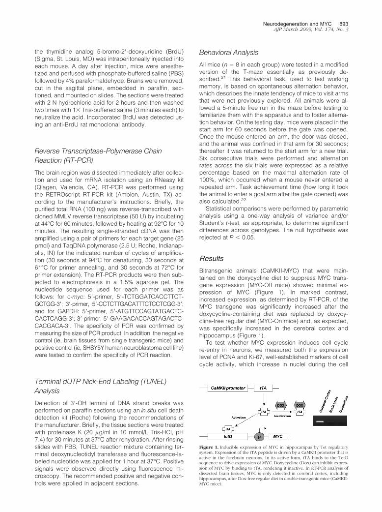
Bitransgenic animals (CaMKII-MYC) that were main-tained on the doxycycline diet to suppress MYC trans-gene expression (MYC-Off mice) showed minimal ex-pression of MYC (Figure 1). In marked contrast,increased expression, as determined by RT-PCR, of theMYC transgene was significantly increased after thedoxycycline-containing diet was replaced by doxycy-cline-free regular diet (MYC-On mice) and, as expected,was specifically increased in the cerebral cortex andhippocampus (Figure 1).
To test whether MYC expression induces cell cyclere-entry in neurons, we measured both the expressionlevel of PCNA and Ki-67, well-established markers of cellcycle activity, which increase in nuclei during the cell
Figure 1. Inducible expression of MYC in hippocampus by Tet regulatorysystem. Expression of the tTA peptide is driven by a CaMKII promoter that isactive in the forebrain neurons. In its active form, tTA binds to the TetOsequence to drive expression of MYC. Doxycycline (Dox) can inhibit expres-sion of MYC by binding to tTA, rendering it inactive. In RT-PCR analysis ofdissected brain tissues, MYC is only detected in cerebral cortex, includinghippocampus, after Dox-free regular diet in double-transgenic mice (CaMKII-MYC mice).
Neurodegeneration and MYC 893AJP March 2009, Vol. 174, No. 3
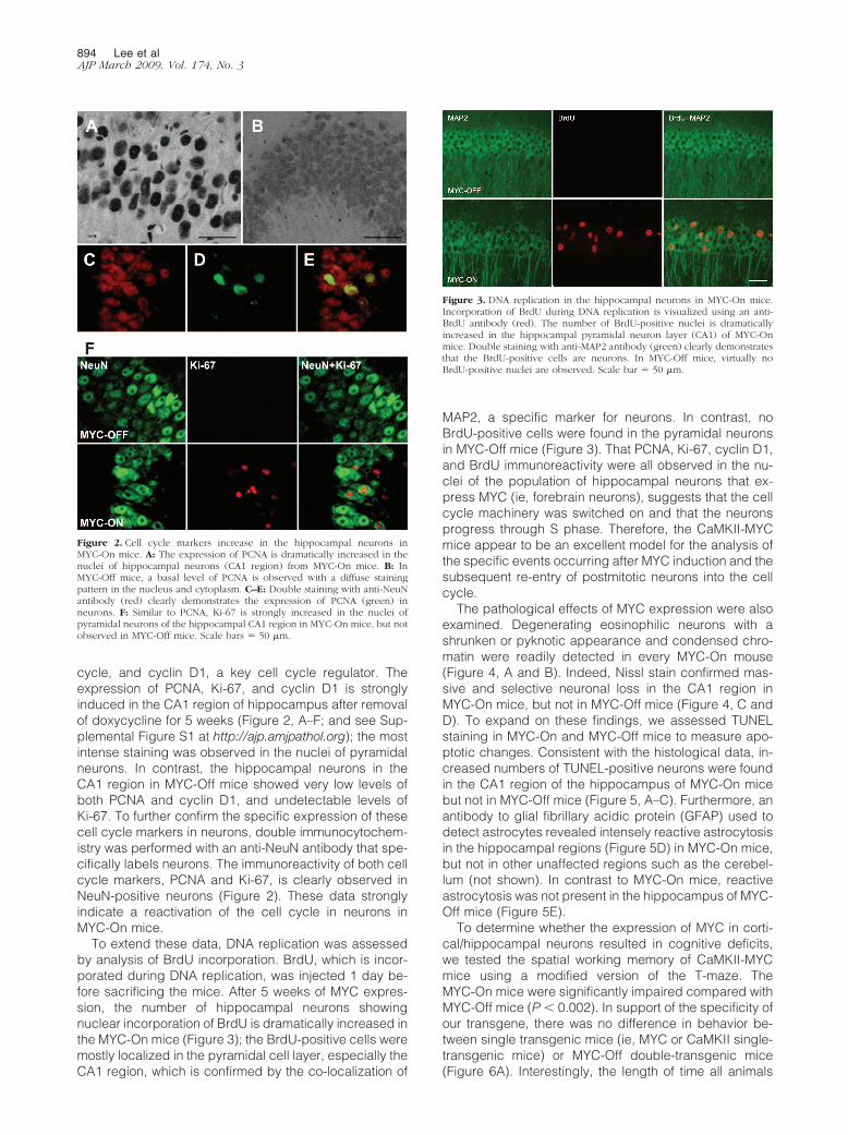
cycle, and cyclin D1, a key cell cycle regulator. Theexpression of PCNA, Ki-67, and cyclin D1 is stronglyinduced in the CA1 region of hippocampus after removalof doxycycline for 5 weeks (Figure 2, A–F; and see Sup-plemental Figure S1 at http://ajp.amjpathol.org); the mostintense staining was observed in the nuclei of pyramidalneurons. In contrast, the hippocampal neurons in theCA1 region in MYC-Off mice showed very low levels ofboth PCNA and cyclin D1, and undetectable levels ofKi-67. To further confirm the specific expression of thesecell cycle markers in neurons, double immunocytochem-istry was performed with an anti-NeuN antibody that spe-cifically labels neurons. The immunoreactivity of both cellcycle markers, PCNA and Ki-67, is clearly observed inNeuN-positive neurons (Figure 2). These data stronglyindicate a reactivation of the cell cycle in neurons inMYC-On mice.
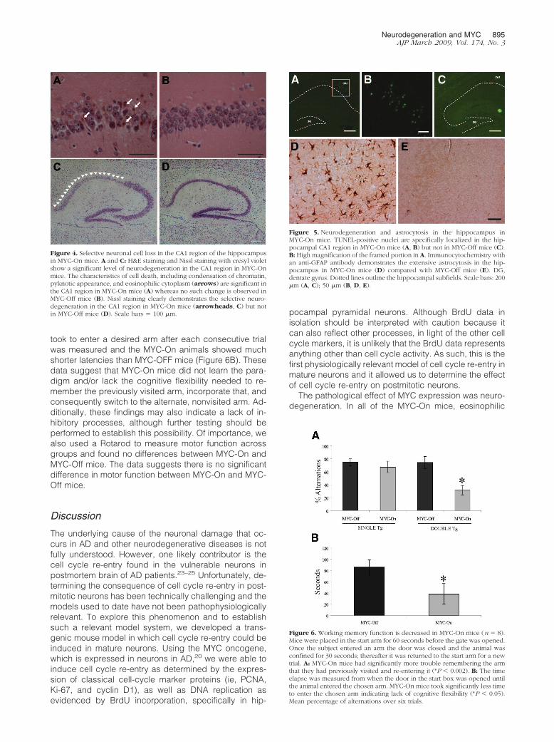
To extend these data, DNA replication was assessedby analysis of BrdU incorporation. BrdU, which is incor-porated during DNA replication, was injected 1 day be-fore sacrificing the mice. After 5 weeks of MYC expres-sion, the number of hippocampal neurons showingnuclear incorporation of BrdU is dramatically increased inthe MYC-On mice (Figure 3); the BrdU-positive cells weremostly localized in the pyramidal cell layer, especially theCA1 region, which is confirmed by the co-localization of
MAP2, a specific marker for neurons. In contrast, noBrdU-positive cells were found in the pyramidal neuronsin MYC-Off mice (Figure 3). That PCNA, Ki-67, cyclin D1,and BrdU immunoreactivity were all observed in the nu-clei of the population of hippocampal neurons that ex-press MYC (ie, forebrain neurons), suggests that the cellcycle machinery was switched on and that the neuronsprogress through S phase. Therefore, the CaMKII-MYCmice appear to be an excellent model for the analysis ofthe specific events occurring after MYC induction and thesubsequent re-entry of postmitotic neurons into the cellcycle.
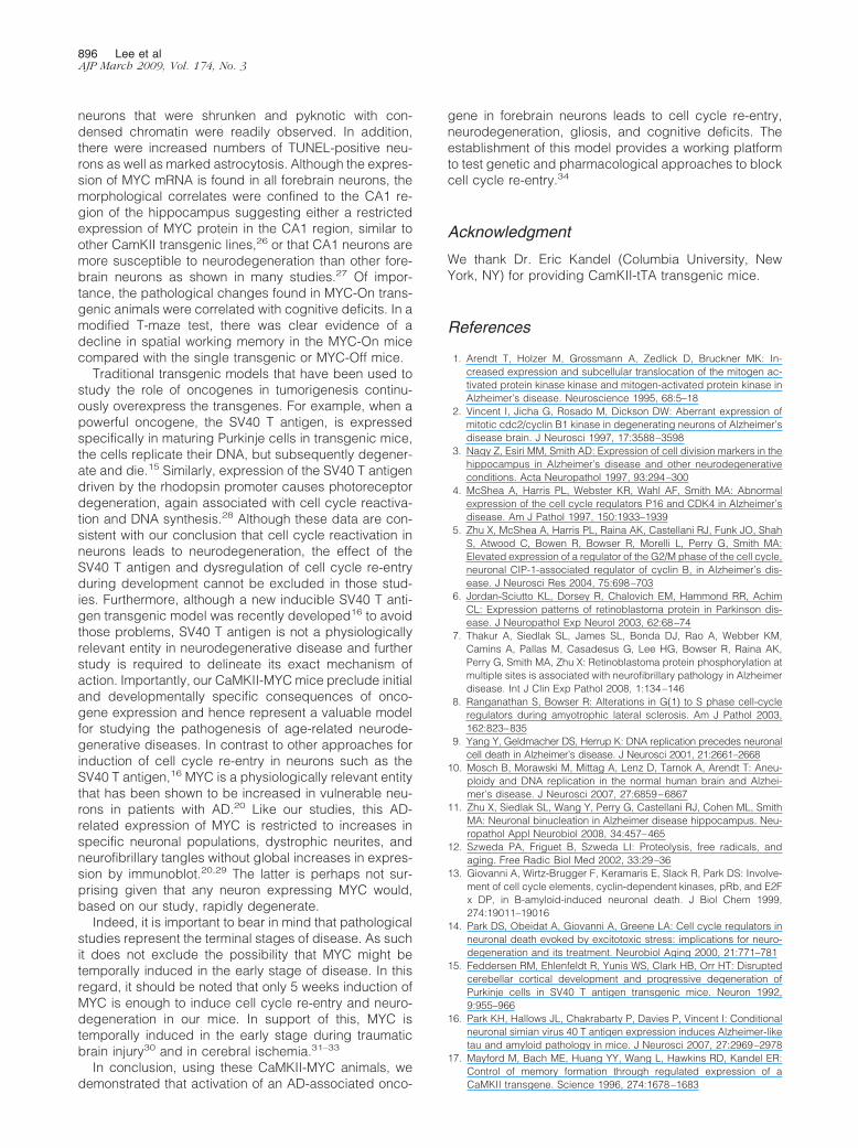
The pathological effects of MYC expression were alsoexamined. Degenerating eosinophilic neurons with ashrunken or pyknotic appearance and condensed chro-matin were readily detected in every MYC-On mouse(Figure 4, A and B). Indeed, Nissl stain confirmed mas-sive and selective neuronal loss in the CA1 region inMYC-On mice, but not in MYC-Off mice (Figure 4, C andD). To expand on these findings, we assessed TUNELstaining in MYC-On and MYC-Off mice to measure apo-ptotic changes. Consistent with the histological data, in-creased numbers of TUNEL-positive neurons were foundin the CA1 region of the hippocampus of MYC-On micebut not in MYC-Off mice (Figure 5, A–C). Furthermore, anantibody to glial fibrillary acidic protein (GFAP) used todetect astrocytes revealed intensely reactive astrocytosisin the hippocampal regions (Figure 5D) in MYC-On mice,but not in other unaffected regions such as the cerebel-lum (not shown). In contrast to MYC-On mice, reactiveastrocytosis was not present in the hippocampus of MYC-Off mice (Figure 5E).
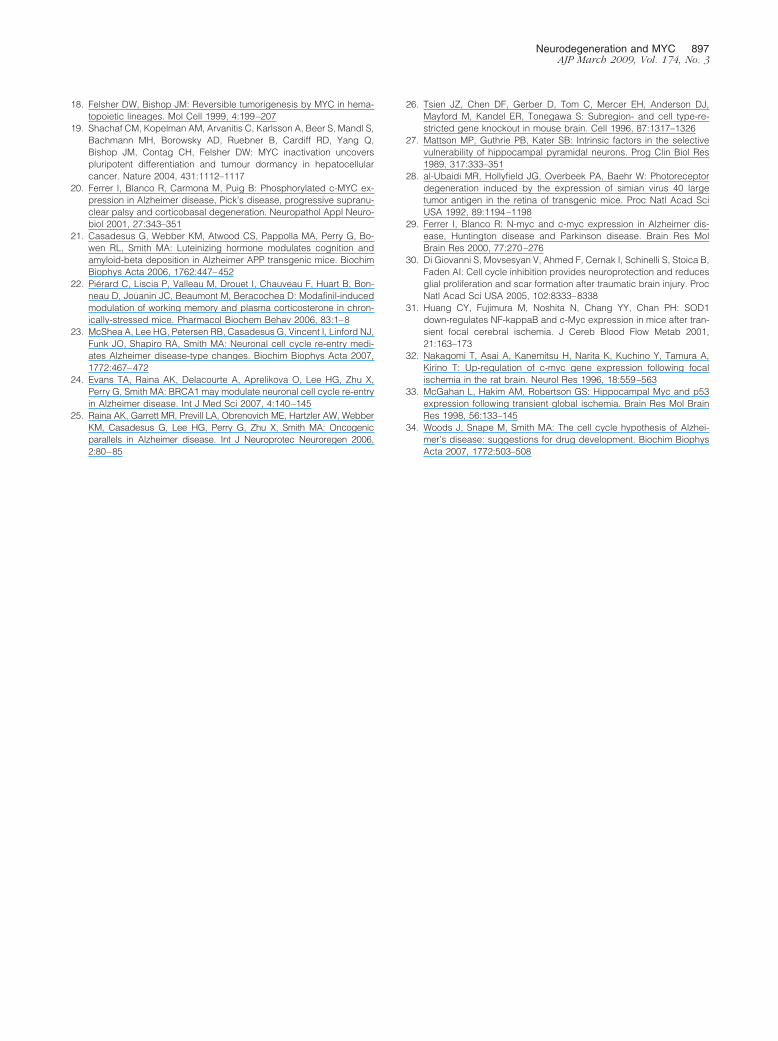
To determine whether the expression of MYC in corti-cal/hippocampal neurons resulted in cognitive deficits,we tested the spatial working memory of CaMKII-MYCmice using a modified version of the T-maze. TheMYC-On mice were significantly impaired compared withMYC-Off mice (P � 0.002). In support of the specificity ofour transgene, there was no difference in behavior be-tween single transgenic mice (ie, MYC or CaMKII single-transgenic mice) or MYC-Off double-transgenic mice(Figure 6A). Interestingly, the length of time all animals
Figure 2. Cell cycle markers increase in the hippocampal neurons inMYC-On mice. A: The expression of PCNA is dramatically increased in thenuclei of hippocampal neurons (CA1 region) from MYC-On mice. B: InMYC-Off mice, a basal level of PCNA is observed with a diffuse stainingpattern in the nucleus and cytoplasm. C–E: Double staining with anti-NeuNantibody (red) clearly demonstrates the expression of PCNA (green) inneurons. F: Similar to PCNA, Ki-67 is strongly increased in the nuclei ofpyramidal neurons of the hippocampal CA1 region in MYC-On mice, but notobserved in MYC-Off mice. Scale bars � 50 �m.
Figure 3. DNA replication in the hippocampal neurons in MYC-On mice.Incorporation of BrdU during DNA replication is visualized using an anti-BrdU antibody (red). The number of BrdU-positive nuclei is dramaticallyincreased in the hippocampal pyramidal neuron layer (CA1) of MYC-Onmice. Double staining with anti-MAP2 antibody (green) clearly demonstratesthat the BrdU-positive cells are neurons. In MYC-Off mice, virtually noBrdU-positive nuclei are observed. Scale bar � 50 �m.
894 Lee et alAJP March 2009, Vol. 174, No. 3
took to enter a desired arm after each consecutive trialwas measured and the MYC-On animals showed muchshorter latencies than MYC-OFF mice (Figure 6B). Thesedata suggest that MYC-On mice did not learn the para-digm and/or lack the cognitive flexibility needed to re-member the previously visited arm, incorporate that, andconsequently switch to the alternate, nonvisited arm. Ad-ditionally, these findings may also indicate a lack of in-hibitory processes, although further testing should beperformed to establish this possibility. Of importance, wealso used a Rotarod to measure motor function acrossgroups and found no differences between MYC-On andMYC-Off mice. The data suggests there is no significantdifference in motor function between MYC-On and MYC-Off mice.
Discussion
The underlying cause of the neuronal damage that oc-curs in AD and other neurodegenerative diseases is notfully understood. However, one likely contributor is thecell cycle re-entry found in the vulnerable neurons inpostmortem brain of AD patients.23–25 Unfortunately, de-termining the consequence of cell cycle re-entry in post-mitotic neurons has been technically challenging and themodels used to date have not been pathophysiologicallyrelevant. To explore this phenomenon and to establishsuch a relevant model system, we developed a trans-genic mouse model in which cell cycle re-entry could beinduced in mature neurons. Using the MYC oncogene,which is expressed in neurons in AD,20 we were able toinduce cell cycle re-entry as determined by the expres-sion of classical cell-cycle marker proteins (ie, PCNA,Ki-67, and cyclin D1), as well as DNA replication asevidenced by BrdU incorporation, specifically in hip-
pocampal pyramidal neurons. Although BrdU data inisolation should be interpreted with caution because itcan also reflect other processes, in light of the other cellcycle markers, it is unlikely that the BrdU data representsanything other than cell cycle activity. As such, this is thefirst physiologically relevant model of cell cycle re-entry inmature neurons and it allowed us to determine the effectof cell cycle re-entry on postmitotic neurons.
The pathological effect of MYC expression was neuro-degeneration. In all of the MYC-On mice, eosinophilic
Figure 4. Selective neuronal cell loss in the CA1 region of the hippocampusin MYC-On mice. A and C: H&E staining and Nissl staining with cresyl violetshow a significant level of neurodegeneration in the CA1 region in MYC-Onmice. The characteristics of cell death, including condensation of chromatin,pyknotic appearance, and eosinophilic cytoplasm (arrows) are significant inthe CA1 region in MYC-On mice (A) whereas no such change is observed inMYC-Off mice (B). Nissl staining clearly demonstrates the selective neuro-degeneration in the CA1 region in MYC-On mice (arrowheads, C) but notin MYC-Off mice (D). Scale bars � 100 �m.
Figure 5. Neurodegeneration and astrocytosis in the hippocampus inMYC-On mice. TUNEL-positive nuclei are specifically localized in the hip-pocampal CA1 region in MYC-On mice (A, B) but not in MYC-Off mice (C).B: High magnification of the framed portion in A. Immunocytochemistry withan anti-GFAP antibody demonstrates the extensive astrocytosis in the hip-pocampus in MYC-On mice (D) compared with MYC-Off mice (E). DG,dentate gyrus. Dotted lines outline the hippocampal subfields. Scale bars: 200�m (A, C); 50 �m (B, D, E).
Figure 6. Working memory function is decreased in MYC-On mice (n � 8).Mice were placed in the start arm for 60 seconds before the gate was opened.Once the subject entered an arm the door was closed and the animal wasconfined for 30 seconds; thereafter it was returned to the start arm for a newtrial. A: MYC-On mice had significantly more trouble remembering the armthat they had previously visited and re-entering it (*P � 0.002). B: The timeelapse was measured from when the door in the start box was opened untilthe animal entered the chosen arm. MYC-On mice took significantly less timeto enter the chosen arm indicating lack of cognitive flexibility (*P � 0.05).Mean percentage of alternations over six trials.
Neurodegeneration and MYC 895AJP March 2009, Vol. 174, No. 3
neurons that were shrunken and pyknotic with con-densed chromatin were readily observed. In addition,there were increased numbers of TUNEL-positive neu-rons as well as marked astrocytosis. Although the expres-sion of MYC mRNA is found in all forebrain neurons, themorphological correlates were confined to the CA1 re-gion of the hippocampus suggesting either a restrictedexpression of MYC protein in the CA1 region, similar toother CamKII transgenic lines,26 or that CA1 neurons aremore susceptible to neurodegeneration than other fore-brain neurons as shown in many studies.27 Of impor-tance, the pathological changes found in MYC-On trans-genic animals were correlated with cognitive deficits. In amodified T-maze test, there was clear evidence of adecline in spatial working memory in the MYC-On micecompared with the single transgenic or MYC-Off mice.
Traditional transgenic models that have been used tostudy the role of oncogenes in tumorigenesis continu-ously overexpress the transgenes. For example, when apowerful oncogene, the SV40 T antigen, is expressedspecifically in maturing Purkinje cells in transgenic mice,the cells replicate their DNA, but subsequently degener-ate and die.15 Similarly, expression of the SV40 T antigendriven by the rhodopsin promoter causes photoreceptordegeneration, again associated with cell cycle reactiva-tion and DNA synthesis.28 Although these data are con-sistent with our conclusion that cell cycle reactivation inneurons leads to neurodegeneration, the effect of theSV40 T antigen and dysregulation of cell cycle re-entryduring development cannot be excluded in those stud-ies. Furthermore, although a new inducible SV40 T anti-gen transgenic model was recently developed16 to avoidthose problems, SV40 T antigen is not a physiologicallyrelevant entity in neurodegenerative disease and furtherstudy is required to delineate its exact mechanism ofaction. Importantly, our CaMKII-MYC mice preclude initialand developmentally specific consequences of onco-gene expression and hence represent a valuable modelfor studying the pathogenesis of age-related neurode-generative diseases. In contrast to other approaches forinduction of cell cycle re-entry in neurons such as theSV40 T antigen,16 MYC is a physiologically relevant entitythat has been shown to be increased in vulnerable neu-rons in patients with AD.20 Like our studies, this AD-related expression of MYC is restricted to increases inspecific neuronal populations, dystrophic neurites, andneurofibrillary tangles without global increases in expres-sion by immunoblot.20,29 The latter is perhaps not sur-prising given that any neuron expressing MYC would,based on our study, rapidly degenerate.
Indeed, it is important to bear in mind that pathologicalstudies represent the terminal stages of disease. As suchit does not exclude the possibility that MYC might betemporally induced in the early stage of disease. In thisregard, it should be noted that only 5 weeks induction ofMYC is enough to induce cell cycle re-entry and neuro-degeneration in our mice. In support of this, MYC istemporally induced in the early stage during traumaticbrain injury30 and in cerebral ischemia.31–33
In conclusion, using these CaMKII-MYC animals, wedemonstrated that activation of an AD-associated onco-
gene in forebrain neurons leads to cell cycle re-entry,neurodegeneration, gliosis, and cognitive deficits. Theestablishment of this model provides a working platformto test genetic and pharmacological approaches to blockcell cycle re-entry.34
Acknowledgment
We thank Dr. Eric Kandel (Columbia University, NewYork, NY) for providing CamKII-tTA transgenic mice.
References
1. Arendt T, Holzer M, Grossmann A, Zedlick D, Bruckner MK: In-creased expression and subcellular translocation of the mitogen ac-tivated protein kinase kinase and mitogen-activated protein kinase inAlzheimer’s disease. Neuroscience 1995, 68:5–18
2. Vincent I, Jicha G, Rosado M, Dickson DW: Aberrant expression ofmitotic cdc2/cyclin B1 kinase in degenerating neurons of Alzheimer’sdisease brain. J Neurosci 1997, 17:3588–3598
3. Nagy Z, Esiri MM, Smith AD: Expression of cell division markers in thehippocampus in Alzheimer’s disease and other neurodegenerativeconditions. Acta Neuropathol 1997, 93:294–300
4. McShea A, Harris PL, Webster KR, Wahl AF, Smith MA: Abnormalexpression of the cell cycle regulators P16 and CDK4 in Alzheimer’sdisease. Am J Pathol 1997, 150:1933–1939
5. Zhu X, McShea A, Harris PL, Raina AK, Castellani RJ, Funk JO, ShahS, Atwood C, Bowen R, Bowser R, Morelli L, Perry G, Smith MA:Elevated expression of a regulator of the G2/M phase of the cell cycle,neuronal CIP-1-associated regulator of cyclin B, in Alzheimer’s dis-ease. J Neurosci Res 2004, 75:698–703
6. Jordan-Sciutto KL, Dorsey R, Chalovich EM, Hammond RR, AchimCL: Expression patterns of retinoblastoma protein in Parkinson dis-ease. J Neuropathol Exp Neurol 2003, 62:68–74
7. Thakur A, Siedlak SL, James SL, Bonda DJ, Rao A, Webber KM,Camins A, Pallas M, Casadesus G, Lee HG, Bowser R, Raina AK,Perry G, Smith MA, Zhu X: Retinoblastoma protein phosphorylation atmultiple sites is associated with neurofibrillary pathology in Alzheimerdisease. Int J Clin Exp Pathol 2008, 1:134–146
8. Ranganathan S, Bowser R: Alterations in G(1) to S phase cell-cycleregulators during amyotrophic lateral sclerosis. Am J Pathol 2003,162:823–835
9. Yang Y, Geldmacher DS, Herrup K: DNA replication precedes neuronalcell death in Alzheimer’s disease. J Neurosci 2001, 21:2661–2668
10. Mosch B, Morawski M, Mittag A, Lenz D, Tarnok A, Arendt T: Aneu-ploidy and DNA replication in the normal human brain and Alzhei-mer’s disease. J Neurosci 2007, 27:6859–6867
11. Zhu X, Siedlak SL, Wang Y, Perry G, Castellani RJ, Cohen ML, SmithMA: Neuronal binucleation in Alzheimer disease hippocampus. Neu-ropathol Appl Neurobiol 2008, 34:457–465
12. Szweda PA, Friguet B, Szweda LI: Proteolysis, free radicals, andaging. Free Radic Biol Med 2002, 33:29–36
13. Giovanni A, Wirtz-Brugger F, Keramaris E, Slack R, Park DS: Involve-ment of cell cycle elements, cyclin-dependent kinases, pRb, and E2Fx DP, in B-amyloid-induced neuronal death. J Biol Chem 1999,274:19011–19016
14. Park DS, Obeidat A, Giovanni A, Greene LA: Cell cycle regulators inneuronal death evoked by excitotoxic stress: implications for neuro-degeneration and its treatment. Neurobiol Aging 2000, 21:771–781
15. Feddersen RM, Ehlenfeldt R, Yunis WS, Clark HB, Orr HT: Disruptedcerebellar cortical development and progressive degeneration ofPurkinje cells in SV40 T antigen transgenic mice. Neuron 1992,9:955–966
16. Park KH, Hallows JL, Chakrabarty P, Davies P, Vincent I: Conditionalneuronal simian virus 40 T antigen expression induces Alzheimer-liketau and amyloid pathology in mice. J Neurosci 2007, 27:2969–2978
17. Mayford M, Bach ME, Huang YY, Wang L, Hawkins RD, Kandel ER:Control of memory formation through regulated expression of aCaMKII transgene. Science 1996, 274:1678–1683
896 Lee et alAJP March 2009, Vol. 174, No. 3
18. Felsher DW, Bishop JM: Reversible tumorigenesis by MYC in hema-topoietic lineages. Mol Cell 1999, 4:199–207
19. Shachaf CM, Kopelman AM, Arvanitis C, Karlsson A, Beer S, Mandl S,Bachmann MH, Borowsky AD, Ruebner B, Cardiff RD, Yang Q,Bishop JM, Contag CH, Felsher DW: MYC inactivation uncoverspluripotent differentiation and tumour dormancy in hepatocellularcancer. Nature 2004, 431:1112–1117
20. Ferrer I, Blanco R, Carmona M, Puig B: Phosphorylated c-MYC ex-pression in Alzheimer disease, Pick’s disease, progressive supranu-clear palsy and corticobasal degeneration. Neuropathol Appl Neuro-biol 2001, 27:343–351
21. Casadesus G, Webber KM, Atwood CS, Pappolla MA, Perry G, Bo-wen RL, Smith MA: Luteinizing hormone modulates cognition andamyloid-beta deposition in Alzheimer APP transgenic mice. BiochimBiophys Acta 2006, 1762:447–452
22. Pierard C, Liscia P, Valleau M, Drouet I, Chauveau F, Huart B, Bon-neau D, Jouanin JC, Beaumont M, Beracochea D: Modafinil-inducedmodulation of working memory and plasma corticosterone in chron-ically-stressed mice. Pharmacol Biochem Behav 2006, 83:1–8
23. McShea A, Lee HG, Petersen RB, Casadesus G, Vincent I, Linford NJ,Funk JO, Shapiro RA, Smith MA: Neuronal cell cycle re-entry medi-ates Alzheimer disease-type changes. Biochim Biophys Acta 2007,1772:467–472
24. Evans TA, Raina AK, Delacourte A, Aprelikova O, Lee HG, Zhu X,Perry G, Smith MA: BRCA1 may modulate neuronal cell cycle re-entryin Alzheimer disease. Int J Med Sci 2007, 4:140–145
25. Raina AK, Garrett MR, Previll LA, Obrenovich ME, Hartzler AW, WebberKM, Casadesus G, Lee HG, Perry G, Zhu X, Smith MA: Oncogenicparallels in Alzheimer disease. Int J Neuroprotec Neuroregen 2006,2:80–85
26. Tsien JZ, Chen DF, Gerber D, Tom C, Mercer EH, Anderson DJ,Mayford M, Kandel ER, Tonegawa S: Subregion- and cell type-re-stricted gene knockout in mouse brain. Cell 1996, 87:1317–1326
27. Mattson MP, Guthrie PB, Kater SB: Intrinsic factors in the selectivevulnerability of hippocampal pyramidal neurons. Prog Clin Biol Res1989, 317:333–351
28. al-Ubaidi MR, Hollyfield JG, Overbeek PA, Baehr W: Photoreceptordegeneration induced by the expression of simian virus 40 largetumor antigen in the retina of transgenic mice. Proc Natl Acad SciUSA 1992, 89:1194–1198
29. Ferrer I, Blanco R: N-myc and c-myc expression in Alzheimer dis-ease, Huntington disease and Parkinson disease. Brain Res MolBrain Res 2000, 77:270–276
30. Di Giovanni S, Movsesyan V, Ahmed F, Cernak I, Schinelli S, Stoica B,Faden AI: Cell cycle inhibition provides neuroprotection and reducesglial proliferation and scar formation after traumatic brain injury. ProcNatl Acad Sci USA 2005, 102:8333–8338
31. Huang CY, Fujimura M, Noshita N, Chang YY, Chan PH: SOD1down-regulates NF-kappaB and c-Myc expression in mice after tran-sient focal cerebral ischemia. J Cereb Blood Flow Metab 2001,21:163–173
32. Nakagomi T, Asai A, Kanemitsu H, Narita K, Kuchino Y, Tamura A,Kirino T: Up-regulation of c-myc gene expression following focalischemia in the rat brain. Neurol Res 1996, 18:559–563
33. McGahan L, Hakim AM, Robertson GS: Hippocampal Myc and p53expression following transient global ischemia. Brain Res Mol BrainRes 1998, 56:133–145
34. Woods J, Snape M, Smith MA: The cell cycle hypothesis of Alzhei-mer’s disease: suggestions for drug development. Biochim BiophysActa 2007, 1772:503–508
Neurodegeneration and MYC 897AJP March 2009, Vol. 174, No. 3
Related Documents