THE NEURAL TISSUE PROVOCATION TEST AS A DIAGNOSTIC TOOL IN THE ASSESSMENT OF CERVICOBRACHIAL PAIN DISORDERS: A CRITICAL APPRAISAL Mariam Hamouda University of Wales College of Medicine A dissertation submitted in partial fulfilment of the requirements for the degree of MSc in Pain Management in the University of Wales College of Medicine, Cardiff July 2003

Welcome message from author
This document is posted to help you gain knowledge. Please leave a comment to let me know what you think about it! Share it to your friends and learn new things together.
Transcript
TTHHEE NNEEUURRAALL TTIISSSSUUEE PPRROOVVOOCCAATTIIOONN TTEESSTT AASS AA DDIIAAGGNNOOSSTTIICC TTOOOOLL IINN TTHHEE AASSSSEESSSSMMEENNTT OOFF
CCEERRVVIICCOOBBRRAACCHHIIAALL PPAAIINN DDIISSOORRDDEERRSS:: AA CCRRIITTIICCAALL AAPPPPRRAAIISSAALL
Mariam Hamouda University of Wales College of Medicine
A dissertation submitted in partial fulfilment of the requirements for the degree of MSc in Pain Management in the University of Wales College of Medicine, Cardiff
July 2003

ii
'(&/$5$7,21�
This work has not previously been accepted in substance for any degree and is not being concurrently submitted in candidature for any degree.
Signed
Date
67$7(0(17���
This dissertation is being submitted in partial fulfilment of the requirements for the degree of MSc in Pain Management.
Signed
Date
67$7(0(17���
This dissertation is the result of my own independent work/investigation, except where otherwise stated. Other sources are acknowledged in brackets giving explicit references. A reference list and bibliography is appended.
Signed
Date
67$7(0(17���
I hereby give consent for my dissertation, if accepted, to be available for photo-copying and for inter-library loan, and for the title and summary to be made available to outside organisations.
Signed
Date
iii
6800$5<�
This thesis will analyse research that is concerned with assessing the reliability and validity of the neural tissue provocation test for the upper quadrant as a diagnostic test. The neural tissue provocation test is a technique that is used to identify the presence of sensitised peripheral nerves. It consists of a sequence of multiple joint movements that may provoke sensory responses in individuals with sensitised neural tissue by elongating the length of the nerve bedding.
In clinical practice patients frequently present with diffuse symptoms in their neck and upper extremity of unknown aetiology, and report of positive symptoms asso-ciated with peripheral nerve injury. Peripheral nerves with relative minor damage to their nerve fibres are characterised by an increased mechanosensitivity and may react to mechanical stimulation with sensory responses and with impaired compli-ance to movement. Assessing the mechanosensitivity of the neural structures by neurodynamic tests is a relatively new but increasingly important technique among orthopaedic physical therapists. In response to contemporary calls for the use of evidence-based-medicine and for professional accountability a growing body of scientific evidence has emerged creating the grounds for the use of this test in clinical practice.
On the basis of the reviewed anatomical and clinical evidence the median biased neural tissue provocation test can be proposed as a useful diagnostic test within the assessment of cervicobrachial pain disorders. However, reports on low reliability are due to handling irregularities when performing this complex multiple joint test, which calls for clearer operational definitions and for a standardisation in its execution.

iv
$&.12:/('*(0(176�
I would like to express my thanks to the tutors of the course MSc Pain Management of the Department of Anaesthetics and Intensive Care Medicine, University of Wales College of Medicine for their support and assistance throughout the course of the study. My thanks go especially to Ann Taylor, who always made me work a little harder and who really pushed me into a critical and analytical approach to facts.
I would also like to thank John Langendoen for supporting me with literature on the early days of neural tissue testing and with his encouragements, Gert-Jan Kleinren-sink for sending me his articles, Brigitte van der Heide and Michel Coppieters for valuable discussions over the internet, and especially Gary Keil who always knew I had it in me. Most of all I want to thank Franz Barrios without whose continual support this work could not have been brought to an end, and who changed my life for the better.
v
7$%/(�2)�&217(176�
DECLARATION .................................................................................................. II
SUMMARY......................................................................................................... III
ACKNOWLEDGEMENTS ................................................................................. IV
TABLE OF CONTENTS..................................................................................... V
LIST OF TABLES.............................................................................................. VI
LIST OF FIGURES .......................................................................................... VII
LIST OF ABBREVIATIONS............................................................................. VIII
1. INTRODUCTION ..........................................................................................1
2. EPIDEMIOLOGY OF CERVICOBRACHIAL PAIN........................................7
3. PATHOBIOLOGICAL ASPECTS OF CERVICOBRACHIAL PAIN .............12
3.1. Anatomical and biomechanical considerations...................................12 3.2. Neurophysiological considerations.....................................................20 3.3. Effects of compression on nerve fibres ..............................................23 3.4. Pain mechanisms as in minor nerve injuries ......................................27
4. THE HISTORY OF NEURAL TISSUE PROVOCATION TESTING ............31
5. CRITICAL ANALYSIS OF ULNT RESEARCH............................................42
5.1. Methodological considerations on ULNT research.............................42 ������ 5HOLDELOLW\�RI�H[DPLQDWLRQ�SURFHGXUHV �������������������������������������� ������ 2SHUDWLRQDO�GHILQLWLRQV�LQ�8/17�UHVHDUFK�������������������������������� ������ &RQWURO�IRU�LQGHSHQGHQW�YDULDEOHV�������������������������������������������� ������ 'HILQLQJ�QRUPDO�UHVSRQVHV������������������������������������������������������ ������ 9DOLGDWLRQ�RI�GLDJQRVWLF�WHVWV ���������������������������������������������������
5.2. Investigating the validity of the ULNT.................................................54 ������ %LRPHFKDQLFDO�ILQGLQJV�GXULQJ�WKH�8/17�LQ�KXPDQ�FDGDYHU�
VWXGLHV ������������������������������������������������������������������������������������� ������ 4XDQWLILDEOH�PHDVXUHPHQWV�LQ�QRUPDWLYH�8/17�VWXGLHV���������� ������ 7KH�XVH�RI�WKH�8/17�LQ�FOLQLFDO�SUDFWLFH ����������������������������������
6. DISCUSSION .............................................................................................83
6.1. Implications for clinical practice..........................................................83 6.2. Limitations ..........................................................................................86
7. CONCLUSION............................................................................................89
7.1. Recommendations .............................................................................91 REFERENCES .................................................................................................93
APPENDIX A ....................................................................................................... I
APPENDIX B ...................................................................................................... II
APPENDIX C ................................................................................................... XII
�
vi
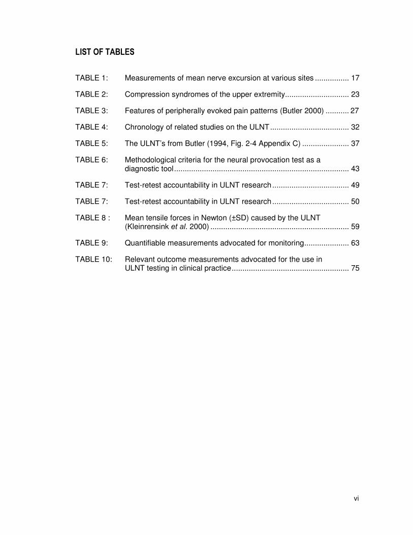
/,67�2)�7$%/(6�
TABLE 1: Measurements of mean nerve excursion at various sites ................ 17
TABLE 2: Compression syndromes of the upper extremity.............................. 23
TABLE 3: Features of peripherally evoked pain patterns (Butler 2000) ........... 27
TABLE 4: Chronology of related studies on the ULNT..................................... 32
TABLE 5: The ULNT’s from Butler (1994, Fig. 2-4 Appendix C) ...................... 37
TABLE 6: Methodological criteria for the neural provocation test as a diagnostic tool.................................................................................. 43
TABLE 7: Test-retest accountability in ULNT research.................................... 49
TABLE 7: Test-retest accountability in ULNT research.................................... 50
TABLE 8 : Mean tensile forces in Newton (±SD) caused by the ULNT (Kleinrensink HW�DO. 2000) ................................................................. 59
TABLE 9: Quantifiable measurements advocated for monitoring..................... 63
TABLE 10: Relevant outcome measurements advocated for the use in ULNT testing in clinical practice....................................................... 75

vii
/,67�2)�),*85(6�
)LJXUH��� The normal sensory responses to the ULNT1. From Keneally HW�DO. (1988) The upper limb tension test: the SLR of the arm.......... 38
)LJXUH��� The nerves, muscles and blood vessels of the forearm. From Butler DS (2000) The Sensitive Nervous System. ........................... 40
)LJXUH��� Diagram of the brachial plexus. From Clemente CD (1995) $QDWRP\��$Q�$WODV�RI�WKH�KXPDQ�ERG\ (4th Ed.) ...............................XII
)LJXUH��� The ULNT1. From Butler DS (2000) The Sensitive Nervous System............................................................................................XIII
)LJXUH��� The ULNT2b. From Butler DS (2000) The Sensitive Nervous System........................................................................................... XIV
)LJXUH��� The ULNT3. From Butler DS (2000) The Sensitive Nervous System............................................................................................ XV
viii
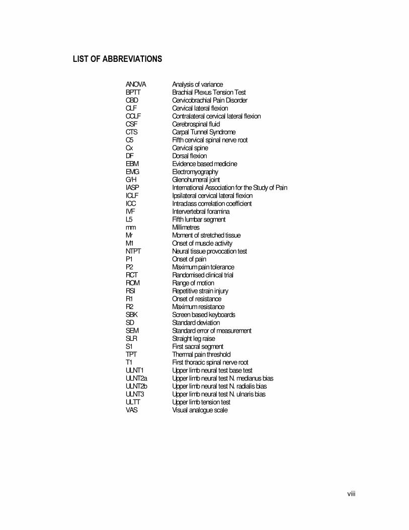
/,67�2)�$%%5(9,$7,216�
ANOVA Analysis of variance BPTT Brachial Plexus Tension Test CBD Cervicobrachial Pain Disorder CLF Cervical lateral flexion CCLF Contralateral cervical lateral flexion CSF Cerebrospinal fluid CTS Carpal Tunnel Syndrome C5 Fifth cervical spinal nerve root Cx Cervical spine DF Dorsal flexion EBM Evidence based medicine EMG Electromyography G/H Glenohumeral joint IASP International Association for the Study of Pain ICLF Ipsilateral cervical lateral flexion ICC Intraclass correlation coefficient IVF Intervertebral foramina L5 Fifth lumbar segment mm Millimetres MU Moment of stretched tissue M1 Onset of muscle activity NTPT Neural tissue provocation test P1 Onset of pain P2 Maximum pain tolerance RCT Randomised clinical trial ROM Range of motion RSI Repetitive strain injury R1 Onset of resistance R2 Maximum resistance SBK Screen based keyboards SD Standard deviation SEM Standard error of measurement SLR Straight leg raise S1 First sacral segment TPT Thermal pain threshold T1 First thoracic spinal nerve root ULNT1 Upper limb neural test base test ULNT2a Upper limb neural test N. medianus bias ULNT2b Upper limb neural test N. radialis bias ULNT3 Upper limb neural test N. ulnaris bias ULTT Upper limb tension test VAS Visual analogue scale
1
��� ,1752'8&7,21�
Pain is central to the practice of physiotherapy being the most frequent symptom for which
patients seek healthcare and the most common cause of physical dysfunction (Simmonds
1999). Therefore an adequate assessment of pain and its underlying pathology is essential for
an efficient therapy. In this respect providing an adequate assessment depends on the
accuracy of the physical examination and on the efficacy of diagnostic tests.
Physiotherapists have been specialising in assessing movement disorders of the neuro-
musculoskeletal system. It is still common belief that the rehabilitation of physical dysfun-
ctions mainly involves treatment of muscles and joints. However, the examination for
normal compliance to movement and abnormal responses to mechanical provocation of the
neural tissue has been an integral part of the experienced physiotherapist’s assessment
(Elvey 1986, Kenneally HW�DO� 1988, Butler 1991). The former theory of considering the exa-
mination and rehabilitation of neural tissue disorders from a biomechanical perspective
(Elvey 1979b/1986/1995, Butler 1989), which has now developed into a more neurophysio-
logical approach (Shacklock 1995, Wright 1999, Butler 2000), has moved into focus and is
currently the subject of investigation. Since many treatment approaches in physiotherapy
have been developed empirically, the need to scientifically validate assessments and treat-
ment regimes has long been recognised.
When patients present with pain conditions in the upper extremities and neck, in which
mechanosensitive neural tissue is considered to be the primary feature, the term cervico-
brachial pain disorder has been advocated (Allison HW�DO� 2002). During patient assessment a
physiotherapist develops hypotheses about possible causes or diagnoses for the presenting
problem. These hypotheses are then tested in the physical examination in which special

2
diagnostic tests are used (Davidson 2002). A diagnostic test seeks to determine whether a
person has a particular condition or whether this condition can be ruled out. In correspon-
dence to the straight leg raise (SLR) of the lower quarter a testing procedure for the neural
tissue has been developed for the upper quarter, the neural tissue provocation test. When
Elvey (1979b) first conceptualised the physical examination of the neural tissue, in the
investigation of arm pain and regional upper quarter pain syndromes, the predominant
thought behind the concept was for a better understanding and differential diagnostics of
upper arm pain. Until then reliable diagnostic procedures for the interpretation of somatic
referred pain to the shoulder and arm had not been defined.
One of these neuromusculoskeletal dysfunctions then under investigation was the whiplash
syndrome, which at that time appeared to be a relatively minor trauma that may progress into
arm pain (Hammacher & van der Werken 1996). Standard clinical examination for possible
referral of pain from the cervical spine comprised muscle power testing, reflex and sensory
testing, as well as nerve conduction testing. If, however, the standard clinical examination
failed to reveal definite positive signs, such as positive neurological deficits or
reproducibility of pain by cervical spine tests, confusion arose as to the probable medical
explanation for the symptoms. Hence, aim was to overcome the difficulty in “determining
the primary pathology” (Elvey 1979a, p.113).
The same difficulty of identifying the primary disorder holds true for minor nerve disorders,
in which normal nerve conduction is not impeded and therefore conduction abnormalities on
electromyographical recordings are not necessarily evident (Dyck 1990), as for example in
the early stages of the Carpal Tunnel Syndrome (CTS). However, it has been shown that
minor nerve disorders, as in injured or inflamed peripheral nerves, are characterised by an
3
increased sensitivity to mechanical load (Greening & Lynn 1998). It is this mechanosensi-
tivity that provides the means of clinical assessment that is utilised by physiotherapists by
the application of neural tissue provocation testing.
Since the original description of the ‘brachial plexus tension test’ by Elvey (1979b) the test
has evolved and is now better known under the terms ‘upper limb neural test’ (Butler 1991),
‘neurodynamic test’ (Shacklock 1995), or ‘neural tissue provocation test’ (Elvey 1995, Hall
HW� DO� 1998, v. der Heide HW� DO� 2001). The provocation tests for the cervicobrachial and
equally for the lumbosacral plexuses have been described in the past and have been used in
clinical practice for the past 20 years (Elvey 1979b, Maitland 1986, Butler 1991), however
most tests had been developed by empirical methods and are now scrutinized for their
scientific and clinical validity.
In response to contemporary calls for the use of evidence-based practice and for professional
accountability an increasing number of randomised clinical trials (RCTs) are appearing in
the literature, which assess the efficacy of manual therapy procedures (Jull & Moore 2002).
Evidence based medicine (EBM) is the conscientious, explicit, and judicious use of current
best evidence in making decisions (Sackett HW� DO� 1996), by integrating individual clinical
practice with the best available external clinical evidence from systematic research. One of
the goals of EBM is to evaluate the accuracy and precision of diagnostic tests, the power of
prognostic markers, and the efficacy and safety of therapeutic, rehabilitative and preventive
regimens (Rosenberg & Donald 1995). Furthermore, there is an ethical responsibility to
recognise and consider the current scientific evidence as it relates to the therapeutic
techniques and interventions we are using on a daily basis (Turner & Whitfield 1997,
Matheson 2000). This way, knowledge of the current literature can help clinicians to avoid

4
the act of performing treatment techniques out of tradition without questioning the rationale
behind their treatment decisions.
In general there is an urgent need to further investigate the effects of manual therapy both to
validate its clinical application, as well as to develop a basis for a neurophysiological model
explaining pain relieving effects (Zusman 1992). Cervicobrachial pain sufferers frequently
seek manual therapy treatment for relief despite little evidence to date to substantiate its
effects or to determine its efficacy (Zusman 1994/1995, Aker HW�DO� 1996, Dreyer & Boden
1998, Gross HW�DO� 2002, Hoving HW�DO� 2002). In this respect there has been an effort in the
last years to find scientific proof for these methods (Zusman 1994, Wright & Vicenzino
1995, Vicenzino HW� DO� 1995/1996, Gifford & Butler 1997, Sterling HW� DO� 2001, Zusman
2002).
In the past years an increasing amount of scientific evidence has emerged that investigated
the validity of the neural tissue provocation test in the assessment of patients with neural
tissue disorder in cervicobrachial pain. The aim of this thesis is to present a critical appraisal
of this research. This work does not claim to be a complete state of the art review but tries to
give an overview on the effort physiotherapy has made to verify this method. The first part
of this paper portrays the relevant neuroanatomical and neurophysiological features that play
a role in the development of cervicobrachial pain disorders. Then a short introduction into
the history of neural tension testing is given followed by the main body of this paper, which
is concerned with the scientific evidence for the use of neural tissue provocation testing for
the upper limb as a diagnostic tool in cervicobrachial pain disorders.
For this purpose the initial proposition that the upper limb neural test can selectively load the
nervous system (Elvey 1986, Selvaratnam HW�DO� 1989), and that limitations in movement are

5
caused by the nervous system as a continuous structure (Yaxely & Jull 1993, Coppieters HW�
DO� 2001b), will be under investigation. Finally, the scientific support for the use of neural
tissue provocation tests in clinical practice and implications will be discussed. To prevent
confusion through different terminologies that are found throughout the literature I will refer
to all upper quarter neural tissue provocation tests in chapter 5 as the Upper Limb Neural
Test (ULNT) except where stated otherwise.
The computerised searches have been conducted via MEDLINE, Winspirs, PeDro, and the
Cochrane Data Base. Additionally conference proceedings, and references from primary
articles were checked. Through the kind help of a co-student I was also able to access
material on early ULNT research, which is dated but was perceived to include key papers.
Inclusion criteria for the literature were German and English language, textbooks and
reviews were accepted for the anatomy section but were kept to a minimum for the ULNT
evaluation. To better relate the research to current practice normative ULNT studies on
asymptomatic and symptomatic subjects were limited to be no older than 1990. The key
search words used singularly and in combination were ‘upper limb tension test’ , ‘brachial
plexus’ , ‘cervicobrachial pain’ , ‘neurogenic pain’ , and ‘manual therapy’ . For this literature
review the critical analysis framework by Rees (1997, Appendix A) was used to quantita-
tively analyse the literature, although it was not possible to describe all aspects of the
framework due to word allowance. A more detailed description of the single studies
discussed in this thesis is presented in the tables in Appendix B.
The time scale used to select the main body of the literature ranges from 1990-2003, but
more dated work was included to insure the development of neural tissue examination in
physiotherapy would be covered adequately. Seminal works from Hromada (1963), and

6
Sunderland (1961/1968) were included as they laid the foundation to most of the
neuroanatomical research today. The greatest part of the literature has been acquired through
the medical and surgical libraries of the University Clinic Charité of the Humboldt-
University Berlin, the state library of Berlin, the UWCM library in Cardiff, and with the help
of colleagues. A limitation to the search presented itself through the fact that most studies
were published in physiotherapy and physiotherapy related journals, which can be valued as
a form of publication bias (Dickersin 1990).
7
��� (3,'(0,2/2*<�2)�&(59,&2%5$&+,$/�3$,1�
Neural tissue provocation testing of the upper limb has been developed to assess the
presence of neural tissue involvement in cervicobrachial pain disorders (v. der Heide HW�DO�
2002). Patients frequently seen in physiotherapy praxis often present with diffuse pain in the
arm and neck without recalling any previous trauma, and first tend to wait for the pain to
regress by itself. From my experience patients finally seek medical treatment after several
weeks of persistent sometimes increasing pain, and are subsequently sent to physiotherapy
after some weeks of initial treatment by their general practitioners.
Due to the lack of population-based studies the precise incidence of cervicobrachial pain is
not known (Hall HW�DO� 1997). The Task Force on Epidemiology of the International Asso-
ciation for the study of Pain (IASP) has so far published individual studies on neck pain and
shoulder pain (Crombie HW�DO� 1999), however, to my knowledge no epidemiological study
has investigated cervicobrachial pain as such. This may be partially due to the wide array of
terms that have been used to describe populations of patients with neck pain and related
disorders, such as ‘upper extremity disorders’ , ‘cervical osteoarthritis’ , ‘tension neck
syndrome’ , ‘cervical spondylosis’ and ‘occupational cervicobrachial disorders’ (Ariëns HW�DO�
1999). As neck pain and cervicobrachial pain (CBP) are closely related, and may even have
been used synonymously in research, some information will be given on the prevalence and
rehabilitative measures of neck pain to help establish a clearer picture on cervicobrachial
pain disorders.
Neck disorders and upper extremity pain are not only common in the general population, but
can be disabling and costly (Dreyer & Boden 1998, Gross HW�DO� 2002). An investigation on
the cost-of-illness of neck pain in The Netherlands in 1996 revealed that the share of these

8
costs were about 1% of the total health care expenditures and 0.1% of the Gross Domestic
Product (Borghouts HW�DO� 1999). There are increasing reports of pain, discomfort and dysfun-
ction of the neck and upper extremities associated with repetitive physical work and stress
(McPhee & Worth 1988, Karjalainen HW�DO� 2000ab). By now it is well accepted that upper
extremity and neck disorders are seen in workers who undertake light repetitive work in
fixed postures, as in keyboard operators, which requires continuous stabilising around the
shoulder girdle, that may even lead to repetitive strain injuries (Quintner & Elvey 1993,
Grant HW�DO� 1995). Even though neck pain is usually not life threatening for the patient, it
may cause pain and restrictions in daily activities. The socioeconomic consequences for
those taking prolonged sick leave or receiving disability pension may also lead to substantial
personal suffering (Jensen HW�DO� 1995, Ariëns HW�DO� 1999).
In general, relatively few consistent data are available on the prevalence of neck pain in the
general population, ranging from as low as 9.5% to 35% (Ariëns HW� DO� 1999). The large
variation in the prevalence estimates of research may be explained by the differences in
definitions and inclusion criteria. In some studies acute and chronic neck pain were included,
in others only chronic pain patients. Also the source of the samples and their age distribution
varied considerably (Ariëns HW� DO� 1999). In the Norwegian population about one third of
adults will experience neck pain in the course of one year (Bovim HW� DO� 1994) with a
prevalence for chronic pain of 13.8%.
Other authors report of a prevalence of neck pain in the general population of The
Netherlands ranging from 10% to 15% (Borghouts HW�DO� 1999). A prospective longitudinal
study of a general population sample initially free of neck pain in the United Kingdom
reported that the 1-year cumulative incidence of neck pain was 17.9% (Croft HW�DO� 2001).
9
Epidemiological studies that have investigated the occurrence of neck pain in the general
population have also shown that women have a higher chance of developing neck pain than
men and that prevalence rises with age (Bovim HW� DO. 1994, Borghouts HW� DO. 1998/1999,
Croft HW�DO� 2001).
Since upper extremity pain often occurs in conjunction with cervical disorders it is difficult
to differentiate and categorise the symptoms into separate clinical disorders. Dwyer HW� DO.
(1990) noted that neck pain is a poorly understood symptom and clinical interpretations most
often ascribe it to putative ‘disc disease’ or ‘soft tissue injury’ . In the cervical region, neck
pain arising from the zygapophysial joints, ligaments, muscles and intervertebral discs may
be accompanied by pain perceived in the head, shoulder girdle, upper limb, and the chest
(Dwyer HW� DO�� 1990, Bogduk 1994, Selvaratnam HW� DO� 1994), thereby providing a more
complex diagnostic problem because pain can arise locally or can be somatically referred
pain. Referred pain is defined as pain perceived in a region separate from the location of the
primary source of pain (Bogduk 1994).
Also muscular pain (Travell & Simons 1983) and pathology from deep somatic tissue may
elicit referred pain obscuring the clinical picture. It is therefore imperative to undertake
differential diagnostics to rule out any serious underlying pathology such as Pancoast
tumour, coronary insufficiencies, fractures after trauma, or systematic inflammatory disor-
ders before referring patients to physiotherapy. The aim of the physiotherapist’ s assessment,
however, is to differentiate between the potential somatic sources that can refer pain into the
neck and upper extremity.
Since there are many treatments available and accepted as standard in the conservative
management of cervicobrachial pain a systematic review assessed the efficacy and

10
effectiveness of treating mechanical neck pain (Aker HW� DO� 1996). 24 RCTs that met the
selection criteria were categorised by their type of intervention (9 manual therapy, 12
physical medicine, 4 drug treatment, 3 patient education). What became clear from this
overview was the lack of evidence for many standard approaches used in health care today.
Even for those treatments that showed some early evidence of support, such as manual
treatments, implications remained inconclusive due to the small number of trials on which
they were based. Apart from the low statistical power of studies differences in defining
diagnostic criteria for cervicobrachial pain syndromes (Koes HW�DO� 1992abc, Levoska HW�DO�
1993, Ekberg HW� DO. 1994) may equally impede comparison of results, thereby limiting
implications that could be made for clinical practice.
Additionally a blinded review study that investigated the effectiveness of manipulation and
mobilisation of the spine for back and neck complaints found that most of the 35 RCTs that
were reviewed showed methodological flaws and were of poor quality (Koes HW�DO. 1991).
The most common problems identified were that diagnostic categories for subject inclusion
were often ill defined and non-specific, treatments lacked operationalising, outcome mea-
sures were not blinded, and that small sample sizes and the lack of describing drop-outs
made it difficult to detect treatment differences. Noticeably, only 5 of the 35 studies were on
neck pain, which additionally had the lowest scores on methodology.
Obviously these two systematic reviews disclose the dilemma of research on the efficacy of
conservative treatment of CBP disorders. First of all the definitions of inclusion criteria are
much too broad and so miss out on subgroups that may profit from specific interventions. On
the other side the interventions themselves are not well defined and often mix specific and
unspecific treatments, such as manual therapy and exercise programmes. This way no

11
conclusive statements can be drawn from these kinds of studies or worse, the lack of
evidence for treatment efficiency portrays the inadequacy of most research designs.
Consequently, physiotherapists will have to develop their own studies designed to fit the
demands of their profession.
12
��� 3$7+2%,2/2*,&$/�$63(&76�2)�&(59,&2%5$&+,$/�3$,1�
Critical to the physiotherapeutic management of patients with upper quarter disorders is an
understanding of the underlying pathophysiological mechanisms. It has been proposed that
increased mechanosensitivity of upper quarter neural tissue plays an important role in the
pathogenesis of cervicobrachial pain (Shacklock 1995, Hall HW�DO� 1997, Greening & Lynn
1998, v. der Heide HW�DO� 2002). This means that any mechanical stimulation, be it pressure or
movement, can trigger a pain response in sensitised neural tissues. Since the neural tissue
provocation test (NTPT) claims to transmit tension from the peripheral nerves to the cervical
nerve roots (Selvaratnam HW�DO� 1989, Elvey 1995, Butler 1991), thereby assessing the neural
tissue as a source of pain, particular emphasis is given to the biomechanical relevant
anatomy of the nerve roots and peripheral nerve trunks, as well as to the movement
relationships within neural tissues and to their surroundings.
����� $1$720,&$/�$1'�%,20(&+$1,&$/�&216,'(5$7,216�
One of the most frequently used interpretations of CBP is, that it may be caused by nerve
root compression (Bogduk 1994). Due to the close proximity of the nerve roots within the
spinal column to the discus intervertebralis and to the bony structures of the intervertebral
foramina (IVF), nerve roots can be subjected to mechanical compression, deformation or
stretch associated i.e. with disc herniation, osteophytes of either the uncovertebral region or
the zygapophyseal joints, or with spinal stenosis.
The spinal nerve roots may be well protected against external trauma by their surrounding
structures, but because they do not possess the same amount of protective connective tissue
as the peripheral nerves, they are more vulnerable to interspinal mechanical deformations.

13
The connective tissues of the cervical nerve root can be distinguished from the peripheral
nerve in two main aspects. In the peripheral nerve the perineurium, which envelopes each
fascicle or nerve bundle, splits into two parts, from which most of it merges with the dura
mater and only a few layers merge with the pia mater to make the sheath of the nerve root
(Haller & Low 1971), a thin membranous structure which is permeable to the cerebrospinal
fluid (CSF). Furthermore, the epineurium of the peripheral nerve is continuous with the dura
mater, leaving only the endoneurium to continue from the peripheral nerve to the nerve root
(Hasue 1993).
Insofar, the nerve root lacks both epineurium and the tough perineurial sheath exposing the
constituent nerve fibres more than those of peripheral nerves, and rendering them more
susceptible to mechanical deformation (Sunderland 1968, Rydevik HW�DO� 1984). Secondly,
nerve roots are more vulnerable to traction and compression injury because their nerve fibres
are arranged in parallel non-plexiform bundles, which are loosely held together by fewer and
finer collagen fibres of the endoneurium compared to those of the peripheral nerve
(Sunderland & Bradley 1961b, Sunderland 1968, Hasue 1993).
The nerve root complex adapts to the movement of the extremities and spine by stretching
and slackening and possibly by some sliding in the IVF (Rydevik HW� DO� 1984). When ap-
proaching the IVF the nerve roots are in close relation to the pedicles (Rydevik HW�DO� 1984),
therefore compression of the nerve roots depends on the effective space available within the
IVF, which might decrease through articular degeneration as well as through physiological
movements of the vertebrae. Moses and Carman (1996) conducted a detailed investigation
on the topography of the fifth, sixth and seventh cervical nerve roots in association to their
structural surroundings. The study showed that significant attachments to the walls of the
14
IVF existed. Posteriorly, at the medial end of the foramina, the nerve roots attach to the
periosteum of the inferior pedicles, and to the capsules of the zygapophysial joints.
Anteriorly, they attach to the vertebral bodies and the intervertebral disc by lateral extensions
of the posterior longitudinal ligament (Moses & Carman 1996).
This means that lateral displacement of the nerve root complex in the IVF is limited
rendering it more vulnerable to mechanical compression within the IVF by space-occupying
pathologies (i.e. osteophytes or disc herniation). If, in case of such a scenario, the nerve root
becomes irritated, the neural tissue provocation test should be able to detect a sensitivity to
mechanical load (more details on the testing procedure in chapter 4). However, the
sensitising of neural tissue can occur without compression through inflammatory or chemical
reactions. A compression type radiculopathy may however, present with paraesthesia and
pain and must not necessarily lead to an impairment of normal compliance to movement (v.
der Heide 2003, personal communication).
Peripheral nerves on the other hand are relatively resistant to mechanical load, which
originates in the highly differentiated construction of their surrounding connective tissue.
The peripheral nerve consists of sensory, motor and sympathetic nerve fibres. Within both
PRWRU� DQG� VHQVRU\� QHUYHV� P\HOLQDWHG� $ -� DQG� $ -fibres, and unmyelinated C-fibres are
present in a ratio of 1:4 (Mackinnon 2002). Myelinated and unmyelinated nerve fibres are
packed within endoneurial connective tissue and bundled into fascicles. These fascicles are
surrounded by the perineurium, a “relatively thin but distinctive lamellated sheath of
connective tissue composed of tightly packed collagenous and elastic fibres, which are
arranged about the fascicles circularly, obliquely and longitudinally” (Sunderland & Bradley
1961a, p.109). The perineurium is the main component giving tensile strength and elasticity

15
to the nerve trunk, and also constitutes a diffusion barrier for several substances including
proteins, which helps to preserve intrafascicular pressure (Sunderland 1968/1990). However,
stretching might damage the perineurium and affect its permeability.
The perineurium is like a tube surrounding the fascicles, which permits some movement of
the nerve fibres inside the fascicle (Sunderland 1968/1990). The connective tissue matrix
that lies between the fascicles is termed the internal epineurium. The external epineurium is
the outer connective tissue that supports and protects all the fascicles of a given nerve, and
carries the vascular vessels (Mumenthaler & Schliak 1977). The entire nerve trunk is
surrounded by the mesoneurium with its extraneural gliding surface called adventitia that
permits excursion of the nerve trunk during joint motion (Sunderland 1968, Mackinnon
2002). This extraneural gliding surface together with the intraneural sliding of fascicles
against each other in deeper layers, present the normal gliding mechanism during joint
motion (Sunderland 1968, Rempel HW� DO� 1999), which are the mechanisms under investi-
gation when applying the NTPT.
Wilgis and Murphy (1986) reported normal gliding properties of the median and ulnar nerve
trunk in fresh intact adult cadaver to be 7.3 mm and 9.8 mm respectively, during full flexion
and extension of the elbow (Table 1). The extent of nerve excursion just proximal to the
wrist was even more pronounced with 14.5 mm (range 11-17 mm) and 13.8 mm (range 10-
15 mm) respectively. Observations on the marked median nerve excursion during upper limb
movement seemed to show consistency with these findings. In an in vivo study by McLellan
and Swash (1976) measurements of the deflections of a needle electrode, inserted through
the skin into the median nerve at a point half way between the elbow and the shoulder,
indicated a mean nerve excursion value of 7.4 mm (range 2.8-20 mm) during wrist and

16
finger extension (Table 1). The authors further estimated that there was 10-15 mm of
excursion at the wrist during wrist and finger hyperextension, but gave no details on the
exact position of the elbow.
Unfortunately, neither one of the studies reported on the position of the shoulder girdle
during measurements, which has been demonstrated to influence the tension in the neural
tissue (Selvaratnam HW�DO� 1989, Kleinrensink HW�DO� 1995b/2000, Coppieters 2002a), nor how
elbow and wrist movements were combined, which leaves interpretation of the data to be
incomplete. Moreover, the shrinkage and stiffness (i.e. rigor mortis) of tissues in human
cadaver have to be taken into account when comparing the results between in situ and in
vivo studies. Although the results seem to show some agreement, and measurements were
taken at similar sites (upper arm or wrist) the position of the elbow during measurement was
not clearly defined so that these data need to be corroborated by studies with exact
operational definitions.
Wright HW�DO� (1996) accounted for these positions and found that in 90º shoulder abduction,
10º elbow extension, and 30º forearm supination the mean total excursion of the median
nerve at the wrist was 19.6 mm (Table 1). Moving the shoulder and the elbow induced
marked excursion of the median nerve at the elbow of 9.1 mm and 12.3 mm respectively, but
not at the site of the wrist. Recent measurements of the longitudinal median nerve motion
distal to the elbow joint using spectral Doppler sonography (Hough HW� DO� 2000) claim to
have confirmed the values of nerve excursion reported by McLellan and Swash (1976) with
a mean of 8.5 mm (range 6.2-13.7 mm). However, the remarkable large variations in the
individual range of measured median nerve excursion reported by McLellan and Swash (2.8-
20 mm) and Hough HW�DO� (6.2-13.7 mm) have not been addressed so far.

17
TABLE 1: Measurements of mean nerve excursion at various sites
6LWH�RI�PHDVXUHPHQW� 1��PHGLDQXV� 1��XOQDULV� 1��UDGLDOLV�
Total longitudinal excursion (LQ�VLWX� measured proximal to the elbow (Wilgis & Murphy 1986)
7.3 mm (during full flexion to extension of the
elbow)
9.8 mm -
Distal longitudinal excursion (LQ�YLYR) measured in the upper arm (McLellan & Swash 1976)
7.4 mm (during wrist/finger
extension to flexion)
- -
Total longitudinal excursion (LQ�VLWX) measured at the wrist proximal to the carpal tunnel (Wilgis & Murphy 1986)
14.5 mm (during full flexion to extension of the
elbow)
13.8 mm 5.8 mm (with ulnar to
radial deviation)
Distal longitudinal excursion (LQ�YLYR) measured at the wrist (McLellan & Swash 1976)
10 -15 mm (during wrist and
finger hyperextension)
- -
Total longitudinal excursion (LQ�VLWX) measured at the wrist proximal to the carpal tunnel (Wright HW�DO� 1996)
19.6 mm (from 60º wrist
extension to 65° wrist flexion)
- -
Distal longitudinal excursion (LQ�YLYR) measured distal to the elbow (Hough HW�DO��2000)
8.5 mm (during wrist
extension from neutral to 60°)
- -
An interesting finding was that when positions of the joints were combined there was a mean
total of 35.4 mm of median nerve excursion (distal and proximal excursion combined) at the
wrist (Wright HW�DO� 1996). If nerve movement was restricted at one location this would lead
to increased neural tension or stretch away from the site of compression, and could explain
the diffuse symptoms frequently reported in entrapment neuropathies such as the CTS
(McLellan & Swash 1976, Wright HW�DO� 1996). Magnetic resonance scans on patients with
work related repetitive strain injuries confirmed this, and showed reduced median nerve
excursion in the carpal tunnel during wrist movement (Greening HW�DO� 1999). Furthermore,
the potential surgical implications of restricted nerve motion caused by adherence of the
nerve to surrounding tissue have been emphasised (Wilgis & Murphy 1986). Therefore, the

18
restoration of the longitudinal sliding mechanism is said to be essential to ensure effective
surgical treatment. These findings have led to the development of specific nerve gliding
exercises in the form of gentle movement of the nerve within its nerve bed that reduce
adhesions (Totten & Hunter 1991, Rozmaryn HW�DO� 1998).
Another important factor in peripheral nerve anatomy is that the fascicles do not run
independently along the entire length of the nerve but are repeatedly dividing and uniting to
form complex fascicular plexuses (Sunderland & Bradley 1961a, Sunderland 1968) provi-
ding the nerve trunk with the capability to endure much higher loads than the nerve root.
Additionally the nerve trunk runs in an undulating course within its nerve bed, as well as the
fasciculi within the epineurium, and the nerve fibres inside the fasciculi (Sunderland 1968).
Under normal circumstances with increasing extension peripheral nerve trunks first
straighten out their resting undulation in the nerve bed, whereby the nerve fibres, which are
arranged in a spiral fashion, are able to untwist without altering the length or tension of the
individual fibres inside the fasciculi (Sunderland 1968/1990). This is then followed by a
slide and glide of the nerve trunk in relation to its nerve bed, adapting to positional changes
of the upper limb (McLellan & Swash 1976). These observations led Zöch (1992) to investi-
gate the lengthening properties of the median nerve in a human cadaver model with the
following questions in mind:
1. What kinds of length differences exist in the nerve bed during full range
movements?
2. Does a peripheral nerve adapt by stretching or by relaxing in the nerve bed?
3. How is the tension distribution in relation to the length of the nerve?

19
The interesting results of this morphological study showed that in maximal wrist and elbow
extension and shoulder abduction the nerve bed was 4% longer than the nerve itself, whereas
in maximal flexion and adduction the nerve bed was 15% shorter. Zöch confirmed that the
median nerve itself adapts to full flexion of the joints by taking up a wavy pattern within the
nerve bed. This procedure accounts for approximately 77% of the length difference of the
median nerve from full flexion to full extension, whilst the missing 23% have to be achieved
by the elastic properties of the nerve itself (Zöch 1992), bearing in mind that this process is
only possible if the gliding movement within the nerve bed is not restricted by adhesions.
However, the lengthening properties of nerves have been stated in the literature with quite
some discrepancy. According to Zöch (1992) the elongation is distributed evenly throughout
the nerve and equals an average of 4% of its original length. The normal maximal stretching
ability has been stated by Zöch to lie at 6%, however, normal daily activity usually never
exceeds the value of 4% stretching. Only in the case of adhesions or fibrosis of the gliding
tissue the even distribution of tension is prevented. This means the nerve distal to the
adhesion has to compensate for the loss of stretching ability and might exceed the critical
value of 6-8%, which in turn might lead to vascular morphological changes (Zöch 1992). As
the fasciculi are stretched, their cross-sectional area is reduced (Sunderland & Bradley
1961a), which leads to an increase in intrafascicular pressure, whereby nerve fibres are
compressed and microcirculation is compromised (Lundborg & Rydevik 1973).
In contrast, Sunderland and Bradley (1961a) have described the mean percentage of
elongation at the elastic limit of the median nerve in 24 unembalmed human specimens to
have a mean average of 14.9 ± 3.9% (range 10-22%). In this study elasticity was defined as
“that property of a material which enables it to return to its original form and shape when the
20
external load is removed” (Sunderland & Bradley 1961a, p.108). They stated that slowly
stretching the nerve leads to considerable lengthening without apparent damage. Greatest
elongation at the elastic limit of a nerve trunk was reported to be in the order of 20%,
whereas complete mechanical failure was observed at 30% stretch of its original length.
The significant differences in results between Zöch (1992) and Sunderland and Bradley
(1961a) are partly due to the different starting lengths used, and partly due to the fact that
Sunderland and Bradley conducted their research on isolated nerves subjected to progres-
sively increasing loads to the point of mechanical failure. Zöch (1992) on the other hand
used an intact nerve in situ without destroying its nerve bed and cutaneous nerve branches
that presented a form of natural fixation to the surrounding, which explains the much lower
values. In general, the data provided through these cadaver studies have to be regarded as
approximate values when compared to the mobility and stretching capabilities of the neural
tissue in living subjects, but nevertheless demonstrate the movement dynamics of neural
tissue.
����� 1(8523+<6,2/2*,&$/�&216,'(5$7,216�
The previous section explained the mechanisms and importance of uncompromised gliding
of the nerve trunk in its adaptation to normal movement of the spine and extremities. If
adhesions hinder this physiological movement and the even distribution of tension in the
nerve, intraneural microcirculation could be impaired. However, nerve function as well as
communication and nutritional transport systems (antegrade and retrograde transport)
depend on an adequate supply of oxygen to the nerve fibres.

21
The peripheral nerve contains a well developed microvascular system with vascular plexuses
in all of its connective tissue layers (Rempel HW� DO� 1999). These vessels have a coiled
configuration so that blood flow is not impaired during normal gliding of the nerve trunk
(Mackinnon 2002). As the vessels reach the nerve trunk they enter the epineurial space,
where there is considerable plexus formation, and run longitudinally in various layers of the
epineurium. The vessels pass the perineurium obliquely to enter the endoneurium where
there is only a fine network of capillaries (Mackinnon 2002). As the endoneurial space has
no lymphatic vessels, oedema within the endoneurium could lead to increased endoneurial
fluid pressure in the fascicles, which could interfere with the vulnerable microcirculation
(Rempel HW�DO� 1999). Nerve roots, ganglia, and spinal nerves on the other hand receive their
blood supply from segmental arteries and medullary vessels. Nutrients may be transported to
the nerve roots both by the intrinsic blood vessels and via diffusion from the CSF (Rydevik
1992, Hasue 1993).
The intraneural blood flow was investigated experimentally by Lundborg and Rydevik
(1973) in a rabbit tibial nerve model. During controlled elongation of the nerve it could be
demonstrated that venular stasis was induced when the nerve was stretched to about 8%
(range 5-10%) over its original length, and if maintained for a longer period of time would
give rise to continuous impairment of intraneural microvascular flow. The “upper stretching
limit” induced a stasis of arterioles and capillaries at 15% elongation (range 11-18%), but is
less important because it lies beyond the critical limit as far as long-term viability of the
nerve is concerned (Lundborg & Rydevik 1973). These findings support Zöch’ s hypothesis
(1992) that maximally stretching a nerve 6-8% of its original length would induce changes in
the microvascular flow.

22
Within this complex microvascular system the endoneurial milieu is protected by a blood-
nerve barrier (Rempel HW�DO� 1999). A breakdown in the blood-nerve barrier will occur with
nerve injury including entrapment or compression, resulting in the accumulation of proteins,
lymphocytes, fibroblasts and macrophages within the endoneurium as a reaction to antigens
in the perineurial space (Mackinnon 2002). This will further initiate inflammation and
eventually scar formation in the peripheral nerve, which in turn will lead to an uneven
distribution of tension during mechanical load.
In contrast, there does not seem to be any blood-nerve barrier in the nerve roots and ganglia,
and intravenously injected substances can leak into the intercellular layers between the nerve
fibres and cell bodies (Rydevik HW� DO� 1984). This means that nerve roots and ganglia are
more susceptible to chemical irritation induced by inflammatory processes of near by
structures, such as the discus intervertebralis or zygapophysial joints, rather than to
mechanical deformation as is commonly assumed.
Since inflammation of the nerve root or nerve trunk in the absence of space-occupying
pathologies are more difficult to diagnose with standard clinical examinations (i.e. radio-
graphy, computer tomography, magnetic resonance imaging, myelography, sonography) than
a radiculopathy induced by disc herniation or osteophytes, many patients with CBP disorders
have pain, of which the underlying mechanisms are not known. Besides, it has been shown
that magnetic resonance imaging has a high percentage of clinically false-positive findings
(Boden HW� DO� 1990). In asymptomatic subjects the abnormal findings, such as herniated
nucleus pulposus, narrowing of disc space or foraminal stenosis, had been shown to be age
related. This means that in patients abnormal anatomical findings may not necessarily be
related to their symptoms. Therefore, imaging diagnostics should not be utilised by

23
themselves to institute a therapy without matching the findings with clinical signs and
symptoms. This is where the NTPT could be used as an additional diagnostic parameter in
the evaluation of the clinical findings by assessing the reaction to mechanical load of
putative inflamed tissues.
����� ())(&76�2)�&2035(66,21�21�1(59(�),%5(6�
The correlation between systemic blood pressure and the function of the nerve has supported
the hypothesis that decreased intraneural microcirculation plays an important role in nerve
compression disorders (Sunderland 1968, Rydevik 1992, Rempel HW� DO� 1999, Mackinnon
2002). When neural tissues are subjected to load or pressure they deform, and pressure
gradients are formed that lead to the redistribution of compressed tissue towards areas of
lower pressure (Rempel HW�DO� 1999). Nerve compression syndromes usually occur at sites
where the nerves pass through a tight tunnel formed by stiff tissue boundaries like sulcuses,
muscles or fascia (Table 2).
TABLE 2: Compression syndromes of the upper extremity
1��UDGLDOLV� 1��PHGLDQXV� 1��XOQDULV�Radial palsy (Saturday night palsy)
Thoracic outlet syndrome Thoracic outlet syndrome
Supinator tunnel syndrome
Pronator teres syndrome Cubital tunnel syndrome
Anterior interosseous nerve syndrome
Guyon’s canal compression
Carpal tunnel syndrome
Animal experiments, of low magnitude extraneural compression, induced by a miniature
inflatable cuff, have demonstrated that the first sign of impairment was the stasis of
24
epineurial vessels appearing at a pressure of 20-30 mmHg (Rydevik HW�DO� 1984), and that
fast as well as slow antegrade and retrograde axonal transport was inhibited (Dahlin HW�DO.
1981). By the time the cuff pressure reached 60-80 mmHg the compressed segment of the
nerve was completely ischaemic. This means that cell nutrition and intraneuronal communi-
cation were compromised at elevated extraneural pressures. Dyck HW�DO� (1990) reported that
extraneural compression of 50 mmHg applied for two minutes can alter the structure of the
myelin sheaths.
These disturbances were usually rapidly reversible after short periods of compression (1-2
hours), but repeated or prolonged compression at these pressure levels may show long
lasting effects. These findings are interesting when related to pressure levels recorded in
patients with CTS. It was found that patients with median nerve compression had an average
of 32 mmHg pressure in their carpal tunnel, while the asymptomatic control group showed
an average of 2.5 mmHg pressure (Gelberman HW�DO� 1981).
Intraneural blood vessels have shown to increase their permeability as a response to nerve
injury. Histological examinations demonstrated rapidly increasing endoneurial pressures in
nerves that had been subjected to short-duration (2 hours) low magnitude extraneural
pressure (30 mmHg), probably due to increased vascular permeability of the endoneurial and
epineurial vessels (Rydevik HW�DO� 1984, Rempel HW�DO� 1999). Also long-term effects after two
hours of low extraneural compression had been shown to result in long-lasting subperineurial
oedema, that later lead to degeneration (demyelination) and regeneration of nerve fibres.
These events seem to be associated with the degree of endoneurial oedema, hence a dose-
response relationship between the duration of compression and the degree of injury can be
observed.

25
Furthermore, inflammation and fibrin deposits have been found to occur within hours after
compression, followed by proliferation of endoneurial fibroblasts and capillary endothelia
cells (Rempel HW� DO� 1999). Even though fibrosis is a normal response to inflammation,
fibrotic adhesions may impair normal nerve gliding, and thus would have significant
consequences on the movement dynamics of the neural tissue, which could appear as a
clinical relevant sign during neural tissue provocation testing.
Morphological changes such as swelling proximal and distal of the ligature, stasis of venous
return, and fibrosis surrounding the nerve have also been observed in animal models of
chronic constrictive nerve injury (Greening & Lynn 1998). Histological evaluations of resec-
ted nerve segments from humans suffering from nerve compression equally showed vascular
sclerosis, epineurial and perineurial oedema, thickening and fibrosis at the site of injury, and
evidence of degeneration and generation of nerve fibres (Rempel HW�DO. 1999), thereby confir-
ming the findings of the previous animal experiments. This cascade of biological responses
to compression mainly affected large diameter myelinated fibres as unmyelinated fibres had
been shown to be spared (Rydevik HW�DO� 1984). A highly significant loss of Aβ fibres was
observed peaking at 2 weeks post constrictive injury, however 8-10 weeks post injury Aβ
fibres still appeared abnormal in respect to their diameter and overall number (Greening &
Lynn 1998).
Due to ethical difficulties in carrying out experiments in humans there is obviously much
less data available on neuropathological changes following nerve injury than from animal
experiments. However, these observed events taking place during peripheral nerve compres-
sion in animal experiments could now be explained and related to clinical symptoms in
patients. Knowledge gained from animal experiments (Lundborg & Rydevik 1973, Dahlin HW�

26
DO� 1981, Rydevik HW� DO��1984, Dyck HW� DO� 1990, Rydevik 1992, Greening & Lynn 1998,
Michaelis & Jänig 1998, Rempel HW�DO� 1999) has also lead to a better understanding of the
pathomechanisms of entrapment neuropathies such as the CTS.
The initial symptoms in CTS are usually intermittent paraesthesia and deficits of sensation
that occur primarily at night. These symptoms are probably due to impairment of the intra-
neural microcirculation associated with endoneurial oedema, which disappear during the day
by movement of the arm (Rempel HW�DO. 1999). With increased compression more severe and
constant symptoms that do not disappear during the day arise. In this stage microcirculation
may be altered during the day, leading to morphological changes such as segmental demye-
lination. Besides entrapment neuropathies metabolic changes as in diabetes mellitus or
during pregnancy may equally cause endoneurial oedema and need to be taken into
consideration during patient assessment.
Alternatively, short-term compression of intact peripheral nerves or nerve roots has been
stated to cause no pain but rather paraesthesia as a result of ischaemia and not through
mechanical nerve fibre deformation (Rydevik HW� DO� 1992, Hasue 1993, Bogduk 1994).
Whereas in the case of an inflamed or irritated nerve or nerve root, compression or minor
mechanical deformation can be the cause of radiating pain (Rydevik HW� DO� 1984, Hasue
1993). Inflammation can for example be caused by the breakdown products of degenerating
nucleus pulposus (Rydevik HW�DO� 1984, Hasue 1993, Zusman 1998), or by long-term or high
magnitude compression (Dyck HW� DO� 1990, Rempel HW� DO. 1999). In so far irritation of the
neural tissue has to be present before mechanical provocation can give rise to pain.
27
����� 3$,1�0(&+$1,606�$6�,1�0,125�1(59(�,1-85,(6�
In clinical practice patients frequently present with diffuse symptoms in their neck and upper
extremity of unknown aetiology. Positive symptoms usually associated with peripheral nerve
injury, such as paraesthesia, hyperalgesia, allodynia, spontaneous pain and impaired function
may be reported. Typical symptoms reported during patient assessment that have been
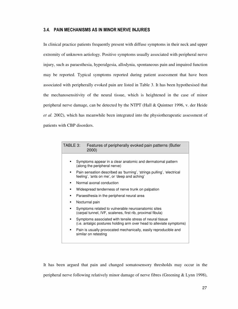
associated with peripherally evoked pain are listed in Table 3. It has been hypothesised that
the mechanosensitivity of the neural tissue, which is heightened in the case of minor
peripheral nerve damage, can be detected by the NTPT (Hall & Quintner 1996, v. der Heide
HW�DO� 2002), which has meanwhile been integrated into the physiotherapeutic assessment of
patients with CBP disorders.
TABLE 3: Features of peripherally evoked pain patterns (Butler 2000)
�� Symptoms appear in a clear anatomic and dermatomal pattern (along the peripheral nerve)
�� Pain sensation described as ‘burning’, ‘strings pulling’, ‘electrical feeling’, ‘ants on me’, or ‘deep and aching’
�� Normal axonal conduction
�� Widespread tenderness of nerve trunk on palpation
�� Paraesthesia in the peripheral neural area
�� Nocturnal pain
�� Symptoms related to vulnerable neuroanatomic sites (carpal tunnel, IVF, scalenes, first rib, proximal fibula)
�� Symptoms associated with tensile stress of neural tissue (i.e. antalgic postures holding arm over head to alleviate symptoms)
�� Pain is usually provocated mechanically, easily reproducible and similar on retesting
It has been argued that pain and changed somatosensory thresholds may occur in the
peripheral nerve following relatively minor damage of nerve fibres (Greening & Lynn 1998),
28
that are not necessarily associated with the loss of axonal conduction and therefore seldom
show neurological deficits such as muscle weakness, depressed reflexes, decreased conduc-
tion velocity, or impaired sensation. An explanations for this may be found in minor nerve
injuries of the chronic neuritis model where axons were found to be undamaged or only
partially damaged and so could account for the normal nerve conduction (Greening & Lynn
1998).
Even in the case of morphologic axonal changes, the clinical limitation of nerve conduction
and electromyography (EMG) in predicting neuropathological abnormalities of single nerve
fibres has been demonstrated (Dyck 1990). Interestingly the pathological inferences that can
be made from EMG studies of large sensory fibres were more limited than those from large
motor fibres. However, patients with CBP disorders usually complain of sensory deficits that
are likely not to show in EMG studies.
Peripheral neurogenic pain as in minor nerve injuries has been attributed to an increased
activity in mechanically or chemically sensitised nociceptors within the nerve sheaths, and is
said to be felt in the course of the peripheral nerve trunk (Hall & Elvey 1999). These mostly
unmyelinated afferents that constitute the intrinsic innervation of the connective tissue
sheaths are known as ‘nervi nervorum’ (Hromada 1963, Bove & Light 1997). These sensory
C fibres of the nerve sheath have been shown to contain neuropeptides such as substance P
and calcitonin gene related peptide suggesting a role in vasodilatation. It is assumed that
local nerve inflammation is mediated by the nervi nervorum, especially in cases with no
intrafascicular axonal damage (Bove & Light 1997).
The nervi nervorum has also been found to be sensitive to excess longitudinal stretch, but not
to stretch within the normal range of motion (Bove & Light 1997), and is assumed to be

29
particularly vulnerable to chronic compression and friction syndromes (Greening & Lynn
1998). However, electrophysiological recordings of the nervi nervorum present considerable
technical difficulties so that their contribution to sensory input remains to be evaluated.
The painful response to nerve trunk palpation has been attributed to the spread of mechano-
sensitivity along the length of the nerve trunk mediated through a neurogenic inflammation
via the nervi nervorum (Hall & Quintner 1995, Bove & Light 1997, Hall & Elvey 1999) and
can be related to secondary hyperalgesia. Experimental studies of primary and secondary
hyperalgesia suggest that there are several different mechanical hyperalgesias characterised
by their location and the primary afferent fibres involved (Treede HW�DO� 1992).
Mechanical hyperalgesia, which is confined to the site of injury (primary hyperalgesia), is
based on the peripheral sensitisation of C fibre nociceptors, i.e. through blunt stimuli.
Whereas secondary hyperalgesia, which reacts to punctuate mechanical stimuli, appears to
be based on central sensitisation of A fibre nociceptor input, and occurs not only in the
injured tissue but spreads to adjacent uninjured tissue (Woolf 1991, Treede HW�DO� 1992). It is
furthermore well known that damaged sensory nerve fibres frequently develop abnormal
impulse generation, which is correlated to the presence of mechanical hyperalgesia.
Paraesthesia has been shown to be induced by spontaneous ectopic sensory discharge
generated from large myelinated afferent fibres that may occur at the damaged site (Nordin
HW�DO� 1984, Zhang HW�DO� 1997, Devor & Selzer 1999).
To sum up, this section has presented the biomechanical and neurophysiological features
pertaining to CBP disorders in respect to impaired movement dynamics of the neural tissue.
It is known that the neural tissue is vulnerable to deformation, compression and entrapment
injuries in its natural surroundings (Table 2), which may lead to restrictions of neural tissue
30
mobility. The neural tissue has the ability to adapt to normal joint motion through a gliding
mechanism that permits movement without putting any strain on the individual nerve fibres
within the fasciculi. Additionally the nerve trunk has the capability to stretch to about 6-8%
of its original length, and is relatively more resistant to mechanical load than the nerve root.
However, increased elongation and compression have shown to impede with neural micro-
vascular circulation, which may lead to perineurial oedema, fibrosis, and degeneration of
nerve fibres.
Peripheral changes caused by partial nerve injury may lead to a lowered threshold of afferent
fibres for mechanical stimulation, as well as to increased firing and spontaneous activity.
Abnormal input from damaged or ischaemic nerve fibres may in turn cause pain and trigger
central sensitisation. Increased mechanosensitivity of neural tissue after minor nerve damage
is said to play an important role in diffuse upper extremity pain that is often seen in clinical
practice. In determining whether the pathology of the neural tissue is associated with clinical
symptoms the NTPT utilises the sensitivity of the neural tissue to mechanical load in the
assessment of CBP disorders.
However, pain should never be understood as an isolated entity, but rather as a summation of
all available information from the outside world and from within our bodies, analysed by the
brain in terms of what action would be appropriate (Wall 1999). Furthermore, alterations of
neural, behavioural and subjective pain responses by arousal, attention and expectation, that
result from the action of the central nervous system networks in modulating the transmission
of nociceptive messages, should equally be taken into account (Fields & Basbaum 1999).

31
��� 7+(�+,6725<�2)�1(85$/�7,668(�35292&$7,21�7(67,1*�
The previous chapter has laid out the basic anatomical features of the neural tissue and the
probable causes for the impediment of its mobility. The rational behind this section is to
show how the initial medical interest on the behaviour of nerves to stretching developed into
a physiotherapeutic approach to assess the neural tissue as a potential source of pain.
Observations on the behaviour of peripheral nerves subjected to stretching date from the
second half of the 19th century and pertained to the therapeutic procedure for the relief of
painful neuralgias of different genesis (Cavafy 1881, Symington 1882, Marshall 1883).
Marshall discussed the mechanisms of pain relief attributing both symptoms and benefits to
the nervi nervorum, a then hypothesised but undescribed structure (Bove & Light 1997).
Apart from the therapeutic approach, investigations on the breaking points and on morpho-
logical changes of stretched peripheral nerves were conducted (i.e. Tillaux 1866, Symington
1882, Vogt 1877, Takimoto 1917, cited in Sunderland & Bradley 1961a p.102).
In the First World War studies on the elasticity of peripheral nerves came into focus due to
the intensified need for nerve reconstruction. Baron and Schreiber (1918) described the
importance of the surrounding tissue, in which the nerve is able to glide. Babcock (1927)
studied the elasticity of peripheral nerves and found them to have less lengthening abilities in
comparison to vessels. These findings were the first to illustrate the importance of an intact
nerve bed for normal nerve mobility, and have set the basis for the concept of neural tissue
dynamics.
The earliest references to upper limb tension testing stem from 1929, in which the most
extended positions of the N. medianus, N. ulnaris and N. radialis were clearly described

32
(Bragard 1929). Bragard also reported, that he could establish what he called the “medianus
phenomena” (tension position for the N. medianus) in patients with plexus brachialis
disorders, and that the affected nerves were sensitive to pressure. Later, tests were described
by Chavaney and Frykholm, who applied traction to the extended, abducted, and supinated
arm, as well as simultaneously tilting the head to the contralateral side (Butler 2000). Since
these early investigations the upper limb neural test has been described with emphasise on
different movement sequences and under changing terminologies (Table 4).
TABLE 4: Chronology of related studies on the ULNT
Bragard (1929) described a series of upper limb tension tests
Chavaney (1934) a version of the ULNT with abducted, elevated and extended arm
Frykholm (1951) a version of the ULNT with abducted, supinated and extended arm
Cyriax (1978) examined wrist dysfunction with extended elbow
Breig (1978) introduced the term adverse mechanical tension
Elvey (1979) first described the brachial plexus tension test
Kenneally HW�DO� (1988) introduced the name upper limb tension test
Butler (1991) introduced the term mobilisation of the nervous system
Selvaratnam HW�DO� (1994) validated Elvey’s brachial plexus tension test
Shacklock (1995) introduced the term neurodynamics
Hall HW�DO� (1998) v. der Heide HW�DO� (2001)
used the term neural tissue provocation test
The seminal work of Elvey (1979b), who initially formulated and described the ‘brachial
plexus tension test’ (BPTT), led to an increased physiotherapeutic interest in the neural
tissue as a potential source of pathology and pain in CBP disorders. Assessing the neural
tissue as part of the physiotherapeutic examination has never been presented to stand alone,
but was always seen as an integral part of existing neurological and orthopaedic assessments.
33
In the early stages of the BPTT the idea of testing for increased neural mechanosensitivity,
by increasing tension within the neural tissue through an ordered set up of joint movements,
was based on the notion that ‘neural tension testing’ accurately reflected the mechanical
function of neural structures (Elvey 1979b, Kenneally HW�DO� 1988, Butler 1991). This implied
that a ‘normal’ neural tension test meant the neural tissue moved correctly, and an
‘abnormal’ test meant it did not. This idea was clearly dominant when Butler published his
work “Mobilisation of the nervous system” in 1991. At that time it was important to clarify
which test components would have an effect on the movement of the nerve in relation to its
surrounding interfacing structures (pathomechanical effect), and which components would
cause ‘tension’ in the nerve itself and might influence its physiological function (patho-
physiological effect).
Deductions like these involved only mechanical factors and were at times inaccurate because
the contribution of neurophysiological factors in the production of symptoms had been over-
looked (Shacklock 1995, Rempel HW� DO� 1999). Therefore, Butler embraced Shacklock’ s
concept (1995) of neurodynamics (the dynamic properties of the neural tissue), because it
allowed a shift away from the pure mechanical thought to include neurophysiological issues
and the inclusion of plasticity changes of the central nervous system. Having an under-
standing of the basic anatomical and physiological science underlying neurodynamics is a
critical part of the clinical reasoning process when treating patients with CBP disorders.
Clinical reasoning is the thinking underlying clinical practice, where an initial working
hypothesis is tested until sufficient information is obtained to make a diagnostic decision that
leads to making a management decision (Jones 1995). Physiotherapists are now increasingly
recognising the importance of integrating basic neuroanatomy and neurophysiology research
34
findings into their clinical practice (Zusman 1992/1994/2002, Wright 1998/1999, Matheson
2000).
So far painful conditions of the upper arm were routinely examined for possible referral of
pain from the cervical spine with muscle power testing, sensation, reflex activity, and nerve
conduction. However, if the standard clinical examination failed to reveal any positive signs,
such as neurological deficits, or reproducibility of pain by cervical spine tests or shoulder
joint tests, confusion arose as to the medical explanation for the pain (Elvey 1979b). Elvey
was a pioneer in conceptualising the physical examination of the neural tissue of the upper
quarter in order to investigate arm pain and regional pain syndromes in the area of the fifth
and sixth cervical dermatome. Pain and movement restrictions felt in the shoulder region or
lateral upper arm can be a result of glenohumeral joint or soft tissue pathology, or can be of
cervical or thoracic origin (Maitland 1991). The shoulder and arm region is innervated by the
C5-6 sclerotome, thus pain in this region could be regarded as referred pain from any
structures supplied by the fifth and sixth nerve roots, provided that other underlying diseases
(i.e. biliary colic, angina pectoris, Pancoast tumour) that may also refer pain into this region
have been ruled out.
The BPTT was then propagated as a diagnostic tool, which may help “differentiate between
intrinsic shoulder symptoms and shoulder symptoms which are referred from the cervical
spine” (Kenneally HW�DO� 1988, p.174), by placing tension on the nerve roots of the brachial
plexus, and thereby testing if there was any neural tissue involvement. It was always clear
that other anatomical structures would come under traction during this test, which could
equally reproduce shoulder pain, therefore it was important to develop ‘sensitising
manoeuvres’ that were able to distinguish between neural and non-neural structures. Aim

35
was to alter the patient’ s symptoms by adding sensitising manoeuvres remote from the site of
pain on the condition that the impact on the nervous system passes beyond the point to which
musculoskeletal structures were loaded.
The early BPTT according to Elvey (1979ab) was composed of three basic movements. The
technique was performed with the patient lying supine on an examination table, head and
neck supported in a resting position.
1. glenohumeral abduction behind the coronal plane, with the elbow extended
2. glenohumeral lateral rotation, and forearm supination
3. while maintaining full forearm supination the elbow was gently flexed
All manoeuvres were performed to the point of pain onset. As each of the movement
components were added it was believed that a progressively greater stretch was transmitted
onto the nerve trunks. The test was thought to be positive if the final movement was able to
reproduce the patient’ s pain (Elvey 1979ab). To verify whether the pain was a reaction to
loading the neural tissue or of non-neural structures to stretch, wrist extension was added to
the final position to further provoke the symptoms, claiming to increase cervical root traction
without implicating other structures. By only moving the wrist, muscles of the forearm or the
glenohumeral joint would not be affected and so could not be held to account for the increase
in symptoms.
Other sensitising manoeuvres included cervical spine movements in flexion as well as in
contralateral flexion, lateral rotation and abduction of the contralateral arm, and bilateral
SLR (Elvey 1979ab, Bell 1987). These manoeuvres were believed to increase symptoms on
the affected side through further stressing the nerve trunks through a traction force in the
36
opposite direction, on the grounds that the nervous system is a continuous structure. The
final reassurance of neural tissue involvement was won by taking up a BPTT position just
short of the patient’ s pain and then adding contralateral cervical lateral flexion (CCLF),
which would load the brachial plexus bringing back the patient’ s pain or symptoms.
All these testing components were developed by Elvey empirically in the course of his
clinical experience with patients who suffered from upper quarter disorders. Elvey’ s
postulated theory soon became popular and accepted but had yet to be validated scienti-
fically. The development of therapeutic techniques in movement-based professions has
traditionally been based on the thoughts and clinical experiences of pioneering clinicians.
However, the increased demand for professional accountability and the lack of subjecting
existing treatments to validation has been a well-recognised deficit. These demands are now
being met by an increasing amount of physiotherapy research, which also reflect the up-
grading of this profession to an academic level.
In 1986 Elvey adapted the BPTT by adding shoulder girdle depression to the starting
position and by changing the final movement component from elbow flexion to extension,
thus transmitting more load to the neural tissues. Keneally HW�DO� (1988), who later renamed
the test ‘upper limb tension test’ , argued that the course of the major nerves in the upper limb
would determine which nerves were most likely to be influenced by the movement
components of the upper limb tension test, and believed the median nerve to be most
affected by this test. Butler (1991) broadened the upper limb tension test into four testing
procedures. The initial idea of testing the tension of a nerve was also revised, so that the
basic test was now called ‘upper limb neural test’ (Butler 1994) namely ULNT1, ULNT2a,
ULNT2b, and ULNT3 (Table 5). In Butler’ s ULNT base test elbow extension was employed

37
as the final component, this way the degree of elbow extension at the point of pain onset or
maximal tolerable pain could be used as the main outcome measure (dependent variable).
For a clear understanding of what a normal response is Kenneally HW� DO�� (1988) listed the
responses to the ULNT1 as seen in 100 normal asymptomatic subjects. (1) A deep stretch or
ache in the cubital fossa (99% of subjects) extending down the anterior and radial aspect of
the forearm and into the radial side of the hand, with a definite tingling sensation in the
thumb and first three fingers in 80% of all subjects. This response was found to be
independent of age and gender of the subjects. (2) A small percentage of subjects may feel
TABLE 5: The ULNT’s from Butler (1994, Fig. 2-4 Appendix C)
1DPH� 0DLQ�VHQVLWLVLQJ�FRPSRQHQW�LQ�EROG� 1HUYH�ELDV�
8/17�� �� shoulder girdle depression (maintained) �� �����*�+�DEGXFWLRQ in the coronal
plane �� forearm supination with wrist/ finger
extension �� G/H lateral rotation �� elbow extension
N. medianus (base test)
8/17�D� �� shoulder girdle depression (maintained) �� 10° G/H abduction in the coronal plane �� elbow extension �� *�+�ODWHUDO�URWDWLRQ���� wrist/ finger extension �� G/H abduction
N. medianus
8/17�E� �� shoulder girdle depression (maintained) �� 10° G/H abduction in the coronal plane �� elbow extension �� *�+�PHGLDO�URWDWLRQ���� wrist/ finger flexion plus ulnar deviation
N. radialis
8/17�� �� wrist/ finger extension �� forearm supination �� full elbow flexion �� shoulder girdle depression �� *�+�DEGXFWLRQ��� head in lateral flexion
N. ulnaris

38
stretch in the anterior shoulder area during elbow extension as the long head of the biceps is
stretched. (3) CCLF increases the response in approximately 90% of the subjects (Figure 1).
)LJXUH��� The normal sensory responses to the ULNT1. From Keneally HW�DO. (1988) The upper limb tension test: the SLR of the arm
The close anatomical and physiological relationship between muscles, joints, neural and
vascular systems sets a high demand on the specificity of these tests (Figure 2). The ULNT
that may have been designed to focus particularly on neural structures, will by its nature
stress tissues and elicit responses in any of these systems. Selvaratnam HW�DO� (1994, Table
III-Appendix B) was the first to test the ability of Elvey’ s BPTT (1979b) to discriminate
between local pain and referred sources of pain from the cervical region. Post-surgery
cardiac patients with a high probability of brachial plexus involvement (n=25) and sports-
Anterior shoulder stretch
Deep painfull stretch
sensation
Definite tingling Slight tingling
Deep painfull stretch
sensation
39
injury patients (n=25) were compared with an asymptomatic control group (n=25). The main
finding was that cardiac patients had significantly greater loss of elbow ROM with CCLF
than the two other groups. Reliability was calculated to be 82,5%, but a high percentage
agreement can be attributed to chance alone and cannot in itself support a claim of high
reliability (Haas 1991b). The large standard error of measurement (SEM) with 16.8º further
questions whether appropriate standardisations were carried out.
Given that the cardiac group were pre-selected to have a higher probability of referred pain
from the cervicothoracic region, these findings support the discriminative validity of the
BPTT. The only difference between the groups was age: the cardiac group being older (mean
age 55.3) than the sports injury group (mean age 26.2); thus, the results may have stemmed
from age-related changes such as osteoarthrosis or possible bony proliferative changes in the
lower cervical spine. In agreement with Elvey (1979b/1986) Selvaratnam HW�DO� (1994) stated
that the brachial plexus was stimulated by the BPTT, but object that the sole involvement of
the brachial plexus cannot be fully determined. It is possible that other cervical and thoracic
structures, particularly the fascia, the subclavian artery or vein (Wilson HW�DO� 1994) that are
affected by the surgical procedure, could be capable of referring pain to the shoulder region.
For an accurate interpretation of the test response it is therefore necessary to examine the
reliability of these testing procedures. The influence of the different test components on the
nervous system and its surrounding has been analysed in several human cadaver studies
(Ginn 1988, Selvaratnam 1989, Wilson HW�DO� 1994, Kleinrensink HW�DO� 1995b/2000, Lewis HW�
DO� 1998), and in clinical studies that describe physical reactions and sensory responses (Bell
1987, Yaxely & Jull 1991, Edgar HW� DO� 1994, Zorn HW� DO� 1995, Balster & Jull 1997,
Coppieters HW�DO� 2001ab/2003ab, v. d. Heide HW�DO� 2001/2002). Obviously clinical studies
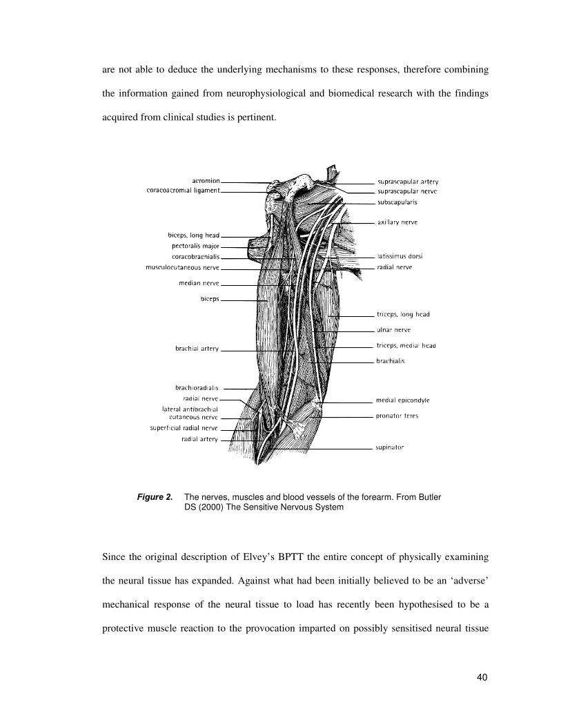
40
are not able to deduce the underlying mechanisms to these responses, therefore combining
the information gained from neurophysiological and biomedical research with the findings
acquired from clinical studies is pertinent.
)LJXUH��� The nerves, muscles and blood vessels of the forearm. From Butler DS (2000) The Sensitive Nervous System
Since the original description of Elvey’ s BPTT the entire concept of physically examining
the neural tissue has expanded. Against what had been initially believed to be an ‘adverse’
mechanical response of the neural tissue to load has recently been hypothesised to be a
protective muscle reaction to the provocation imparted on possibly sensitised neural tissue
41
(Balster & Jull 1997). However, no correlation between muscle activity and pain perception
was evident in this study, therefore the hypothesised mechanisms have yet to be confirmed.
The initial concept of examining the extensibility of neural tissue was further challenged by
an investigating on the normal compliance of neural tissue to the straight leg raise (SLR) test
in L5/S1 radiculopathy patients. Hall HW�DO� (1998) proposed that the onset of muscle activity
rather than the onset of resistance seems to represent more accurately an increased neural
sensitivity to mechanical load (see section 5.2.2). V. der Heide (2002) confirmed this pattern
of reactive muscle activity in a case study of three subjects with cervical radiculopathy,
where trapezius muscle activity was found to be in close association to the onset of pain,
however, the small sample size precludes any generalising of the findings so that future
investigations with larger patient populations need to be conducted that are statistically more
powerful (Hicks 1999).
The original name brachial plexus tension test tends to “convey an incorrect impression of
the whole thought process behind the assessment of conditions when using physical exami-
nation tests of neural tissues” (Elvey 1995, p.115). The need for a correct terminology has
lead to the description of the whole concept of testing manoeuvres as responses to ‘neural
tissue provocation testing’ (Hall HW� DO� 1998, Coppieters HW� DO� 2001a, v. der Heide HW� DO�
2001) or to neurodynamic testing (Shacklock 1995). In this sense physiotherapy has taken a
step back from giving preliminary explanations of the mechanisms behind the reactions to
neural tissue testing, and instead has engaged in the process of evidence-based reasoning by
trying to establish a knowledge base through normative studies. This way the normal
reactions of asymptomatic subjects to this test can be used as a standard against which
findings in a clinical setting can be compared.
42
��� &5,7,&$/�$1$/<6,6�2)�8/17�5(6($5&+�
In this main section of the thesis observations and assumptions underling the NTPT will be
critically evaluated. For this purpose it is necessary to first discuss some methodological
criteria pertaining to the ULNT research. In the following sections, the ULNT’ s construct
validity will be investigated by looking at biomechanical findings, and by assessing the
reliability of outcome variables measured during neural tissue provocation testing in normal
subjects. The last sections of this chapter will investigate scientific and clinical support for
the use of the ULNT in a clinical setting. As the ULNT has been developed as a diagnostic
test all research presented will be concerned with issues of its validation. The evaluation of
neural mobilisation techniques will not be included as it goes beyond the scope of this study.
����� 0(7+2'2/2*,&$/�&216,'(5$7,216�21�8/17�5(6($5&+�
In physiotherapy there is an increasing attention to the evidence-based medicine approach,
partly stimulated by the demands of society to show efficacy and cost-effective physio-
therapy (Koes & Hoving 1998). Many physiotherapist however, work in environments
where research facilities and the support for research are lacking (Matheson 2000), impeding
the desire to be part of the scientific community. Nevertheless, physiotherapy is realising this
deficit and research designs are being developed that acknowledge scientific demands as
well as the potentials that lie in physiotherapy. Traditionally the randomised clinical trial
(RCT) is considered to be the most valid design because of its potential to control various
forms of bias (Colditz HW� DO� 1989, Schulz 1996, Koes & Hoving 1998). However, one
limitation is that RCTs give only little insight into the proposed working mechanisms and
generally only evaluate the efficacy of existing interventions compared to new ones, but play
no role in the development of new treatment strategies. With regards to neural tissue testing

43
the ULNT is a diagnostic test and cannot be subject of an RCT. To test the validity of a test
it has to be compared with a criterion standard or golden standard. The problem with the
ULNT is that there is no gold standard against which it can be compared. Electrodiagnostic
tests for example evaluate nerve conduction and neuromuscular diseases, but are unable to
assess increased neuromechanosensitivity, a cardinal sign in minor peripheral nerve injuries.
Despite these obstacles physiotherapist have recognised the importance of investigating the
diagnostic validity of the ULNT. Research from the past ten years not only reflects this
increasing urge, but also reflects the evolving quality in terms of scientific merit. There has
been a change in physiotherapy from an empirically based approach towards a much greater
emphasis on scientifically based practice, and the neural tension tests are probably the “most
thoroughly evaluated group of assessment procedures” among the physiotherapists’ arma-
mentarium (Wright 1998, p.1). To evaluate the subsequent research a number of methodo-
logical criteria have been adopted pertaining to the ULNT as a diagnostic tool (Table 6).
TABLE 6: Methodological criteria for the neural provocation test as a diagnostic tool
1. How reliable is the test? ��intra-examiner, inter-examiner reliability and repeatability
2. How clear are the operational definitions in the studies? ��starting position of the subjects (fixation of the shoulder girdle) ��sequence of test movements ��what end position was used and under what criteria
(pain, resistance) ��what are the quantifiable measurements (instrumentation)
3. How are the independent variables controlled for? ��sensitising manoeuvres (wrist extension, CCLF)
4. How is the normal response in asymptomatic subjects defined? ��sensory area of response ��normal ROM ��reactive muscle activity
5. How valid is the test? ��sensitivity and specificity of the test

44
5.1.1. Reliability of examination procedures
Research into the use of manual examination techniques as the ULNT is essential particu-
larly in determining if physiotherapists are successful in achieving the objectives of validity,
reliability, and repeatability for these techniques. Such research requires that an accurate
diagnostic test is available as a standard against which findings of the test response can be
compared (Philips & Twomey 1996). This also means a diagnostic test has to show consis-
tency in order to be reliable. Reliability has been defined as “the degree to which measure-
ments are error-free and the degree to which repeated measurements will agree” (Rothstein
HW� DO� 1991, p.52). In the past there have been investigations on the reliability of manual
examination techniques of the vertebral joints (Maitland 1986) that are commonly used by
manual therapist in the assessment of spinal disorders.
A single-blinded crossover study by Jull HW� DO. (1988) demonstrated that one experienced
manipulative therapist was able to locate the symptomatic cervical zygapophysial joints,
prior and post to diagnostic blocks that established the diagnosis, with a 100% sensitivity
and specificity. Sensitivity meant that from the 15 patients that had zygapophysial pain
syndromes all 15 were detected correctly, and reciprocally specificity related to the 5
patients without zygapophysial pain syndromes, that were equally identified correctly. The
diagnostic test in this case involved manual testing of the mechanical properties of all
cervical joints in search for “perceived stiffness properties”. Since movement abnormalities
are palpable even in asymptomatic joints, the criteria for identifying a symptomatic joint
were: abnormal ‘end feel’ , abnormal quality of resistance to motion, and reproduction of
pain.

45
The findings of this study suggest that this set of criteria is useful in identifying symptomatic
joints, but before such conclusions can be made with assurance, similar research considering
other regions of the spine is necessary, and the inter-examiner reliability of these manual
techniques would have to be established. Furthermore, it should be noted that since the
examiner was highly skilled, results from this study are not typical for the majority of
physiotherapists and cannot be generalised, but emphasise the importance of high quality
manual training for orthopaedic physical therapists.
In the quest for standardised manual examination techniques, a more recent randomised
crossover study by Philips and Twomey (1996) raised the question whether manual exami-
nation alone is sufficient for accurate segmental diagnosis in low back pain patients, or
whether it must be accompanied by a verbal pain response. In the patient group that was
examined prior to a spinal diagnostic block the manual therapist’ s diagnosis was correct in
16 out of 17 subjects (94.12% sensitivity) for verbal responses, and in 9 out of 17 (52.9%
sensitivity) for non-verbal responses. The identification of subjects with no history of low
back pain was 5 out of 5 for verbal responses (100% specificity), and 4 out of 5 for non-
verbal responses (80% specificity). These results demonstrate that the patient’ s response was
important in identifying the symptomatic segment or in identifying true-positives. These
findings are concordant with the earlier propagated set of criteria (Jull HW� DO� 1988) for
identifying symptomatic joints, and indicate that manual examination techniques alone are
not a reliable measurement, but that the reproduction of pain or/and the verbal response of
the patient is necessary for an accurate diagnosis.
Percentage agreement rates were calculated to assess inter-examiner reliability for passive
intervertebral movements and tissue responses, and showed a range between as low as 43%
46
to a 100% agreement. However, as a measure for reliability, percentage agreement does not
take into account the agreement that is expected to occur due to chance alone, and since a
high agreement can be attributed to chance cannot in itself support a claim of good reliability
(Haas 1991b). Hence, the weak inter-examiner reliability on the passive manual techniques
remains to pose a considerable problem in the attempt to standardise manual diagnostic tests.
Implicating these results to the evaluation of the ULNT’ s reliability obviously leads to the
following considerations. In the first place, the testing procedure of the ULNT is much more
complex than the spinal joint tests involving multiple joint movements and handling
techniques. This results in a large number of independent variables that are difficult to
control. Secondly, as with the spinal joints, a normal range of movement respectively a
normal response to the ULNT has to be established against which abnormal test responses
can be compared. Thirdly, the consistency of the measurement tool, in this case corres-
ponding to the inter-examiner reliability, needs to be evaluated. Finally, as has been shown
to be important for spinal manual diagnostic tests, verbal responses and reproduction of pain
are equally a necessary part of neural tissue testing procedures.
It has been proposed that one of the most important steps in establishing the efficacy of any
diagnostic procedure is the investigation of its reliability (Haas 1991b). In Table 7 several
ULNT studies are shown, that have accounted for their test-retest reliability. Yaxely and Jull
(1991), for example calculated a high inter-examiner reliability and repeatability of 94.9%
and 99.9% respectively between two raters for the range of glenohumeral abduction at R2
(maximal resistance). In contrast, Hines HW�DO� (1993) showed that assessing elbow extension
at R1 (onset of resistance) among four raters was unreliable, which is not surprising as it is
almost impossible to manually pick up on the first point of increase in tension. On the other
47
hand excellent intra- and inter-examiner reliability for elbow ROM at submaximal pain
tolerance (Coppieters HW�DO. 2001b) were shown, as well as high reliability coefficients for the
intra-examiner reliability for pain onset (P1) and maximal pain tolerance (P2), and for
muscle activity during elbow extension (v. der Heide HW�DO� 2001). These measurements were
clearly described and calculated with the one-way ANOVA intraclass correlation coefficient
(ICC), which is the statistic of choice for the reliability of examiners for continuous data
(Haas 1991a).
Selvaratnam HW� DO� (1994) demonstrated a moderate to high intra-examiner reliability of
82.5% measuring elbow-wrist extension at P1 by correlating data acquired from two patient
groups. No significant differences were found in intra-examiner repeatability for trapezius
length and the ULNT, and in inter-examiner reliability for shoulder girdle depression (Edgar
HW� DO. 1994). Grant HW� DO� (1995) computed only a fair inter-examiner reliability (0.505)
between two raters for glenohumeral abduction at R2. The intra-examiner repeatability
however, was calculated with a percent agreement of 2.57% and not the usual indices of –1
to +1 (Hicks 1999), which made interpretations inconclusive.
No statistical descriptions were given for the high percent agreement in the area of sensory
response (Zorn HW�DO� 1995), and no reports on examiner reliability were given by Quintner
(1990), and Balster and Jull (1997). Obviously only weak implications can be made for the
use of these parameters in clinical practice, as long as it is not ensured that only relevant
outcome measures are analysed and that the appropriate statistics are being used. The
significance of this will be discussed in more detail for individual studies in sections 5.2.2.
and 5.2.3.
48
In summary, reliability measurements were made for range of motion at R1 or R2, for pain
intensity and sensory area of response, and for P1 and P2. Apart from the fact that these
studies differed widely in their quality for demonstrating their statistical analysis most of
these studies endeavoured to establish the reliability of diagnostic procedures. However, the
appropriateness of statistical methods and their interpretations of results were not always
clear. Hass (1991a) stated that the ANOVA and the W-test are least appropriate for assessing
reliability, because they test rater performance for significant differences from chance alone,
which could lead to concealing large inter-examiner disagreements. In addition most studies
report on both intra- and inter-examiner reliability and usually find greater concordance
within raters than between them.
One reason for this is the difficulty to ensure sufficient blinding of the rater to accurately
assess intra-examiner reliability. Secondly, even though there might be a strong self-
consistency of each examiner, there could be disagreement between raters, which results in a
lower inter-examiner reliability. However, high intra-examiner reliability does not rule out
that the measurements stem from a consistency of error (Haas 1991a). Therefore inter-
examiner concordance weighs more heavily in the evaluation of reliability measures, and
should be included in every research evaluating diagnostic procedures or examination
techniques especially if the testing procedures are not standardised.

TABLE 7: Test-retest accountability in ULNT research
6WXG\� 1HXUDO�WHVW� 1� $JH� :KDW�ZDV�PHDVXUHG"�
6HQVLWLVLQJ�PDQRHXYUHV�
5HVXOWV� ,QWUD���LQWHU�H[DPLQHU�UHOLDELOLW\�
Yaxely & Jull 1991
ULNT2b (radial bias)
50 normal
18-30 G/H abduction and sensory response at R2
__ Normal response = 40º of G/H abduction, and stretch felt over radial aspect of forearm
High inter-examiner reliability >95% for G/H abduction at R2 (ANOVA)
Hines HW�DO� 1993
ULNT1 (base test)
25 normal
19-50 onset of R1 in elbow extension
__ The range of elbow extension at R1 differed significantly between 4 raters
Low inter-examiner reliability for elbow ROM at R1 (ANOVA)
Edgar HW�DO. 1994
ULNT1 60 normal
17-25 shoulder depression during ULNT, trapezius length
contralateral CLF
Lesser extensibility of neural tissue and trapezius length are related
Good inter-examiner reliability for shoulder depression and intra-examiner repeatability for trapezius length and ULNT (ANOVA)
Selvaratnam HW�DO��1994
ULNT1 25 symptomatic 25 symptomatic
25 asymptomatic
22-70 16-51 19-73
elbow-wrist extension at P1
contralateral CLF and
ipsilateral CLF
BPTT is able to identify referred pain from brachial plexus in symptomatic subjects
Intra-examiner reliability 82,5% for performing the ULNT1 (ANOVA)
Grant HW�DO� 1995
ULNT2b 15 symptomatic 10 asymptomatic
17-55 mean
28
G/H abduction, sensory response at P?
contralateral CLF
Patient group exhibited decreased range of G/H abduction in ULNT in comparison to normal subjects
Report of high intra- and inter-examiner reliability (p=0.004) for G/H abduction (ANOVA)

�
TABLE 7: Test-retest accountability in ULNT research
6WXG\� 1HXUDO�WHVW� 1� $JH� :KDW�ZDV�PHDVXUHG"�
6HQVLWLVLQJ�PDQRHXYUHV�
5HVXOWV� ,QWUD���LQWHU�H[DPLQHU�UHOLDELOLW\�
Zorn HW�DO� 1995
ULNT1 90 normal
18-60 Location of sensory response at P?
__ Sequencing from distal to proximal produces fewer proximal responses
93% of agreement for area of sensory response (no statistics)
Balster & Jull 1997
ULNT2a (median
bias)
20 normal
18-30 Trapezius activity, elbow extension at P1
contralateral CLF
Greater magnitude of trapezius activity found in lesser extensible neural tissue group
Not stated
Coppieters HW�DO. 2001a
ULNT1 35 normal
20-28 Shoulder girdle elevation force
wrist extension, contralateral
CLF
Gradual increase in shoulder girdle elevation force is a normal sign during neurodynamic testing
Excellent reliability coefficient for intra-examiner (0.93-0.97) and for inter-examiner (0.93-0.96) reliability (one-way ANOVA ICC)
Coppieters HW�DO. 2001b
ULNT1 35 normal
20-28 Elbow extension, sensory response at P2
wrist extension, contralateral
CLF
Elbow ROM is markedly reduced when adding test components and 80% of subjects reported paraesthesia in hand
Excellent reliability coefficient for intra-examiner (0.95-0.98) and for inter-examiner (0.91-0.97) reliability (one-way ANOVA ICC)
v. der Heide HW DO��2001
ULNT2a 20 normal
43 (mean)
P1 and P2 during elbow extension
contralateral CLF
Normal muscular response to trapezius activity at P1. CCLF increases pain and muscle response
Onset of P1 and P2 both have high intra-examiner reliability coefficients (one-way ANOVA ICC)

51
5.1.2. Operational definitions in ULNT research
To allow an experimental study to be replicated and results corroborated a highly repro-
ducible test protocol and a detailed description of operational definitions should be provided.
An operational definition is a set of procedures that guides the process of obtaining a mea-
surement and includes descriptions of the attribute that is to be measured, the conditions
under which the measurement is to be taken, and the actions that are taken in order to obtain
the measurement (Rothstein HW�DO� 1991).
There has been an effort to ensure a standardised application by operationalising the starting
position of the ULNT1 in 5 out of the 8 normative studies (Edgar HW�DO� 1994, Coppieters HW�DO� 2001ab, Balster & Jull 1997, v. der Heide HW�DO� 2001, Table II-Appendix B). The subjects
were placed in a supine position with the head in a neutral position. The arm to be tested was
positioned in 90° glenohumeral abduction supported by an armrest. The shoulder girdle was
additionally gently depressed with an initial force of 30 Newton prior to adding the other test
components to neutralise the shoulder girdle elevation that is caused by the abduction of the
arm. To lessen the variation of this variable a pressure sensor pre-inflated to 20 mmHg was
placed on the superior part of the subjects shoulder and used to monitor the amount of force
applied by the examiner, such that a pressure increase of 40 mmHg was recorded. The inter-
examiner reliability of this measurement device had been analysed with a one-way ANOVA,
and indicated no significant differences (Edgar HW�DO� 1994). These studies demonstrate that it
is possible to employ clear operational definitions, and to use quantifiable measurements
aiding the comparison and evaluation of test results.
Stating in what end position outcome measures were taken (elbow extension/flexion, G/H
abduction, range of CCLF) and under what criteria (pain, resistance) is another important

52
operational definition. In the method section of each study the quantifiable measurements
(pain, resistance, ROM) and their respective instrumentation (i.e. goniometer, VAS) should
also be clearly described with indications to their validity (see 5.2.2). In addition it is impor-
tant to accurately describe and operationalise the sequencing of each ULNT (Table 5), so
that results of identical tests can be related to each other.
Sensory responses of different ULNT components and sequences have been investigated in
90 asymptomatic subjects (Zorn HW�DO. 1995, Table II). Comparing the consequences of three
different ULNT sequences disclosed that the proximal to distal build up and the middle
sequence showed similar sensory responses proximal to the elbow. However, distal to
proximal build up produced symptoms distal to and including the elbow. Previous studies
have shown that the longitudinal excursion of peripheral nerves depends upon the location
and degree of joint motion (McLellan & Swash 1976, Shaw & Wilgis 1986). In this respect
the proximal or distal build up may be valuable when examining patients with a suspected
proximal or distal neural component to their shoulder or upper limb pain.
5.1.3. Control for independent variables
Another important criteria for the evaluation of the ULNT is the control for sensitising
manoeuvres that play a critical role in confirming neural tissue involvement in CBP
disorders. Will the experimental setting provide that each testing component can be added
without any deviation from the previous position? In human cadaver studies, that
investigated the influence of CCLF on the tension of the brachial plexus cords, this was
easier achieved as the critical parts were fixed (Selvaratnam HW�DO� 1989, Lewis HW�DO. 1998,
Kleinrensink HW� DO� 2000, Table I). In contrast, demands on in vivo studies are not only
focused on rigorous measurements, but also controlling the sensitising manoeuvres plays a
53
decisive role when assessing neural tissue involvement in symptomatic subjects (Yaxely &
Jull 1993, Selvaratnam HW�DO� 1994, Grant HW�DO. 1995, v. der Heide HW�DO� 2002, Table III). To
prevent evasive movements when adding the test components some studies have used splints
to fix the head in a neutral position and/or seatbelts around the hips and thorax (Yaxely &
Jull 1991/1993, Selvaratnam HW�DO� 1994, Zorn HW�DO� 1995), but most studies had to keep the
cervical spine free for the CCLF manoeuvres.
5.1.4. Defining normal responses
Normal responses in asymptomatic subjects should be established so they can be used as a
standard reaction against which test responses from symptomatic subjects can be compared.
The main dependent variables that produce quantifiable measurements (see 5.2.2) are range
of motion at a defined end position, type and area of sensory response, reactive muscle
activity, and response to sensitising manoeuvres (Yaxely & Jull 1991, Edgar HW� DO. 1994,
Zorn HW�DO��1995, Balster & Jull 1997, Hall HW�DO� 1998, Coppieters HW�DO� 2001ab, v. der Heide
HW�DO� 2001, Table II).
5.1.5. Validation of diagnostic tests
Validity is defined as the degree to which a meaningful interpretation can be inferred from a
measurement (Rothstein HW�DO� 1991). To do so the ULNT’ s construct validity, which is the
theoretical basis for inferring an interpretation from the measurements, has to be stated. This
also includes the evaluation of the ULNT’ s content validity, which means ‘does the test
measure what we think it will measure’ . In the next sections the following questions should
therefore be answered :

54
1. What does a positive test indicate?
2. What limits the movement during testing?
3. Can the test truly discriminate between different sources of pathology?
����� ,19(67,*$7,1*�7+(�9$/,',7<�2)�7+(�8/17�
In the subsequent sections research investigating the ULNT’ s anatomical and clinical
validity and reflections on some limitations will be presented. Neural tissue provocation
testing was originally based exclusively on a neuromechanical construct. The nervous
system was believed to respond to mechanical induced stresses by distributing forces
throughout the spinal cord, meninges, nerve roots and peripheral nerves. This was based on
the fact that neural tissue has elasticity and movement relationships with adjacent tissues
(Sunderland & Bradley 1961ab, McLellan & Swash 1976, Wilgis & Murphy 1986, Zöch
1992). It had been demonstrated that injured or inflamed nerves, are characterised by an
increased sensitivity to mechanical load (Bove & Light 1997, Greening & Lynn 1998,
Rempel HW� DO� 1999, Mackinnon 2002), and could react to mechanical stimulation with an
impaired compliance to movement and sensory responses. The original idea of the ULNT
was that an ordered set of joint movements could be used to selectively increase tension
within the neural tissue and their connective tissue sheaths (Elvey 1979b), thereby testing for
increased mechanosensitivity.
To test the construct validity of this original hypothesis it is necessary to quantify the amount
of tension transmitted onto the neural tissues during the ULNT. Additional points needing
consideration include if the test response corresponds to the presumed tissues under test or to
other structures, and what is the test’ s predictive value. In order to test the accuracy and
55
reliability of a diagnostic test, research that assesses the predictive value of positive and
negative tests is necessary. A good diagnostic test minimises the probability of an occurrence
of either a false-positive or false-negative result. To determine the accuracy of the neural
tissue provocation test in clinical practice two different conditional probabilities are relevant.
Firstly the test VHQVLWLYLW\, which determines the probability of testing positive in a truly
present lesion, would be greater the more tension a test caused in the intended nerve.
Secondly, the VSHFLILFLW\, which determines the probability of testing negative in the absence
of a lesion in the intended nerve, would be determined by knowing whether the test caused
significant tension in other nerves than the intended one. This would mean that pain and
symptoms could be due to increased tension in other nerves, and would lead to a false-
positive response. It is therefore important to investigate whether a ULNT causes the highest
tension in the nerve for which it was intended.
In clinical practice, however, it is often impossible to calculate the sensitivity or specificity
of a diagnostic test because of confounding factors. For example the probability that a
positive test indicates a true-positive finding, determines the sensitivity or positive predictive
value of the test. In pain studies, false-positive responses could arise due to central
sensitisation and secondary hyperalgesia (Woolf 1989/1991, Gracely HW�DO� 1992). Moreover,
the probability that a negative response represents a ‘true-negative response,’ or the negative
predictive value, can be complicated because some peripheral neuropathies can lead to
decreased mechanosensitivity, thus leading to false-negative results (Shacklock 1996, Woolf
& Mannion 1999).
56
5.2.1. Biomechanical findings during the ULNT in human cadaver studies
Initially it was believed that manipulating the distal parts of an extremity could help in
differentiating between peripheral nerve trunk and nerve root lesions (Elvey 1979a, Keneally
HW� DO� 1988, Butler 1991). However, as it was not known how the tensile forces were
distributed along the peripheral nerve, or whether they were actually transmitted all the way
up to the nerve root, the interpretation of a positive test response remained difficult. To
clarify these questions and to validate the diagnostic value of the ULNT, human cadaver
studies investigated the distribution of tension along the median, ulnar and radial nerves.
Although informative findings from cadaver studies are not likely representative of measure-
ments from living subjects because of post-mortem tissue changes, these studies contribute
to the understanding of neural tissue dynamics, and provide a framework for the clinical
findings presented in the next sections.
Selvaratnam HW� DO� (1989, Table I-Appendix B) was the first to test Elvey’ s BPTT by
conducting an anatomical study on 5 unembalmed human cadavers to confirm that adding
CCLF to elbow and wrist extension produced a greater strain on the brachial plexus nerve
roots. The highest tensile forces were measured between markers on the spinal nerve roots
and the nerve trunks. The C5 nerve root displayed the most tension followed by the C6, C7,
C8, and T1 nerve roots. An important finding was that elbow extension with CCLF produced
a greater strain on the nerve roots than with ipsilateral CLF, which lead the authors to
recommend the use of CCLF in differentiating between disorders of the upper quarter with
and without brachial plexus involvement (Selvaratnam HW� DO� 1989). The robustness of the
obtained data is however questionable as the authors used a camera to document the changes
of spatial location of the nerve roots during the ULNT, which were then calculated into
strain scores, without establishing the reliability of this method.

57
Lewis HW�DO� (1998, Table I) later confirmed these findings in a randomised, single-blinded
study of 5 human specimens that only the elbow and wrist extension components of the
ULNT1 produced a significant increase in the median nerve tension, which could be further
increased by adding CCLF. The ULNT sequencing was operationalised and followed the
protocol described by Keneally HW� DO� (1988). Buckle force transducers were used for the
measurement of tension, and were prior tested in a pilot study to be highly reliable (r=0.998).
Shoulder girdle depression, and glenohumeral lateral rotation in contrast have been found to
not significantly increase tension in the median nerve (Lewis HW�DO� 1998), although it was
not clear if these components were added in the final ULNT position. Earlier Ginn (1988,
Table I) even reported of a decrease in the tension of all the brachial plexus cords during
glenohumeral lateral rotation. However, these findings are not representative because they
were based on a single specimen, and no reliability measures of the buckle force transducer
or the procedures were given, so that the study cannot be replicated to test for its rigor. The
results should therefore be interpreted with caution.
Kleinrensink HW� DO� (1995b, Table I) investigated the distribution of tensile forces along 5
human median nerves. Since a positive correlation had been established between the
measurements of tensile forces in embalmed and unembalmed human specimens a combined
analysis of the data was accepted (Kleinrensink HW�DO� 1995a). The effect of 22 arm positions
in the normal range of motion (ROM) on the median nerve tension was measured with
buckle force transducers at the site of the axilla, elbow, and wrist. All 22 joint positions were
studied three times as test and retest, and after simulating an obstruction of the gliding
mechanism of the median nerve through the pronator teres muscle. The transducer had a
high test-retest reliability and was calibrated after each measurement. The elbow and hand

58
position were clearly described, however no reference to the position of the shoulder girdle
was given, which has been defined as an integral part of the ULNT (Butler 1994, Table 5).
With the elbow in full extension and hand in neutral position, the authors found that altering
the position of the shoulder to maximal abduction, retroflexion and lateral rotation
significantly influenced the tension of the median nerve at the level of the axilla and elbow,
while tension at the level of the wrist was not influenced. With the shoulder in 90° abduc-
tion, wrist extension combined with an extended elbow increased tension in all parts of the
nerve, whereas wrist extension combined with elbow flexion only caused an increase in the
distal part of the median nerve. Manipulating joints in the distal part of the upper extremity
can increase tension in the most proximal part of the median nerve, however, changing the
shoulder joint position cannot alter the tension in the distal part of the median nerve. Thus,
there is anatomical evidence that differentiating between lesions in the upper and lower part
of the median nerve may be possible by manipulating specific joints, but this needs to be
confirmed by clinical investigations.
A more recent anatomical and biomechanical study by Kleinrensink HW�DO� (2000, Table I)
clarified the specificity of the three basic ULNTs by Butler (Table 5). The outcome variables
were the mechanical tension assessed with a calibrated buckle force transducer on the
medial, lateral and posterior cords of the brachial plexus measured beneath the clavicle, as
well as on the proximal parts of the median, ulnar and radial nerves. A fixed protocol was
used for the sequence of arm positions and each measurement was taken twice to be
analysed with a multiple regression analysis.
All three ULNTs caused a higher tension in the medial cord than in the lateral or posterior
cords (Figure 3, Appendix C). Only the median biased ULNT1 had shown to be both

59
sensitive and specific for the median nerve and was recommended as a valid test by the
authors. The radial biased ULNT2b caused more tension in the median nerve than in the
radial nerve itself, and was not specific. Equally the ulnar biased ULNT3 was not specific
since there was no significant difference between the tension caused in the ulnar, median and
radial nerves (Table 8). The proposed selective stress on specific nerves and cords of the
brachial plexus could therefore not be confirmed by this study.
TABLE 8 : Mean tensile forces in Newton (±SD) caused by the ULNT (Kleinrensink HW�DO. 2000)
� 8/17����PHGLDQXV��
8/17����XOQDULV��
8/17�E���UDGLDOLV��
0HGLDQ�QHUYH�� 10.88 (5.88) 0.59 (0.29) 8.52 (5.19)
8OQDU�QHUYH� 1.18 (0.98) 3.29 (3.04) 1.76 (1.57)
5DGLDO�QHUYH� 1.86 (0.59) 2.94 (2.55) 5.88 (2.35)
0HGLDO�FRUG� 9.31 (6.17) 4.21 (4.80) 8.13 (5.88)
/DWHUDO�FRUG� 5.10 (4.21) 0.49 (0.69) 4.41 (3.42)
3RVWHULRU�FRUG� 1.57 (1.47) 2.55 (2.06) 5.00 (2.74)
Although the results by Kleinrensink HW�DO� (2000) indicated the use of only the ULNT1 as a
valid test, there has been a contradicting report on the specificity of the ULNT3 from a
single case report of a proven ulnar neuropathy (Shacklock 1996). In this case report
Shacklock described that the ULNT3 was able to reproduce the patient’ s symptoms while
the ULNT1 was not. One reason for this discrepancy could be the fact that Kleinrensink HW�DO� (2000) conducted a cadaver study where tension was measured as the outcome variable,
whereas in the clinical setting the main outcome measurements were pain response and
ROM.
60
Another difference influencing the results might be a deviation between the movement
sequencing. The median ULNT1 appears to be the most specific test in transferring tensile
forces to its corresponding nerve, however, the ULNT3 produces more tension in the ulnar
nerve than the median biased ULNT1 (Table 8). This could explain why, despite the weaker
tensile forces in total, the ULNT3 was still specific for the ulnar nerve. Clinically this means
that if a patient presented with an isolated ulnar neuropathy the ULNT3 would most likely be
positive, but if a patient had both ulnar and median nerve pathology the ULNT3 could not be
specific in isolating the ulnar neuropathy.
A basic assumption of the ULNT is based upon the ability to selectively move neural tissue
in the absence of mechanically affecting neighbouring non-neural tissue. To test this it is
important to verify which components of the test move the neural tissue and which
components cause tension in non-neural structures. Anatomical connections in the cervical
region suggest that upper limb neural testing may equally stimulate the intervertebral discs,
interspinous ligaments, zygapophyseal joints and muscles as these structures have shown to
possess nociception and could therefore refer pain to the upper limb (Dwyer HW� DO� 1990,
Bogduk 1994, Moses & Carman 1996). During neural tissue provocation testing, especially
in the end range position, many non-neural structures are stretched.
A clinical study on the subclavian artery of 2 embalmed human specimens demonstrated that
CCLF with elbow extension alone or with additional wrist extension induced a stretch on the
first and third segment of the subclavian artery (Wilson HW�DO� 1994, Table I). The authors
also stated that the strain on the lateral cord of the brachial plexus was greater than on the
first part of the subclavian artery. Because no positions of the shoulder girdle or the
glenohumaral joint were described no specific comparisons can be made to the ULNT.

61
Furthermore, these findings contradict Elvey (1979b, 1995), who observed no movement in
the subclavian artery during CCLF. Even though Elvey produced no quantitative data to
support his statement, the results from Wilson HW�DO� (1994) may equally be flawed because
the subclavian artery was prepared by freeing it from the clavicle and sternocleidomastoid
muscle destroying its natural surroundings, which may have presented some form of natural
fixation thereby leading to a systematic bias of the obtained data.
To sum up, the initial idea of manipulating the distal parts of an extremity to help different-
tiate between peripheral nerve trunk and nerve root lesions has not been confirmed. What the
cadaver studies have shown is that the main components of the ULNT capable of inducing
tension changes from the proximal parts of the median nerve to the brachial plexus cords are
wrist and elbow extension, and that this tension can be intensified by adding CCLF
(Kleinrensink HW�DO� 1995b/2000, Lewis HW�DO� 1998). Selvaratnam HW�DO� (1989) showed that
adding CCLF to the ULNT selectively increased the tension in the brachial plexus nerve
roots, with the highest tension at C5 and the lowest at T1. Interestingly the C5 and C6 nerve
roots supply the upper trunk of the brachial plexus, which leads into the lateral and posterior
cords (Figure 3). In contrast, Kleinrensink HW�DO� (2000) found the medial cord to be under
most tension when adding contralateral cervical rotation to the ULNT. This discrepancy
might be due to the different sites of measurement (nerve roots vs. distal brachial plexus
cords).
Unexpectedly Kleinrensink HW�DO� (2000) also found that when adding contralateral cervical
rotation to the ULNT the tension in the medial cord remained the same while tension in the
lateral and posterior cords increased up to 50% when compared to ULNT with the head in
neutral. This supports the idea that the brachial plexus plays a role in the distribution of
62
tensile forces (Butler 1991). Furthermore, it has been shown that the median ULNT1 is the
most specific test in transmitting tension to the proximal part of the corresponding nerve, and
is the only ULNT that can be recommended as a valid test (Kleinrensink HW� DO� 2000).
However, according to the research at hand the original idea that the ULNT can selectively
isolate neural tissue from non-neural tissue has further to be questioned.
Although the information gained from these anatomical studies is valuable there are some
methodological limitations that have to be considered, such as the small sample size
(maximal 5 specimens), and the degenerative changes associated with the higher age of the
samples. Apart from changes post mortem and the effect of conservation on the tissues, it is
also not know what existing pathologies and diseases of the specimens may have influenced
the measurements. As these studies are purely biomechanical investigations, they have
shown that tension is transmitted to the proximal parts of the nerves and the cords of the
brachial plexus; however, so far no relationship between the amount of tension produced in a
nerve and its sensory response has been established. Therefore the next step in the evaluation
of the test’ s efficacy is to investigate the normal reactions to the ULNT in asymptomatic
subjects.
5.2.2. Quantifiable measurements in normative ULNT studies
To standardise the ULNT as a diagnostic test quantifiable outcome measurements, detected
during the testing procedure in asymptomatic subjects, have to be determined (Table 9).
These measurements also include the criteria that determine the end of the test, which can be
either resistance (Yaxely & Jull 1991, Hines HW� DO� 1993) or pain (Balster & Jull 1997,
Coppieters HW�DO� 2001b/2002b/2003ab, v. der Heide HW�DO. 2001). However, determining the

63
end condition relies heavily on the perception and manual skills of the examiner, and on the
report made by the subject.
TABLE 9: Quantifiable measurements advocated for monitoring�
2XWFRPH�0HDVXUH� 0HWKRG�2I�'DWD�&ROOHFWLRQ�
1. Compliance to movement: ��onset of resistance (R1) ��maximal tolerable resistance (R2) ��range of motion (ROM) ��reactive muscle activity ��shoulder girdle elevation
1. Compliance to movement: ��manual skills of examiner ��manual skills of examiner ��standard/electro-goniometer ��EMG ��load cell
2. Sensory responses: ��type and area of sensory response ��onset of pain (P1) ��maximal tolerable pain (P2) ��reproduction of the patient’s symptoms ��pain intensity
2. Sensory responses: ��verbal report of patient ��verbal report of patient ��verbal report of patient ��verbal report of patient ��numeric pain scale
3. Effects of sensitising manoeuvres: �� increase of sensory responses �� reproduction of the patient’s symptoms
3. Effects of sensitising manoeuvres: ��numeric pain scale ��verbal report of patient
The onset of resistance (R1) and its clinically acceptable maximum (R2) may be at any point
within the normal passive range of motion (Maitland 1991). If the neural tissue is sensitised
normal movement could cause a provocative mechanical stimuli that may lead to a pain
response and/or to non-compliance to movement (Hall & Elvey 1999). One feature of this
non-compliance is proposed to lie in the increased and reactive tissue stiffness offered by
muscle spasm (Maitland 1986) caused by muscles antagonistic to the painful direction of the
movement. The other feature is an increased through range tissue stiffness, which is thought
to result from pathological articular or connective tissue. However, no studies have deter-
mined whether a physiotherapist can differentiate between tissue resistance and muscle
spasm (Hall HW�DO� 1998).
64
Some studies have questioned whether the onset of resistance or pain truly reflects what is
occurring when testing the neural tissue (Wright HW�DO� 1994, Hall & Quintner 1996, Balster
& Jull 1997, Hall HW�DO� 1998). The following study quantified the ability of physiotherapists
to determine the onset of resistance during the SLR test in an age and subject matched design
of 20 subjects with no previous history of back or leg pain, and 20 subjects with validated
L5/S1 radiculopathy (Hall HW�DO� 1998, Table III-Appendix B). The study compared the range
of SLR at R1, the electromyographical (EMG) activity of the hamstring muscles during the
SLR test, and the moment of the stretched tissues (MU) between the two groups. An impor-
tant finding was that R1 was significantly earlier in the range of SLR than the first increase
of muscle activity, and that there was no significant difference between R1 in the control and
the radiculopathy group, which means that R1 is not a reliable dependent variable when
assessing differences in neural mechanosensitivity between symptomatic and asymptomatic
subjects.
In all the subjects with radiculopathy the onset of muscle activity (M1) was significantly
earlier during the SLR test than in the control group, and probably accounted for the increase
in MU at that point rather than the increase in neural tissue tension. The authors concluded
that M1 was a more accurate measurement representing neural mechanosensitivity than R1.
The accuracy and reliability of the electrogoniometer and the device to measure MU were
assessed in a pilot study and were found to be excellent (r=1.0). However, as the Pearson’ s U tends to overestimate reliability and does not account for systematic observer bias (Haas
1991b) these values need to be reassessed. The intra-examiner reliability was established for
the repeated measurements of R1 and MU and was calculated to be good to excellent (ICC
value of 0.75-0.98), but as the examiner was not blinded the result may only be a reflection
65
of a strong self consistency, therefore the inter-examiner reliability needs to be established
before further implications can be made.
As M1 has been shown to be a clinically more significant measure than R1 the subsequent
normative studies were interested in the relationship between muscle activity and neural
mechanosensitivity in asymptomatic subjects. Bearing in mind that in normal subjects
abnormal age-related changes of the cervical spine can be found (Boden HW� DO� 1990) the
following studies included only asymptomatic subjects, that had no previous history of CBP
disorders or systemic diseases related to neuropathy.
Balster and Jull (1997) investigated the relationship between the ULNT1, upper trapezius
muscle activity and the range of neural tissue extensibility in 20 young male asymptomatic
subjects (Table II). The ULNT1 was performed according to Elvey (1986), and shoulder
girdle depression was operationalised (Edgar HW� DO� 1994). Reliability measures were only
performed for the EMG activity and revealed no significant difference between trials, but the
examiner reliability for elbow ROM was omitted. Two groups with greater and lesser neural
tissue extensibility were formed by baseline measurements of their elbow extension in the
final ULNT1 position.
In comparison the two groups showed no difference in perceived pain levels rated through a
verbal analogue scale. The EMG measurements revealed that the lesser extensible group
exhibited a significant greater trapezius muscle activity at the onset of pain (p=0.01), at the
limit of elbow extension (p=0.01), and at the limit of CCLF (p=0.006). However, neither the
criteria for the limit of elbow extension and CCLF were defined (pain or resistance) nor were
any ranges presented in the analysis. Thus, the repeatability of these measurements as well as
66
comparison of the data to similar research is impeded. These methodological deficiencies
therefore weaken the reliability of this study.
The hypothesised mechanisms explaining this neuromusculoskeletal interaction were that the
muscle activity and pain response are either attributed to a heightened flexion withdrawal
reflex elicited by afferent input upon sensitised spinal neurons (Woolf 1989, Wright HW�DO� 1994) or by nociceptive input from tension sensitive neural tissue such as the nervi nervorum
(Bove & Light 1997). Considering that both groups experienced the same level of pain but
displayed a different muscle response indicated that the nociceptive mediated withdrawal
reflex may not be the only mechanism involved in protecting the neural structures (Balster &
Jull 1997).
Rather the authors suggested that stretch receptors in the neural tissue were responsible for
this protective muscle action, on the grounds of recently identified stretch receptors in the
phial ligaments supporting the spinal arteries. However, the existence of stretch receptors in
the neural tissue has not been confirmed. Noteworthy is that no correlation was found
between the increase in muscle activity and the increase in pain intensity in asymptomatic
subjects (Balster & Jull 1997), which contradicts the hypothesised mechanism. If a corre-
lation can be found in symptomatic subjects still needs to be investigated, however.
The purported theory of a nociceptive mediated withdrawal reflex has further to be recon-
sidered in view of the results obtained by Edgar HW� DO� (1994). This correlation study
investigated the relationship between the extensibility of the upper quarter neural tissue
tested via the ULNT1 and the muscle length of the upper trapezius muscle in 60
asymptomatic young male volunteers (Table II). Two groups (n=30) were formed in the
same manner as in the study by Balster and Jull (1997). Shoulder girdle depression was
67
standardised (see section 5.1.2) such that a pressure increase from 20 mmHg to 60 mmHg
was performed on each subject, before adding the other test components.
No significant differences between trials were found for inter-examiner reliability of
shoulder girdle depression, or for the intra-examiner repeatability of trapezius length and
ULNT1 measurements. Although statistical calculations were not adequately presented, the
procedure for obtaining these measurements was described in detail. The authors found that
the length index of the trapezius muscle was significantly less in the subjects with lesser
neural tissue extensibility independent of elbow extension or flexion, and concluded that
neural tissue extensibility should be assessed prior to interpreting the results of length tests
of the upper trapezius muscle in patients that present with pain in the upper quarter.
While Balster and Jull (1997) concluded that the heightened trapezius muscle activity was a
protective reaction to the decreased extensibility of neural tissue, Edgar HW�DO� (1994) found
that in subjects with lesser neural tissue extensibility the trapezius muscle was equally lesser
extensible. This is not surprising as subjects with decreased mobility will exhibit this charac-
teristic in all of their tissues and not just in their neural tissue. In this respect it will be
difficult to distinguish whether the nociceptive reflex is elicited to protect the less extensible
neural tissue or musculature.
However, these normal findings should also be interpreted with regards to the painful
reactions of the trapezius muscles in disorders of the cervicobrachial region. Patients
suffering from cervical radiculopathy have been found to react with a reduced muscle
tension (EMG recording), due to the decreased microcirculation of the trapezius muscle
(Löfgren HW�DO� 2001), whereas in chronic neck pain populations increased values of muscle
68
tension were shown (Larsson HW�DO� 1999). In so far the trapezius length should be considered
when assessing the neural tissue of chronic neck pain patients.
The second group of quantifiable measures used when testing the ULNT are the sensory
responses (Table 9). In a same-subject design study by van der Heide HW� DO� (2001) the
correlation between pain and muscle activity responses to the ULNT1 were examined in 20
asymptomatic subjects (Table II). The starting position (Edgar HW�DO� 1994) and the testing
procedure were operationalised, and all statistical analyses for the measurements were
clearly described. The test was performed on both arms with the cervical spine in neutral,
and in CCLF as a sensitising manoeuvre. P1 and P2 were used as indicators to cease the
elbow extension, which the subjects determined by using an external trigger. Elbow exten-
sion was measured with a calibrated electrogoniometer, and EMG recordings determined the
muscle activity. There was no statistical significant difference for the intra-examiner relia-
bility at P1 and P2, and the onset of pain showed a high reliability between the three trials.
The results displayed that pain responses and muscle activity of the trapezius muscle were
evoked in the majority of all subjects and could be defined as a normal physiological
response. A total of 59% (n=11.8) of the subjects had an onset of muscle activity at P1, but
as Balster and Jull (1997) did not state the degree of elbow extension at P1 these results
cannot be compared. In some subjects trapezius activity was measured before the onset of
pain. Additionally adding CCLF had a sensitising effect in 18 subjects, who felt an increased
intensity of their sensory responses. Of the 37.5% that had no onset of pain with the head in
neutral, CCLF had a sensitising effect on the onset of pain and muscle activity.
Even though there was a strong correlation between trapezius activity and pain, approxi-
mately 16% (n=3.2) of the total study population showed trapezius activity without pain, but

69
experienced symptoms such as stretching sensation or paraesthesia in the arm. If muscle
activity occurred as a response to symptoms other than pain, this would contradict the
hypothesis of a protective motor reaction in healthy subjects, but a study with a larger
sample size would be needed to confirm this. The large variation in responses may partly be
due to the variation of pain responses. Pain is a sensory-emotional phenomenon and quanti-
tative measurements do not account for the affective perception of pain that has an impact on
pain intensity.
As proposed by Elvey (1979b/1986) and Keneally HW�DO� (1988) an abnormal response to the
ULNT may not only involve just a pain response but also the reproduction of the patient’ s
exact symptoms. To decide when a response to the ULNT is abnormal, it is important to
know the standard response to neural tissue provocation testing in asymptomatic subjects.
For the therapist to accurately interpret a neurodynamic test the difference between a motion
restriction indicative of a dysfunction and a limitation to movement, which can be consi-
dered as a normal response, has to be established.
Yaxely and Jull (1991, Table II) used the radial biased ULNT2b to demonstrate the normal
sensory response at R2 in 50 asymptomatic young subjects (25 female, 25 male). A stretch
felt over the radial aspect of the proximal forearm at 41.45° (± 4.06°) glenohumeral abduc-
tion and wrist flexion was reported in 84% of all responses (left and right arm combined).
The abduction range, measured with a standard goniometer, was not influenced by the
gender of the subjects nor by which side was tested. Excellent inter-examiner reliability
(p=0.949) and repeatability (p=0.999) for the mean range of glenohumeral abduction in the
ULNT2b were calculated.

70
An increase in “ arm symptoms” with the addition of CCLF as a sensitising manoeuvre to the
final test position was recorded, in 86% for the right arm and 90% for the left arm. However,
the authors did not state if the area was identical to the initially reported stretch over the
radial aspect of the arm. To what extent the sensory responses really reflect the reaction of
the stretched radial nerve have further to be questioned in the light of the results by
Kleinrensink HW�DO� (2000, Table 8), who demonstrated that ULNT2b caused more tension in
the median nerve than in the radial nerve, therefore the arm symptoms could have be elicited
through stretching the median nerve. Another limitation appears to be that for the ULNT2b
no standardisation for the starting position in an experimental setting has yet been defined.
As in the ULNT1 the shoulder girdle was initially depressed but with what amount of force
has not been stated.
To analyse the effect of different ULNT1 components on the limitation of elbow range of
motion and on the provocation of sensory responses Coppieters HW�DO� (2001b) investigated
four test variations on 35 asymptomatic young male subjects (Table II). The starting position
was operationalised (Edgar HW�DO� 1994), and baseline measurements of the elbow and wrist
joints were taken prior to testing. Elbow extension in a non-ULNT position was 182.6º
(±3.5º). The addition of each test component resulted in a significantly reduced elbow ROM
(ULNT1 in neutral 179.5º ±8.8º; ULNT1 + wrist extension 169.0 ±13.9º; ULNT1 + CCLF
154.7º ±13.2º; ULNT1 + wrist extension + CCLF 143.9 ±16.1º).
The sequence of the test variants was randomly allocated, and the analysis of variance
revealed no significant difference between the three repetitions. Sensory responses were
described as a (painful) stretch or paraesthesia predominantly evoked in the region of the
added components. The relatively high incidence of paraesthesia (76%) suggests that at least

71
some responses are neurogenic in origin. The average numeric pain rating (0-10) increased
progressively as the test components were added from 4.4 to 6.6. The distribution of sensory
responses revealed that when CCLF was added sensory responses in the more proximal areas
of the upper quarter were reported than without CCLF.
The excellent inter-examiner reliability measures (ICC 0.94-0.97) imply that elbow ROM
may be used as a reliable comparable sign in the clinical evaluation of the ULNT1. The
individual test components had a cumulative effect on limiting the elbow ROM when added
simultaneously. Furthermore, the elbow ROM was significantly influenced by the position of
the cervical spine, which could not have been caused by the mono- and biarticular structures
around the elbow joint. The authors suggested that the only continuous structure that could,
at least partly, influence the range of elbow extension is the nervous system, since blood
vessels, skin or the lymphatic system are unlikely to distribute such restrictive forces.
However, the potential role of the fascia to restrict movement will need to be investigated in
the future. The correspondence between the regions of added components and the area of
sensory responses implicate that reproducing symptoms in patients and altering these by
changing distant components of the test such as wrist extension or CCLF, will be essential
for the structural differentiation between neural and non-neural tissues.
One of the signs advocated for monitoring during ULNT1 in subjects with upper quarter
disorders is the involuntary elevation of the shoulder girdle (Elvey 1994). Coppieters HW�DO� (2001a) investigated the shoulder girdle elevation force during five variants of the ULNT1 in
35 young asymptomatic male subjects (Table II). In agreement to Edgar HW� DO� (1994) the
shoulder girdle was depressed with an initial force of 30 Newton prior to adding the ancillary
manoeuvres of the wrist and/or cervical spine. Extension of the elbow and wrist measured
72
with two electrogoniometers were stopped when the participant reported a substantial
discomfort (P2).
A calibrated load cell was used to measure the amount of shoulder girdle elevation force that
had been tested reliable in a former study (Coppieters HW�DO� 1999). The sequence of the test
variants were randomly allocated to balance possible effects of repeated testing. A
significant increase in force at the end of range for all test variants was observed, and
attributed to the probable loading of neural structures and their intimate surroundings.
Although the exact mechanisms causing the elevation remain unexamined, the authors
support the hypothesis of a protective muscle activity (Wright HW� DO� 1994, Balster & Jull
1997) as a reaction to loading the neural tissue beyond its physiological range.
In a subsequent experimental study Coppieters HW�DO� (2002a, Table II) additionally investi-
gated the influence of the shoulder position on the ULNT1 components. Operational defini-
tions and measuring devices were the same as described in the studies by Coppieters HW�DO� (2001ab). When the ULNT1 with wrist extension and CCLF was performed in 90º gleno-
humeral abduction the mean range of elbow extension was 180.2º (SD 6.7), but decreased
significantly to 144.3º (SD 12.6) when the shoulder was additionally laterally rotated. These
findings suggest that adding glenohumeral lateral rotation to the ULNT1 increases the
tension on the neural tissue thereby affecting the elbow ROM.
However, contradicting opinions exist on the importance of the glenohumeral lateral rotation
component in exerting tension on the median nerve. In cadaver studies by Ginn (1988) and
Lewis HW�DO� (1998) lateral rotation was said to have no effect on the tension of the median
nerve. On the other hand Kleinrensink HW�DO� (1995b) demonstrated that altering the shoulder
joint to maximal abduction, retrofelxion and lateral rotation resulted in a significant increase

73
in the median nerve tension, but only at the level of the axilla and the elbow joint. To resolve
this discrepancy future anatomical studies should be conducted under the operational defini-
tions and the sequencing protocol that have been established by physiotherapists. This way
biomechanical findings are more likely to be comparable to the findings made in clinical
practice and may contribute to a better understanding of the processes under investigation.
In conclusion the response to neural tissue provocation testing is assessed by measuring the
ROM (elbow extension for the median biased ULNT1 and glenohumeral abduction for the
radial biased ULNT2b) at a defined end position (M1, R1, R2, P1, P2). The onset of muscle
activity (M1) has been found to be a more accurate measurement representing neural
mechanosensitivity than R1 (Hall HW� DO� 1998). Only a few studies have actually demon-
strated the presence of muscle activity during neurodynamic testing (Balster & Jull 1997,
Hall HW�DO� 1998, v. der Heide HW�DO� 2002). The possible mechanisms for this neuromusculo-
skeletal interaction have not been fully understood, but are currently under further investi-
gation. Due to weaknesses in the study design the clinical validity of the ULNT2b remains
questionable, as findings did not support it to be specific for the radial nerve.
A gradual increase in shoulder girdle elevation during ULNT1 was observed and regarded as
a normal sign in the interpretation of the test (Coppieters HW�DO� 2001a). Sensory responses
have been shown to be in the area of the (added) ULNT1 test components, and are thought to
be partially of neurogenic origin (Coppieters HW�DO� 2001b). CCLF and wrist extension are
sensitising manoeuvres that increase the sensory responses and can be used for the structural
differentiation between neural and non-neural tissues. The strong correlation between increa-
sed pain intensity and decreased ROM (Coppieters HW� DO� 2001b) suggests that ROM as a
comparable sign can be used as a reliable dependent variable.

74
The large variability between subject responses, however, challenges the suggestion of the
use of an DEVROXWH�QRUP of one response to define an impairment (i.e. standardised degree of
elbow extension). Therefore, research needs to analyse whether the ROM of the uninvolved
side can be used as a relative norm to differentiate between abnormality and normality.
Before implications can be generalised from these findings the following limitations need to
be considered. 1) The small sample size (Balster & Jull 1997, Coppieters HW�DO� 2001a, v. der
Heide HW�DO� 2001) may not have provided a sufficient power calculation. 2) Due to the lack
of blinding the examiners the amount of experimenter bias is not known. 3) Most studies
have incomplete pain measurements of either the quantitative (sensory/physiological) or the
qualitative (emotional/affective) aspect. 4) Clinical experience has show that cervicobrachial
pain syndromes are more common after the age of 40 (Persson HW�DO� 1997), with a higher
prevalence in women than in men (Borghouts HW�DO� 1998, Croft HW�DO� 2001). However, the
results reflect the normal responses of a very young in the majority male population (Table
II), which may be a confounding factor. It is therefore necessary to obtain data from an
asymptomatic population that is gender and age matched with the patient group of interest to
better account for these dependent variables.
5.2.3. The use of the ULNT in clinical practice
Sensory responses and reactions in normal subjects to the provocation of neural tissue have
been described in the previous section. To standardise the application of the ULNT, testing
procedures have been operationalised (Edgar HW� DO� 1994) and reliability of outcome mea-
sures as P1, P2 and elbow ROM have been established (Coppieters HW�DO� 2001b, v. der Heide
HW�DO� 2001). However, to use the ULNT as a diagnostic test the clinician has to know what
the indications are for a positive test. The relevant signs monitored during diagnostic testing
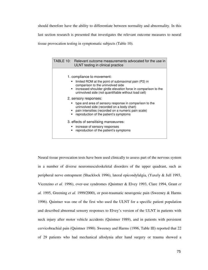
75
should therefore have the ability to differentiate between normality and abnormality. In this
last section research is presented that investigates the relevant outcome measures to neural
tissue provocation testing in symptomatic subjects (Table 10).
TABLE 10: Relevant outcome measurements advocated for the use in ULNT testing in clinical practice
1. compliance to movement: ��limited ROM at the point of submaximal pain (P2) in
comparison to the uninvolved side ��increased shoulder girdle elevation force in comparison to the
uninvolved side (not quantifiable without load cell)
2. sensory responses: ��type and area of sensory response in comparison to the
uninvolved side (recorded on a body chart) ��pain intensities (recorded on a numeric pain scale) ��reproduction of the patient’s symptoms
3. effects of sensitising manoeuvres: ��increase of sensory responses ��reproduction of the patient’s symptoms
Neural tissue provocation tests have been used clinically to assess part of the nervous system
in a number of diverse neuromusculoskeletal disorders of the upper quadrant, such as
peripheral nerve entrapment (Shacklock 1996), lateral epicondylalgia, (Yaxely & Jull 1993,
Vicenzino HW�DO� 1996), over-use syndromes (Quintner & Elvey 1993, Clare 1994, Grant HW�DO� 1995, Greening HW�DO. 1999/2000), or post-traumatic neurogenic pain (Sweeney & Harms
1996). Quintner was one of the first who used the ULNT for a specific patient population
and described abnormal sensory responses to Elvey’ s version of the ULNT in patients with
neck injury after motor vehicle accidents (Quintner 1989), and in patients with persistent
cervicobrachial pain (Quintner 1990). Sweeney and Harms (1996, Table III) reported that 22
of 29 patients who had mechanical allodynia after hand surgery or trauma showed a

76
significant difference of sensory responses and elbow ROM between the involved and
uninvolved side during ULNT1 testing. More recently ultrasound imaging showed a lack of
median nerve movement during wrist flexion in RSI patients to painfully limited elbow
ROM during ULNT1 testing (Greening HW�DO� 2000).
Investigating abnormal responses to the ULNT2b the neural tissue extensibility has been
investigated in 15 screen based keyboard (SBK) operators (Grant HW�DO� 1995, Table III), and
in 20 patients suffering from unilateral epicondylitis (Yaxely & Jull 1993, Table III). The
involved side exhibited an average of 12º lesser range in glenohumeral abduction at R2 in
comparison to the uninvolved side. The inter-examiner reliability for glenohumeral abduc-
tion at R2 in asymptomatic subjects had been shown in an earlier study (Yaxely & Jull
1991). The testing sequence was performed in accordance to Butler (1991) in that the
shoulder girdle was depressed to an end range and maintained during the whole procedure.
The main outcome measure, glenohumeral abduction, was measured with a standard
goniometer at the point were tissue resistance limited further range. Grant HW� DO. (1995)
reported that all 15 female SBK operators experienced a strong stretch on the proximal radial
aspect of the forearm in the final ULNT2b position. However, 90% (n=9) of the control
group reported the same sensory response, which has previously been shown to be a normal
response in asymptomatic subjects (Yaxely & Jull 1991). Adding CCLF as a sensitising
manoeuvre increased symptoms in 73% of the SBK operators but also in 70% of the control
group (Grant HW�DO� 1995). In only 55% of the cases with epicondylitis (Yaxely & Jull 1993)
symptoms were reproduced at R2 but no correlation between pain intensity and R2 was
presented.

77
In both studies glenohumeral ROM was reduced in comparison to the asymptomatic group
or side, and was claimed to be a sign of reduced neural tissue extensibility. This purely
mechanistic approach leaves other possible limiting factors to be unaccounted for, such as
the more recent findings of a possible protective motor response of the shoulder girdle
musculature (Balster & Jull 1997, Coppieters HW� DO� 2001a, v. der Heide HW� DO� 2002). The
small sample size and the inappropriate reliability calculations (Grant HW� DO��1995) further
question the statistical power of these results (see section 5.1.1). While the areas of sensory
responses were similar between the asymptomatic and symptomatic sides the intensities
elicited by the ULNT2b were different (Yaxely & Jull 1993, Grant HW�DO� 1995). However,
when assessing painful conditions it is more sensible to measure limited ROM at P2 and not
at the clinically insignificant point of maximal resistance.
As neurodynamic tests are often performed repeatedly within one treatment session to assess
the immediate effects of treatment, the patient’ s ability to reliably indicate the moment of
pain is an essential criterion for the clinical use of neural tissue provocation tests. The
stability and reliability of the occurrence of ‘pain onset’ and ‘submaximal pain’ throughout
the ROM during the ULNT1 was analysed in a laboratory and clinical setting (Coppieters HW�DO� 2002b, Table III). The ULNT1 was performed in a single session on a total of 27 patients
with unilateral and bilateral neurogenic CBP disorders. In addition, two examiners perfor-
med the ULNT1 with the cervical spine in neutral, with wrist extension, with CCLF, and
with both wrist extension and CCLF on 10 asymptomatic subjects in laboratory conditions
only. The operationalised starting position (Edgar HW�DO. 1994) and a calibrated load cell for
shoulder girdle depression (Coppieters HW�DO. 1999) were used only in the laboratory setting.
In the clinical setting the ULNT1 was performed following the operational definition by

78
Butler (2000), which means the shoulder girdle was controlled in a neutral position. Corres-
ponding angles of elbow ROM were measured with an electrogoniometer in both settings.
Intra- and inter-tester reliability coefficients were greater than 0.95 (SEM �����º) for P1 and
P2 in the asymptomatic group. The reliability coefficients for the symptomatic group in the
laboratory setting for P1 was greater than 0.98 (SEM �����º), and in the clinical settings for
P2 it was greater than 0.98 (SEM �� ���º). Although pain threshold and tolerance levels
varied widely between subjects the study demonstrated the moment of pain can be detected
reliably, an essential criterion when using neurodynamic tests in clinical practice. Further-
more, the absence of additional devices, used for operationalising the starting position in an
experimental setting, can be compensated with sound operational definitions and skilful
handling in a clinical setting.
The increasing importance of neural tissue involvement in minor peripheral neurogenic pain
disorders has lead to the development of treatment strategies as the cervical glide technique
(Elvey 1986, Vicenzino HW�DO� 1994). This mobilisation technique was developed on the basis
of Elvey’ s concept of impaired neural dynamics in upper limb disorders, and has been shown
to improve pain intensity, symptom provocation and range of motion (Vicenzino HW� DO� 1995/1996, Hall HW�DO� 1997, Cowell & Phillips 2002, Coppieters HW�DO� 2003b). According to
a comprehensive review of the literature up through 1997, Elvey developed a set of clinical
criteria to identify patients with minor peripheral neurogenic pain disorders amenable to
physiotherapy management: (1) the presence of an active movement restriction that is related
to a disorder of a specific nerve trunk, (2) a passive movement restriction that correlates with
the active movement dysfunction, (3) an abnormal response to neural tissue provocation
testing, (4) painful nerve trunk palpation, and (5) signs of a local musculoskeletal dysfunc-

79
tion responsive to physiotherapy, such as cervical segmental motion restriction indicating a
cause of neurogenic disorder.
In a recent single-blinded RCT (Coppieters HW� DO� 2003a, Table III) 20 patients with non-
acute unilateral or bilateral neurogenic cervicobrachial pain were initially assessed with the
five criteria described by Elvey (1997), and randomly allocated into two matched groups.
The shoulder girdle elevation force during the ULNT1, and the effects following a cervical
mobilisation technique were investigated. The operational definitions for the starting
position, and the standardisation of the measurement devices established in earlier studies
(Edgar HW�DO. 1994, Coppieters HW�DO� 1999/2001a) were used. Outcome measures were elbow
ROM at the point of substantial discomfort (P2) as reported by the patients, and the shoulder
girdle elevation force during ULNT1. The highest pain intensity was recorded using a
numeric pain intensity rating scale (0-10).
The experimental condition consisted of a cervical contralateral glide technique at one or
more segments (Elvey 1986, Vicenzino HW� DO� 1994) with the involved arm in a neural
preloaded position, in which several components of the ULNT1 were applied. A lateral
translatory movement away from the involved side was performed, with the patient in a
supine position minimising gross cervical side flexion or rotation. The technique is described
to either selectively move the nerve root complex within the spinal intervertebral canal, or to
move the spinal intervertebral canal in relation to the nerve root complex when it is held
tense by means of shoulder girdle fixation (Elvey 1986).
The control condition consisted of pulsed ultrasound applied for 5 minutes over the most
painful area, during which the arm was positioned in an unloaded position. To reduce the
“ therapist effect” both the experimental and the control interventions were performed by the

80
same therapist. The results showed a significant increase in shoulder girdle elevation force
during ULNT1. However, the sudden increase in shoulder girdle elevation force occurred
earlier in range on the involved side than on the uninvolved side. The immediate effect of the
cervical mobilisation treatment on the involved side was that an increase in shoulder girdle
elevation force occurred later in range and that the amount of end force was significantly
larger than before treatment. These findings were regarded as a normalisation in force
generation and were associated with the significant decrease in pain intensity and increase in
ROM on the involved side. For the control group no treatment effects relating to end force,
ROM, or pain intensity were observed.
The immediate hypoalgesic effect of the cervical mobilisation technique (Wright 1995,
Wright & Vicenzino 1995, Vicenzino HW� DO� 1998) may be a plausible explanation for a
retarded nociceptive flexor withdrawal reflex proposed in earlier studies. However, future
electromyographical studies need to confirm if changes in force generation correlate with
changes in muscle activity. In a subsequent RCT Coppieters HW�DO. (2003b Table III) used the
ULNT1 to additionally assess the distribution of elicited sensory responses. The same
methods and operational definitions were used as described in the former study (Coppieters
HW�DO. 2003a). Significant differences between the involved and uninvolved side were found
for the range of elbow extension (mean difference 25.6º), for pain intensity (mean difference
3.1 points on VAS), and for the symptom area, which was approximately 2.9 times larger.
These results support the use of the uninvolved side as a relative norm against which the
clinical signs of the involved side can be compared.
One requirement demanded of a diagnostic test is its ability to predict pathology. Evaluating
the predictive validity of the ULNT is problematic because surgical verification that neural

81
tissue pathology is the cause of the symptoms usually cannot be obtained. A case study by
Shacklock (1996) analysed the response to the ULNT3 (ulnar bias) in a case of a surgically
proven ulnar neuropathy. A 25-year-old female receptionist presented with intermittent, deep
and burning pain located in her left medial elbow. There was also a cold tingling feeling
extending from the medial elbow to the little finger. Physical examination revealed no
abnormality of nerve conduction (light touch, pin prick, thermal sensation, vibration), and
the neurological examination, ultrasound scanning and cervical spine X-rays were negative.
Passive and active elbow, shoulder and neck movements were pain free and full range. The
ULNT3 with glenohumeral abduction and CCLF reproduced the patient’ s pain, and an
increased pain reaction in comparison to the asymptomatic side was seen with the ULNT1
(Shacklock 1996). Subsequent surgery revealed a tight tendinous band crossing the ulnar
nerve that caused increased pressure and mechanical irritation.
The negative results from the neurological examination and ultrasound scanning may have
occurred for several reasons. During the neurological examination in a comfortable position
the nerve may not have been sufficiently ischaemic to show conduction abnormalities,
because symptoms were only provoked under working load when the nerve was in an
elongated position (typing position) unmasking the neuropathy. Hence, the neurological
examination and the ULNT3 may not have tested the same parameters (nerve conduction
versus mechanosensitivity). The author further stated that the specificity of the test has been
demonstrated by the capability of the ULNT3 to reproduce the patient’ s clinical symptoms
while ULNT1 did not, although the ULNT1 has been shown to produce more than double
the amount of strain on the medial cord than the ULNT3 (Kleinrensink HW�DO� 2000). Despite
the fact that it is apparently not the strain on the medial cord that produced the symptoms,

82
the positive response to the ULNT3 cannot not be disputed. However, these findings need to
be verified in a larger patient population, as single case studies have no statistical power.
To sum up the main points, evidence is still lacking for the use of the ULNT2b in clinical
practice because glenohumeral abduction as the relevant outcome measure has not been
effectively measured at a clinical relevant end point (Yaxely & Jull 1993, Grant HW�DO� 1995).
On the other hand excellent intra- and inter-examiner reliability for the ULNT1 provide the
evidence for the use of pain onset and submaximal pain as relevant parameters in clinical
practice (Coppieters HW� DO� 2002b). Furthermore, significant differences exist between the
involved and uninvolved side for shoulder girdle elevation force (Coppieters HW�DO. 2003a),
for pain intensity, elbow ROM, and for distribution of sensory responses (Coppieters HW�DO� 2003b).
These parameters monitored during diagnostic testing have the ability to differentiate
between normality and abnormality. In this respect a positive test would imply an abnormal
response to the test on the involved side in comparison to the uninvolved side, which implies
a non-compliance of mechanosensitive neural tissue to movement. However, it should be
stressed that a positive test cannot identify the site of pathology or predict a diagnosis. To
show its predictive validity the ULNT would need to be compared to other diagnostic test
that assess the mechanosensitivity of neural tissue, or to pre-diagnosed peripheral neurogenic
disorders (Shacklock 1996).
The small sample size of most studies remains a problem in achieving sufficient statistical
power (Hicks 1999). However, the strength of evidence has clearly increased due to the
improved quality of recent studies, and gives hope for future high quality research in
physiotherapy.

83
��� ',6&866,21�
Ä7KH� QHUYRXV� V\VWHP�ZDV� SHUKDSV� WKH� ODVW� RI� WKH�PRELOH� WLVVXHV�ZKRVH� PHFKDQLFDO� DELOLWLHV� DQG� DVVRFLDWHG� VHQVLWLYLWLHV� KDG� QRW�EHHQ� FRQVLGHUHG�� DOWKRXJK� WKH� IDFW� WKDW� WKH� QHXUDO� WLVVXH� PXVW�SK\VLFDOO\�UHVSRQG�WR�PRYHPHQW�LV�VHOI�HYLGHQW´�
%XWOHU������
����� ,03/,&$7,216�)25�&/,1,&$/�35$&7,&(�
When the ULNT was first conceptualised the emphasis was on the mechanical aspects of the
test. The hypothesis then was that an ordered set of joint movements could be used to
selectively increase tension within mechanosensitive neural tissue that would react to
mechanical load with non-compliance to movement and with sensory responses (Elvey
1979b). Orthopaedic physiotherapists quickly welcomed this new approach and assessment
strategy because, except for the straight leg raise, there was no neurodynamic test for the
upper quarter. Although at that time no scientific research was available that investigated the
reliability and validity of this diagnostic test, physiotherapists accepted it as a useful tool.
After 20 years of looking into the ULNT from a clinical and scientific perspective, there is
now a growing body of research from countries like the UK, the USA, Australia, Belgium
and The Netherlands. The current understanding of neurodynamic tests incorporates both the
basic knowledge of pathomechanics and neurophysiology. Unfortunately, although the
amount of clinically-based research is increasing, physiotherapists tend to chose their
treatment techniques directly from what they were taught as students or in postgraduate
training courses, but seldom base their treatment on clinical research (Turner & Whitfield
1997). This might be a reflection of the more passive approach of physiotherapists to deliver

84
certain treatment protocols rather than to make independent decisions. Moreover, physio-
therapy is still not an academic profession in countries like Germany, therefore questioning
traditional treatment strategies and doing research is not very common. Nevertheless, it is the
responsibility of practicing clinicians to seek evidence supporting the efficacy of new and
current treatment regimes (Matheson 2000).
As a movement-based profession, physiotherapy identifies structural dysfunctions through
the examination of compliance or non-compliance to active or passive movements. Patients
with upper quarter pain, in whom overt neurological deficits are not present and few medical
investigative tests are definitive in the diagnosis of cervicobrachial pain syndromes, are
frequently referred to physiotherapy. When assessing these patients physiotherapists usually
focus on finding the musculoskeletal structure ‘at fault’ . The routine functional assessment
for upper quarter pain syndromes includes a patient interview, active and passive tests of the
cervical and thoracic spine and the glenohumeral joint, palpations of the soft tissues, bones
and nerve trunks, muscle function and length tests, and neurological examination of reflexes
and sensibility. However, new insight gained in basic neurophysiological science has
changed the physiotherapist’ s understanding of neurogenic pain disorders. Since the deve-
lopment of the NTPT, the physical assessment of the neural tissue has been integrated into
the clinical assessment of CBP disorders.
Whether the NTPT is able to discriminate between different sources of pathology, i.e. nerve
root or nerve trunk pathology, has recently been investigated. While there is no scientific
evidence that it is possible to identify specific structural disorders, identifying the existence
of an neurogenic disorder may be possible by integrating clues and information acquired
from the preceding patient interview and from findings of the clinical assessment. To do so,

85
the knowledge of pain mechanisms and electrophysiological investigations need to be
combined with the analysis of clinical features within a clinical reasoning framework, in
which an initial working hypothesis is tested until sufficient information is obtained (Jones
1995). It is the clinician’ s responsibility to incorporate the entire range of relevant infor-
mation to produce a working hypothesis. In this respect the issue of a test’ s validity should
be broadened to the “ analysis of the degree to which a meaningful interpretation can be
inferred from the integrated findings from both the patient interview and the physical
examination” (Coppieters & Butler 2001c, p.520), of which the ULNT is only a part.
Therefore, the ULNT in isolation will not distinguish which pathology is the cause of a
positive test (Shacklock 1996), but the solution lies in the analysis of the entire information.
Furthermore, in clinical practice a single test is never used in isolation to make a diagnosis
especially not in patients with complex neuromusculoskeletal disorders so often referred to
physiotherapy.
Designing causal treatment strategies are difficult as many different tissues and anatomical
structures might be involved in a pain syndrome (Coppieters HW�DO��2003b). Pain states are
currently categorised by their duration (acute/chronic), causes (whiplash, repetitive strain
injury), or the body parts involved (cervicobrachial pain, epicondylitis). This method of
classifying pain, however, does not help predict outcome nor does it help identify physio-
logical subcategories. Butler (2000) proposed that pain experiences should be categorised
clinically into operant mechanisms on the basis of known pathophysiology, clinical patterns
and logic. This approach corresponds with the recent developments in basic pain research for
a mechanism-based classification (Woolf HW�DO� 1998). Pain that manifests in distinct diseases
may operate through common mechanisms. On the other hand, no pain mechanism is an
inevitable consequence of a particular disease process (Woolf & Mannion 1999). It is
86
therefore important to differentiate between central and peripheral processes (Gifford &
Butler 1997) when deciding on a therapeutic strategy.
����� /,0,7$7,216�
Many non-neural structures are stretched during neural tissue provocation testing and adding
sensitising manoeuvres “ does not help to localise the tissue at fault because other structures
are moving with the nerves during these procedures” (Di Fabio 2001, p.224). However, most
clinical tests in orthopaedic physiotherapy are not able to exclusively load specific struc-
tures, i.e. testing muscle length or joint mobility. “ If the utility of a treatment modality would
depend on its ability to independently mobilise one structure, very few interventions in
orthopaedic physiotherapy would withstand” (Coppieters & Butler 2001c, p.521). None-
theless, the adding of remote sensitising manoeuvres to the ULNT has shown to be a
valuable tool in assessing neural tissue involvement to the patient’ s symptoms (Selvaratnam
HW�DO� 1994, v. der Heide HW�DO� 2001, Coppieters HW�DO. 2001b). For example, if a patient’ s
local shoulder pain was reproduced or intensified by adding wrist extension, neurogenic
involvement may be reasoned as part of the disorder, as the impact of the nervous system
passes beyond the point to which musculoskeletal structures are loaded.
Adding wrist extension to the extended elbow and abducted shoulder has been shown to
transmit tension along the median nerve at least up to the level of the axilla (Kleinrensink HW�DO� 1995b). Components like CCLF and wrist extension are thought to elongate the nerve
bedding, and, when combined, the available ROM of elbow extension is markedly reduced,
with sensory responses elicited through the entire arm (Coppieters HW�DO. 2001b, 2002b). No
doubt non-neural structures will be stressed during neurodynamic testing, but adding
87
sensitising manoeuvres to a final test position has shown to increase symptoms that cannot
be related to the provocation of non-neural structures. With the addition of individual test
components an increased pain intensity has been reported, despite a decreased elbow range
of motion and decreased loading of surrounding musculature and articular structures
(Coppieters HW� DO��2002a). This limited range of elbow extension is thought to be, at least
partly, a result of the limited elasticity of the neural tissue; this has also been attributed to a
protective motor response of the antagonistic musculature, however.
It has to be emphasised that a limitation in elbow extension does not equal limited neural
tissue elasticity. Therefore, treatment cannot be aimed at ‘mobilising the neural tissue.’ A
rather limited ROM is a clinical sign indicating a probable neurogenic contribution to the
presenting symptoms. In this respect a positive test would imply aiming the treatment at
improving or restoring longitudinal gliding necessary for full limb motion, reduction of
adhesions, and improvement of neurophysiological processes (McLellan & Swash 1976,
Wilgis & Murphy 1986, Wright HW�DO� 1996, Totten & Hunter 1991, Rozmaryn HW�DO� 1998).
One of the limitations in analysing the research in the current thesis was the insufficient
consensus as to what principle signs should be monitored during the NTPT. This made it
difficult to compare studies and identify discrepancies. Some authors consider the repro-
duction of symptoms, difference in ROM, and altered through range and end-feel
(resistance) as the most important signs during NTPT. Others state that the differences in
ROM are insufficient and that abnormal reactions may be shown in a reactive muscle
activity of the upper trapezius, the latter being regarded as a nociceptively mediated flexor
withdrawal reflex to protect the respective nerve from harmful elongation (Wright HW� DO� 1994, Balster & Jull 1997, Coppieters HW�DO� 2001a, v. d. Heide HW�DO� 2002).

88
Also the use of different techniques to standardise testing methods poses a problem in
comparing reliability measures from individual research. It is therefore necessary, for the
researchers to conduct control intra- and inter-examiner reliability studies as long as testing
procedures are not standardised. Only with standardised protocols will researchers be able to
compare ULNT studies and interpret the results with clarity.

89
���&21&/86,21�
The seminal work of Elvey (1979b) initially describing the brachial plexus tension test led to
an increased interest in neural tissue as a source of pathology and pain. Presently, the
neuromeningeal approach for the examination and treatment of dysfunctions and pain is
widely accepted and integrated into the clinical practice of physiotherapists. The underlying
mechanisms of how these effects are exerted are poorly understood, however, and they
remain inadequately researched. Early research focused on the conceptual idea of testing the
mechanical properties of the neural tissue by testing extensibility. This testing paradigm
gradually changed as there was an increasing recognition for the important changes within
the central nervous system potentially responsible for many of the signs and symptoms in
patients with CBP disorders seen in clinical practice.
In reviewing the literature concerning the evaluation of the neural tissue provocation test as a
diagnostic tool the following can be stated: The NTPT has been developed to assess probable
neurogenic involvement in CBP disorders, and can be used as a diagnostic tool, or as an
outcome measure to evaluate the effect of treatment. Minor peripheral nerve injuries, as in
nerve entrapment syndromes, are characterised by an increased sensitivity to mechanical
stimulation. The NTPT is a sequence of movements designed to assess the compliance of
sensitised neural tissue to mechanical load by elongating the length of the nerve bedding.
Biomechanical studies have only found the median biased ULNT1 to be specific in
transferring tension to the corresponding nerve and recommend its use as a valid test
(Kleinrensink HW�DO� 2000).
A positive test implies at least a partial reproduction of the patient’ s symptoms, in which
symptoms increase and decrease with a varying amount of nerve provocation by changing

90
distant test components (sensitising manoeuvres) that have no direct structural link with the
symptomatic area except via the nervous system. It is not possible, however, to differentiate
between proximal or distal dysfunctions by a different sequencing of the test components, or
to make any inference on the type of pathology. To judge when a response to the NTPT is
abnormal the uninvolved arm can be used as a standard against which the clinical findings
from the involved side can be compared. Reliability of the occurrence of pain onset and
submaximal pain throughout elbow ROM for the ULNT1 for both intra- and inter-examiner
reliability are highly reliable (Coppieters HW�DO� 2002b) and can be used as a reliable outcome
measure.
Many non-neural structures are stretched during neural tension testing and may account for
the elicited responses. However, the relatively high incidence of paraesthesia in asympto-
matic as well as symptomatic subjects suggests that at least some of the responses are
neurogenic in origin (Coppieters HW� DO� 2001b). The primary factor limiting movement is
believed to be the nervous system that triggers a protective muscle reaction to prevent
harmful elongation; however, electromyographical studies need to confirm this in larger
patient populations.
Although most measurements acquired from studies have been performed in a laboratory
setting, conflicting results have been reported with regard to the reliability of the NTPT. This
is partly due to the lack of standardisations of the testing procedure that in turn leads to
handling irregularities when performing this complex multiple joint test. The other limiting
factor lies in the variation of the criteria for the end point at which the outcome measures
were taken. Onset of resistance for example is not a reliable criteria, instead the onset of
muscle activity has been found to be a more relevant sign (Hall HW�DO� 1998). To date, only

91
the starting position for the ULNT1 with an initial shoulder girdle depression has been
operationalised (Edgar HW� DO� 1994). Gentle glenohumeral fixation throughout the ULNT1,
and glenohumeral lateral rotation are furthermore crucial components of the test that warrant
the mechanical loading of the brachial plexus and the median nerve and should not be
omitted (Coppieters HW�DO� 2003a).
Reliability in a clinical setting was not lower than in optimal laboratory conditions
(Coppieters HW� DO. 2002b), suggesting that, with adequate handling and sound operational
definitions, physiotherapists are able to perform these tests in a standardised and reliable
way. However, it should be appreciated that subtle adverse responses monitored during
NTPT in a clinical setting without any measuring devices remains a “ complex task that
requires multifaceted skills” (Coppieters HW�DO� 2003, p. 105).
����� 5(&200(1'$7,216�
³:H� EHOLHYH� WKDW� ZH� FDQQRW� WRWDOO\� GLVPLVV� WKH� JUHDW� FOLQLFDO�LPSRUWDQFH«RI�QHXUDO�SURYRFDWLRQ� WHVWLQJ«VLPSO\�EHFDXVH�RI�D�ODFN�RI�VFLHQWLILF�VWXGLHV�DGGUHVVLQJ�WKLV�WRSLF�´�
� � � �&RSSLHWHUV��%XWOHU�����F��S������
On the basis of the analysed research, the median biased ULNT1 can be a useful diagnostic
test in disorders that lack clear physical signs of nerve injury or inflammation. Moreover, the
test is extremely cost-effective when compared with nerve conduction studies, and should be
performed as an integral part of the physiotherapists’ assessment of upper quadrant dis-
orders. Methodological deficiencies of many studies and the incomplete presentation and
analysis of the data strongly suggests more rigorous studies with standardised procedures

92
and evaluation criteria. Future research should provide more insight in to the possible
mechanisms that lead to limited range of motion. To establish the predictive validity of the
NTPT, systematic research is necessary that shows how reliable the test can predict any
neurogenic contribution in comparison with traditional diagnostic methods.
93
5()(5(1&(6��
Aker PD, Gross AR, Goldsmith CH, Peloso P (1996) Conservative management of mechanical neck pain: systematic overview and meta-analysis. %ULWLVK�0HGLFDO�-RXUQDO ���: 1291-1296
Allison GT, Nagy BM, Hall T (2002) A randomised clinical trial of manual therapy for cervico-brachial pain syndrome- a pilot study. 0DQXDO�7KHUDS\ �(2): 95-102
$UL QV�*$0��%RUJKRXWV�-$-��.RHV�%:��������1HFN�SDLQ��,Q�&URPELH�,.�HW�DO. (Eds.): (SLGHPLRORJ\�RI�3DLQ. 7DVN�)RUFH�RQ�(SLGHPLRORJ\, IASP Press, Seattle, pp 235-255
Babcock WW (1927) A standard technique for operations on peripheral nerves. 6*2 ��: 364-378
Balster S, Jull G (1997) Upper trapezius muscle activity during the brachial plexus tension test in asymptomatic subjects. 0DQXDO�7KHUDS\ �(3): 144-149
Baron A, Schreiber W (1918) Über die direkte Nervenvereinigung bei großen Nervendefekten. 0�QFKQHU�0HGL]LQLVFKH�:RFKHQVFKULIW ��: 446-449, cited in Zöch (1992), p 13
Bell A (1987) The upper limb tension test - bilateral straight leg raising - a validating manoeuvre for the upper limb tension test. 3URFHHGLQJV� RI� WKH� )LIWK� %LHQQLDO� &RQIHUHQFH� RI� WKH� 0DQLSXODWLYH�7KHUDSLVWV�$VVRFLDWLRQ�RI�$XVWUDOLD, Melbourne, pp 106-114
Boden SD, McCowin PR, Davis DO, Dina TS, Mark AS, Wiesel S (1990) Abnormal magnetic-resonance scans of the cervical spine in asymptomatic subjects. A prospective investigation. -RXUQDO�RI�%RQH�DQG�-RLQW�6XUJHU\ ��: 1178-1184
Bogduk N (1994) Innervation and pain patterns of the cervical spine. In Grant R (Ed.): &OLQLFV� LQ�SK\VLFDO�WKHUDS\��3K\VLFDO�WKHUDS\�RI�WKH�FHUYLFDO�DQG�WKRUDFLF�VSLQH (2nd ed.), Churchill Livingstone, Edinburgh, pp 65-76
Borghouts JAJ, Koes BW, Bouter LM (1998) The clinical course and prognostic factors of non-specific neck pain: a systematic review. 3DLQ ��: 1-13
Borghouts JAJ, Koes BW, Vondeling H, Bouter LM (1999) Cost-of-illness of neck pain in The Netherlands in 1996. 3DLQ ��: 629-636
Bove G, Light A (1997) The nervi nervorum: Missing link for neuropathic pain? 3DLQ�)RUXP �(3): 181-190
Bovim G, Schrader H, Sand T (1994) Neck pain in the general population. 6SLQH���(12): 1307-1309
Bragard K (1929) Die Nervendehnung als diagnostisches Prinzip ergibt eine Reihe neuer Nerven-phänomene. 0�QFKQHU�0HGL]LQLVFKH�:RFKHQVFKULIW ��(48): 1999-2003
Butler DS (1989) Adverse mechanical tension in the nervous system: a model for assessment and treatment. 7KH $XVWUDOLDQ�-RXUQDO�RI�3K\VLRWKHUDS\ ��: 227-238
Butler DS (1991) 0RELOLVDWLRQ�RI�WKH�QHUYRXV�V\VWHP, Churchill Livingstone, Melbourne
Butler DS (1994) The upper limb tension test revisited. In Grant R (Ed.):�&OLQLFV�LQ�SK\VLFDO�WKHUDS\� 3K\VLFDO� WKHUDS\ RI� WKH�FHUYLFDO�DQG�WKRUDFLF�VSLQH (2nd ed.), Churchill Livingstone, Edinburgh, pp 217-244
Butler DS (1998) Adverse mechanical tension in the nervous system: a model for assessment and treatment. Commentary. In Maher C (Ed.):�7KH $XVWUDOLDQ�-RXUQDO�RI�3K\VLRWKHUDS\�0RQRJUDSK�1R����³$GYHUVH�QHXUDO�WHQVLRQ´�UHFRQVLGHUHG, Melbourne,�pp 33-36
Butler DS (2000) 7KH�VHQVLWLYH�QHUYRXV�V\VWHP, Noigroup Publications, Adelaide
Cavafy J (1881) A case of sciatic nerve-stretching in locomotor ataxy: with remarks on the operation. 7KH�%ULWLVK�0HGLFDO�-RXUQDO (Dec.) ��: 973-974, cited in Butler (2000), p 13

94
Clare HA (1989) The clinical testing of upper limb neural tissue in repetitive strain injury 3URFHHGLQJV� RI� WKH� 6L[WK %LHQQLDO� &RQIHUHQFH� RI� WKH � 0DQLSXODWLYH� 7KHUDSLVWV� $VVRFLDWLRQ� RI�$XVWUDOLD� Adelaide, pp 28-33
Colditz GA, Miller JN, Mosteller K (1989) How study design affects outcomes in comparison of therapy I. 6WDWLVWLFV�LQ�0HGLFLQH� �: 441-451
Coppieters MW, Stappaerts KH, Everaert DG, Staes FF (1999) A qualitative assessment of shoulder girdle elevation during the upper limb tension test 1.�0DQXDO�7KHUDS\ �(1): 33-38
Coppieters MW, Stappaerts KH, Staes FF, Everaert DG (2001a) Shoulder girdle elevation during neurodynamic testing: an assessable sign? 0DQXDO�7KHUDS\ �(2): 88-96.
Coppieters MW, Stappaerts KH, Everaert DG, Staes FF (2001b) Addition of test components during neurodynamic testing: effect on range of motion and sensory responses. -RXUQDO�RI�2UWKRSDHGLF��6SRUWV�3K\VLFDO�7KHUDS\���(5): 226-237
Coppieters MW, Butler DS (2001c) In defence of neural mobilisation. -RXUQDO� RI�2UWKRSDHGLF��6SRUWV�3K\VLFDO�7KHUDS\���(9): 520-521
Coppieters MW, van de Velde M, Staeppaerts KH (2002a) Positioning in anesthesiology. Toward a better understanding of stretch-induced perioperative neuropathies. $QHVWKHVLRORJ\ ��: 75-81
Coppieters MW, Stappaerts KH, Koen J, Jull G (2002b) Reliability of detecting ‘onset of pain’ and ‘submaximal pain’ during neural provocation testing of the upper quadrant. 3K\VLRWKHUDS\�5HVHDUFK�,QWHUQDWLRQDO �(3): 146-156
Coppieters MW, Butler DS (2002c) In defence of neural mobilisation: Part 2. -RXUQDO�RI�2UWKRSDHGLF��6SRUWV�3K\VLFDO�7KHUDS\���(3): 125-126
Coppieters MW, Stappaerts KH, Wouters LL, Koen J (2003a) Aberrant protection force generation during neural provocation testing and the effect of treatment in patients with neurogenic cervicobrachial pain. -RXUQDO�RI�0DQLSXODWLYH�3K\VLRORJLFDO�7KHUDS\ ��: 99-106
Coppieters MW, Stappaerts KH, Wouters LL, Janssens K (2003b) The immediate effects of a cervical lateral glide treatment technique in patients with neurogenic cervicobrachial pain. -RXUQDO�RI�2UWKRSDHGLF��6SRUWV�3K\VLFDO�7KHUDS\ ��(7): 369-378 in press
Cowell IM, Phillips DR (2002) Effectiveness of manipulative physiotherapy for the treatment of a neurogenic cervicobrachial pain syndrome: a single case study-experimental design. 0DQXDO�7KHUDS\��(1): 31-38
Croft PR, Lewis M, Papageorgiou AC, Thomas E, Jayson MIV, Mcfarlane GJ, Silman AJ (2001) Risk factors for neck pain: a longitudinal study in the general population. 3DLQ ��: 317-325
Crombie IK, Croft PR, Linton SJ, LeResche L, Von Korff M (Eds.) (1999) (SLGHPLRORJ\�RI�3DLQ. 7DVN�)RUFH�RQ�(SLGHPLRORJ\, IASP Press, Seattle
Dahlin LB, Danielsen N, McLean WG, Rydevik B, Sjöstrand J (1981) Critical pressure level for impairment of fast axonal transport during experimental compression of rabbit vagus nerve. -RXUQDO�RI�3K\VLRORJ\ ��3, cited in Rydevik HW�DO� (1984) p 11
Davidson M (2002) The interpretation of diagnostic tests: a primer for physiotherapists. $XVWUDOLDQ�-RXUQDO�RI�3K\VLRWKHUDS\ ��: 227-233
Devor M, Seltzer Z (1999) Pathophysiology of damaged nerves in relation to chronic pain: In Wall PD and Melzack R (Eds.): 7H[WERRN�RI�3DLQ (4th ed.), Churchill Livingstone, London, pp 129-164
Di Fabio RP (2001) Neural mobilisation: the impossible. -RXUQDO�RI�2UWKRSDHGLF��6SRUWV�3K\VLFDO�7KHUDS\ ��(5): 224-225
Dickersin K (1990) The existence of publication bias and risk factors for its occurrence. -RXUQDO�RI�WKH�$PHULFDQ�0HGLFDO�$VVRFLDWLRQ����: 1385-1389

95
Dreyer S, Boden S (1998) Nonoperative treatment of neck and arm pain.�6SLQH���(24): 2746-2754
Dwyer A, Aprill C, Bogduk N (1990) Cervical zygapophyseal joint patterns I: a study in normal volunteers. 6SLQH���(6): 453-457
Dyck PJ (1990) Invited review: Limitations in predicting pathologic abnormality of nerves from EMG examination. 0XVFOH��1HUYH���: 371-375
Dyck PJ, Lais AC, Giannini C, Engelstad JK (1990) Structural alterations of nerve during cuff compression. 3URFHHGLQJV�RI�WKH�1DWLRQDO�$FDGHPLF�RI�6FLHQFH ��: 9828-9832, cited in Rempel HW�DO� (1999) p 1602
Edgar D, Jull G, Sutton S (1994) Relationship between upper trapezius muscle length and upper quadrant neural tissue extensibility. 7KH $XVWUDOLDQ�-RXUQDO�RI�3K\VLRWKHUDS\ ��(2): 99-103
Ekberg K, Björkqvist B, Malm P, Bjerre-Kiely B, Axelson O (1994) Controlled two year follow up of rehabilitation for disorders in the neck and shoulders. 2FFXSDWLRQDO�DQG�(QYLURQPHQWDO�0HGLFLQH ��: 833-838
Elvey RL (1979a) Painful restriction of shoulder movement - a clinical observational study. 3URFHHGLQJV� RI GLVRUGHUV� RI� NQHH�� DQNOH� DQG� VKRXOGHU. 0DQLSXODWLYH� $VVRFLDWLRQ� RI� $XVWUDOLD�&RQIHUHQFH, Perth, pp 113-126
Elvey RL (1979b) Brachial plexus tension tests and the pathoanatomical origin of arm pain. In Glasgow EF and Twomey L (Eds.): $VSHFWV�RI�0DQLSXODWLYH�7KHUDS\��Melbourne Lincoln Institute of Health Sciences, pp 105-110
Elvey RL (1986) Treatment of arm pain associated with abnormal brachial plexus tension. 7KH $XVWUDOLDQ�-RXUQDO�RI�3K\VLRWKHUDS\ ��: 225-230
Elvey RL (1994) The investigation of arm pain: signs of adverse responses to the physical examination of the brachial plexus and related neural tissues. In Boyling JD, Palastanga N (Eds.): *ULHYH¶V�0RGHUQ�0DQXDO�7KHUDS\, Churchill Livingstone, Edinburgh, pp 577-585
Elvey RL (1995) Peripheral neuropathic disorders and neuromusculoskeletal pain. In Shacklock MO (Ed.): 0RYLQJ�LQ�RQ�SDLQ, Buttersworth-Heinemann, Australia, pp 115-122
Elvey RL (1997) Physical evaluation of the peripheral nervous system in disorders of pain and dysfunction. -RXUQDO�RI�+DQG�7KHUDS\ ��(2): 122-129, cited by Coppieters HW�DO� (2002b), p 148
Fields HL, Basbaum AI (1999) Central nervous system mechanisms of pain modulation. In Wall PD and Melzack R (Eds.): 7H[WERRN�RI�3DLQ (4th ed.), Churchill Livingston, Edinburgh, pp. 309-330
Gelberman RH, Hergenroeder PT, Hargens AR, Lundborg GN, Akeson WH (1981) The carpal tunnel syndrome. A study of carpal canal pressures. -RXUQDO�RI�%RQH�DQG�-RLQW�6XUJHU\ ��$: 380-383
Gifford LS, Butler DS (1997) The integration of pain sciences into clinical practice. -RXUQDO�RI�+DQG�7KHUDS\, April-June: 86-95
Ginn K (1988) An investigation of tension development in upper limb soft tissues during the upper limb tension test. 3URFHHGLQJV� RI� WKH� ,QWHUQDWLRQDO� )HGHUDWLRQ� RI� 2UWKRSDHGLF� 0DQLSXODWLYH�7KHUDSLVWV, England, Cambridge, May: 25-26
Gracely RH, Lynch SA, Bennett GJ (1992) Painful neuropathy: altered central processing maintained dynamically by peripheral input. 3DLQ ��:175-194
Grant R, Forrester C and Hides J (1995) Screen based keyboard operation: the adverse effects on the neural system. $XVWUDOLDQ�-RXUQDO�RI�3K\VLRWKHUDS\ ��: 99-107
Greening J, Lynn B (1998) Minor peripheral nerve injuries: an underestimated source of pain? 0DQXDO�7KHUDS\ �(4): 187-194

96
Greening J, Smart S, Leary R, Hall-Craggs M, O’ Higgins P, Lynn B (1999) Reduced movement of median nerve in carpal tunnel during wrist flexion in patients with non-specific arm pain. /DQFHW ���: 217-218
Greening J, Lynn B, Leary R, Warren L, O’ Higgins P, Hall-Craggs M (2000) The use of ultrasound imaging to demonstrate reduced movement of the median nerve during wrist flexion in patients with RSI. In Singer KP (Ed.): $EVWUDFWV�RI�WKH�� � � �6FLHQWLILF�&RQIHUHQFH�RI�WKH�,)207�LQ�FRQMXQFWLRQ�ZLWK�WKH�%LHQQLDO�&RQIHUHQFH�RI�WKH�03$$, Perth, p 55
Gross AR, Kay T, Hondras M, Goldsmith C, Haines T, Peloso P, Kennedy C, Hoving J (2002) Manual Therapy for mechanical neck disorders: a systematic review. 0DQXDO�7KHUDS\ �(3): 131-149
Haas M (1991a) Statistical methodology for reliability studies. -RXUQDO� RI� 0DQLSXODWLYH� DQG�3K\VLRORJLFDO�7KHUDSHXWLFV���(2): 119-128
Haas M (1991b) The reliability of reliability. -RXUQDO� RI� 0DQLSXODWLYH� DQG� 3K\VLRORJLFDO�7KHUDSHXWLFV���(3): 199-208
Hall TM, Quintner J (1995) Mechanically evoked EMG responses in peripheral neuropathic pain: a single case study.� 3URFHHGLQJV� RI� WKH� 1LQWK� %LHQQLDO� &RQIHUHQFH� RI� WKH� 0DQLSXODWLYH�3K\VLRWKHUDSLVWV�$VVRFLDWLRQ�RI�$XVWUDOLD, Gold Coast, pp 42-47
Hall TM, Quintner J (1996) Response to mechanical stimulation of the upper limb in painful cervical radiculopathy. $XVWUDOLDQ�-RXUQDO�RI�3K\VLRWKHUDS\ ��(4): 277-285
Hall TM, Elvey RL, Davies N, Dutton L, Moog M (1997) Efficacy of manipulative physiotherapy for the treatment of cervicobrachial pain. 3URFHHGLQJV� RI� WKH� 7HQWK� %LHQQLDO� &RQIHUHQFH� RI� WKH�0DQLSXODWLYH�3K\VLRWKHUDSLVWV�$VVRFLDWLRQ�RI�$XVWUDOLD, Melbourne, pp 73-74
Hall TM, Zusman M, Elvey RL (1998) Adverse mechanical tension in the nervous system? Analysis of straight leg raise. 0DQXDO�7KHUDS\ �(3): 140-146
Hall TM, Elvey RL (1999) Nerve trunk pain: Physical diagnosis and treatment. 0DQXDO 7KHUDS\ �(2): 63-73
Haller FR, Low FN (1971) The fine structure of the peripheral nerve root sheath in the subarachnoid space in the rat and other laboratory animals. $PHULFDQ�-RXUQDO�RI�$QDWRP\����: 1-20, cited in Hasue (1993), p 2053
Hammacher ER, van der Werken C (1996) Acute neck sprain: ‘whiplash’ reappraised. ,QMXU\���(7): 463-466
Hasue M (1993) Pain and the nerve root. 6SLQH ��(14): 2053-2058
Hicks�CM (1999) 5HVHDUFK�0HWKRGV�IRU�&OLQLFDO�7KHUDSLVWV (3rd ed.), Churchill Livingstone, London
Hines T, Noakes R, Manners B (1993) The upper limb tension test: inter-tester reliability for assessing the onset of passive resistance R1. 7KH�-RXUQDO�RI�0DQXDO��0DQLSXODWLYH�7KHUDS\ �(3): 95-98
Hough AD, Moore AP, Jones MP (2000) Peripheral nerve motion measurement with spectral Doppler sonography: a reliability study. -RXUQDO�RI�+DQG�6XUJHU\ ��%(6): 585-589
Hoving JL, Koes BW, de Vet HCW, HW�DO� (2002) Manual therapy, physical therapy, or continued care by a general practitioner for patients with neck pain. $QQDOV�RI�,QWHUQDO�0HGLFLQH ���: 713-722
Hromada J (1963) On the nerve supply of the connective tissue of some peripheral nervous system components. $FWD�$QDWRPLFD ��: 343-351
Jensen I, Nygren A, Gamberle F, HW� DO. (1995) The role of the psychologist in multidisciplinary treatments for chronic neck and shoulder pain: a controlled cost-effectiveness study. 6FDQGLQDYLDQ�-RXUQDO�RI�5HKDELOLWDWLRQ�0HGLFLQH ��: 19-26
97
Jones M (1995) Clinical reasoning and pain. In Shacklock MO (Ed.): 0RYLQJ� LQ� RQ� SDLQ, Buttersworth-Heinemann, Australia, pp 104-114
Jull GA, Bogduk N, Marsland A (1988) The accuracy of manual diagnosis for cervical zygapophysial joint pain syndromes. 0HGLFDO�-RXUQDO�RI�$XVWUDOLD ���: 233-236
Jull GA, Moore A (2002) Editorial. Are manipulative therapy approaches the same? 0DQXDO�7KHUDS\ �(2): 63
Karjalainen K, Malmivaara A, van Tudler M, Roine R, Jauhiainen M, Hurri H, Koes BW (2000a) Multidisciplinary biopsychosocial rehabilitation for neck and shoulder pain among working age adults (Cochrane Review). In: 7KH�&RFKUDQH�/LEUDU\, Issue 3, Oxford: Update Software
Karjalainen K, Malmivaara A, van Tudler M, Roine R, Jauhiainen M, Hurri H, Koes BW (2000b) Biopsychosocial rehabilitation for upper limb repetitive strain injuries in working age adults (Cochrane Review). In: 7KH�&RFKUDQH�/LEUDU\, Issue 3, Oxford: Update Software
Kenneally M, Rubenach H, Elvey RL (1988) The upper limb tension test: the SLR of the arm. In Grant R (Ed.): &OLQLFV� LQ�SK\VLFDO� WKHUDS\��3K\VLFDO� WKHUDS\�RI� WKH�FHUYLFDO�DQG WKRUDFLF�VSLQH�(1st ed.), Churchill Livingstone, New York, pp 167-194
Kleinrensink GJ, Stoeckart R, Vleeming A, Snijders C, Mulder P, van Wingerden JP (1995a) Peripheral nerve tension due to joint motion. A comparison between embalmed and unembalmed human bodies. &OLQLFDO�%LRPHFKDQLFV ��(5): 235-239
Kleinrensink GJ, Stoeckart R, Vleeming A, Snijders C, Mulder P (1995b) Mechanical tension in the median nerve. The effects of joint positions. &OLQLFDO�%LRPHFKDQLFV ��(5): 240-244
Kleinrensink GJ, Stoeckart R, Mulder PGH, van der Hoek G, Broek TH Vleeming A, Snijders CJ, (2000) Upper limb tension tests as tools in the diagnosis of nerve and plexus lesions. Anatomical and biomechanical aspects. &OLQLFDO�%LRPHFKDQLFV ��: 9-14
Koes BW, Assendelft WJJ, van der Heijden GJ, Bouter LM, Knipshild PG (1991) Spinal manipulation and mobilisation for back and neck pain: a blinded review. %ULWLVK�0HGLFDO� -RXUQDO ���: 1298-1303
Koes BW, Bouter LM, van Mameren H, Esser AHM, HW�DO� (1992a) A blinded randomised clinical trial of manual therapy and physiotherapy for chronic back and neck complaints: physical outcome measures. -RXUQDO�RI 0DQLSXODWLYH�DQG�3K\VLRORJLFDO�7KHUDSHXWLFV ��(1): 16-23
Koes BW, Bouter LM, van Mameren H, Esser AHM, HW� DO� (1992b) The effectiveness of manual therapy, physiotherapy, and treatment by the general practitioner for non-specific back and neck complaints. A randomised clinical trial. 6SLQH ��(1): 28-35
Koes BW, Bouter LM, van Mameren H, Esser AHM, HW� DO� (1992c) Randomised clinical trial of manipulative therapy and physiotherapy for persistent back and neck complaints: results of one year follow up. %ULWLVK�0HGLFDO�-RXUQDO ���: 601-605
Koes BW, Hoving (1998) The value of the randomised clinical trial in the field of physiotherapy. 0DQXDO�7KHUDS\ �(4): 179-186
Larsson R, Öberg PA, Larsson SE (1999) Changes of trapezius muscle blood flow and electromyo-graphy in chronic neck pain due to trapezius myalgia. 3DLQ ��: 45-50
Levoska S, Keinänen-Kiukaanniemi S (1993) Active or passive physiotherapy for occupational cervicobrachial disorders? A comparison of two treatment methods with a 1 year follow-up. $UFKLYHV�RI�3K\VLFDO�5HKDELOLWDWLRQ�0HGLFLQH���: 425-430
Lewis J, Ramot R, Green A (1998) Changes in mechanical tension in the median nerve: possible implications for the upper limb tension test. 3K\VLRWKHUDS\ ��(6): 254-261

98
Löfgren H, Larsson R, Larson SE (2001) Outcome of surgery for cervical radiculopathy evaluated by determination of trapezius muscle microcirculation and electromyography. (XURSHDQ�-RXUQDO�RI�3DLQ �: 39-4
Lundborg G, Rydevik B (1973) Effects of stretching the tibial nerve of the rabbit. A preliminary study of the intraneural circulation and the barrier function of the perineurium. -RXUQDO�RI�%RQH�DQG�-RLQW�6XUJHU\ ��%(2): 390-401
Mackinnon SE (2002) Pathophysiology of nerve compression. +DQG�&OLQLF ��: 231-241
Maitland GD (1986) 9HUWHEUDO�PDQLSXODWLRQ (5th ed.), Butterworth-Heinemann, London
Maitland GD (1991) 3HULSKHUDO�0DQLSXODWLRQ (3rd ed.), Butterworth-Heinemann, London
Marshall J (1883) Nerve-stretching for the relief of pain. /DQFHW �: 1029-1036, cited in Bove and Light (1997), p 181
Matheson JW (2000) Research and neurodynamics: Is neurodynmics worthy of scientific merit? In Butler DS (Ed.): 7KH�VHQVLWLYH�QHUYRXV�V\VWHP, Noigroup Publications, Adelaide, pp 342-366
McLellan DL, Swash M (1976) Longitudinal sliding of the median nerve during movements of the upper limb. -RXUQDO�RI�1HXURORJ\��1HXURVXUJHU\��DQG�3V\FKLDWU\ ��: 566-570
McPhee B, Worth DR (1994) Neck and upper extremity pain in the workplace. In Grant R (Ed.): &OLQLFV�LQ�SK\VLFDO�WKHUDS\��3K\VLFDO�WKHUDS\�RI� WKH�FHUYLFDO�DQG�WKRUDFLF�VSLQH�(2nd ed.), Churchill Livingstone, Edinburgh, pp 379-407
Michaelis M, Jänig W (1998) Pathophysiologische Mechanismen und Erklärungsansätze aus der tierexperimentellen Forschung. 6FKPHU]���:261-271
Moses A, Carman J (1996) Anatomy of the cervical spine: implications for the upper limb tension test. $XVWUDOLDQ�-RXUQDO�RI�3K\VLRWKHUDS\���: 31-35
Mumenthaler M, Schliack H (Eds.) (1977) /lVLRQHQ�SHULSKHUHU�1HUYHQ��'LDJQRVWLN�XQG�7KHUDSLH (3rd ed.), Georg Thieme Verlag, Stuttgart
Nordin M, Nyström B, Wallin U, Hagbarth KE (1984) Ectopic sensory discharge and paresthesiae in patients with disorders of peripheral nerves, dorsal roots and dorsal columns. 3DLQ ��: 231-245
Persson LC, Moritz U, Brandt L, Carlson CA (1997) Cervical radiculopathy: pain, muscle weakness and sensory loss in patients with cervical radiculopathy treated with surgery, physiotherapy or cervical collar. A prospective controlled study. (XURSHDQ�6SLQH�-RXUQDO��(4): 256-266
Phillips DR, Twomey LT (1996) A comparison of manual diagnosis with a diagnosis established by a uni-level lumbar spinal block procedure. 0DQXDO�7KHUDS\ �: 82-87
Quintner JL (1989) A study of upper limb pain and paraesthesiae following neck injury in motor vehicle accidents: assessment of the brachial plexus tension test of Elvey. %ULWLVK -RXUQDO� RI�5KHXPDWRORJ\ ��: 528-533
Quintner JL (1990) Stretch-induced cervicobrachial pain syndrome. $XVWUDOLDQ� -RXUQDO� RI�3K\VLRWKHUDS\���: 99-103
Quintner JL, Elvey RL (1993) Understanding ‘RSI’ : a review of the role of peripheral neural pain and hyperalgesia. 7KH�-RXUQDO�RI�0DQXDO��0DQLSXODWLYH�7KHUDS\ �(3): 99-105
Rees C (1997) $Q�LQWURGXFWLRQ�WR�UHVHDUFK�IRU�PLGZLIHV, Book for Midwives Press, Cheshire
Rempel D, Dahlin L, Lundborg G (1999) Pathophysiology of nerve compression syndromes: response of peripheral nerves to loading. -RXUQDO�RI�%RQH�DQG�-RLQW�6XUJHU\ ��$: 1600-1610
Rosenberg W, Donald A (1995) Evidence based medicine: an approach to clinical problem solving. %ULWLVK�0HGLFDO�-RXUQDO����: 1122-1126
99
Rothstein JM, Campbell SK, Echternach JL, Jette AM, Knecht HG, Rose SJ (1991) Standards for tests and measurements in physical therapy practice. 3K\VLFDO�7KHUDS\ ��(8): 589-622
Rozmaryn LM, Dovelle S, Rothamn ER HW� DO� (1998) Nerve and tendon gliding exercises and the conservative management of carpal tunnel syndrome. -RXUQDO�RI�+DQG�7KHUDS\ ��: 171-179
Rydevik BL, Brown MD, Lundborg G (1984) Pathoanatomy and pathophysiology of nerve root compression. 6SLQH �(1): 7-15
Rydevik BL (1992) The effects of compression on the physiology of nerve roots. -RXUQDO� RI 0DQLSXODWLYH�DQG�3K\VLRORJLFDO�7KHUDSHXWLFV ��(1): 62-66
Sackett DL, Rosenberg WMC, Gray JAM, Haynes RB, Richardson WS (1996) Evidence based medicine: what it is and what it isn’ t. %ULWLVK�0HGLFDO -RXUQDO ���: 71-72
Schulz KF (1996) Randomised trials, human nature, and reporting guidelines. /DQFHW ���: 596-598
Selvaratnam PJ, Glasgow EF, Matyas T (1989) Differential strain produced by the brachial plexus tension test on C5 to T1 nerve roots. 3URFHHGLQJV� RI� WKH� 6L[WK� %LHQQLDO� &RQIHUHQFH� RI� WKH�0DQLSXODWLYH�3K\VLRWKHUDSLVWV�$VVRFLDWLRQ�RI�$XVWUDOLD��Victoria, pp 167-172
Selvaratnam PJ, Matyas TA, Glasgow EF (1994) Noninvasive discrimination of brachial plexus involvement in upper limb pain. 6SLQH���(1): 26-33
Shacklock MO (1995) Clinical applications of neurodynamics. In Shacklock MO (Ed.): 0RYLQJ�LQ�RQ�SDLQ, Buttersworth-Heinemann, Australia, pp 123-131
Shacklock MO (1996) Positive upper limb tension test in a case of surgically proven neuropathy: analysis and validity. 0DQXDO�7KHUDS\ �(3): 154-161
Simmonds MJ (1999) Pain Management: How are we doing? How do we know? (guest editorial) 3K\VLFDO�7KHUDS\�5HYLHZV �: 3-6
Sterling M, Jull G, Wright a (2001) Cervical mobilisation: concurrent effects on pain, sympathetic nervous system activity and motor activity. 0DQXDO�7KHUDS\ �(2): 72-81
Sunderland S, Bradley KC (1961a) Stress-strain phenomena in human peripheral nerve trunks. %UDLQ ��: 102-119
Sunderland S, Bradley KC (1961b) Stress-strain phenomena in human spinal nerve roots. %UDLQ ��� 120-124
Sunderland S (Ed.) (1968) 1HUYHV�DQG�QHUYH�LQMXULHV�(1st ed.), E & S Livingstone LTD, Edinburgh
Sunderland S (1990) The anatomy and physiology of nerve injury. 0XVFOH��1HUYH���: 771-784
Sweeney J, Harms A (1996) Persistent mechanical allodynia following injury of the hand. Treatment through mobilisation of the nervous system. -RXUQDO�RI�+DQG�7KHUDS\ �: 328-338
Symington J (1882) The physics of nerve-stretching. 7KH�%ULWLVK�0HGLFDO� -RXUQDO (May) ��: 770-771, cited in Butler (2000), p 13
Totten PA, Hunter JM (1991) Therapeutic techniques to enhance nerve gliding in thoracic outlet syndrome and carpal tunnel syndrome. +DQG�&OLQLFV �(3): 505-520
Travell JG, Simons DG (1983) 0\RIDVFLDO�SDLQ�DQG�G\VIXQFWLRQ��WKH�WULJJHU�SRLQW�PDQXDO, Williams and Wilkens, Baltimore
Treede RD (1998) Pathophysiologie und Diagnostik von sensiblen Störungen bei sympathikus-abhängigen Schmerzen. 6FKPHU] ��: 250-260
Turner P, Whitfield TWA (1997) Physiotherapists’ use of evidence based practice: a cross-national study. 3K\VLRWKHUDS\�5HVHDUFK�,QWHUQDWLRQDO �(1): 17-29
Van der Heide B, Allison GT, Zusman M (2001) Pain and muscular responses to a neural tissue provocation test in the upper limb: 0DQXDO�7KHUDS\ �(3): 154-162
100
Van der Heide B, Allison GT, Zusman M (2002) Physiologische Reaktion auf einen Provokationstest der Neuralstrukturen bei Patienten mit klinischer Diagnose einer zervikalen Radikulopathie. 3 Fallstudien. 0DQXHOOH�7KHUDSLH �: 131-138
Vicenzino B, Collins D, Wright T (1994) Sudomotor changes induced by neural mobilisation techniques in asymptomatic subjects. 7KH�-RXUQDO�RI�0DQXDO��0DQLSXODWLYH�7KHUDS\ �(2): 66-74
Vicenzino B, Gutschlag F, Collins D, Wright A (1995) An investigation of the effects of spinal manual therapy on forequarter pressure and thermal pain thresholds and sympathetic nervous system activity in asymptomatic subjects: a preliminary report. In Shacklock MO (Ed.): 0RYLQJ�LQ�RQ�SDLQ, Buttersworth-Heinemann, Australia, pp 185-193
Vicenzino B, Collins D, Wright A (1996) The initial effects of a cervical spine manipulative physiotherapy treatment on the pain and dysfunction of lateral epicondylalgia. 3DLQ ��: 69-74
Vicenzino B, Collins D, Benson H, Wright A (1998) An investigation of the interrelationship between manipulative therapy-induced hypoalgesia and sympathoexcitation. -RXUQDO�RI 0DQLSXODWLYH�DQG�3K\VLRORJLFDO�7KHUDSHXWLFV ��(7): 448-453
Wall PD (1999) 3DLQ��7KH�VFLHQFH�RI�VXIIHULQJ��Weidenfeld & Nicolson, London
Wilgis EFS, Murphy R (1986) The significance of longitudinal excursion in peripheral nerves. +DQG�&OLQLFV��(4): 761-766
Wilson S, Selvaratnam P, Briggs C (1994) Strain at the subclavian artery during the upper limb tension test. $XVWUDOLDQ�-RXUQDO�RI�3K\VLRWKHUDS\ ��: 243-248
Woolf CJ (1989) Recent advances in the pathophysiology of acute pain. %ULWLVK� -RXUQDO� RI $QDHVWKHVLD ��: 139-146
Woolf CJ (1991) Generation of acute pain: central mechanisms. %ULWLVK�0HGLFDO�%XOOHWLQ ��: 523-533
Woolf CJ, Bennett GJ, Doherty M, Dubner R, Kidd B HW� DO. (1998) Towards a mechanism-based classification of pain? 3DLQ���: 227-229
Woolf CJ, Mannion RJ (1999) Neuropathic pain: aetiology, symptoms, mechanisms, and management. /DQFHW����: 1995-1964
Wright A, Thurnwald P, O’ Callaghan J, Smith J, Vicenzino B (1994) Hyperalgesia in tennis elbow patients. -RXUQDO�RI�0XVFXORVNHOHWDO�3DLQ �(4): 83-97
Wright A (1995) Hypoalgesia post-manipulative therapy: a review of a potential neurophysiological mechanism. 0DQXDO�7KHUDS\ �(1): 11-16
Wright A, Vicenzino B (1995) Cervical mobilisation techniques, sympathetic nervous system effects and their relationship to analgesia. In Shacklock MO (Ed.): 0RYLQJ� LQ� RQ� SDLQ, Buttersworth-Heinemann, Australia, pp 164-173
Wright A (1998) A reappraisal of the adverse neural tension phenomenon. In Maher C (Ed.): $XVWUDOLDQ�-RXUQDO�RI�3K\VLRWKHUDS\�0RQRJUDSK�1R����³$GYHUVH�QHXUDO�WHQVLRQ´�UHFRQVLGHUHG��pp 1-4
Wright A (1999) Recent concepts in the neurophysiology of pain. 0DQXDO�7KHUDS\ �(4): 196-202
Wright TW, Glowczewskie F, Wheeler D, Miller G, Cowin D (1996) Excursion and strain of the median nerve. -RXUQDO�RI�%RQH�DQG�-RLQW�6XUJHU\ ���$(12): 1897-1903
Yaxley GA, Jull GA (1991) A modified upper limb tension test: an investigation of responses in normal subjects. $XVWUDOLDQ�-RXUQDO�RI�3K\VLRWKHUDS\ ��: 143-151
Yaxely GA, Jull GA (1993) Adverse tension in the neural system: a preliminary study of tennis elbow. In Maher C (Ed.): $XVWUDOLDQ�-RXUQDO�RI�3K\VLRWKHUDS\�0RQRJUDSK�1R����³$GYHUVH�QHXUDO�WHQVLRQ´�UHFRQVLGHUHG� pp 47-55

101
Zhang JM, Song XJ, La Motte RH (1997) An in vitro study of ectopic discharge generation and adrenergic sensitivity in the intact, nerve injured rat dorsal root ganglion. 3DLQ ��: 51-57
Zöch G (1992) Über die Anpassung der peripheren Nerven an die Bewegungen der Extremitäten durch Gleiten und Dehnung: Untersuchungen am Nervus medianus. $FWD�&KLUXUJLFD�$XVWULDFD�Vol. 24 Supplement Nr. ��: 3-16
Zorn PM (1995) The effect of sequencing the movements of the upper limb tension test on the area of symptom production. 3URFHHGLQJV� RI� WKH� 1LQWK� %LHQQLDO� &RQIHUHQFH� RI WKH� 0DQLSXODWLYH�3K\VLRWKHUDSLVWV�$VVRFLDWLRQ�RI�$XVWUDOLD, Gold Coast, pp 166-167
Zusman M (1992) Central nervous system contribution to mechanically produced motor and sensory responses. $XVWUDOLDQ�-RXUQDO�RI�3K\VLRWKHUDS\���(4): 245-255
Zusman M (1994) What does manual therapy do? The need for basic research. In Boyling J and Palastanga N (Eds.): *ULHYH¶V�0RGHUQ�0DQXDO�7KHUDS\ (2nd ed.), Churchill Livingtone, Edinburgh, pp 651-659
Zusman M (1995) Sympathetic responses to passive movement manoeuvres. 3URFHHGLQJV� RI� WKH�1LQWK� %LHQQLDO� &RQIHUHQFH� RI� WKH� 0DQLSXODWLYH� 3K\VLRWKHUDSLVWV� $VVRFLDWLRQ� RI� $XVWUDOLD, Gold Coast, pp 168-175
Zusman M (1998) Irritability. 0DQXDO�7KHUDS\ �(4): 195-202
Zusman M (2002) Forebrain-mediated sensitisation of central pain pathways: ‚non-specific’ pain and a new image for MT. 0DQXDO�7KHUDS\ �(2): 80-88
Bibliography Bortz J, Lienert GA (1998) .XU]JHIDVVWH� 6WDWLVWLN� I�U� GLH� NOLQLVFKH� )RUVFKXQJ. Springer Verlag, Berlin-Heidelberg
Crombie IK (1998) The limits of evidence-based medicine. 3DLQ�)RUXP �(1): 63-65
Easterbrook PJ, Berlin JA, Gopalan R, Mathews DR (1991) Publication bias in clinical research��/DQFHW ���: 867-872
Eriator I I (1998) Evidence-based practice, reasoning, and statistics. 3DLQ�)RUXP �(4): 172-173
Greenhalgh T (1997) How to read a paper: papers that summarise other papers (systematic reviews and meta-analysis). %ULWLVK�0HGLFDO�-RXUQDO ���: 672-675
Guyatt GH, Sackett DL, Cook DJ (1993) User’ s guide to the medical literature: II How to use an article about therapy or prevention. -RXUQDO�RI�WKH�$PHULFDQ�0HGLFDO�$VVRFLDWLRQ ���: 2598-2601
Merskey H, Bogduk N (1994) IASP pain terminology. From: &ODVVLILFDWLRQ�RI�FKURQLF SDLQ. ,$63�7DVN�)RUFH�RQ�7D[RQRP\, pp 209-214
Oxman AD, Cook DJ, Guyatt GH (1994) User’ s guide to the medical literature: VI How to use an overview. -RXUQDO�RI�WKH�$PHULFDQ�0HGLFDO�$VVRFLDWLRQ ���: 1367-1371
Shekelle PG, Andersson G, Bombardier C, Cherkin D, Deyo R, Keller R HW� DO. (1994) A brief introduction to the critical reading of the clinical literature. 6SLQH ��(18S): 2028S-2031S

I
$33(1',;�$�
Framework for critiquing quantitative research (C. Rees 1997)
FOCUS In broad terms what is the theme of the article? What are the key words you would file this under? Are the key words in the title a clue to the focus? How important is this clue for clinical practice? BACKGROUND What argument or evidence does the researcher provide that suggests this topic is worthwhile exploring? Is there a critical review of previous literature on the subject? Are gaps in the literature or inadequacies with previous methods highlighted? Are local problems or changes that justify the study presented? Is there a trigger that answers the question ‘why did they do it then?’. Is there a theoretical or conceptual framework? TERMS OF REFERENCE What is the aim of the research? This will usually start with the word ‘to’, e.g. the aim of this research was to examine/ determine/ compare/ establish/ ect. If relevant, is there a hypothesis? If there is, what are the dependent and independent variables? Are there concept and operational definitions for the key concepts? STUDY DESIGN What is the broad research approach? Is it experimental? Descriptive? Action research or audit? Is it quantitative or qualitative? Is the study design appropriate to the terms of reference? DATA COLLECTION METHOD What tool of data collection has been used? Has a single method been used or triangulation? Has the author addressed the issues of reliability and validity? Has a pilot study been conducted? Have limitations to the tool been recognized by the author? ETHICAL CONSIDERATIONS Were the issue of informed consent, and confidentiality addressed? Was any harm or discomfort to individuals balanced against any benefits? Was the study considered by a local research ethics committee? SAMPLE Who or what makes up the sample? Are there clear inclusion and exclusion criteria? What method of sampling was used? Are those in the sample typical and representative, or are there any obvious elements of bias? On how many people/ things/ events are the results based? DATA PRESENTATION In what form are the results presented: tables, graphs, bar-charts, pie-charts, raw figures, percentages? Does the author explain and comment on these? Has the author used correlation to establish whether certain variables are associated with each other? Have tests of significance been used to establish to what extent any differences between groups/ variables could have happened by chance? Can you make sense of the way the results have been presented, or could the author have provided more explanation? MAIN FINDINGS Which are the most important results that relate to the terms of reference/ hypothesis/ research question? Think of this as putting the results in priority order; that is the most important result followed by the next most important result, ect. There may only be a small number of these). CONCLUSION AND RECOMMENDATIONS Using the author’s own words, what is the answer to the terms of reference/ research question? If relevant, was the hypothesis accepted or rejected? Are the conclusions based on and supported by the results? What recommendations are made for practice? Are these relevant specific and feasible? READABILITY How readable is it? Is it written in a clear, interesting, or ‘heavy style’? Does it assume a lot of technical knowledge about the subject and/or research procedures (i.e. is there much jargon)? PRACTICE IMPLICATIONS How could the results be related to practice? What is the answer to the question ‘so what?’ Who might find it relevant and in what way? What questions does it raise for practice and further study?

$33(1',;�%�
TABLE I: NORMATIVE ULNT HUMAN CADAVER STUDIES
AUTHOR/YEAR & STUDY DESIGN
TERMS OF REFERENCE
POPULATION (n =)
METHOD (intervention) DATA COLLECTION VALIDITY&
RELIABILITY RESULTS/
COMMENTS Ginn 1988 (Proceedings)
To investigate tension changes in some muscles of the shoulder region and of the brachial plexus cords
1 unembalmed male cadaver (67 years)
ULNT1 with -elimination of G/H lateral rotation and shoulder girdle depression -full G/H horizontal extension -decrease of G/H abduction to 45°
��buckle force transducer on pectoralis major, biceps brachii, and brachial plexus cords below the clavicle
No reliability statistics were calculated
-G/H horizontal extension and lateral rotation decrease tension in the brachial plexus cords -lateral rotation produced dramatic decrease of tension in the biceps brachii. -shoulder girdle depression had no effect on tension of muscles or cords
Selvaratnam HW�DO� 1989 (Proceedings) experimental design (parametric data)
To examine if the ULNT1 can differentially stimulate the brachial plexus with CCLF and ICLF
5 unembalmed cadavers (5-54 hours post mortem) Exclusion: subjects with rigor mortis
ULNT1: Shoulder depression, 110º G/H abduction and max. lateral rotation, forearm supination, elbow/wrist extension with ipsilateral and contralateral CLF
��cable ties on nerve roots C5-T1 (upper, middle, lower trunks)
��cable ties on median, ulnar, radial, musculocutaneus nerves
Changes in spatial location of marked points were obtained by photographs by a computer digitising pad
No reliability statistics were calculated
-elbow extension with CCLF produced greater strain on C5/6 nerve root than did with ICLF. -variation between CCLF and ICLF for C7-T1 less apparent Hypothesis: BPTT with CCLF produces selective strain on nerve roots
Wilson HW�DO� 1994 (peer-reviewed) experimental design (parametric data)
Pilot study to examine the strain at the subclavian artery during the ULNT1 in human cadavers
2 embalmed cadavers (in their seventh decade) Clavicle and strenocleidomastoid were removed
ULNT1: Shoulder depression, 110º G/H abduction and max. lateral rotation, forearm supination, elbow/wrist extension with ipsilateral and contralateral cervical flexion
��changes in spatial location of marked points were obtained by photographs using a compass divider
No reliability statistics were calculated
ULNT1 with CCLF is able to produce strain on segments of the subclavian artery, but strain in the lateral cord of the brachial plexus is greater Hypothesis: arterial baroreceptors might lead to nociception

AUTHOR/YEAR & STUDY DESIGN
TERMS OF REFERENCE
POPULATION (n =)
METHOD (intervention) DATA COLLECTION VALIDITY&
RELIABILITY RESULTS/
COMMENTS Kleinrensink HW�DO� 1995b (peer-reviewed) experimental design, same-subject (repeated-measure)
To investigate the distribution of tensile forces along the median nerve of 22 positions of the arm
5 embalmed cadavers Exclusion: subjects with rigor mortis
18 positions in normal ROM, ULNT1, ULNT2a, ULNT2b, modified ULNT (Kleinrensink HW�DO. 1993) in neutral cervical position
��tensile forces measured with buckle force transducer (axilla, elbow, wrist)
Transducer tested to have a high test-retest reliability, and was calibrated after each measurement
-extended elbow (0°) and DF of hand cause increased tension in all three parts of the median nerve. -G/H position does not alter tension in distal median nerve
Lewis HW�DO� 1998 (peer-reviewed) randomised, single-blinded, same-subject protocol
To investigate tension changes in the median nerve during ULNT1 in unembalmed human cadavers
2 female, 3 male (mean age 43,4 yrs) unembalmed cadavers (10 hours after death) Exclusion: subjects with rigor mortis
ULNT1: ipsilateral G/H depression, G/H abduction to 90°, forearm fully supinated, G/H external rotation, elbow extension, wrist/finger extension as final component Sensitising manoeuvres: head CLF, contralateral arm in ULNT, ipsilateral leg and bilateral SLR (70°)
��buckle force transducer attached in a 90° angle to the median nerve 2 cm distal to the axilla
��goniometer for 90° shoulder abduction
Reliability of the equipment was tested in a pilot study. Pearson correlation was r=0.9983 (highly reliable)
-elbow/wrist extension, and CCLF significantly increased median nerve tension during ULNT1. -shoulder depression and bilateral SLR did not significantly increase tension -G/H lateral rotation does not increase tension in the median nerve
Kleinrensink HW�DO� 2000 (peer-reviewed) experimental design, same-subject (repeated-measure)
To analyse the quantitative validity of the three ULNTs
6 arms of embalmed human specimens Exclusion: subjects with rigor mortis
ULNT for the median, ulnar and radial nerve single and combined with cervical contralateral rotation and lateral bend
��buckle force transducers attached to the medial, lateral and posterior cords of the brachial plexus beneath the clavicle
��buckle force transducers attached to the proximal part of the median, ulnar and radial nerve
Transducer tested to have a high test-retest reliability, and was calibrated after each measurement
-exclusively the ULNT for the median nerve was found to be sensitive and specific -highest tension was found in the medial cord in all three tests and additional cervical lateral rotation further increased tension
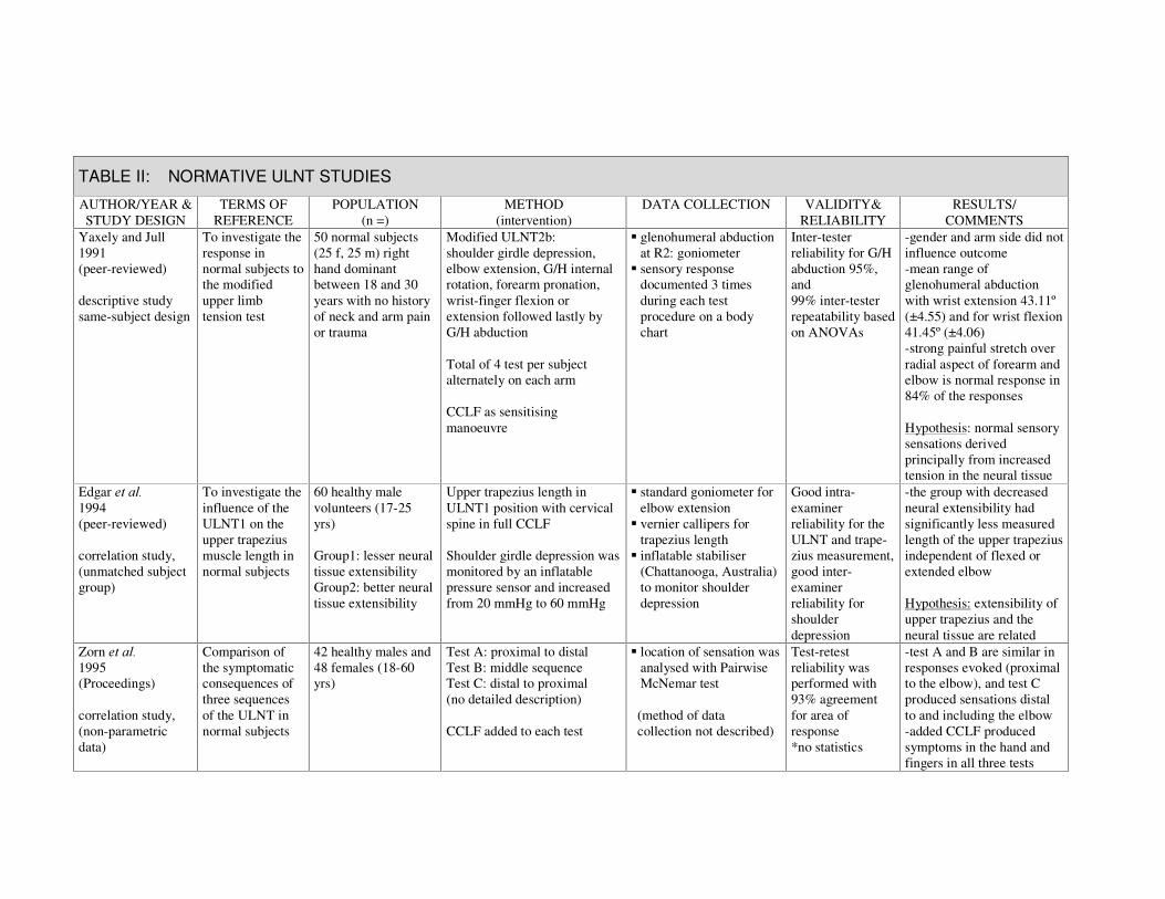
TABLE II: NORMATIVE ULNT STUDIES
AUTHOR/YEAR & STUDY DESIGN
TERMS OF REFERENCE
POPULATION (n =)
METHOD (intervention)
DATA COLLECTION VALIDITY& RELIABILITY
RESULTS/ COMMENTS
Yaxely and Jull 1991 (peer-reviewed) descriptive study same-subject design
To investigate the response in normal subjects to the modified upper limb tension test
50 normal subjects (25 f, 25 m) right hand dominant between 18 and 30 years with no history of neck and arm pain or trauma
Modified ULNT2b: shoulder girdle depression, elbow extension, G/H internal rotation, forearm pronation, wrist-finger flexion or extension followed lastly by G/H abduction Total of 4 test per subject alternately on each arm CCLF as sensitising manoeuvre
��glenohumeral abduction at R2: goniometer
��sensory response documented 3 times during each test procedure on a body chart
Inter-tester reliability for G/H abduction 95%, and 99% inter-tester repeatability based on ANOVAs
-gender and arm side did not influence outcome -mean range of glenohumeral abduction with wrist extension 43.11º (±4.55) and for wrist flexion 41.45º (±4.06) -strong painful stretch over radial aspect of forearm and elbow is normal response in 84% of the responses Hypothesis: normal sensory sensations derived principally from increased tension in the neural tissue
Edgar HW�DO� 1994 (peer-reviewed) correlation study, (unmatched subject group)
To investigate the influence of the ULNT1 on the upper trapezius muscle length in normal subjects
60 healthy male volunteers (17-25 yrs) Group1: lesser neural tissue extensibility Group2: better neural tissue extensibility
Upper trapezius length in ULNT1 position with cervical spine in full CCLF Shoulder girdle depression was monitored by an inflatable pressure sensor and increased from 20 mmHg to 60 mmHg
��standard goniometer for elbow extension
��vernier callipers for trapezius length
��inflatable stabiliser (Chattanooga, Australia) to monitor shoulder depression
Good intra-examiner reliability for the ULNT and trape-zius measurement, good inter-examiner reliability for shoulder depression
-the group with decreased neural extensibility had significantly less measured length of the upper trapezius independent of flexed or extended elbow Hypothesis: extensibility of upper trapezius and the neural tissue are related
Zorn HW�DO� 1995 (Proceedings) correlation study, (non-parametric data)
Comparison of the symptomatic consequences of three sequences of the ULNT in normal subjects
42 healthy males and 48 females (18-60 yrs)
Test A: proximal to distal Test B: middle sequence Test C: distal to proximal (no detailed description) CCLF added to each test
��location of sensation was analysed with Pairwise McNemar test
(method of data collection not described)
Test-retest reliability was performed with 93% agreement for area of response *no statistics
-test A and B are similar in responses evoked (proximal to the elbow), and test C produced sensations distal to and including the elbow -added CCLF produced symptoms in the hand and fingers in all three tests
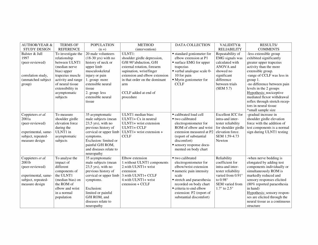
AUTHOR/YEAR & STUDY DESIGN
TERMS OF REFERENCE
POPULATION (n =)
METHOD (intervention)
DATA COLLECTION VALIDITY& RELIABILITY
RESULTS/ COMMENTS
Balster & Jull 1997 (peer-reviewed) correlation study, (unmatched subject group)
To investigate the relationship between ULNT1 (median nerve bias) upper trapezius muscle activity and range of neural tissue extensibility in asymptomatic subjects
20 male volunteers (18-30 yrs) with no history of neck or upper limb musculoskeletal injury or pain 1. group: more extensible neural tissue 2. group: less extensible neural tissue
ULNT1: shoulder girdle depression, G/H 90°abduction, G/H external rotation, forearm supination, wrist/finger extension and elbow extension in that order on the dominant arm CCLF added at end of procedure
��standard goniometer for elbow extension at P1
��surface EMG for upper trapezius
��verbal analogue scale 0-10 for pain
��Myrin goniometer for CCLF
Repeatability of EMG signals was calculated with ANOVA and showed no significant difference between trials (SEM 5.7)
-less extensible group exhibited significantly greater upper trapezius activity than the more extensible group. -range of CCLF was less in group 1. -no difference between pain levels in the 2 groups Hypothesis: nociceptive mediated flexor withdrawal reflex through stretch recep-tors in neural tissue *small sample size
Coppieters HW�DO� 2001a (peer-reviewed) experimental, same-subject, repeated-measure design
To measure shoulder girdle elevation force during the ULNT1 in asymptomatic subjects
35 asymptomatic male subjects (mean 23,5 yrs), with no previous history of cervical or upper limb symptoms. Exclusion: limited or painful G/H ROM, and diseases relate to neuropathy
ULNT1 median bias: ULNT1+ Cx in neutral ULNT1+ wrist extension ULNT1+ CCLF ULNT1+ wrist extension + CCLF
��calibrated load cell ��two calibrated
electrogoniometer for ROM of elbow and wrist extension measured at P2 (report of substantial discomfort)
��sensory response docu-mented on body chart
Excellent ICC for intra-and inter-tester reliability for shoulder girdle elevation force. SEM 1.59-4.73 Newton
-gradual increase in shoulder girdle elevation force with the addition of test components is a normal sign during ULNT1 testing
Coppieters HW�DO� 2001b (peer-reviewed) experimental, same-subject, repeated-measure design
To analyse the impact of different components of the ULNT1 (median bias) on the ROM of elbow and wrist in a normal population
35 asymptomatic male subjects (mean 23,5 yrs), with no previous history of cervical or upper limb symptoms. Exclusion: limited or painful G/H ROM, and diseases relate to neuropathy
Elbow extension 1.without ULNT1 components 2.with ULNT1+ wrist extension 3.with ULNT1+ CCLF 4.with ULNT1+ wrist extension + CCLF
��two calibrated electrogoniometer for elbow/wrist extension
��numeric pain intensity scale
��stretch and paraesthesia recorded on body chart
��criteria to end elbow extension: P2 (report of substantial discomfort)
Reliability coefficient for intra-and inter-tester reliability varied from 0.91° to 0.98° SEM varied from 1.7° to 2.5°
-when nerve bedding is elongated by adding test components individually or simultaneously ROM is markedly reduced and sensory responses elicited (80% reported paraesthesia in hand) Hypothesis: sensory respon-ses are elicited through the neural tissue as a continuous structure

AUTHOR/YEAR & TERMS OF REFERENCE
POPULATION METHOD DATA COLLECTION VALIDITY& RESULTS/
V. der Heide HW�DO� 2001 (peer-reviewed) experimental, same-subject design (multiple variables)
To investigate the presence and onset of pain and muscle activity during ULNT1 (median nerve bias) in the normal population
10 male, 10 female asymptomatic subjects (mean age 43 yrs) with no previous history of cervical or upper limb symptoms. Exclusion: limited passive range of upper limb movement, and diseases relate to neuropathy
Stage I: ULNT1 with cervical spine in neutral, P1 and P2 as indicated by the subject during elbow extension Stage II: ULNT1 with CCLF prior to elbow extension, P1 and P2 as indicated by the subject during elbow extension
��calibrated electro-goniometer for elbow extension
��surface electromyography for trapezius muscle activity
��all measurements at stage I and II were taken three times on both arms
90° shoulder abduction, upper arm splint and pressure sensor tested for reliability in a study by Edgar HW�DO� (1994) onset of P1 showed high reliability between trials
-trapezius activity is a normal response to ULNT1 and related to pain onset. -no significant difference in the range of elbow extension at P1 between both arms. -18 subjects responded with an increase in sensory symptoms and muscular response with the addition of CCLF (not indicated at what end point) *small sample size
Coppieters HW�DO��2002a (peer-reviewed) single-blinded, same-subject repeated-measure design
To quantify the impact of different shoulder positions on the ULNT1
25 male volunteers with no history of cervicobrachial pain (mean age 23.4 years) Exclusion: limited or painful G/H ROM, and diseases relate to neuropathy
Elbow extension was performed with ULNT1 in: 1)wrist and Cx in neutral 2)wrist extension 3)CCLF 4)wrist extension + CCLF 4 ULNT1 variations were performed in 3 G/H positions: 1)abduction and lateral rotation, shoulder girdle in neutral 2)abduction + shoulder girdle in neutral 3)only abduction
��calibrated electro-goniometer for elbow extension
��numeric pain intensity scale
��stretch and paraesthesia recorded on body chart
��criteria to end elbow extension: P2 (report of substantial discomfort)
Mean intra-tester reliability for elbow ROM among 12 testers was 0.96º (SD 1.8º), mean inter-tester reliability among 12 tests was 0.93º (SEM 2.3º)
-ULNT1 plus wrist extension and CCLF both decrease elbow ROM. -G/H lateral rotation decreases elbow ROM in all 4 variations of the ULNT1. -pain intensity and paraesthesia increased when more ULNT1 components were added
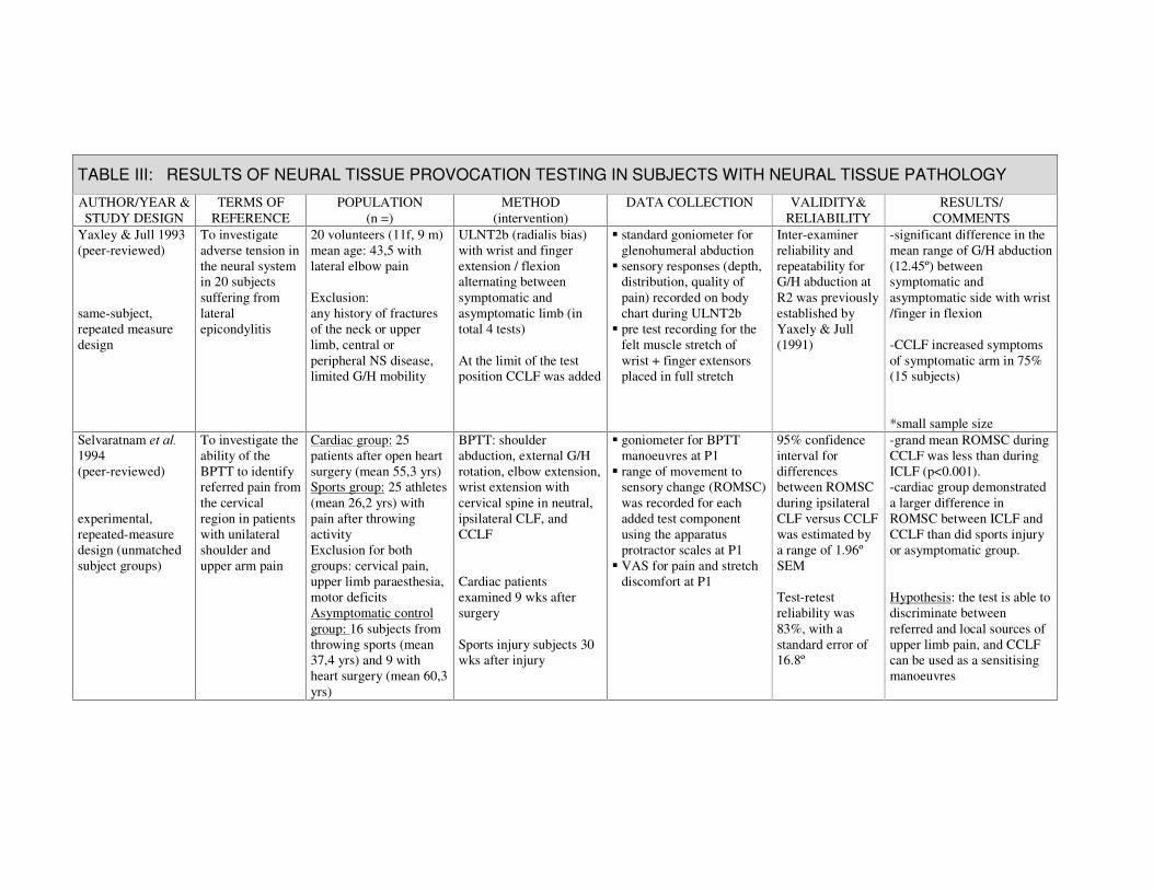
TABLE III: RESULTS OF NEURAL TISSUE PROVOCATION TESTING IN SUBJECTS WITH NEURAL TISSUE PATHOLOGY
AUTHOR/YEAR & STUDY DESIGN
TERMS OF REFERENCE
POPULATION (n =)
METHOD (intervention)
DATA COLLECTION VALIDITY& RELIABILITY
RESULTS/ COMMENTS
Yaxley & Jull 1993 (peer-reviewed) same-subject, repeated measure design
To investigate adverse tension in the neural system in 20 subjects suffering from lateral epicondylitis
20 volunteers (11f, 9 m) mean age: 43,5 with lateral elbow pain Exclusion: any history of fractures of the neck or upper limb, central or peripheral NS disease, limited G/H mobility
ULNT2b (radialis bias) with wrist and finger extension / flexion alternating between symptomatic and asymptomatic limb (in total 4 tests) At the limit of the test position CCLF was added
��standard goniometer for glenohumeral abduction
��sensory responses (depth, distribution, quality of pain) recorded on body chart during ULNT2b
��pre test recording for the felt muscle stretch of wrist + finger extensors placed in full stretch
Inter-examiner reliability and repeatability for G/H abduction at R2 was previously established by Yaxely & Jull (1991)
-significant difference in the mean range of G/H abduction (12.45º) between symptomatic and asymptomatic side with wrist /finger in flexion -CCLF increased symptoms of symptomatic arm in 75% (15 subjects) *small sample size
Selvaratnam HW�DO��1994 (peer-reviewed) experimental, repeated-measure design (unmatched subject groups)
To investigate the ability of the BPTT to identify referred pain from the cervical region in patients with unilateral shoulder and upper arm pain
Cardiac group: 25 patients after open heart surgery (mean 55,3 yrs) Sports group: 25 athletes (mean 26,2 yrs) with pain after throwing activity Exclusion for both groups: cervical pain, upper limb paraesthesia, motor deficits Asymptomatic control group: 16 subjects from throwing sports (mean 37,4 yrs) and 9 with heart surgery (mean 60,3 yrs)
BPTT: shoulder abduction, external G/H rotation, elbow extension, wrist extension with cervical spine in neutral, ipsilateral CLF, and CCLF Cardiac patients examined 9 wks after surgery Sports injury subjects 30 wks after injury�
��goniometer for BPTT manoeuvres at P1
��range of movement to sensory change (ROMSC) was recorded for each added test component using the apparatus protractor scales at P1
��VAS for pain and stretch discomfort at P1
95% confidence interval for differences between ROMSC during ipsilateral CLF versus CCLF was estimated by a range of 1.96º SEM Test-retest reliability was 83%, with a standard error of 16.8º
-grand mean ROMSC during CCLF was less than during ICLF (p<0.001). -cardiac group demonstrated a larger difference in ROMSC between ICLF and CCLF than did sports injury or asymptomatic group. Hypothesis: the test is able to discriminate between referred and local sources of upper limb pain, and CCLF can be used as a sensitising manoeuvres

AUTHOR/YEAR & STUDY DESIGN
TERMS OF REFERENCE
POPULATION (n =)
METHOD (intervention)
DATA COLLECTION VALIDITY& RELIABILITY
RESULTS/ COMMENTS
Grant HW�DO�� 1995 (peer-reviewed) single blinded study (unmatched subject groups)
To investigate the response on ULNT2b (radial bias) in screen based keyboard (SBK) workers
15 female volunteers (17-55yrs) working >4 hours at SBK; 80% experiencing some discomfort in neck and arm that did not interrupt daily activity 10 female controls with an occupation that did not involve SBK (mean 28 yrs) with no discomfort in the upper quadrant
ULNT2b with CCLF added as sensitising manoeuvre at the final ULNT position
��verbal sensory response ��goniometer for G/H
abduction
High intra- and inter-examiner reliability for G/H abduction (p=0.004) *inappropriate statistics
-SKB group exhibited decreased range in G/H abduction (mean 12.05º) in comparison with normal subjects -additional CCLF increased symptoms to 70% in control and 73% in SKB group *small sample size, weak statistics
Sweeney & Harms 1996 (peer-reviewed) same-subject, repeated measure design
To investigate the relationship between the ULNT1 and mechanical allodynia following nerve injury of the hand
29 patients (22 m, 7 f) aged 21-77 years, with injury to their hand. Time since injury or surgery: 4 weeks to 41 years Exclusion: limited shoulder mobility, and infection to the wound site
1. ULNT1 (Butler 1994) with CCLF added to final test position tested on the uninvolved side first 2. ULNT1 as a home mobilisation exercise for 2 weeks
��elbow ROM with standard goniometer at R2 or to the point of symptom reproduction
��area of maximum sensory response recorded
��VAS for deep touch
Not stated pre mobilisation: -in 22 patients the ULNT1 reproduced their symptoms. -22 patients reported a significant difference in elbow ROM between affected and unaffected side, and 7 subjects reported normal sensory response to the ULNT1 post mobilisation: -ULNT1 evoked symptoms in 10 patients -26 subjects achieved full elbow ROM on the affected side, and 19 subjects reported normal sensory response to the ULNT1

AUTHOR/YEAR & STUDY DESIGN
TERMS OF REFERENCE
POPULATION (n =)
METHOD (intervention)
DATA COLLECTION VALIDITY& RELIABILITY
RESULTS/ COMMENTS
Hall HW�DO��1998 (peer-reviewed) correlation study (matched subject design)
To investigate the effects of ankle dorsiflexion and cervical spine flexion on compliance of neural tissue to the straight leg raise (SLR)
20 normal subjects (no history of back pain) 20 age and sex matched subjects with a validated lumbar or sacral radiculopathy (mean duration 1.6 years)
SLR with ankle dorsiflexion SLR with cervical spine flexion SLR in neutral ankle and cervical position
��EMG activity of hamstrings and gluteus
��SLR range at onset of resistance (R1)
��SLR range at first increase of momentum of stretched tissue (MU)
��SLR at onset of muscle activity (M1)
Accuracy and reliability of electrogoniometer and device to measure MU was assessed in a pilot study Intra-examiner reliability was established
-R1 has no discriminative power or validity to measure non-compliance of neural tissue. -ankle dorsiflexion and cervical flexion had insignificant effects on MU. -MU was significantly greater in the radiculopathy group in SLR range post M1
Coppieters HW�DO��2002b�(peer-reviewed)� correlation study (repeated measure design within and between sessions)
To analyse the stability and reliability of P1 and P2 throughout the ROM during the ULNT1 in a laboratory and clinical setting
Experiment I: 15 (11 f, 4 m) patients with unilateral neurogenic CBP (mean age 47.7 years) Experiment II: 10 (5 f, 5 m) asymptomatic subjects with no previous history of CBP (mean age 23.4 years) Experiment III: 12 patients with CBP (10 f, 2 m) 3 patients with bilateral symptoms (mean age 41.0 years) Exclusion: limited G/H mobility
Experiment I + III for only P2: 1.ULNT1 with wrist extension Experiment II for P1 and P2: 1.ULNT1+ Cx in neutral 2.ULNT1+ wrist extension 3.ULNT1+ CCLF 4.ULNT1+ wrist extension + CCLF were added before extending the elbow
��Elbow extension: calibrated electrogonio-meter
��P1: subject pressing hand held switch
��P2: verbal signal of subjects
Experiment II: Intra-tester reliability for P1 0.98 and P2 0.98. Inter-tester reliability for P1 0.86 and P2 0.89 (lab.setting) Experiment I: Intra-tester reliability for P1 0.98 (lab.setting) Experiment III: Intra-tester reliability for P2 0.98. Inter-tester reliability for P2 0.98. (clin.setting)
-the reliability of the occurrence of P1 and P2 throughout the range of elbow ROM during ULNT1 testing was good to excellent in asymptomatic subjects in a laboratory and clinical setting. -in the patient group reliability was excellent and comparable to the reliability obtained in asymptomatic subjects when tested within the same session. -reliability in a clinical setting was not lower than in optimal laboratory conditions.

AUTHOR/YEAR & STUDY DESIGN
TERMS OF REFERENCE
POPULATION (n =)
METHOD (intervention)
DATA COLLECTION VALIDITY& RELIABILITY
RESULTS/ COMMENTS
v. der Heide HW�DO. 2002 (peer-reviewed)
To investigate the reaction to the ULNT1 in patients with cervical radiculopathy
3 patients (39-41) with C6/7 radiculopathy
In a ULNT1 position starting from 90º elbow flexion elbow was extended with Cx in neutral
��elbow extension: calibrated electrogonio-meter at P1 and P2 as indicated by the subjects
��surface electromyo-graphy for trapezius muscle at P1 and P2
��sensory responses reported on body chart
��all measurements were taken three times
High reliability between trials for onset of pain established earlier (v. der Heide HW�DO� 2001)
-ULNT1 reproduced the symptoms in 2 patients -trapezius activity was evident around the point of P1 in all 3 patients -in 2 patients P1 was significantly earlier in elbow ROM than on the asymptomatic side *small sample size no statistical power
Coppieters HW�DO� 2003a (peer-reviewed) single-blinded RCT
To analyse whether aberrations in shoulder girdle elevation force during ULNT1 can be demonstrated in patients with CBP, and if they can be normalised following cervical mobilisation in a laboratory setting
Experimental group: 10 patients (8 f, 2 m, mean age: 49.1) Control group: 10 patients (8 f, 2 m, mean age: 48.1) with non-acute (2 weeks to 6 months) unilateral or bilateral CBP related to the median nerve, volunteered to participate
3 repetitions of the ULNT1 Experimental condition: cervical lateral glide technique at 1 or more segments (C5-T1) with the involved arm in a neural preloaded position Control condition: 5 minutes of pulsed ultrasound over the most painful area in an unloaded position (both interventions performed by the same therapist, blinded to the patient allocation)
��calibrated load cell for shoulder elevation force
��calibrated electrogonio-meters for ROM of elbow extension measured at P2 (report of substantial discomfort)
��numeric pain intensity scale
No systematic changes could be observed when the amount of end force of 3 consecutive repetitions of the UNT1 was analysed
-the amount of force at the end of the test on the unin-volved side was significantly larger than on the involved side. The sudden increase in shoulder girdle elevation force on the involved side occurred earlier in range than on the uninvolved side. The immediate effect of treatment: increase in shoulder girdle elevation force occurred later in the ROM; the amount of end force was significantly larger than before treat-ment; pain intensity was significantly decreased; available ROM was significantly increased -No treatment effects observed after control intervention

AUTHOR/YEAR & STUDY DESIGN
TERMS OF REFERENCE
POPULATION (n =)
METHOD (intervention)
DATA COLLECTION VALIDITY& RELIABILITY
RESULTS/ COMMENTS
Coppieters HW�DO��2003b (in press) single blinded RCT
To analyse the immediate treatment effect of cervical mobilisation and therapeutic ultrasound in patients with neurogenic CBP disorders
20 patients (volunteers) with subacute (2 weeks–6 months) uni- or bilateral cervico-brachial pain Inclusion criteria formulated by Elvey (1997)
3 repetitions of the ULNT1 Experimental condition: cervical lateral glide technique at 1 or more segments (C5-T1) with the involved arm in a neural preloaded position Control condition: 5 minutes of pulsed ultrasound over the most painful area in an unloaded position (both interventions performed by the same therapist, blinded to the patient allocation)
��elbow ROM with electro-goniometer at P2
��body chart for symptom distribution during ULNT1
��numeric pain intensity scale during ULNT1
��load cell was used to standardise shoulder girdle depression
Good to excellent intra- and inter-tester reliability for P1 and P2 previously established (Coppieters HW�DO��2002b)
-for the cervical mobilisation group a significant reduction in pain intensity, an improve-ment in elbow ROM, and a 43.4% reduction in area of symptom provocation was noted. -in the ultrasound group no significant changes were found. -a significant difference in available elbow ROM, elicited pain intensity, and area of symptom distribution between the uninvolved and involved side was evident.
One way-ANOVA ICC index (Coppieters HW�DO� 2002b)
• ‘Poor’ = ICC < 0.40 • ‘Fair’ = 0.04 ��,&&������� • ‘Good’ = 0.70 ��,&&������� • ‘Excellent’ = ICC ������
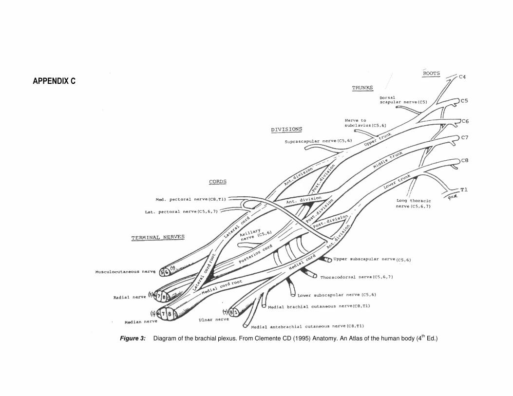
$33(1',;�&�
� )LJXUH��� Diagram of the brachial plexus. From Clemente CD (1995) Anatomy. An Atlas of the human body (4th Ed.)

XIII
ULNT1-A: Starting position ULNT1- E: Shoulder lateral rotation
ULNT1-B: Shoulder abduction ULNT1-F: Elbow extension
ULNT1-C: Wrist extension ULNT1-G: Contralateral cervical lateral
flexion
ULNT1-D: Forearm supination ULNT1-H: Ipsilateral cervical lateral flexion
)LJXUH��� The ULNT1. From Butler DS (2000) The Sensitive Nervous System
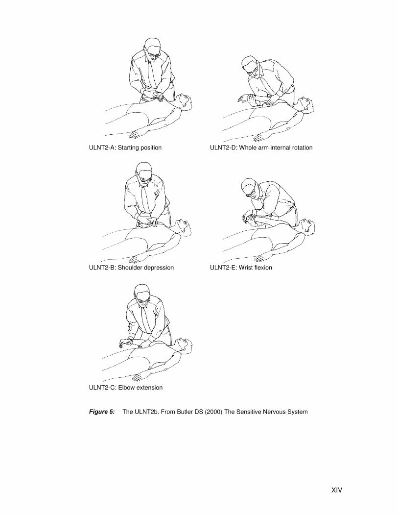
XIV
ULNT2-A: Starting position ULNT2-D: Whole arm internal rotation
ULNT2-B: Shoulder depression ULNT2-E: Wrist flexion
ULNT2-C: Elbow extension
)LJXUH��� The ULNT2b. From Butler DS (2000) The Sensitive Nervous System

XV
ULNT3-A: Starting position ULNT3-E: Shoulder lateral rotation
ULNT3-B: Wrist extension ULNT3-F: Shoulder girdle depression
ULNT3-C: Forearm pronation ULNT3-G: Shoulder abduction
ULNT3-D: Elbow flexion
)LJXUH��� The ULNT3. From Butler DS (2000) The Sensitive Nervous System
Related Documents


