University of Pennsylvania University of Pennsylvania ScholarlyCommons ScholarlyCommons Cognitive Neuroscience Publications Center for Cognitive Neuroscience 2009 The Neural Basis for Spatial Relations The Neural Basis for Spatial Relations Prin X. Amorapanth University of Pennsylvania Page Widick University of Pennsylvania Anjan Chatterjee University of Pennsylvania, [email protected] Follow this and additional works at: https://repository.upenn.edu/cog_neuro_pubs Part of the Neuroscience and Neurobiology Commons Recommended Citation Recommended Citation Amorapanth, P. X., Widick, P., & Chatterjee, A. (2009). The Neural Basis for Spatial Relations. Retrieved from https://repository.upenn.edu/cog_neuro_pubs/10 Suggested Citation: Amorapanth, P.X., P. Widick and A. Chatterjee. (2009). "The Neural Basis for Spatial Relations." Journal of Cognitive Neuroscience. Vol. 22(8). pp. 1739-1753. © 2009 Massachusetts Institute of Technology http://www.mitpressjournals.org/loi/jocn This paper is posted at ScholarlyCommons. https://repository.upenn.edu/cog_neuro_pubs/10 For more information, please contact [email protected].

Welcome message from author
This document is posted to help you gain knowledge. Please leave a comment to let me know what you think about it! Share it to your friends and learn new things together.
Transcript

University of Pennsylvania University of Pennsylvania
ScholarlyCommons ScholarlyCommons
Cognitive Neuroscience Publications Center for Cognitive Neuroscience
2009
The Neural Basis for Spatial Relations The Neural Basis for Spatial Relations
Prin X. Amorapanth University of Pennsylvania
Page Widick University of Pennsylvania
Anjan Chatterjee University of Pennsylvania, [email protected]
Follow this and additional works at: https://repository.upenn.edu/cog_neuro_pubs
Part of the Neuroscience and Neurobiology Commons
Recommended Citation Recommended Citation Amorapanth, P. X., Widick, P., & Chatterjee, A. (2009). The Neural Basis for Spatial Relations. Retrieved from https://repository.upenn.edu/cog_neuro_pubs/10
Suggested Citation: Amorapanth, P.X., P. Widick and A. Chatterjee. (2009). "The Neural Basis for Spatial Relations." Journal of Cognitive Neuroscience. Vol. 22(8). pp. 1739-1753.
© 2009 Massachusetts Institute of Technology http://www.mitpressjournals.org/loi/jocn
This paper is posted at ScholarlyCommons. https://repository.upenn.edu/cog_neuro_pubs/10 For more information, please contact [email protected].

The Neural Basis for Spatial Relations The Neural Basis for Spatial Relations
Abstract Abstract Studies in semantics traditionally focus on knowledge of objects. By contrast, less is known about how objects relate to each other. In an fMRI study, we tested the hypothesis that the neural processing of categorical spatial relations between objects is distinct from the processing of the identity of objects. Attending to the categorical spatial relations compared with attending to the identity of objects resulted in greater activity in superior and inferior parietal cortices (especially on the left) and posterior middle frontal cortices bilaterally. In an accompanying lesion study, we tested the hypothesis that comparable areas would be necessary to represent categorical spatial relations and that the hemispheres differ in their biases to process categorical or coordinate spatial relations. Voxelbased lesion symptom mapping results were consistent with the fMRI observations. Damage to a network comprising left inferior frontal, supramarginal, and angular gyri resulted in behavioral impairment on categorical spatial judgments. Homologous right brain damage also produced such deficits, albeit less severely. The reverse pattern was observed for coordinate spatial processing. Right brain damage to the middle temporal gyrus produced more severe deficits than left hemisphere damage. Additional analyses suggested that some areas process both kinds of spatial relations conjointly and others distinctly. The left angular and inferior frontal gyrus processes coordinate spatial information over and above the categorical processing. The anterior superior temporal gyrus appears to process categorical spatial information uniquely. No areas within the right hemisphere processed categorical spatial information uniquely. Taken together, these findings suggest that the functional neuroanatomy of categorical and coordinate processing is more nuanced than implied by a simple hemispheric dichotomy.
Disciplines Disciplines Life Sciences | Neuroscience and Neurobiology
Comments Comments Suggested Citation: Amorapanth, P.X., P. Widick and A. Chatterjee. (2009). "The Neural Basis for Spatial Relations." Journal of Cognitive Neuroscience. Vol. 22(8). pp. 1739-1753.
© 2009 Massachusetts Institute of Technology http://www.mitpressjournals.org/loi/jocn
This journal article is available at ScholarlyCommons: https://repository.upenn.edu/cog_neuro_pubs/10

The Neural Basis for Spatial Relations
Prin X. Amorapanth, Page Widick, and Anjan Chatterjee
Abstract
■ Studies in semantics traditionally focus on knowledge ofobjects. By contrast, less is known about how objects relateto each other. In an fMRI study, we tested the hypothesis thatthe neural processing of categorical spatial relations betweenobjects is distinct from the processing of the identity of objects.Attending to the categorical spatial relations compared withattending to the identity of objects resulted in greater activityin superior and inferior parietal cortices (especially on theleft) and posterior middle frontal cortices bilaterally. In anaccompanying lesion study, we tested the hypothesis thatcomparable areas would be necessary to represent categoricalspatial relations and that the hemispheres differ in their biasesto process categorical or coordinate spatial relations. Voxel-based lesion symptom mapping results were consistent withthe fMRI observations. Damage to a network comprising leftinferior frontal, supramarginal, and angular gyri resulted in
behavioral impairment on categorical spatial judgments. Ho-mologous right brain damage also produced such deficits, albeitless severely. The reverse pattern was observed for coordinatespatial processing. Right brain damage to the middle temporalgyrus produced more severe deficits than left hemispheredamage. Additional analyses suggested that some areas pro-cess both kinds of spatial relations conjointly and others dis-tinctly. The left angular and inferior frontal gyrus processescoordinate spatial information over and above the categoricalprocessing. The anterior superior temporal gyrus appears toprocess categorical spatial information uniquely. No areaswithin the right hemisphere processed categorical spatial in-formation uniquely. Taken together, these findings suggest thatthe functional neuroanatomy of categorical and coordinateprocessing is more nuanced than implied by a simple hemi-spheric dichotomy. ■
INTRODUCTION
Cognitive neuroscience investigations of semantics tradi-tionally focus on knowledge of objects (Borgo & Shallice,2001; Moore & Price, 1999; Tranel & Damasio, 1999;Caramazza&Shelton,1998; Binder et al., 1997; Gonnerman,Andersen, Devlin, Kempler, & Seidenberg, 1997; Tranel,Logan, Frank, & Damasio, 1997; Vandenberghe, Price,Wise, Josephs, & Frackowiak, 1996; Capitani, Laiacona,Barbarotto, & Trivelli, 1994; Damasio, Damasio, Tranel,& Brandt, 1990). Although these investigations havecertainly been fruitful, confining semantics to the studyof objects leaves us with an impoverished understandingof the world. Until recently, relatively little attention waspaid to our knowledge of how objects act in the worldand how they are related to each other. Investigatingthe neural basis of knowledge of actions, events, andrelations between objects would enrich our understand-ing of the human semantic system.In this report, we focused on the neural basis for a
specific kind of relational knowledge, that is, spatial rela-tions. Spatial relations between objects can be organizedin different ways. These relations can be organized in theform of continuous metrics (distance, orientation, etc.)or as a discrete set of relations (such as those that canbe labeled verbally by prepositions in English). For exam-
ple, a pen might be 5 or 10 cm to the right of a notebook.These would represent different coordinate relations butnot different categorical relations. The pen would haveto be on top of the notebook to represent a different cate-gorical relationship. Slobin (1996, 2000) proposed thatthinking for speaking might be different than thinkingfor other reasons. Along these lines, thinking about spa-tial relationships for speaking might be different thanthinking about spatial relations for other reasons. Thus,coordinate (continuous metric) representations are criticalto guide movements such as reaching or navigation butmay be less relevant to language. They can be describedby a large and flexible set of open class terms, such asdistance in centimeters, inches, or miles. By contrast, cate-gorical representations form a more coarsely organizedset of spatial relations that are encoded in a closed classset of terms such as “on” or “in.”
The neural instantiation of spatial relations might beexpected to follow naturally from the “what” versus“where” distinction (Ungerleider & Mishkin, 1982). On thiscentral tenet of visual neuroscience from monkey stud-ies, visual processing is divided into dorsal or occipito-parietal and ventral or occipito-temporal streams. Thedorsal visual stream specializes in spatial processing,and the ventral stream specializes in object processing(Haxby et al., 1991). Although the streams undoubtedlyinteract at multiple levels (Schiller, 1996; Ferrera, Rudolph,& Maunsell, 1994), this processing division of labor byThe University of Pennsylvania
© 2009 Massachusetts Institute of Technology Journal of Cognitive Neuroscience 22:8, pp. 1739–1753

the nervous system has been corroborated in humans(Chatterjee, 2003; Martin, Ungerleider, & Haxby, 2000;Aguirre & DʼEsposito, 1997; Farah, 1990). Occipito-temporal lesions are associated with various visual agno-sias, in which individuals have trouble recognizing objectsor faces, and posterior parietal lesions are associated withspatial deficits such as hemispatial neglect or simultanag-nosia. Imaging studies also confirm this general distinction.Thus, the fusiform gyrus is associated with processingfaces (Kanwisher, McDermott, & Chun, 1997), the para-hippocampal gyrus with processing places and buildings(Epstein & Kanwisher, 1998; Aguirre & DʼEsposito, 1997),and the lateral occipital complex with processing objectsin general (Kanwisher, Woods, Iacoboni, & Mazziotta,1997; Malach et al., 1995). Although the posterior parietalcortex is implicated in shifts of spatial attention (Corbetta,Shulman, Miezin, & Peterson, 1995), relatively little directfunctional imaging evidence supports the role of the pari-etal cortex in processing spatial relations.
Spatial relations relevant to language are likely to becategorical. Kosslyn et al. (1989) initially suggested thedistinction between categorical and coordinate spatialrelations. They proposed a left hemisphere advantagefor categorical visual processing and a right hemisphereadvantage for coordinate visual processing (Kosslyn &Ochsner, 1994; Kosslyn et al., 1989). However, the empir-ical bases for these neural claims were limited, derivedprimarily from visual hemifield stimuli presentationsand computer simulations. The specific results obtainedby Kosslyn et al. were subsequently challenged as anartifact of a bias in spatial resolution of sensory process-ing (Sergent, 1991) or as an artifact of task difficulty(Slotnick, Moo, Tesoro, & Hart, 2001). Furthermore, theapproach of using lateralized stimuli presentation to inferlateralized brain function might itself be suspect (Efron,1990). In a subsequent PET study, Kosslyn, Thompson,Gitelman, and Alpert (1998) failed to find consistent lefthemisphere activation for categorical spatial judgments.However, in an fMRI study, Baciu et al. (1999) found thatleft angular gyrus (AG), within the inferior parietal lobe, dis-played greater activity than the right AG when subjectsjudged whether a dot was located above or below a bar.
Despite the limited evidence for the neural basis forcategorical and coordinate spatial processing, this distinc-tion seems plausible (for reviews, see Postma & Laeng,2006; Jager & Postma, 2003). Damage to right parietalcortex appears to impair performance on tasks that relyon precise metrics (Laeng, 1994; Hannay, 1976; Warrington& Taylor, 1973). Left posterior parietal lesions are asso-ciated with left/right confusion, a hallmark symptom ofGerstmann syndrome (Mayer et al., 1999). To our knowl-edge, patients with Gerstmann syndrome per se havenot been tested more comprehensively on categoricalspatial judgments. Left–right judgments may be a class ofspatial judgment, as evidenced by left hemispatial neglect(Chatterjee, 2003) or mirror reading and writing (Gottfried,Sancar, & Chatterjee, 2003), that implicates a special
role for the horizontal axis in processing space. Whetherleft–right processing generalizes to categorical processingis not known.Laeng (1994, 2006) has reported data consistent with
the hemispheric specialization hypothesis. Patients withfocal brain damage were shown drawings of two objects.After a short delay, they were asked to identify the draw-ing when it was paired with a drawing of the same objectstransformed in their categorical or coordinate spatial re-lations. He found that patients with right hemispheredamage (RHD) were more likely to make coordinate re-lation errors and patients with left hemisphere damage(LHD) were more likely to make categorical memoryerrors. The anatomic analyses of patients in this studywere rudimentary. Counterintuitively, behavior differ-ences were found when patients had parietal damageonly and not when they also had temporal or frontal dam-age in addition to the parietal damage. More relevant toour study is a report by Tranel and Kemmerer (2004).They examined a large group of patients on a variety oftasks probing knowledge of locative relations. In an “oddman out” task, participants viewed groups of three pic-tures, each with two objects. They were asked to pointto the picture that depicted a different categorical rela-tionship than the other two. They found that damage un-derlying the white matter of the left parietal and frontalopercula was associated with deficits in matching catego-rical spatial relations.The studies reviewed served as the backdrop to our
investigations of the neural basis of spatial relations usingboth functional imaging in young healthy participantsand behavioral studies in participants with focal brainlesions. Both approaches, now central methods in cogni-tive neuroscience, have complementary strengths andweaknesses (Chatterjee, 2005; Fellows et al., 2005; Rorden& Karnath, 2004). Imaging studies in young normal sub-jects offer insights into the processing of the unalteredbrain but remain fundamentally a correlational method.Lesion studies are inferentially stronger but are limited byuncertainties of neuronal reorganization, not to mentionvariables such as age, education, medications, and comor-bid conditions in an elderly population. In principle, con-verging evidence from bothmethods would provide strongsupport for brain–behavior relations.Our first experiment used fMRI to investigate the neu-
ral systems activated by the identification of categoricalspatial relations, as compared with the identification ofobjects being located. By categorical relations, we meanbetween-object relations rather than within-object rela-tions (such as the handle at the top of the briefcase). Ourexperiment relies on subjects making different judgmentson the same set of stimuli. Thus, different neural activationpatterns could not arise from differences in the perceptualproperties of the stimuli themselves. The second experi-ment used participants with left or right focal brain lesionsto assess their abilities to make categorical and coordinatespatial judgments. We used a variant of voxel-based lesion
1740 Journal of Cognitive Neuroscience Volume 22, Number 8
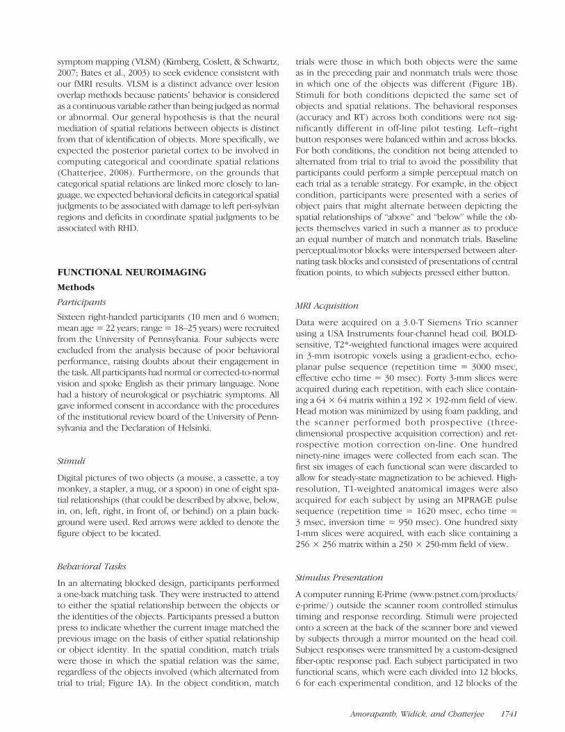
symptommapping (VLSM) (Kimberg, Coslett, & Schwartz,2007; Bates et al., 2003) to seek evidence consistent withour fMRI results. VLSM is a distinct advance over lesionoverlap methods because patientsʼ behavior is consideredas a continuous variable rather than being judged as normalor abnormal. Our general hypothesis is that the neuralmediation of spatial relations between objects is distinctfrom that of identification of objects. More specifically, weexpected the posterior parietal cortex to be involved incomputing categorical and coordinate spatial relations(Chatterjee, 2008). Furthermore, on the grounds thatcategorical spatial relations are linked more closely to lan-guage, we expected behavioral deficits in categorical spatialjudgments to be associated with damage to left peri-sylvianregions and deficits in coordinate spatial judgments to beassociated with RHD.
FUNCTIONAL NEUROIMAGING
Methods
Participants
Sixteen right-handed participants (10 men and 6 women;mean age = 22 years; range = 18–25 years) were recruitedfrom the University of Pennsylvania. Four subjects wereexcluded from the analysis because of poor behavioralperformance, raising doubts about their engagement inthe task. All participants had normal or corrected-to-normalvision and spoke English as their primary language. Nonehad a history of neurological or psychiatric symptoms. Allgave informed consent in accordance with the proceduresof the institutional review board of the University of Penn-sylvania and the Declaration of Helsinki.
Stimuli
Digital pictures of two objects (a mouse, a cassette, a toymonkey, a stapler, a mug, or a spoon) in one of eight spa-tial relationships (that could be described by above, below,in, on, left, right, in front of, or behind) on a plain back-ground were used. Red arrows were added to denote thefigure object to be located.
Behavioral Tasks
In an alternating blocked design, participants performeda one-back matching task. They were instructed to attendto either the spatial relationship between the objects orthe identities of the objects. Participants pressed a buttonpress to indicate whether the current image matched theprevious image on the basis of either spatial relationshipor object identity. In the spatial condition, match trialswere those in which the spatial relation was the same,regardless of the objects involved (which alternated fromtrial to trial; Figure 1A). In the object condition, match
trials were those in which both objects were the sameas in the preceding pair and nonmatch trials were thosein which one of the objects was different (Figure 1B).Stimuli for both conditions depicted the same set ofobjects and spatial relations. The behavioral responses(accuracy and RT) across both conditions were not sig-nificantly different in off-line pilot testing. Left–rightbutton responses were balanced within and across blocks.For both conditions, the condition not being attended toalternated from trial to trial to avoid the possibility thatparticipants could perform a simple perceptual match oneach trial as a tenable strategy. For example, in the objectcondition, participants were presented with a series ofobject pairs that might alternate between depicting thespatial relationships of “above” and “below” while the ob-jects themselves varied in such a manner as to producean equal number of match and nonmatch trials. Baselineperceptual/motor blocks were interspersed between alter-nating task blocks and consisted of presentations of centralfixation points, to which subjects pressed either button.
MRI Acquisition
Data were acquired on a 3.0-T Siemens Trio scannerusing a USA Instruments four-channel head coil. BOLD-sensitive, T2*-weighted functional images were acquiredin 3-mm isotropic voxels using a gradient-echo, echo-planar pulse sequence (repetition time = 3000 msec,effective echo time = 30 msec). Forty 3-mm slices wereacquired during each repetition, with each slice contain-ing a 64 × 64 matrix within a 192 × 192-mm field of view.Head motion was minimized by using foam padding, andthe scanner performed both prospective (three-dimensional prospective acquisition correction) and ret-rospective motion correction on-line. One hundredninety-nine images were collected from each scan. Thefirst six images of each functional scan were discarded toallow for steady-state magnetization to be achieved. High-resolution, T1-weighted anatomical images were alsoacquired for each subject by using an MPRAGE pulsesequence (repetition time = 1620 msec, echo time =3 msec, inversion time = 950 msec). One hundred sixty1-mm slices were acquired, with each slice containing a256 × 256 matrix within a 250 × 250-mm field of view.
Stimulus Presentation
A computer running E-Prime (www.pstnet.com/products/e-prime/ ) outside the scanner room controlled stimulustiming and response recording. Stimuli were projectedonto a screen at the back of the scanner bore and viewedby subjects through a mirror mounted on the head coil.Subject responses were transmitted by a custom-designedfiber-optic response pad. Each subject participated in twofunctional scans, which were each divided into 12 blocks,6 for each experimental condition, and 12 blocks of the
Amorapanth, Widick, and Chatterjee 1741

baseline condition. There were 13 stimuli per block, withstimulus durations of 1200 msec, ISIs of 415 msec, and aninterval of 3000 msec between blocks.
Preprocessing
Data processing was performed off-line using softwaredeveloped at the University of Pennsylvania (www.voxbo.org). After reconstructing images from the raw data, datawere sinc interpolated in time to correct for staggered sliceacquisition, realigned to the first image acquired for eachsubjects using a six-parameter motion-correction algo-rithm, and thresholded to excluded extraparenchymalvoxels from subsequent analyses. Within each subject, avoxelwise analysis was performed using a modified generallinear model that included covariates modeling task condi-tions as well as sine and cosine regressors for frequenciesbelow those of the task and for frequencies in the elevatedrange of the noise spectrum. Task covariates were boxcarwaveforms convolved with an estimate of the BOLD hemo-dynamic transfer function, which was empirically derivedfrom motor cortex in a large group of subjects (Aguirre,Zarahn, & DʼEsposito, 1998). Data were also smoothed in
time with the hemodynamic transfer function to controlthe false-positive rate (Aguirre, Zarahn, & DʼEsposito,1997; Zarahn, Aguirre, & DʼEsposito, 1997). Group activa-tion maps were constructed by a random-effects analysis ofbeta-values for each covariate at each voxel.
Definition of ROIs
Anatomical ROIs were derived from the automated ana-tomical labeling map of the Montreal Neurological Insti-tute (MNI) single-subject brain (Tzourio-Mazoyer et al.,2002). After translation to the appropriate file format, re-gions were resampled to the correct dimensions andmanually adjusted to compensate for resampling errors.Anatomical ROIs were (a) superior parietal lobules, (b)inferior parietal lobules (IPLs) encompassing AG andsupramarginal gyrus), (c) lateral occipital cortex, (d) mid-dle temporal cortex, (e) inferior temporal cortex, (f ) fusi-form gyri (both occipital and temporal portions), and(g) parahippocampal gyri. These ROIs were chosen onthe basis of findings in the literature reviewed earlier,suggesting possible roles for these areas in the process-ing of the cognitive aspects of both of these tasks.
Figure 1. Examples of thesequence of stimuli used inthe fMRI study, (A) spatialcondition and (B) objectcondition. Images labeledfor purposes of this figure;images used in the scannerwere unlabeled and in color.The arrow indicated thefigure object in the spatialrelations condition.
1742 Journal of Cognitive Neuroscience Volume 22, Number 8

Figure 2. (A–G) Averaged time series for ROIs displaying significant differences between conditions. Time series were averaged across all blocksfor each subject and then across all subjects for each experiment. Solid line depicts the average; dotted lines depict average ± SE.
Amorapanth, Widick, and Chatterjee 1743

Hypotheses about activity differences were testedusing the following approach. Voxels were initially identi-fied in which activity was significant for the main effectof spatial relations and object matching compared withthe baseline condition ( p < .05, Bonferroni-correctedfor multiple comparisons within an ROI). As describedpreviously (Kable, Kan, Wilson, Thompson-Schill, &Chatterjee, 2005; Kable, Lease-Spellmeyer, & Chatterjee,2002), the baseline condition was not necessarily matchedto the conditions of interest; rather, the goal was to reducethe number of voxels within which the contrasts of inter-est would be queried. The fMRI time series was averagedfor all voxels significant for the main effect within a definedROI in each subject. Next, a measure of the effect size forthe orthogonal contrast between conditions of interest(spatial relations–object matching) was extracted fromthe spatially averaged ROI time series in each subject.We used t values as a measure of effect size rather thanpercent signal change because the residual error term inthe denominator of the t value most effectively correctsthe effect size for scaling effects because of differencesin signal intensity across scanning sessions (Postle, Zarahn,& DʼEsposito, 2000). Finally, paired t tests addressedwhether this effect was consistently greater for one condi-tion or the other in that ROI across subjects. This analysistrades spatial resolution for sensitivity by reducing the ef-
fect size in each ROI to a single value through averagingacross all active voxels within an ROI.
Results
Behavioral Results
Participants did not differ in accuracy (89% ± 3% vs.87% ± 3%) or RTs (702 ± 32 vs. 661 ± 32 msec) forthe spatial relation and object identification conditions,respectively.
Imaging Results
Within each ROI, the averaged time series across allvoxels that showed activity for the main effect of bothspatial relations and object matching compared withthe baseline condition is shown in Figure 2. For eachaveraged time series, we calculated a measure of thedifference in activity between the spatial relation andthe object matching conditions and then tested whetherthis difference was significantly different from zero acrosssubjects for each ROI (a random-effects test, see Table 1).Across participants, there was consistently greater ac-
tivation bilaterally for judgments of spatial relations inthe superior parietal lobules, left, t(11) = 3.7, p < .005,
Table 1. Functional MRI Results
Spatial Relations–Object Matching Average Voxels
Left Hemisphere
Superior parietal lobe 1.84 ± 0.49 520
Inferior parietal lobe 2.07 ± 0.49 374
Lateral occipital complex −0.78 ± 0.39 520
Middle temporal gyrus 0.88 ± 0.37 149
Inferior temporal gyrus 0.25 ± 0.49 77
Fusiform gyrus −0.78 ± 0.28 325
Parahippocampal gyrus −0.47 ± 0.23 124
Right Hemisphere
Superior parietal lobe 2.04 ± 0.62 520
Inferior parietal lobe 1.39 ± 0.55 166
Lateral occipital complex −0.39 ± 0.39 414
Middle temporal gyrus 0.67 ± 0.24 189
Inferior temporal gyrus 0.55 ± 0.41 209
Fusiform gyrus −0.37 ± 0.28 380
Parahippocampal gyrus −0.08 ± 0.22 124
Effect sizes are given as mean ± SE for contrasts between conditions within each ROI. Effect sizes that are consistently different from zero acrossconditions ( p < .05) are in boldface.
1744 Journal of Cognitive Neuroscience Volume 22, Number 8
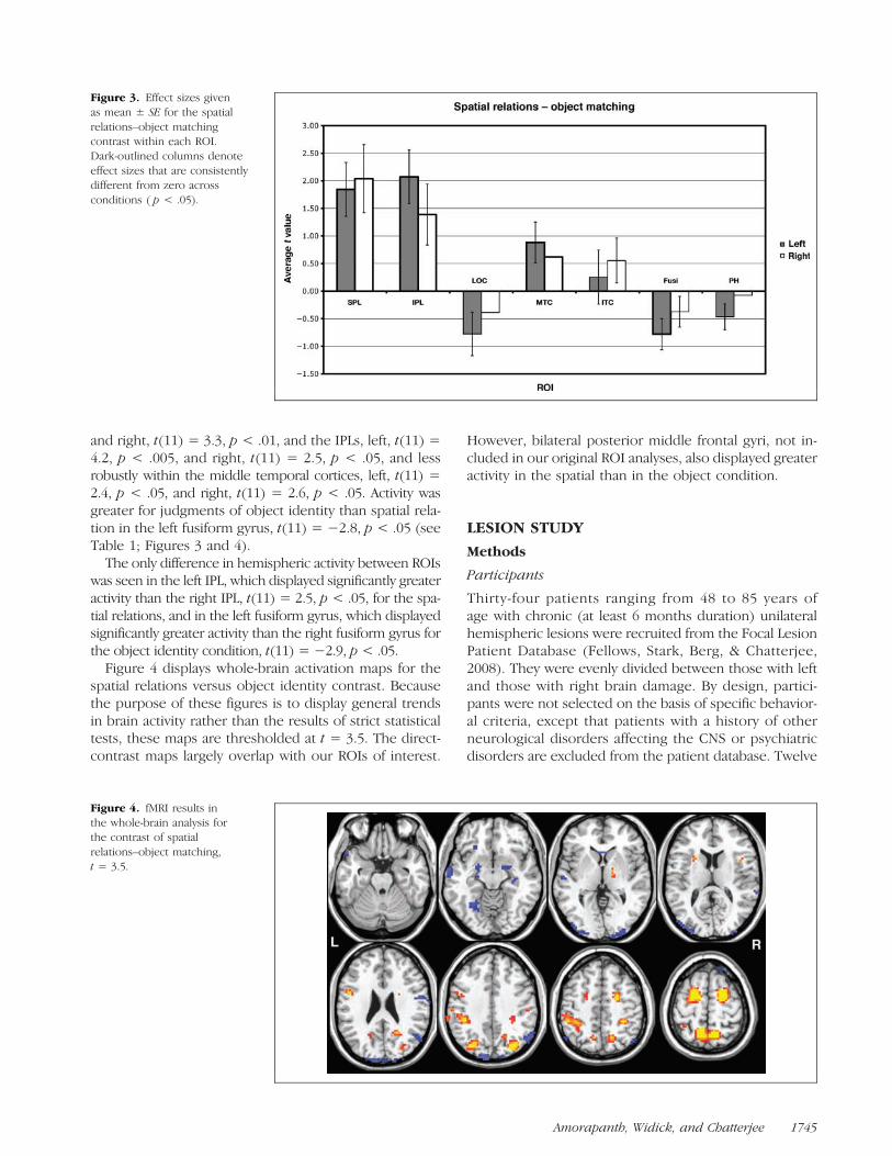
and right, t(11) = 3.3, p < .01, and the IPLs, left, t(11) =4.2, p < .005, and right, t(11) = 2.5, p < .05, and lessrobustly within the middle temporal cortices, left, t(11) =2.4, p < .05, and right, t(11) = 2.6, p < .05. Activity wasgreater for judgments of object identity than spatial rela-tion in the left fusiform gyrus, t(11) = −2.8, p < .05 (seeTable 1; Figures 3 and 4).The only difference in hemispheric activity between ROIs
was seen in the left IPL, which displayed significantly greateractivity than the right IPL, t(11) = 2.5, p < .05, for the spa-tial relations, and in the left fusiform gyrus, which displayedsignificantly greater activity than the right fusiform gyrus forthe object identity condition, t(11) = −2.9, p < .05.Figure 4 displays whole-brain activation maps for the
spatial relations versus object identity contrast. Becausethe purpose of these figures is to display general trendsin brain activity rather than the results of strict statisticaltests, these maps are thresholded at t = 3.5. The direct-contrast maps largely overlap with our ROIs of interest.
However, bilateral posterior middle frontal gyri, not in-cluded in our original ROI analyses, also displayed greateractivity in the spatial than in the object condition.
LESION STUDY
Methods
Participants
Thirty-four patients ranging from 48 to 85 years ofage with chronic (at least 6 months duration) unilateralhemispheric lesions were recruited from the Focal LesionPatient Database (Fellows, Stark, Berg, & Chatterjee,2008). They were evenly divided between those with leftand those with right brain damage. By design, partici-pants were not selected on the basis of specific behavior-al criteria, except that patients with a history of otherneurological disorders affecting the CNS or psychiatricdisorders are excluded from the patient database. Twelve
Figure 3. Effect sizes givenas mean ± SE for the spatialrelations–object matchingcontrast within each ROI.Dark-outlined columns denoteeffect sizes that are consistentlydifferent from zero acrossconditions ( p < .05).
Figure 4. fMRI results inthe whole-brain analysis forthe contrast of spatialrelations–object matching,t = 3.5.
Amorapanth, Widick, and Chatterjee 1745

elderly control subjects ranging from 58 to 79 years ofage were also tested on these tasks.
All participants were native English speakers and right-handed. Written informed consent in accordance withthe procedures of the institutional review board ofthe University of Pennsylvania and the Declaration ofHelsinki was obtained for each participant. A nativeEnglish speaker delivered all spoken materials. If a par-ticipant was unable to complete testing in one session, ad-ditional sessions were scheduled at least one week later.
Stimuli
Images consisted of digital color photographs (specificstimuli may be obtained from the corresponding author).The objects in these images consisted of small set ofrelatively common household/office items that couldfunction as figure and/or ground objects for the locativerelations being tested. In the images, a red arrow pointedto the figure object. Spatial relationships that could be
described by in and on, which are considered topologicrelations, and those that could be described by above,below, to the left of, and to the right of, which are con-sidered projective relations, were used as stimuli. We rec-ognized the polysemous nature of prepositions. Withtopological relationships, each exemplar for “in” depicteda containment relationship and for “on” a contact rela-tionship of the figure to ground along the vertical y-axis.For the topological relationships, the verbal descrip-tors refer to an allocentric reference frame (note thatthe stimuli themselves only used pictures; for examples,see Figure 5).
Screening Tasks
All patients were screened for unilateral neglect. Althoughneglect usually recovers within 3 months (Chatterjee,1995), we wanted to ensure that severe attentional bi-ases would not impair their ability to see or to respondto specific stimuli. All patients were given the gray scale testfor neglect (Mattingley et al., 2004). We also wished toensure that subjects recognized the objects being pictured.All subjects identified the series of objects that were usedin the spatial tasks. Twenty-six trials were presented inwhich the name of an object was presented along with fourphotographic images (one image depicting the objectnamed). Participants indicated which one of the four pic-tures depicted the correct answer either by pointing orby reading the letter underneath a particular image. Thelocation of correct response (top/bottom and left/right)was counterbalanced across the trials.
Categorical Spatial Matching
This task was designed to assess patientsʼ ability to matchcategorical spatial concepts across different photographicrepresentations. The patients were presented with 40 trialsin which a probe image containing one pair of objects in aparticular spatial relationship was to be matched with oneof four images containing a different pair of objects. Of thefour choices, one depicted the correct categorical spatialrelationship, and the foils could depict within- or across-class categorical relations (Figure 5A). Twenty-four itemshad a correct topological match (12 with pictures thatcould be described by in and 12 that could be describedby on). Sixteen items had a correct projective match (4 thatcould be described by in front of, 4 behind, 4 above, 2 tothe left of, and 2 to the right of ). Subjects indicated whichof the four pictures depicted the correct answer either bypointing or by reading the letter underneath the image oftheir choice.
Coordinate Spatial Matching
This task was designed to assess patientsʼ abilities tomatch images on the basis of metric spatial distance.The patients were presented with 32 trials in which a
Figure 5. Example stimuli for the (A) categorical matching and the (B)coordinate matching tasks. Figure object being located indicated by thearrow. Original in color.
1746 Journal of Cognitive Neuroscience Volume 22, Number 8
probe image containing one pair of objects in a particularspatial relationship was to be matched with one of fourimages containing the same pair of objects in the samecoordinate spatial relationship (Figure 5B). Subject indi-cated which of the four pictures depicted the correctanswer either by pointing or by reading the letter under-neath the image of their choice.
Neuroanatomical Analysis
Lesions were segmented and coregistered using a manualprocedure with MRIcro (Rorden & Brett, 2000). A T1-weighted MNI template image was first rotated pitchwiseto correspond with the patientʼs scans. An experiencedneurologist outlined the lesions on the rotated template,resulting in a map in which each voxel was labeled either0 (intact) or 1 (lesioned). Finally, the lesion maps wererotated back into a canonical MNI orientation, usingnearest-neighbor interpolation to restrict the map valuesto 0 and 1 using an automated feature of MRIcro. Formost of the subjects, lesions were drawn on a 2 × 2 ×2-mm template. For those originally drawn at higher res-olution, the lesions were resampled to 2 × 2 × 2 mmfor the purposes of this test. All of the thresholds belowcorrespond to an alpha criterion of .05.
Voxel-based Lesion Symptom Mapping/Analysis
We used permutation analyses to establish VLSM (Bateset al., 2003; Nichols & Holmes, 2002). In contrast to fMRI,where the dependent value is the signal value in a givenvoxel and behavior the independent value, VLSMswitches the roles, with lesion status comprising the in-dependent variable and behavior the dependent variable.However, as with fMRI, the problem of multiple compar-isons remains. Given the large number of comparisonsacross the brain, it is likely that voxels will display as sig-nificantly associated with impairment strictly by chance.Extant methods of correcting for this problem, such asBonferroni correction, yield overly conservative thresh-olds and do not account for the spatial coherence oflesion data (the lesion status of one voxel is well pre-dicted by neighboring voxels). Permutation testing pro-vides a solution to this by correcting exactly for thenumber of independent comparisons within a volume,without making assumptions about the spatial structureof the data. Generally, the permutation method entailsthe creation of a null distribution for a given test statistic(here, a t statistic) by generating a large number (>1000)of random permutations of the independent and depen-dent variables (in this case, lesion status and behavioraldata, respectively) and by recalculating the test statisticfor each permutation. A maximum statistic across thebrain is calculated for each permutation, and thresholdsfor significance are calculated from the 95th percentile ofthis distribution to ensure a family-wise error rate of 0.05.This yields a threshold t value that would be exceeded
somewhere in the brain in only 5% of the permutations(Nichols & Holmes, 2002). When the empirically ob-tained pairing results in an extreme t value relative tothe other permutations and exceeds this threshold, thenull hypothesis is rejected. To eliminate effects producedby a single participant outlier, only voxels that were dam-aged in at least two participants were queried. For furtherdetails of this logic, see Kimberg et al. (2007), and for arecent application, see Wu, Waller, and Chatterjee (2007).
Results
Screening Tasks
As a group, RHD and LHD subjects displayed mild atten-tional biases on the gray scales test. None of the patientshad obvious neglect. All participant groups performedat ceiling in the object identification task, ensuring thaterrors on subsequent tasks were not influenced by objectrecognition deficits.
Categorical and Coordinate Spatial Matching:Behavioral Analyses
The elderly control subjects generally performed wellon both tasks, 89% accurate for categorical matchingand 93% accurate for coordinate matching. These perfor-mances showed a trend toward being significantly differ-ent (Wilcoxon signed ranks test, p = .08).
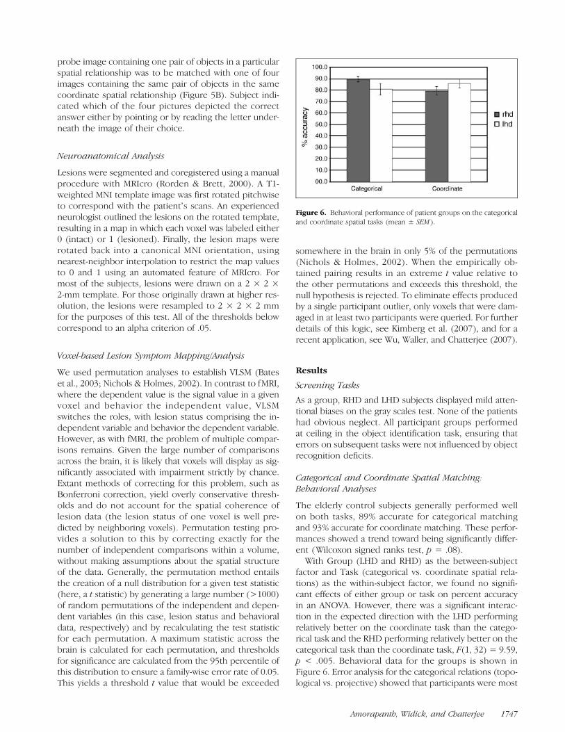
With Group (LHD and RHD) as the between-subjectfactor and Task (categorical vs. coordinate spatial rela-tions) as the within-subject factor, we found no signifi-cant effects of either group or task on percent accuracyin an ANOVA. However, there was a significant interac-tion in the expected direction with the LHD performingrelatively better on the coordinate task than the catego-rical task and the RHD performing relatively better on thecategorical task than the coordinate task, F(1, 32) = 9.59,p < .005. Behavioral data for the groups is shown inFigure 6. Error analysis for the categorical relations (topo-logical vs. projective) showed that participants were most
Figure 6. Behavioral performance of patient groups on the categoricaland coordinate spatial tasks (mean ± SEM ).
Amorapanth, Widick, and Chatterjee 1747
likely to make within-class errors (84.2% as comparedwith 45.8% if choosing randomly) than across-class errors( p < .001 using test of proportions).
The lack of group differences in performances of theRHD and LHD groups on the categorical or coordinatetasks does not necessarily disconfirm the hypothesis thatthe left hemisphere is biased toward categorical spatial
judgments and the right to coordinate spatial judgments.By design, greater behavioral variability within groups isdesirable for VLSM methods to identify specific brain be-havior correlations. This greater behavioral variabilitywithin each group maximizes the likelihood of findingstatistically robust differences within the group and mini-mizes the likelihood of finding differences across groups.
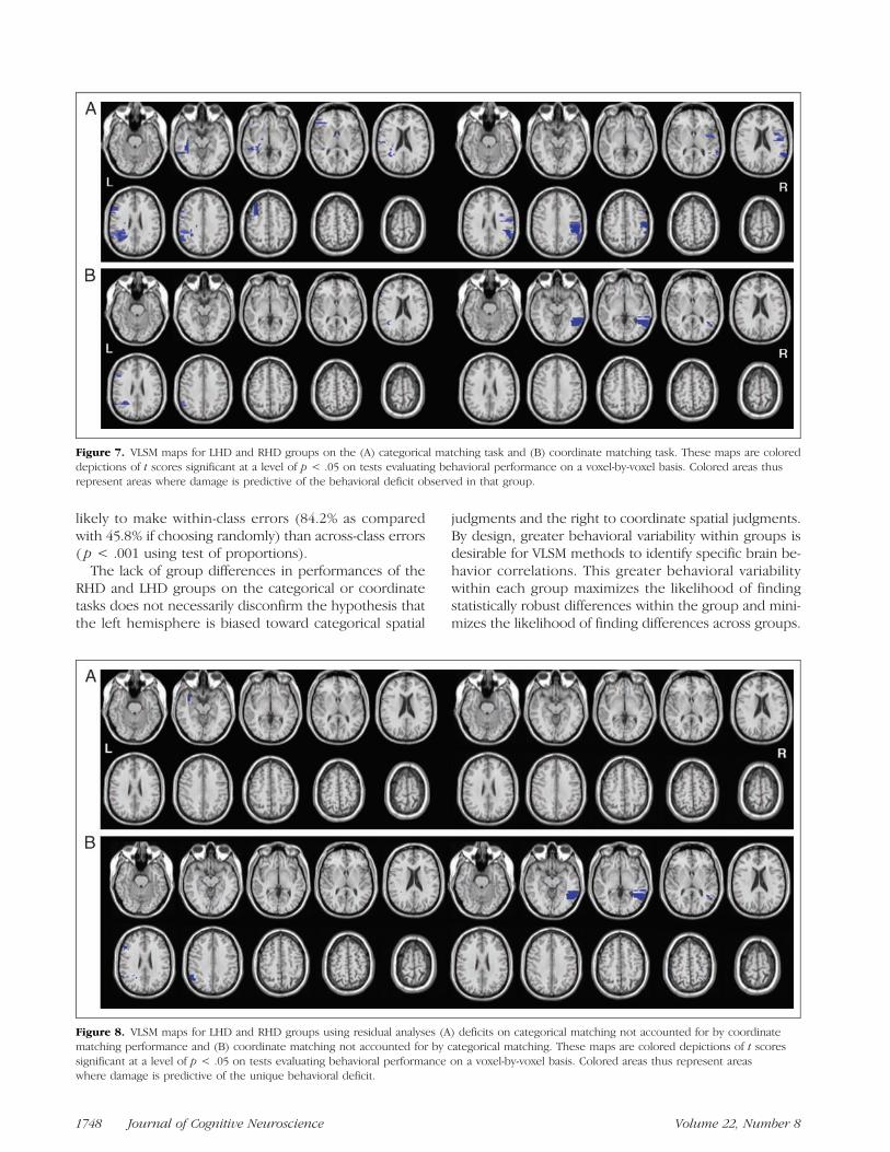
Figure 7. VLSM maps for LHD and RHD groups on the (A) categorical matching task and (B) coordinate matching task. These maps are coloreddepictions of t scores significant at a level of p < .05 on tests evaluating behavioral performance on a voxel-by-voxel basis. Colored areas thusrepresent areas where damage is predictive of the behavioral deficit observed in that group.
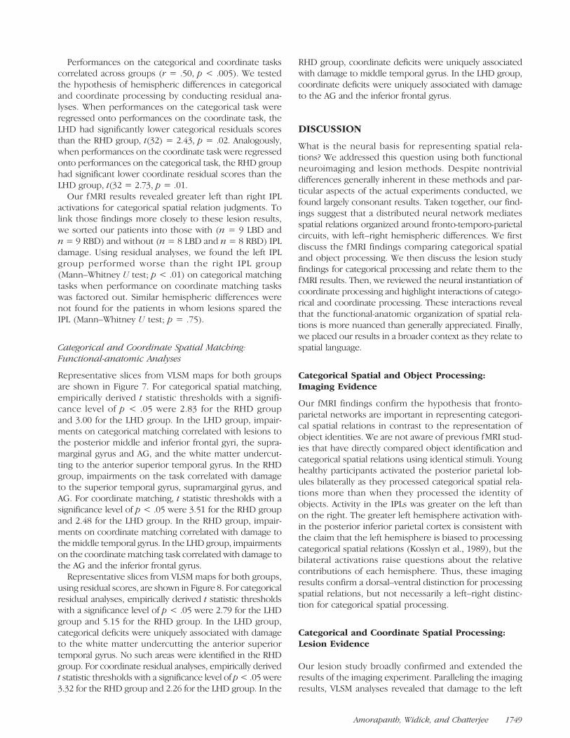
Figure 8. VLSM maps for LHD and RHD groups using residual analyses (A) deficits on categorical matching not accounted for by coordinatematching performance and (B) coordinate matching not accounted for by categorical matching. These maps are colored depictions of t scoressignificant at a level of p < .05 on tests evaluating behavioral performance on a voxel-by-voxel basis. Colored areas thus represent areaswhere damage is predictive of the unique behavioral deficit.
1748 Journal of Cognitive Neuroscience Volume 22, Number 8
Performances on the categorical and coordinate taskscorrelated across groups (r = .50, p < .005). We testedthe hypothesis of hemispheric differences in categoricaland coordinate processing by conducting residual ana-lyses. When performances on the categorical task wereregressed onto performances on the coordinate task, theLHD had significantly lower categorical residuals scoresthan the RHD group, t(32) = 2.43, p = .02. Analogously,when performances on the coordinate task were regressedonto performances on the categorical task, the RHD grouphad significant lower coordinate residual scores than theLHD group, t(32 = 2.73, p = .01.Our fMRI results revealed greater left than right IPL
activations for categorical spatial relation judgments. Tolink those findings more closely to these lesion results,we sorted our patients into those with (n = 9 LBD andn = 9 RBD) and without (n = 8 LBD and n = 8 RBD) IPLdamage. Using residual analyses, we found the left IPLgroup performed worse than the right IPL group(Mann–Whitney U test; p < .01) on categorical matchingtasks when performance on coordinate matching taskswas factored out. Similar hemispheric differences werenot found for the patients in whom lesions spared theIPL (Mann–Whitney U test; p = .75).
Categorical and Coordinate Spatial Matching:Functional-anatomic Analyses
Representative slices from VLSM maps for both groupsare shown in Figure 7. For categorical spatial matching,empirically derived t statistic thresholds with a signifi-cance level of p < .05 were 2.83 for the RHD groupand 3.00 for the LHD group. In the LHD group, impair-ments on categorical matching correlated with lesions tothe posterior middle and inferior frontal gyri, the supra-marginal gyrus and AG, and the white matter undercut-ting to the anterior superior temporal gyrus. In the RHDgroup, impairments on the task correlated with damageto the superior temporal gyrus, supramarginal gyrus, andAG. For coordinate matching, t statistic thresholds with asignificance level of p < .05 were 3.51 for the RHD groupand 2.48 for the LHD group. In the RHD group, impair-ments on coordinate matching correlated with damage tothemiddle temporal gyrus. In the LHD group, impairmentson the coordinate matching task correlated with damage tothe AG and the inferior frontal gyrus.Representative slices from VLSM maps for both groups,
using residual scores, are shown in Figure 8. For categoricalresidual analyses, empirically derived t statistic thresholdswith a significance level of p < .05 were 2.79 for the LHDgroup and 5.15 for the RHD group. In the LHD group,categorical deficits were uniquely associated with damageto the white matter undercutting the anterior superiortemporal gyrus. No such areas were identified in the RHDgroup. For coordinate residual analyses, empirically derivedt statistic thresholds with a significance level of p< .05 were3.32 for the RHD group and 2.26 for the LHD group. In the
RHD group, coordinate deficits were uniquely associatedwith damage to middle temporal gyrus. In the LHD group,coordinate deficits were uniquely associated with damageto the AG and the inferior frontal gyrus.
DISCUSSION
What is the neural basis for representing spatial rela-tions? We addressed this question using both functionalneuroimaging and lesion methods. Despite nontrivialdifferences generally inherent in these methods and par-ticular aspects of the actual experiments conducted, wefound largely consonant results. Taken together, our find-ings suggest that a distributed neural network mediatesspatial relations organized around fronto-temporo-parietalcircuits, with left–right hemispheric differences. We firstdiscuss the fMRI findings comparing categorical spatialand object processing. We then discuss the lesion studyfindings for categorical processing and relate them to thefMRI results. Then, we reviewed the neural instantiation ofcoordinate processing and highlight interactions of catego-rical and coordinate processing. These interactions revealthat the functional-anatomic organization of spatial rela-tions is more nuanced than generally appreciated. Finally,we placed our results in a broader context as they relate tospatial language.
Categorical Spatial and Object Processing:Imaging Evidence
Our fMRI findings confirm the hypothesis that fronto-parietal networks are important in representing categori-cal spatial relations in contrast to the representation ofobject identities. We are not aware of previous fMRI stud-ies that have directly compared object identification andcategorical spatial relations using identical stimuli. Younghealthy participants activated the posterior parietal lob-ules bilaterally as they processed categorical spatial rela-tions more than when they processed the identity ofobjects. Activity in the IPLs was greater on the left thanon the right. The greater left hemisphere activation with-in the posterior inferior parietal cortex is consistent withthe claim that the left hemisphere is biased to processingcategorical spatial relations (Kosslyn et al., 1989), but thebilateral activations raise questions about the relativecontributions of each hemisphere. Thus, these imagingresults confirm a dorsal–ventral distinction for processingspatial relations, but not necessarily a left–right distinc-tion for categorical spatial processing.
Categorical and Coordinate Spatial Processing:Lesion Evidence
Our lesion study broadly confirmed and extended theresults of the imaging experiment. Paralleling the imagingresults, VLSM analyses revealed that damage to the left
Amorapanth, Widick, and Chatterjee 1749

supramarginal gyrus and AG, the posterior middle and in-ferior frontal gyri, and the white matter undercutting thesuperior temporal gyrus was associated with deficits incategorical spatial judgments. Damage to the right supe-rior temporal gyrus, supramarginal gyrus, and AG pro-duced milder deficits in categorical spatial judgments.VLSM analyses also revealed that damage to the rightmiddle temporal gyrus produced deficits in coordinatespatial processing, whereas damage to the left AG and in-ferior frontal gyrus produced milder deficits in coordi-nate spatial processing. Thus, damage to the left AGand inferior frontal gyrus produced deficits in both coor-dinate and categorical processing.
In our brain-damaged participants, performances oncategorical and coordinate spatial tasks were correlated.Thus, some variance in performance on each of thesetasks could be accounted for by deficits on the comple-mentary task. To test the hypothesis that damage tosome brain regions contributes uniquely to categoricalor to coordinate deficits, we conducted residual analyses.Specifically, performances on categorical tasks were re-gressed on coordinate performances, and the derivedresiduals were subjected to analysis. Analogously, coordi-nate performances were regressed on categorical perfor-mances to derive residuals then subjected to analysis.
The LHD patients were more impaired than the RHDgroup on the categorical task beyond the variance in perfor-mances accounted for by coordinate deficits. Analogously,the RHD patients were more impaired than the LHD groupon the coordinate tasks beyond variance in performanceaccounted for by categorical deficits. These relative im-pairments cannot be explained by difficulty in recognizingstimuli, nor can they be explained by the hypothesis thatthe left hemisphere processes relatively easy spatial relationsand the right more difficult ones (Slotnick et al., 2001).
Can our lesion results be linked more directly to thefMRI data? The fMRI study revealed greater activity inthe left IPL than that in the right for categorical spatialprocessing than for object identity. If these relative acti-vations are causally related to categorical processing,then patients with left IPL damage should be worse thanpatients with right IPL damage on categorical tasks. Wetested this hypothesis by first sorting patients on the basisof whether they had IPL damage. Nine patients had leftIPL damage and nine had right IPL damage. Using residualanalyses, we found that patients with damaged left IPL hadgreater deficits than patients with damaged right IPL oncategorical spatial tasks. The same analysis with the otherpatients (n = 8 each) did not reveal similar differences.Thus, we have strong converging evidence for the impor-tance of the left IPL on categorical spatial processing.
Our VLSM results demonstrate that the functional neu-roanatomy of categorical and coordinate spatial relationsis more nuanced than that suggested by the straightfor-ward hypothesis that the left hemisphere processes catego-rical spatial information and the right processes coordinatespatial information. Damage to left posterior parietal and
dorsolateral prefrontal structures produced deficits in bothcategorical and coordinate processing. However, damagewithin the left AG and inferior frontal gyrus produced def-icits in coordinate processing (although these were milddeficits) not explained by categorical deficits. Similarly,damage to the white matter undercutting the left anteriorsuperior temporal gyrus uniquely produced deficits incategorical processing not accounted for by coordinate def-icits. Although RHD also produced deficits in both catego-rical and coordinate processing, no damaged areas wereuniquely associated with categorical deficits. By contrast,damage to the right middle temporal gyrus uniquely pro-duced coordinate spatial deficits.These patterns of lesion and behavioral correlations
suggest that categorical and coordinate spatial processingare quite intertwined. The right hemisphere processescoordinate spatial information preferentially within theposterior temporal cortex. By contrast, the left hemi-sphere processes both categorical and coordinate infor-mation conjointly within fronto-parietal circuits as wellas distinctly within the AG, inferior frontal gyrus for coor-dinate processing, and anterior superior temporal gyrusfor categorical processing.Our categorical spatial task used stimuli that depicted
both topological and projective spatial relations. Wenoted that when subjects erred on this task, they werelikely to choose within-class foils. That is, if the targetwas topological, they were more likely to choose a topo-logical foil and vice versa for projective spatial relations.Thus, despite categorical spatial deficits, some knowl-edge of the kind of categorical spatial relationships ap-pears to have been retained. These observations raisethe possibility that the neural underpinnings of topolog-ical and projective spatial relations might segregatefurther within networks instantiating categorical spatialrelations. Future studies designed and powered to testthis hypothesis will be needed.
Categorical Spatial Processing and Language
A major motivation for anticipating that the left hemi-sphere is biased to processing categorical spatial rela-tions is its relationship to language (Chatterjee, 2008;Kemmerer, 2006). Prepositions comprise only a small setof the infinite possible spatial relations. As Landau andJackendoff (1993) and Talmy (1983) pointed out someyears ago, prepositions discard much of the geometricrichness of objects in favor of relatively coarse spatialnotions like “containment” or “contact.” They suggestedthat any aspect of space that could be expressed in lan-guage must have nonlinguistic spatial representations.Similarly, Regier (1995) used computational models toargue that spatial language is grounded in spatial per-ception. Thus, one might infer that categorical spatialrelations, which are limited in number and in which de-tails of object features are reduced to general shapesand axes, would serve as the perceptual counterpart to
1750 Journal of Cognitive Neuroscience Volume 22, Number 8
locative prepositions. At an anatomic level, one would ex-pect the processing of these discrete spatial representa-tions linked to language, to be instantiated in the lefthemisphere of right-handed individuals, close to or over-lapping with the processing of spatial prepositions.As an aside, we recently reported a fronto-parietal
network involved in processing paths of movement(Wu, Morganti, & Chatterjee, 2008). This finding is rele-vant to the current study because path of motion in Eng-lish is described using prepositional phrases (e.g., runningthrough the fields, jumping across the puddle). Thus, theperceptual counterparts of locative prepositions, whetherdynamic or static, may be instantiated in similar fronto-parietal networks.Few studies have examined the neural bases of loca-
tive prepositions directly. Using PET imaging, Emmoreyet al. (2002) and Damasio et al. (2001) found that namingspatial locations was associated with the left supramar-ginal gyrus and the inferior prefrontal cortical activations.Noordzij, Neggers, Ramsey, and Postma (2008) also foundleft supramarginal gyrus activity in subjects who wereasked to process terms denoting left and right, but whetherleft–right judgments generalize broadly to other categor-ical relations is not known. Limited electrophysiologicdata are consistent with the view that the parietal corti-ces instantiate the use of spatial terms (Carlson, Robert,Taylor, & Herndon, 2002). Occasional case studies of def-icits with prepositions in aphasics have been reported, butthese have not been informative about their neural under-pinnings (Miozzo, Simon, & Postman, 2008; Chatterjee &Maher, 2000; Grodzinsky, 1988; Frederici, 1982; Schwartz,Saffran, & Marin, 1980). An important exception in this re-gard is a study by Tranel and Kemmerer (2004), which di-rectly examined the neural correlates of deficits in locativeprepositions in a large group of brain-damaged individ-uals. They found the greatest overlap of lesions in patientswith such deficits within the left supramarginal gyrus andsubjacent white matter as well as the frontal opercula. Wealso found that damage to the posterior temporo-parietalcortex, anterior superior temporal gyrus, and inferior pFCwas most closely associated with deficits in matching loc-ative sentences to pictures depicting categorical spatial re-lations (Wu et al., 2007). The emerging view from thesestudies is that left peri-sylvian cortices involving the supra-marginal gyrus, the anterior superior temporal gyrus, andthe inferior prefrontal regions mediate locative preposi-tions. Areas mediating spatial categorical relations overlapor are closely aligned with these regions, consistent withthe view that these regions serve as perceptual points ofentry for their lexicalizations (Chatterjee, 2008; Wu et al.,2007).In summary, we found converging evidence from both
imaging and lesion investigations of the neural basis forspatial relations. A bilateral network comprising the pos-terior parietal, posterior middle and superior temporal,and posterior middle and inferior frontal gyri mediatesspatial relations. The left hemisphere is biased toward
processing categorical spatial relations and the right towardprocessing coordinate spatial relations, but these distinc-tions are relative rather than absolute. The left hemisphereappears to mediate both categorical and coordinate pro-cessing, whereas we did not find evidence that the righthemisphere processes categorical relations independentof its coordinate processing.
Acknowledgments
This work was supported by NIH RO1 HD050199 and RO1DC008779 and a subcontract under NSF SBE0541957. Theauthors thank Eileen Cardillo, Alexander Kranjec, and GwendaSchmidt for helpful feedback on an earlier draft of this manu-script. They also appreciate very helpful input from DavidKemmerer and a second anonymous reviewer.
Reprint requests should be sent to Anjan Chatterjee, Depart-ment of Neurology, The Hospital of The University of Pennsyl-vania, 3 West Gates, 3400 Spruce Street, Philadelphia, PA 19104,or via e-mail: [email protected].
REFERENCES
Aguirre, G., & DʼEsposito, M. (1997). Environmental knowledgeis subserved by separable dorsal/ventral neural areas. Journalof Neuroscience, 17, 2512–2518.
Aguirre, G., Zarahn, E., & DʼEsposito, M. (1998). The variabilityof human BOLD hemodynamic response. Neuroimage, 8,360–369.
Aguirre, G. K., Zarahn, E., & DʼEsposito, M. (1997). Empiricalanalysis of BOLD fMRI statistics: II. Spatially smoothed datacollected under the null-hypothesis and experimentalconditions. Neuroimage, 5, 199–212.
Baciu, M., Olivier, K., Vernier, M.-P., Bedoin, N., Runin, C., &Segebarth, C. (1999). Catergorical and coordinate spatialrelations: fMRI evidence for hemispheric specialization.NeuroReport, 10, 1373–1378.
Bates, E., Wilson, S., Saygin, A., Dick, F., Sereno, M., Knight, R.,et al. (2003). Voxel-based lesion symptom mapping. NatureNeuroscience, 6, 448–450.
Binder, J., Frost, J., Hammeke, T., Cox, R., Rao, S., & Prieto, T.(1997). Human language areas identified by functionalmagnetic resonance imaging. Journal of Neuroscience,17, 353–362.
Borgo, F., & Shallice, T. (2001). When living things and other“sensory quality” categories behave in the same fashion: Anovel category specificity effect. Neurocase, 7, 201–220.
Capitani, E., Laiacona, M., Barbarotto, R., & Trivelli, C. (1994).Living and non-living categories. Is there a “normal”asymmetry? Neuropsychologia, 32, 1453–1463.
Caramazza, A., & Shelton, J. (1998). Domain-specific knowledgesystems in the brain: The animate-inanimate distinction.Journal of Cognitive Neuroscience, 10, 1–34.
Carlson, L., Robert, W., Taylor, H., & Herndon, R. (2002). Neuralcorrelates of spatial term use. Journal of ExperimentalPsychology: Human Perception and Performance, 28,1391–1407.
Chatterjee, A. (1995). Unilateral spatial neglect: Assessment andrehabilitation strategies. NeuroRehabilitation, 5, 115–128.
Chatterjee, A. (2003). Neglect. A disorder of spatial attention. InM. DʼEsposito (Ed.), Neurological foundations of cognitiveneuroscience (pp. 1–26). Cambridge, MA: MIT Press.
Chatterjee, A. (2005). A madness to the methods in cognitiveneuroscience? Journal of Cognitive Neuroscience, 17,847–849.
Amorapanth, Widick, and Chatterjee 1751
Chatterjee, A. (2008). The neural organization of spatial thoughtand language. Seminars in Speech and Language, 29,226–238.
Chatterjee, A., & Maher, L. (2000). Grammar and agrammatism.In L. Gonzalez Rothi, B. Crosson, & S. Nadeau (Eds.), Aphasiaand language: Theory to practice (pp. 133–156). New York:Guilford Publications.
Corbetta, M., Shulman, G. L., Miezin, F. M., & Peterson, S. E.(1995). Superior parietal cortex activation during spatialattention shifts and visual feature conjunction. Science,270, 802–805.
Damasio, A. R., Damasio, H., Tranel, D., & Brandt, J. P. (1990).Neural regionalization of knowledge access: Preliminaryevidence. Cold Springs Harbor Symposia on QuantitativeBiology, LV, 1039–1047.
Damasio,H., Grabowski, T. J., Tranel, D., Ponto, L. L., Hichwa, R.D.,& Damasio, A. R. (2001). Neural correlates of naming actionsand of naming spatial relations. Neuroimage, 13, 1053–1064.
Efron, R. (1990). The decline and fall of hemisphericspecialization. Hillsdale, NJ: Lawrence Earlbaum Associates.
Emmorey, K., Damasio, H., McCullough, S., Grabowski, T.,Ponto, L. L., Hichwa, R. D., et al. (2002). Neural systemsunderlying spatial language in American Sign Language.Neuroimage, 17, 812–824.
Epstein, R., & Kanwisher, N. (1998). A cortical representation ofthe local visual environment. Nature, 392, 598–601.
Farah, M. J. (1990). Visual agnosia. Cambridge, MA: MIT Press.Fellows, L. K., Heberlein, A. S., Morales, D. A., Shivde, G.,
Waller, S., & Wu, D. H. (2005). Method matters: An empiricalstudy of impact in cognitive neuroscience. Journal ofCognitive Neuroscience, 17, 850–858.
Fellows, L. K., Stark, M., Berg, A., & Chatterjee, A. (2008).Patient registries in cognitive neuroscience research:Advantages, challenges, and practical advice. Journal ofCognitive Neuroscience, 20, 1107–1113.
Ferrera, V. P., Rudolph, K. K., & Maunsell, J. H. R. (1994).Responses of neurons in the parietal and temporal visualpathways during a motion task. Journal of Neuroscience,14, 6171–6186.
Frederici, A. (1982). Syntactic and semantic processing inaphasic deficits: The availability of prepositions. Brainand Language, 15, 249–258.
Gonnerman, L. M., Andersen, E. S., Devlin, J. T., Kempler, D., &Seidenberg, M. S. (1997). Double dissociation of semanticcategories in Alzheimerʼs disease. Brain & Language, 57,254–279.
Gottfried, J. A., Sancar, F., & Chatterjee, A. (2003). Acquiredmirror writing and reading: Evidence for reflected graphemicrepresentations. Neuropsychologia, 41, 96–107.
Grodzinsky, Y. (1988). Syntactic representations in agrammaticaphasia: The case of prepositions. Language and Speech,31, 115–134.
Hannay, H. (1976). Visual localization in patients with unilateralbrain disease. Journal of Neurology, Neurosurgery, andPsychiatry, 39, 307–313.
Haxby, J. V., Grady, C. L., Horwitz, B., Ungerleider, L. G.,Mishkin, M., Carson, R. E., et al. (1991). Dissociation ofobject and spatial visual processing pathways in humanextrastriate cortex. Proceedings of the NationalAcademy of Sciences, U.S.A., 88, 1621–1625.
Jager, G., & Postma, A. (2003). On the hemispheric specializationfor categorical and coordinate spatial relations: A review ofthe current evidence. Neuropsychologia, 41, 504–515.
Kable, J. K., Lease-Spellmeyer, J., & Chatterjee, A. (2002). Neuralsubstrates of action event knowledge. Journal of CognitiveNeuroscience, 14, 795–804.
Kable, J. W., Kan, I., Wilson, A., Thompson-Schill, S., &Chatterjee, A. (2005). Conceptual representations of
action in lateral temporal cortex. Journal of CognitiveNeuroscience, 17, 1855–1870.
Kanwisher, N., McDermott, J., & Chun, M. (1997). Thefusiform face area: A module in human extrastriate cortexspecialized for perception of faces. Journal of Neuroscience,17, 4302–4311.
Kanwisher, N., Woods, R. P., Iacoboni, M., & Mazziotta, J. C.(1997). A locus in human extrastriate cortex for visual shapeanalysis. Journal of Cognitive Neuroscience, 9, 133–142.
Kemmerer, D. (2006). The semantics of space: Integratinglinguistic typology and cognitive neuroscience.Neuropsychologia, 44, 1607–1621.
Kimberg, D. Y., Coslett, H. B., & Schwartz, M. F. (2007). Powerin voxel-based lesion-symptom mapping. Journal ofCognitive Neuroscience, 19, 1067–1080.
Kosslyn, S., Koenig, O., Barrett, C., Cave, C., Tang, J., &Gabrielli, J. (1989). Evidence for two types of spatialrepresentations: Hemispheric specialization forcategorical and coordinate relations. Journal ofExperimental Psychology: Human Perception andPerformance, 15, 723–735.
Kosslyn, S., Thompson, W., Gitelman, D., & Alpert, N. (1998).Neural systems that encode categorical versus coordinatespatial relations: PET investigations. Psychobiology,26, 333–347.
Kosslyn, S. M., & Ochsner, K. N. (1994). In search of occipitalactivation during visual mental imagery. Trends inNeurosciences, 17, 290–292.
Laeng, B. (1994). Lateralization of categorical and coordinatespatial function: A study of unilateral stroke patients. Journalof Cognitive Neuroscience, 6, 189–203.
Laeng, B. (2006). Constructional apraxia after left or rightunilateral stroke. Neuropsychologia, 44, 1595–1606.
Landau, B., & Jackendoff, R. (1993). “What” and “where” inspatial language and spatial cognition. Behavioral andBrain Sciences, 16, 217–265.
Malach, R., Reppas, J., Benson, R., Kwong, K., Jiang, H.,Kennedy, W., et al. (1995). Object related activity revealed byfunctional magnetic resonance imaging in human occipitalcortex. Proceedings of the National Academy of Sciences,U.S.A., 92, 8135–8139.
Martin, A., Ungerleider, L., & Haxby, J. (2000). The sensory/motor model of semantic representation of objects. InM. Gazzaniga (Ed.), The new cognitive neurosciences(2nd ed., pp. 1023–1036). Cambridge, MA: MIT Press.
Mattingley, J., Berberovic, N., Corben, L., Slavin, M., Nicholls,M., & Bradshaw, J. (2004). The greyscales task: A perceptualmeasure of attentional bias following unilateral hemisphericdamage. Neuropsychologia, 42, 387–394.
Mayer, E., Martory, M.-D., Pegna, A. J., Landis, T., Delavelle, J., &Annoni, J.-M. (1999). A pure case of Gerstmann syndromewith a subangular lesion. Brain, 122, 1107–1120.
Miozzo, M., Simon, F.-B., & Postman, J. (2008). Knowing wherebut not what: Impaired thematic roles and spatial language.Cognitive Neuropsychology, 25, 853–873.
Moore, C. J., & Price, C. J. (1999). A functional neuroimagingstudy of the variables that generate category-specific objectprocessing differences. Brain, 122, 943–962.
Nichols, T., & Holmes, A. (2002). Nonparametric tests forfunctional neuroimaging: A primer with examples. HumanBrain Mapping, 15, 1–25.
Noordzij, M. L., Neggers, S. F. W., Ramsey, N. F., & Postma, A.(2008). Neural correlates of locative prepositions.Neuropsychologia, 46, 1576–1580.
Postle, B. R., Zarahn, E., & DʼEsposito, M. (2000). Usingevent-related fMRI to assess delay-period activity duringperformance of spatial and nonspatial working memory tasks.Brain Research Protocols, 5, 57–66.
1752 Journal of Cognitive Neuroscience Volume 22, Number 8
Postma, A., & Laeng, B. (2006). New insights in categorical andcoordinate processing of spatial relations. Neuropsychologia,44, 1515–1518.
Regier, T. (1995). A model of human capacity for categorizingspatial relations. Cognitive Linguistics, 6, 63–88.
Rorden, C., & Brett, M. (2000). Stereotaxic display of brainlesions. Behavioral Neurology, 12, 191–200.
Rorden, C., & Karnath, H.-O. (2004). Using human brain lesionsto infer function: A relic from a past era in the fMRI age?Nature Reviews Neuroscience, 5, 813–819.
Schiller, P. H. (1996). On the specificity of neurons and visualareas. Behavioural Brain Research, 76, 21–35.
Schwartz, M. F., Saffran, E. M., & Marin, O. S. M. (1980). Theword order problem in agrammatism: I. Comprehension.Brain and Language, 10, 249–262.
Sergent, J. (1991). Judgments of relative position and distanceon representations of spatial relations. Journal ofExperimental Psychology: Human Perception andPerformance, 17, 762–789.
Slobin, D. (1996). From “thought and language” to “thinking forspeaking.” In J. Gumperz & S. Levinsohn (Eds.), Rethinkinglinguistic relativity (pp. 70–96). New York: CambridgeUniversity Press.
Slobin, D. (2000). Verbalized events. In S. Niemeier & R. Dirven(Eds.), Evidence for linguistic relativity (pp. 107–138).Amsterdam: John Benjamins Publishing Company.
Slotnick, S. D., Moo, L. R., Tesoro, M. A., & Hart, J. (2001).Hemispheric asymmetry in categorical versus coordinatevisuospatial processing revealed by temporary corticaldeactivation. Journal of Cognitive Neuroscience, 13,1088–1096.
Talmy, L. (1983). How language structures space. In H. Pick &L. Acredolo (Eds.), Spatial orientation: Theory, research andapplication. New York: Plenum Press.
Tranel, D., & Damasio, A. R. (1999). The neurobiology ofknowledge retrieval. Behavioral and Brain Sciences,22, 303.
Tranel, D., & Kemmerer, D. (2004). Neuroanatomical correlatesof locative prepositions. Cognitive Neuropsychology, 21,719–749.
Tranel, D., Logan, C. G., Frank, R. J., & Damasio, A. R. (1997).Explaining category-related effects in the retrieval ofconceptual and lexical knowledge for concrete entities:Operationalization and analysis of factors. Neuropsychologia,35, 1329–1339.
Tzourio-Mazoyer, N., Landeau, B., Papathanassiou, D., Crivello,F., Etard, O., Delcroix, N., et al. (2002). Automatedanatomical labeling of activations in SPM using amacroscopic anatomical parcellation of the MNI MRIsingle-subject brain. Neuroimage, 15, 273–289.
Ungerleider, L. G., & Mishkin, M. (1982). Two cortical visualsystems. In D. J. Ingle, M. A. Goodale, & R. J. W. Mansfield(Eds.), Analysis of visual behavior (pp. 549–586).Cambridge, MA: MIT Press.
Vandenberghe, R., Price, C., Wise, R., Josephs, O., &Frackowiak, R. (1996). Functional anatomy of a commonsemantic system for words and pictures. Nature, 383, 254–256.
Warrington, E. K., & Taylor, A. M. (1973). The contribution ofthe right parietal lobe to object recognition. Cortex, 9, 152–164.
Wu, D. H., Morganti, A., & Chatterjee, A. (2008). Neuralsubstrates of processing path and manner information of amoving event. Neuropsychologia, 46, 704–713.
Wu, D. H., Waller, S., & Chatterjee, A. (2007). The functionalneuroanatomy of thematic role and locative relationalknowledge. Journal of Cognitive Neuroscience, 19,1542–1555.
Zarahn, E., Aguirre, G., & DʼEsposito, M. (1997). A trial-basedexperimental design for fMRI. Neuroimage, 6, 122–138.
Amorapanth, Widick, and Chatterjee 1753
Related Documents