THE NERVOUS SYSTEM BASIC DIVISION, STRUCTURE AND FUNCTIONS

Welcome message from author
This document is posted to help you gain knowledge. Please leave a comment to let me know what you think about it! Share it to your friends and learn new things together.
Transcript
THE NERVOUS SYSTEM
BASIC DIVISION, STRUCTURE AND FUNCTIONS

-

-

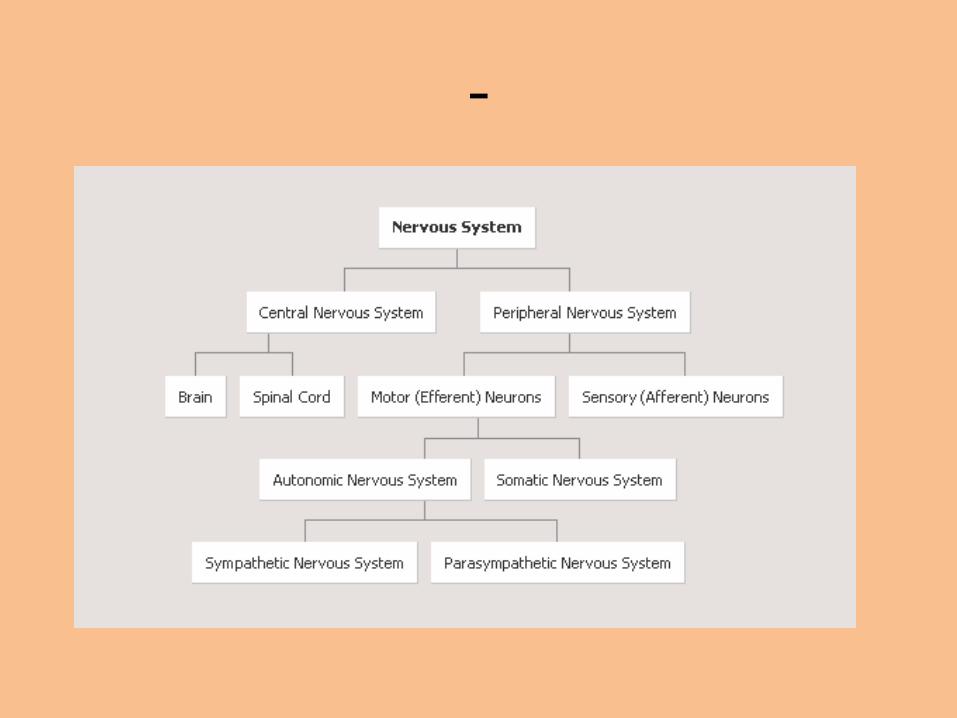
Division - Illustration

-

DIVISION
• The nervous system is the most important control system in the human body. The other system involved in organ control is the endocrine system, but whereas this uses hormones as messengers, the nervous system uses electrical stimuli which travel a great deal faster. Nerves provide the wiring through which electrical impulses are received from and sent to virtually all parts of the body.
• The nervous system consists of the central nervous system (CNS) and the peripheral nervous system (PNS).
-

THE CENTRAL NERVOUS SYSTEM
• The central nervous system consists of the brain and the spinal cord. It is in the brain that the higher senses, both cognitive and emotional, are found. It is also responsible for producing sensations and controlling movements. The brain acts as a computer, integrating all incoming information, selecting an appropriate response, then instructing the involved body parts to take appropriate action. Thus the nervous system forms a vital link, allowing communication and coordination of interaction between the various tissues in the body as well as with the outside world.
THE PERIPHERAL NERVOUS SYSTEM
• The peripheral nervous system consists of all the nervous tissue outside the central nervous system: the peripheral nerves that inervate muscles and organs.
• Nervous tissue consists of an intricate , interconnected network of specialized cells called neurons, which are enclosed within a supportive tissue that has the same function in the nervous system that connective tissue fulfils everywhere. The characteristic support cells of the brain are known generally as glia and include astrocytes and oligodendrocytes.

-

NEURONS
• The functional and structural unit of the nervous system is the neuron. Although neurons vary widely in form and size they all share the same basic structure, consisting of:
1) The cell body, or soma (contains the nucleus surrounded by cytplasm)
2) The dendrites (numerous multi-branched extensions or processes that make contact with other neurons – convey impulses to the cell body).

-

-
3) The axon (Usually a long single process which acts as the neuron`s transmitter, conducting impulses away from the cell body). Near its end, an axon splits into numerous branches, which are called axon terminals or terminal fibrils. The tips of these terminals are dilated into tiny bulbs known as the synaptic knobs. These knobs house numerous vesicles (sacs) filled with chemicals, known as neurotransmitters, that are used for communication between a neuron and another cell.

-

-
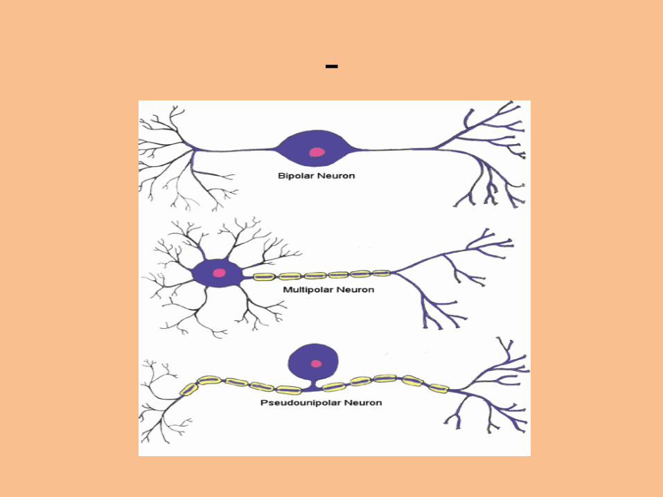
There are three types of neurons: a) Unipolar (a single axon divides into two
branches) b) Bipolar (two axons originate from
differentt points of the neuron cell body) c) Multipolar (an axon and many dendrites
originate from the cell body)

-
-

THE NERVE IMPULSE(how it is generated and how it travels through a neuron)
• A nerve impulse – an electrical charge – is the signal that passes from one neuron to the next and finally to an end organ.
Resting Membrane Potential: The cell membrane of a neuron at rest has a
negative electrical potential of about -70mV. That means that the electrical charges found inside the cell and the charges found outside the cell differ by 70mV, and that the inside is more negative relative to the outside.

-
-
• This potential difference is known as the resting membrane potential, or RMP. It is caused by a separation of charges across the membrane. When the charges along the membrane differ, the membrane is polarized. This polarization is due to a high concentration of potassium ions (K+) on the inside and a high concentration of sodium ions (Na+) on the outside because the sodium-potassium pump actively moves sodium out of the cell and potassium into it.

-
Depolarization and Hyperpolarization If the middle of the cell becoms less negative relative
to the ouside, the potential difference across the membrane will decrease. The membrane is now depolarized. This depolarization occurs anytime the change difference becomes less than the RMP of -70mV, moving closer to zero. The opposite can also occur. If the charge difference across the membrane increases, then the membrane becomes more polarized. This is known as hyperpolarization.

-
• Graded potentials Graded potentials are localized changes in the
membrane potential. They can be either depolarizations or hyperporizations. Graded potentials are triggered by a change in the neuron`s local environment. A graded potential is usually just a local event, and the depolarization does not spread very far along the neuron. To travel the full distance, an impulse must generate an action potential.

-
Action Potentials An action potential is a rapid and substantial
depolarization of the neuron`s membrane. It usually lasts only about 1 ms. Typically, the membrane potential changes from RMP of -70 mV to a value of 30 mV, then rapidly returns to its resting value.

-
-
Threshold and the All-Or-None Principle All action potentials begin as graded potentials. When
enough stimulation occurs to cause a depolarization of at least 15 to 20 mV, an action potential results. That means if the membrane depolarizes from the RMP of -70 mV to a value of -50 to -55 mV, the cell will experience an action potential. The minimum depolarization required to produce an action potential is called the threshold.
Repolarization is when the neuron returns to its normal resting state

-

-
Propagation of the Action Potential The Myelin Sheath The axons of most neurons are myelinated, covered with a sheath
formed by myelin, a fatty substance that insulates the cell membrane. In the peripheral nervous system, this sheath is formed by Schwann cells.
The sheath is not continuous. It exibits gaps between adjacent Schwann cells, leaving the axon uninsulated at those points. These gaps are referred to as nodes of Ranvier. The action potential appears to jump from one node to the next as it traverses the myelinated fiber. This is referred to as saltatory conduction, a much faster rate of conduction than in unmyelinated fibers

-

-
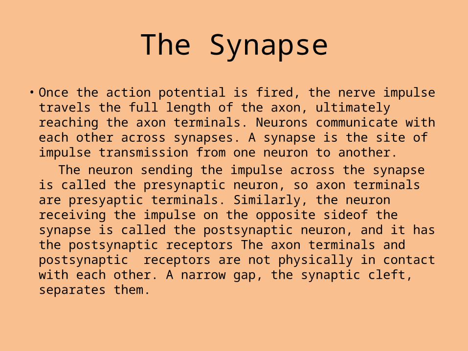
The Synapse
• Once the action potential is fired, the nerve impulse travels the full length of the axon, ultimately reaching the axon terminals. Neurons communicate with each other across synapses. A synapse is the site of impulse transmission from one neuron to another.
The neuron sending the impulse across the synapse is called the presynaptic neuron, so axon terminals are presyaptic terminals. Similarly, the neuron receiving the impulse on the opposite sideof the synapse is called the postsynaptic neuron, and it has the postsynaptic receptors The axon terminals and postsynaptic receptors are not physically in contact with each other. A narrow gap, the synaptic cleft, separates them.

-

-
• A nerve impulse can be transmitted across a synapse only in one direction: from the axon terminals of the presynaptic neuron to the postsynaptic receptors usually on the dendrites of the postsynaptic neuron.
• The presynaptic terminals of the axon contain a large number of sac-like structures, called synaptic vesicles. These sacs contain neurotransmitter chemicals. When the impulse reaches the presynaptic terminals, the synaptic vesicles respond by dumping their chemicals into the synaptic cleft. The neurotransmitters then diffuse across the synaptic cleft to the postsynaptic neurons receptors. The postsynaptic receptors bind the neurotransmitter once it diffuses across the synaptic cleft.

-
• When this binding occurs, the impulse has been transmitted successfully to the next neuron and can be transmitted onward. More than 40 neurotransmitters have been identified. Acetylcholine and norepinephrine are the two major neurotransmitters. Neurotransmitters can have either excitatory or inhibitory effects, or both.
• Once the neurotransmitter binds to the postsynaptic receptor, the nerve impulse has been successfully transmitted. The neurotransmitter is then either destroyed by enzymes or actively transported back into the presynaptic terminals for reuse when the next impulse arrives.

-

THE NERVES
• The nerves are responsible for conveying nerve stimuli in the peripheral nervous system. They form bundles and some are long, extending from the spinal cord to the tip of a finger or toe.
• There are two types of nerves, defined according to the function: the somatic nerves, which are involved in volunatary functions and are the type that stimulate the muscles to produce movement; and the autonomic nerves, which control involuntary functions such as the functioning of the different organs.
• One of the most important nerves of the autonomic nervous system is the vagus nerve, which controls many vital functions such as heart rate, digestion, and breathing.

Structures of the Nerves• Nerves are structures of different thickness and length. The cell
bodies of the neuronal axons that form the nerves are situated in the central nervous system or in the collections of cell bodies (ganglia) that lie next to the spinal cord.
• Each nerve is formed by one or more bundles of nerve fibers. Each individual nerve fiber is the axon of a neuron which is covered by the cytoplasm of a supporting cell known as a Schwann cell. Large-diameter fibers are covered by several concentric layers of Schwann cells, which form a sheath of myelin.
• Each bundle of nerve fibers is surrounded by a layer of connective tissue called the perineurium; if the nerve contains many bundles, these are surrounded by another layer known as the epineurium.

-

-

THE CENTRAL NERVOUS SYSTEM
The central nervous system is made up of the brain and spinal cord. The brain functions to receive nerve impulses from the spinal cord and cranial nerves. The spinal cord contains the nerves that carry messages between the brain and the body

The Brain
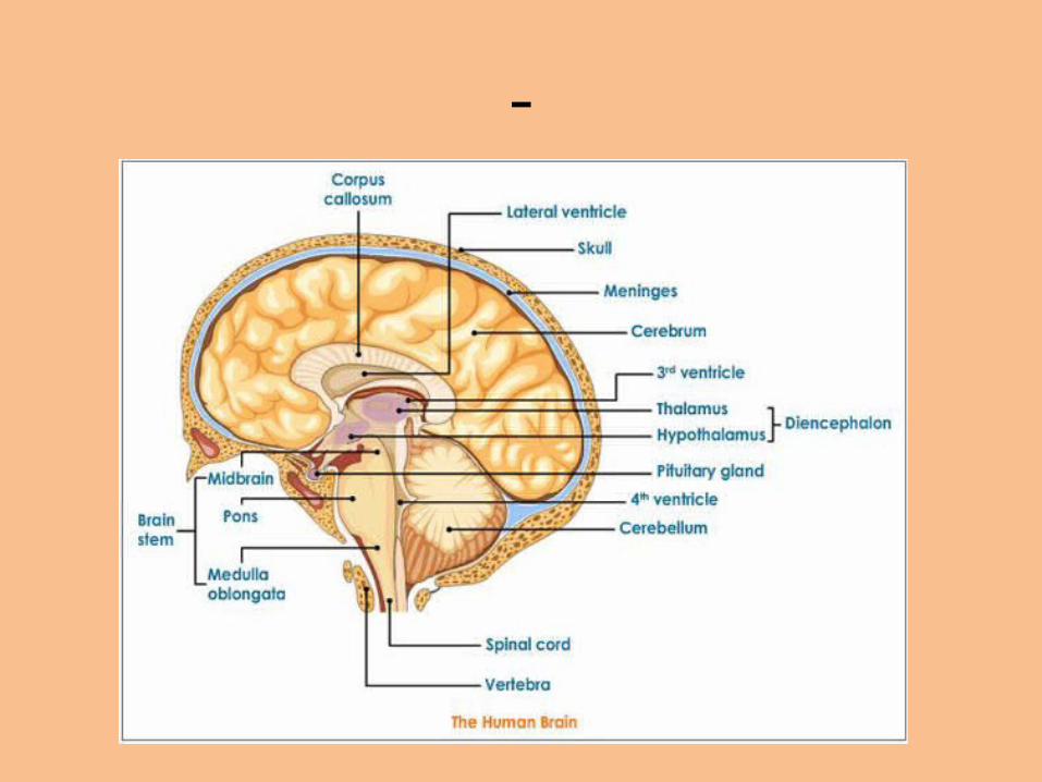
The brain is composed of four major parts: 1. The Cerebrum 2. The Diencephalon 3. The Cerebellum 4. The Brain Stem

Male –Female Brain 1
Male –Female Brain 2

Male Brain

Male –Female Brain 3

-
-

The Cerebrum
• The cerebrum is composed of the right and left cerebral hemispheres. These are connected to each other by fiber bundles referred to as the corpus callosum alowing the two hemispheres to communicate with each other. The cerebral cortex forms the outer portion of the cerebral hemispheres and has been referred to as the site of the mind and intellect. It is also called the gray matter, which simply reflects its distinctive color resulting from lack of myelin on the cell bodies located in this area. The cerebral cortex is our conscious brain. It allows us to think, to be aware of sensory stimuli, and to voluntarily control our movements.

-

-

-

-
• The cerebrum consists of five lobes – four outer lobes and the central insula. The four outer lobes are named after the bones that lie directly above them.
• Numerous folds, or convolutions, called gyri are found in the cerebrum surface. These are separated by furrows called fissures or sulci.
• The remainder of the cerebrum is composed primarily of white matter (myelinated axons)

-

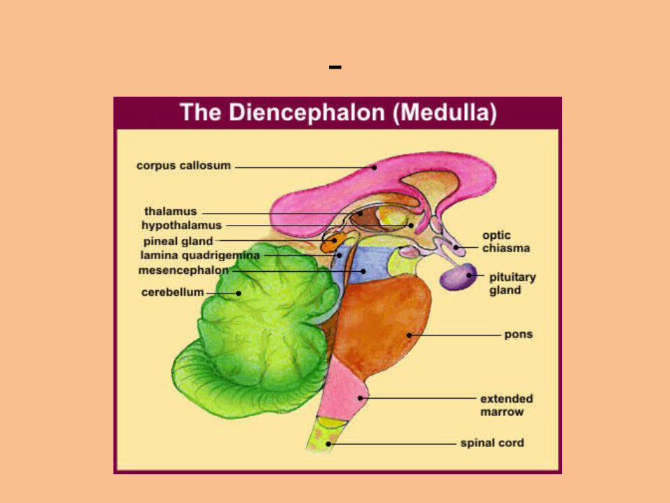
The Diencephalon
• This region of the brain is mostly composed of the thalamus and hypothalamus. The thalamus is an important sensory integration center. All sensory input (except smell) enters the thalamus and is relayed to the appropriate area of the cortex. The thalamus regulates what sensory input reaches our conscious brain, and thus is very important for motor control.
• The hypothalamus, directly below the thalamus, is responsible for maintaining homeostasis by regulating almost all processes that affect the body`s internal environment.
-
The Cerebellum
• The cerebelum is located behind the brain stem. It is connected to numerous parts of the brain and has a crucial role in controlling movement. When the cerebrum initiates muscular movement, the cerebellum coordinates and refines the movement. The cerebellum also maintains the equilibrium and balance of the body

-

The Brain Stem
• The brain stem composed of the middle brain (mesencephalon), the pons, and the medulla oblongata, is the stalk of the brain, connecting the brain and the spinal cord. All sensory and motor nerves pass through the brain stem as they relay information between the brain and the spinal cord. This is site of origin for 10 of the 12 pairs of cranial nerves. The brain stem also contains the major autonomic regulatory centers that exert control over respiratory and cardiovascular systems.


THE SPINAL CORD
• The lowest part of the brain stem, the medulla oblongata, is continuous below with the spinal cord. The spinal cord is composed of tracts of nerve fibers that allow two-way conduction of nerve impulses. The sensory (afferent) fibers carry neural signals from sensory receptors, such as those in the muscles and joints, to the upper levels of the CNS. Motor (efferent) fibers from the brain and upper spinal cord travel down to the organs.

-

Meninges
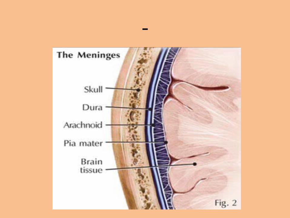
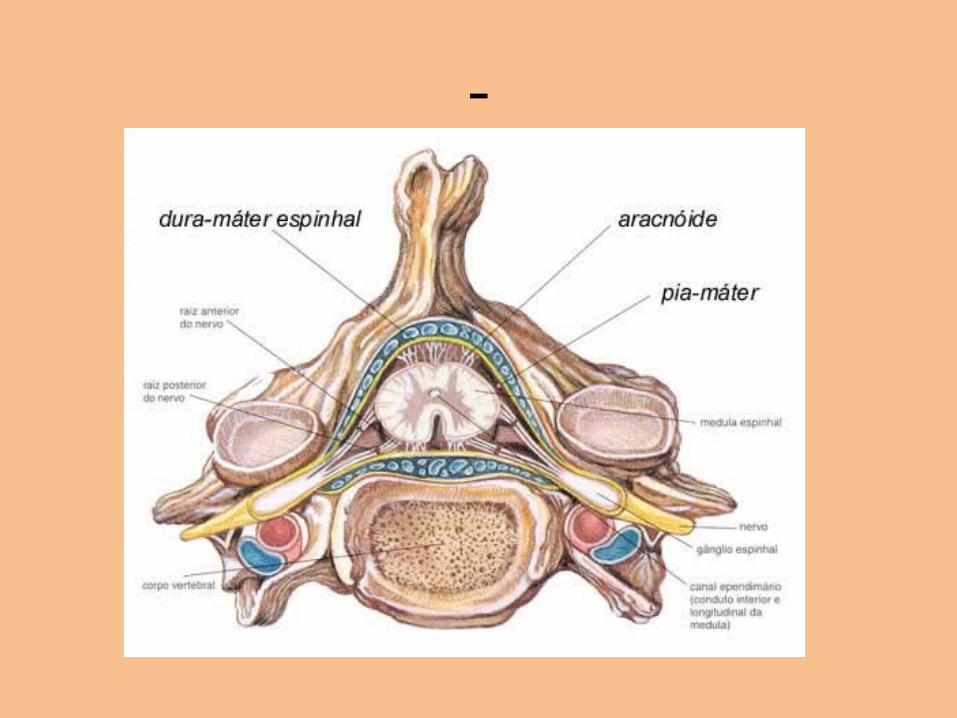
• Both the brain and the spinal cord are protected against injury by bones. The brain is enclosed within the skull and the spinal cord is enclosed within the vertebral column. In addition, both the brain and the spinal cord receive limited protection from a set of three coverings called meninges. The outermost coat, the dura mater, is tough and fibrous. Immediately beneath the dura mater is a cavity called the subdural space. It is filled with serous fluid. The next layer of the meninges is the arachnoid.
-
-
• A subarachnoid space filled with cerebrospinal fluid, provides additional protection for the brain and spinal cord by acting as a shock absorber. Finally, the innermost layer, the pia mater, contains numerous blood vessels and lymphatics, which provide nourishment for the underlying tissues. Cerebral fluid circulates around the spinal cord and the brain and through spaces called ventricles. These ventricles are located within the inner portion of the brain.
-

THE PERIPHERAL NERVOUS SYSTEM
• The peripheral nervous system contains 43 pairs of nerves: 12 pairs of cranial nerves that connect with tthe brain and 31 pairs of spinal nerves that connect with the spinal cord. Spinal nerves directly supply the spinal muscles. Functionally, the peripheral nervous system has two major divisioons: the sensory division and the motor division.

-
The Sensory Division
• The sensory division of the peripheral nervous system carries sensory information toward the central nervous system. Sensory neurons originate in such areas as: blood and lymph vessels, internal organs, organs of special sense (taste, touch, smell, hearing, vision), the skin and muscles and tendons.

-

The Motor Division
• The central nervous system transmits information out to various parts of the body through the motor, or efferent, division of the peripheral nervous system. Once the CNS has processed the information it receives from the sensory division, it decides how the body should respond to that input. From the brain and spinal cord, intricate networks of neurons go out to all parts of the body providing detailed instructions to the target areas.

THE AUTONOMIC NERVOUS SYSTEM
• The autonomic nervous system, often considered part of the motor division of the PNS, controls our body`s involuntary internal functions such as: heart rate, blood pressure, respiration...
• The autonomic nervous system has two major divisions: the sympathetic nervous system and the parasympathetic nervous system. These originate from different sections of the spinal cord and from the base of the brain. The effects of the two systems are often antagonistic, but both systems always function together.

Illustration

The Sympathetic Nervous System
• The sympathetic nervous system is our fight-or-flight system – it prepares the body to face a crisis. When we are excited, our sympathetic nervous system produces a massive discharge through the body, preparing us for action: increased heart rate, vasodilation, increased blood pressure, bronchodilation, increased metabolic rate, release of glucose from the liver...
• These basic alternations in body functions facilitate our motor response, demonstrating the importance of the autonomic nervous system in preparing us for acute stress or physical activity

Illustration

The Parasympathetic Nervous System
• The parasympathetic nervous system is our body`s housekeeping system. It has a major role in carrying out such processes as digestion, urination, glandular secretion, and conservation of energy. The system is more active when we are calm and at rest. Its effects tend to oppose those of the sympathetic system. It causes: decreased heart rate, constriction of coronary vessels, and bronchoconstriction.

Finally: THE END
Related Documents