The NECAP PHear domain increases clathrin accessory protein binding potential Brigitte Ritter 1,3 , Alexei Yu Denisov 2,3 , Jacynthe Philie 1 , Patrick D Allaire 1 , Valerie Legendre-Guillemin 1,4 , Peter Zylbergold 1 , Kalle Gehring 2, * and Peter S McPherson 1, * 1 Department of Neurology and Neurosurgery, Montreal Neurological Institute, McGill University, Montreal, Quebec, Canada and 2 Department of Biochemistry, McGill University, Montreal, Quebec, Canada AP-2 is a key regulator of the endocytic protein machinery driving clathrin-coated vesicle (CCV) formation. One cri- tical function, mediated primarily by the AP-2 a-ear, is the recruitment of accessory proteins. NECAPs are a-ear-bind- ing proteins that enrich on CCVs. Here, we have solved the structure of the conserved N-terminal region of NECAP 1, revealing a unique module in the pleckstrin homology (PH) domain superfamily, which we named the PHear domain. The PHear domain binds accessory proteins bear- ing FxDxF motifs, which were previously thought to bind exclusively to the AP-2 a-ear. Structural analysis of the PHear domain reveals the molecular surface for FxDxF motif binding, which was confirmed by site-directed muta- genesis. The reciprocal analysis of the FxDxF motif in amphiphysin I identified distinct binding requirements for binding to the a-ear and PHear domain. We show that NECAP knockdown compromises transferrin uptake and establish a functional role for NECAPs in clathrin- mediated endocytosis. Our data uncover a striking con- vergence of two evolutionarily and structurally distinct modules to recognize a common peptide motif and pro- mote efficient endocytosis. The EMBO Journal (2007) 26, 4066–4077. doi:10.1038/ sj.emboj.7601836; Published online 30 August 2007 Subject Categories: membranes & transport; structural biology Keywords: AP-2; clathrin; endocytosis; NMR; PH domain Introduction The formation of endocytic clathrin-coated vesicles (CCVs) is a complex multistep process requiring coordination of a low- affinity interaction network based on protein–protein and protein–lipid interplay (Ritter and McPherson, 2004; Traub, 2005). These interactions are mediated through protein modules that specifically recognize lipid head groups and/ or peptide motifs in target molecules (McPherson and Ritter, 2005; Traub, 2005). One hub for the organization of this machinery is the a-ear of the heterotetrameric clathrin adap- tor AP-2 (Owen et al, 2004; Honing et al, 2005). The a-ear, which is connected to the AP-2 trunk by a flexible linker, extends into the surrounding cytosol to recruit endocytic accessory proteins through N-terminal platform and C-term- inal sandwich subdomains that each present a conserved protein-binding interface. Through these interactions, the a- ear functions as a major organizer for numerous endocytic accessory proteins including Eps15, epsins, amphiphysin I and II, AP180, connecdenn, NECAP 1 and 2, synaptojanin, and AAK1 (McPherson and Ritter, 2005; Allaire et al, 2006). The platform subdomain utilizes overlapping binding sites to interact with DPF/W, FxDxF, and FxxFxxL peptide motifs (Owen et al, 1999; Traub et al, 1999; Brett et al, 2002; Praefcke et al, 2004). Recently, we and others identified a spatially distinct binding site on the sandwich subdomain that recruits proteins through WxxF-acidic motifs (Ritter et al, 2003; Jha et al, 2004; Mishra et al, 2004; Praefcke et al, 2004; Ritter and McPherson, 2004; Walther et al, 2004). The NECAPs are a family of endocytic proteins that we identified through a proteomic analysis of CCVs isolated from rat brain (Wasiak et al, 2002; Ritter et al, 2003; Blondeau et al, 2004). NECAP 1 expression levels are highest in the brain and dominant-negative NECAP 1 constructs disrupt synaptic vesicle endocytosis (Murshid et al, 2006). NECAP 1 is also expressed in non-neuronal tissues and cells and NECAP 2 is ubiquitously expressed. In contrast to most endocytic acces- sory proteins, both NECAP family members are enriched on CCVs together with AP-2 and clathrin (Mills et al, 2003; Ritter et al, 2003; Blondeau et al, 2004). The primary NECAP sequences are evolutionarily conserved but lack homology to other proteins. Our initial studies identified and character- ized C-terminal peptide-binding motifs for AP-2 and the Golgi/endosome-associated clathrin adaptor proteins AP-1/ GGAs (Ritter et al, 2003, 2004; Mattera et al, 2004). Here, we have solved the structure of the conserved N- terminal region in NECAP 1, revealing a new module in the pleckstrin homology (PH) domain superfamily. The PH do- main superfamily is one of the largest domain superfamilies and includes in addition to PH domains, phosphotyrosine- binding (PTB), Ena/VASP homology 1 (EVH1)/WASP homol- ogy 1 (WH1), and Ran-binding domains (RanBD) (Blomberg et al, 1999). The PH superfold is a seven-stranded b-barrel that is closed on one side by a C-terminal a-helix. PH domains were first characterized as phospholipid-binding modules, but further studies revealed that only a few have high affinity for phospholipids (Lemmon, 2004; DiNitto and Lambright, 2006). In budding yeast, for example, only one of 33 PH domains in the genome strongly binds phospho- lipids (Yu et al, 2004). Many PH domains have now been Received: 11 April 2007; accepted: 30 July 2007; published online: 30 August 2007 *Corresponding authors. K Gehring, Department of Biochemistry, McGill University, 3655 Promenade Sir William Osler, Montreal, Canada H3G 1Y6. Tel.: þ 514 398 7287; Fax: þ 514 847 0220; E-mail: [email protected] or PS McPherson, Department of Neurology and Neurosurgery, Montreal Neurological Institute, McGill University, 3801 University Street, Montreal, Canada H3A 2B4. Tel.: þ 514 398 7355; Fax: þ 514 398 8106; E-mail: [email protected] 3 These authors contributed equally to this work 4 Present address: De ´partement des Sciences Fondamentales, Universite ´ du Que ´bec a ` Chicoutimi, Chicoutimi, G7H 2B1, Canada The EMBO Journal (2007) 26, 4066–4077 | & 2007 European Molecular Biology Organization | All Rights Reserved 0261-4189/07 www.embojournal.org The EMBO Journal VOL 26 | NO 18 | 2007 & 2007 European Molecular Biology Organization EMBO THE EMBO JOURNAL THE EMBO JOURNAL 4066

Welcome message from author
This document is posted to help you gain knowledge. Please leave a comment to let me know what you think about it! Share it to your friends and learn new things together.
Transcript

The NECAP PHear domain increases clathrinaccessory protein binding potential
Brigitte Ritter1,3, Alexei Yu Denisov2,3,Jacynthe Philie1, Patrick D Allaire1,Valerie Legendre-Guillemin1,4,Peter Zylbergold1, Kalle Gehring2,*and Peter S McPherson1,*1Department of Neurology and Neurosurgery, Montreal NeurologicalInstitute, McGill University, Montreal, Quebec, Canada and 2Departmentof Biochemistry, McGill University, Montreal, Quebec, Canada
AP-2 is a key regulator of the endocytic protein machinery
driving clathrin-coated vesicle (CCV) formation. One cri-
tical function, mediated primarily by the AP-2 a-ear, is the
recruitment of accessory proteins. NECAPs are a-ear-bind-
ing proteins that enrich on CCVs. Here, we have solved the
structure of the conserved N-terminal region of NECAP 1,
revealing a unique module in the pleckstrin homology
(PH) domain superfamily, which we named the PHear
domain. The PHear domain binds accessory proteins bear-
ing FxDxF motifs, which were previously thought to bind
exclusively to the AP-2 a-ear. Structural analysis of the
PHear domain reveals the molecular surface for FxDxF
motif binding, which was confirmed by site-directed muta-
genesis. The reciprocal analysis of the FxDxF motif in
amphiphysin I identified distinct binding requirements
for binding to the a-ear and PHear domain. We show
that NECAP knockdown compromises transferrin uptake
and establish a functional role for NECAPs in clathrin-
mediated endocytosis. Our data uncover a striking con-
vergence of two evolutionarily and structurally distinct
modules to recognize a common peptide motif and pro-
mote efficient endocytosis.
The EMBO Journal (2007) 26, 4066–4077. doi:10.1038/
sj.emboj.7601836; Published online 30 August 2007
Subject Categories: membranes & transport; structural biology
Keywords: AP-2; clathrin; endocytosis; NMR; PH domain
Introduction
The formation of endocytic clathrin-coated vesicles (CCVs) is
a complex multistep process requiring coordination of a low-
affinity interaction network based on protein–protein and
protein–lipid interplay (Ritter and McPherson, 2004; Traub,
2005). These interactions are mediated through protein
modules that specifically recognize lipid head groups and/
or peptide motifs in target molecules (McPherson and Ritter,
2005; Traub, 2005). One hub for the organization of this
machinery is the a-ear of the heterotetrameric clathrin adap-
tor AP-2 (Owen et al, 2004; Honing et al, 2005). The a-ear,
which is connected to the AP-2 trunk by a flexible linker,
extends into the surrounding cytosol to recruit endocytic
accessory proteins through N-terminal platform and C-term-
inal sandwich subdomains that each present a conserved
protein-binding interface. Through these interactions, the a-
ear functions as a major organizer for numerous endocytic
accessory proteins including Eps15, epsins, amphiphysin I
and II, AP180, connecdenn, NECAP 1 and 2, synaptojanin,
and AAK1 (McPherson and Ritter, 2005; Allaire et al, 2006).
The platform subdomain utilizes overlapping binding sites to
interact with DPF/W, FxDxF, and FxxFxxL peptide motifs
(Owen et al, 1999; Traub et al, 1999; Brett et al, 2002;
Praefcke et al, 2004). Recently, we and others identified a
spatially distinct binding site on the sandwich subdomain
that recruits proteins through WxxF-acidic motifs (Ritter et al,
2003; Jha et al, 2004; Mishra et al, 2004; Praefcke et al, 2004;
Ritter and McPherson, 2004; Walther et al, 2004).
The NECAPs are a family of endocytic proteins that we
identified through a proteomic analysis of CCVs isolated from
rat brain (Wasiak et al, 2002; Ritter et al, 2003; Blondeau et al,
2004). NECAP 1 expression levels are highest in the brain and
dominant-negative NECAP 1 constructs disrupt synaptic
vesicle endocytosis (Murshid et al, 2006). NECAP 1 is also
expressed in non-neuronal tissues and cells and NECAP 2 is
ubiquitously expressed. In contrast to most endocytic acces-
sory proteins, both NECAP family members are enriched on
CCVs together with AP-2 and clathrin (Mills et al, 2003; Ritter
et al, 2003; Blondeau et al, 2004). The primary NECAP
sequences are evolutionarily conserved but lack homology
to other proteins. Our initial studies identified and character-
ized C-terminal peptide-binding motifs for AP-2 and the
Golgi/endosome-associated clathrin adaptor proteins AP-1/
GGAs (Ritter et al, 2003, 2004; Mattera et al, 2004).
Here, we have solved the structure of the conserved N-
terminal region in NECAP 1, revealing a new module in the
pleckstrin homology (PH) domain superfamily. The PH do-
main superfamily is one of the largest domain superfamilies
and includes in addition to PH domains, phosphotyrosine-
binding (PTB), Ena/VASP homology 1 (EVH1)/WASP homol-
ogy 1 (WH1), and Ran-binding domains (RanBD) (Blomberg
et al, 1999). The PH superfold is a seven-stranded b-barrel
that is closed on one side by a C-terminal a-helix. PH
domains were first characterized as phospholipid-binding
modules, but further studies revealed that only a few have
high affinity for phospholipids (Lemmon, 2004; DiNitto and
Lambright, 2006). In budding yeast, for example, only one of
33 PH domains in the genome strongly binds phospho-
lipids (Yu et al, 2004). Many PH domains have now beenReceived: 11 April 2007; accepted: 30 July 2007; published online: 30August 2007
*Corresponding authors. K Gehring, Department of Biochemistry,McGill University, 3655 Promenade Sir William Osler, Montreal, CanadaH3G 1Y6. Tel.: þ 514 398 7287; Fax: þ 514 847 0220;E-mail: [email protected] or PS McPherson, Department ofNeurology and Neurosurgery, Montreal Neurological Institute, McGillUniversity, 3801 University Street, Montreal, Canada H3A 2B4.Tel.: þ 514 398 7355; Fax: þ 514 398 8106;E-mail: [email protected] authors contributed equally to this work4Present address: Departement des Sciences Fondamentales, Universitedu Quebec a Chicoutimi, Chicoutimi, G7H 2B1, Canada
The EMBO Journal (2007) 26, 4066–4077 | & 2007 European Molecular Biology Organization | All Rights Reserved 0261-4189/07
www.embojournal.org
The EMBO Journal VOL 26 | NO 18 | 2007 &2007 European Molecular Biology Organization
EMBO
THE
EMBOJOURNAL
THE
EMBOJOURNAL
4066
recognized to bind proteins (Lemmon, 2004). Besides the
common fold, the superfamily is characterized by low
sequence similarity between family members and high varia-
bility in the length of the loops connecting the core structural
elements (DiNitto and Lambright, 2006). This flexibility
allows the stable PH superfold to morph into interfaces
with the electrostatic potential and molecular surface neces-
sary to adopt variable functions. Characterization of the
PH-like domain of NECAPs identified this module as a
protein-binding interface that mimics the FxDxF motif bind-
ing properties of the a-ear. We have thus named the module
the PHear (PH fold with ear-like function) domain. As
NECAPs themselves are stable components of CCVs, the
PHear domain provides a means to regulate the access of
FxDxF motif-bearing proteins to the assembling clathrin coat,
thereby fine-tuning clathrin-mediated endocytosis.
Results
The PHear fold
The N-terminal half of NECAPs is well conserved throughout
evolution (Figure 1A and Supplementary Table I). Analysis of15N-1H HSQC spectra of an 15N-enriched NECAP 1 construct
encoding amino acids 1–178 revealed good signal dispersion
for amino acids 1–132, while HSQC signals for amino acids
134–178 were located between 7.8–8.3 p.p.m. for 1H nuclear
magnetic resonance (NMR) and their heteronuclear 15N{1H}-
NOEs were negative (data not shown), indicating that only
residues 1–132 are folded. We thus focused on a shorter
NECAP 1 construct encoding amino acids 1–133, which
showed good signal dispersion (5A).
The fold of amino acids 1–133 was determined using
triple-resonance, multidimensional NMR spectroscopy on15N- and 13C-,15N-labeled samples. The structural statistics
of the analysis are given in Table I and a ribbon presentation
and backbone superposition for the deposited structure is
shown in Figure 1B and C. The NECAP PHear domain
possesses a b-barrel fold, with seven b-strands packed to-
gether to form an antiparallel b-sandwich, closed by a long C-
terminal a-helix (Figure 1B and C). A second, short helical
structure is formed by amino acids 33–35 in the loop between
b-strands 1 and 2 (Figure 1B and C). The hydrophobic core of
the fold is formed primarily by residues V17, V19, L47, I49,
L58, I60, V74, V82, I94, I96, and I106. A comparison of the
Ca-atomic coordinates of the NECAP 1 PHear domain to
known protein structures using the DALI program revealed
31 similar structures in the PDB database, with a Z-factor in
the range of 5.0 to 6.6 (a total number of 84–110 equivalent
residues; Supplementary Table II), indicating a high statistical
significance for these structural domain alignments. The
selected structures all share the common PH superfold
(Blomberg et al, 1999). Within the PH domain superfamily,
the NECAP fold best resembles PH domains.
PH domains were initially characterized as phospholipid-
binding modules. NMR titrations of the PHear domain with
sodium phosphate and inositol-1,4,5-tris-phosphate revealed
C
AP-2
AP-1
22
VHS
GGA
1 133 178 275 amino acids
GAT
GA
E
1
1
Conserved region
PHear domain
A
NECAP 1
2
1
C
N
B
Figure 1 The NECAP fold. (A) Schematic representation of the organization of NECAP 1. The evolutionarily conserved N-terminal region(amino acids 1–178) is represented in green and the portion encoding the PHear fold (amino acids 1–133) is indicated by the purple line. TheC-terminal binding motifs for AP-1/GGAs (blue) and AP-2 (brown) are assigned to the corresponding adaptor protein. (B) Ribbonrepresentation of the solution structure of the NECAP 1 PHear domain with b-strands and a-helices indicated. (C) Stereoview of the backbonesuperposition of 10 low-energy structures.
NECAPs recruit FxDxF motif proteinsB Ritter et al
&2007 European Molecular Biology Organization The EMBO Journal VOL 26 | NO 18 | 2007 4067
only low-affinity interactions (Kd B10 mM) for residues in the
spacial proximity of R90 and R113, which are outside the
signature phospholipid binding motif (Lemmon and
Ferguson, 2001) (data not shown). Structural alignment of
the NECAP PHear domain with phospholipid-binding PH
domains shows that most of the critical amino acids for
phospholipid interaction are not conserved (Supplementary
Figure 1A). Moreover, we failed to detect phospholipid bind-
ing using PIP strips, despite binding of the epsin 1 ENTH
domain (Supplementary Figure 1B), and lipid sedimentation
assays failed to detect interaction of the PHear domain with
purified brain lipids (Supplementary Figure 1C). We did,
however, detect a weak interaction with brain lipids supple-
mented with 20% PtdIns(4,5)P2. Further studies will be
needed to determine the biological significance of this inter-
action, but taken together, these studies indicate that phos-
pholipids are unlikely the primary target of the NECAP PHear
domain.
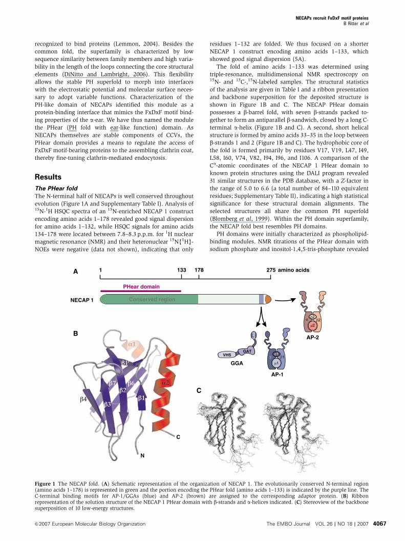
Interaction with amphiphysin I
To search for protein-binding partners in an unbiased screen,
we performed affinity selection experiments from rat brain
extracts with purified GST-NECAP 1 PHear domain and MS/
MS analysis identified amphiphysin I as an interaction
partner. Amphiphysin I is involved in clathrin-mediated
endocytosis and forms homodimers as well as heterodimers
with amphiphysin II (Butler et al, 1997; Ramjaun et al, 1997,
1999; Wigge et al, 1997; Di Paolo et al, 2002). Both isoforms
contain an N-terminal BAR domain that senses membrane
curvature and targets the protein to the neck of a forming
vesicle (Peter et al, 2004), and a C-terminal SH3 domain that
recruits synaptojanin and dynamin during vesicle formation
(Ramjaun et al, 1997; McPherson, 1999). The interaction of
the PHear domain with amphiphysin was readily detected by
Western blot (Figure 2A). In pull-down assays from brain
extract, amphiphysin binding is similar for the PHear domain
and the longer NECAP 1 construct encoding amino acids
1–178, while the region of amino acids 129–178, which com-
prise the evolutionarily conserved region in NECAP 1 outside
Table I Structural statistics for the NECAP 1 PHear domain
Restraints for structure calculationsIntra-residue NOEs 258
Sequential NOEs 291Medium- and long-range NOEs 148Hydrogen bonds 42f Backbone angles 118NH residual dipolar couplings 110
Final energies (kcal/mol)Etotal 329720Ebond 13.371.6Eangle 8377Eimpr 9.471.4Erepel 152723Enoe 10.673.1Ecdih 0.470.3Esani 3076Ecoup 3176
R.m.s.d. from idealized geometryBond (A) 0.002570.0002Bond angles (deg.) 0.3870.02Improper torsions (deg.) 0.2370.02
R.m.s.d. for experimental restraintsDistances (A) 0.01470.002Dihedral angles (deg.) 0.1270.08
Residual dipolar couplingsR.m.s.d. (Hz) 2.470.2Q-value 0.1670.02
Coordinate r.m.s.d. from average structure (A)a
Backbone atoms (N, Ca, C0) 0.7070.07All heavy atoms 1.3770.07
Ramachandran analysis (%)Residues in most favored regions 66.174.4Residues in additional allowed regions 26.176.1Residues in generously allowed regions 5.672.2
aFor residues 10–22, 32–61, and 69–129.
SM
GS
T
GS
T-N
EC
AP
1aa
1–1
78G
ST-
NE
CA
P 2
aa 1
–177
Amphiphysin I (brain)
Amphiphysin II (brain)
120
90
(kDa)
SM
Amphiphysin I
Amphiphysin II
NECAP 1S
M
Moc
k
Moc
k
α-A
mph
iphy
sin
α-N
EC
AP
1
120
90
(kDa)
40
C
B
GS
T
GS
T-N
EC
AP
1P
Hea
rG
ST-
NE
CA
P 1
aa 1
–178
SM
Amphiphysin I (brain)
Amphiphysin I (purified)
Amphiphysin II (brain)
120
120
90
(kDa)
A
GS
T-N
EC
AP
1aa
129
–178
Amphiphysin I/II IP NECAP 1 IP
Figure 2 NECAPs interact with amphiphysins. (A, B) GST fusionproteins encoding the NECAP 1 PHear domain, NECAP 1 aa 1–178,NECAP 1 aa 129–178, NECAP 2 aa 1–177, or GSTalone pre-coupledto glutathione–Sepharose were incubated with Triton X-100-solubi-lized rat brain extract or purified amphiphysin I as indicated, andamphiphysin binding was analyzed by Western blot. (C) Co-im-munoprecipitation of endogenous NECAP 1 and amphiphysins fromTriton X-100-solubilized rat brain extract using antibodies directedagainst amphiphysin (1874, which recognizes both amphiphysin Iand II) or NECAP 1. Immunoprecipitated proteins were detected byWestern blot. For all binding studies, 10% of the protein amountused in each reaction is analyzed as starting material (SM).
NECAPs recruit FxDxF motif proteinsB Ritter et al
The EMBO Journal VOL 26 | NO 18 | 2007 &2007 European Molecular Biology Organization4068

the PHear fold (Figure 1A), does not contribute to amphi-
physin binding (Figure 2A). Similar binding behavior was
observed using purified GST-amphiphysin I, from which the
GST tag had been removed before the binding assay, demon-
strating that the NECAP PHear domain directly interacts with
amphiphysin I (Figure 2A). Moreover, amphiphysin binding
is conserved between NECAP 1 and NECAP 2 (Figure 2B).
The NECAP 1/amphiphysin interaction was verified by
co-immunoprecipitation of endogenous proteins in both
directions (Figure 2C).
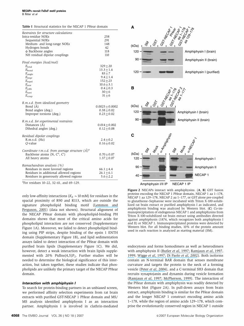
The PHear domain targets FxDxF motifs
To identify the region in amphiphysin I responsible for PHear
domain binding, we generated a series of GST-amphiphysin I
C- and N-terminal deletion constructs (Figure 3A), which
were used in pull-down assays from rat brain extract. NECAP
1 binding was observed for all constructs containing the
amphiphysin insert domain, similar to what was seen for
clathrin (Figure 3B). The insert domain is present in brain-
specific splice variants of amphiphysins and contains in
addition to clathrin-binding motifs (LLDLD and PWDLW),
peptide motifs for interaction with SH3 domain proteins
(PxxP) and the a-ear of AP-2 (FxDxF and DPF; Figure 3C)
(McMahon et al, 1997; Micheva et al, 1997; Ramjaun and
McPherson, 1998; Slepnev et al, 2000; Miele et al, 2004). We
used a series of N- and C-terminal deletion constructs of the
insert domain (Figure 3C) to define the PHear domain-bind-
ing region. The constructs showed the expected binding
pattern for clathrin, as constructs containing a single
clathrin-binding motif showed weak interaction while con-
structs containing both motifs showed strong clathrin
binding (Figure 3D). AP-2 interaction was detected for all
constructs containing the FxDxF motif, either alone or to-
gether with the single DPF motif (Figure 3D). AP-2 binding
was not observed for constructs containing the DPF motif
alone, due to the low affinity of a single DPF motif.
Surprisingly, NECAP 1 demonstrated the same binding pat-
tern as AP-2 (Figure 3D). In particular, both proteins bound to
amphiphysin I constructs encoding amino acids 291–329
and 324–445, and the small overlapping region of residues
324–329 encodes the FxDxF motif known to target the a-ear
of AP-2 (Brett et al, 2002). To verify that this region mediates
the interaction with NECAP 1, we used a synthetic peptide
containing the amphiphysin I FxDxF motif (CSFFEDNFPE) in
competition binding studies and indeed, the synthetic FxDxF
peptide interferes with amphiphysin binding to the NECAP 1
PHear domain (Figure 3E).
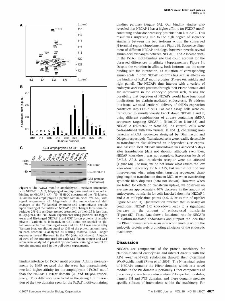
We used 2D-NMR spectroscopy to further characterize the
PHear domain-binding motif in amphiphysin I. Significant
chemical shift changes (40.1 p.p.m.) in the 1H-15N HSQC
spectra of an 15N-labeled amphiphysin I construct encoding
amino acids 291–329 were found for residues 321–328
(ISFFEDNF; Figure 4A and B). Additional signal broadening
for residues S322 to F328 suggests their involvement in ligand
binding. To further investigate the contribution of single
amino acids, we performed an alanine screen for positions
320–328 within the context of amino acids 311–329 and tested
for binding of purified His-tagged a-ear and NECAP 1 to the
various amphiphysin I mutants. This screen revealed inter-
esting differences in the binding properties of the a-ear and
NECAP 1. For the FxDxF motif core, binding of both proteins
was affected by mutation of F324 and F328, whereas muta-
tion of E325 and N327 did not inhibit binding (Figure 4C). A
major difference in binding behavior is observed when
changing the center core position D326 to alanine. This
mutation strongly reduces binding to the a-ear but does not
affect NECAP 1 binding (Figure 4C). D326 has been shown to
directly contact the a-ear (Brett et al, 2002), but the extended
definition of the motif, Fx[D/N/S]x[F/L] (Mishra et al, 2001),
indicates a higher variability allowed for this position. The
lack of influence of the D326A mutation on NECAP 1 binding
reveals differences in binding requirements for the two
domains, suggesting a higher tolerance of the PHear domain
for variation of the middle position of the motif core. Our
NMR study also detected significant chemical shift changes
for positions preceding the motif core and for S322, concur-
rent signal broadening further suggests a direct contribution
to binding. Mutation of this position to alanine selectively
interferes with NECAP 1 binding and does not affect the
interaction with the a-ear, whereas changing I321 to alanine
does not influence interaction with either domain. Together,
these data demonstrate that while both the a-ear and PHear
domain use the FxDxF motif as targeting sequence, some
positions within and surrounding the core contribute to
affinity and binding specificity in a domain-specific manner.
The PHear domain-binding site
We used NMR to identify the binding site for FxDxF motifs on
the PHear domain. Chemical shift changes in the 1H-15N
HSQC spectra of the 15N-labeled NECAP 1 PHear domain
upon interaction with the amphiphysin I insert domain or the
synthetic peptide containing the amphiphysin I FxDxF motif
were observed primarily for residues located in b-strands 1, 4,
6, and 7 (Figure 5A and B). The shifted residues are indicated
in Figure 5C in yellow (0.1oDdo0.2 p.p.m.) and red
(Dd40.2 p.p.m.). When plotted onto the fold, these residues
reveal the binding surface of the PHear domain (Figure 5D).
Point mutations of amino acids predicted to be involved
in FxDxF motif binding identified five residues essential for
interaction (Figure 5C and E) and revealed differences in the
way they contribute to the interaction. For example, binding
is lost when R21, R95, and R102 are exchanged to alanine or
glutamic acid, indicating a need for positive charge at these
positions. Moreover, substitution of R95 by lysine promotes
binding whereas the R102K substitution abolishes inter-
action, indicating different sterical restrictions for presenta-
tion of the positive charge (Figure 5E). Similarly, while the
negative charge presented by D98 appears negligible for
binding since D98E is not active, binding by D98N but not
D98A or D98K suggests a requirement for a b-carbonyl group,
possibly as a hydrogen bond acceptor (Figure 5E).
The PHear domain displays a distinct interface for FxDxF
motifs
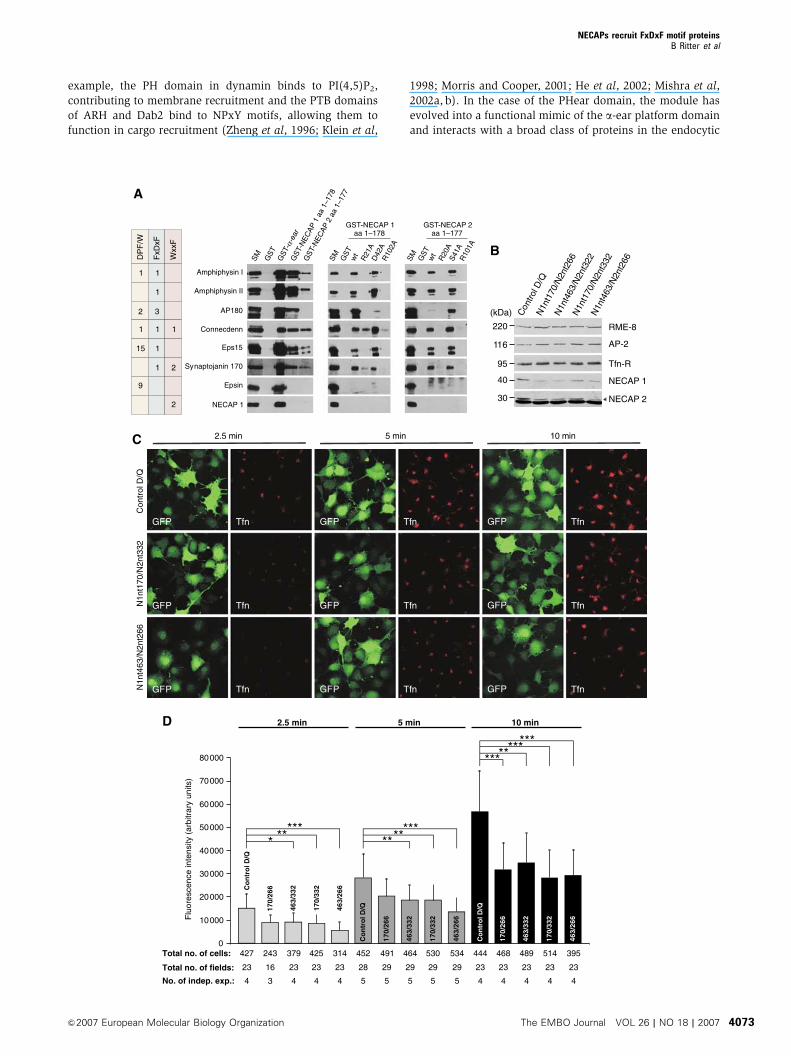
To address if the PHear domain binds selectively to amphi-
physin I or functions as a general platform for FxDxF motifs,
we compared the binding properties of the conserved
N-terminal regions of NECAP 1 and 2 to that of the a-ear
(Figure 6A). NECAP 1 (one WxxF-acidic motif) and epsin
(multiple DPW motifs) bound exclusively to the a-ear, while
all proteins containing at least one FxDxF motif bound to the
a-ear and NECAP 1 and 2 (amphiphysin I and II, AP180,
connecdenn, Eps15, and synaptojanin 170; Figure 6A). We
also used GST-tagged FxDxF motifs from various proteins
NECAPs recruit FxDxF motif proteinsB Ritter et al
&2007 European Molecular Biology Organization The EMBO Journal VOL 26 | NO 18 | 2007 4069
(amphiphysin I, AP180, disabled-2, HIP1, connecdenn, sy-
naptojanin 170, and CALM) to pull down purified His-tagged
NECAP 1 (Supplementary Figure 2). Most bound, despite the
low affinity of FxDxF motifs for the PHear domain (100 mM),
and the presentation of the motifs as short, unrestrained
peptides. The PHear domain thus functions as a general
BAR
INSERT
SH3
BAR
BAR
BARINSERT
INSERT
INSERT SH3
SH3
SH3
1
1
1
1
297
469
615
296
468
614
695
695
695
695
291PxxP FxDxF DPF PWDLWLLDLD
291291291291291
309324
338351
375406
445
445445445445445445445
411380
360329
C
170
40
(kDa) SM
GS
T
1–
296
1–
468
1–
614
1–
695
297
–69
546
9–
695
615
–69
5
GST-amphiphysin I aa...
Clathrin
NECAP 1
B
SM
GS
T29
1–
329
291
–36
029
1–
380
291
–41
129
1–
445
309
–44
5
338
–44
535
1–
445
375
–44
540
6–
445
GST-amphiphysin I aa...
324
–44
5
Clathrin
AP-2
NECAP 140
116
170
(kDa)
D
(kDa)
SM
GS
T
GST-NECAP 1 PHear
1000
:175
0:1
500:
125
0:1
100:
110
:11:
1
FxDxF peptide
Amphiphysin I
Amphiphysin II
120
95
E
Figure 3 Identification of the NECAP 1-binding region in amphiphysin I. (A) Domain structure of amphiphysin I indicating the N-terminal BARdomain followed by the neuronal insert domain, a linker region, and the C-terminal SH3 domain, and schematic presentation of N- andC-terminal deletion constructs. Amino-acid positions in front and behind the fragment indicate the protein borders. (B) Pull-down experimentswith Triton X-100-solubilized rat brain extract using GST fusion proteins of the amphiphysin I variants indicated in panel A or GST alone pre-coupled to glutathione–Sepharose. Binding of clathrin and NECAP 1 was analyzed by Western blot. (C) Schematic presentation of bindingmotifs for endophilin A1 (PxxP), clathrin (LLDLD, PWDLW), and AP-2 (FxDxF, DPF) within the neuronal insert domain of amphiphysin I, andoverview of N- and C-terminal deletion constructs with amino-acid positions indicating the protein borders. (D) Pull-down experiments withTriton X-100-solubilized rat brain extract using GST fusion proteins of the amphiphysin I variants indicated in panel C or GSTalone pre-coupledto glutathione–Sepharose. Binding of clathrin, AP-2, and NECAP 1 was analyzed by Western blot. The faint signal for NECAP 1 with the GSTfusion protein encoding amino acids 291–380 of amphiphysin I is due to co-migration of NECAP 1 with the fusion protein. (E) Aliquots(100 pmole) of the NECAP 1 PHear domain fused to GSTor GSTalone pre-coupled to glutathione–Sepharose were incubated with Triton X-100-solubilized rat brain extract in the absence or molar excess of a synthetic peptide encoding the amphiphysin I FxDxF motif (CSFFEDNFPE) asindicated. Binding of amphiphysin I and II was analyzed by Western blot. For all binding studies, 10% of the protein amount used in eachreaction is analyzed as starting material (SM).
NECAPs recruit FxDxF motif proteinsB Ritter et al
The EMBO Journal VOL 26 | NO 18 | 2007 &2007 European Molecular Biology Organization4070
binding interface for FxDxF motif proteins. Affinity measure-
ments by NMR revealed that the a-ear has approximately
two-fold higher affinity for the amphiphysin I FxDxF motif
than the NECAP 1 PHear domain (40 and 100 mM, respec-
tively). This difference is reflected in the strength of interac-
tion of the two domains seen for the FxDxF motif-containing
binding partners (Figure 6A). Our binding studies also
revealed that NECAP 1 has a higher affinity for FXDXF motif-
containing endocytic accessory proteins than NECAP 2. This
result was surprising due to the high degree of sequence
similarity between the two isoforms within the conserved
N-terminal region (Supplementary Figure 3). Sequence align-
ment of different NECAP orthologs, however, reveals several
amino-acid exchanges between NECAP 1 and 2 located with-
in the FxDxF motif-binding site that could account for the
observed differences in affinity (Supplementary Figure 3).
Despite the variation in affinity, both isoforms use the same
binding site for interaction, as mutation of corresponding
amino acids in both NECAP isoforms has similar effects on
the binding of FxDxF motif proteins (Figure 6A, middle and
right panel). The NECAPs thus interact with a variety of
endocytic accessory proteins through their PHear domain and
are interwoven in the endocytic protein web, raising the
possibility that depletion of NECAPs would have functional
implications for clathrin-mediated endocytosis. To address
this issue, we used lentiviral delivery of shRNA expression
constructs into COS-7 cells. For each assay, cells were co-
transduced to simultaneously knock down NECAP 1 and 2,
using different combinations of viruses containing shRNA
sequences targeting NECAP 1 (N1nt170 or N1nt463) and
NECAP 2 (N2nt266 or N2nt332). As control, cells were
co-transduced with two viruses, D and Q, containing non-
targeting shRNA sequences designed by Dharmacon and
Qiagen, respectively. Transduced cells were readily detectable
as transduction also delivered an independent GFP expres-
sion cassette. Best NECAP knockdown was achieved 5 days
after transduction (data not shown), although even then,
NECAP knockdown was not complete. Expression levels of
RME-8, AP-2, and transferrin receptor were not affected
(Figure 6B). For now, we do not know what causes the low
knockdown efficiency for NECAPs, but we did not find any
improvement when using other targeting sequences, chan-
ging length of transduction time or MOI, or when transfecting
synthetic RNA duplexes (data not shown). However, when
we tested for effects on transferrin uptake, we observed on
average an approximately 40% decrease in the amount of
endocytosed transferrin for cells knocked down for NECAP 1
and 2 at multiple time points (2.5, 5, or 10 min of uptake;
Figure 6C and D). Quantification revealed that in nearly all
conditions, NECAP 1/2 knockdown leads to a significant
decrease in the amount of endocytosed transferrin
(Figure 6D). These data show a functional role for NECAPs
in clathrin-mediated endocytosis and support the idea that
the PHear domain serves as an interaction module within the
endocytic protein web, promoting efficiency of the endocytic
machinery.
Discussion
NECAPs are components of the protein machinery for
clathrin-mediated endocytosis and interact directly with the
AP-2 a-ear sandwich subdomain through their C-terminal
WxxF-acidic motif (Ritter et al, 2004). The N-terminal region
of NECAPs contains the PHear domain, which is a novel
module in the PH domain superfamily. Other components of
the endocytic machinery also contain PH superfold modules,
namely PH and PTB domains, and these domains mediate
specific subsets of interactions within the machinery. For
(p.p.m.)
(p.p.m.)8.08.28.48.6
110
112
114
116
118
120
122
124
G300
T312
T310
T297
S295 N327
N319 S322
G(–1)
I320
I321
F323
F324
E325
D326F328
V303V309
L315
L306
E318
K299
K313
R298
K308Q316
Q317
Q296E314
0.1
0.3
0.5
0.7
312 314 316 318 320 322 324 326 328Residue number
T K E
E
E
L N
NI
I
V
F
F
FD
S
B
C
His-α-ear
His-NECAP 1
26
40
(kDa) SM
GS
Tw
tI3
20A
I321
AS
322A
F323
AF3
24A
E32
5AD
326A
N32
7AF3
28A
GST-amphiphysin I aa 311–329
GST proteins(Coomassie)
∆ C
hem
ical
shi
ft (p
.p.m
.)
Figure 4 The FXDXF motif in amphiphysin I mediates interactionwith NECAP 1. (A, B) Mapping of amphiphysin residues involved inbinding to NECAP 1. (A) 15N–1H HSQC spectrum of the 15N-labeled39-amino-acid amphiphysin I peptide (amino acids 291–329) withsignal assignments. (B) Magnitude of the amide chemical shiftchanges of the 15N-labeled 39-amino-acid amphiphysin peptideupon binding of the unlabeled NECAP 1 (the changes for N-terminalresidues 291–311 residues are not presented, as their Dd is less than0.03 p.p.m.). (C) Pull-down experiments using purified His-taggeda-ear and His-tagged NECAP 1 and GST fusion proteins of amphi-physin I variants as indicated, or GST alone pre-coupled to glu-tathione–Sepharose. Binding of a-ear and NECAP 1 was analyzed byWestern blot. An aliquot equal to 10% of the protein amount usedin each reaction is analyzed as starting material (SM). Longerexposures reveal His-a-ear in the SM (data not shown). Aliquotsof 30% of the amounts used for each GST fusion protein and GSTalone were analyzed in parallel by Coomassie staining to control forprotein amounts used in the pull-down experiments.
NECAPs recruit FxDxF motif proteinsB Ritter et al
&2007 European Molecular Biology Organization The EMBO Journal VOL 26 | NO 18 | 2007 4071

SM
GS
Tw
tD
98A
D98
ED
98K
D98
NR
102A
R10
2ER
102K
F105
AF1
05Y
Amphiphysin IAmphiphysin II
12590
(kDa)
GST-NECAP 1 PHear
SM
GS
Tw
tR
21A
R21
ED
42A
D42
ED
42K
D42
NA
70I
R95
AR
95E
R95
K
Amphiphysin IAmphiphysin II
12590
(kDa)
GST-NECAP 1 PHearE
C D
(p.p.m.)
(p.p.m.)
106 Amphiphysin I (291–445)
CSFFEDNFPE
0.6
0.4
0.2
0.6
0.4
0.2
Residue number20 40 60 80 100 120
∆ C
hem
ical
shi
ft (p
.p.m
.)110
114
118
122
126
130
10.0 9.0 8.0 7.0
Figure 5 Identification of the NECAP 1 binding site for FxDxF motifs. (A) Comparison of 15N–1H HSQC spectra of 15N-labeled NECAP 1 in theabsence (black) or presence (red) of a synthetic amphiphysin I peptide (CSFFEDNFPE) (at a 2:1 peptide–protein ratio). (B) Magnitude of theamide chemical shift changes of the NECAP 1 residues upon binding of the amphiphysin I insert domain (amino acids 291–445) or the syntheticpeptide. (C) Sequence of NECAP 1 with positions of b-strands and a-helices indicated. Color shading represents the size of the amide chemicalshift changes (red, Dd40.2 and yellow, 0.24Dd 40.1 p.p.m.) upon binding of the amphiphysin I insert domain. Asterisks mark residues thatwhen mutated affect binding. (D) Backbone trace of NECAP 1 colored according to the size of the chemical shift changes indicated in panel C.(E) GST fusion proteins encoding wild-type NECAP 1 PHear domain and point mutations as indicated, or GSTalone pre-coupled to glutathione–Sepharose were incubated with Triton X-100-solubilized rat brain extract and interaction with amphiphysin I and II was analyzed by Westernblot. A total of 10% of the protein amount used in each reaction is analyzed as starting material (SM).
Figure 6 The PHear domain is a general FxDxF motif-binding site implicated in the regulation of clathrin-mediated endocytosis. (A) Pull-downexperiments with Triton X-100-solubilized rat brain extract using GST fusion proteins of the a-ear and NECAP variants as indicated, or GSTalone pre-coupled to glutathione–Sepharose. Binding of various endocytic accessory proteins was analyzed by Western blot and the motifsinvolved in a-ear and PHear domain binding are indicated. For all binding studies, 10% of the protein amount used in each reaction is analyzedas starting material (SM). (B) Equal protein amounts of Triton-X100-solubilized COS-7 cells transduced with various combinations of shRNAconstructs for simultaneous knockdown of NECAP 1 and 2 or with control shRNA constructs (control D/Q) were analyzed by Western blot forexpression levels of various proteins as indicated. The arrowhead indicates the NECAP 2 signal, the band underneath represents a cross-reaction of the antibody. (C) Immunofluorescence analysis for uptake of fluorescently labeled transferrin (Tfn) in COS-7 cells transduced withvarious combinations of shRNA constructs for simultaneous knockdown of NECAP 1 and 2 or with control shRNA constructs (control D/Q).Cells were allowed to endocytose for 2.5, 5, or 10 min, as indicated. Transduced cells express GFP from an independent expression cassette. (D)Quantification of the transferrin uptake assay described in panel C. The number of independent experiments (No. of indep. exp.), total numberof microscopic fields analyzed (total no. of fields), and total number of cells analyzed (total # of cells) are indicated for each time-point andknockdown condition underneath the corresponding bar. *Po0.05, **Po0.01, and ***Po0.001.
NECAPs recruit FxDxF motif proteinsB Ritter et al
The EMBO Journal VOL 26 | NO 18 | 2007 &2007 European Molecular Biology Organization4072
example, the PH domain in dynamin binds to PI(4,5)P2,
contributing to membrane recruitment and the PTB domains
of ARH and Dab2 bind to NPxY motifs, allowing them to
function in cargo recruitment (Zheng et al, 1996; Klein et al,
1998; Morris and Cooper, 2001; He et al, 2002; Mishra et al,
2002a, b). In the case of the PHear domain, the module has
evolved into a functional mimic of the a-ear platform domain
and interacts with a broad class of proteins in the endocytic
BG
ST
GST
GST
GST
-α-e
ar
wt
wt
SM SM SMGST
-NEC
AP1
aa 1
–178
R21
A
R20
A
D42
A
S41A
GST
-NEC
AP2
aa 1
–177
R10
2A
R10
1A
GST-NECAP 1aa 1–178
GST-NECAP 2aa 1–177
DP
F/W
FxD
xF
Wxx
F
11
1
32
11 1
15 1
21
9
2
Amphiphysin I
Amphiphysin II
AP180
Connecdenn
Eps15
Synaptojanin 170
Epsin
NECAP 1
Con
trol D
/QN
1nt1
70/N
2nt2
66
N1n
t463
/N2n
t322
N1n
t170
/N2n
t332
N1n
t463
/N2n
t266
RME-8
AP-2
Tfn-R
NECAP 1
NECAP 2
C
D
463/
332
170/
3 32
4 63/
266
170/
2 66
463/
332
1 70 /
332
463 /
266
Co
ntr
ol D
/Q
170/
2 66
463/
332
170/
332
463 /
2 66
Co
ntr
ol D
/Q
Co
ntr
ol D
/Q
170/
2 66
0
10000
20000
30000
40000
50000
60000
70000
80000
2.5 min 5 min 10 min
**
******
*********
*******
427 243 379 425 314 452 491 464 530 534 444 468 489 514 395
23 16 23 23 23 28 29 29 29 29 23 23 23 23 23
4 3 4 4 4 5 5 5 5 5 4 4 4 4 4
Total no. of cells:
No. of indep. exp.:
Total no. of fields:
Con
trol
D/Q
N1n
t170
/N2n
t332
2.5 min 5 min 10 min
GFP GFP GFP
GFP GFP GFP
GFP GFP GFP
Tfn Tfn Tfn
Tfn Tfn Tfn
Tfn Tfn TfnN1n
t463
/N2n
t266
(kDa)
220
116
95
40
30
Flu
ores
cenc
e in
tens
ity (
arbi
trar
y un
its)
NECAPs recruit FxDxF motif proteinsB Ritter et al
&2007 European Molecular Biology Organization The EMBO Journal VOL 26 | NO 18 | 2007 4073

machinery. The a-ear and PHear domain share no significant
sequence or structural homology and therefore expose dis-
tinct molecular surfaces for FxDxF motif binding (Figure 7A
and B). This provides a likely explanation for the different
requirements observed for some amino-acid positions within
and surrounding the FxDxF motif core (Figure 4C), but only
future extensive mutagenesis for each single position would
allow the designation of a-ear- and PHear domain-specific
variations of the FxDxF motif. Nonetheless, both domains
target FxDxF motifs to select common binding partners. This
represents an intriguing example of convergent molecular
evolution, in which two distinct domains have acquired a
common function, that is, to bind to FxDxF motifs. It is
tempting to speculate that it results from the colocalization
of the a-ear and the PHear domain within a functional
microdomain.
The AP-2 a-ear recruits accessory proteins from the
surrounding cytosol to sites of CCV assembly. The b2-ear
also recruits accessory proteins during early steps of vesicle
formation and proteins that engage a- and b2-ear simulta-
neously have higher avidity for AP-2. Clathrin recruitment to
the maturing vesicle by the b2-ear and linker displaces the
accessory proteins, many of which have FxDxF motifs, from
the b2-ear (Owen et al, 2000; Edeling et al, 2006; Schmid
et al, 2006). NECAPs target the a-ear sandwich subdomain
through their C-terminal WxxF-acidic motif, thereby placing
the PHear domain into the environment of the a-ear. This
could provide a means to maximize the sampling radius
covered by the a-ear and to increase the avidity for FxDxF
motif proteins by locally increasing the number of available
binding interfaces either throughout vesicle formation or at
specific steps of vesicle maturation, for example, when acces-
sory protein binding to the b2-ear is disrupted by clathrin.
Knockdown of AP-2 leads to a drastic reduction of transferrin
endocytosis (Conner and Schmid, 2003; Hinrichsen et al,
2003; Motley et al, 2003; Huang et al, 2004), attesting to
Figure 7 Comparison of the molecular surfaces of the PHear domain and AP-2 a-ear. (A, B) Presentation of the FxDxF motif-binding surfacefor the NECAP PHear domain (left) and the a-ear of AP-2 (right). The orientation of the NECAP PHear domain is corresponding to Figure 5D.(A) The surface of both proteins is color-coded, with red indicating negative electrostatic potential and blue indicating positive potential. Theamphiphysin I SFFEDNFVP peptide is shown in green. The atomic coordinates for the a-ear/FxDxF motif complex were taken from PDB entry1KY7. (B) Color-coding (green) highlights amino acids implicated in FxDxF motif binding by NMR for the PHear domain, or shown to contactthe motif by co-crystallization for the a-ear. Mutational analysis of amino acids labeled in orange verified their contribution to FxDxF motifbinding (this study and Brett et al, 2002).
NECAPs recruit FxDxF motif proteinsB Ritter et al
The EMBO Journal VOL 26 | NO 18 | 2007 &2007 European Molecular Biology Organization4074

the critical role of AP-2 within the endocytic machinery. Here
we observed a moderate reduction in transferrin endocytosis
following NECAP knockdown. This suggests that NECAPs
function supplementary to AP-2 and are involved in the fine-
tuning of the endocytic machinery. In this scenario, one could
imagine how NECAP knockdown could cause an imbalance
in the recruitment of FxDxF motif proteins during vesicle
formation, resulting in a decreased endocytic efficiency.
NECAPs also use a FxxF motif in their C-terminal region
to bind the clathrin adaptor proteins AP-1 and GGAs, which
function at endosomes and the trans-Golgi network (TGN)
(Mattera et al, 2004; Ritter et al, 2004). The g-ear and GAE
domains of these adaptors serve to recruit accessory proteins,
analogous to the a-ear (Lui et al, 2003). In contrast to the a-
ear, however, these ears only contain a fold equivalent to the
a-ear sandwich subdomain (Kent et al, 2002; Collins et al,
2003). NECAP binding to the ears of AP-1 and GGAs would
provide a means to recruit FxDxF motif proteins to sites of
vesicle formation at endosomes and the TGN.
The prevailing idea is that the peptide motifs that allow for
targeting of endocytic proteins to the a-ear interact exclu-
sively with this domain. The PHear domain is the first
evidence that one endocytic motif can target alternative
protein domains. Moreover, to the best of our knowledge,
the binding of FxDxF motifs to the a-ear and the PHear
domain represents the only known example where a single
defined peptide motif targets two distinct protein modules.
Our characterization of the NECAP PHear domain has thus
revealed a surprising twist on domain/motif specificity, sug-
gesting even more complex ways to regulate vesicle forma-
tion. Time will tell if the PHear domain alone evolved to
provide an alternative mechanism of accessory protein reg-
ulation, or if this is only the first of many examples to come.
Materials and methods
Expression constructs, antibodies, and peptidesA description of the expression constructs used can be found in theSupplementary data online. Antibodies for amphiphysin I and II(1874, detects both isoforms), connecdenn, NECAP 1, NECAP 2,and RME-8 were previously described (Ramjaun et al, 1997; Ritteret al, 2003, 2004; Girard et al, 2005; Allaire et al, 2006). Monoclonalantibodies for a-adaptin and clathrin were from BD TransductionLaboratories. Monoclonal antibodies for transferrin receptor andthe tetra-His tag were from Zymed and Qiagen, respectively.Polyclonal serum against Eps15 was from Santa Cruz. Antibodiesagainst synaptojanin 170, epsin, and AP180 (F1-20) were generousgifts from Drs P De Camilli, L Traub, and E Lafer, respectively. Thesynthetic amphiphysin I peptide was purchased from HHMI/KeckBiotechnology Resource Laboratory, Yale University.
Knockdown experiments and transferrin uptake assaysA detailed description of the shRNA constructs and the virusproduction procedure can be found in the Supplementary dataonline. COS-7 cells were plated in 24-well plates with 29 000 cells/well in regular medium. After 12–14 h, the medium was replacedwith 500ml/well of transduction medium and the two controlviruses or combinations of NECAP 1 and 2 viruses were added at anMOI of 10 (for each virus). The next day, 1 ml of regular mediumwas added to each well. Three days after transduction, the cellswere trypsinized and plated in parallel in six-well plates andpoly-L-lysine-coated glass coverslips in 24-well plates (three cover-slips for each virus combination). Five days after transduction, thecells in the six-well plate were analyzed by Western blot. The cellsin the 24-well plate were serum-starved for at least 2 h and Alexa546-labeled transferrin (Molecular Probes) in pre-warmed DMEM(5 mg/ml) was added at 371C. After endocytosis was allowed toproceed at 371C for 2.5, 5, and 10 min, the cells were chilled on ice
and washed with ice-cold PBS. Surface-bound transferrin wasremoved by incubating the cells on ice in ice-cold acid wash (0.2 Macetic acid, 0.5 M NaCl). The cells were rinsed, fixed for 30 min onice with ice-cold 3% paraformaldehyde in PBS, incubated 1 min in50 mM NH4Cl in PBS, washed, and mounted for analysis on a ZeissLSM 510 confocal microscope. Acquisition settings were keptconstant for all conditions analyzed within each experiment andbetween experiments analyzed. For each time point and trans-duction condition, 5–6 pictures of randomly chosen fields of cellswere taken. Transferrin uptake was quantified using NIH ImageJsoftware. For each field taken, all cells were outlined by hand andthe total intensity of transferrin signal for the selected area wasmeasured and divided by the number of measured cells to obtainthe average intensity/cell for each field. Statistical analysis of theresults was performed using the Kruskal–Wallis test, followed byDunn’s post test.
NMR spectroscopy and structure calculationsGST fusion proteins of NECAP 1 (residues 1–133 and 1–178) wereexpressed in the Escherichia coli strain BL21. Cultures were grown at371C in M9-media supplemented with 15N ammonium chloride and13C-enriched glucose to produce uniformly 15N- or 15N-, 13C-labeledproteins. Following 4 h of induction with 1 mM IPTG at 251C, GSTfusion proteins were purified, cleaved with thrombin in PBS, andthrombin was removed using benzamidine Sepharose. The NMRsamples contained 0.2–1.4 mM protein in 90% H2O/10% D2O,25 mM sodium phosphate buffer (pH 7.2), 75 mM NaCl, 0.5 mMEDTA, and 3 mM DTT.
NMR spectra were acquired at 301C on a Bruker DRX-600 andVarian Unity Inova 800 MHz spectrometer equipped with tripleresonance probes and pulsed field gradients. The following 3Dexperiments were used for backbone and side-chain 1H, 13C, and15N resonance assignments: HNCACB, CBCA(CO)HN, HNCA,HNCO, 1H-TOCSY-(CO)HN, 13C-TOCSY-(CO)HN, 15N-edited-TOCSY,and 15N-edited-NOESY (Cavanagh et al, 1996). NMR spectra wereprocessed using NMRPIPE (Delaglio et al, 1995) and XWINNMR(Bruker) software, and analyzed with XEASY (Bartels et al, 1995).Values of 3J(HNHa) were estimated from HNHA experiments(Kuboniva et al, 1994). Amide heteronuclear 15N{1H}-NOEs weremeasured and used for determination of high-mobility regions ofthe proteins (Peng and Wagner, 1994). 1H–15N residual dipolarcouplings (RDC) with a precision of 71.5 Hz were extracted from(IPAP)-HSQC experiments (Ottiger et al, 1998) on an isotropicsample and on a sample containing 8 mg/ml Pf1-phage. The NMRassignments and values of RDC for the NECAP 1 PHear domain(residues 1–133) have been deposited in the BioMagnetic Reso-nance Bank under the BMRB accession number 6354.
Regions of a-helical or b-strand secondary structure weredetermined based on Ca-chemical shifts (Wishart and Sykes,1994), values of 3J(HNHa) coupling constants, and the NOE patterns(Wuthrich, 1986). The protein structure was refined using standardprotocol in CNS version 1.1 (Brunger et al, 1998). The numbers ofdifferent types of constraints are presented in Table I. Manuallyassigned NOE distance constraints were classified according to thepeak intensities as strong (1.8–3.0 A), medium (2.0–4.0 A), or weak(2.5–5.0 A). Hydrogen bonds were given bounds of 1.7–2.3 A (HN–O). The atomic coordinates of NECAP 1 PHear domain have beendeposited in the Protein Data Bank under PDB accession code1TQZ. The pairwise coordinate r.m.s.d. comparisons betweendifferent proteins were obtained by the DALI program (Holm andSander, 1993). Structural images were generated with MOLMOL(Koradi et al, 1996) and PyMOL (DeLano, 2002).
Detailed analysis of peptide binding to the NECAP 1 PHeardomain was carried out by comparison of chemical shifts forbackbone amide signals in 15N–1H HSQC spectra. GST fusionprotein of amphiphysin I residues 291–445 or 291–329 wasexpressed in the E. coli strain BL21. For 3D-NMR studies, the 15N-labeled 39-amino-acid peptide was additionally purified by reverse-phase chromatography on a C-18 column (Vydac), lyophilized, andresuspended in the buffer at pH 6.2. The 15N-edited-NOESYand 15N-edited-TOCSY experiments were used for the assignment of amidesignals in the 15N–1H HSQC spectra at 301C. Due to strongbroadening of some HSQC signals of the 40-amino-acid peptide incomplex with unlabeled NECAP 1 (in region between S322-V329),2D chemical shift changes were measured at 1:2 protein–peptideratio and calculated as {(D1H shift)2þ (D15N shift x 0.2)2}1/2 inp.p.m. for totally bound complex.
NECAPs recruit FxDxF motif proteinsB Ritter et al
&2007 European Molecular Biology Organization The EMBO Journal VOL 26 | NO 18 | 2007 4075
Binding studiesA detailed description for the PIP strips, lipid sedimentation, andprotein binding studies is given in Supplementary data.
Supplementary dataSupplementary data are available at The EMBO Journal Online(http://www.embojournal.org).
Acknowledgements
This paper is dedicated to Hubert Ritter (December 1, 1934–March18, 2005). We thank Drs Linton Traub and Pietro De Camilli for
the gift of antibodies and Dr Eileen Lafer for the gift of anti-bodies and an AP180 full-length expression construct, LyneBourbonniere for excellent technical assistance, and Rachel Katfor assistance with the statistical analysis. NMR experimentswere recorded at the Quebec/Eastern Canada High Field (QANUC)NMR Facility. This research was supported by Canadian Institutesof Health Research (CIHR) Grants MOP-43967 to KG and MOP-13461 to PSM. BR was supported by a CIHR fellowship. PSMis a Fonds de la recherche en sante du Quebec (FRSQ) SeniorScholar and holds the James McGill Chair. KG is an FRSQChercheur National. The authors declare no competing financialinterests.
References
Allaire PD, Ritter B, Thomas S, Burman JL, Denisov AY, Legendre-Guillemin V, Harper SQ, Davidson BL, Gehring K, McPherson PS(2006) Connecdenn, a novel DENN domain-containing protein ofneuronal clathrin-coated vesicles functioning in synaptic vesicleendocytosis. J Neurosci 26: 13202–13212
Bartels C, Xia TH, Billeter M, Guntert P, Wuthrich K (1995) Theprogram XEASY for computer-supported NMR spectral analysis ofbiological macromolecules. J Biomol NMR 6: 1–10
Blomberg N, Baraldi E, Nilges M, Saraste M (1999) The PH super-fold: a structural scaffold for multiple functions. Trends BiochemSci 24: 441–445
Blondeau F, Ritter B, Allaire PD, Wasiak S, Girard M, Hussain NK,Angers A, Legendre-Guillemin V, Roy L, Boismenu D, Kearney RE,Bell AW, Bergeron JJ, McPherson PS (2004) Tandem MS analysisof brain clathrin-coated vesicles reveals their critical involve-ment in synaptic vesicle recycling. Proc Natl Acad Sci USA 101:3833–3838
Brett TJ, Traub LM, Fremont DH (2002) Accessory protein recruit-ment motifs in clathrin-mediated endocytosis. Structure 10:797–809
Brunger AT, Adams PD, Clore GM, DeLano WL, Gros P, Grosse-Kunstleve RW, Jiang JS, Kuszewski J, Nilges M, Pannu NS, ReadRJ, Rice LM, Simonson T, Warren GL (1998) Crystallography &NMR system: a new software suite for macromolecular structuredetermination. Acta Crystallogr D Biol Crystallogr 54: 905–921
Butler MH, David C, Ochoa GC, Freyberg Z, Daniell L, Grabs D,Cremona O, De Camilli P (1997) Amphiphysin II (SH3P9; BIN1),a member of the amphiphysin/Rvs family, is concentrated in thecortical cytomatrix of axon initial segments and nodes of Ranvierin brain and around T tubules in skeletal muscle. J Cell Biol 137:1355–1367
Cavanagh J, Fairbrother WJ, Palmer AG, Skelton NJ (1996) ProteinNMR Spectroscopy: Principles and Practice. San Diego: AcademicPress
Collins BM, Praefcke GJ, Robinson MS, Owen DJ (2003) Structuralbasis for binding of accessory proteins by the appendage domainof GGAs. Nat Struct Biol 10: 607–613
Conner SD, Schmid SL (2003) Differential requirements for AP-2 inclathrin-mediated endocytosis. J Cell Biol 162: 773–779
Delaglio F, Grzesiek S, Vuister GW, Zhu G, Pfeifer J, Bax A (1995)NMRPipe: a multidimensional spectral processing system basedon UNIX pipes. J Biomol NMR 6: 277–293
DeLano WL (2002) The PyMOL Molecular Graphics System. DeLanoScientific: San Carlos, CA
Di Paolo G, Sankaranarayanan S, Wenk MR, Daniell L, Perucco E,Caldarone BJ, Flavell R, Picciotto MR, Ryan TA, Cremona O, DeCamilli P (2002) Decreased synaptic vesicle recycling efficiencyand cognitive deficits in amphiphysin 1 knockout mice. Neuron33: 789–804
DiNitto JP, Lambright DG (2006) Membrane and juxtamembranetargeting by PH and PTB domains. Biochim Biophys Acta 1761:850–867
Edeling MA, Mishra SK, Keyel PA, Steinhauser AL, Collins BM,Roth R, Heuser JE, Owen DJ, Traub LM (2006) Molecularswitches involving the AP-2 beta2 appendage regulate endo-cytic cargo selection and clathrin coat assembly. Dev Cell 10:329–342
Girard M, Poupon V, Blondeau F, McPherson PS (2005) TheDnaJ-domain protein RME-8 functions in endosomal trafficking.J Biol Chem 280: 40135–40143
He G, Gupta S, Yi M, Michaely P, Hobbs HH, Cohen JC (2002) ARHis a modular adaptor protein that interacts with the LDL receptor,clathrin, and AP-2. J Biol Chem 277: 44044–44049
Hinrichsen L, Harborth J, Andrees L, Weber K, Ungewickell EJ(2003) Effect of clathrin heavy chain- and alpha-adaptin-specificsmall inhibitory RNAs on endocytic accessory proteins andreceptor trafficking in HeLa cells. J Biol Chem 278: 45160–45170
Holm L, Sander C (1993) Protein structure comparison by alignmentof distance matrices. J Mol Biol 233: 123–138
Honing S, Ricotta D, Krauss M, Spate K, Spolaore B, Motley A,Robinson M, Robinson C, Haucke V, Owen DJ (2005) Phospha-tidylinositol-(4,5)-bisphosphate regulates sorting signal recognitionby the clathrin-associated adaptor complex AP2. Mol Cell 18: 519–531
Huang F, Khvorova A, Marshall W, Sorkin A (2004) Analysis ofclathrin-mediated endocytosis of epidermal growth factor recep-tor by RNA interference. J Biol Chem 279: 16657–16661
Jha A, Agostinelli NR, Mishra SK, Keyel PA, Hawryluk MJ, TraubLM (2004) A novel AP-2 adaptor interaction motif initiallyidentified in the long-splice isoform of synaptojanin 1, SJ170.J Biol Chem 279: 2281–2290
Kent HM, McMahon HT, Evans PR, Benmerah A, Owen DJ (2002)Gamma-adaptin appendage domain: structure and binding sitefor Eps15 and gamma-synergin. Structure 10: 1139–1148
Klein DE, Lee A, Frank DW, Marks MS, Lemmon MA (1998) Thepleckstrin homology domains of dynamin isoforms require oli-gomerization for high affinity phosphoinositide binding. J BiolChem 273: 27725–27733
Koradi R, Billeter M, Wuthrich K (1996) MOLMOL: a program fordisplay and analysis of macromolecular structures. J Mol Graph14: 51–55
Kuboniva H, Grzesiek S, Delaglio F, Bax A (1994) Measurement ofHN-H alpha J couplings in calcium-free calmodulin using new 2Dand 3D water-flip-back methods. J Biomol NMR 4: 871–878
Lemmon MA (2004) Pleckstrin homology domains: not just forphosphoinositides. Biochem Soc Trans 32: 707–711
Lemmon MA, Ferguson KM (2001) Molecular determinants inpleckstrin homology domains that allow specific recognition ofphosphoinositides. Biochem Soc Trans 29: 377–384
Lui WW, Collins BM, Hirst J, Motley A, Millar C, Schu P, Owen DJ,Robinson MS (2003) Binding partners for the COOH-terminalappendage domains of the GGAs and gamma-adaptin. Mol BiolCell 14: 2385–2398
Mattera R, Ritter B, Sidhu SS, McPherson PS, Bonifacino JS (2004)Definition of the consensus motif recognized by gamma-adaptinear domains. J Biol Chem 279: 8018–8028
McMahon HT, Wigge P, Smith C (1997) Clathrin interacts specifi-cally with amphiphysin and is displaced by dynamin. FEBS Lett413: 319–322
McPherson PS (1999) Regulatory role of SH3 domain-mediatedprotein–protein interactions in synaptic vesicle endocytosis. CellSignal 11: 229–238
McPherson PS, Ritter B (2005) Peptide motifs: building the clathrinmachinery. Mol Neurobiol 32: 73–87
Micheva KD, Ramjaun AR, Kay BK, McPherson PS (1997) SH3domain-dependent interactions of endophilin with amphiphysin.FEBS Lett 414: 308–312
Miele AE, Watson PJ, Evans PR, Traub LM, Owen DJ (2004) Twodistinct interaction motifs in amphiphysin bind two independentsites on the clathrin terminal domain beta-propeller. Nat StructMol Biol 11: 242–248
NECAPs recruit FxDxF motif proteinsB Ritter et al
The EMBO Journal VOL 26 | NO 18 | 2007 &2007 European Molecular Biology Organization4076

Mills IG, Praefcke GJ, Vallis Y, Peter BJ, Olesen LE, Gallop JL, ButlerPJ, Evans PR, McMahon HT (2003) EpsinR: an AP1/clathrininteracting protein involved in vesicle trafficking. J Cell Biol160: 213–222
Mishra SK, Agostinelli NR, Brett TJ, Mizukami I, Ross TS, Traub LM(2001) Clathrin- and AP-2-binding sites in HIP1 uncover a generalassembly role for endocytic accessory proteins. J Biol Chem 276:46230–46236
Mishra SK, Hawryluk MJ, Brett TJ, Keyel PA, Dupin AL, Jha A,Heuser JE, Fremont DH, Traub LM (2004) Dual engagementregulation of protein interactions with the AP-2 adaptor alphaappendage. J Biol Chem 279: 46191–46203
Mishra SK, Keyel PA, Hawryluk MJ, Agostinelli NR, Watkins SC,Traub LM (2002a) Disabled-2 exhibits the properties of a cargo-selective endocytic clathrin adaptor. EMBO J 21: 4915–4926
Mishra SK, Watkins SC, Traub LM (2002b) The autosomal recessivehypercholesterolemia (ARH) protein interfaces directly with theclathrin-coat machinery. Proc Natl Acad Sci USA 99: 16099–16104
Morris SM, Cooper JA (2001) Disabled-2 colocalizes with the LDLRin clathrin-coated pits and interacts with AP-2. Traffic 2: 111–123
Motley A, Bright NA, Seaman MN, Robinson MS (2003) Clathrin-mediated endocytosis in AP-2-depleted cells. J Cell Biol 162:909–918
Murshid A, Srivastava A, Kumar R, Presley JF (2006) Charac-terization of the localization and function of NECAP 1 in neurons.J Neurochem 98: 1746–1762
Ottiger M, Delaglio F, Bax A (1998) Measurement of J and dipolarcouplings from simplified two-dimensional NMR spectra. J MagnReson 131: 373–378
Owen DJ, Collins BM, Evans PR (2004) Adaptors for clathrin coats:structure and function. Annu Rev Cell Dev Biol 20: 153–191
Owen DJ, Vallis Y, Noble ME, Hunter JB, Dafforn TR, Evans PR,McMahon HT (1999) A structural explanation for the binding ofmultiple ligands by the alpha-adaptin appendage domain. Cell 97:805–815
Owen DJ, Vallis Y, Pearse BM, McMahon HT, Evans PR (2000) Thestructure and function of the beta 2-adaptin appendage domain.EMBO J 19: 4216–4227
Peng JW, Wagner G (1994) Investigation of protein motions viarelaxation measurements. Methods Enzymol 239: 563–596
Peter BJ, Kent HM, Mills IG, Vallis Y, Butler PJ, Evans PR, McMahonHT (2004) BAR domains as sensors of membrane curvature: theamphiphysin BAR structure. Science 303: 495–499
Praefcke GJ, Ford MG, Schmid EM, Olesen LE, Gallop JL, Peak-Chew SY, Vallis Y, Babu MM, Mills IG, McMahon HT (2004)Evolving nature of the AP2 alpha-appendage hub during clathrin-coated vesicle endocytosis. EMBO J 23: 4371–4383
Ramjaun AR, McPherson PS (1998) Multiple amphiphysin II splicevariants display differential clathrin binding: identification of twodistinct clathrin-binding sites. J Neurochem 70: 2369–2376
Ramjaun AR, Micheva KD, Bouchelet I, McPherson PS (1997)Identification and characterization of a nerve terminal-enrichedamphiphysin isoform. J Biol Chem 272: 16700–16706
Ramjaun AR, Philie J, de Heuvel E, McPherson PS (1999) The Nterminus of amphiphysin II mediates dimerization and plasmamembrane targeting. J Biol Chem 274: 19785–19791
Ritter B, Denisov AY, Philie J, Deprez C, Tung EC, Gehring K,McPherson PS (2004) Two WXXF-based motifs in NECAPsdefine the specificity of accessory protein binding to AP-1 andAP-2. EMBO J 23: 3701–3710
Ritter B, McPherson PS (2004) Molecular mechanisms in clathrin-mediated membrane budding. In: Keraenen S, Jantti J (eds). Topicsin Current Genetics. Regulatory Mechanisms of IntracellularMembrane Transport. Berlin, Heidelberg: Springer-Verlag pp 9–37
Ritter B, Philie J, Girard M, Tung EC, Blondeau F, McPherson PS(2003) Identification of a family of endocytic proteins thatdefine a new alpha-adaptin ear-binding motif. EMBO Rep 4:1089–1095
Schmid EM, Ford MG, Burtey A, Praefcke GJ, Peak-Chew SY, MillsIG, Benmerah A, McMahon HT (2006) Role of the AP2 beta-appendage hub in recruiting partners for clathrin-coated vesicleassembly. PLoS Biol 4: e262
Slepnev VI, Ochoa GC, Butler MH, De Camilli P (2000) Tandemarrangement of the clathrin and AP-2 binding domains in amphi-physin 1 and disruption of clathrin coat function by amphiphysinfragments comprising these sites. J Biol Chem 275: 17583–17589
Traub LM (2005) Common principles in clathrin-mediated sorting atthe Golgi and the plasma membrane. Biochim Biophys Acta 1744:415–437
Traub LM, Downs MA, Westrich JL, Fremont DH (1999) Crystalstructure of the alpha appendage of AP-2 reveals a recruitmentplatform for clathrin-coat assembly. Proc Natl Acad Sci USA 96:8907–8912
Walther K, Diril MK, Jung N, Haucke V (2004) Functional dissectionof the interactions of stonin 2 with the adaptor complex AP-2 andsynaptotagmin. Proc Natl Acad Sci USA 101: 964–969
Wasiak S, Legendre-Guillemin V, Puertollano R, Blondeau F,Girard M, de Heuvel E, Boismenu D, Bell AW, Bonifacino JS,McPherson PS (2002) Enthoprotin: a novel clathrin-associatedprotein identified through subcellular proteomics. J Cell Biol 158:855–862
Wigge P, Kohler K, Vallis Y, Doyle CA, Owen D, Hunt SP, McMahonHT (1997) Amphiphysin heterodimers: potential role in clathrin-mediated endocytosis. Mol Biol Cell 8: 2003–2015
Wishart DS, Sykes BD (1994) Chemical shifts as a tool for structuredetermination. Methods Enzymol 239: 363–392
Wuthrich K (1986) NMR of Proteins and Nucleic Acids. John Wiley &Sons: New York
Yu JW, Mendrola JM, Audhya A, Singh S, Keleti D, DeWald DB,Murray D, Emr SD, Lemmon MA (2004) Genome-wide analysis ofmembrane targeting by S. cerevisiae pleckstrin homology do-mains. Mol Cell 13: 677–688
Zheng J, Cahill SM, Lemmon MA, Fushman D, Schlessinger J,Cowburn D (1996) Identification of the binding site for acidicphospholipids on the pH domain of dynamin: implications forstimulation of GTPase activity. J Mol Biol 255: 14–21
NECAPs recruit FxDxF motif proteinsB Ritter et al
&2007 European Molecular Biology Organization The EMBO Journal VOL 26 | NO 18 | 2007 4077
Related Documents