The Myeloid Transcription Factor KLF2 Regulates the Host Response to Polymicrobial Infection and Endotoxic Shock Ganapati H. Mahabeleshwar 1 , Daiji Kawanami 1 , Nikunj Sharma 1 , Yoichi Takami 1 , Guangjin Zhou 1 , Hong Shi 1 , Lalitha Nayak 1 , Darwin Jeyaraj 1 , Robert Grealy 3 , Mary White 3 , Ross McManus 3 , Thomas Ryan 3 , Patrick Leahy 2 , Zhiyong Lin 1 , Saptarsi M. Haldar 1 , G. Brandon Atkins 1 , Hector R. Wong 4 , Jerry B. Lingrel 5 , and Mukesh K. Jain *,1 1 Case Cardiovascular Research Institute, Department of Medicine, Harrington-McLaughlin Heart and Vascular Institute, Case Western Reserve University School of Medicine, University Hospitals Case Medical Center, Cleveland, OH 44106, USA 2 Case Comprehensive Cancer Center, Case Western Reserve University, Cleveland, OH 44106 3 Department of Clinical Medicine, Institute of Molecular Medicine, Trinity College Dublin, Ireland 4 Division of Critical Care Medicine, Cincinnati Children’s Research Foundation and Department of Pediatrics, University of Cincinnati College of Medicine 5 Department of Molecular Genetics, Biochemistry and Microbiology, University of Cincinnati College of Medicine, Cincinnati, OH 45267–0524 SUMMARY Precise control of myeloid cell activation is required for optimal host defense. However, this activation process must be under exquisite control to prevent uncontrolled inflammation. Herein, we identify the Kruppel-like transcription factor 2 (KLF2) as a potent regulator of myeloid cell activation in vivo. Exposure of myeloid cells to hypoxia and/or bacterial products reduced KLF2 expression while inducing hypoxia indusable factor-1α (HIF-1α), findings that were recapitulated in human septic patients. Myeloid KLF2 was found to be a potent inhibitor of nuclear factor- kappaB (NFκB)-dependent HIF-1α transcription and, consequently, a critical determinant of outcome in models of polymicrobial infection and endotoxemia. Collectively, these observations identify KLF2 as a tonic repressor of myeloid cell activation in vivo and an essential regulator of the innate immune system. INTRODUCTION Cells of the myeloid lineage are the principal effectors of the innate immune response to pathogen challenge. Under physiologic conditions, these cells circulate in the bloodstream in a quiescent state. However, in response to an infectious stimulus, these phagocytes are quickly activated and recruited to sites of injury where they engage in the elimination of invading microorganisms (Serbina et al., 2008; Serbina and Pamer, 2008). Indeed, myeloid cell activation is an exquisitely robust biological response that involves transcriptional © 2011 Elsevier Inc. All rights reserved. * Address correspondence to: Mukesh K. Jain M.D., 2103 Cornell Road, Room 4-522, Cleveland, OH 44106, Tel: (216) 368-3607, Fax: (216) 368-0556, [email protected]. Publisher's Disclaimer: This is a PDF file of an unedited manuscript that has been accepted for publication. As a service to our customers we are providing this early version of the manuscript. The manuscript will undergo copyediting, typesetting, and review of the resulting proof before it is published in its final citable form. Please note that during the production process errors may be discovered which could affect the content, and all legal disclaimers that apply to the journal pertain. Supplementary Supplemental information includes extended Experimental Procedure, six figures and associated figure legends. NIH Public Access Author Manuscript Immunity. Author manuscript; available in PMC 2012 May 27. Published in final edited form as: Immunity. 2011 May 27; 34(5): 715–728. doi:10.1016/j.immuni.2011.04.014. NIH-PA Author Manuscript NIH-PA Author Manuscript NIH-PA Author Manuscript

Welcome message from author
This document is posted to help you gain knowledge. Please leave a comment to let me know what you think about it! Share it to your friends and learn new things together.
Transcript
The Myeloid Transcription Factor KLF2 Regulates the HostResponse to Polymicrobial Infection and Endotoxic Shock
Ganapati H. Mahabeleshwar1, Daiji Kawanami1, Nikunj Sharma1, Yoichi Takami1, GuangjinZhou1, Hong Shi1, Lalitha Nayak1, Darwin Jeyaraj1, Robert Grealy3, Mary White3, RossMcManus3, Thomas Ryan3, Patrick Leahy2, Zhiyong Lin1, Saptarsi M. Haldar1, G. BrandonAtkins1, Hector R. Wong4, Jerry B. Lingrel5, and Mukesh K. Jain*,1
1Case Cardiovascular Research Institute, Department of Medicine, Harrington-McLaughlin Heartand Vascular Institute, Case Western Reserve University School of Medicine, UniversityHospitals Case Medical Center, Cleveland, OH 44106, USA 2Case Comprehensive CancerCenter, Case Western Reserve University, Cleveland, OH 44106 3Department of ClinicalMedicine, Institute of Molecular Medicine, Trinity College Dublin, Ireland 4Division of Critical CareMedicine, Cincinnati Children’s Research Foundation and Department of Pediatrics, University ofCincinnati College of Medicine 5Department of Molecular Genetics, Biochemistry andMicrobiology, University of Cincinnati College of Medicine, Cincinnati, OH 45267–0524
SUMMARYPrecise control of myeloid cell activation is required for optimal host defense. However, thisactivation process must be under exquisite control to prevent uncontrolled inflammation. Herein,we identify the Kruppel-like transcription factor 2 (KLF2) as a potent regulator of myeloid cellactivation in vivo. Exposure of myeloid cells to hypoxia and/or bacterial products reduced KLF2expression while inducing hypoxia indusable factor-1α (HIF-1α), findings that were recapitulatedin human septic patients. Myeloid KLF2 was found to be a potent inhibitor of nuclear factor-kappaB (NFκB)-dependent HIF-1α transcription and, consequently, a critical determinant ofoutcome in models of polymicrobial infection and endotoxemia. Collectively, these observationsidentify KLF2 as a tonic repressor of myeloid cell activation in vivo and an essential regulator ofthe innate immune system.
INTRODUCTIONCells of the myeloid lineage are the principal effectors of the innate immune response topathogen challenge. Under physiologic conditions, these cells circulate in the bloodstream ina quiescent state. However, in response to an infectious stimulus, these phagocytes arequickly activated and recruited to sites of injury where they engage in the elimination ofinvading microorganisms (Serbina et al., 2008; Serbina and Pamer, 2008). Indeed, myeloidcell activation is an exquisitely robust biological response that involves transcriptional
© 2011 Elsevier Inc. All rights reserved.*Address correspondence to: Mukesh K. Jain M.D., 2103 Cornell Road, Room 4-522, Cleveland, OH 44106, Tel: (216) 368-3607,Fax: (216) 368-0556, [email protected]'s Disclaimer: This is a PDF file of an unedited manuscript that has been accepted for publication. As a service to ourcustomers we are providing this early version of the manuscript. The manuscript will undergo copyediting, typesetting, and review ofthe resulting proof before it is published in its final citable form. Please note that during the production process errors may bediscovered which could affect the content, and all legal disclaimers that apply to the journal pertain.SupplementarySupplemental information includes extended Experimental Procedure, six figures and associated figure legends.
NIH Public AccessAuthor ManuscriptImmunity. Author manuscript; available in PMC 2012 May 27.
Published in final edited form as:Immunity. 2011 May 27; 34(5): 715–728. doi:10.1016/j.immuni.2011.04.014.
NIH
-PA Author Manuscript
NIH
-PA Author Manuscript
NIH
-PA Author Manuscript
alterations in gene expression affecting a substantial part of the cellular genome (Kellam andWeiss, 2006). The transcriptional modules that drive this response fashion the phagocytewith a multipronged armamentarium against invading microorganisms that includes theelaboration of numerous antimicrobial peptides, cytokines, chemokines, and reactivenitrogen and oxygen species (Kolls et al., 2008). Successful containment of the pathogentypically leads to resolution followed by tissue repair. However, if these initial efforts areunsuccessful, endotoxins produced by the pathogen can lead to overexuberant activation ofphagocytes that can rapidly become deleterious to the host (Gordon and Martinez, 2010;Martinez et al., 2009). This scenario is seen clinically in the context of the host response tobacterial infection(Munford, 2006). If successful containment is not achieved, bacterialproducts such as lipopolysaccharides (LPS) can lead to uncontrolled myeloid cell activationand culminate in a cytokine storm that leads to tissue damage, vascular collapse, multi-organfailure and death. These observations suggest that while a robust myeloid response isnecessary for pathogen clearance, it can be detrimental to the host if left unchecked. Thus,endogenous mechanisms must exist to strictly maintain cell quiescence yet allow for rapidcellular activation with precise spatiotemporal control.
Because sites of bacterial infection are characterized by both hypoxia and high amounts ofbacterial products, there has been intense interest in understanding how thismicroenvironment induces myeloid cell activation. Elegant studies have identified asynergistic and interdependent relationship between key transcriptional pathways of thehypoxic and innate immune response in governing myeloid cell activation - namelyhypoxia-inducible factor-1α (HIF-1α) and Nuclear Factor-KappaB (NFκB) (Rius et al.,2008). HIF-1 is a heterodimeric helix-loop-helix transcription factor whose expression istightly regulated at both the mRNA and protein expression. The importance of HIF-1α inmyeloid cell biology is best highlighted by loss-of-function studies which show that HIF-1αis necessary for myeloid activation. Cramer and colleagues demonstrated that, HIF-1αdeficient myeloid cells exhibited reduced glycolysis and ATP production, and a profoundimpairment of cellular motility, invasiveness, and bacterial killing(Cramer et al., 2003).Subsequent work from the same group and others has verified the importance of HIF-1α inmyeloid cell bactericidal capacity in vivo (Bayele et al., 2007; Peyssonnaux et al., 2005).These studies also revealed that bacteria are a potent stimulus for HIF-1α accumulation evenunder normoxic conditions. Efforts to understand the molecular basis for this observation ledto an appreciation of an intimate and synergistic relationship between HIF-1α and the NFκBpathway (Nizet and Johnson, 2009). HIF-1α has been shown to mediate NFκB activation inneutrophils and promote the expression of NFκB regulated proinflammatory cytokines(Walmsley et al., 2005). Conversely, both hypoxia and bacterial products (e.g. LPS) induceHIF-1α mRNA accumulation in an NFκB dependent manner (Rius et al., 2008). Thisinduction of HIF-1α mRNA is a crucial precursor to the post-transcriptional stabilization andaccumulation of HIF-1α protein that occurs in the hypoxic microenvironment. Thus, thecombination of hypoxia and bacterial products (e.g. LPS), as seen at sites of infection, canlead to robust induction of the NFκB-HIF-1α module, thereby resulting in myeloid cellactivation (Nizet and Johnson, 2009). As the intersection of NFκB-HIF-1α signalingrepresents a key nodal point in myeloid activation, factors that inhibit this module might becritical for maintenance of the quiescent state. In the current study, we identify Kruppel-likefactor 2 (KLF2) as such a factor.
Emerging evidence implicates the Kruppel-like factor (KLF) family of zinc-fingertranscription factors as endogenous regulators of cellular activation (Cao et al., 2010).Kruppel-like factor 2 (KLF2) was initially identified and termed Lung Kruppel-like factorowing to its high expression in lung tissues (Anderson et al., 1995). However, the in vivorole for this factor in the context of myeloid cell biology has not been elucidated. Herein, weidentify myeloid KLF2 as an essential tonic repressor of myeloid cell activation.
Mahabeleshwar et al. Page 2
Immunity. Author manuscript; available in PMC 2012 May 27.
NIH
-PA Author Manuscript
NIH
-PA Author Manuscript
NIH
-PA Author Manuscript
Furthermore, we provide evidence that KLF2-mediated regulation of the NFκB-HIF-1α axistitrates cellular activation and is a critical determinant of the organism’s response toinfection and endotoxic shock.
RESULTSMyeloid-specific KLF2 Deficiency Results in Spontaneous Pro-inflammatory Activation
Myeloid specific deletion of KLF2 was achieved through crossing the Klf2fl/fl mouse to theLyz2cre line. Extensive deletion of KLF2 was observed in myeloid cells and no substantialeffect was seen on other major hematopoietic lineages, tissues, or monocytic subsets (FigureS1). Previous work from our lab and others implicated KLF2 as a critical transcription factorthat maintains cellular quiescence (Bista et al., 2008; Buckley et al., 2001; Dekker et al.,2006; SenBanerjee et al., 2004). Therefore we hypothesized that deficiency of KLF2 maypromote derepression of inflammatory cytokine gene expression under non-inflammatoryconditions. Intriguingly, basal expression of several pro-inflammatory cytokines wasinduced significantly (p<0.05) while anti-inflammatory cytokines were unaltered (Figure1A). These data suggest that KLF2 deficiency results in spontaneous myeloid cell activation.
Myeloid KLF2 Deficiency is Protective Against Polymicrobial InfectionActivation of myeloid cells is requisite for optimal bactericidal activity(Serbina and Pamer,2008). As such, we hypothesized that the heightened basal activation observed inLyz2creKlf2fl/fl mice may confer a greater capacity to withstand bacterial infection. Toaddress this consideration, Lyz2cre and Lyz2creKlf2fl/fl mice were subjected to apolymicrobial infection by cecal ligation and puncture (CLP)(Buras et al., 2005). As shownin Figure 1B, survival of the Lyz2creKlf2fl/fl mice was significantly (p<0.01) higher than thatof Lyz2cre mice. Lyz2creKlf2fl/fl mice were also protected from polymicrobial infectioninduced hypotension and hypothermia (Figure 1C and D). Enumeration of bacterial coloniesfrom blood and homogenates of several major organs revealed significantly lower (p<0.05)colony forming units in Lyz2creKlf2fl/fl mice (Figure. 1E–H). Consistent with this reductionin bacterial colonization, Lyz2creKlf2fl/fl mice demonstrated moderately elevated expressionof several proinflammatory cytokines (Figure 1I–L). Collectively, these findings indicatethat myeloid specific deficiency of KLF2 leads to reduced bacterial burden and increasedhost survival in the setting of polymicrobial infection.
KLF2 Deficiency Enhances Myeloid Cell Bactericidal ActivityThe reduced bacterial burden observed in Lyz2creKlf2fl/fl mice suggested that myeloid cellsfrom these animals may exhibit enhanced bactericidal capability. To test this directly, weperformed antibiotic protection assays (APA) in primary macrophages and neutrophils fromLyz2cre and Lyz2creKlf2fl/fl mice. As shown in Figure 2 A and B, Lyz2creKlf2fl/fl myeloidcells exhibited significantly enhanced (p<0.002) bactericidal capacity. This effect was notdue to an alteration in the rate of bacterial uptake or phagocytosis as assessed bydeconvolution microscopy (Figure S2A) and Fluorescent Activated Cell Sorting (FACS)analysis (Figure S2B and C). In agreement with these loss-of-function observations,adenoviral overexpression of KLF2 in the RAW 264.7 macrophage cell line significantlyattenuated (p<0.00216) bactericidal capacity (Figure S2 D and E).
As KLF2 expression did not directly affect phagocytic capacity, we focused attention onmechanisms of intracellular pathogen killing. As shown in Figure 2 C and D, KLF2deficient macrophages and neutrophils express significantly higher mRNA encoding bothCRAMP and iNOS, two agents with potent antimicrobial activity (p<0.003). Consistent withthese regulatory effects on iNOS mRNA, parallel effects were observed for iNOS proteinaccumulation and NO production (Figure. 2E and F). Conversely, overexpression of KLF2
Mahabeleshwar et al. Page 3
Immunity. Author manuscript; available in PMC 2012 May 27.
NIH
-PA Author Manuscript
NIH
-PA Author Manuscript
NIH
-PA Author Manuscript
in RAW264.7 cells strongly attenuated the LPS-induced upregulation of CRAMP and iNOSmRNA as well as iNOS protein expression and activity (Figure. S2F–I).
In addition to the elaboration of antimicrobial substances, optimal myeloid cell functionduring inflammatory challenge requires alterations in cellular metabolism(Krauss et al.,2001). As shown in Figure 2G and H, KLF2 deficiency led to a significant increase(p<0.009) in LPS-induced glycolysis as evidenced by higher lactate and intracellular ATPamounts. Furthermore, enhanced expression of Glut1 and PGK were observed (Figure 2 Iand J). Conversely, overexpression of KLF2 in RAW 264.7 cells led to a reduction in lactateproduction, ATP amounts, and Glut1, PGK expression after LPS stimulation (Figure S2J–M). Collectively, these results indicate that myeloid-specific deficiency of KLF2 results inaltered expression of anti-microbial and metabolic genes in a manner conducive to enhancedbactericidal activity.
Myeloid KLF2 Deficiency Renders Animals Susceptible to Endotoxic ShockWhereas increased inflammation can be advantageous in the context of bacterial killing,unbridled inflammation can lead to septic shock and death(Russell, 2006). We hypothesizedthat Lyz2creKlf2fl/fl mice, which are resistant to polymicrobial infection and exhibit aproinflammatory phenotype, might be more susceptible to direct challenge with endotoxin.As shown in Figure 3A, LPS challenge of Lyz2creKlf2fl/fl mice produced 100% mortality by72 h, whereas Lyz2cre mice experienced only 20% mortality. In addition, Lyz2creKlf2fl/fl
mice exhibited all the cardinal features of endotoxic shock including hypothermia (Figure3B), hypotension (Figure 3C), and elevated shock index (Figure 3D). By comparison toLyz2cre mice, LPS-treated Lyz2creKlf2fl/fl mice had significantly elevated (p<0.03) amountsof plasma cytokines (Figure 3 E–K). Of note, the amounts of many of these cytokines weredramatically higher than that observed in the CLP studies and are consistent with the factthat this model of LPS-induced endotoxic shock produces a cytokine storm. Concordanteffects were seen following ex vivo stimulation of Lyz2cre and Lyz2creKlf2fl/fl primarymacrophages with LPS (Figure S3 A – K). Collectively, these results strongly suggest thatenhanced inflammation seen in the Lyz2creKlf2fl/fl mice is protective with respect tobacterial clearance, it is detrimental in the face of direct endotoxin challenge.
Myeloid KLF2 is regulated by hypoxia and inflammatory stimuliWe next sought to understand the molecular mechanism that rendered Lyz2creKlf2fl/fl miceresistant to polymicrobial infection and susceptible to LPS-induced endotoxemia.Intriguingly, previous studies in the myeloid-specific gene ablation of HIF-1α reportedobservations in contrast to those made in our Lyz2creKlf2fl/fl line. For example, myeloiddeficiency of HIF-1α has been shown to reduce myeloid cell pro-bactericidal-pro-inflammatory gene expression, glycolysis (Cramer et al., 2003; Peyssonnaux et al., 2005),and mortality in the face of an LPS-challenge (Peyssonnaux et al., 2007). Therefore, wefocused our attention on HIF-1α, a central integrator of both hypoxic and innate immunestimulation in myeloid cells.
As a first step, the expression profile of KLF2 and HIF-1α in wild-type primary peritonealmacrophages incubated with heat-inactivated gram positive (S. Aureus) or gram negative (E.Coli) extracts in combination with hypoxia was assessed. As expected, HIF-1α expressionwas induced by both hypoxia and heat inactivated extracts of gram positive or negativebacteria (Figure 4A and S4B). A more robust induction of HIF-1α was seen using dualstimulation of macrophages with hypoxia and bacterial extracts. By contrast, KLF2expression was reduced by hypoxia and exposure to bacterial products at the protein (Figure4A) and mRNA expression (Figure S4A). As hypoxia has been shown to alter KLF2expression in a time-dependent fashion in endothelial cells (Kawanami et al., 2009), we
Mahabeleshwar et al. Page 4
Immunity. Author manuscript; available in PMC 2012 May 27.
NIH
-PA Author Manuscript
NIH
-PA Author Manuscript
NIH
-PA Author Manuscript
examined the kinetics of KLF2 expression in macrophages following hypoxia. Our resultsdemonstrate that hypoxia induces KLF2 expression at 1 hour followed by a markedreduction in expression by 2 hours (Figure S4C). At later time points (6, 12, and 24 hours)KLF2 protein is essentially undetectable (Figure S4C). Similar results were observed at theRNA expression (Figure S4D). Finally, we sought to understand the molecular basis for howhypoxia reduced KLF2 expression. We note a previous publication from our lab showed thatKLF2 expression is negatively regulated by activation of NFκB in the context of cytokinestimulation (Kumar et al., 2005). In addition, a recent report from Culver et. al. (Culver etal., 2010) demonstrated that hypoxia activates NFκB and is a critical component oftranscriptional response to hypoxia. Therefore we hypothesized that NFκB may regulateKLF2 expression under hypoxic conditions. To test this hypothesis, we exposed wild-typeprimary peritoneal macrophages to hypoxia in the presence and absence of a NFκB specificpeptide inhibitor, SN-50. Hypoxia alone significantly reduced (p<0.02) KLF2 expression inmacrophages. However, in the presence of SN-50, this reduction is strongly attenuated atboth the mRNA and protein expression (Figures S4E and F).
Finally, to determine if these changes in macrophage KLF2 expression were observed inpatients who were exposed to bacterial infection and developed sepsis. Whole blood-derivedRNA was generated from 98 patients with septic shock and 32 normal pediatric controls(Wong et al., 2009). As shown in Figure 4B, septic patients exhibited a significant reductionin KLF2 and increase in HIF-1α mRNA expression (p<0.002). These data suggest that theexpression pattern of KLF2 and HIF-1α observed in cultured cells (Figure 4A) isrecapitulated in human sepsis.
KLF2 Inhibits NFκB Mediated HIF-1α ExpressionThese observations prompted us to directly examine the role of KLF2 in the molecularregulation of HIF-1α following LPS stimulation. Consistent with previous reports (Blouin etal., 2004), LPS induced HIF-1α protein accumulation (Figure 4C). Further, actinomycin Dstudies confirmed that this induction was at the transcriptional level (data not shown). KLF2overexpression in RAW264.7 cell line attenuated HIF-1α mRNA expression (Figure 4D)while Lyz2creKlf2fl/fl macrophages and neutrophils exhibited enhanced HIF-1α mRNAexpression both at baseline and following LPS stimulation (Figure 4E and F). To gaingreater insight into how KLF2 regulates HIF-1α mRNA, we undertook gene reporter assaysusing a HIF-1α promoter-luciferase construct. Recent studies indicate that NFκB is a directtranscriptional regulator of HIF-1α (Rius et al., 2008). Thus we hypothesized that KLF2 mayreduce HIF-1α transcription via inhibition of NFκB activity. Indeed, KLF2 attenuated theability of p65 to induce the HIF-1α promoter under both basal and LPS-stimulatedconditions (Figure 4G). However, chromatin immunoprecipitation (ChIP) studies revealedthat neither overexpression nor deficiency of KLF2 altered p65 recruitment to theendogenous HIF-1α promoter (Figure 4H and I). Thus, we reasoned that the recruitment ofkey co-activators required for optimal NFκB activity may be affected by KLF2. Indeed,KLF2 overexpression attenuated and deficiency augmented recruitment of critical NFκBtranscriptional co-activators p300 and PCAF to the HIF-1α promoter (Figure 4H and I). Asimilar conclusion was gleaned from studies on the expression of IκBα, a well-establishedNFκB target (Figure S4G). To gain a more global understanding of KLF2 targets, weperformed Affymetrix microarray analyses (accession number GSE26727) using primaryperitoneal macrophages from Lyz2cre and Lyz2creKlf2fl/fl mice stimulated with LPS for 6hours. The full microarray data set has been submitted to Gene Expression Omnibus (GEO),a public functional genomics data repository. However, we note that, in addition toconfirming an increase in pro-inflammatory target genes (e.g. increase in NOS2), themicroarray revealed a number of novel targets relevant to the inflammatory response [e.g.reduced expression of anti-inflammatory targets such as arginase-1(Arg-1) and resistin-like
Mahabeleshwar et al. Page 5
Immunity. Author manuscript; available in PMC 2012 May 27.
NIH
-PA Author Manuscript
NIH
-PA Author Manuscript
NIH
-PA Author Manuscript
molecule alpha (Retnla)]. A subset of genes relevant to inflammation is shown in FigureS4H.
Finally, to determine whether the effects of KLF2 on HIF-1α mRNA translated into proteinexpression, immunoblot analyses were performed. KLF2 deficiency enhanced basal andLPS stimulated increase in HIF-1α protein expression in primary peritoneal macrophagesand neutrophils (Figure 4J–L). Conversely, adenoviral overexpression of KLF2 inRAW264.7 cells strongly attenuated LPS induced HIF-1α protein expression (Figure S4I).Collectively, these studies indicate that KLF2 negatively regulates LPS induced HIF-1αexpression through modulating recruitment of critical coactivators of NFκB to the promoterof HIF-1α.
Alterations in Lyz2creKlf2fl/fl Myeloid Cell Gene Expression and Function are HIF-1αDependent
We next sought to determine whether the pro-inflammatory phenotype observed inLyz2creKlf2fl/fl mice was HIF-1α dependent using both pharmacologic and geneticapproaches. Echinomycin is a well-established inhibitor of HIF-1α (Kong et al., 2005). Asshown in Figure S5A and B, echinomycin treatment strongly attenuated LPS-stimulated cellmigration and invasion in Lyz2creKlf2fl/fl cells. Further, echinomycin treatment alsoattenuated the LPS-induced hyperinduction of numerous inflammatory gene products(Figure S5C– S5F), glycolytic targets (Glut-1 and PGK) and metabolite markers ofglycolysis (lactate and ATP concentreation) (Figure S5 G–J).
To substantiate the role of HIF-1α inhibition in vivo, we generated compound mutant micein which both KLF2 and HIF-1α (Lyz2cre Setd2fl/fl) were deleted in a myeloid-specificfashion (termed Lyz2creKlf2fl/flSetd2fl/fl). Dual deficiency of KLF2 and HIF-1α wasconfirmed at the mRNA and protein expression (Figure S5K and S5L) and did not alter anyhematologic parameters (Figure S1 D , E and K). Consistent with our observations usingpharmacologic HIF-1α inhibition, genetic ablation of HIF-1α completely rescued LPSinduced effects on cellular migration and invasion observed in Lyz2creKlf2fl/flmacrophages (Figure 5A and B). Further, the heightened expression of numerous geneproducts that are critically involved in various aspects of macrophage biology such asCOX2, ADM-1, MMP2, TNF-α, IL-6 and IL-1β expression was attenuated by HIF-1αdeficiency in Lyz2creKlf2fl/fl macrophages (Figure 5C, E, G, I, K and L). Concordanteffects were seen in neutrophils (Figure 5D, F, H and J). Finally, analysis of glycolyticfunction following LPS stimulation indicated that deficiency of HIF-1α in Lyz2creKlf2fl/flmacrophages also rescued enhanced glycolytic gene expression and activity as determinedby Glut-1,PGK expression and lactate,intracellular ATP concentreation, respectively (Figure5M–P). Together, these results indicate that critical components of the pro-inflammatoryphenotype observed in KLF2 deficient macrophages are HIF-1α dependent.
Response of KLF2-deficient Mice to Infection and Endotoxic Shock are HIF-1α DependentWe next sought to determine if the HIF-1α dependency observed in KLF2-deficient myeloidcells in ex vivo assays (Figure 5) was maintained in the intact organism. Examination ofbasal plasma cytokine concentreation revealed that the increased expression of several pro-inflammatory cytokines seen in the Lyz2creKlf2fl/fl mice was abrogated in Lyz2creKlf2fl/flSetd2fl/fl mice (Figure 6A). Next, we subjected Lyz2cre, Lyz2creKlf2fl/fl, Lyz2creSetd2fl/fland Lyz2creKlf2fl/flSetd2fl/fl mice to both bacterial infection and endotoxic shock models.As observed previously, Lyz2creKlf2fl/fl mice were resistant to polymicrobial infection andexhibited increased rates of survival compared to control mice. Strikingly, dual deficiency ofmyeloid KLF2 and HIF-1α (Lyz2creKlf2fl/flSetd2fl/fl) rendered animals highly susceptibleto polymicrobial infection reaching 100% mortality within 48 hours of CLP (Figure 6B).
Mahabeleshwar et al. Page 6
Immunity. Author manuscript; available in PMC 2012 May 27.
NIH
-PA Author Manuscript
NIH
-PA Author Manuscript
NIH
-PA Author Manuscript
Enumeration of bacterial culture from blood and tissue homogenates revealed that myeloiddeficiency of KLF2 significantly reduced the number of live aerobic bacteria while thedouble deficient KLF2 and HIF-1α line (Lyz2creKlf2fl/flSetd2fl/fl) had a significantly higher(p<0.03) number of live bacteria in the blood and peripheral organs (Figure 6C and D,Figure S6 A – C). Consistent with this observation, deficiency of HIF-1α in Lyz2creKlf2fl/flmacrophages significantly reduced (p<0.002) bactericidal properties observed inLyz2creKlf2fl/fl macrophages and was associated with a five-fold increase in survival ofintracellular bacteria in Lyz2creKlf2fl/flSetd2fl/fl macrophages (Figure 6E). Analysis ofmyeloid antimicrobial genes indicated that deficiency of HIF-1α in Lyz2creKlf2fl/flmacrophages and neutrophils resulted in significantly attenuated (p<0.02) expression ofiNOS and CRAMP following LPS stimulation (Figure 6 F–I).
Next, these same lines of mice were subjected to LPS-induced endotoxemia. Following LPSadministration, Lyz2creKlf2fl/fl mice exhibited 100% mortality within 60 hours while thecontrol line exhibited only 50% mortality at 96 hours. By contrast, genetic deficiency ofHIF-1α in Lyz2creKlf2fl/fl mice provided a potent protective effect in the face of LPS-induced endotoxic shock (as assessed by blood pressure and body temperature; Figure 6Jand K) and mortality (Figure 6L). Analysis of circulating plasma cytokines following LPSinduced endotoxemia indicated that compound deficiency of HIF-1α and KLF2 significantly(p<0.002) attenuated plasma concentreation of several key cytokines (Figure 6 M–P). Thesedata are congruent with our ex vivo studies (Figure 5) and demonstrate that the response ofLyz2creKlf2fl/fl mice to both polymicrobial infection and endotoxic shock are HIF-1α-dependent.
DISCUSSIONThe central finding of this study is that KLF2 inhibits HIF-1α dependent myeloid cellactivation. Specifically, we find that: (1) KLF2 expression is regulated by hypoxia andbacterial products in a manner that is anti-parallel to that of HIF-1α, (2) this anti-parallelpattern of expression is recapitulated in human subjects with sepsis, (3) KLF2transcriptionally inhibits the NFκB-HIF-1 axis and attendant myeloid cell functions, (4)myeloid deficiency of KLF2 confers a pro-inflammatory milieu that offers protection in thecontext of polymicrobial infection but is deleterious in the setting of LPS-induced endotoxicshock, and (5) the phenotype of Lyz2creKlf2fl/fl mice is rescued by inhibition or ablation ofHIF-1α. Collectively, these observations identify KLF2 expression and function as a criticalcomponent of the innate immune response to bacterial infection and endotoxic shock.
Our studies provide important insights regarding the role of KLF2 as a transcriptionalregulator of both myeloid cell quiescence and activation. On the basis of these observations,we propose the model outlined in the Graphical Abstract, available online. Circulatingmyeloid cells, which are in the quiescent state, express robust levels of KLF2 and lowexpression of HIF-1α. As a consequence, circulating concentration of pro-inflammatorycytokines are held in check at vanishingly low. The importance of this basal KLF2expression is underscored by the fact that its deficiency leads to spontaneous activation ofmyeloid cells – which is manifest as elevated serum concentreation of several pro-inflammatory cytokines in Lyz2creKlf2fl/fl mice. Our studies suggest that this modestincrease in basal cytokines is secondary to derepression of HIF-1α. In support, a modest butappreciable increase in basal HIF-1α mRNA and protein are observed in KLF2-deficientmacrophages. Furthermore, the increase in basal pro-inflammatory cytokines is completelyabolished by compound deficiency of KLF2 and HIF-1α.
Our studies also indicate that release from KLF2-mediated repression is critical for optimalmyeloid cell activation. Upon egress into tissue, myeloid cells encounter hypoxic tissues as
Mahabeleshwar et al. Page 7
Immunity. Author manuscript; available in PMC 2012 May 27.
NIH
-PA Author Manuscript
NIH
-PA Author Manuscript
NIH
-PA Author Manuscript
well as foreign pathogens. Importantly, we find that both hypoxia and bacterial productsreduce KLF2 expression while robustly activating HIF-1α expression and/or activity. Thesefindings may be clinically relevant as a reduction in KLF2 mRNA and enhanced HIF-1αmRNA expression were seen in circulating human myeloid cells from patients with sepsis.This reduction in KLF2 expression appears to be required for optimal HIF-1α activation assustained expression of KLF2 strongly attenuates the NFκB mediated induction of HIF-1αmRNA and protein. The coordinated reduction in KLF2 and induction of HIF-1α allows foroptimal bactericidal activity. Consistent with this idea, KLF2 deficient myeloid cells are“primed” for activation in the basal state and exhibit robust bacterial killing in antibioticprotection assays and confer improved survival following CLP with reduced bacterialburden in blood and peripheral tissues. The enhanced expression of HIF-1α observed inKLF2-null cells is clearly an important mediator of the observed phenotype as myeloid cellsdeficient in both KLF2 and HIF-1α lose bactericidal activity and succumb more readilyfollowing CLP challenge. Our observations from the LPS- induced endotoxic shockexperiments represent an extension of this line of reasoning. In the event that initial hostdefense efforts fail, bacterial products can leach out into the circulation and induce anoverwhelming inflammatory response. In this case, it is anticipated that KLF2 expressionwill fall both in circulating and non-circulating myeloid cells leading to an exaggeratedinduction of HIF-1α and an inflammatory storm. Consistent with this model, Lyz2creKlf2fl/flmice exhibit a profound intolerance to LPS-induced sepsis. Further, exceptionally highconcentration of cytokines (especially IL-1β, MCP-1, IL-17 and TNF-α) was observed afterLPS challenge, rendering animals unable to sustain themselves against elevatedconcentration of bacterial endotoxins. Again, the importance of HIF-1α in this setting isunderscored by the fact that Lyz2creKlf2fl/flSetd2fl/fl mice exhibit enhanced survival afterLPS challenge. The latter finding is also consistent with the observations of Pessyoneux andcolleagues who demonstrated that myeloid HIF-1α deficiency renders rodents resistant toLPS-induced sepsis (Peyssonnaux et al., 2007). Collectively these studies identify a KLF2-HIF-1 axis as critical in regulating the balance between myeloid quiescence and activation.
Our mechanistic insights suggest that in the context of inflammatory stimuli such as LPS,KLF2’s ability to inhibit HIF-1α expression occurs primarily through the inhibition ofNFκB-dependent induction of HIF-1α mRNA expression. Our data coupled with previousobservations (Das et al., 2006; SenBanerjee et al., 2004) suggest that KLF2 does not affectthe NFκB pathway at the cytosolic signaling or the recruitment of NFκB to the endogenousHIF-1α promoter. Intriguingly, the main effect lies at the level of co-activator recruitment.Sustained expression of KLF2 inhibits while deficiency augments p300 and PCAFrecruitment to the HIF-1α promoter. Previous studies by our group and others reveal thatKLF2 can interact directly with both p300 and PCAF and thus, in the setting of sustainedKLF2 expression, this likely constitutes the molecular basis for preventing p300-PCAFrecruitment to NFκB (Ahmad and Lingrel, 2005; SenBanerjee et al., 2004). This type ofcontrol at the target gene transcription is atypical and provides a particularly elegant andimportant “molecular brake” that titrates the inflammatory transcriptional response. AsKLF2 expression fall, unbound p300-PCAF can more freely interact with NFκB and inducetarget genes. We note, however, that in the setting of an infected tissue, HIF-1α expressionare induced not only through de novo transcription but also secondary to proteinstabilization. In this regard, a recent report by Kawanami and colleagues showed that inhypoxic endothelial cells KLF2 can also reduce HIF-1α protein stability by disruptinginteraction with its chaperone Hsp90 (Kawanami et al., 2009). Whether a similar mechanismis operative in hypoxic myeloid cells is an important area for future investigation.Collectively, our observations along with previous work, suggest that KLF2 negativelyregulates HIF-1α at both transcriptional and post-transcriptional levels.
Mahabeleshwar et al. Page 8
Immunity. Author manuscript; available in PMC 2012 May 27.
NIH
-PA Author Manuscript
NIH
-PA Author Manuscript
NIH
-PA Author Manuscript
We note that although the survival phenotype observed in myeloid KLF2 deficient animalssupports a dominant role for HIF-1α, there is evidence that additional mechanisms may alsobe operative. For example, because myeloid deficiency of HIF-1α, KLF2 and HIF-1α micedisplay similar rates of survival in experimental models of sepsis one may also expectminimal differences in gene expression or functional responses in cell and/or animalsbearing these two genotypes. Whereas this appears to be the case for many key myeloidgenes functions, several other parameters demonstrate differential responses between theLyz2creSetd2fl/fl and Lyz2creKlf2fl/flSetd2fl/fl genotypes. Additionally, although much ofour study highlights the induction of pro-inflammatory targets following KLF2 depletion,our microarrays also show reduced expression of numerous factors with potent anti-inflammatory properties. These factors have diverse cellular functions and include enzymes(e.g. arginase-1 and TIMP3), growth factors (e.g. TGFβ superfamily members), secretedmolecules (e.g. Retnla and Cyr61), and transcription factors (e.g. PPARδ) (Barish et al.,2008; Gill et al., 2010; Nair et al., 2009). We note that because our microarray study wasconducted at a single time point, one may be underestimating the full spectrum of KLF2-regulated targets. Indeed, kinetic microarray and/or RNA-seq approaches (Wang et al.,2009) will be helpful towards gaining additional insights regarding KLF2 action in myeloidbiology. The importance of these additional regulatory pathways will require additionalinvestigation and serve as the focus of future studies.
Finally, the observations presented in this study coupled with previous studies of KLF2biology bear important implications for the clinical syndrome of sepsis. We note that themulti-organ failure that typifies end-stage sepsis occurs not only through exuberant myeloidcell activation but also secondary to widespread endothelial damage (Aird, 2003).Characteristically, endothelial injury in sepsis leads to diffuse vascular dysfunction manifestas enhanced permeability, intravascular coagulation, and loss of vascular tone (Schouten etal., 2008). A robust literature indicates that endothelial KLF2 is an essential regulator ofendothelial homeostasis (Atkins and Jain, 2007) and confers an anti-inflammatory, anti-thrombotic, and anti-adhesive phenotype to the vessel wall (Dekker et al., 2006; Lin et al.,2006; Lin et al., 2005). Indeed, KLF2+/− mice exhibit a pro-inflammatory (Atkins et al.,2008) and pro-permeable vasculature (Lin et al., 2010). Collectively, these observationsalong with findings presented in the current study, suggest that a reduction in endothelialand myeloid KLF2 expression may be functionally important in regulating the organism’sresponse to sepsis. As such, manipulation of KLF2 expression may offer new opportunitiesfor therapeutic gain. Indeed, agents known to induce KLF2 expression such as statins havebeen shown to ameliorate experimental sepsis and are being considered for clinicalapplication(Liappis et al., 2001).
EXPERIMENTAL PROCEDUREGeneration of Myeloid Specific Deletion of KLF2 and HIF-1α Mice
Mouse line expressing lysozyme M promoter driven Cre recombinase (Lyz2cre) and HIF-1αfloxed (Setd2fl/fl) mice were obtained from The Jackson Laboratory. KLF2 floxed (Klf2fl/fl)mice were created in Jerry Lingrel's laboratory at the University of Cincinnati as describedbefore (Weinreich et al., 2009). Klf2fl/fl mice were crossed with the Lyz2cre mice togenerate myeloid specific KLF2 deficient mice. Similarly, Setd2fl/fl mice were crossed withLyz2cre mice to generate myeloid specific HIF-1α deficient mice. Finally, Lyz2creKlf2fl/fland Lyz2creSetd2fl/fl were crossed to generate KLF2 and HIF1α myeloid specific doubledeficient mice. All mice colonies were maintained in a clean animal facility and all animalexperimentation was approved by the Case Western Reserve University IACUC committee.
Mahabeleshwar et al. Page 9
Immunity. Author manuscript; available in PMC 2012 May 27.
NIH
-PA Author Manuscript
NIH
-PA Author Manuscript
NIH
-PA Author Manuscript
Bacterial Uptake and Killing AssayE.Coli was transformed with the GFP-expressing plasmid pTreacer-B. These clones ofbacteria were grown to logarithmic phase in Luria-broth medium (at O.D.600, 0.4= 10e8cfu/ml). These bacterial cultures were pelleted, washed in PBS and diluted with DMEMsupplemented with 0.1% BSA to desired concentration. These live or inactivated bacteriawere added to monolayer of Lyz2cre, Lyz2creKlf2fl/fl macrophages and RAW 264.7 cellsinfected with Ad-GFP or Ad-KLF2 to assess intracellular bacterial killing and cellularbacterial uptake respectively as described before (Cramer et al., 2003).
Cecal Ligation and PunctureCLP was performed as described before (Rittirsch et al., 2009). Briefly, mice wereanesthetized and a midline incision was performed on abdominal side. The cecum wasexteriorized and ligature was placed at center of the cecum. The ligated cecal stump waspunctured twice with a 21-gauge needle and small amount of stool was extruded. Next, thececum was placed back into its intra-abdominal position and closed abdomen withcombination of running suture and stainless steel wound clips. All animals were subjected topostoperative fluid resuscitation with 1ml pre-warmed normal saline. Following 12 hours ofCLP, rectal temperature, blood pressure were recorded. These mice were anesthetized bloodcollected by inferior vena cava injection and lung, liver, heart, kidney were isolated andhomogenized. Serial dilutions of this homogenate were plated on tryptic soya agar plates forenumerations of bacterial colony forming units. For survival studies, a separate set of micewere observed for 6 days following CLP. Survival data were analyzed by the construction ofKaplan-Meier plots and use of the log-rank test.
LPS Induced Endotoxic ShockMice (8–10 weeks old) were injected intraperitoneally with 21mg/kg bodyweight of LPS orsaline solution. Mice were monitored for 8 days following LPS injection and their rectaltemperature, blood pressure and heart beat rates recorded. Blood samples were drawn byretro-orbital bleeding on separate set of mice for cytokine analysis. Shock index wascalculated using following formula, Shock Index=Heart Rate/Systolic Blood Pressure.Survival data were analyzed by the construction of Kaplan-Meier plots and use of the log-rank test.
Quantification of Plasma CytokinesBlood samples were collected after four hours of LPS or saline injection or twelve hoursafter CLP or sham surgery by retro-orbital bleeding. Indicated mouse plasma were analyzedusing the MILLIPLEX MAP mouse cytokine or chemokine panel from Millipore.
Real-time Quantitative RT-PCRTotal RNA was extracted from primary macrophages, neutrophils or RAW 264.7 cellsfollowing indicated treatment using TRIzol® Reagent (Invitrogen) and 2 µg of total RNAwas reverse transcribed using M-MuLV reverse transcriptase (New England Biolabs Inc.)and mixture of random and oligo-dT primers. Real-Time PCR was performed usingUniversal SYBR Green PCR Master Mix on Applied Biosystems Step One Real-Time PCRSystem (Applied Biosystems) using gene specific primers.
Statistical AnalysisAll data, unless indicated are presented as the mean ± SD. The statistical significance ofdifferences between two groups was analyzed with Student’s t test. Values of P < 0.05 wereconsidered significant.
Mahabeleshwar et al. Page 10
Immunity. Author manuscript; available in PMC 2012 May 27.
NIH
-PA Author Manuscript
NIH
-PA Author Manuscript
NIH
-PA Author Manuscript
Supplementary MaterialRefer to Web version on PubMed Central for supplementary material.
AcknowledgmentsThis work was supported, in whole or in part, by National Institutes of Health Grants HL72952, HL75427,HL76754, HL086548, HL084154, and P01 HL048743 (M. K. J.); HL097023 (G.H.M) HL 78806 (J. L.); HL087595(Z. L.); HL088740 (G. B. A.); HL086614 (S.M.H); HL094660 (D.J); GM064619 and HL100474 (H.R.W.); aRobert Wood Johnson/Harold Amos Medical Faculty Development grant (G. B. A.), a Dominic Visconsi ScholarAward (S.M.H, G.B.A.), American Heart Association Grants 0725297B (D.K); 09POST2060203 (N.S.), SankyoFoundation of Life Science grants (Y.T.), and a Kanae Foundation for the Promotion of Medical Science grant(Y.T. and D. K.). The authors declare no competing conflict-of financial interests.
REFERENCESAhmad N, Lingrel JB. Kruppel-like factor 2 transcriptional regulation involves heterogeneous nuclear
ribonucleoproteins and acetyltransferases. Biochemistry. 2005; 44:6276–6285. [PubMed:15835916]
Aird WC. The role of the endothelium in severe sepsis and multiple organ dysfunction syndrome.Blood. 2003; 101:3765–3777. [PubMed: 12543869]
Anderson KP, Kern CB, Crable SC, Lingrel JB. Isolation of a gene encoding a functional zinc fingerprotein homologous to erythroid Kruppel-like factor: identification of a new multigene family. MolCell Biol. 1995; 15:5957–5965. [PubMed: 7565748]
Atkins GB, Jain MK. Role of Kruppel-like transcription factors in endothelial biology. Circ Res. 2007;100:1686–1695. [PubMed: 17585076]
Atkins GB, Wang Y, Mahabeleshwar GH, Shi H, Gao H, Kawanami D, Natesan V, Lin Z, Simon DI,Jain MK. Hemizygous deficiency of Kruppel-like factor 2 augments experimental atherosclerosis.Circ Res. 2008; 103:690–693. [PubMed: 18757824]
Barish GD, Atkins AR, Downes M, Olson P, Chong LW, Nelson M, Zou Y, Hwang H, Kang H,Curtiss L, et al. PPARdelta regulates multiple proinflammatory pathways to suppressatherosclerosis. Proc Natl Acad Sci U S A. 2008; 105:4271–4276. [PubMed: 18337509]
Bayele HK, Peyssonnaux C, Giatromanolaki A, Arrais-Silva WW, Mohamed HS, Collins H, GiorgioS, Koukourakis M, Johnson RS, Blackwell JM, et al. HIF-1 regulates heritable variation and alleleexpression phenotypes of the macrophage immune response gene SLC11A1 from a Z-DNA formingmicrosatellite. Blood. 2007; 110:3039–3048. [PubMed: 17606764]
Bista P, Mele DA, Baez DV, Huber BT. Lymphocyte quiescence factor Dpp2 is transcriptionallyactivated by KLF2 and TOB1. Mol Immunol. 2008; 45:3618–3623. [PubMed: 18555530]
Blouin CC, Page EL, Soucy GM, Richard DE. Hypoxic gene activation by lipopolysaccharide inmacrophages: implication of hypoxia-inducible factor 1alpha. Blood. 2004; 103:1124–1130.[PubMed: 14525767]
Buckley AF, Kuo CT, Leiden JM. Transcription factor LKLF is sufficient to program T cellquiescence via a c-Myc--dependent pathway. Nat Immunol. 2001; 2:698–704. [PubMed:11477405]
Buras JA, Holzmann B, Sitkovsky M. Animal models of sepsis: setting the stage. Nat Rev DrugDiscov. 2005; 4:854–865. [PubMed: 16224456]
Cao Z, Sun X, Icli B, Wara AK, Feinberg MW. Role of Kruppel-like factors in leukocytedevelopment, function, and disease. Blood. 2010; 116:4404–4414. [PubMed: 20616217]
Cramer T, Yamanishi Y, Clausen BE, Forster I, Pawlinski R, Mackman N, Haase VH, Jaenisch R,Corr M, Nizet V, et al. HIF-1alpha is essential for myeloid cell-mediated inflammation. Cell.2003; 112:645–657. [PubMed: 12628185]
Culver C, Sundqvist A, Mudie S, Melvin A, Xirodimas D, Rocha S. Mechanism of hypoxia-inducedNF-kappaB. Mol Cell Biol. 2010; 30:4901–4921. [PubMed: 20696840]
Mahabeleshwar et al. Page 11
Immunity. Author manuscript; available in PMC 2012 May 27.
NIH
-PA Author Manuscript
NIH
-PA Author Manuscript
NIH
-PA Author Manuscript
Das H, Kumar A, Lin Z, Patino WD, Hwang PM, Feinberg MW, Majumder PK, Jain MK. Kruppel-like factor 2 (KLF2) regulates proinflammatory activation of monocytes. Proc Natl Acad Sci U SA. 2006; 103:6653–6658. [PubMed: 16617118]
Dekker RJ, Boon RA, Rondaij MG, Kragt A, Volger OL, Elderkamp YW, Meijers JC, Voorberg J,Pannekoek H, Horrevoets AJ. KLF2 provokes a gene expression pattern that establishes functionalquiescent differentiation of the endothelium. Blood. 2006; 107:4354–4363. [PubMed: 16455954]
Gill SE, Huizar I, Bench EM, Sussman SW, Wang Y, Khokha R, Parks WC. Tissue inhibitor ofmetalloproteinases 3 regulates resolution of inflammation following acute lung injury. Am JPathol. 2010; 176:64–73. [PubMed: 20008147]
Gordon S, Martinez FO. Alternative activation of macrophages: mechanism and functions. Immunity.2010; 32:593–604. [PubMed: 20510870]
Kawanami D, Mahabeleshwar GH, Lin Z, Atkins GB, Hamik A, Haldar SM, Maemura K, LamannaJC, Jain MK. Kruppel-like factor 2 inhibits hypoxia-inducible factor 1alpha expression andfunction in the endothelium. J Biol Chem. 2009; 284:20522–20530. [PubMed: 19491109]
Kellam P, Weiss RA. Infectogenomics: insights from the host genome into infectious diseases. Cell.2006; 124:695–697. [PubMed: 16497580]
Kolls JK, McCray PB Jr, Chan YR. Cytokine-mediated regulation of antimicrobial proteins. Nat RevImmunol. 2008; 8:829–835. [PubMed: 18949018]
Kong D, Park EJ, Stephen AG, Calvani M, Cardellina JH, Monks A, Fisher RJ, Shoemaker RH,Melillo G. Echinomycin, a small-molecule inhibitor of hypoxia-inducible factor-1 DNA-bindingactivity. Cancer Res. 2005; 65:9047–9055. [PubMed: 16204079]
Krauss S, Brand MD, Buttgereit F. Signaling takes a breath--new quantitative perspectives onbioenergetics and signal transduction. Immunity. 2001; 15:497–502. [PubMed: 11672532]
Kumar A, Lin Z, SenBanerjee S, Jain MK. Tumor necrosis factor alpha-mediated reduction of KLF2 isdue to inhibition of MEF2 by NF-kappaB and histone deacetylases. Mol Cell Biol. 2005; 25:5893–5903. [PubMed: 15988006]
Liappis AP, Kan VL, Rochester CG, Simon GL. The effect of statins on mortality in patients withbacteremia. Clin Infect Dis. 2001; 33:1352–1357. [PubMed: 11565076]
Lin Z, Hamik A, Jain R, Kumar A, Jain MK. Kruppel-like factor 2 inhibits protease activatedreceptor-1 expression and thrombin-mediated endothelial activation. Arterioscler Thromb VascBiol. 2006; 26:1185–1189. [PubMed: 16514085]
Lin Z, Kumar A, SenBanerjee S, Staniszewski K, Parmar K, Vaughan DE, Gimbrone MA Jr,Balasubramanian V, Garcia-Cardena G, Jain MK. Kruppel-like factor 2 (KLF2) regulatesendothelial thrombotic function. Circ Res. 2005; 96:e48–e57. [PubMed: 15718498]
Lin Z, Natesan V, Shi H, Dong F, Kawanami D, Mahabeleshwar GH, Atkins GB, Nayak L, Cui Y,Finigan JH, Jain MK. Kruppel-like factor 2 regulates endothelial barrier function. ArteriosclerThromb Vasc Biol. 2010; 30:1952–1959. [PubMed: 20651277]
Martinez FO, Helming L, Gordon S. Alternative activation of macrophages: an immunologicfunctional perspective. Annu Rev Immunol. 2009; 27:451–483. [PubMed: 19105661]
Munford RS. Severe sepsis and septic shock: the role of gram-negative bacteremia. Annu Rev Pathol.2006; 1:467–496. [PubMed: 18039123]
Nair MG, Du Y, Perrigoue JG, Zaph C, Taylor JJ, Goldschmidt M, Swain GP, Yancopoulos GD,Valenzuela DM, Murphy A, et al. Alternatively activated macrophage-derived RELM-{alpha} is anegative regulator of type 2 inflammation in the lung. J Exp Med. 2009; 206:937–952. [PubMed:19349464]
Nizet V, Johnson RS. Interdependence of hypoxic and innate immune responses. Nat Rev Immunol.2009; 9:609–617. [PubMed: 19704417]
Peyssonnaux C, Cejudo-Martin P, Doedens A, Zinkernagel AS, Johnson RS, Nizet V. Cutting edge:Essential role of hypoxia inducible factor-1alpha in development of lipopolysaccharide-inducedsepsis. J Immunol. 2007; 178:7516–7519. [PubMed: 17548584]
Peyssonnaux C, Datta V, Cramer T, Doedens A, Theodorakis EA, Gallo RL, Hurtado-Ziola N, NizetV, Johnson RS. HIF-1alpha expression regulates the bactericidal capacity of phagocytes. J ClinInvest. 2005; 115:1806–1815. [PubMed: 16007254]
Mahabeleshwar et al. Page 12
Immunity. Author manuscript; available in PMC 2012 May 27.
NIH
-PA Author Manuscript
NIH
-PA Author Manuscript
NIH
-PA Author Manuscript
Rittirsch D, Huber-Lang MS, Flierl MA, Ward PA. Immunodesign of experimental sepsis by cecalligation and puncture. Nat Protoc. 2009; 4:31–36. [PubMed: 19131954]
Rius J, Guma M, Schachtrup C, Akassoglou K, Zinkernagel AS, Nizet V, Johnson RS, Haddad GG,Karin M. NF-kappaB links innate immunity to the hypoxic response through transcriptionalregulation of HIF-1alpha. Nature. 2008; 453:807–811. [PubMed: 18432192]
Russell JA. Management of sepsis. N Engl J Med. 2006; 355:1699–1713. [PubMed: 17050894]Schouten M, Wiersinga WJ, Levi M, van der Poll T. Inflammation, endothelium, and coagulation in
sepsis. J Leukoc Biol. 2008; 83:536–545. [PubMed: 18032692]SenBanerjee S, Lin Z, Atkins GB, Greif DM, Rao RM, Kumar A, Feinberg MW, Chen Z, Simon DI,
Luscinskas FW, et al. KLF2 Is a novel transcriptional regulator of endothelial proinflammatoryactivation. J Exp Med. 2004; 199:1305–1315. [PubMed: 15136591]
Serbina NV, Jia T, Hohl TM, Pamer EG. Monocyte-mediated defense against microbial pathogens.Annu Rev Immunol. 2008; 26:421–452. [PubMed: 18303997]
Serbina NV, Pamer EG. Coordinating innate immune cells to optimize microbial killing. Immunity.2008; 29:672–674. [PubMed: 19006691]
Walmsley SR, Print C, Farahi N, Peyssonnaux C, Johnson RS, Cramer T, Sobolewski A, CondliffeAM, Cowburn AS, Johnson N, Chilvers ER. Hypoxia-induced neutrophil survival is mediated byHIF-1alpha-dependent NF-kappaB activity. J Exp Med. 2005; 201:105–115. [PubMed: 15630139]
Wang Z, Gerstein M, Snyder M. RNA-Seq: a revolutionary tool for transcriptomics. Nat Rev Genet.2009; 10:57–63. [PubMed: 19015660]
Weinreich MA, Takada K, Skon C, Reiner SL, Jameson SC, Hogquist KA. KLF2 transcription-factordeficiency in T cells results in unrestrained cytokine production and upregulation of bystanderchemokine receptors. Immunity. 2009; 31:122–130. [PubMed: 19592277]
Wong HR, Cvijanovich N, Lin R, Allen GL, Thomas NJ, Willson DF, Freishtat RJ, Anas N, Meyer K,Checchia PA, et al. Identification of pediatric septic shock subclasses based on genome-wideexpression profiling. BMC Med. 2009; 7:34. [PubMed: 19624809]
Mahabeleshwar et al. Page 13
Immunity. Author manuscript; available in PMC 2012 May 27.
NIH
-PA Author Manuscript
NIH
-PA Author Manuscript
NIH
-PA Author Manuscript
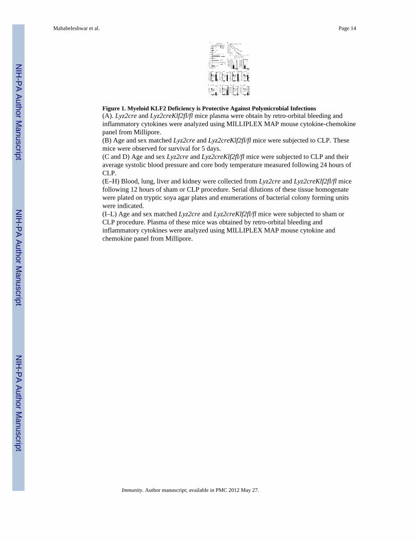
Figure 1. Myeloid KLF2 Deficiency is Protective Against Polymicrobial Infections(A). Lyz2cre and Lyz2creKlf2fl/fl mice plasma were obtain by retro-orbital bleeding andinflammatory cytokines were analyzed using MILLIPLEX MAP mouse cytokine-chemokinepanel from Millipore.(B) Age and sex matched Lyz2cre and Lyz2creKlf2fl/fl mice were subjected to CLP. Thesemice were observed for survival for 5 days.(C and D) Age and sex Lyz2cre and Lyz2creKlf2fl/fl mice were subjected to CLP and theiraverage systolic blood pressure and core body temperature measured following 24 hours ofCLP.(E–H) Blood, lung, liver and kidney were collected from Lyz2cre and Lyz2creKlf2fl/fl micefollowing 12 hours of sham or CLP procedure. Serial dilutions of these tissue homogenatewere plated on tryptic soya agar plates and enumerations of bacterial colony forming unitswere indicated.(I–L) Age and sex matched Lyz2cre and Lyz2creKlf2fl/fl mice were subjected to sham orCLP procedure. Plasma of these mice was obtained by retro-orbital bleeding andinflammatory cytokines were analyzed using MILLIPLEX MAP mouse cytokine andchemokine panel from Millipore.
Mahabeleshwar et al. Page 14
Immunity. Author manuscript; available in PMC 2012 May 27.
NIH
-PA Author Manuscript
NIH
-PA Author Manuscript
NIH
-PA Author Manuscript
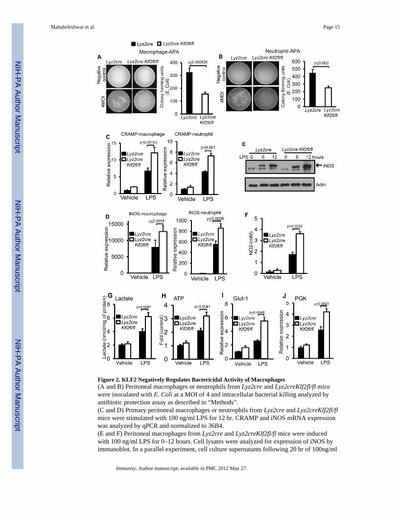
Figure 2. KLF2 Negatively Regulates Bactericidal Activity of Macrophages(A and B) Peritoneal macrophages or neutrophils from Lyz2cre and Lyz2creKlf2fl/fl micewere inoculated with E. Coli at a MOI of 4 and intracellular bacterial killing analyzed byantibiotic protection assay as described in “Methods”.(C and D) Primary peritoneal macrophages or neutrophils from Lyz2cre and Lyz2creKlf2fl/flmice were stimulated with 100 ng/ml LPS for 12 hr. CRAMP and iNOS mRNA expressionwas analyzed by qPCR and normalized to 36B4.(E and F) Peritoneal macrophages from Lyz2cre and Lyz2creKlf2fl/fl mice were inducedwith 100 ng/ml LPS for 0–12 hours. Cell lysates were analyzed for expression of iNOS byimmunoblot. In a parallel experiment, cell culture supernatants following 20 hr of 100ng/ml
Mahabeleshwar et al. Page 15
Immunity. Author manuscript; available in PMC 2012 May 27.
NIH
-PA Author Manuscript
NIH
-PA Author Manuscript
NIH
-PA Author Manuscript
of LPS treatment were analyzed for nitrite concentration by Griess assay as an index ofiNOS activity.(G and H) Lyz2cre and Lyz2creKlf2fl/fl mice peritoneal macrophages were stimulated with100ng/ml LPS. Lactate concentration from these cell culture supernatants and intracellularATP concentration from cell lysates were quantified and normalized to total protein content.(I and J) Lyz2cre and Lyz2creKlf2fl/fl mice were stimulated with 100 ng/ml LPS. Total RNAwas isolated and mRNA expression of Glut-1 and PGK was analyzed by qPCR.
Mahabeleshwar et al. Page 16
Immunity. Author manuscript; available in PMC 2012 May 27.
NIH
-PA Author Manuscript
NIH
-PA Author Manuscript
NIH
-PA Author Manuscript
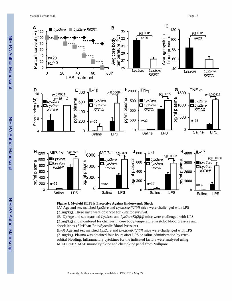
Figure 3. Myeloid KLF2 is Protective Against Endotoxemic Shock(A) Age and sex matched Lyz2cre and Lyz2creKlf2fl/fl mice were challenged with LPS(21mg/kg). These mice were observed for 72hr for survival.(B–D) Age and sex matched Lyz2cre and Lyz2creKlf2fl/fl mice were challenged with LPS(21mg/kg) and monitored for changes in core body temperature, systolic blood pressure andshock index (SI=Heart Rate/Systolic Blood Pressure).(E–J) Age and sex matched Lyz2cre and Lyz2creKlf2fl/fl mice were challenged with LPS(21mg/kg). Plasma was obtained four hours after LPS or saline administration by retro-orbital bleeding. Inflammatory cytokines for the indicated factors were analyzed usingMILLIPLEX MAP mouse cytokine and chemokine panel from Millipore.
Mahabeleshwar et al. Page 17
Immunity. Author manuscript; available in PMC 2012 May 27.
NIH
-PA Author Manuscript
NIH
-PA Author Manuscript
NIH
-PA Author Manuscript
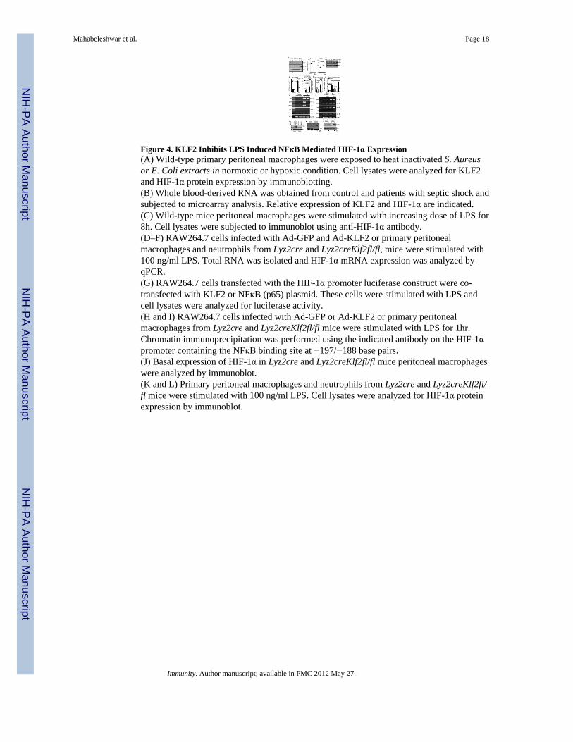
Figure 4. KLF2 Inhibits LPS Induced NFκB Mediated HIF-1α Expression(A) Wild-type primary peritoneal macrophages were exposed to heat inactivated S. Aureusor E. Coli extracts in normoxic or hypoxic condition. Cell lysates were analyzed for KLF2and HIF-1α protein expression by immunoblotting.(B) Whole blood-derived RNA was obtained from control and patients with septic shock andsubjected to microarray analysis. Relative expression of KLF2 and HIF-1α are indicated.(C) Wild-type mice peritoneal macrophages were stimulated with increasing dose of LPS for8h. Cell lysates were subjected to immunoblot using anti-HIF-1α antibody.(D–F) RAW264.7 cells infected with Ad-GFP and Ad-KLF2 or primary peritonealmacrophages and neutrophils from Lyz2cre and Lyz2creKlf2fl/fl, mice were stimulated with100 ng/ml LPS. Total RNA was isolated and HIF-1α mRNA expression was analyzed byqPCR.(G) RAW264.7 cells transfected with the HIF-1α promoter luciferase construct were co-transfected with KLF2 or NFκB (p65) plasmid. These cells were stimulated with LPS andcell lysates were analyzed for luciferase activity.(H and I) RAW264.7 cells infected with Ad-GFP or Ad-KLF2 or primary peritonealmacrophages from Lyz2cre and Lyz2creKlf2fl/fl mice were stimulated with LPS for 1hr.Chromatin immunoprecipitation was performed using the indicated antibody on the HIF-1αpromoter containing the NFκB binding site at −197/−188 base pairs.(J) Basal expression of HIF-1α in Lyz2cre and Lyz2creKlf2fl/fl mice peritoneal macrophageswere analyzed by immunoblot.(K and L) Primary peritoneal macrophages and neutrophils from Lyz2cre and Lyz2creKlf2fl/fl mice were stimulated with 100 ng/ml LPS. Cell lysates were analyzed for HIF-1α proteinexpression by immunoblot.
Mahabeleshwar et al. Page 18
Immunity. Author manuscript; available in PMC 2012 May 27.
NIH
-PA Author Manuscript
NIH
-PA Author Manuscript
NIH
-PA Author Manuscript
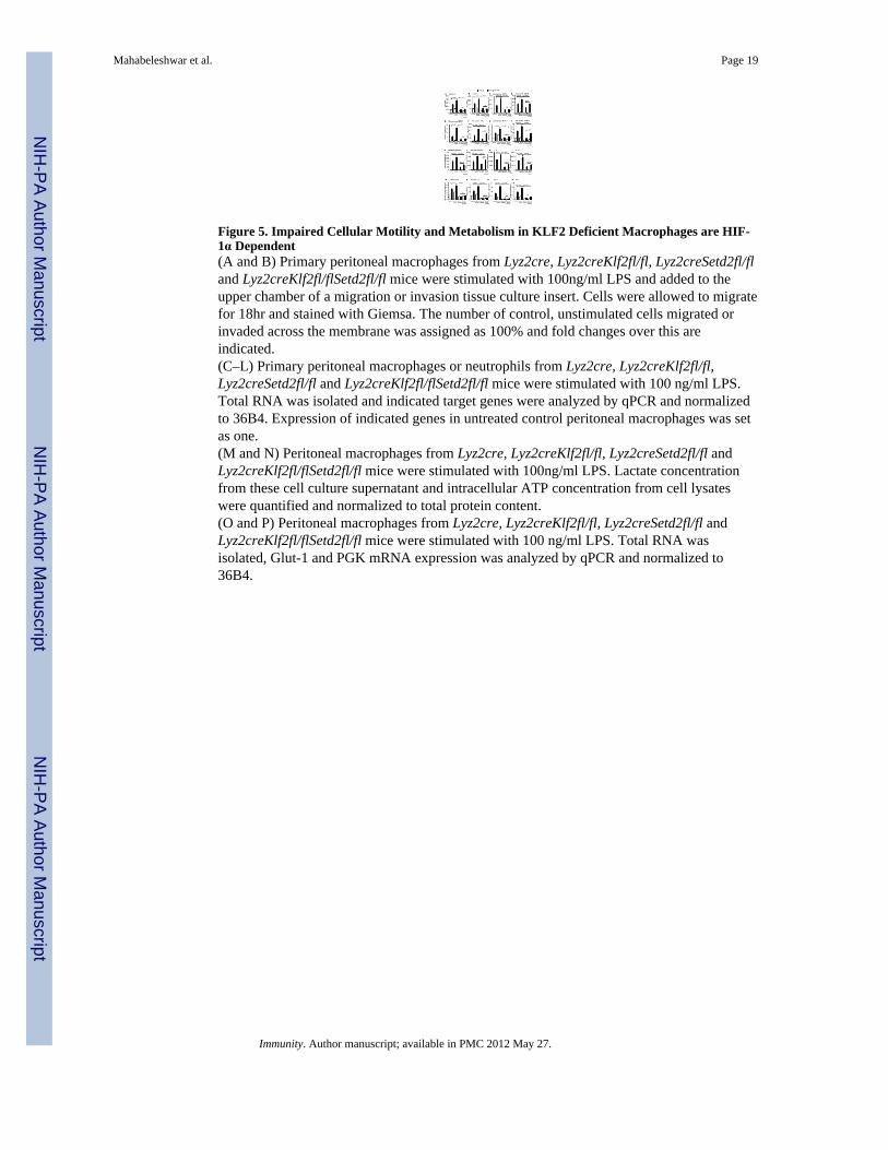
Figure 5. Impaired Cellular Motility and Metabolism in KLF2 Deficient Macrophages are HIF-1α Dependent(A and B) Primary peritoneal macrophages from Lyz2cre, Lyz2creKlf2fl/fl, Lyz2creSetd2fl/fland Lyz2creKlf2fl/flSetd2fl/fl mice were stimulated with 100ng/ml LPS and added to theupper chamber of a migration or invasion tissue culture insert. Cells were allowed to migratefor 18hr and stained with Giemsa. The number of control, unstimulated cells migrated orinvaded across the membrane was assigned as 100% and fold changes over this areindicated.(C–L) Primary peritoneal macrophages or neutrophils from Lyz2cre, Lyz2creKlf2fl/fl,Lyz2creSetd2fl/fl and Lyz2creKlf2fl/flSetd2fl/fl mice were stimulated with 100 ng/ml LPS.Total RNA was isolated and indicated target genes were analyzed by qPCR and normalizedto 36B4. Expression of indicated genes in untreated control peritoneal macrophages was setas one.(M and N) Peritoneal macrophages from Lyz2cre, Lyz2creKlf2fl/fl, Lyz2creSetd2fl/fl andLyz2creKlf2fl/flSetd2fl/fl mice were stimulated with 100ng/ml LPS. Lactate concentrationfrom these cell culture supernatant and intracellular ATP concentration from cell lysateswere quantified and normalized to total protein content.(O and P) Peritoneal macrophages from Lyz2cre, Lyz2creKlf2fl/fl, Lyz2creSetd2fl/fl andLyz2creKlf2fl/flSetd2fl/fl mice were stimulated with 100 ng/ml LPS. Total RNA wasisolated, Glut-1 and PGK mRNA expression was analyzed by qPCR and normalized to36B4.
Mahabeleshwar et al. Page 19
Immunity. Author manuscript; available in PMC 2012 May 27.
NIH
-PA Author Manuscript
NIH
-PA Author Manuscript
NIH
-PA Author Manuscript
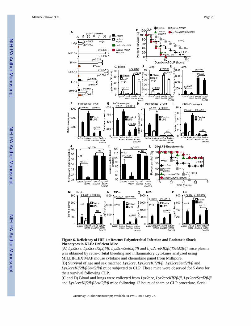
Figure 6. Deficiency of HIF-1α Rescues Polymicrobial Infection and Endotoxic ShockPhenotypes in KLF2 Deficient Mice(A) Lyz2cre, Lyz2creKlf2fl/fl, Lyz2creSetd2fl/fl and Lyz2creKlf2fl/flSetd2fl/fl mice plasmawas obtained by retro-orbital bleeding and inflammatory cytokines analyzed usingMILLIPLEX MAP mouse cytokine and chemokine panel from Millipore.(B) Survival of age and sex matched Lyz2cre, Lyz2creKlf2fl/fl, Lyz2creSetd2fl/fl andLyz2creKlf2fl/flSetd2fl/fl mice subjected to CLP. These mice were observed for 5 days fortheir survival following CLP.(C and D) Blood and lungs were collected from Lyz2cre, Lyz2creKlf2fl/fl, Lyz2creSetd2fl/fland Lyz2creKlf2fl/flSetd2fl/fl mice following 12 hours of sham or CLP procedure. Serial
Mahabeleshwar et al. Page 20
Immunity. Author manuscript; available in PMC 2012 May 27.
NIH
-PA Author Manuscript
NIH
-PA Author Manuscript
NIH
-PA Author Manuscript
dilutions of blood and lung homogenate were plated on tryptic soya agar plates andenumerations of bacterial colony forming units were indicated.(E) Peritoneal macrophages from Lyz2cre, Lyz2creKlf2fl/fl, Lyz2creSetd2fl/fl andLyz2creKlf2fl/flSetd2fl/fl mice were inoculated with E. Coli at a MOI of 2 and intracellularbacterial killing analyzed by antibiotic protection assay.(F–I) Primary peritoneal macrophages and neutrophils from Lyz2cre, Lyz2creKlf2fl/flLyz2creSetd2fl/fl and Lyz2creKlf2fl/flSetd2fl/fl mice were stimulated with 100 ng/ml LPS.Total RNA was isolated and both iNOS and CRAMP mRNA expression analyzed by qPCRand normalized to 36B4.(J–L) Lyz2cre, Lyz2creKlf2fl/fl, Lyz2creSetd2fl/fl and Lyz2creKlf2fl/flSetd2fl/fl mice weremonitored for core body temperature, systolic blood pressure and survival after LPSchallenge.(M–P) Age and sex matched Lyz2cre, Lyz2creKlf2fl/fl, Lyz2creSetd2fl/fl and Lyz2creKlf2fl/flSetd2fl/fl mice were challenged with LPS (21mg/kg). Plasma of these mice were obtainfour hours after LPS or saline administration by retro-orbital bleeding. Inflammatorycytokines for the indicated factors were analyzed using MILLIPLEX MAP mouse cytokine-chemokine panel from Millipore.
Mahabeleshwar et al. Page 21
Immunity. Author manuscript; available in PMC 2012 May 27.
NIH
-PA Author Manuscript
NIH
-PA Author Manuscript
NIH
-PA Author Manuscript
Related Documents


