The Myelin-associated Glycoproteins: Membrane Disposition, Evidence of a Novel Disulfide Linkage between Immunoglobulin-like Domains, and Posttranslational Palmitylation Liliana Pedraza,* Geoffrey C. Owens,* L. A. David Green,§ and James L. Salzer*ll Departments of *Cell Biology, § Pharmacology, and II Neurology, New York University Medical School, New York 10016; * Department of Anatomy and Neurobiology, Washington University School of Medicine, St. Louis, Missouri 63110 Abstract. The myelin-associated glycoproteins (MAG) are members of the immunoglobulin gene superfamily that function in the cell interactions of myelinating glial cells with axons. In this paper, we have charac- terized the structural features of these proteins. The disposition of MAG in the bilayer as a type 1 integral membrane protein (with an extracellularly disposed amino terminus, single transmembrane segment, and cytoplasmic carboxy terminus) was demonstrated in protease protection studies of MAG cotranslationally inserted into microsomes in vitro and in im- munofluorescent studies with site specific antibodies. A genetically engineered MAG cDNA, which lacks the putative membrane spanning segment, was con- structed and shown to encode a secreted protein. These results confirm the identity of this hydrophobic sequence as the transmembrane segment. Sequencing of the secreted protein demonstrated the presence of a cleaved signal sequence and the site of signal pepti- dase cleavage. To characterize the disulfide linkage pattern of the ectodomain, we cleaved MAG with cy- anogen bromide and used a panel of antibodies to coprecipitate specific fragments under nonreducing conditions. These studies provide support for a novel disulfide linkage between two of the immunoglobulin domains of the extracellular segment. Finally, we re- port that MAG is posttranslationally palmitylated via an intramembranous thioester linkage. Based on these studies, we propose a model for the conformation of MAG, including its RGD sequence, which is consid- ered with regard to its function as a cell adhesion molecule. T hE myelin sheath is formed by Schwann cells in the peripheral nervous system and the oligodendrocytes in the central nervous system, as a concentric, mul- tilamellar spiral around an axon. Myelin forms in close ap- position to, and its formation is regulated by contact of the glial cell with the surface of the axon (44). Cell adhesion molecules present on the extending glial process, are thought to mediate the interactions of the glial cell with the axon (36). The myelin-associated glycoproteins (MAGs) 1 are considered likely candidates for mediating these interactions (31). This suggested role is consistent with their localization to the periaxonal membrane (23, 50) and their ability to bind to large caliber axons that are destined to become myelinated (35). Two isoforms of MAG, designated large MAG (L-MAG) and small MAG (S-MAG), have been identified (19, 37). Both are heavily glycosylated integral membrane proteins (•30% by weight) that comigrate on SDS-PAGE as a broad band of 100 kD. Distinct functions for each isoform is sug- gested by their differential expression during development: L-MAG is synthesized during the rapid phase of myelination 1. Abbreviations used in this paper: IgSF, immunoglobulin family; MAG, myelin-associated glycoproteins. and S-MAG is synthesized predominately in the adult (8, 19). These isoforms are also differentially phosphorylated in vivo (6) and in vitro (1). cDNAs for both MAG isoforms have been isolated and characterized (19, 37). They are encoded by a single gene whose RNA transcript is alternatively spliced. An analysis of their deduced amino acid sequences, has suggested poten- tial structure-function correlates (37). Both proteins are predicted to have an identical, large extracellular disposed amino-terminai segment and a single transmembrane seg- ment (e.g., they are type 1 integral membrane proteins). They differ in their carboxy-terminal, presumptive cyto- plasmic sequences. The extracellular segment, which is ex- pected to mediate binding to the axon, contains five immu- noglobulin homology units. The MAG proteins are therefore members of the immunoglobulin gene superfamily (IgSF) (4, 20, 37). They are homologous to a subset of the IgSF that includes several well-characterized cell adhesion molecules, including the neural cell adhesion molecule (NCAM), L1, and I-CAM 1 (52). Also present in the ectodomain of MAG is the tripeptide recognition sequence Arg-Gly-Asp (RGD). RGD sequences are frequently an important determinant of the binding site of cell recognition and extracellular matrix © The Rockefeller University Press, 0021-9525/90/12/2651/11 $2.00 The Journal of Cell Biology, Volume 111 (No. 6, Pt. 1), Dec. 19902651-2661 2651 on February 22, 2015 jcb.rupress.org Downloaded from Published December 1, 1990

Welcome message from author
This document is posted to help you gain knowledge. Please leave a comment to let me know what you think about it! Share it to your friends and learn new things together.
Transcript
The Myelin-associated Glycoproteins: Membrane Disposition, Evidence of a Novel Disulfide Linkage between Immunoglobulin-like Domains, and Posttranslational Palmitylation Liliana Pedraza,* Geoffrey C. Owens,* L. A. David Green,§ and James L. Salzer*ll
Departments of *Cell Biology, § Pharmacology, and II Neurology, New York University Medical School, New York 10016; * Department of Anatomy and Neurobiology, Washington University School of Medicine, St. Louis, Missouri 63110
Abstract. The myelin-associated glycoproteins (MAG) are members of the immunoglobulin gene superfamily that function in the cell interactions of myelinating glial cells with axons. In this paper, we have charac- terized the structural features of these proteins. The disposition of MAG in the bilayer as a type 1 integral membrane protein (with an extracellularly disposed amino terminus, single transmembrane segment, and cytoplasmic carboxy terminus) was demonstrated in protease protection studies of MAG cotranslationally inserted into microsomes in vitro and in im- munofluorescent studies with site specific antibodies. A genetically engineered MAG cDNA, which lacks the putative membrane spanning segment, was con- structed and shown to encode a secreted protein. These results confirm the identity of this hydrophobic
sequence as the transmembrane segment. Sequencing of the secreted protein demonstrated the presence of a cleaved signal sequence and the site of signal pepti- dase cleavage. To characterize the disulfide linkage pattern of the ectodomain, we cleaved MAG with cy- anogen bromide and used a panel of antibodies to coprecipitate specific fragments under nonreducing conditions. These studies provide support for a novel disulfide linkage between two of the immunoglobulin domains of the extracellular segment. Finally, we re- port that MAG is posttranslationally palmitylated via an intramembranous thioester linkage. Based on these studies, we propose a model for the conformation of MAG, including its RGD sequence, which is consid- ered with regard to its function as a cell adhesion molecule.
T hE myelin sheath is formed by Schwann cells in the peripheral nervous system and the oligodendrocytes in the central nervous system, as a concentric, mul-
tilamellar spiral around an axon. Myelin forms in close ap- position to, and its formation is regulated by contact of the glial cell with the surface of the axon (44). Cell adhesion molecules present on the extending glial process, are thought to mediate the interactions of the glial cell with the axon (36). The myelin-associated glycoproteins (MAGs) 1 are considered likely candidates for mediating these interactions (31). This suggested role is consistent with their localization to the periaxonal membrane (23, 50) and their ability to bind to large caliber axons that are destined to become myelinated (35). Two isoforms of MAG, designated large MAG (L-MAG) and small MAG (S-MAG), have been identified (19, 37). Both are heavily glycosylated integral membrane proteins (•30% by weight) that comigrate on SDS-PAGE as a broad band of 100 kD. Distinct functions for each isoform is sug- gested by their differential expression during development: L-MAG is synthesized during the rapid phase of myelination
1. Abbreviations used in this paper: IgSF, immunoglobulin family; MAG, myelin-associated glycoproteins.
and S-MAG is synthesized predominately in the adult (8, 19). These isoforms are also differentially phosphorylated in vivo (6) and in vitro (1).
cDNAs for both MAG isoforms have been isolated and characterized (19, 37). They are encoded by a single gene whose RNA transcript is alternatively spliced. An analysis of their deduced amino acid sequences, has suggested poten- tial structure-function correlates (37). Both proteins are predicted to have an identical, large extracellular disposed amino-terminai segment and a single transmembrane seg- ment (e.g., they are type 1 integral membrane proteins). They differ in their carboxy-terminal, presumptive cyto- plasmic sequences. The extracellular segment, which is ex- pected to mediate binding to the axon, contains five immu- noglobulin homology units. The MAG proteins are therefore members of the immunoglobulin gene superfamily (IgSF) (4, 20, 37). They are homologous to a subset of the IgSF that includes several well-characterized cell adhesion molecules, including the neural cell adhesion molecule (NCAM), L1, and I-CAM 1 (52). Also present in the ectodomain of MAG is the tripeptide recognition sequence Arg-Gly-Asp (RGD). RGD sequences are frequently an important determinant of the binding site of cell recognition and extracellular matrix
© The Rockefeller University Press, 0021-9525/90/12/2651/11 $2.00 The Journal of Cell Biology, Volume 111 (No. 6, Pt. 1), Dec. 19902651-2661 2651
on February 22, 2015
jcb.rupress.orgD
ownloaded from
Published December 1, 1990
molecules that bind to a family of heterodimeric surface receptors, the integrins (11, 33). Determination of whether the RGD sequence of MAG is an important functional ele- ment therefore, has clear implications as to whether its ax- onal receptor is an integrin or whether, like most IgSF mem- bers, its axonal receptor is immunoglobulin related.
While the disposition of MAG within the lipid bilayer has been predicted to be conventional, several features of its se- quence are atypical for IgSF members. MAG has 14 cys- teines in its ectodomain distributed amongst its five immuno- globulin domains. This suggests that either MAG contains free cysteines or that it has more than the single intradomain disulfide linkage that is typical of immunoglobulin domains. There is an additional cysteine within the predicted mem- brane segment, that may also be covalently linked.
In this paper, we have characterized the structural features of MAG. We have confirmed its disposition as a type 1 inte- gral membrane protein and identified the site of signal pep- tide cleavage. In addition, we have investigated the covalent linkages of the cysteines present in the ectodomain and demonstrated the existence of a novel disulfide linkage be- tween immunoglobulin domains of MAG. Finally, we have shown that a cysteine within the lipid bilayer is posttransla- tionally palmitylated. Based on these studies, we propose a model for the conformation of MAG, including the location of its RGD sequence. This model is considered with regard to the function of MAG as a cell adhesion molecule.
Materials and Methods
In Vitro Transcription and Translation
An L-MAG cDNA was subcloned into the plasmid, pGEM4 (Promega Bio- tec, Madison, WI). Complementary RNA was transcribed using SP6 poly- merase in the presence of the RNA cap analogue m7G(5')ppp(5~G (Pharma- cia Fine Chemicals, Piscataway, NJ) according to the directions of the manufacturer. 1 #1 of the transcription reaction was used to program a rabbit reticulocyte translation system (New England Nuclear, Boston, MA) con- taining [3SS]methionine (New England Nuclear) for 30 rain at 37°C with or without canine pancreatic microsomes. For protease protection studies, translation rgi~tures were divided into equal aliquots: one was treated on ice for 45"~'min with proteinase K(Boehringer-Mannheim Biochemicals) at 0.5 rng/ml final concentration and the other served as a control. A 10-fold excess of ice-cold methanol was then added to precipitate proteins and pellets were collected at 13,000 g. Immunoprecipitation procedures are de- scribed below.
Generation of anti-MAG Antibodies Rabbit polyclonals were prepared against two MAG segments: a 20-mer corresponding to amino acids 109-128 of the published amino acid se- quence of MAG(37), which includes the RGD sequence (peptide 1) and the carboxy terminal 20 amino acids of L-MAG (peptide 2). Both peptides were synthesized with carboxy terminal cysteines and were coupled, by cross- linking with m-maleimidobenzoyl-N-hydroxysulfosuccinimide ester (Pierce Chemical Co., Rockford, IL) after the manufacturer's instructions, to key- hole limpet hemocyanin (KLH). Rabbits were inoculated with the coupled peptides as previously described (37).
Expression of MAG Isoforms in a Schwann Cell Line Construction of a recombinant retrovirus encoding L-MAG has been previ- ously described (27). Construction of an S-MAG encoding retrovirus was performed in a similar manner. Briefly, Hind HI linkers were added to the 2087 bp Apa 1 cDNA fragment of S-MAG, corresponding to the complete coding sequence and this cDNA was subcloned into the Hind HI site of the retroviral vector, pMV-7(16). This construct was introduced into the ecotropic packaging cell Psi2 by calcium phosphate-mediated DNA trans-
fection (51). Viral stocks shed by the transfected psi 2 cells were used to infect an immortalized Schwann cell line (29) as described (16). Individual clones that were resistant to the neomycin analogue, G418 (Gibco Labora- tories, Grand Island, NY) at a concentration of 800 #g/ml, were selected and checked for their expression of MAG by immunofluorescent staining.
lndirect Immunofluorescence Permanently transfected L-MAG Schwann cells, plated onto glass cover- slips, were washed twice with PBS, fixed for 10 min with 4% formaldehyde and, in some cases, were then permeabilized with 0.05 % Triton X-100 in PBS for 5 min. Cells were washed twice with PBS and then incubated for 30 rain in PBS/5% normal goat serum to block nonspecific binding. The primary antibodies used were the polyclonal antibodies to peptide 1 and peptide 2, used at a 1:100 dilution in PBS/5% normal goat serum, and an anti-MAG mAb (28), (a gift of Dr. M. Schachner, ETH, Zurich) used as undiluted hybridoma culture supernatant. Ceils were incubated for 1 h at room temperature then washed, and a secondary antibody, fluoresceinated goat anti-rabbit or antimouse (Cappel Laboratories, Malvern, PA), was added at 1:100 in PBS/5% normal goat serum. Coverslips were washed three times in PBS, mounted in a PBS-gelatin solution, and viewed by epifluorescence microscopy.
Construction of a Recombinant cDNA Corresponding to the Ectodomain of MAG To generate a secreted form of MAG, we introduced a translation stop codon amino terminal to the predicted membrane spanning segment of an L-MAG cDNA (after nucleotide position 1,665 of the published MAG sequence [37]). A sense oligunucleotide and a complementary antisense oligunucleo- tide were synthesized (model #380A DNA Synthesizer; Applied Binsys- terns, Inc., Foster City, CA), gel purified, and annealed together. The result- ing double stranded DNA fragment shown below contained an Xho I-compatible overhang 5' on the sense (top) strand, the coding sequences of the 15 amino acids proximal to the predicted membrane spanning seg- ment, a translation stop codon (TGA), and an Eco RI-compatible overhang (3' on the sense strand) to facilitate subcloning into an expression vector.
5 '-TC GAG CTGC C TTTC CAGGGAGCACACC GACTGATG TGGGC CAA ATGAG- 3 ' 3'-CGACGGAA AGGTCCC TCGTGTGG CTGACTACACCC GGTT TAC TCTTAA-5'
This double stranded fragment was then ligated to the 1,619-bp Eco RI-Xhol fragment that encodes the major portion of the ectodomain of MAG. The resultant cDNA, which encoded the entire extracellular domain of MAG, was subcloned into the Eco RI site of the expression vector pMT2 (15) to create pMT2-ECD. A similar strategy was also used recently to generate a soluble form of MAG in transfected cells (13).
COS Cell Expression and Purification of Soluble MAG COS-7 ceils were transfected with pMT2-ECD by the DEAE-dextran (22). 72 h after transfection, culture media was harvested. In most experiments, cells were grown under low serum conditions (DMEM with 0.5% Serum Plus; Gibco Laboratories), which greatly simplified subsequent purifica- tion of the secreted MAG protein because of the reduced level of proteins in the media without adversely affecting the level of MAG secretion. The media was then concentrated by ultrafiltration over an Amicoti Filter (Ami- con Corp., Danvers, MA) (tool mass cutoff Of 30 kD), and the soluble MAG was purified by lentil lectin chromatography, elution with 0.5 M methyl mannoside, followed by fast protein liquid chromatography on a Mono Q anion exchange column (Pharmacia Fine Chemicals, Piscataway, NJ). The secreted MAG eluted in phosphate buffered saline using a gradient of NaCI, at a concentration of 0.2 M.
Peptide Sequencing Purified soluble MAG was sequenced by automated gas-phase protein se- quencing (model 470; Applied Biosystems, Inc.).
Immunoprecipitation and Immunobiotting Total cell lysates were prepared by scraping up 106 transfected cells and boiling in 2 % SDS. Samples were diluted fourfold with PBS containing Triton-X-100 (0.5%) and 1 ml samples were precleared with protein A cou- pled to Sepharose CL4B (Sigma Chemical Co.) by incubating with 10 #1 of beads for 2 h and briefly centrifuging at 13,000 g. The supernatants were
The Journal of Cell Biology, Volume 111, 1990 2652
on February 22, 2015
jcb.rupress.orgD
ownloaded from
Published December 1, 1990
incubated overnight at 4°C with 30 #1 of the anti-peptide antibodies. Im- mtmoprecipitates were collected with protein A/Sepharose, released by boiling for 2 min in SDS-PAGE sample buffer, and fractionated by elec- trophoresis on 7.5-15% polyacrylamide gradient gel (18). The gels were fixed in 30% methanol, 10% acetic acid, 60% water, treated with EN3HANCE following the manufacturer's instructions (New England Nu- clear), and fluorographed. Western blotting of samples was carded out by described procedures (47) and antibodies were visualized with 125I-Protein A (ICN Biochemicals, lrvine, CA).
Alkylation of Cysteines Purified soluble MAG or immunoprecipitated L-MAG (from transfected Schwann cells) were alkylated with neutralized 14C iodoacetic acid (5/~Ci/ ml) (ICN Biochemicals) at room temperature in the dark in a solution consisting of 0.2 M Tris-HC1 (pH 8.1), 2 mM EDTA, and 6 M guanidine hydroeloride. Samples were either alkylated directly or alkylated after prereduction with a t00-fold molar excess of DTT for l h at 37°C. After 60 min, the alkylation was stopped by quenching with an excess of ~-mer- captoethanol. The samples were dialyzed against 0.1 M Tris, pH 7.4 and analyzed by SDS-PAGE.
Metabolic Labeling of Cells Confluent T-75 flasks of transfected Schwann cells were labeled with either [35S]cysteine, [35S]methionine, or [3H]palmitate. To label MAG with [35S]cysteine, cells were preincubated in cysteine-free DME for 30 rain, and then labeled with 100/~Ci/ml of [3~S]eysteine in eysteine-free DME (G-ibco Laboratories) for 2 h at 370C, followed by an additional 2-h chase in DME supplemented with 10% FCS. For the palmitytation studies, the transfected Schwann cells were labeled with 400 /~Ci/ml of 9-10- [3H]palmitic acid (30 mCi/mmol; New England Nuclear) in DME with 5 mM Na pyruvate plus 1% FCS for 5 h at 370C. In all cases, at the end of the labeling period the cells were washed twice with PBS, lysed in 2% SDS, scraped into a conical tube, sonicated for 60 s and boiled for 2 min before immunoprecipitation with anti-MAG antibodies.
Cyanogen Bromide Cleavage 5/zl of a cyanogen bromide (CNBr) solution (2 g/ml in DMSO) were added to [3sS]Cysteine-labeled L-MAG immunoprecipitates resuspended in 0.5 ml of 70% formic acid. The reaction mixture was incubated in the dark overnight at room temperature. To control for partial cleavage of Asp-Pro bonds resulting from incubation in the acidic medium, additional samples were incubated in 70% formic acid without CNBr. The reaction mixture was then diluted 10-fold with water and lyophilized. The lyophilized sam- pies were sohibilized in sample buffer (without B-mereaptoethanol), soni- cated for 30 s, and boiled for 2 min. Samples were then immunoprecipitated with antiMAG antibodies and analyzed by SDS-PAGE with or without /3-mercaptoethanol present.
Characterization of the Palmitoyl Moiety To analyze the sensitivity of the MAG-palmityl bond to neutral hydroxyl- amine, MAG labeled with [3H]palmitate was separated by SDS-PAGE and the gels were soaked for 4 h at room temperature in at least 10 vol of 1 M Tris (pH 7.0) or 1 M hydroxylamine (pH 7.0). The gels were then washed for 1 h in water and prepared for fluorography. Quantitation of fluorographs was performed by optical densitometry.
To identify the nature of the acyl group, the immunoprecipitates of the MAG labeled with [3H]palmitic acid were washed in deionized water and then in methanol. The immunoprecipitates were incubated for 4 h in 1 ml 0.l M KOH in methanol. The supernatant yeas removed and extracted with 2 vol of chloroform/water (1:1); the upper (aqueous) phase was reextracted with 1 vol of chloroform. The organic phases were combined and washed three times with chloroform/methanol/water (1:10:10). The final organic phase was dried under a nitrogen stream, redisselved in methanol, and ana- lysed by reverse-phase HPLC on a CI 8 column (Supelcosil LC-318, 4.6 mm × 25 cm). The mobile phase was acetonitrile/water (85:15, vol/vol); with a flow rate~f I ml/min. Standards consisting of myristic acid, palmitic acid, methyl myristate, and methyl palmitate (Sigma Chemical Co.) were in- jected with the radioactive sample. Fractions were collected at I min inter- vals while monitoring at 210 nm by UV absorption, and radioactivity in in- dividual fractions was determined by scintillation counting. Data analysis and graphics were done on a Macintosh II computer.
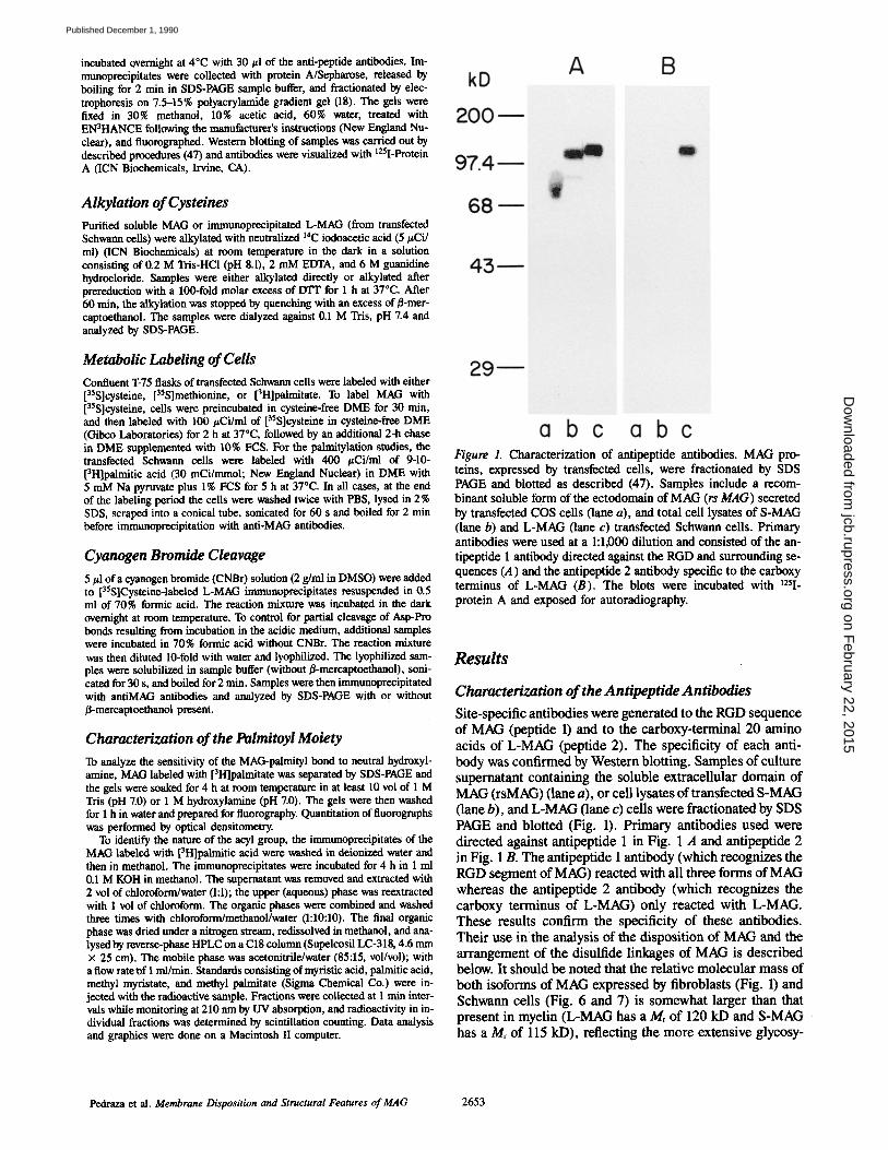
Figure 1. Characterization of antipeptide antibodies. MAG pro- teins, expressed by transfected cells, were fractionated by SDS PAGE and blotted as described (47). Samples include a recom- binant soluble form of the ectodomain of MAG (rs MAG) secreted by transfected COS cells (lane a), and total cell lysates of S-MAG (lane b) and L-MAG (lane c) transfected Schwann cells. Primary antibodies were used at a 1:1,000 dilution and consisted of the an- tipeptide 1 antibody directed against the RGD and surrounding se- quences (A) and the antipeptide 2 antibody specific to the carboxy terminus of L-MAG (B). The blots were incubated with 125I- protein A and exposed for autoradiography.
Results
Characterization of the Antipeptide Antibodies Site-specific antibodies were generated to the RGD sequence of MAG (peptide 1) and to the carboxy-terminal 20 amino acids of L-MAG (peptide 2). The specificity of each anti- body was confirmed by Western blotting. Samples of culture supernatant containing the soluble extracellular domain of MAG (rsMAG) (lane a), or cell lysates of transfected S-MAG (lane b), and L-MAG (lane c) cells were fractionated by SDS PAGE and blotted (Fig. 1). Primary antibodies used were directed against antipeptide 1 in Fig. 1 A and antipeptide 2 in Fig. 1 B. The antipeptide 1 antibody (which recognizes the RGD segment of MAG) reacted with all three forms of MAG whereas the antipeptide 2 antibody (which recognizes the carboxy terminus of L-MAG) only reacted with L-MAG. These results confirm the specificity of these antibodies. Their use inthe analysis of the disposition of MAG and the arrangement of the disulfide linkages of MAG is described below. It should be noted that the relative molecular mass of both isoforms of MAG expressed by fibroblasts (Fig. 1) and Schwann cells (Fig. 6 and 7) is somewhat larger than that present in myelin (L-MAG has a M, of 120 kD and S-MAG has a Mr of 115 kD), reflecting the more extensive glycosy-
Pedraza et al. Membrane Disposition and Structural Features of MAG 2653
on February 22, 2015
jcb.rupress.orgD
ownloaded from
Published December 1, 1990
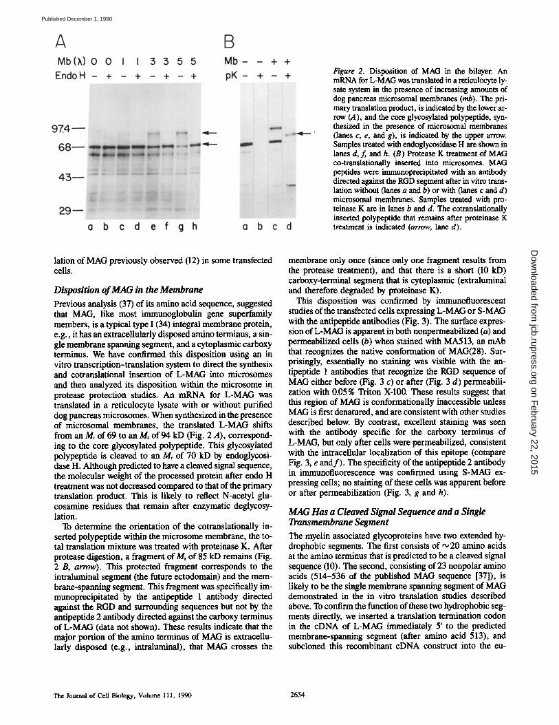
Figure 2. Disposition of MAG in the bilayer. An mRNA for L-MAG was translated in a reticulocyte ly- sate system in the presence of increasing amounts of dog pancreas microsomal membranes (rob). The pri- mary translation product, is indicated by the lower ar- row (A), and the core glycosylated polypeptide, syn- thesized in the presence of microsomal membranes (lanes c, e, and g), is indicated by the upper arrow. Samples treated with endoglycosidase H are shown in lanes d, f, and h. (B) Protease K treatment of MAG co-translationally inserted into microsomes. MAG peptides were immunoprecipitated with an antibody directed against the RGD segment after in vitro trans- lation without (lanes a and b) or with (lanes c and d) microsomal membranes. Samples treated with pro- teinase K are in lanes b and d. The cotranslationally inserted polypeptide that remains after proteinase K treatment is indicated (arrow, lane d).
lation of MAG previously observed (12) in some transfected cells.
Disposition of MAG in the Membrane
Previous analysis (37) of its amino acid sequence, suggested that MAG, like most immunoglobulin gene superfamily members, is a typical type I (34) integral membrane protein, e.g., it has an extraceUularly disposed amino terminus, a sin- gle membrane spanning segment, and a cytoplasmic carboxy terminus. We have confirmed this disposition using an in vitro transcription-translation system to direct the synthesis and cotranslational insertion of L-MAG into microsomes and then analyzed its disposition within the microsome in protease protection studies. An mRNA for L-MAG was translated in a reticulocyte lysate with or without purified dog pancreas microsomes. When synthesized in the presence of microsomai membranes, the translated L-MAG shifts from an Mr of 69 to an Mr of 94 kD (Fig. 2 A), correspond- ing to the core glycosylated polypeptide. This glycosylated polypeptide is cleaved to an Mr of 70 kD by endoglycosi- dase H. Although predicted to have a cleaved signal sequence, the molecular weight of the processed protein after endo H treatment was not decreased compared to that of the primary translation product. This is likely to reflect N-acetyl glu- cosamine residues that remain after enzymatic deglycosy- lation.
To determine the orientation of the cotranslationaily in- serted polypeptide within the microsome membrane, the to- tal translation mixture was treated with proteinase K. After protease digestion, a fragment of Mr of 85 kD remains (Fig. 2 B, arrow). This protected fragment corresponds to the intraluminal segment (the future ectodomain) and the mem- brane-spanning segment. This fragment was specifically im- munoprecipitated by the antipeptide 1 antibody directed against the RGD and surrounding sequences but not by the antipeptide 2 antibody directed against the carboxy terminus of L-MAG (data not shown). These results indicate that the major portion of the amino terminus of MAG is extracellu- larly disposed (e.g., intraluminal), that MAG crosses the
membrane only once (since only one fragment results from the protease treatment), and that there is a short (10 kD) carboxy-terminal segment that is cytoplasmic (extraluminai and therefore degraded by proteinase K).
This disposition was confirmed by immunofluorescent studies of the transfected cells expressing L-MAG or S-MAG with the antipeptide antibodies (Fig. 3). The surface expres- sion of L-MAG is apparent in both nonpermeabilized (a) and permeabilized cells (b) when stained with MA513, an mAb that recognizes the native conformation of MAG(28). Sur- prisingly, essentially no staining was visible with the an- tipeptide 1 antibodies that recognize the RGD sequence of MAG either before (Fig. 3 c) or after (Fig. 3 d) permeabili- zation with 0.05 % Triton X-100. These results suggest that this region of MAG is conformationally inaccessible unless MAG is first denatured, and are consistent with other studies described below. By contrast, excellent staining was seen with the antibody specific for the carboxy terminus of L-MAG, but only after cells were permeabilized, consistent with the intracellular localization of this epitope (compare Fig. 3, e and f ) . The specificity of the antipeptide 2 antibody in immunofluorescence was confirmed using S-MAG ex- pressing cells; no staining of these cells was apparent before or after permeabilization (Fig. 3, g and h).
MAG Has a Cleaved Signal Sequence and a Single Transmembrane Segment
The myelin associated glycoproteins have two extended hy- drophobic segments. The first consists of '~ 20 amino acids at the amino terminus that is predicted to be a cleaved signal sequence (10). The second, consisting of 23 nonpolar amino acids (514-536 of the published MAG sequence [37]), is likely to be the single membrane spanning segment of MAG demonstrated in the in vitro translation studies described above. To confirm the function of these two hydrophobic seg- ments directly, we inserted a translation termination codon in the cDNA of L-MAG immediately 5' to the predicted membrane-spanning segment (after amino acid 513), and subcloned this recombinant cDNA construct into the eu-
The Journal of Cell Biology, Volume 111, 1990 2654
on February 22, 2015
jcb.rupress.orgD
ownloaded from
Published December 1, 1990
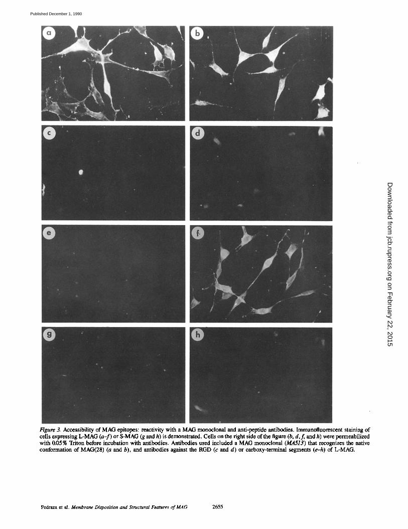
Figure 3. Aecessibility of MAG epitopes: reactivity with a MAG monoclonal and anti-peptide antibodies. Immunofluorescent staining of cells expressing L-MAG (a-f) or S-MAG (g and h) is demonstrated. Cells on the right side of the figure (b, d,f , and h) were permeabilized with 0.05% Triton before incubation with antibodies. Antibodies used included a MAG monoclonal (MA513) that recognizes the native conformation of MAG(28) (a and b), and antibodies against the RGD (c and d) or carboxy-terminal segments (e-h) of L-MAG.
Pedraza et al. Membrane Disposition and Structural Features of MAG 2655
on February 22, 2015
jcb.rupress.orgD
ownloaded from
Published December 1, 1990
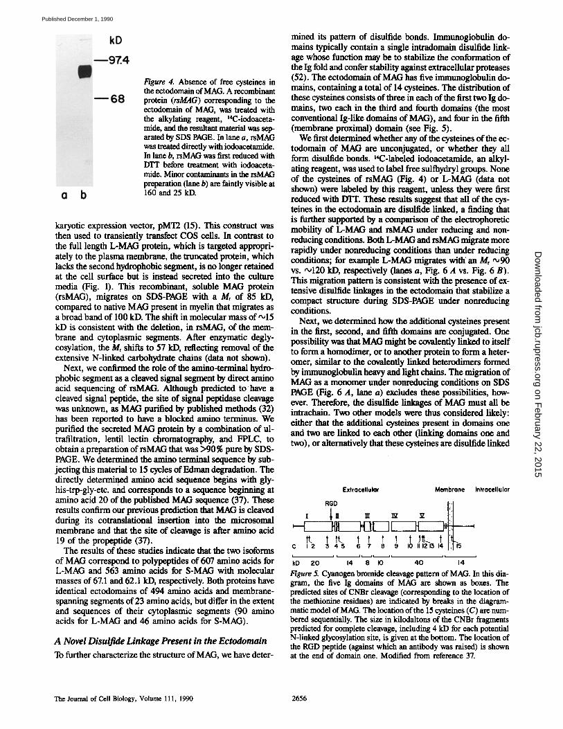
Figure 4. Absence of free cysteines in the e~etodomain of MAG. A recombinant protein (rsMAG) corresponding to the ectodomain of MAG, was treated with the alkylating reagent, ~4C-iodoaeeta- mide, and the resultant material was sep- arated by SDS PAGE. In lane a, rsMAG was treated directly with iodoacetamide. In lane b, rsMAG was first reduced with ITI'T before treatment with iodoaceta- mide. Minor contaminants in the rsMAG preparation (lane b) are faintly visible at 160 and 25 kD.
karyotic expression vector, pMT2 (15). This construct was then used to transiently transfect COS cells. In contrast to the full length L-MAG protein, which is targeted appropri- ately to the plasma membrane, the truncated protein, which lacks the second hydrophobic segment, is no longer retained at the cell surface but is instead secreted into the culture media (Fig. 1). This recombinant, soluble MAG protein (rsMAG), migrates on SDS-PAGE with a Mr of 85 kD, compared to native MAG present in myelin that migrates as a broad band of 100 kD. The shift in molecular mass of,~15 kD is consistent with the deletion, in rsMAG, of the mem- brane and cytoplasmic segments. After enzymatic degly- cosylation, the Mr shifts to 57 kD, reflecting removal of the extensive N-linked carbohydrate chains (data not shown).
Next, we confirmed the role of the amino-terminal hydro- phobic segment as a cleaved signal segment by direct amino acid sequencing of rsMAG. Although predicted to have a cleaved signal peptide, the site of signal peptidase cleavage was unknown, as MAG purified by published methods (32) has been reported to have a blocked amino terminus. We purified the secreted MAG protein by a combination of ul- trafiltration, lentil lectin chromatography, and FPLC, to obtain a preparation of rsMAG that was >90% pure by SDS- PAGE. We determined the amino terminal sequence by sub- jecting this material to 15 cycles of Edman degradation. The directly determined amino acid sequence begins with gly- his-trp-gly-etc, and corresponds to a sequence beginning at amino acid 20 of the published MAG sequence 07) . These results confirm our previous prediction that MAG is cleaved during its cotranslational insertion into the microsomal membrane and that the site of cleavage is after amino acid 19 of the propeptide (37).
The results of these studies indicate that the two isoforms of MAG correspond to polypeptides of 607 amino acids for L-MAG and 563 amino acids for S-MAG with molecular masses of 67.1 and 62.1 kD, respectively. Both proteins have identical ectodomains of 494 amino acids and membrane- spanning segments of 23 amino acids, but differ in the extent and sequences of their cytoplasmic segments (90 amino acids for L-MAG and 46 amino acids for S-MAG).
A Novel Disulfule Linkage Present in the Ectodomain
To further characterize the structure of MAG, we have deter-
mined its pattern of disulfide bonds. Immunoglobulin do- mains typically contain a single intradomain disulfide link- age whose function may be to stabilize the conformation of the Ig fold and confer stability against extracellular proteases (52). The ectodomain of MAG has five immunoglobulin do- mains, containing a total of 14 cysteines. The distribution of these cysteines consists of three in each of the first two Ig do- mains, two each in the third and fourth domains (the most conventional Ig-like domains of MAG), and four in the fifth (membrane proximal) domain (see Fig. 5).
We first determ'med whether any of the cysteines of the ec- todomain of MAG are unconjugated, or whether they all form disulfide bonds. ~4C-labeled iodoacetamide, an alkyl- ating reagent, was used to label free sulfhydryl groups. None of the cysteines of rsMAG (Fig. 4) or L-MAG (data not shown) were labeled by this reagent, unless they were first reduced with DTT. These results suggest that all of the cys- teines in the ectodomain are disulfide linked, a finding that is further supported by a comparison of the electrophoretic mobility of L-MAG and rsMAG under reducing and non- reducing conditions. Both L-MAG and rsMAG migrate more rapidly under nonreducing conditions than under reducing conditions; for example L-MAG migrates witth an Mr '~90 vs. 'o120 kD, respectively (lanes a, Fig. 6 A vs. Fig. 6 B). This migration pattern is consistent with the presence of ex- tensive disulfide linkages in the ectedomain that stabilize a compact structure during SDS-PAGE under nonreducing conditions.
Next, we determined how the additional cysteines present in the first, second, and fifth domains are conjugated. One possibility was that MAG might be covalently linked to itself to form a homodimer, or to another protein to form a heter- omer, similar to the covalently linked heterodimers formed by immunoglobulin heavy and light chains. The migration of MAG as a monomer under nonreducing conditions on SDS PAGE (Fig. 6 A, lane a) excludes these possibilities, how- ever. Therefore, the disulfide linkages of MAC; must all be intrachain. Two other models were thus considered likely: either that the additional cysteines present in domains one and two are linked to each other Oinking domains one and two), or alternatively that these cysteines are disulfide linked
Extracellular Membrane Intracellular
t~. t tL t t t t t ~ C 1 2 3 4 5 6 7 8 9 I0 I11213 1
I I I l~,~l II it i
kD ZO 14 8 I0 40 14
Figure 5. Cyanogen bromide cleavage pattern of MAG. In this dia- gram, the five Ig domains of MAG are shown as boxes. The predicted sites of CNBr cleavage (corresponding to the location of the methionine residues) are indicated by breaks in the diagram- matic model of MAG. The location of the 15 cysteines (C) are num- bered sequentially. The size in kilodaltons of the CNBr fragments predicted for complete cleavage, including 4 kD for each potential N-linked glycosylation site, is given at the bottom. The location of the RGD peptide (against which an antibody was raised) is shown at the end of domain one. Modified from reference 37.
The Journal of Cell Biology, Volume 111, 1990 2656
on February 22, 2015
jcb.rupress.orgD
ownloaded from
Published December 1, 1990
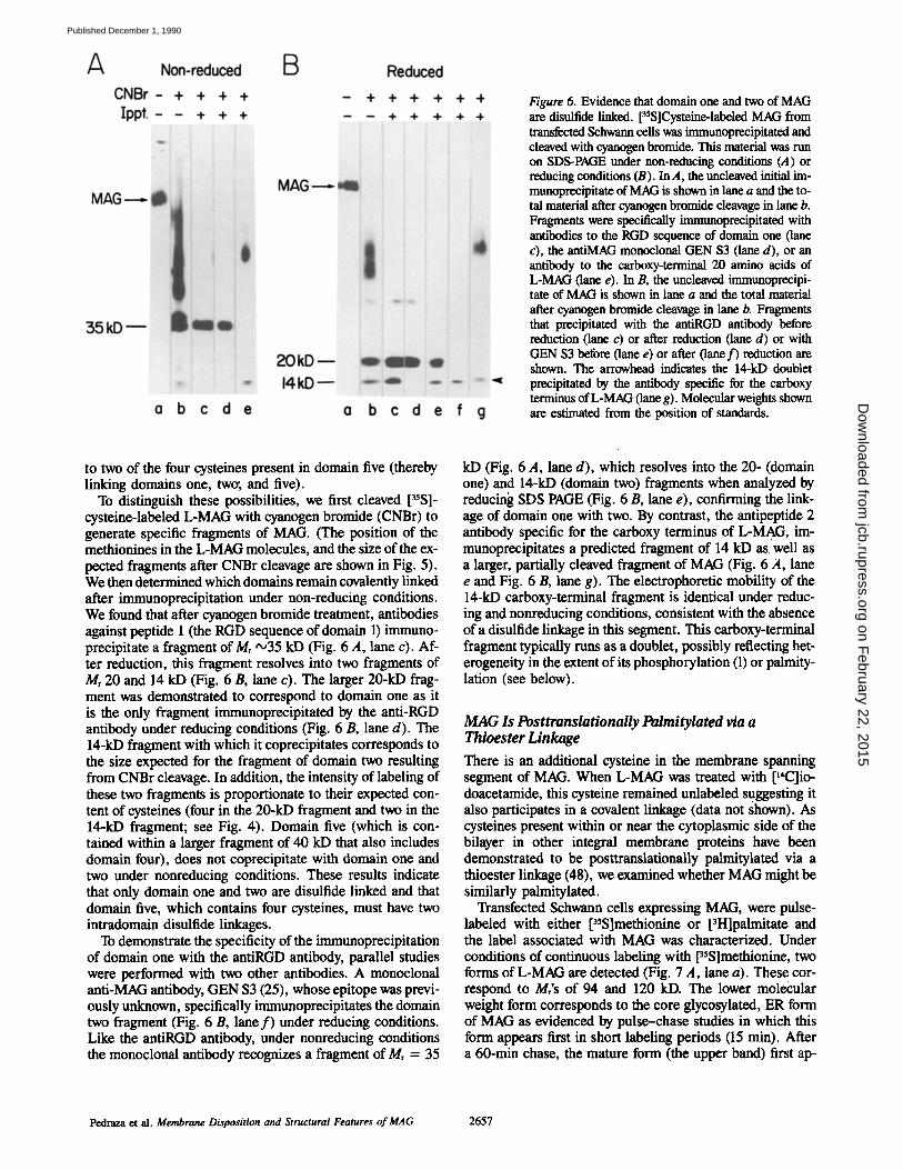
Figure 6. Evidence that domain one and two of MAG are disulfide linked. [35S]Cysteine-labeled MAG from transfected Schwarm cells was immunoprecipitated and cleaved with cyanogen bromide. This material was run on SDS-PAGE under non-reducing conditions (A) or reducing conditions (B). In A, the uncleaved initial im- mtmoprecipitate of MAG is shown in lane a and the to- tal material after cyanogen bromide cleavage in lane b. Fragments were specifically immunoprecipitated with antibodies to the RGD sequence of domain one (lane c), the antiMAG monoclonal GEN $3 (lane d), or an antibody to the carboxy-terminal 20 amino acids of L-MAG (lane e). In B, the uncleaved immunoprecipi- tare of MAG is shown in lane a and the total material after cyanogen bromide cleavage in lane b. Fragments that precipitated with the antiRGD antibody before reduction (lane c) or after reduction (lane d) or with GEN $3 before (lane e) or after (lane f ) reduction are shown. The arrowhead indicates the 14-kD doublet precipitated by the antibody specific for the carboxy terminus of L-MAG (lane g). Molecular weights shown are estimated from the position of standards.
to two of the four cysteines present in domain five (thereby linking domains one, twos, and five).
To distinguish these possibilities, we first cleaved [35S]- cysteine-labeled L-MAG with cyanogen bromide (CNBr) to generate specific fragments of MAG. (The position of the methionines in the L-MAG molecules, and the size of the ex- pected fragments after CNBr cleavage are shown in Fig. 5). We then determined which domains remain covalently linked after immunoprecipitation under non-reducing conditions. We found that after cyanogen bromide treatment, antibodies against peptide 1 (the RGD sequence of domain 1) immuno- precipitate a fragment of Mr ,,o35 kD (Fig. 6 A, lane c). Af- ter reduction, this fragment resolves into two fragments of Mr 20 and 14 kD (Fig. 6 B, lane c). The larger 20-kD frag- ment was demonstrated to correspond to domain one as it is the only fragment immunoprecipitated by the anti-RGD antibody under reducing conditions (Fig. 6 B, lane d). The 14-kD fragment with which it coprecipitates corresponds to the size expected for the fragment of domain two resulting from CNBr cleavage. In addition, the intensity of labeling of these two fragments is proportionate to their expected con- tent of cysteines (four in the 20-kD fragment and two in the 14-kD fragment; see Fig. 4). Domain five (which is con- tained within a larger fragment of 40 kD that also includes domain four), does not coprecipitate with domain one and two under nonreducing conditions. These results indicate that only domain one and two are disulfide linked and that domain five, which contains four cysteines, must have two intradomain disulfide linkages.
To demonstrate the specificity of the immunoprecipitation of domain one with the antiRGD antibody, parallel studies were performed with two other antibodies. A monoclonal anti-MAG antibody, GEN $3 (25), whose epitope was previ- ously unknown, specifically immunoprecipitates the domain two fragment (Fig. 6 B, lane f ) under reducing conditions. Like the antiRGD antibody, under nonreducing conditions the monoclonal antibody recognizes a fragment of Mr = 35
kD (Fig. 6 A, lane d), which resolves into the 20- (domain one) and 14-kD (domain two) fragments when analyzed by reducing SDS PAGE (Fig. 6 B, lane e), confirming the link- age of domain one with two. By contrast, the antipeptide 2 antibody specific for the carboxy terminus of L-MAG, im- munoprecipitates a predicted fragment of 14 kD as well as a larger, partially cleaved fragment of MAG (Fig. 6 A, lane e and Fig. 6 B, lane g). The electrophoretic mobility of the 14-kD carboxy-terminal fragment is identical under reduc- ing and nonreducing conditions, consistent with the absence of a disulfide linkage in this segment. This carboxy-terminal fragment typically runs as a doublet, possibly reflecting het- erogeneity in the extent of its phosphorylation (1) or paimity- lation (see below).
M A G Is PosttranslationaUy Palmitylated via a Thioester Linkage
There is an additional cysteine in the membrane spanning segment of MAG. When L-MAG was treated with [14C]io- doacetamide, this cysteine remained unlabeled suggesting it also participates in a covalent linkage (data not shown). As cysteines present within or near the cytoplasmic side of the bilayer in other integral membrane proteins have been demonstrated to be posttranslationally palmitylated via a thioester linkage (48), we examined whether MAG might be similarly palmitylated.
Transfected Schwann cells expressing MAG, were pulse- labeled with either [35S]methionine or [3H]palmitate and the label associated with MAG was characterized. Under conditions of continuous labeling with [35S]methionine, two forms of L-MAG are detected (Fig. 7 A, lane a). These cor- respond to Mr's of 94 and 120 kD. The lower molecular weight form corresponds to the core glycosylated, ER form of MAG as evidenced by pulse-chase studies in which this form appears first in short labeling periods (15 min). After a 60-min chase, the mature form (the upper band) first ap-
Pedraza et al. Membrane Disposition and Structural Features of MAG 2657
on February 22, 2015
jcb.rupress.orgD
ownloaded from
Published December 1, 1990
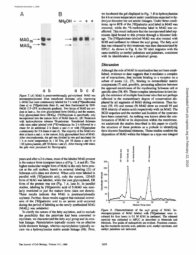
Figure 7. (A) MAG is posttrramlationally palmitylated. MAG was immunoprecipitated from transfected Schwann cells expressing L-MAG that were continuously labeled for 5 h with [35S]methionine (lane a) or [3I-I]palmitate (lane b), and then fractionated by SDS- PAGE (7.5-15% acrylamide gradient). Two forms of MAG are evi- dent in lane a, the core glycosylated form (MAGo) and the mature, fully glycosylated form (MAGi). [3H]Palmitate is specifically only incorporated into the mature form of MAG (lane b). (B) Treatment with hydroxylamine releases 3H-palmitate. Transfected Schwann cells were pulse labeled with [35S]methionine for 2 h followed by a chase for an additional 2 h (lanes a and c) or with [3H]palmitate continuously for 5 h (lanes b and d). The majority of the MAG evi- dent in lanes a and c, is the mature, fully glycosylated form of MAG. After electrophoresis, the gel was divided in two and incubated for 1 h at room temperature in 1 M Tris, pH 7.0 (lanes a and b) or 1 M hydroxylamine, pH 7.0 (lanes c and d). After rinsing with water, the gels were processed for fluorography.
pears and after a 2-h chase, most of the labeled MAG present is the mature form (compare lanes a of Fig. 7, A and B). The higher molecular weight form of MAG is the only form pres- ent at the cell surface, based on external labeling (21) of Schwann cells (data not shown). When cells were labeled in parallel with pH]palmitic acid, only the mature, 120-kD form of MAG was labeled, while the core glycosylated, ER form of the protein was not (Fig. 7 A, lane b). In parallel studies, labeling by PH]palmitic acid of S-MAG was simi- larly restricted to just the mature form (data not shown). These results indicate that MAG is posttranslationally acylated. Further, these results suggest that minimal conver- sion of the pH]palmitic acid to an amino acid occurred during the period of labeling as the newly synthesized MAG (MAGo) was unlabeled.
To clarify the nature of the fatty acylation, and to exclude the possibility that the palmitate had been converted to myristate, we characterized the fatty acyl group and its cova- lent linkage. Palmitylation typically occurs via an alkaline labile thioester linkage, whereas myristylation typically oc- curs via a hydroxylamine stable amide linkage (48). First,
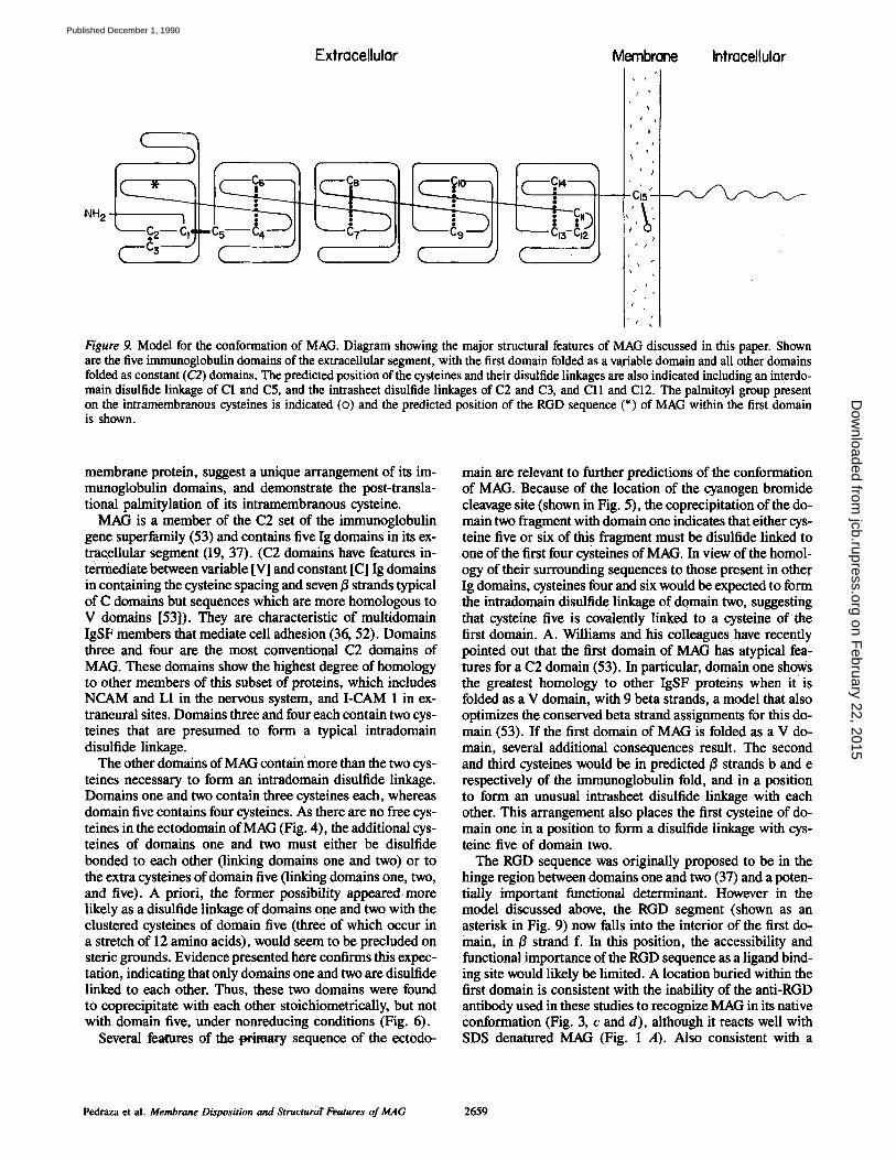
we incubated the gel displayed in Fig. 7 B in hydroxylamine for 4 h at room temperature under conditions expected to hy- drolyze thioester but not amide linkages. Under these condi- tions, up to 80% of the [3H]palmitic acid label in MAG was removed, while the 3~S-methionlne label in MAG was un- affected. This result indicates that the incorporated label rep- resents lipid bound to this protein through a thioester link- age. The [3H]palmitate-labeled MAG was also treated with KOH and methanol to release the acyl group. The fatty acid that was released by this treatment was then characterized by HPLC. As shown in Fig. 8, the 3H label migrates with the same mobility as methyl-palmitate and palmitate, consistent with its identification as a palmitoyl group.
Discussion
Although the role of MAG in myelination has not been estab- lished, evidence to date suggests that it mediates a complex set of interactions, that include binding to a receptor on a subset of axons (12, 27), binding to extracellular matrix components (7) and, possibly, promoting adhesion between the apposed membranes of the myelinating Schwann cell at specific sites (38, 49). These complex interactions in turn im- ply the existence of multiple functional sites that are perhaps reflected in the extraordinary degree of conservation dis- played by all segments of MAG during evolution. Thus hu- man (39, 43) and mouse (9) MAG show an overall 95 and 98 % identity of amino acids respectively with rat MAG. The RGD sequence and the number and location of the cysteines have been conserved. As nothing was known about the con- formation of MAG or its disposition within the membrane, we undertook the studies described in this paper to clarify the structure of these proteins as a prelude to identifying their discrete functional elements. These studies confirm the disposition of MAG within the bilayer as a type one integral
o o - '
3o0- + + ~' +
.2+1 ++°t I II + +o+ + ,
t
0 10 20 30 40 Elution Time (rain)
Figure 8. Characterization of the acyl group of MAG. Im- munoprecipitates of MAG labeled with [3H]palmitate were in- cubated for four hours in 0.1 M KOH in methanol. The released material was subjected to HPLC as described in Materials and Methods. Two peaks of radioactivity are evident. Fractions contain- ing the standards myristic acid, palmitic acid, methyl myristate, and methyl palmitate are indicated.
The Journal of Cell Biology, Volume l 11, 1990 2658
on February 22, 2015
jcb.rupress.orgD
ownloaded from
Published December 1, 1990
)
Extracellular Membrane Intracellular
C J C J C J C . )
t •
Ci
#
#
i
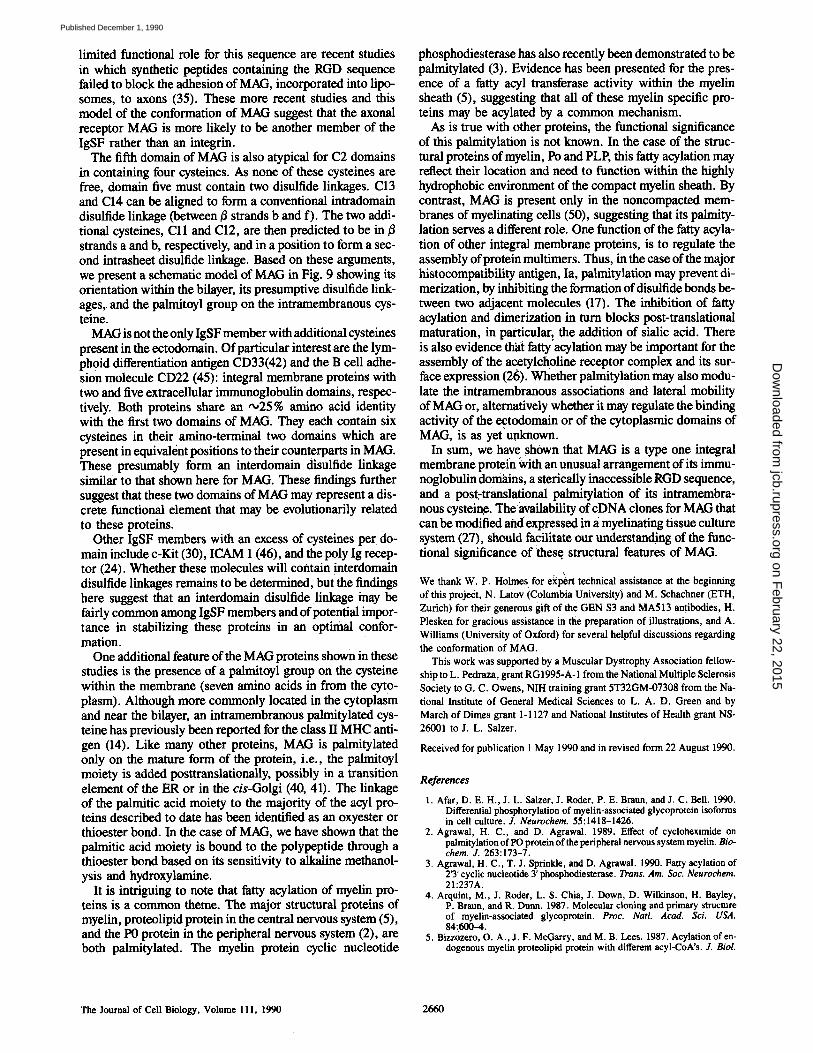
Figure 9. Model for the conformation of MAG. Diagram showing the major structural features of MAG discussed in this paper. Shown are the five immunoglobulin domains of the extracellular segment, with the first domain folded as a variable domain and all other domains folded as constant (C2) domains. The predicted position of the cysteines and their disulfide linkages axe also indicated including an interdo- main disulfide linkage of C1 and C5, and the intrasheet disulfide linkages of C2 and C3, and Cll and C12. The palmitoyl group present on the intramembranous cysteines is indicated (o) and the predicted position of the RGD sequence (*) of MAG within the first domain is shown.
membrane protein, suggest a unique arrangement of its im- munoglobulin domains, and demonstrate the post-transla- tional palmitylation of its intramembranous cysteine.
MAG is a member of the C2 set of the immunoglobulin gene superfamily (53) and contains five Ig domains in its ex- tra~ellular segment (19, 37). (C2 domains have features in- termediate between variable [V] and constant [C] Ig domains in containing the cysteine spacing and seven/~ strands typical of C domains but sequences which are more homologous to V domains [53]). They are characteristic of multidomain IgSF members that mediate cell adhesion (36, 52). Domains three and four are the most conventional C2 domains of MAG. These domains show the highest degree of homology to other members of this subset of proteins, which includes NCAM and L1 in the nervous system, and I-CAM 1 in ex- traneural sites. Domains three and four each contain two cys- teines that are presumed to form a typical intradomain disulfide linkage.
The other domains of MAG containmore than the two cys- teines necessary to form an intradomain disulfide linkage. Domains one and two contain three cysteines each, whereas domain five contains four cysteines. As there are no free cys- teines in the ectodomain of MAG (Fig. 4), the additional cys- teines of domains one and two must either be disulfide bonded to each other (linking domains one and two) or to the extra cysteines of domain five (linking domains one, two, and five). A priori, the former possibility appeared more likely as a disulfide linkage of domains one and two with the clustered cysteines of domain five (three of which occur in a stretch of 12 amino acids), would seem to be precluded on steric grounds. Evidence presented here confirms this expec- tation, indicating that only domains one and two are disulfide linked to each other. Thus, these two domains were found to coprecipitate with each other stoichiometrically, but not with domain five, under nonreducing conditions (Fig. 6).
Several features of tim t~rimary sequence of the ectodo-
main are relevant to further predictions of the conformation of MAG. Because of the location of the cyanogen bromide cleavage site (shown in Fig. 5), the coprecipitation of the do- main two fragment with domain one indicates that either cys- teine five or six of thi s fragment must be disulfide linked to one of the first four cysteines of MAG. In view of the homol- ogy of their surrounding sequences to those present in other Ig domains, cysteines four and six would be expected to form the intradomain disulfid e linkage of domain two, suggesting that cysteine five is covalently linked to a cysteine of the first domain. A. Williams and his colleagues have recently pointed out that the first domain of MAG has atypical fea- tures for a C2 domain (53). In particular, domain one shows the greatest homology to other IgSF proteins when it' is folded as a V domain, with 9 beta strands, a model that also optimizes the conserved beta strand assignments for this do- main (53). If the first domain of MAG is folded as a V do- main, several additional consequences result. The second and third cysteines would be in predicted t3 strands b and e respectively of the immunoglobulin fold, and in a position to form an unusual intrasheet disulfide linkage with each other. This arrangement also places the first cysteine of do- main one in a position to form a disulfide linkage with cys- teine five of domain two.
The RGD sequence was originally proposed to be in the hinge region between domains one and two (37) and a poten- tially important functional determinant. However in the model discussed above, the RGD segment (shown as an asterisk in Fig. 9) now falls into the interior of the first do- main, in/3 strand f. In this position, the accessibility and functional importance of the RGD sequence as a ligand bind- ing site would likely be limited. A location buried within the first domain is consistent with the inability of the anti-RGD antibody used in these studies to recognize MAG in its native conformation (Fig. 3, c and d), although it reacts well with SDS denatured MAG (Fig. 1 A). Also consistent with a
Pedraza et al. Membrane Disposition and StructuraTFeatures of MAG 2659
on February 22, 2015
jcb.rupress.orgD
ownloaded from
Published December 1, 1990
limited functional role for this sequence are recent studies in which synthetic peptides containing the RGD sequence failed to block the adhesion of MAG, incorporated into lipo- somes, to axons (35). These more recent studies and this model of the conformation of MAG suggest that the axonal receptor MAG is more likely to be another member of the IgSF rather than an integrin.
The fifth domain of MAG is also atypical for C2 domains in containing four cysteines. As none of these cysteines are free, domain five must contain two disulfide linkages. C13 and C14 can be aligned to form a conventional intradomain disulfide linkage (between/3 strands b and f). The two addi- tional cysteines, Cll and C12, are then predicted to be in 13 strands a and b, respectively, and in a position to form a sec- ond intrasheet disulfide linkage. Based on these arguments, we present a schematic model of MAG in Fig. 9 showing its orientation within the bilayer, its presumptive disulfide link- ages, and the palmitoyl group on the intramembranous cys- teine.
MAG is not the only IgSF member with additional cysteines present in the ectodomain. Of particular interest are the lym- phoid differentiation antigen CD33(42) and the B cell adhe- sion molecule CD22 (45): integral membrane proteins with two and five extracellular immunoglobulin domains, respec- tively. Both proteins share an ,,025% amino acid identity with the first two domains of MAG. They each contain six cysteines in their amino-terminal two domains which are present in equivalent positions to their counterparts in MAG. These presumably form an interdomain disulfide linkage similar to that shown here for MAG. These findings further suggest that these two domains of MAG may represent a dis- crete functional element that may be evolutionarily related to these proteins.
Other IgSF members with an excess of cysteines per do- main include c-Kit (30), ICAM 1 (46), and the poly Ig recep- tor (24). Whether these molecules will contain interdomain disulfide linkages remains to be determined, but the findings here suggest that an interdomain disulfide linkage may be fairly common among IgSF members and of potential impor- tance in stabilizing these proteins in an optimal confor- mation.
One additional feature of the MAG proteins shown in these studies is the presence of a palmitoyl group on the cysteine within the membrane (seven amino acids in from the cyto- plasm). Although more commonly located in the cytoplasm and near the bilayer, an intramembranous palmitylated cys- teine has previously been reported for the class II MHC anti- gen (14). Like many other proteins, MAG is palmitylated only on the mature form of the protein, i.e., the palmitoyl moiety is added posttranslationally, possibly in a transition element of the ER or in the cis-Golgi (40, 41). The linkage of the palmitic acid moiety to the majority of the acyl pro- teins described to date has been identified as an oxyester or thioester bond. In the case of MAG, we have shown that the palmitic acid moiety is bound to the polypeptide through a thioester bond based on its sensitivity to alkaline methanol- ysis and hydroxylamine.
It is intriguing to note that fatty acylation of myelin pro- teins is a common theme. The major structural proteins of myelin, proteolipid protein in the central nervous system (5), and the P0 protein in the peripheral nervous system (2), are both palmitylated. The myelin protein cyclic nucleotide
phosphodiesterase has also recently been demonstrated to be palmitylated (3). Evidence has been presented for the pres- ence of a fatty acyl transferase activity within the myelin sheath (5), suggesting that all of these myelin specific pro- teins may be acylated by a common mechanism.
As is true with other proteins, the functional significance of this palmitylation is not known. In the case of the struc- tural proteins of myelin, Po and PLP, this fatty acylation may reflect their location and need to function within the highly hydrophobic environment of the compact myelin sheath. By contrast, MAG is present only in the noncompacted mem- branes of myelinating cells (50), suggesting that its palmity- lation serves a different role. One function of the fatty acyla- tion of other integral membrane proteins, is to regulate the assembly of protein multimers. Thus, in the case of the major histocompatibility antigen, Ia, palmitylation may prevent di- merization, by inhibiting the formation of disulfide bonds be- tween two adjacent molecules (17). The inhibition of fatty acylation and dimerization in turn blocks post-translational maturation, in particular~ the addition of sialic acid: There is also evidence their fatty acylation may be important for the assembly of the a cetylcholine receptor complex and its sur- face expression (26). Whether palmitylation may also modu- late the intramembranous associations and lateral mobility of MAG or, alternatively whether it may regulate the binding activity of the ectodomain or of the cytoplasmic domains of MAG, is as yet unknown.
In sum, we have shown that MAG is a type one integral membrane protein ~;vith an unusual arrangement of its immu- noglobulin doniains, a sterically inaccessible RGD sequence, and a post-translational palmitylation of its intramembra- nous cysteine. The availability of eDNA clones for MAG that can be modified and expressed in ~i myelinating tissue culture system (27), should facilitate our understanding of the func- tional significance of these structural features of MAG.
We thank W. P. Holmes for e~l~rt technical assistance at the beginning of this project, N. Latov iColambia University) and M. Schachner (ETH, Zurich) for their generous gift of the GEN $3 and MA513 antibodies, H. Plesken for gracious assistance in the preparation of illustrations, and A. Williams (University of Oxford) for several helpful discussions regarding the conformation of MAG.
This work was supported by a Muscular Dystrophy Association fellow- ship to L. Pedraza, grant RGI995-A- 1 from the National Multiple Sclerosis Society to G. C. Owens, NIH training grant 5T32GM-07308 from the Na- tional Institute of General Medical Sciences to L. A. D. Green and by March of Dimes grant 1-1127 and National Institutes of Health grant NS- 26001 to J. L. Salzer.
Received for publication 1 May 1990 and in revised form 22 August 1990.
References
1. Afar, D. E. H., J. L. Seizer, J. Roder, P. E. Braun, and J. C. Bell. 1990. Differential phosphorylation of myelin-associated glycoprotein isoforms in cell culture. J. Neurochem. 55:1418-1426.
2. Agrawal, H. C., and D, Agrawal. 1989. Effect of cycloheximide on palmitylation of PO protein of the peripheral nervous system myelin. Bio- chem. J. 263:173-7.
3. Agrawal, H. C., T. J. Sprinkle, and D. Agrawal. 1990. Fatty acylation of 2'3' cyclic nucleotide 3' phosphodiesterase. Trans. Am. Soc. Neurochem. 21:237A.
4. Arquint, M., J. Roder, L. S. Chia, J. Down, D. Wilkinson, H. Bayley, P. Braun, and R. Dunn. 1987. Molecular cloning and primary structure of myelin-associated glycoprotein. Proc. Natl. Acad. Sci. USA. 84:600--4.
5. Bizzozero, O. A., J. F. McGarry, and M. B. Lees. 1987. Acylation of en- dogenous myelin proteolipid protein with different acyl-CoA's. J. Biol.
The Journal of Cell Biology, Volume 111, 1990 2660
on February 22, 2015
jcb.rupress.orgD
ownloaded from
Published December 1, 1990
Chem. 262:2138-2145. 6. Edwards, A. M., P. E. Braun, and J. C. Ball. 1989. Phosphorylation of
myelin-associated glycoprotein in vivo and in vitro occurs only in the cy- toplasmic domain of the large isoform. J. Neurochem. 52:317-320.
7. Fahrig, T., C. Landa, P. Pesheva, K. Kuhn, and M. Schachner. 1987. Characterization of binding properties of the myelin-associated glycopro- tein to extracellular matrix constituents. EMBO (Eur. Mol. Biol. Organ.) J. 6:2875-2883.
8. Frail, D. E., and P. E. Braun. 1984. Two developmentally regulated mes- senger RNAs differing in their coding regions may exist for the myelin- associated glycoprotein. J. Biol. Chem. 259:14857-14862.
9. Fujita, N., S. Sato, T. Kurihara, R. Kuwano, K. Sakimura, T. Inuzuka, Y. Takahashi, and T. Miyatake. 1989. cDNA cloning of mouse myelin- associated glycoprotein: a novel alternative splicing pattern. Biochem. Biophys. Res. Commun. 165:1162-9.
10. Heijne, G. V. 1983. Patterns of amino acids near signal-cleavage sites. Fur. J. Biochem. 133:17-21.
11. Hynes, R. O. 1987. lntegrins: a family of cell surface receptors. Cell. 48:549-554.
12. Johnson, P. W., W. Abramow-Newerly, B. Seilheimer, R. Sadoul, M. B. Tropak, M. Arquint, R. J. Duma, M. Schachner, and J. C. Roder. 1989. Recombinant myelin-associated glycoprotein confers neural adhesion and neurite outgrowth function. Neuron. 3:377-385.
13. Johnson, P. W., J. Attica, C. D. Richardson, J. C. Roder, and R. J. Dunn. 1989. Synthesis of soluble myelin-associated-glycoprotein in insect and mammalian cells. Gene (Amst.). 77:287-296.
14. Kaufman, J. F., M. S. Krangel, and J. L. Strominger. 1984. Cysteines in the transmembrane region of major histocompatibility complex antigens are fatty acylated via thioester bonds. J. Biol. Chem. 259:7230-7238.
15. Kaufman, R. J., P. Murtha, and M. V. Davies. 1987. Translation efficiency of polycistronic mRNAs and their utilization to express heterologous genes in mammalian cells. EMBO (Eur. MoL Biol. Organ.) J. 6:187-193.
16. Kirschraeier, P. T., G. M. Housey, M. D. Johnson, A. S. Perkins, and I. B. Weinstein. 1988. Construction and characterization of a retroviral vector demonstrating efficient expression of cloned cDNA sequences. DNA (NY). 7:219-225.
17. Koch, N., and G. J. Hammerling. 1985. Ia-associated invariant chain is fatty acylated before addition of sialic acid. Biochemistry. 24:6185-6190.
18. Laemmli, U. K. 1970. Cleavage of structural proteins during the assembly of the head of bacteriophage T4. Nature (Lond.). 227:680-685.
19. Lai, C., M. A. Brow, K.-A. Nave, A. B. Noronha, R. H. Quarles, F. E. Bloom, R. J. Milner, and J. G. Sutcliffe. 1987. Two forms of IB236/my- elin associated glycoprotein, a cell adhesion molecule for postnatal devel- opment, are produced by alternate splicing. Proc. Natl. Acad. Sci. USA. 84:4337--4341.
20. Lai, C., J. B. Watson, F. E. Bloom, J. G. Sutcliffe, and R. J. Milner. 1987. Neural protein lB236/Myelin-associated glycoprotein (MAG) defines a subgroup of the immnnoglobulin gene supeffamily. Immunol. Rev. 100:129-151.
21. Lisanti, M. P., M. Sargiacomo, L. Graeve, A. R. Saltiel, and E. Rodriguez-Boulan. 1988. Polarized apical distribution of glycosyl- pbosphatidylinositol-anchored proteins in a renal epithelial cell line. Proc. Natl. Acad. Sci. USA. 85:9557-9561.
22. Lopata, M. A., D. W. Cleveland, and B. Sollner-Webb. 1984. High-level expression of a chloramphenicol acetyltransferase gene by DEAE- dextran-mediated DNA transfection coupled with a dimethyl sulfoxide or glycerol shock treatment. Nucleic Acids Res. 12:5707-5717.
23. Martini, R., and M. Schechner. 1986. Immunoelectron microscopic local- ization of neural call adhesion molecules (L1, N-CAM, and MAG) and their shared carbohydrate epitope and myelin basic protein in developing sciatic nerve. J. Cell Biol. 103:2439-2448.
24. Mostov, K. E., M. Friedlander, and G. Blobel. 1984. The receptor for trans- epithelial transport of IgA and IgM contains multiple immunoglobulin- like domains. Nature (Lond.). 308:37-43.
25. Nobile-Orazio, E., A. P. Hays, N. Latov, G. Perman, J. Golier, M. E. Shy, and L. Freddo. 1984. Specificity of mouse and human monoclonal anti- bodies to myelin-associated glycoprotein. Neurology. 34:1336-1342.
26. Olson, E. N., L. Glaser, and J. P. Merlie. 1984. ¢ and/3 subunits of the nicotinic acetylcholine receptor contain covalently bound lipid. J. Biol. Chem. 259:5364-5367.
27. Owens, G. C., C. J. Boyd, R. P. Bunge, andJ. L. Salzer. 1990. Expression of recombinant myelin-associated glycoprotein in primary Schwann cells promotes the initial investment of axons by myelinating Schwann cells. J. Cell Biol. In press.
28. Poltorak, M., R. Sadoul, G. Keilhauer, C. Landa, T. Fahrig, and M. Schachner. 1987. Myelin-associated glycoprotein, a member of the L2/HNK-1 family of neural cell adhesion molecules, is involved in neuron-oligodendrocyte and oligodendrocyte-oligodendrocyte interac-
tions. J. Cell Biol. 105:1893-1899. 29. Porter, S., L. Giaser, and R. P. Bunge. 1987. Release of autocrine growth
factor by primary and immortalized Schwann cells. Proc. Natl. Acad. Sci. USA. 84:7768-7772.
30. Quarles, R. H. 1983184. Myelin-associated glycoprotein in development and disease. Dev. Neurosci. 6:285-303.
31. Quarles, R. H., and C. F. Pasnak. 1977. A rapid procedure for selectively isolating the major glycoprotein from purified rat brain myelin. Biochem. J. 1977:635-637.
32. Qin, F., P. Ray, K. Brown, P. E. Barker, S. Jhanwar, F. H. Ruddle, and P. Besmer. 1988. Primary structure of c-Kit: relationship with the CSF- I/PDGF receptor kinase family: oncogehic activation of v-Kit involves deletion of extracellular domain and C terminus. EMBO (Eur. Mol. Biol. Organ.) J. 7:1003-1011.
33. Ruoslahti, E., and M. D. Pierschbacher. 1987. New perspectives in cell adhesion: RGD and Integrins. Science (Wash. DC). 238:491--497.
34. Sabatini, D. D., and M. B. Adesnik. 1989. The biogenesis of membranes and organelles. In The Metabolic Basis of Inherited Disease. C. R. Scriver, A. L. Beaudet, W. S. Sly, and D. Valle, editors. McGraw-Hill Book Co., New York. 177-223.
35. Sadoul, R., T. Fahring, U. Bartig, and M. Schachner. 1990. Binding prop- erties of liposomes containing the myelin-associated glyeoprotein MAG to neural cell cultures. J. Neurasci. Res. 25:1-13.
36. Salzer, J. L., and D. R. Colman. 1989. Mechanisms of cell adhesion in the nervous system: role of the immunoglobulin gene superfamily. Dev. Neu- rosci. 11:377-390.
37. Salzer, J. L., W. P. Holmes, and D. R. Colman. 1987. The amino acid sequences of the myelin-associated glycoproteins: homology to the im- munoglobulin gene superfamily. J. Cell Biol. 104:957-965.
38. Salzer, J. L., L. Pedraza, M. Brown, A. Struyk, D. Afar, andJ. Ball. 1990. Structure-function studies of the myelin-associated glyeoproteins. Ann. NY Acad. Sci. 605:302-312.
39. Sato, S., N. Fujita, T. Kurihara, R. Kuwano, K. Sakimura, Y. Takahashi, and T. Miyatake. 1989. cDNA cloning and amino acid sequence for hu- man myelin-associated glycoprotein. Biochem. Biophys. Res. Commun. 163:1473-1480.
40. Schmidt, J. W., and W. A. Catterall. 1987. Palmitylation, sulfation, and glycosylation of the alpha subunit of the sodium channel. Role of post- translational modifications in channel assembly. J. Biol. Chem. 262: 13713-13723.
41. Schmidt, M. F. G., and M. J. Schlesinger. 1980. Relation of fatty acid at- tachment to the translation and maturation of vesicular stomatitis and Sindbis virus membrane glyeoproteins. J. Biol. Chem. 255:3334-3339.
42. Simmons, D., and B. Seed. 1988. Isolation of a cDNA encoding CD33, a differentiation antigen of myeloid progenitor cells. J. Immunol. 141:2797-2800.
43. Spagnol, G., M. Williams, J. Srinivasan, J. Golier, D. Bauer, R. V. Lebo, and N. Latov. 1989. Molecular cloning of human myelin-associated gly- coprotein. J. Neurosci. Res. 24:137-142.
44. Spencer, P. S., and H. J. Weinberg. 1978. Axonal specification of Schwann cell expression and myelination. In Physiology and Pathobiotogy of Axons. S. G. Waxman, editor. Raven Press, New York 389-405.
45. Stamenkovic, L, and B. Seed. 1990. The B-cell antigen CD22 mediates monocyte and erythrocyte adhesion. Nature (Lond.). 345:74-77.
46. Stannton, D. E., S. D. Martin, C. Stratowa, M. L. Dustin, and T. A. Springer. 1988. Primary structure of ICAM-1 demonstrates interaction between members of the immunoglobniin and integrin supergene fami- lies. Cell. 52:925-933.
47. Towbin, H., T. Staehelin, and J. Gordon. 1979. Electrophoretic transfer of proteins from polyacrylamide gels to nitrocellulose sheets: procedure and some applications. Proc. Natl. Acad. Sci. USA. 76:4350-4354.
48. Towler, D. A., J. I. Gordon, S. P. Adams, and L. Glaser. 1988. The biol- ogy and enzymology of eukaryotic protein acylation. Annu. Rev. Bio- chem. 57:69-99.
49. Trapp, B. D. 1988. Distribution of the myelin-associated glycoprotein and P0 protein during myelin compaction in quaking mouse peripheral nerve. J. Cell Biol. 107:675-685.
50. Trapp, B. D., and R. H. Quarles. 1982. Presence of the myelin-associated glycoprotein correlates with alterations in the periodicity of peripheral myelin. J. Cell Biol. 92:877-882.
51. Wigler, M., S. Silverstein, L.-S. Lee, A. Pellicer, Y.-C. Cheng, and R. Axel. 1977. Transfer of purified herpes virus thymidine kinase gene to cultured mouse cells. Cell. 11:223-232.
52. Williams, A. F., and A. N. Barclay. 1988. The immunoglobniin superfa- rally: domains for cell surface recognition. Annu. Rev. lmmunol. 6: 381-405.
53. Williams, A. F., S. J. Davis, Q. He, and A. N. Barclay. 1989. Structural diversity of the immunoglobulin family. Cold. Spring Harbor Syrup. Quant. Biol. 54:637-647.
Pedraza et al. Membrane Disposition and Structural Features of MAG 2661
on February 22, 2015
jcb.rupress.orgD
ownloaded from
Published December 1, 1990
Related Documents


![Posttranslational Modifications of FERREDOXIN …...Posttranslational Modifications of FERREDOXIN-NADP+ OXIDOREDUCTASE in Arabidopsis Chloroplasts1[W][OPEN] Nina Lehtimäki2, Minna](https://static.cupdf.com/doc/110x72/5f0d9b3d7e708231d43b3018/posttranslational-modiications-of-ferredoxin-posttranslational-modiications.jpg)