cells Review The Multiple Interactions of RUNX with the Hippo–YAP Pathway Linda Shyue Huey Chuang and Yoshiaki Ito * Citation: Chuang, L.S.H.; Ito, Y. The Multiple Interactions of RUNX with the Hippo–YAP Pathway. Cells 2021, 10, 2925. https://doi.org/10.3390/ cells10112925 Academic Editors: Wanjin Hong, Lanfen Chen and Kay-Dietrich Wagner Received: 14 September 2021 Accepted: 26 October 2021 Published: 28 October 2021 Publisher’s Note: MDPI stays neutral with regard to jurisdictional claims in published maps and institutional affil- iations. Copyright: © 2021 by the authors. Licensee MDPI, Basel, Switzerland. This article is an open access article distributed under the terms and conditions of the Creative Commons Attribution (CC BY) license (https:// creativecommons.org/licenses/by/ 4.0/). NUS Centre for Cancer Research, Cancer Science Institute of Singapore, Yong Loo Lin School of Medicine, National University of Singapore, 14 Medical Drive, #12-01, Singapore 117599, Singapore * Correspondence: [email protected] Abstract: The Hippo–YAP signaling pathway serves roles in cell proliferation, stem cell renewal/ maintenance, differentiation and apoptosis. Many of its functions are central to early development, adult tissue repair/regeneration and not surprisingly, tumorigenesis and metastasis. The Hippo path- way represses the activity of YAP and paralog TAZ by modulating cell proliferation and promoting differentiation to maintain tissue homeostasis and proper organ size. Similarly, master regulators of development RUNX transcription factors have been shown to play critical roles in proliferation, differentiation, apoptosis and cell fate determination. In this review, we discuss the multiple interac- tions of RUNX with the Hippo–YAP pathway, their shared collaborators in Wnt, TGFβ, MYC and RB pathways, and their overlapping functions in development and tumorigenesis. Keywords: RUNX1; RUNX2; RUNX3; YAP; Hippo; TAZ; cancer 1. Introduction RUNX transcription factors are well-established master regulators of development. They play critical roles in directing cell proliferation, lineage specification and differen- tiation. The three mammalian RUNX paralogs RUNX1, RUNX2 and RUNX3 have been shown to regulate hematopoiesis, bone formation and neuronal development, respec- tively [1–7]. RUNX proteins are strongly influenced by interacting proteins, which may enhance their transcriptional activities or toggle RUNX’s properties between transcription activation and inhibition. The first indication that RUNX contributes to the Hippo pathway came from an early 1999 report showing the physical interaction of all human RUNX pro- teins with the transcriptional coactivator Yes-associated protein 1 (YAP), and the resultant strong enhancement of RUNX’s transactivation ability [8]. This interaction is mediated by the PPxY sequence (also known as the PY motif), which is evolutionarily conserved in the C-terminal domain of the mammalian RUNX (Figure 1), to the WW domain of the YAP protein. Since then, YAP has been recognized as a key downstream effector of the Hippo pathway, an attractive target for cancer therapy as well as a promising option for regenerative medicine [9–11]. First discovered as a key regulator of organ size, the Hippo signaling cascade has now been mechanistically linked to cell proliferation, tissue homeostasis, differentiation and apoptosis [12]. The Hippo pathway is remarkable for its numerous components of WW domain proteins (e.g., YAP, TAZ and SAV1) and PY motif-containing proteins (e.g., LATS1/2 and AMOTs). WW domains may occur in isolation or in tandem, which could bestow a strong and specific binding to PY motifs [13]. Furthermore, tyrosine phosphorylation of the WW proteins and the PY motif have been shown to regulate binding affinity [14]. In mammals, the core components of the Hippo pathway are the STE20-like kinases MST1/2, the SAV1 and MOB1 scaffold/adaptor proteins, which regulate MST1/2 activities on the large tumor suppressor 1 and 2 (LATS1/2) kinases, and the transcription factor complexes comprising coactivators YAP or TAZ and their main DNA- binding partners TEAD1–4 [12]. Paralogs YAP and TAZ serve functionally redundant Cells 2021, 10, 2925. https://doi.org/10.3390/cells10112925 https://www.mdpi.com/journal/cells

Welcome message from author
This document is posted to help you gain knowledge. Please leave a comment to let me know what you think about it! Share it to your friends and learn new things together.
Transcript
cells
Review
The Multiple Interactions of RUNX with theHippo–YAP Pathway
Linda Shyue Huey Chuang and Yoshiaki Ito *
�����������������
Citation: Chuang, L.S.H.; Ito, Y. The
Multiple Interactions of RUNX with
the Hippo–YAP Pathway. Cells 2021,
10, 2925. https://doi.org/10.3390/
cells10112925
Academic Editors: Wanjin Hong,
Lanfen Chen and Kay-Dietrich
Wagner
Received: 14 September 2021
Accepted: 26 October 2021
Published: 28 October 2021
Publisher’s Note: MDPI stays neutral
with regard to jurisdictional claims in
published maps and institutional affil-
iations.
Copyright: © 2021 by the authors.
Licensee MDPI, Basel, Switzerland.
This article is an open access article
distributed under the terms and
conditions of the Creative Commons
Attribution (CC BY) license (https://
creativecommons.org/licenses/by/
4.0/).
NUS Centre for Cancer Research, Cancer Science Institute of Singapore, Yong Loo Lin School of Medicine,National University of Singapore, 14 Medical Drive, #12-01, Singapore 117599, Singapore* Correspondence: [email protected]
Abstract: The Hippo–YAP signaling pathway serves roles in cell proliferation, stem cell renewal/maintenance, differentiation and apoptosis. Many of its functions are central to early development,adult tissue repair/regeneration and not surprisingly, tumorigenesis and metastasis. The Hippo path-way represses the activity of YAP and paralog TAZ by modulating cell proliferation and promotingdifferentiation to maintain tissue homeostasis and proper organ size. Similarly, master regulatorsof development RUNX transcription factors have been shown to play critical roles in proliferation,differentiation, apoptosis and cell fate determination. In this review, we discuss the multiple interac-tions of RUNX with the Hippo–YAP pathway, their shared collaborators in Wnt, TGFβ, MYC and RBpathways, and their overlapping functions in development and tumorigenesis.
Keywords: RUNX1; RUNX2; RUNX3; YAP; Hippo; TAZ; cancer
1. Introduction
RUNX transcription factors are well-established master regulators of development.They play critical roles in directing cell proliferation, lineage specification and differen-tiation. The three mammalian RUNX paralogs RUNX1, RUNX2 and RUNX3 have beenshown to regulate hematopoiesis, bone formation and neuronal development, respec-tively [1–7]. RUNX proteins are strongly influenced by interacting proteins, which mayenhance their transcriptional activities or toggle RUNX’s properties between transcriptionactivation and inhibition. The first indication that RUNX contributes to the Hippo pathwaycame from an early 1999 report showing the physical interaction of all human RUNX pro-teins with the transcriptional coactivator Yes-associated protein 1 (YAP), and the resultantstrong enhancement of RUNX’s transactivation ability [8]. This interaction is mediatedby the PPxY sequence (also known as the PY motif), which is evolutionarily conservedin the C-terminal domain of the mammalian RUNX (Figure 1), to the WW domain of theYAP protein. Since then, YAP has been recognized as a key downstream effector of theHippo pathway, an attractive target for cancer therapy as well as a promising option forregenerative medicine [9–11].
First discovered as a key regulator of organ size, the Hippo signaling cascade hasnow been mechanistically linked to cell proliferation, tissue homeostasis, differentiationand apoptosis [12]. The Hippo pathway is remarkable for its numerous componentsof WW domain proteins (e.g., YAP, TAZ and SAV1) and PY motif-containing proteins(e.g., LATS1/2 and AMOTs). WW domains may occur in isolation or in tandem, whichcould bestow a strong and specific binding to PY motifs [13]. Furthermore, tyrosinephosphorylation of the WW proteins and the PY motif have been shown to regulatebinding affinity [14]. In mammals, the core components of the Hippo pathway are theSTE20-like kinases MST1/2, the SAV1 and MOB1 scaffold/adaptor proteins, which regulateMST1/2 activities on the large tumor suppressor 1 and 2 (LATS1/2) kinases, and thetranscription factor complexes comprising coactivators YAP or TAZ and their main DNA-binding partners TEAD1–4 [12]. Paralogs YAP and TAZ serve functionally redundant
Cells 2021, 10, 2925. https://doi.org/10.3390/cells10112925 https://www.mdpi.com/journal/cells
Cells 2021, 10, 2925 2 of 13
as well as nonoverlapping roles in transcription coactivation [15]. They are structurallysimilar, sharing about 60% amino acid sequence homology [15]. YAP appears to exert astronger influence than TAZ on cellular physiology in HEK293A cells (e.g., proliferationand migration) [15]. The properties of YAP are diversified by multiple mRNA splicevariants. Eight YAP protein isoforms have been characterized—the main differences arethe presence of one or two WW domains, and the presence of an intact or disrupted leucinezipper domain, which mediates interaction with TAZ and pro-oncogenic phosphataseSHP2 [16–18]. The retention of the leucine zipper was shown to be influenced by oncogenicKRAS regulation of splicing factor SRSF3 [16]. While four of the YAP isoforms possessone WW domain, the other four possess two. These differences afforded the isoformsdistinct interacting partners and transcriptional activities in regulating cell proliferation,differentiation and oncogenic potential. YAP isoforms are differentially expressed basedon cell type, as well as levels of pluripotency and differentiation [17]. TAZ possessesone WW domain [15]. Moreover, TAZ contains an additional phosphodegron, indicatingdifferences in regulation of protein stability between YAP and TAZ [19]. LATS-mediatedphosphorylation of YAP and TAZ regulate YAP/TAZ subcellular localization as well assusceptibility to SCFβ-TRCP-mediated proteosomal degradation [12].
Cells 2021, 10, x FOR PEER REVIEW 2 of 14
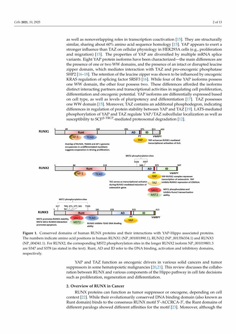
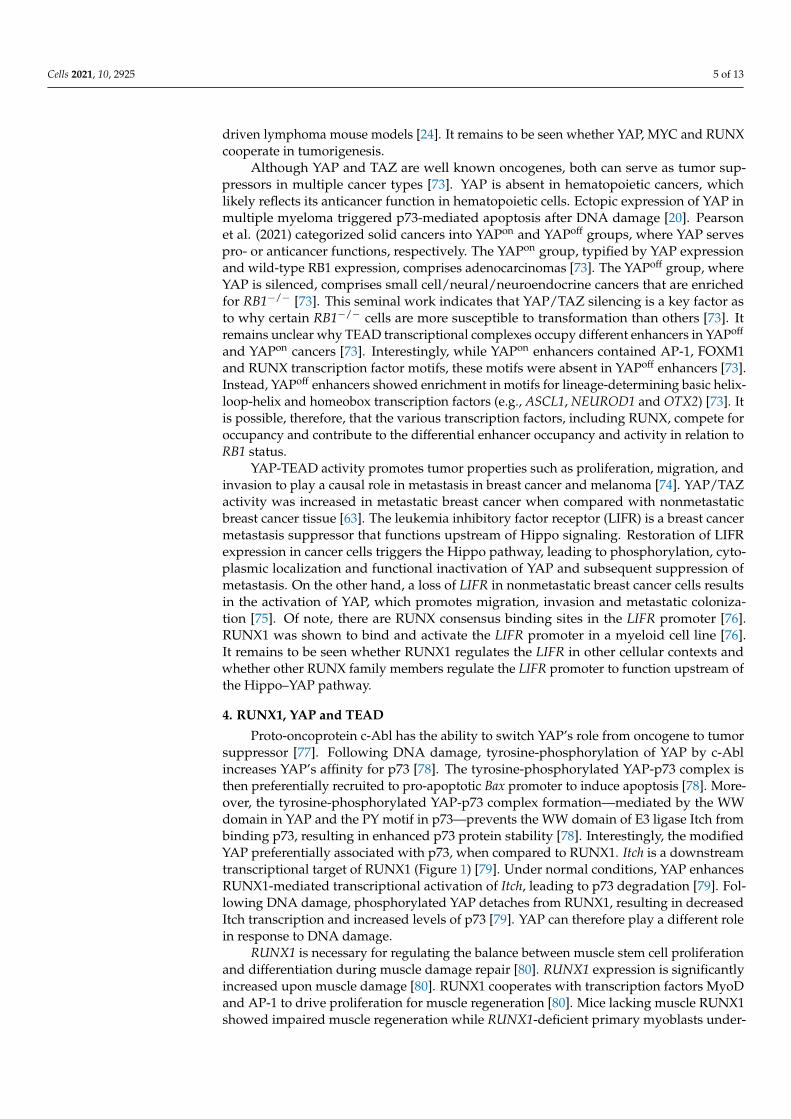
Figure 1. Conserved domains of human RUNX proteins and their interactions with YAP-Hippo associated proteins. The numbers indicate amino acid positions in human RUNX1 (NP_001001890.1), RUNX2 (NP_001356334.1) and RUNX3 (NP_004341.1). For RUNX2, the corresponding MST2 phosphorylation sites in the longer RUNX2 isoform NP_001019801.3 are S347 and S378 (as stated in the text). Runt, AD and ID refer to the DNA binding, activation and inhibitory domains, respectively.
First discovered as a key regulator of organ size, the Hippo signaling cascade has now been mechanistically linked to cell proliferation, tissue homeostasis, differentiation and apoptosis [12]. The Hippo pathway is remarkable for its numerous components of WW domain proteins (e.g., YAP, TAZ and SAV1) and PY motif-containing proteins (e.g., LATS1/2 and AMOTs). WW domains may occur in isolation or in tandem, which could bestow a strong and specific binding to PY motifs [13]. Furthermore, tyrosine phosphory-lation of the WW proteins and the PY motif have been shown to regulate binding affinity [14]. In mammals, the core components of the Hippo pathway are the STE20-like kinases MST1/2, the SAV1 and MOB1 scaffold/adaptor proteins, which regulate MST1/2 activities on the large tumor suppressor 1 and 2 (LATS1/2) kinases, and the transcription factor complexes comprising coactivators YAP or TAZ and their main DNA-binding partners TEAD1–4 [12]. Paralogs YAP and TAZ serve functionally redundant as well as nonover-lapping roles in transcription coactivation [15]. They are structurally similar, sharing about 60% amino acid sequence homology [15]. YAP appears to exert a stronger influence than TAZ on cellular physiology in HEK293A cells (e.g., proliferation and migration) [15]. The properties of YAP are diversified by multiple mRNA splice variants. Eight YAP pro-tein isoforms have been characterized—the main differences are the presence of one or two WW domains, and the presence of an intact or disrupted leucine zipper domain, which mediates interaction with TAZ and pro-oncogenic phosphatase SHP2 [16–18]. The retention of the leucine zipper was shown to be influenced by oncogenic KRAS regulation of splicing factor SRSF3 [16]. While four of the YAP isoforms possess one WW domain, the other four possess two. These differences afforded the isoforms distinct interacting partners and transcriptional activities in regulating cell proliferation, differentiation and oncogenic potential. YAP isoforms are differentially expressed based on cell type, as well as levels of pluripotency and differentiation [17]. TAZ possesses one WW domain [15]. Moreover, TAZ contains an additional phosphodegron, indicating differences in regula-tion of protein stability between YAP and TAZ [19]. LATS-mediated phosphorylation of YAP
Figure 1. Conserved domains of human RUNX proteins and their interactions with YAP-Hippo associated proteins.The numbers indicate amino acid positions in human RUNX1 (NP_001001890.1), RUNX2 (NP_001356334.1) and RUNX3(NP_004341.1). For RUNX2, the corresponding MST2 phosphorylation sites in the longer RUNX2 isoform NP_001019801.3are S347 and S378 (as stated in the text). Runt, AD and ID refer to the DNA binding, activation and inhibitory domains,respectively.
YAP and TAZ function as oncogenic drivers in various solid cancers and tumorsuppressors in some hematopoietic malignancies [20,21]. This review discusses the collabo-ration between RUNX and various components of the Hippo pathway in cell fate decisionssuch as proliferation, regeneration and differentiation.
2. Overview of RUNX in Cancer
RUNX proteins can function as tumor suppressor or oncogene, depending on cellcontext [22]. While their evolutionarily conserved DNA binding domain (also known asRunt domain) binds to the consensus RUNX motif 5′-ACCRCA-3′, the Runt domains ofdifferent paralogs showed different affinities for the motif [23]. Moreover, although the
Cells 2021, 10, 2925 3 of 13
divergent C-termini of the three mammalian RUNX paralogs allow for distinct interac-tomes and functions, there are some structural similarities such as the transactivation andinhibitory domains, as well as the PY and VWRPY motifs (Figure 1). The VWRPY motifis necessary for RUNX interaction with transcriptional corepressors, including TLE1 [24].RUNX can act as transcription activator or repressor, depending on interacting proteinsand post-translational modifications [24]. RUNX proteins interact with a medley of tran-scription regulators/chromatin modifiers, which include coactivators and corepressors.RUNX1, 2 and 3 have been reported to interact with acetyltransferase p300, histone deacety-lases (HDAC) and corepressor mSin3A; RUNX1 and 3 bind to histone methyltransferaseSUV39H1; RUNX2 binds to NAD-dependent histone deacetylase and tumor suppressorSIRT6 [25–31].
RUNX1 is a key player in definitive hematopoietic stem cell formation and is frequentlymutated in leukemia [32,33]. Mutations in the Runt domain are frequently observed inacute myeloid leukemia (AML) and myelodysplastic syndromes. Recurrent mutations inRUNX1 have been observed in estrogen receptor-positive luminal breast cancer, which,similarly to leukemia, may be considered as a stem-cell disorder [34–36]. Conversely,RUNX1 serves as a key component of the core transcriptional TAL1–GATA3–RUNX1complex to support the malignant state of human T cell acute lymphoblastic leukemia(T-ALL) [37]. Dominant oncogenicity for all mouse RUNX genes was earlier shown usingretroviral mutagenesis in CD2-MYC mice. Retroviral insertions that led to RUNX1, RUNX2and RUNX3 overexpression were frequently observed in virus-accelerated lymphomas [24].
RUNX2 is the master regulator of osteogenic development—it regulates the osteopro-genitor proliferation and osteoblast differentiation. Heterozygous mutations in the Runtdomain of RUNX2 play a causal role in cleidocranial dysplasia, an autosomal dominantheritable skeletal disease [33,38]. Of note, RUNX2 expression is frequently elevated in os-teosarcoma [39]. RUNX2 establishes osteoblasts in a terminally differentiated state throughcooperation with retinoblastoma tumor suppressor protein (pRb) and cyclin-dependentkinase inhibitor p27KIP1, and disruption of this cooperation is associated with dedifferenti-ation in high-grade osteosarcomas [40]. RUNX2 has also been functionally implicated inmetastatic breast and prostate cancer cell lines as well as their metastasis to the bone [41,42].
RUNX3 is a versatile tumor suppressor gene that has been shown to cooperate withsignaling pathways such as TGFβ and Wnt to inhibit growth [22]. The frequent hyper-methylation and silencing of RUNX3 in solid tumors—including breast cancer, gastriccancer and hepatocellular carcinoma—indicates a prominent role in solid tumor suppres-sion [22,43,44]. RUNX3-deficient mice are associated with tumor predisposition in thegastrointestinal tract [45,46]. The stomach of RUNX3 knockout mice exhibited reducedchief cell population, indicating differentiation defects [47]. Using an oncogenic K-ras lungcancer mouse model system, Lee et al. observed that RUNX3 inactivation is a key earlyevent during lung adenocarcinoma development [48]. Mechanistically, RUNX3 interactswith TGFβ effectors SMAD2/3 to induce the transcription of cell cycle inhibitor CDKN1A(also known as p21CIP1) and proapoptotic BIM genes [49,50]. RUNX3 can also attenuatethe activity of Wnt effectors TCF4-β-catenin, resulting in decreased transcription of Wntpathway genes such as MYC and cyclin D1 [46]. Furthermore, RUNX3 plays a key rolein the regulation of the restriction point to defend against transformation [51]. Duringthe restriction point—when cells decide on cell fate choices such as differentiation andG1–S transition—RUNX3 transiently forms a complex with pRb and BRD2, resulting in thesynergistic induction of a key regulator of the restriction point CDKN1A [51,52].
Paradoxically, RUNX3 serves oncogenic functions in ovarian cancer and naturalkiller/T-cell lymphoma [53–55]. Moreover, RUNX3 was shown to function as both tu-mor suppressor and tumor promoter in pancreatic ductal adenocarcinoma [56]. WhileRUNX3 can inhibit proliferation, highly elevated levels of RUNX3 in pancreatic ductaladenocarcinoma can direct a metastatic program to promote cell migration, invasion anddistant colonization [56]. In addition, there is increasing evidence that RUNX proteinsplay non-transcriptional roles during DNA repair and mitosis [57–59]. Not surprisingly,
Cells 2021, 10, 2925 4 of 13
dysregulation of RUNX genes has been heavily implicated in disease states such as cancerand autoimmune disorders.
3. Overview of YAP/TAZ in Cancer
The Hippo pathway regulates the nucleocytoplasmic shuttling of YAP/TAZ. LATS-mediated phosphorylation of YAP/TAZ blocks their nuclear accumulation and activity. Inthe nucleus, YAP/TAZ bind to the TEAD transcription factor, which is responsible for mostof the YAP/TAZ transcriptional output [60]. Overexpression of YAP induces hyperprolifer-ation of undifferentiated stem/progenitor cells in mouse tissues such as the gastrointestinaltract, liver and skin [61,62]. Moreover, increased TAZ/YAP activity in poorly differentiatedbreast tumors were associated with enrichment of stem cell signature, suggesting thatTAZ/YAP bestowed cancer stem cell-like properties on breast cancer cells [63]. YAP andTAZ maintain self-renewal and pluripotency in somatic stem cells [64]. The introductionof YAP and TAZ into terminally differentiated cells can induce reprogramming into astem/progenitor cell-like state [64]. Therefore, abnormally elevated YAP/TAZ activity andthe subsequent enhancement of stem-like properties might promote tumorigenesis.
Activation of YAP/TAZ is a key characteristic of many human cancers [65]. It wassuggested that increased YAP expression might be a common event in the development ofsolid tumors such as colonic adenocarcinoma, lung adenocarcinoma and ovarian serouscystadenocarcinoma [66]. Moreover, YAP and TAZ are important contributors to tissuerepair following injury [65]. For example, the gp130–Src–YAP signaling module servesas a critical link between inflammation and epithelial regeneration after injury [67]. Theinjury-related activation of YAP and the YAP-dependent inflammation response indicatethat YAP is a common denominator driving proliferation in both epithelial tissue repairand cancer [65].
To promote transcription for oncogenic growth, YAP and TAZ collaborate with TEADDNA binding proteins and activator protein-1 (AP-1, dimeric complex comprising JUNand FOS proteins) [68]. YAP/TAZ, TEAD and AP-1 form a complex at enhancers thatharbor TEAD and AP-1 motifs to synergistically activate genes involved in the control ofS-phase entry and mitosis [68]. Interestingly, this work also revealed a low but significantenrichment of RUNX motifs at YAP/TAZ peaks [68]. Earlier, RUNX1 and 2 were shownto interact with AP-1; RUNX2 cooperatively bound to AP-1 to activate the collagenase-3promoter (Figure 1) [69]. Whether AP-1 binds to RUNX and YAP/TEAD simultaneously orin a mutually exclusive manner remains unclear. Moreover, YAP physically interacts withBET (bromodomain and extra-terminal) transcriptional coactivators BRD2 and BRD4 [70].YAP, TAZ, TEAD1 and BRD4 are found in a multiprotein nuclear complex. YAP/TAZrecruits BRD4 to enhancers of growth-related genes to boost their expression, therebymediating transcriptional addiction in cancer cells [70]. Small molecule BET inhibitorswere able to mediate regression of YAP/TAZ-addicted neoplastic lesions [70]. RUNX3has been shown to interact with BRD2. It would be interesting to examine whether theYAP–TEAD–BRD4 multiprotein complex includes RUNX proteins.
MYC and YAP–TEAD cooperate to regulate proliferation-related genes, such as thoseessential for cell cycle entry, organ growth, and tumorigenesis [71]. Activation of MYCresults in its extensive association with genomic sites, most of which were already occupiedby TEAD [71]. Subsequent recruitment of YAP to MYC–TEAD-occupied promoters requirespre-bound MYC and is followed by full transcriptional activation [71]. YAP/TAZ activationwas reported to play a crucial role in the initiation of gastric cancer, both in mouse andhuman. In mice, Lgr5-targeted YAP/TAZ activation—via conditional knockouts of LATS1and LATS2—in pyloric stem cells induced dysplastic changes and, in time, neoplasia in thepyloric epithelia [72]. MYC was also identified to be a downstream target of YAP via bothtranscriptional and post-transcriptional regulation [72]. Moreover, a significant correlationbetween YAP and MYC expression was also observed in human gastric cancer [72]. Asdiscussed earlier, RUNX genes have been shown to be collaborating oncogenes in MYC-
Cells 2021, 10, 2925 5 of 13
driven lymphoma mouse models [24]. It remains to be seen whether YAP, MYC and RUNXcooperate in tumorigenesis.
Although YAP and TAZ are well known oncogenes, both can serve as tumor sup-pressors in multiple cancer types [73]. YAP is absent in hematopoietic cancers, whichlikely reflects its anticancer function in hematopoietic cells. Ectopic expression of YAP inmultiple myeloma triggered p73-mediated apoptosis after DNA damage [20]. Pearsonet al. (2021) categorized solid cancers into YAPon and YAPoff groups, where YAP servespro- or anticancer functions, respectively. The YAPon group, typified by YAP expressionand wild-type RB1 expression, comprises adenocarcinomas [73]. The YAPoff group, whereYAP is silenced, comprises small cell/neural/neuroendocrine cancers that are enrichedfor RB1−/− [73]. This seminal work indicates that YAP/TAZ silencing is a key factor asto why certain RB1−/− cells are more susceptible to transformation than others [73]. Itremains unclear why TEAD transcriptional complexes occupy different enhancers in YAPoff
and YAPon cancers [73]. Interestingly, while YAPon enhancers contained AP-1, FOXM1and RUNX transcription factor motifs, these motifs were absent in YAPoff enhancers [73].Instead, YAPoff enhancers showed enrichment in motifs for lineage-determining basic helix-loop-helix and homeobox transcription factors (e.g., ASCL1, NEUROD1 and OTX2) [73]. Itis possible, therefore, that the various transcription factors, including RUNX, compete foroccupancy and contribute to the differential enhancer occupancy and activity in relation toRB1 status.
YAP-TEAD activity promotes tumor properties such as proliferation, migration, andinvasion to play a causal role in metastasis in breast cancer and melanoma [74]. YAP/TAZactivity was increased in metastatic breast cancer when compared with nonmetastaticbreast cancer tissue [63]. The leukemia inhibitory factor receptor (LIFR) is a breast cancermetastasis suppressor that functions upstream of Hippo signaling. Restoration of LIFRexpression in cancer cells triggers the Hippo pathway, leading to phosphorylation, cyto-plasmic localization and functional inactivation of YAP and subsequent suppression ofmetastasis. On the other hand, a loss of LIFR in nonmetastatic breast cancer cells resultsin the activation of YAP, which promotes migration, invasion and metastatic coloniza-tion [75]. Of note, there are RUNX consensus binding sites in the LIFR promoter [76].RUNX1 was shown to bind and activate the LIFR promoter in a myeloid cell line [76].It remains to be seen whether RUNX1 regulates the LIFR in other cellular contexts andwhether other RUNX family members regulate the LIFR promoter to function upstream ofthe Hippo–YAP pathway.
4. RUNX1, YAP and TEAD
Proto-oncoprotein c-Abl has the ability to switch YAP’s role from oncogene to tumorsuppressor [77]. Following DNA damage, tyrosine-phosphorylation of YAP by c-Ablincreases YAP’s affinity for p73 [78]. The tyrosine-phosphorylated YAP-p73 complex isthen preferentially recruited to pro-apoptotic Bax promoter to induce apoptosis [78]. More-over, the tyrosine-phosphorylated YAP-p73 complex formation—mediated by the WWdomain in YAP and the PY motif in p73—prevents the WW domain of E3 ligase Itch frombinding p73, resulting in enhanced p73 protein stability [78]. Interestingly, the modifiedYAP preferentially associated with p73, when compared to RUNX1. Itch is a downstreamtranscriptional target of RUNX1 (Figure 1) [79]. Under normal conditions, YAP enhancesRUNX1-mediated transcriptional activation of Itch, leading to p73 degradation [79]. Fol-lowing DNA damage, phosphorylated YAP detaches from RUNX1, resulting in decreasedItch transcription and increased levels of p73 [79]. YAP can therefore play a different rolein response to DNA damage.
RUNX1 is necessary for regulating the balance between muscle stem cell proliferationand differentiation during muscle damage repair [80]. RUNX1 expression is significantlyincreased upon muscle damage [80]. RUNX1 cooperates with transcription factors MyoDand AP-1 to drive proliferation for muscle regeneration [80]. Mice lacking muscle RUNX1showed impaired muscle regeneration while RUNX1-deficient primary myoblasts under-
Cells 2021, 10, 2925 6 of 13
went G1 phase arrest, followed by differentiation [80]. Interestingly, TEADs were shownto be required for normal primary myoblast differentiation and muscle regeneration [81].In undifferentiated myoblasts, TEAD4-occupied sites were enriched in RUNX and AP-1motifs, indicating cooperation between TEAD4, RUNX1 and AP-1 in driving proliferation(Figure 1) [81]. In differentiated cells, TEAD4 binding sites showed poor overlap with Jun,but better co-occupancy with RUNX and MyoD1/Myog [81].
5. RUNX2, YAP, TAZ, MST2, SAV1 and SNAIL/SLUG
The Hippo/YAP pathway is involved in the regulation of immature osteoblasts andtheir maturation into osteoblasts. RUNX2 is well established as an important regulator ofosteoblast differentiation. YAP/TAZ can also function as transcription corepressors [82].The interaction of osteogenic master gene RUNX2 with YAP1 in osteoblastic cells resultsin the suppression of RUNX2 transcriptional activity [83]. In the osteoblasts, Src andYes tyrosine kinases phosphorylate YAP to promote YAP-RUNX2 complex formation.RUNX2 then recruits YAP to the bone-specific osteocalcin promoter, leading to the sup-pression of promoter activity [83]. The ability of YAP to modulate RUNX2 transcriptionalregulation of osteoblast-related genes indicates the importance of the Src–YAP–RUNX2axis in the regulation of osteoblast differentiation [83]. RUNX2 and YAP1 cooperateto promote transformation—coexpression of RUNX2 and YAP1 significantly increasesanchorage-independent growth [84]. Overexpression of YAP1 inhibits the ability of RUNX2to suppress the promoter of cell cycle inhibitor p21CIP1 [84]. RUNX2 interacts with TAZto regulate oncogenic soluble E-Cadherin levels and tumorsphere formation in breastcancer cells (Figure 1) [85]. RUNX2 also interacts with TAZ, which serves as transcriptionalactivator during RUNX2-mediated induction of osteocalcin gene expression [86,87]. Duringspecification of mesenchymal stem cell fate, TAZ interacts with RUNX2 and coactivatesRUNX2-dependent gene transcription to promote osteoblast differentiation while inhibit-ing adipocyte differentiation [87]. The phosphorylation of the tyrosine residue in the PYmotif is likely to negatively impact the interaction of the PY motif with the WW domain [88].Recently, tyrosine kinase ABL was reported to phosphorylate RUNX2 at multiple tyrosineresidues, including Y412 at the PY motif. This ABL–RUNX2 interaction is necessary for thetranscriptional induction of a major determinant of invasion in breast cancer, MMP13 (alsoknown as collagenase-3) [89].
YAP/TAZ nucleocytoplasmic shuttling are strongly influenced by the composition ofthe extracellular matrix [90,91]. In stem cells, mechanical cues from the extracellular envi-ronment can instruct on the decision to maintain stemness or promote differentiation [91].Increased matrix stiffness promotes the translocation of YAP/TAZ into the nucleus, wherethey can interact with transcription factors [91]. For example, the length of culture time onstiff substrates has been shown to affect the activation of YAP, TAZ and RUNX2 in mes-enchymal stem cells [92]. On soft hydrogels (2 kPa), both YAP and RUNX2 were excludedfrom the nucleus; on 10 kPa hydrogels, YAP and RUNX2 were primarily nuclear [92].Extended culture on stiff substrata may therefore influence stem cell fate toward osteogenicdifferentiation via persistent RUNX2-TAZ-YAP activation [92].
Bone-marrow-derived skeletal stem/stromal cells (SSC) are necessary for skeletaldevelopment and homeostasis. SSC can differentiate into osteoblasts, chondrocytes oradipocytes. The cooperation of Snail, Slug, YAP/TAZ/TEAD and RUNX2 is importantfor SSC homeostasis and osteogenesis [93]. Snail and Slug were reported to interactwith YAP/TAZ to regulate SSC function [93]. Snail/Slug-deficient SSCs failed to engagedifferentiation programs downstream of RUNX2 [93]. Interesting, Snail and Slug werefound in TAZ-Runx2 multiprotein complexes [93]. The combination of TAZ with eitherSnail or Slug synergistically enhances RUNX2 transcriptional activity [93]. Snail and Slughave roles in epithelial–mesenchymal transition, as well as the maintenance of the stemcell-like properties in tumor cells. It remains to be seen whether RUNX2 cooperates withSnail and Slug during tumorigenesis and metastasis.
Cells 2021, 10, 2925 7 of 13
An analysis of gastric cancer patients revealed elevated RUNX2 expression in earlycancer stages and high RUNX2 expression correlated with poor prognosis [94]. RUNX2was found to be involved in the maintenance of self-renewal properties and malignantpotential in gastric cancer cell lines [94]. A xenograft model using primary diffuse typegastric cancer cell line XN0422 with shRNA-mediated depletion of RUNX2 expressionshowed a significant reduction of tumor size, when compared to control cells [94]. Ectopicexpression of RUNX2 in gastric cancer cell line MGC803 correlated with increased YAPexpression, while depletion of RUNX2 in XN0422 reduced the YAP mRNA expression [94].
Aside from YAP and TAZ, the ternary complex formation of RUNX2–MST2–SAV1has been reported [95]. The MST2–RUNX2 interaction is facilitated by the C-terminaldomain containing the PY motif of RUNX2 and the WW domain of SAV1 [95]. MST2phosphorylates mouse RUNX2 at Ser-339 (Ser-347 in human RUNX2) and Ser-370 (Ser-378in human RUNX2), resulting in the inhibition of the RUNX2 transactivation ability in C2C12mouse myoblast cells (Figure 1). The MST2-mediated phosphorylation and inhibition ofRUNX2 activity might be important in osteoblast differentiation [95].
6. RUNX3, YAP, TEAD, SAV1 and MST2
The TEAD–YAP complex drives oncogenic growth in gastric epithelial cells by stronginduction of proliferative genes [96]. Elevated TEAD–YAP expression correlates with poorprognosis in gastric cancer patients [96]. RUNX3 suppresses cancer growth by interactingwith the TEAD–YAP complex and inhibiting its transcriptional activity in gastric cancercell lines [96]. The Runt domain of RUNX3 binds directly to a region within the DNArecognition helix (denoted as α3 helix) of TEAD, resulting in the abrogation of TEAD’s DNAbinding ability (Figure 1) [96,97]. Interestingly, RUNX proteins show different affinities forTEADs, with RUNX1 and RUNX3 binding stronger to TEADs, relative to RUNX2 [96].
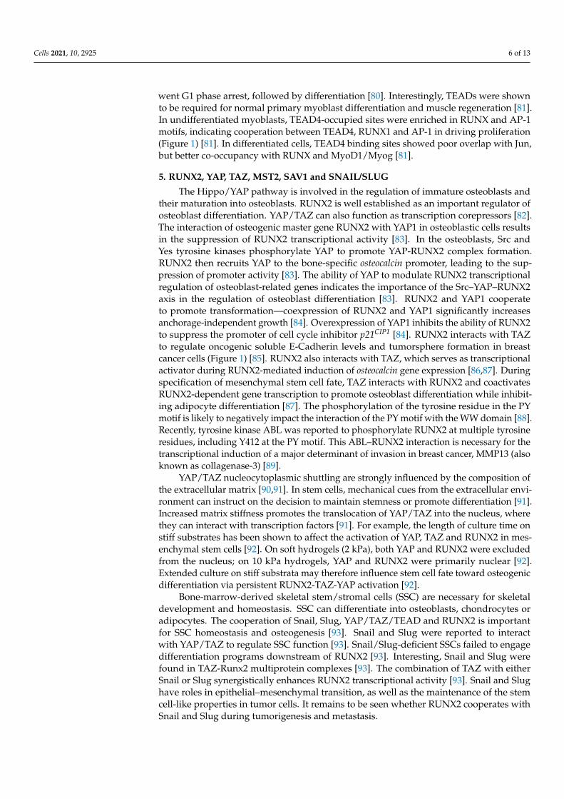
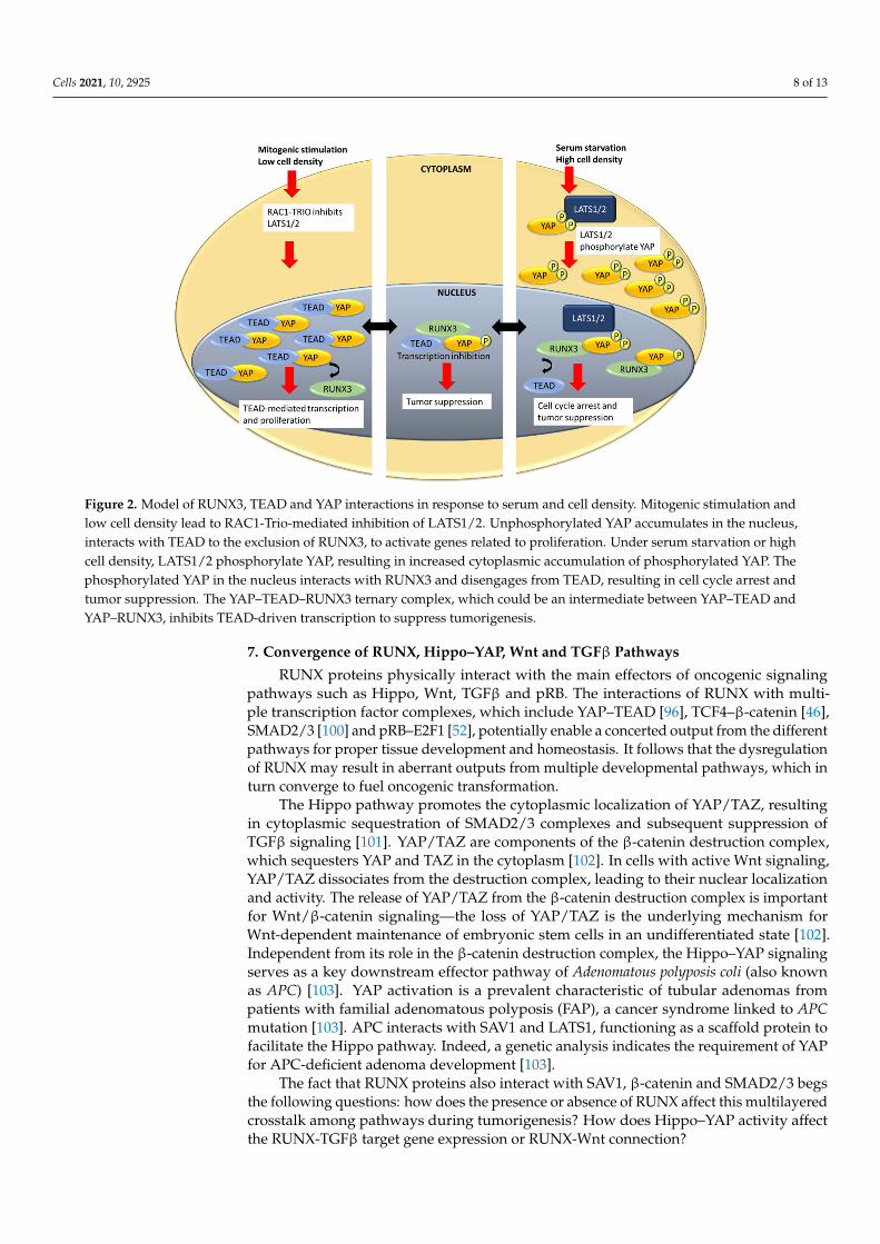
The interactions between YAP, TEAD and RUNX were found to be sensitive to serumdeprivation [98]. Serum deprivation is associated with the inactivation of RAC (memberof the Rho family of small GTPases) signaling, subsequent LATS1/2 activation and YAPphosphorylation. YAP phosphorylation results in a marked reduction of YAP–TEAD4 inter-action, and an increased YAP–RUNX3 interaction [98] (Figure 2). Moreover, YAP–TEAD4complex formation mainly occurs at a low cell density, while YAP–RUNX3 interactionpredominates at high cell density [98] (Figure 2). This work suggests that the YAP–TEAD4–RUNX3 ternary complex is an intermediate when YAP switches partners, for TEAD4 orRUNX3 [98]. Furthermore, it was proposed that RUNX3 suppresses growth in gastriccancer cells by changing the partner of YAP from TEAD4 to RUNX3 [98].
Aside from YAP–TEAD, RUNX3 also interacts with SAV1 in a MST2-dependent man-ner [99]. Similar to its interaction with TEAD, the Runt domain of RUNX3 is essential forinteraction with the first WW domain of SAV1; the PY motif of RUNX3 is not required for in-teraction (Figure 1) [99]. Through SAV1, MST2 interacts and colocalizes with RUNX3 in thenucleus [99]. MST2 phosphorylates RUNX3 at residues Ser-17, Thr-69, Ser-71, Ser-77, Ser-81and Thr-153, with the latter 4 amino acids located within the Runt domain (Figure 1) [99].MST2 functions in conjunction with SAV1 to interfere with Smurf1-mediated RUNX3 degra-dation and promote RUNX3 stability [99]. MST1/2 kinases serve proapoptotic functions.Depletion of RUNX3 abrogates MST-mediated reduction of cell viability, suggesting thatMST2, SAV1 and RUNX3 cooperate synergistically to promote cell death [99].
Cells 2021, 10, 2925 8 of 13Cells 2021, 10, x FOR PEER REVIEW 8 of 14
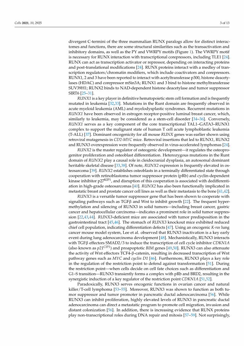
Figure 2. Model of RUNX3, TEAD and YAP interactions in response to serum and cell density. Mi-togenic stimulation and low cell density lead to RAC1-Trio-mediated inhibition of LATS1/2. Un-phosphorylated YAP accumulates in the nucleus, interacts with TEAD to the exclusion of RUNX3, to activate genes related to proliferation. Under serum starvation or high cell density, LATS1/2 phos-phorylate YAP, resulting in increased cytoplasmic accumulation of phosphorylated YAP. The phos-phorylated YAP in the nucleus interacts with RUNX3 and disengages from TEAD, resulting in cell cycle arrest and tumor suppression. The YAP–TEAD–RUNX3 ternary complex, which could be an intermediate between YAP–TEAD and YAP–RUNX3, inhibits TEAD-driven transcription to sup-press tumorigenesis.
Aside from YAP–TEAD, RUNX3 also interacts with SAV1 in a MST2-dependent manner [99]. Similar to its interaction with TEAD, the Runt domain of RUNX3 is essential for interaction with the first WW domain of SAV1; the PY motif of RUNX3 is not required for interaction (Figure 1) [99]. Through SAV1, MST2 interacts and colocalizes with RUNX3 in the nucleus [99]. MST2 phosphorylates RUNX3 at residues Ser-17, Thr-69, Ser-71, Ser-77, Ser-81 and Thr-153, with the latter 4 amino acids located within the Runt domain (Figure 1) [99]. MST2 functions in conjunction with SAV1 to interfere with Smurf1-mediated RUNX3 degradation and promote RUNX3 stability [99]. MST1/2 kinases serve proapoptotic func-tions. Depletion of RUNX3 abrogates MST-mediated reduction of cell viability, suggesting that MST2, SAV1 and RUNX3 cooperate synergistically to promote cell death [99].
7. Convergence of RUNX, Hippo–YAP, Wnt and TGFβ Pathways RUNX proteins physically interact with the main effectors of oncogenic signaling
pathways such as Hippo, Wnt, TGFβ and pRB. The interactions of RUNX with multiple transcription factor complexes, which include YAP–TEAD [96], TCF4–β-catenin [46], SMAD2/3 [100] and pRB–E2F1 [52], potentially enable a concerted output from the differ-ent pathways for proper tissue development and homeostasis. It follows that the dysreg-ulation of RUNX may result in aberrant outputs from multiple developmental pathways, which in turn converge to fuel oncogenic transformation.
The Hippo pathway promotes the cytoplasmic localization of YAP/TAZ, resulting in cytoplasmic sequestration of SMAD2/3 complexes and subsequent suppression of TGFβ signaling [101]. YAP/TAZ are components of the β-catenin destruction complex, which sequesters YAP and TAZ in the cytoplasm [102]. In cells with active Wnt signaling, YAP/TAZ dissociates from the destruction complex, leading to their nuclear localization and activity. The release of YAP/TAZ from the β-catenin destruction complex is important for Wnt/β-catenin signaling—the loss of YAP/TAZ is the underlying mechanism for Wnt-
Figure 2. Model of RUNX3, TEAD and YAP interactions in response to serum and cell density. Mitogenic stimulation andlow cell density lead to RAC1-Trio-mediated inhibition of LATS1/2. Unphosphorylated YAP accumulates in the nucleus,interacts with TEAD to the exclusion of RUNX3, to activate genes related to proliferation. Under serum starvation or highcell density, LATS1/2 phosphorylate YAP, resulting in increased cytoplasmic accumulation of phosphorylated YAP. Thephosphorylated YAP in the nucleus interacts with RUNX3 and disengages from TEAD, resulting in cell cycle arrest andtumor suppression. The YAP–TEAD–RUNX3 ternary complex, which could be an intermediate between YAP–TEAD andYAP–RUNX3, inhibits TEAD-driven transcription to suppress tumorigenesis.
7. Convergence of RUNX, Hippo–YAP, Wnt and TGFβ Pathways
RUNX proteins physically interact with the main effectors of oncogenic signalingpathways such as Hippo, Wnt, TGFβ and pRB. The interactions of RUNX with multi-ple transcription factor complexes, which include YAP–TEAD [96], TCF4–β-catenin [46],SMAD2/3 [100] and pRB–E2F1 [52], potentially enable a concerted output from the differentpathways for proper tissue development and homeostasis. It follows that the dysregulationof RUNX may result in aberrant outputs from multiple developmental pathways, which inturn converge to fuel oncogenic transformation.
The Hippo pathway promotes the cytoplasmic localization of YAP/TAZ, resultingin cytoplasmic sequestration of SMAD2/3 complexes and subsequent suppression ofTGFβ signaling [101]. YAP/TAZ are components of the β-catenin destruction complex,which sequesters YAP and TAZ in the cytoplasm [102]. In cells with active Wnt signaling,YAP/TAZ dissociates from the destruction complex, leading to their nuclear localizationand activity. The release of YAP/TAZ from the β-catenin destruction complex is importantfor Wnt/β-catenin signaling—the loss of YAP/TAZ is the underlying mechanism forWnt-dependent maintenance of embryonic stem cells in an undifferentiated state [102].Independent from its role in the β-catenin destruction complex, the Hippo–YAP signalingserves as a key downstream effector pathway of Adenomatous polyposis coli (also knownas APC) [103]. YAP activation is a prevalent characteristic of tubular adenomas frompatients with familial adenomatous polyposis (FAP), a cancer syndrome linked to APCmutation [103]. APC interacts with SAV1 and LATS1, functioning as a scaffold protein tofacilitate the Hippo pathway. Indeed, a genetic analysis indicates the requirement of YAPfor APC-deficient adenoma development [103].
The fact that RUNX proteins also interact with SAV1, β-catenin and SMAD2/3 begsthe following questions: how does the presence or absence of RUNX affect this multilayeredcrosstalk among pathways during tumorigenesis? How does Hippo–YAP activity affectthe RUNX-TGFβ target gene expression or RUNX-Wnt connection?
Cells 2021, 10, 2925 9 of 13
8. Discussion
It is interesting that knockout mouse models of individual Hippo pathway genes wereinsufficient to induce tumor in tissues such as lung, breast and pancreas [65]. Moreover, ge-netic alterations of Hippo components were generally low in cancer [104]. It was proposedthat YAP/TAZ’s oncogenic effects require additional events [65]. It is therefore conceivablethat transcriptional cooperation of YAP/TAZ with developmental transcription factorssuch as RUNX modulate YAP/TAZ-driven oncogenic growth at various stages of cancerdevelopment. The ability of RUNX to interact with various components of the Hippo–YAPpathway suggests a feedback mechanism to safeguard the different stages of the Hippo sig-naling cascade. The activation of YAP/TAZ is strongly associated with stem-like behaviorin cancer cells, proliferation, inflammation, chemoresistance and metastasis. RUNX genesare intimately involved in proliferation, stem cell regulation, and immunity [22,105]. Asdescribed above, RUNX genes have been implicated in cancer initiation, inflammation aswell as metastasis. These overlaps in biological processes are indicative of the shared rolesof RUNX and the Hippo–YAP pathway and the inappropriate outcomes, should eitherbe deregulated.
Further exploring the interaction of RUNX with the YAP-Hippo pathway is likely toyield insights on regenerative medicine. As described earlier, RUNX1 is a key determinantof muscle repair [80]. Studying the synergistic effect of RUNX1 with AP-1, TEAD and YAPmay promote the development of effective strategies for muscle regeneration due to severeinjury or congenital muscle diseases. Moreover, the multipotential mesenchymal stem cellsare frequently used for regenerative medicine. The fact that extracellular matrix stiffnessmodulates the activity of the RUNX2–YAP/TAZ axis in mesenchymal stem cells [92]indicates potential for bone/skeletal tissue repair.
So far, the research on RUNX interaction with the YAP–Hippo pathway has raisedmany exciting possibilities for stem cell studies, tissue regeneration and cancer treatment.Further in-depth studies will, in time, expand our knowledge on the regulation of stem cellfate and cancer behavior.
Author Contributions: Writing–original draft preparation, review and editing, L.S.H.C. and Y.I.;funding acquisition, Y.I. All authors have read and agreed to the published version of the manuscript.
Funding: This work is supported by the National Research Foundation Singapore and the SingaporeMinistry of Education under its Research Centres of Excellence initiative, the Singapore Ministry ofHealth’s National Medical Research Council under its Translational and Clinical Research FlagshipProgram (NMRC/TCR/009-NUHS/2013), under its Clinician-Scientist Individual Research Grant(NMRC/CIRG/1452/2016), the Open Fund Large Collaborative Grant (OFLCG18May-0023), aswell as the Singapore Ministry of Education Academic Research Fund Tier 2 (MOE2015-T2-2-038)provided to YI.
Institutional Review Board Statement: Not applicable.
Informed Consent Statement: Not applicable.
Data Availability Statement: Not applicable.
Conflicts of Interest: The authors declare no conflict of interest.
References1. Okuda, T.; van Deursen, J.; Hiebert, S.W.; Grosveld, G.; Downing, J.R. AML1, the Target of Multiple Chromosomal Translocations
in Human Leukemia, Is Essential for Normal Fetal Liver Hematopoiesis. Cell 1996, 84, 321–330. [CrossRef]2. Wang, Q.; Stacy, T.; Miller, J.D.; Lewis, A.F.; Gu, T.-L.; Huang, X.; Bushweller, J.; Bories, J.-C.; Alt, F.W.; Ryan, G.; et al. The CBFβ
Subunit Is Essential for CBFα2 (AML1) Function In Vivo. Cell 1996, 87, 697–708. [CrossRef]3. Komori, T.; Yagi, H.; Nomura, S.; Yamaguchi, A.; Sasaki, K.; Deguchi, K.; Shimizu, Y.; Bronson, R.; Gao, Y.-H.; Inada, M.; et al.
Targeted Disruption of Cbfa1 Results in a Complete Lack of Bone Formation owing to Maturational Arrest of Osteoblasts. Cell1997, 89, 755–764. [CrossRef]
4. Mundlos, S.; Otto, F.; Mundlos, C.; Mulliken, J.; Aylsworth, A.; Albright, S.; Lindhout, D.; Cole, W.; Henn, W.; Knoll, J.; et al.Mutations Involving the Transcription Factor CBFA1 Cause Cleidocranial Dysplasia. Cell 1997, 89, 773–779. [CrossRef]
Cells 2021, 10, 2925 10 of 13
5. Otto, F.; Thornell, A.P.; Crompton, T.; Denzel, A.; Gilmour, K.C.; Rosewell, I.R.; Stamp, G.W.; Beddington, R.S.; Mundlos, S.;Olsen, B.R.; et al. Cbfa1, a Candidate Gene for Cleidocranial Dysplasia Syndrome, Is Essential for Osteoblast Differentiation andBone Development. Cell 1997, 89, 765–771. [CrossRef]
6. Levanon, D.; Bettoun, D.; Harris-Cerruti, C.; Woolf, E.; Negreanu, V.; Eilam, R.; Bernstein, Y.; Goldenberg, D.; Xiao, C.; Fliegauf,M.; et al. The Runx3 transcription factor regulates development and survival of TrkC dorsal root ganglia neurons. EMBO J. 2002,21, 3454–3463. [CrossRef]
7. Inoue, K.-I.; Ozaki, S.; Shiga, T.; Ito, K.; Masuda, T.; Okado, N.; Iseda, T.; Kawaguchi, S.; Ogawa, M.; Bae, S.-C.; et al. Runx3controls the axonal projection of proprioceptive dorsal root ganglion neurons. Nat. Neurosci. 2002, 5, 946–954. [CrossRef]
8. Yagi, R.; Chen, L.-F.; Shigesada, K.; Murakami, Y.; Ito, Y. A WW domain-containing Yes-associated protein (YAP) is a noveltranscriptional co-activator. EMBO J. 1999, 18, 2551–2562. [CrossRef]
9. Huang, J.; Wu, S.; Barrera, J.; Matthews, K.; Pan, D. The Hippo Signaling Pathway Coordinately Regulates Cell Proliferation andApoptosis by Inactivating Yorkie, the Drosophila Homolog of YAP. Cell 2005, 122, 421–434. [CrossRef]
10. Dey, A.; Varelas, X.; Guan, K.-L. Targeting the Hippo pathway in cancer, fibrosis, wound healing and regenerative medicine. Nat.Rev. Drug Discov. 2020, 19, 480–494. [CrossRef]
11. Moya, I.M.; Halder, G. Hippo–YAP/TAZ signalling in organ regeneration and regenerative medicine. Nat. Rev. Mol. Cell Biol.2018, 20, 211–226. [CrossRef]
12. Zhao, B.; Li, L.; Lei, Q.-Y.; Guan, K.-L. The Hippo-YAP pathway in organ size control and tumorigenesis: An updated version.Genes Dev. 2010, 24, 862–874. [CrossRef]
13. Lin, Z.; Yang, Z.; Xie, R.; Ji, Z.; Guan, K.; Zhang, M. Decoding WW domain tandem-mediated target recognitions in tissue growthand cell polarity. eLife 2019, 8. [CrossRef]
14. Reuven, N.; Shanzer, M.; Shaul, Y. Tyrosine phosphorylation of WW proteins. Exp. Biol. Med. 2015, 240, 375–382. [CrossRef]15. Plouffe, S.W.; Lin, K.C.; Moore, J.L.; Tan, F.E.; Ma, S.; Ye, Z.; Qiu, Y.; Ren, B.; Guan, K.-L. The Hippo pathway effector proteins
YAP and TAZ have both distinct and overlapping functions in the cell. J. Biol. Chem. 2018, 293, 11230–11240. [CrossRef]16. Ben, C.; Wu, X.; Takahashi-Kanemitsu, A.; Knight, C.T.; Hayashi, T.; Hatakeyama, M. Alternative splicing reverses the cell-intrinsic
and cell-extrinsic pro-oncogenic potentials of YAP1. J. Biol. Chem. 2020, 295, 13965–13980. [CrossRef]17. Vrbský, J.; Vinarský, V.; Perestrelo, A.R.; De La Cruz, J.O.; Martino, F.; Pompeiano, A.; Izzi, V.; Hlinomaz, O.; Rotrekl, V.; Sudol,
M.; et al. Evidence for discrete modes of YAP1 signaling via mRNA splice isoforms in development and diseases. Genomics 2021,113, 1349–1365. [CrossRef]
18. Gaffney, C.J.; Oka, T.; Mazack, V.; Hilman, D.; Gat, U.; Muramatsu, T.; Inazawa, J.; Golden, A.; Carey, D.J.; Farooq, A.; et al.Identification, basic characterization and evolutionary analysis of differentially spliced mRNA isoforms of human YAP1 gene.Gene 2012, 509, 215–222. [CrossRef]
19. Huang, W.; Lv, X.; Liu, C.; Zha, Z.; Zhang, H.; Jiang, Y.; Xiong, Y.; Lei, Q.-Y.; Guan, K.-L. The N-terminal Phosphodegron TargetsTAZ/WWTR1 Protein for SCFβ-TrCP-dependent Degradation in Response to Phosphatidylinositol 3-Kinase Inhibition. J. Biol.Chem. 2012, 287, 26245–26253. [CrossRef]
20. Cottini, F.; Hideshima, T.; Xu, C.; Sattler, M.; Dori, M.; Agnelli, L.; Hacken, E.T.; Bertilaccio, M.T.; Antonini, E.; Neri, A.; et al.Rescue of Hippo coactivator YAP1 triggers DNA damage–induced apoptosis in hematological cancers. Nat. Med. 2014, 20,599–606. [CrossRef]
21. Grieve, S.; Wajnberg, G.; Lees, M.; Chacko, S.; Weir, J.; Crapoulet, N.; Reiman, T. TAZ functions as a tumor suppressor in multiplemyeloma by downregulating MYC. Blood Adv. 2019, 3, 3613–3625. [CrossRef]
22. Ito, Y.; Bae, S.-C.; Chuang, L.S.H. The RUNX family: Developmental regulators in cancer. Nat. Rev. Cancer 2015, 15, 81–95.[CrossRef]
23. Bruno, L.; Ramlall, V.; Studer, R.A.; Sauer, S.; Bradley, D.; Dharmalingam, G.; Carroll, T.; Ghoneim, M.; Chopin, M.; Nutt, S.L.;et al. Selective deployment of transcription factor paralogs with submaximal strength facilitates gene regulation in the immunesystem. Nat. Immunol. 2019, 20, 1372–1380. [CrossRef]
24. Blyth, K.; Cameron, E.R.; Neil, J.C. The runx genes: Gain or loss of function in cancer. Nat. Rev. Cancer 2005, 5, 376–387. [CrossRef]25. Reed-Inderbitzin, E.; Moreno-Miralles, I.; Vanden-Eynden, S.K.; Xie, J.; Lutterbach, B.; Durst-Goodwin, K.L.; Luce, K.S.; Irvin, B.J.;
Cleary, M.L.; Brandt, S.J.; et al. RUNX1 associates with histone deacetylases and SUV39H1 to repress transcription. Oncogene2006, 25, 5777–5786. [CrossRef]
26. Lutterbach, B.; Westendorf, J.J.; Linggi, B.; Isaac, S.; Seto, E.; Hiebert, S.W. A Mechanism of Repression by Acute MyeloidLeukemia-1, the Target of Multiple Chromosomal Translocations in Acute Leukemia. J. Biol. Chem. 2000, 275, 651–656. [CrossRef]
27. Jin, Y.-H.; Jeon, E.-J.; Li, Q.-L.; Lee, Y.H.; Choi, J.-K.; Kim, W.-J.; Lee, K.-Y.; Bae, S.-C. Transforming Growth Factor-β Stimulatesp300-dependent RUNX3 Acetylation, Which Inhibits Ubiquitination-mediated Degradation. J. Biol. Chem. 2004, 279, 29409–29417.[CrossRef]
28. Yamaguchi, Y.; Kurokawa, M.; Imai, Y.; Izutsu, K.; Asai, T.; Ichikawa, M.; Yamamoto, G.; Nitta, E.; Yamagata, T.; Sasaki, K.;et al. AML1 Is Functionally Regulated through p300-mediated Acetylation on Specific Lysine Residues. J. Biol. Chem. 2004, 279,15630–15638. [CrossRef]
29. Imai, Y.; Kurokawa, M.; Yamaguchi, Y.; Izutsu, K.; Nitta, E.; Mitani, K.; Satake, M.; Noda, T.; Ito, Y.; Hirai, H. The CorepressormSin3A Regulates Phosphorylation-Induced Activation, Intranuclear Location, and Stability of AML1. Mol. Cell. Biol. 2004, 24,1033–1043. [CrossRef]
Cells 2021, 10, 2925 11 of 13
30. Westendorf, J.J. Transcriptional co-repressors of Runx2. J. Cell. Biochem. 2006, 98, 54–64. [CrossRef]31. Choe, M.; Brusgard, J.L.; Chumsri, S.; Bhandary, L.; Zhao, X.F.; Lu, S.; Goloubeva, O.G.; Polster, B.M.; Fiskum, G.M.; Girnun, G.D.;
et al. The RUNX2 Transcription Factor Negatively Regulates SIRT6 Expression to Alter Glucose Metabolism in Breast CancerCells. J. Cell. Biochem. 2015, 116, 2210–2226. [CrossRef] [PubMed]
32. Osato, M. Point mutations in the RUNX1/AML1 gene: Another actor in RUNX leukemia. Oncogene 2004, 23, 4284–4296.[CrossRef] [PubMed]
33. Matheny, C.J.; Speck, M.; Cushing, P.R.; Zhou, Y.; Corpora, T.; Regan, M.; Newman, M.; Roudaia, L.; Speck, C.L.; Gu, T.-L.; et al.Disease mutations in RUNX1 and RUNX2 create nonfunctional, dominant-negative, or hypomorphic alleles. EMBO J. 2007, 26,1163–1175. [CrossRef] [PubMed]
34. Ellis, M.J.; Ding, L.; Shen, D.; Luo, J.; Suman, V.J.; Wallis, J.W.; Van Tine, B.A.; Hoog, J.; Goiffon, R.; Goldstein, T.C.; et al.Whole-genome analysis informs breast cancer response to aromatase inhibition. Nature 2012, 486, 353–360. [CrossRef]
35. Banerji, S.; Cibulskis, K.; Rangel-Escareno, C.; Brown, K.; Carter, S.L.; Frederick, A.M.; Lawrence, M.S.; Sivachenko, A.Y.; Sougnez, C.;Zou, L.; et al. Sequence analysis of mutations and translocations across breast cancer subtypes. Nature 2012, 486, 405–409. [CrossRef]
36. Ciriello, G.; Gatza, M.; Beck, A.H.; Wilkerson, M.D.; Rhie, S.; Pastore, A.; Zhang, H.; McLellan, M.; Yau, C.; Kandoth, C.; et al.Comprehensive Molecular Portraits of Invasive Lobular Breast Cancer. Cell 2015, 163, 506–519. [CrossRef]
37. Sanda, T.; Lawton, L.N.; Barrasa, M.I.; Fan, Z.P.; Kohlhammer, H.; Gutierrez, A.; Ma, W.; Tatarek, J.; Ahn, Y.; Kelliher, M.A.; et al.Core Transcriptional Regulatory Circuit Controlled by the TAL1 Complex in Human T Cell Acute Lymphoblastic Leukemia.Cancer Cell 2012, 22, 209–221. [CrossRef]
38. Yoshida, T.; Kanegane, H.; Osato, M.; Yanagida, M.; Miyawaki, T.; Ito, Y.; Shigesada, K. Functional Analysis of RUNX2 Mutationsin Japanese Patients with Cleidocranial Dysplasia Demonstrates Novel Genotype-Phenotype Correlations. Am. J. Hum. Genet.2002, 71, 724–738. [CrossRef]
39. Martin, J.W.; Zielenska, M.; Stein, G.S.; Van Wijnen, A.J.; Squire, J.A. The Role of RUNX2 in Osteosarcoma Oncogenesis. Sarcoma2010, 2011, 282745. [CrossRef]
40. Thomas, D.; Johnson, S.A.; Sims, N.; Trivett, M.K.; Slavin, J.L.; Rubin, B.P.; Waring, P.; McArthur, G.A.; Walkley, C.; Holloway, A.J.;et al. Terminal osteoblast differentiation, mediated by runx2 and p27KIP1, is disrupted in osteosarcoma. J. Cell Biol. 2004, 167,925–934. [CrossRef]
41. Pratap, J.; Wixted, J.J.; Gaur, T.; Zaidi, K.; Dobson, J.; Gokul, K.D.; Hussain, S.; Van Wijnen, A.J.; Stein, J.L.; Stein, G.S.; et al. Runx2Transcriptional Activation of Indian Hedgehog and a Downstream Bone Metastatic Pathway in Breast Cancer Cells. Cancer Res.2008, 68, 7795–7802. [CrossRef]
42. Akech, J.; Wixted, J.J.; Bedard, K.; Van Der Deen, M.; Hussain, S.; Guise, T.; Van Wijnen, A.J.; Stein, J.L.; Languino, L.; Altieri, D.C.;et al. Runx2 association with progression of prostate cancer in patients: Mechanisms mediating bone osteolysis and osteoblasticmetastatic lesions. Oncogene 2009, 29, 811–821. [CrossRef]
43. Kim, T.Y.; Lee, H.J.; Hwang, K.S.; Lee, M.; Kim, J.-W.; Bang, Y.-J.; Kang, G.H. Methylation of RUNX3 in various types of humancancers and premalignant stages of gastric carcinoma. Lab. Investig. 2004, 84, 479–484. [CrossRef]
44. Lau, Q.C.; Giafis, N.; Katsoulidis, E.; Sassano, A.; Tallman, M.S.; Higgins, L.S.; Nebreda, A.R.; Davis, R.J.; Platanias, L.C. RUNX3Is Frequently Inactivated by Dual Mechanisms of Protein Mislocalization and Promoter Hypermethylation in Breast Cancer.Cancer Res. 2006, 66, 6512–6520. [CrossRef]
45. Li, Q.; Ito, K.; Sakakura, C.; Fukamachi, H.; Inoue, K.-I.; Chi, X.-Z.; Lee, K.-Y.; Nomura, S.; Lee, C.-W.; Han, S.-B.; et al. CausalRelationship between the Loss of RUNX3 Expression and Gastric Cancer. Cell 2002, 109, 113–124. [CrossRef]
46. Ito, K.; Lim, A.C.-B.; Salto-Tellez, M.; Motoda, L.; Osato, M.; Chuang, L.S.H.; Lee, C.W.L.; Voon, D.; Koo, J.K.W.; Wang, H.; et al.RUNX3 Attenuates β-Catenin/T Cell Factors in Intestinal Tumorigenesis. Cancer Cell 2008, 14, 226–237. [CrossRef]
47. Ito, K.; Chuang, L.S.H.; Ito, T.; Chang, T.L.; Fukamachi, H.; Salto–Tellez, M.; Ito, Y. Loss of Runx3 Is a Key Event in InducingPrecancerous State of the Stomach. Gastroenterology 2011, 140, 1536–1546.e8. [CrossRef]
48. Lee, Y.-S.; Lee, J.-W.; Jang, J.-W.; Chi, X.-Z.; Kim, J.-H.; Li, Y.-H.; Kim, M.-K.; Kim, D.-M.; Choi, B.-S.; Kim, E.-G.; et al. Runx3Inactivation Is a Crucial Early Event in the Development of Lung Adenocarcinoma. Cancer Cell 2013, 24, 603–616. [CrossRef]
49. Yano, T.; Ito, K.; Fukamachi, H.; Chi, X.-Z.; Wee, H.-J.; Inoue, K.-I.; Ida, H.; Bouillet, P.; Strasser, A.; Bae, S.-C.; et al. The RUNX3Tumor Suppressor Upregulates Bim in GastricEpithelial Cells Undergoing Transforming Growth Factorβ-Induced Apoptosis.Mol. Cell. Biol. 2006, 26, 4474–4488. [CrossRef]
50. Chi, X.-Z.; Yang, J.-O.; Lee, K.-Y.; Ito, K.; Sakakura, C.; Li, Q.; Kim, H.-R.; Cha, E.-J.; Lee, Y.-H.; Kaneda, A.; et al. RUNX3Suppresses Gastric Epithelial Cell Growth by Inducing p21 WAF1 / Cip1 Expression in Cooperation with Transforming GrowthFactor β-Activated SMAD. Mol. Cell. Biol. 2005, 25, 8097–8107. [CrossRef]
51. Chi, X.-Z.; Lee, J.-W.; Lee, Y.-S.; Park, I.Y.; Ito, Y.; Bae, S.-C. Runx3 plays a critical role in restriction-point and defense againstcellular transformation. Oncogene 2017, 36, 6884–6894. [CrossRef]
52. Lee, J.-W.; Kim, D.-M.; Jang, J.-W.; Park, T.-G.; Song, S.-H.; Lee, Y.-S.; Chi, X.-Z.; Park, I.Y.; Hyun, J.-W.; Ito, Y.; et al. RUNX3regulates cell cycle-dependent chromatin dynamics by functioning as a pioneer factor of the restriction-point. Nat. Commun. 2019,10, 1897. [CrossRef]
53. Selvarajan, V.; Osato, M.; Nah, G.S.S.; Yan, J.; Chung, T.-H.; Voon, D.; Ito, Y.; Ham, M.F.; Salto-Tellez, M.; Shimizu, N.; et al.RUNX3 is oncogenic in natural killer/T-cell lymphoma and is transcriptionally regulated by MYC. Leukemia 2017, 31, 2219–2227.[CrossRef]
Cells 2021, 10, 2925 12 of 13
54. Lee, C.W.L.; Chuang, L.S.H.; Kimura, S.; Lai, S.K.; Ong, C.W.; Yan, B.; Salto-Tellez, M.; Choolani, M.; Ito, Y. RUNX3 functions asan oncogene in ovarian cancer. Gynecol. Oncol. 2011, 122, 410–417. [CrossRef]
55. Nevadunsky, N.S.; Barbieri, J.S.; Kwong, J.; Merritt, M.A.; Welch, W.R.; Berkowitz, R.S.; Mok, S.C. RUNX3 protein is overexpressedin human epithelial ovarian cancer. Gynecol. Oncol. 2009, 112, 325–330. [CrossRef]
56. Whittle, M.C.; Izeradjene, K.; Rani, P.G.; Feng, L.; Carlson, M.A.; DelGiorno, K.E.; Wood, L.D.; Goggins, M.; Hruban, R.H.; Chang,A.; et al. RUNX3 Controls a Metastatic Switch in Pancreatic Ductal Adenocarcinoma. Cell 2015, 161, 1345–1360. [CrossRef]
57. Chuang, L.S.H.; Khor, J.M.; Lai, S.K.; Garg, S.; Krishnan, V.; Koh, C.-G.; Lee, S.H.; Ito, Y. Aurora kinase-induced phosphorylationexcludes transcription factor RUNX from the chromatin to facilitate proper mitotic progression. Proc. Natl. Acad. Sci. USA 2016,113, 6490–6495. [CrossRef]
58. Tay, L.S.; Krishnan, V.; Sankar, H.; Chong, Y.L.; Chuang, L.S.H.; Tan, T.Z.; Kolinjivadi, A.M.; Kappei, D.; Ito, Y. RUNX Poly(ADP-Ribosyl)ation and BLM Interaction Facilitate the Fanconi Anemia Pathway of DNA Repair. Cell Rep. 2018, 24, 1747–1755.[CrossRef]
59. Wang, C.Q.; Krishnan, V.; Tay, L.S.; Chin, D.W.L.; Koh, C.P.; Chooi, J.Y.; Nah, G.S.S.; Du, L.; Jacob, B.; Yamashita, N.; et al.Disruption of Runx1 and Runx3 Leads to Bone Marrow Failure and Leukemia Predisposition due to Transcriptional and DNARepair Defects. Cell Rep. 2014, 8, 767–782. [CrossRef]
60. Zhao, B.; Ye, X.; Yu, J.; Li, L.; Li, W.; Li, S.; Lin, J.D.; Wang, C.-Y.; Chinnaiyan, A.M.; Lai, Z.-C.; et al. TEAD mediates YAP-dependentgene induction and growth control. Genes Dev. 2008, 22, 1962–1971. [CrossRef]
61. Camargo, F.D.; Gokhale, S.; Johnnidis, J.B.; Fu, D.; Bell, G.W.; Jaenisch, R.; Brummelkamp, T.R. YAP1 Increases Organ Size andExpands Undifferentiated Progenitor Cells. Curr. Biol. 2007, 17, 2054–2060. [CrossRef] [PubMed]
62. Schlegelmilch, K.; Mohseni, M.; Kirak, O.; Pruszak, J.; Rodriguez, J.R.; Zhou, D.; Kreger, B.T.; Vasioukhin, V.; Avruch, J.;Brummelkamp, T.R.; et al. Yap1 Acts Downstream of α-Catenin to Control Epidermal Proliferation. Cell 2011, 144, 782–795.[CrossRef] [PubMed]
63. Cordenonsi, M.; Zanconato, F.; Azzolin, L.; Forcato, M.; Rosato, A.; Frasson, C.; Inui, M.; Montagner, M.; Parenti, A.R.; Poletti,A.; et al. The Hippo Transducer TAZ Confers Cancer Stem Cell-Related Traits on Breast Cancer Cells. Cell 2011, 147, 759–772.[CrossRef] [PubMed]
64. Panciera, T.; Azzolin, L.; Fujimura, A.; Di Biagio, D.; Frasson, C.; Bresolin, S.; Soligo, S.; Basso, G.; Bicciato, S.; Rosato, A.; et al.Induction of Expandable Tissue-Specific Stem/Progenitor Cells through Transient Expression of YAP/TAZ. Cell Stem Cell 2016,19, 725–737. [CrossRef]
65. Zanconato, F.; Cordenonsi, M.; Piccolo, S. YAP/TAZ at the Roots of Cancer. Cancer Cell 2016, 29, 783–803. [CrossRef]66. Steinhardt, A.A.; Gayyed, M.F.; Klein, A.P.; Dong, J.; Maitra, A.; Pan, D.; Montgomery, E.A.; Anders, R.A. Expression of
Yes-associated protein in common solid tumors. Hum. Pathol. 2008, 39, 1582–1589. [CrossRef]67. Taniguchi, K.; Wu, L.-W.; Grivennikov, S.I.; de Jong, P.R.; Lian, I.; Yu, F.-X.; Wang, K.; Ho, S.B.; Boland, B.S.; Chang, J.; et al. A
gp130–Src–YAP module links inflammation to epithelial regeneration. Nature 2015, 519, 57–62. [CrossRef]68. Zanconato, F.; Forcato, M.; Battilana, G.; Azzolin, L.; Quaranta, E.; Bodega, B.; Rosato, A.; Bicciato, S.; Cordenonsi, M.; Piccolo, S.
Genome-wide association between YAP/TAZ/TEAD and AP-1 at enhancers drives oncogenic growth. Nat. Cell Biol. 2015, 17,1218–1227. [CrossRef]
69. D’Alonzo, R.C.; Selvamurugan, N.; Karsenty, G.; Partridge, N. Physical Interaction of the Activator Protein-1 Factors c-Fos andc-Jun with Cbfa1 for Collagenase-3 Promoter Activation. J. Biol. Chem. 2002, 277, 816–822. [CrossRef]
70. Zanconato, F.; Battilana, G.; Forcato, M.; Filippi, L.; Azzolin, L.; Manfrin, A.; Quaranta, E.; Di Biagio, D.; Sigismondo, G.;Guzzardo, V.; et al. Transcriptional addiction in cancer cells is mediated by YAP/TAZ through BRD4. Nat. Med. 2018, 24,1599–1610. [CrossRef]
71. Croci, O.; De Fazio, S.; Biagioni, F.; Donato, E.; Caganova, M.; Curti, L.; Doni, M.; Sberna, S.; Aldeghi, D.; Biancotto, C.; et al.Transcriptional integration of mitogenic and mechanical signals by Myc and YAP. Genes Dev. 2017, 31, 2017–2022. [CrossRef]
72. Choi, W.; Kim, J.; Park, J.; Lee, D.-H.; Hwang, D.; Kim, J.-H.; Ashktorab, H.; Smoot, D.T.; Kim, S.-Y.; Choi, C.; et al. YAP/TAZInitiates Gastric Tumorigenesis via Upregulation of MYC. Cancer Res. 2018, 78, 3306–3320. [CrossRef]
73. Pearson, J.D.; Huang, K.; Pacal, M.; McCurdy, S.R.; Lu, S.; Aubry, A.; Yu, T.; Wadosky, K.M.; Zhang, L.; Wang, T.; et al. Binarypan-cancer classes with distinct vulnerabilities defined by pro- or anti-cancer YAP/TEAD activity. Cancer Cell 2021, 39, 1115–1134.[CrossRef]
74. Lamar, J.M.; Stern, P.; Liu, H.; Schindler, J.W.; Jiang, Z.-G.; Hynes, R.O. The Hippo pathway target, YAP, promotes metastasisthrough its TEAD-interaction domain. Proc. Natl. Acad. Sci. USA 2012, 109, E2441–E2450. [CrossRef]
75. Chen, D.; Sun, Y.; Wei, Y.; Zhang, P.; Rezaeian, A.H.; Teruya-Feldstein, J.; Gupta, S.; Liang, H.; Lin, H.-K.; Hung, M.-C.; et al.LIFR is a breast cancer metastasis suppressor upstream of the Hippo-YAP pathway and a prognostic marker. Nat. Med. 2012, 18,1511–1517. [CrossRef]
76. Qadi, A.A.; Taberlay, P.; Phillips, J.L.; Young, A.; West, A.C.; Brettingham-Moore, K.; Dickinson, J.; Holloway, A.F. The LeukemiaInhibitory Factor Receptor Gene Is a Direct Target of RUNX1. J. Cell. Biochem. 2015, 117, 49–58. [CrossRef]
77. Keshet, R.; Reuven, N.; Shaul, Y. c-Abl forces YAP to switch sides. Mol. Cell. Oncol. 2015, 2, e995006. [CrossRef]78. Levy, D.; Adamovich, Y.; Reuven, N.; Shaul, Y. Yap1 Phosphorylation by c-Abl Is a Critical Step in Selective Activation of
Proapoptotic Genes in Response to DNA Damage. Mol. Cell 2008, 29, 350–361. [CrossRef]
Cells 2021, 10, 2925 13 of 13
79. Levy, D.; Reuven, N.; Shaul, Y. A Regulatory Circuit Controlling Itch-mediated p73 Degradation by Runx. J. Biol. Chem. 2008, 283,27462–27468. [CrossRef]
80. Umansky, K.B.; Gruenbaum-Cohen, Y.; Tsoory, M.; Feldmesser, E.; Goldenberg, D.; Brenner, O.; Groner, Y. Runx1 TranscriptionFactor Is Required for Myoblasts Proliferation during Muscle Regeneration. PLoS Genet. 2015, 11, e1005457. [CrossRef]
81. Joshi, S.; Davidson, G.; Le Gras, S.; Watanabe, S.; Braun, T.; Mengus, G.; Davidson, I. TEAD transcription factors are required fornormal primary myoblast differentiation in vitro and muscle regeneration in vivo. PLoS Genet. 2017, 13, e1006600. [CrossRef]
82. Kim, M.; Kim, T.; Johnson, R.L.; Lim, D.-S. Transcriptional Co-repressor Function of the Hippo Pathway Transducers YAP andTAZ. Cell Rep. 2015, 11, 270–282. [CrossRef]
83. Zaidi, K.; Sullivan, A.J.; Medina, R.F.; Ito, Y.; Van Wijnen, A.J.; Stein, J.L.; Lian, J.B.; Stein, G.S. Tyrosine phosphorylation controlsRunx2-mediated subnuclear targeting of YAP to repress transcription. EMBO J. 2004, 23, 790–799. [CrossRef]
84. Vitolo, M.I.; Anglin, I.E.; Mahoney, W.M.; Renoud, K.J.; Gartenhaus, R.B.; Bachman, K.E.; Passaniti, A. The RUNX2 transcriptionfactor cooperates with the YES-associated protein, YAP65, to promote cell transformation. Cancer Biol. Ther. 2007, 6, 856–863.[CrossRef]
85. Brusgard, J.L.; Choe, M.; Chumsri, S.; Renoud, K.; MacKerell, A.D., Jr.; Sudol, M.; Passaniti, A. RUNX2 and TAZ-dependentsignaling pathways regulate soluble E-Cadherin levels and tumorsphere formation in breast cancer cells. Oncotarget 2015, 6,28132–28150. [CrossRef]
86. Cui, C.B.; Cooper, L.; Yang, X.; Karsenty, G.; Aukhil, I. Transcriptional Coactivation of Bone-Specific Transcription Factor Cbfa1 byTAZ. Mol. Cell. Biol. 2003, 23, 1004–1013. [CrossRef]
87. Hong, J.-H.; Hwang, E.S.; McManus, M.T.; Amsterdam, A.; Tian, Y.; Kalmukova, R.; Mueller, E.; Benjamin, T.; Spiegelman, B.M.;Sharp, P.A.; et al. TAZ, a Transcriptional Modulator of Mesenchymal Stem Cell Differentiation. Science 2005, 309, 1074–1078.[CrossRef]
88. Sudol, M.; Hunter, T. NeW Wrinkles for an Old Domain. Cell 2000, 103, 1001–1004. [CrossRef]89. He, F.; Matsumoto, Y.; Asano, Y.; Yamamura, Y.; Katsuyama, T.; La Rose, J.; Tomonobu, N.; Komalasari, N.L.G.Y.; Sakaguchi, M.;
Rottapel, R.; et al. RUNX2 Phosphorylation by Tyrosine Kinase ABL Promotes Breast Cancer Invasion. Front. Oncol. 2021, 11.[CrossRef]
90. Stanton, A.E.; Tong, X.; Yang, F. Extracellular matrix type modulates mechanotransduction of stem cells. Acta Biomater. 2019, 96,310–320. [CrossRef]
91. Naqvi, S.M.; McNamara, L.M. Stem Cell Mechanobiology and the Role of Biomaterials in Governing Mechanotransduction andMatrix Production for Tissue Regeneration. Front. Bioeng. Biotechnol. 2020, 8. [CrossRef] [PubMed]
92. Yang, C.; Tibbitt, M.W.; Basta, L.; Anseth, K.S. Mechanical memory and dosing influence stem cell fate. Nat. Mater. 2014, 13,645–652. [CrossRef] [PubMed]
93. Tang, Y.; Feinberg, T.; Keller, E.T.; Li, X.-Y.; Weiss, S.J. Snail/Slug binding interactions with YAP/TAZ control skeletal stem cellself-renewal and differentiation. Nature 2016, 18, 917–929. [CrossRef] [PubMed]
94. Guo, Z.; Zhou, K.; Wang, Q.; Huang, Y.; Ji, J.; Peng, Y.; Zhang, X.; Zheng, T.; Zhang, Z.; Chong, D.; et al. The transcription factorRUNX2 fuels YAP1 signaling and gastric cancer tumorigenesis. Cancer Sci. 2021, 112, 3533–3544. [CrossRef]
95. Won, G.-W.; Sung, M.; Lee, Y.; Lee, Y.H. MST2 kinase regulates osteoblast differentiation by phosphorylating and inhibitingRunx2 in C2C12 cells. Biochem. Biophys. Res. Commun. 2019, 512, 591–597. [CrossRef]
96. Qiao, Y.; Lin, S.J.; Chen, Y.; Voon, D.; Zhu, F.; Chuang, L.S.H.; Wang, T.; Tan, P.; Lee, S.C.; Yeoh, K.G.; et al. RUNX3 is a novelnegative regulator of oncogenic TEAD–YAP complex in gastric cancer. Oncogene 2015, 35, 2664–2674. [CrossRef]
97. Anbanandam, A.; Albarado, D.C.; Nguyen, C.T.; Halder, G.; Gao, X.; Veeraraghavan, S. Insights into transcription enhancer factor1 (TEF-1) activity from the solution structure of the TEA domain. Proc. Natl. Acad. Sci. USA 2006, 103, 17225–17230. [CrossRef]
98. Jang, J.-W.; Kim, M.-K.; Lee, Y.-S.; Lee, J.-W.; Kim, D.-M.; Song, S.-H.; Choi, B.-Y.; Min, B.; Chi, X.-Z.; Bae, S.-C. RAC-LATS1/2signaling regulates YAP activity by switching between the YAP-binding partners TEAD4 and RUNX3. Oncogene 2016, 36,999–1011. [CrossRef]
99. Min, B.; Kim, M.-K.; Zhang, J.-W.; Kim, J.; Chung, K.-C.; Oh, B.-C.; Stein, G.S.; Lee, Y.-H.; van Wijnen, A.J.; Bae, S.-C. Identificationof RUNX3 as a component of the MST/Hpo signaling pathway. J. Cell. Physiol. 2011, 227, 839–849. [CrossRef]
100. Hanai, J.-I.; Chen, L.F.; Kanno, T.; Ohtani-Fujita, N.; Kim, W.Y.; Guo, W.-H.; Imamura, T.; Ishidou, Y.; Fukuchi, M.; Shi, M.-J.; et al.Interaction and Functional Cooperation of PEBP2/CBF with Smads. J. Biol. Chem. 1999, 274, 31577–31582. [CrossRef]
101. Varelas, X.; Samavarchi-Tehrani, P.; Narimatsu, M.; Weiss, A.; Cockburn, K.; Larsen, B.G.; Rossant, J.; Wrana, J.L. The CrumbsComplex Couples Cell Density Sensing to Hippo-Dependent Control of the TGF-β-SMAD Pathway. Dev. Cell 2010, 19, 831–844.[CrossRef]
102. Azzolin, L.; Panciera, T.; Soligo, S.; Enzo, E.; Bicciato, S.; Dupont, S.; Bresolin, S.; Frasson, C.; Basso, G.; Guzzardo, V.; et al.YAP/TAZ Incorporation in the β-Catenin Destruction Complex Orchestrates the Wnt Response. Cell 2014, 158, 157–170. [CrossRef]
103. Cai, J.; Maitra, A.; Anders, R.A.; Taketo, M.M.; Pan, D. β-Catenin destruction complex-independent regulation of Hippo–YAPsignaling by APC in intestinal tumorigenesis. Genes Dev. 2015, 29, 1493–1506. [CrossRef]
104. Wang, Y.; Xu, X.; Maglic, D.; Dill, M.; Mojumdar, K.; Ng, P.K.-S.; Jeong, K.J.; Tsang, Y.H.; Moreno, D.; Bhavana, V.H.; et al.Comprehensive Molecular Characterization of the Hippo Signaling Pathway in Cancer. Cell Rep. 2018, 25, 1304–1317.e5.[CrossRef]
105. Seo, W.; Taniuchi, I. The Roles of RUNX Family Proteins in Development of Immune Cells. Mol. Cells 2020, 43, 107–113. [CrossRef]
Related Documents