1 The motor hierarchy: from kinematics to goals and intentions Antonia F de C. Hamilton 1 , Scott T. Grafton 2 1) Department of Psychological and Brain Sciences, Dartmouth College, Hanover, NH. 2) Department of Psychology, University of California Santa Barbara, Santa Barbara, CA Chapter for Attention & Performance 22, Y Rosetti, M Kawato, P Haggard (Eds) Corresponding Author: Dr Antonia Hamilton, Dept of Psychological and Brain Sciences, Dartmouth College, Hanover, NH, 03755, USA [email protected]

Welcome message from author
This document is posted to help you gain knowledge. Please leave a comment to let me know what you think about it! Share it to your friends and learn new things together.
Transcript
1
The motor hierarchy:
from kinematics to goals and intentions
Antonia F de C. Hamilton1, Scott T. Grafton2
1) Department of Psychological and Brain Sciences, Dartmouth College, Hanover, NH.
2) Department of Psychology, University of California Santa Barbara, Santa Barbara, CA
Chapter for Attention & Performance 22, Y Rosetti, M Kawato, P Haggard (Eds)
Corresponding Author:
Dr Antonia Hamilton,
Dept of Psychological and Brain Sciences, Dartmouth College, Hanover, NH, 03755,
USA

2
The idea of hierarchical organisation in the motor system has a long history, but the
different components of the hierarchy have not been easy to localise in the human brain.
We have recently developed a one-back paradigm for inducing repetition suppression in
response to observed actions in fMRI, which allows different levels of the motor
hierarchy to be examined independently within the same stimulus set. 31 participants
viewed video clips of a hand taking a wine-bottle or a dumb-bell with either a precision or
whole hand grip and performed one of four attentional tasks while fMRI images were
acquired with a 3T scanner. We found suppression for repeated goals compared to
novel goals in the anterior intraparietal sulcus (aIPS), cerebellum and basal ganglia,
replicating previous results (Hamilton & Grafton, 2006). Suppression for repeated
grasps compared to novel grasps was found in the lateral occipital cortex (LOC), middle
intraparietal sulcus and inferior frontal gyrus (IFG). An analysis of task revealed no main
effects and no interactions between task and repetition suppression in any of the grasp
or goal clusters. We suggest that these results are compatible with a model of the motor
representational hierarchy in which aIPS represents goals or intentions and is placed at
the top of the hierarchy. In this model, LOC provides a visual analysis of grasp, while
IFG provides a motor analysis of grasp, both subsidiary to the goal representation in
aIPS. We discuss this model in relation to other hierarchical control models, and in
relation to the processing of actions for the self and others in the human mirror neuron
system.
Introduction
An ordinary action such as a pouring a glass of wine can be understood on many
levels. The barman will reach for and grasp the bottle, then transport it and tilt it over the
glass, carefully controlling the angle to avoid spilling. These kinematic components each
require a precisely orchestrated pattern of muscle activity guided by proprioceptive and
visual feedback. But a drinker waiting for the barman to finish pouring would likely
consider only the goal of the action – to provide some refreshment. Three broad levels of
description can be distinguished in this example – the goal level incorporates the
intention of the actor and the outcome of the action; the kinematic level describes the
shape of the hand and the movement of the arm in space and time; while the muscle level
describes the patterns of activity in over thirty hand and arms muscles which contribute to
the action.

3
These three levels have a hierarchical relationship, and are relatively independent
of one another. That is, the same goal may be accomplished with several different
kinematic components, each of which could be accomplished by a variety of muscle
activation sequences. Conversely, one particular pattern of muscle activity or one
particular kinematic component may contribute to different goals in different contexts.
The aim of the current paper is to determine if this descriptive hierarchy has a real
cognitive counterpart, and if so, can the different levels of the hierarchy be localised in
the human brain?
Background
The idea that the motor system is organised hierarchically to achieve particular
goals has a long and broad history. 100 years ago, Sherrington (1906) distinguished
between upper and lower motor neurons, while Jackson (1889) referred to higher motor
centres controlling simple movements. From a psychological point of view, William
James suggested that voluntary movement is secondary to reflexive actions, and involves
“an anticipatory image … of the sensorial consequences of a movement” (James, 1890).
This image precedes the action and guides performance, acting as a goal or target state
which the action should aim towards. These concepts came together in the work of
Donald Hebb, who argued that our theories of different levels of description for
behaviours, including abstract concepts such as goal, should also have a counterpart in
different types of representation in the brain (Hebb, 1949).
More recently, theories of motor hierarchies have become more specific and have
been applied to all levels of the motor system. For example, Keele (1990) emphasised
the ordered nature of high level motor programs for feedforward control of movements,
in particular in sequential tasks such as writing or typing. Hierarchical ordering is found
implicitly in more detailed computational models of motor control, which always include
some ‘goal’ parameters which specifies the desired output of the system. This may be
termed the ‘desired trajectory’ (Wolpert et al., 1998) or ‘cost function’ (Hamilton and
Wolpert, 2002) or an ‘instruction stimulus’ (Arbib et al., 2000), but in every case, the
goal is assumed to exist at a level of control above the detailed model.
Wohlschlager (2003) provides experimental evidence for a hierarchy of goals in
children, showing that when a child imitates another person, he or she tends to copy goals

4
rather than subgoals. In this model, objects and outcomes are considered to be higher
goals than actions or movement paths, and goals are defined according to the ideomotor
principles of William James. At an even more abstract level, Pachiere (2006) provides a
philosophical analysis of action and intention which distinguishes motor-intentions,
present-intentions and future-intentions in a hierarchical scheme. All of these theories
see motor control in terms of a refinement of information from an abstract goal (‘to pour
the wine’) to a more detailed motor plan (‘lift the bottle, tilt the bottle over the glass’) to a
precise specification of the reaching and grasping actions required to achieve each goal,
and finally the activation of specific muscles in a coordinated sequence. The hierarchy
for motor control parallels work into executive function which suggests a similar
organisation in terms of goals and subgoals in both problem solving tasks (Dehaene and
Changeux, 1997, Norman and Shallice, 1986) and in everyday life (Shallice and Burgess,
1991).
In addition to this linear planning, the majority of theories recognise the essential
role of feedback loops in motor control. The role of spinal reflexes in eliciting and
controlling action has been studied for over 100 years, and it is now clear that
proprioceptive and motor systems are intimately linked in control loops at every level of
the motor system. Though it has in the past been argued that the delays between a
sensory event and a motor response severely limit the use of feedback control (Keele et
al., 1990), newer computational work reveals mechanisms which can largely mitigate
these delays. In particular, forward models provide a mechanism for predicting and
cancelling the sensory consequences of actions (Blakemore et al., 1998, Wolpert and
Flanagan, 2001). The idea of predictive forward models has recently been incorporated
into a more sophisticated computational framework, where a system of multiple parallel
forward – inverse model pairs are able to provide accurate control of action in a variety of
contexts, in a model called MOSAIC (Wolpert and Kawato, 1998). Furthermore, it has
been suggested that MOSAIC could be organised in a hierarchical fashion (Haruno et al.,
2003), and could even provide for understanding other people’s actions (Wolpert et al.,
2003).
Thus, the idea emerges of a hierarchical system of loops, in which information
flows in both directions between the different goal, kinematic and muscle levels. The

5
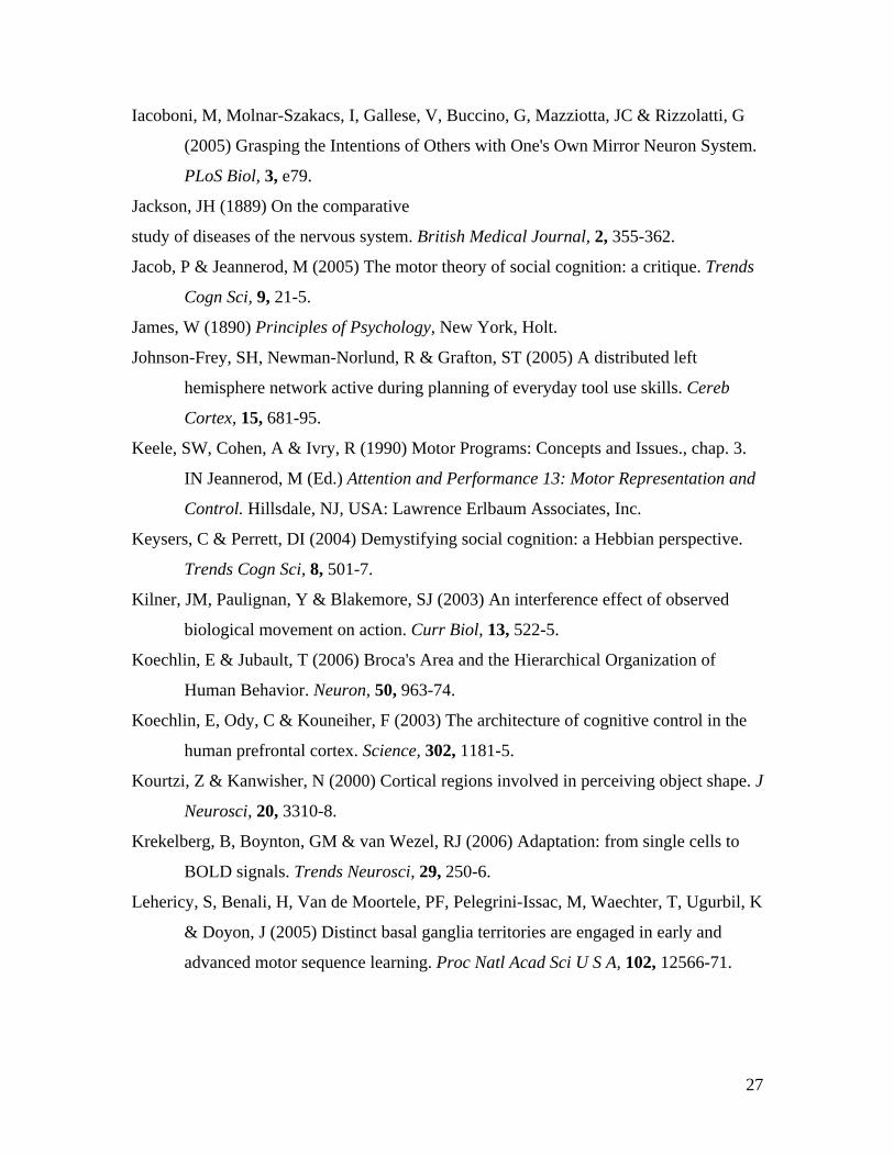
cognitive structure of this system is illustrated in Figure 1, which presents a rough
schema of a visual-motor loop for the control of hand actions. We distinguish the 3
major of motor control levels – goal, kinematic and muscles, together with two levels of
visual analysis – a general early analysis followed by more specific visual representations
of moving body parts and the objects they interact with, and we assume that information
flows in both directions between every level. This sketch is dramatically simplified and
ignores the important role of proprioceptive feedback, as well as the more subtle
distinctions within different levels, but it does sets out the basis components we are
interested in. Similar models have been proposed for both language (Geschwind, 1965)
and hand actions (Tessari and Rumiati, 2004), but the localisation of the different
components in the human brain remains uncertain. In the current paper, we will focus on
the visual analysis of action, the goal representation and the kinematic levels, and ask -
can these three components of the visual-motor hierarchy be localised in the human
brain?
-------------- FIGURE 1 ABOUT HERE -------------
The motor hierarchy in the brain
The inputs and outputs of the motor system ground the hierarchy in the brain.
Thus, we should begin by assuming the primary and secondary visual cortex provides the
low level visual analysis system, while primary motor cortex processes outputs to the
spinal cord and thus the muscles (Lemon et al., 1998). The higher levels of kinematics
and goal representations are likely to be found within the neural systems involved in
visually guided actions, which include the premotor cortex, supplementary motor areas,
parietal cortex, cerebellum and the basal ganglia. Extensive electrophysiological
recordings from these regions in awake behaving primates give some clues to their
functions.
Single unit recordings in the inferior frontal cortex of the monkey have revealed
neurons coding for grasp configuration and object shape (Rizzolatti et al., 1988) and
some of these are also responsive to the observation of actions (di Pellegrino et al., 1992,
Gallese et al., 1996). In particular, it has been claimed that some cells in area F5 encode
the goal of an action, because they respond when an action is inferred to have taken place

6
out of sight (Umilta et al., 2001). Similarly, single unit recordings in the anterior
intraparietal sulcus (AIP) in the monkey have found neuronal coding of object shape and
grasp (Sakata et al., 1995), while it has been reported that neurons in monkey inferior
parietal lobule (IPL) fire when a grasp and place action sequence is performed or
observed (Fogassi et al., 2005). These data have been interpreted in terms of neural
coding for performed and observed intentions in the IPL. In contrast, studies of more
abstract, symbolic goals implicate the frontal cortex in goal planning and control (Saito et
al., 2005, Shima et al., 1996, Mushiake et al., 2006). Thus, the monkey
neurophysiological studies do not provide a consensus on the localisation of goals or
grasps in the brain.
Studies of the monkey brain are limited by the fact that in general, only one brain
region, and often only one neuron, is tested at a time. This means that it is very difficult
to get an overall picture of the motor hierarchy. Furthermore, the tasks used in different
studies can be quite variable, and do not necessarily systematically separate the different
levels of representation. In particular, studies of object grasping in monkeys have not
systematically distinguished hand configuration from object identity (Sakata et al., 1995,
di Pellegrino et al., 1992). Finally, while there are homologies between the human and
monkey brain, there are also major differences, which are matched by behavioural
differences in planning, flexibility of action and the ability to infer other people’s
intentions, all of which are limited or absent in monkeys.
Human neuroimaging studies have the potential to examine the whole brain
during more flexible and varied tasks than can be performed by monkeys, and have
provided evidence for an extensive neural system for visual control of action,
encompassing primary motor cortex, premotor cortex, inferior parietal cortex, SMA,
cerebellum and basal ganglia (Grafton et al., 1996, Grafton et al., 1992). Many of these
studies used simple finger tapping or tracking tasks, because the use of fMRI to examine
more complex visually guided actions is severely limited by the scanner environment and
the need to avoid movement artefacts. These environmental limitations can be avoided
by studying imagined or observed actions rather than performed actions. There is
increasing evidence that action observation activates similar brain structures to those

7
involved in motor performance (Buccino et al., 2001, Rizzolatti and Craighero, 2004), so
observation can be taken, with caveats, as a proxy for performance.
Nevertheless, few human neuroimaging studies have even attempted to
distinguish different levels of the motor hierarchy in either performance or observation.
This is because any single task involving motor performance, imagined action or
observation of action requires processing at all levels at once, which means that it is not
easy to separate different components by means of a subtraction design. For example, if
participants observe a video clip of a hand action during fMRI (e.g. (Buccino et al.,
2001)), brain regions involved in processing visual motion, hand kinematics, goals and
intentions will all be activated, and a straight forward subtraction experiment cannot
distinguish the different levels.
Some studies have attempted to segment these systems, for example, by showing
videos of actions without an object and thus without a clear goal or intention, in
comparison to videos of goal directed actions (Pelphrey et al., 2004). These authors
report increases STS activation for ‘unintended’ actions, but it remains unclear if
participants really see a movement towards empty space as a movement without an
intention, or just as a movement with a more complex intention, for example,
communicating something to the observer. Similar problems would arise if ‘accidental’
actions were used as stimuli, because even children are able to perceive the true intention
underlying the action (Meltzoff, 1995), so a subtraction between accidental and correct
actions reveals brain regions involved in coding errors (Manthey et al., 2003) but not
those coding intentions. While subtraction designs have been used to separate reach and
grasp within the kinematic level (Culham et al., 2003), and to localise visual processing
of human body parts (Downing et al., 2001), it does not seem possible to examine goals,
intentions or kinematics across levels in the motor hierarchy by a traditional subtraction
design.
The aim of the current paper is to localise the different components of the motor
hierarchy, using a repetition suppression (RS) design to examine each level
independently, as detailed below. We use action observation rather than performance,
and thus rely on the assumption that performed and observed actions involve the same
neural systems (Rizzolatti and Craighero, 2004). Though this assumption restricts the

8
conclusions we can draw about motor performance, the use of action observation allows
us to address the question of the neural basis of action understanding and inferences
about other people’s intentions. This approach allows us to take a broader view of the
motor hierarchy, examining several different levels of representation to obtain an overall
model of action representation.
The current paper has two specific aims. First, we plan to replicate our previous
study (Hamilton and Grafton, 2006), which suggested that action goals are represented in
the anterior intraparietal sulcus (aIPS). This result is at odds with several previous
studies which give frontal regions a role in goals (Koechlin and Jubault, 2006) and
intentions (Iacoboni et al., 2005), and thus requires replication. As before, we focus
specifically on object goals, where the goal of the action is to take a particular object and
thus the goal is defined by the identity of the object taken. Second, we aim to localise the
kinematic levels of the hierarchy, which have received less attention in the past. In
particular, we will look for evidence of a dual representation of kinematics, in both visual
and motor terms. Understanding these lower levels of the system will allow a more
complete model of the visuo-motor control loop to be constructed, taking into account the
known connectivity between brain regions as well as the fMRI activations.
The repetition suppression approach
The experimental method we use measures a phenomenon known as repetition
suppression (RS), which is not widely used in motor studies. However, RS has been
extensively studied in the context of visual representations (Grill-Spector and Malach,
2001, Kourtzi and Kanwisher, 2000), where it is more commonly known as fMRI-
adaptation. The term repetition suppression will be used here as a more succinct and
general descriptor, because reduced responses to repeated stimuli are not unique to fMRI.
There are three major advantages to the RS approach. First, it allows us to look for
changes within a class of stimuli or a level of the hierarchy rather than between classes.
Thus, different levels of representation for the same stimulus can be analysed
independently. Second, RS has been linked to reaction time priming (Wig et al., 2005,
Maccotta and Buckner, 2004), though we do not make use of this link here. Third, RS
data can be interpreted in terms of neuronal population coding, in accordance with two
key principles:

9
1. Suppression from stimulus A to stimulus B occurs within a brain region only if some
feature of both A and B is encoded in the same population of neurons in that region.
2. If stimulus B caused suppression in a region, release from suppression on presentation
of stimulus C occurs only if some feature of B and C is encoded in different
populations within that region.
-------------- FIGURE 2 ABOUT HERE -------------
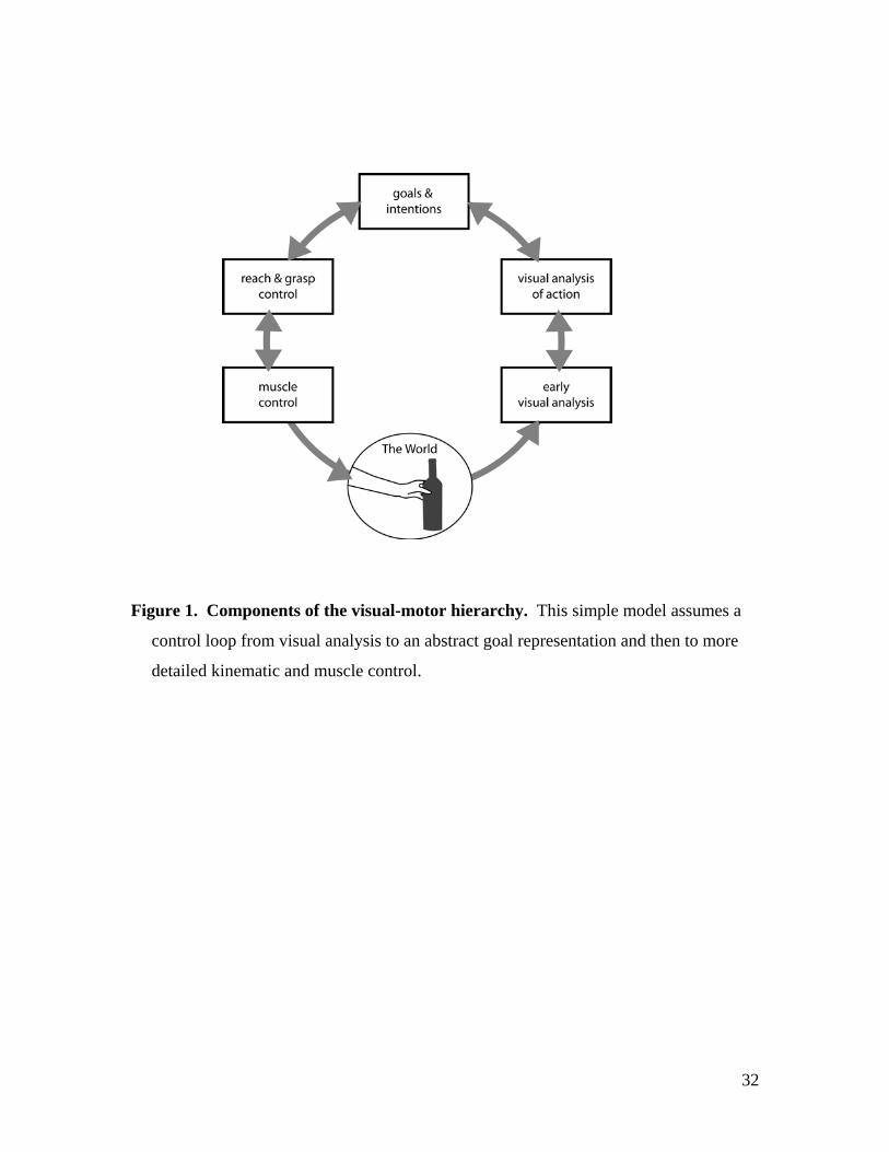
Figure 2 gives a concrete example of these principles in a hypothetical brain area
representing animals. In this region, one subpopulation of neurons encode ‘dog’, and a
different population encode ‘cat’. These two populations are interspersed and each
contain the same total number of neurons, so they cannot be distinguished by a traditional
fMRI subtraction design, but they can be discerned by measuring RS.
On seeing stimulus A, a Dalmatian, the neuronal population encoding ‘dog’ will
fire vigorously and a robust BOLD response will be recorded. When a picture of a
Daschund is shown next (B), the total activity in the ‘dog’ population is reduced and a
suppressed BOLD response is recorded for the repeated stimulus. If the third stimulus
(C) shows a tabby cat, the neuronal population encoding ‘cat’ will now respond robustly
giving a strong BOLD signal. Thus, the presence of the predicted BOLD pattern in
response to this stimulus sequence is evidence for distinct neuronal populations encoding
‘dog’ and ‘cat’ within a brain region. Note that this pattern of activation would not be
seen if the brain area represented either a broader category (e.g. mammals), which would
result in suppression over all stimuli, or a narrower category (e.g. each breed of dog in a
different population), which would not give any suppression between breeds. Thus, RS
can reveal the level of neural representation in different brain regions.
The validity of this interpretation depends on two simple assumptions. First is the
assumption of population coding within brain regions, for which there is extensive
evidence in many parts of the cortex (Georgopoulos et al., 1982, Britten et al., 1993).
Second, the population response must be suppressed when the same stimulus feature is
repeated. The precise pattern of suppression, which could be an overall reduction or a
sharpening of neuronal tuning curves, remains a matter of debate (Grill-Spector et al.,
2006), as does the mechanism by which suppression might occur (Krekelberg et al.,
2006). However, the plausibility of population suppression to repeated stimuli is no
10
longer in doubt, and thus the principle of measuring RS in order to infer neuronal
population coding appears to be sound.
A final issue which must be addressed before employing RS in motor studies is
the generality of the phenomena. The vast majority of studies have examined RS in
visual regions such as the lateral occipital complex and fusiform face area (Henson,
2003). However, there is also evidence for RS in frontal (Buckner et al., 1998) and
parietal (Shmuelof and Zohary, 2005) regions, and RS studies have been used to examine
for semantic (Thompson-Schill et al., 1999), syntactic (Noppeney and Price, 2004) and
numeric (Pinel et al., 2001) representations in the brain. Thus, this phenomenon seems to
be general to most of the cortex, and could plausibly apply to motor representations too.
By measuring suppression in response to repeated features at different levels of the motor
hierarchy, it should be possible to localise the representations involved in the human
brain.
Applying RS to the motor hierarchy
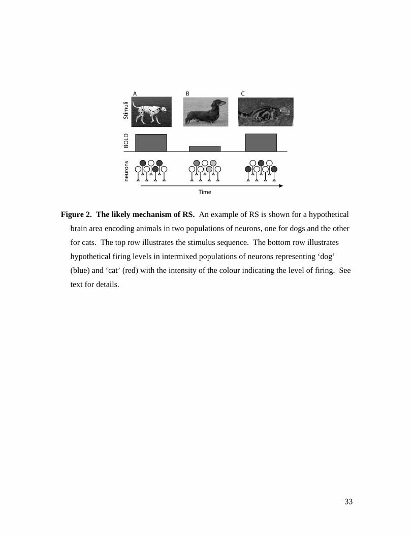
To examine the motor hierarchy in the human brain, we used a sequence of
stimuli designed to induce RS at three distinct levels: 1) The goal object of a reaching
action, 2) The type of grasp used to reach the object, and 3) the weight of the object as
revealed by lift velocity. Unlike many other RS studies (Grill-Spector and Malach,
2001), we use a one-back RS design (Figure 3), where each stimulus is defined as novel
or repeated relative to the one stimulus before it. This approach is motivated by the fact
that RS is largest on a single repeated trial immediately following the prime stimulus and
amount of suppression does not increase after approximately 8 stimuli (Grill-Spector et
al., 2006). Thus, a one-back design provides an efficient and flexible approach to
inducing and measuring RS within a single set of stimuli. We measured RS for three
stimulus characteristics: goal-object, grasp and weight; independently in a 2x2x2 factorial
design where each factor can be either Novel or Repeated.
-------------- FIGURE 3 ABOUT HERE -------------
Based on previous work, we have predictions for RS at each level. The goal level
was previously examined in an RS experiment (Hamilton and Grafton, 2006), which
revealed that a region of the left anterior intraparietal sulcus (aIPS) encodes the goal of
another person’s action. Here we define a goal by the identity of the object which a

11
person is reaching for, and note that our results do not necessarily generalise to all types
of goal, such as tool use, or social goals. Nevertheless, the goal of reaching for an object
is often a sub-goal in other more complex tasks, and provides an ideal entry-point for the
study of high level action representation. The goal-object analysis here will be an
attempt to replicate the previous study, in a more powerful scanner and with a more
precisely controlled stimulus set. Thus, we predict that the same brain region, aIPS, will
show RS for goal object.
The grasp level of representation could be localised to several regions, and in
particular, we hope to distinguish visual analysis of grasp from a motor preparatory
representation of grasp. Possible regions for motor grasp include aIPS which was
traditionally considered a ‘grasp area’(Sakata et al., 1995) and IFG on the basis of the
equivalence of this region to monkey F5, which has direct connections to primary motor
cortex and neurons specialising for different grasp configurations (Rizzolatti et al., 1988).
Visual analysis of grasp could be carried out in a variety of extrastriate regions in the
middle temporal gyrus, lateral occipital cortex or superior temporal sulcus, all of which
have been associated with processing observed body parts (Downing et al., 2001) and
biological motion (Grossman et al., 2000).
Object weight representations are likely to be similar to grasp representations, as
both fall at the kinematic level of the hierarchy. Weight judgments are made based on
the velocity of the actor’s lifting action (Hamilton et al., 2005), which could be expected
to involve visual and biological motion regions, including middle temporal gyrus, lateral
occipital cortex and superior temporal sulcus. However, inferior frontal gyrus is also
required for accurate weight judgment (Pobric and Hamilton, 2006), and both this region
and primary motor cortex show activity related to the biasing effect of box lifting on
weight judgment (Hamilton et al., 2006). Thus we predict that weight representations,
like grasp, would be likely to be found in the extrastriate visual regions and premotor
cortex.
The final factor we will examine is the effect of attention to different tasks on the
level of RS in each region. That is, does top-down processing of one element of the
scene influence the amount of RS to different elements in the scene? This is a critical
question for the interpretation of RS studies, in particular those that report suppression in
12
parietal or frontal regions, where the suppression could be interpreted in terms of
attention rather than a specific neural population code for the stimulus characteristics.
We examine task as a between subjects factor to ensure that each participant focuses on
only one task during the experiment, and predict no effects of task on RS.
Methods
Sixteen unique video clips were generated, each 4.5 seconds long, depicting a
hand reaching for and taking either a dumb-bell or a wine bottle. Each of the objects
could be grasped with either a precision grip on the neck of the bottle or middle of the
dumb-bell, or with a power grip on the body of the bottle or the top of the dumb-bell.
The object dimensions were precisely matched (neck / middle = 2.5 cm diameter; body /
top = 7.7 cm diameter) so that the motor properties of each grip would be identical.
Furthermore, each object could either be empty, weighing 0.5kg, or filled with water /
lead shot to a weight of 1kg. These changes in weight did not alter the appearance of the
object, so weight could only be judged by careful examination of the speed of the lifting
action. Finally, each object could be placed on the far left of the table, or slightly behind
and to the right, with the other object in the alternate location. Thus, the clips filled a
2x2x2 factorial design, with factors: Goal-object (dumb-bell / wine), Grasp (power /
precision), Weight (Heavy / Light), and clips were ordered to obtain RS for all of these
factors simultaneously.
31 right-handed participants gave their informed consent to take part. In the
scanner, the participants watched sets of video clips in a sequence determined by a one-
back repetition suppression design as illustrated in
Figure 3. In this design, each movie is defined as ‘Novel’ or ‘Repeated’ on each
of the three stimulus dimensions (goal, grasp and weight) relative to the one movie
before. Thus, each movie contributes to the analysis on its own account, but also acts as
a ‘prime’ for the next movie in the sequence. Movies were organised into sets, which
each began with a new movie which primes the RS movie on the next trial but was not
analysed. Each set contained between 5 and 10 RS movies, and then ended with a
question trial, followed by a short rest before the next set. All participants saw 8 new
movies, 8 questions and 64 RS movies in a single functional run.
13
Different questions were assigned randomly to individual participants to assess
the effects of task in a between subjects design. 3 participants were asked to determine
the identity of the object taken on each trial (Object-task), 11 were asked to determine the
grip used on each trial (Grip-task), 11 were asked to determine the object weight on each
trial (Weight-task) and the remaining 6 were asked to determine the location of the
grasped object (Location task). These groups allow us to examine the influence of top-
down control on RS at each level of the motor hierarchy. All participants were instructed
to attend to every video and mentally perform the task for that clip. After a set of 5 to 10
videos, the task question appeared written on the screen and participants were required to
answer as quickly and accurately as possible. These question trials ensured that
participants were awake and on-task during the scanning but were not analysed further.
The experiment was carried out in a 3T Philips Intera scanner using an eight
channel phased array coil and 36 slices per TR (4 mm thickness, 0.5 mm gap); TR: 2000
ms; TE: 35 ms; flip angle:90o; field of view: 24cm; matrix 80x80. The first two brain
images of the functional run were discarded, then 233 images were collected and stored.
Raw data for each participant was realigned, unwarped and normalised to the MNI
template with a resolution of 2x2x2mm in SPM2. A design matrix was fitted for each
subject, with each movie modeled as a boxcar convolved with the standard
haemodynamic response function. The design matrix weighted each raw image
according to its overall variability to reduce the impact of movement artifacts
(Diedrichsen and Shadmehr, 2005). After estimation, 9mm smoothing was applied to the
beta images.
We calculated contrasts for novel object > repeated object, novel grip > repeated
grip and novel weight > repeated weight. An exploratory analysis of interactions
between object RS and grip RS was also conducted within a mask of the main effects.
Results were analysed first over the whole group (regardless of task) and we report
regions which survive a threshold of p<0.001 uncorrected and 10 voxels over the whole
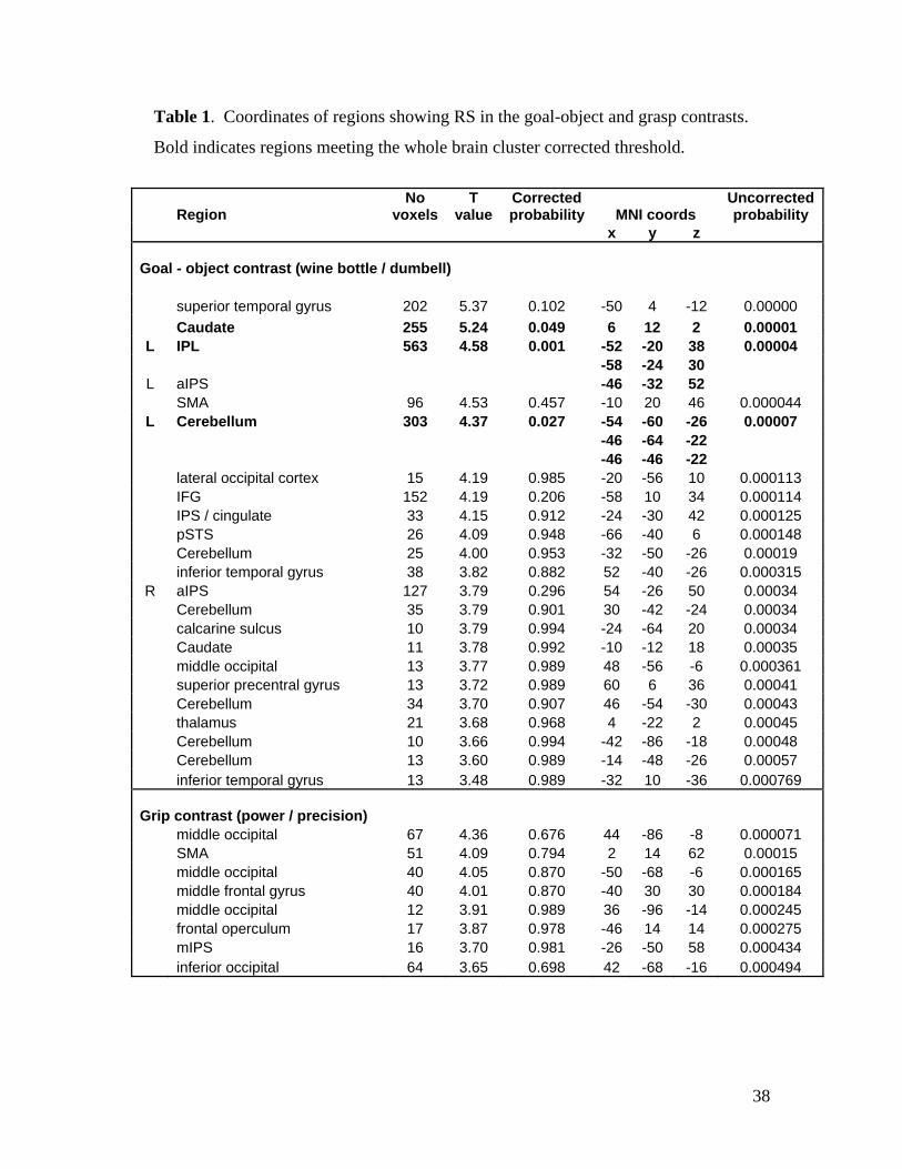
brain in Table 1. However, we limit the discussion of results to clusters which fall within
the predicted action representation network, or which survive the cluster level correction
for multiple comparisons. To examine the effect of cognitive task on RS in the action
representation system, a secondary analysis was carried out on the main clusters reported
14
in Table 1. Mean BOLD signal over the cluster in each condition for each participant
was extracted and subjected to a repeated measures ANOVA in SPSS with factors Grasp
and Object (both novel or repeated), and a between subjects factor of Task (object, grasp,
location or weight tasks). We looked for main effects of task or interactions of task with
RS, and consider only effects which survive Bonferroni correction for the number of
clusters tested to be significant, resulting in thresholds of p<0.0083 for the object cluster
set, and p<0.0063 for the grasp cluster set.
Results
The neural representation of object weight
In the contrast of novel weight > repeated weight over all participants, no regions
survived the p<0.001 threshold. At a more liberal p<0.005, cluster of 5 voxels was found
in the left inferior occipital cortex (-38, -74, 4). This region is close to the lateral
occipital location which is involved in the biasing effect of box lifting on weight
judgement (Hamilton et al., 2006) and is within the predicted visual action regions. Thus,
this result hints at a visual analysis of object weight. The effect may be weak because it
was not easy to judge object weight in the clips used, and participants who were not alert
to the different possible weights may not have discriminated object weight at all. More
precisely controlled stimuli, for example from a high speed video camera (Hamilton et
al., 2005), might be needed to obtain a more robust localisation of object weight. In
particular, the question of IFG involvement in weight judgement, as suggested previously
(Hamilton et al., 2006, Pobric and Hamilton, 2006), remains open.
The neural representation of hand grasp
Main effects of novel grasps compared to repeated grasps were found in three
clusters in the inferior and middle occipital regions, a single cluster in the inferior frontal
gyrus, and a region of the middle IPS. All these regions passed the p<0.001 uncorrected
threshold, and are listed in the top part of Table 1. Figure 4 illustrates the locations of the
clusters and the RS effects. Weaker responses to repeated grasps (orange and pale blue
bars) compared to novel grasps (red and dark blue bars) provide evidence for distinct
neuronal populations encoding whole hand grips and precision grips in these regions.
The RS effect appears weak in the plots due to individual differences in the absolute level
15
of response, which do not have an impact on the repeated-measures analysis conducted in
SPM. Outside the predicted network, RS for grasp was also seen in the SMA and middle
frontal gyrus, but there was no evidence of RS for grasp in the more anterior portion of
IPS or in IPL on either the left or right. These results are compatible with the idea of
several grasp representations in the brain. In particular, the lateral occipital regions could
contribute to a visual analysis of grasp, and the inferior frontal region to motor grasp
representation, as hypothesised.
-------------- FIGURE 4 ABOUT HERE -------------
The neural representation of a goal object
The analysis of RS for goal object revealed a robust and extensive brain network
(Figure 5 and Table 1). Three regions survived the whole brain cluster corrected
threshold in the grasp analysis, demonstrating stronger responses to novel goal objects
compared to repeated goal objects. These were the left aIPS extending into the left IPL, a
large cluster in the basal ganglia extending bilaterally through the caudate and putamen,
and the left cerebellum. These findings provide a clear replication of (Hamilton and
Grafton, 2006), where aIPS, basal ganglia and cerebellum were all reported. Additional
regions showing RS for grasp over the p<0.001 uncorrected threshold include SMA, IFG
and STS, as listed in Table 1.
-------------- FIGURE 5 ABOUT HERE -------------
Interactions between goal object and hand grasp
An exploratory analysis of interactions between RS for goal and RS for grasp was
carried out at a p<0.001 uncorrected threshold and 10 voxel minimum cluster size within
a mask of the main effects of goal and grasp. No brain regions showed a positive
interaction between Goal and Grasp. A negative interaction was found in a small cluster
of 32 voxels in the left aIPS (MNI coordinates -44 -34 56). This region was located
within the much large cluster of 563 voxels, which spanned the inferior parietal lobule
and aIPS and showed a strong main effect of RS for goal. The interaction cluster showed
stronger activity in the novel Goal – repeated Grip and repeated Goal – novel Grip
conditions compared to the novel Goal – novel Grip and repeated Goal – repeated Grip
conditions. This pattern of activation implies that this region is sensitive to the
association of a particular grasp with a particular object, and responds more robustly
16
when that association changes. However, interactions in repetition suppression studies
cannot easily be interpreted in terms of population coding, and further studies will be
required to establish the robustness and meaning of RS interactions.
The effects of task
The effects of task were examined in as a between-subjects factor in a repeated
measures ANOVA in each of 8 grip clusters (those with more than 10 voxels) and 6
object clusters (those with more than 100 voxels). Within the grip clusters, there were no
main effects of task (all p>0.1) and no grip by task interactions (all p>0.1). Similarly,
analysis of the object clusters did not reveal any main effects of task (min p=0.097) or
object by task interactions (all p>0.1). These results indicate that directing participant’s
attention towards different aspects of the video clips does not influence the RS obtained
to different motor components. This provides evidence that RS is unrelated to visual
attention or cognitive factors, but is an obligatory part of processing action information.
-------------- TABLE 1 ABOUT HERE -----------
Discussion
Using a repetition suppression experiment, we have been able to localise the
different levels of the visual-motor hierarchy in the human brain. We found evidence
that lateral occipital cortex and IFG support a neural representation of hand grasp, while
aIPS, basal ganglia and cerebellum contain a neural representation of the goal of an
action. These clusters all fall within the predicted parietal – premotor action network,
and are coherent with our previous data (Hamilton and Grafton, 2006). In particular, we
now have evidence for a dual representation of grasps in both occipital and premotor
regions, which we can map onto the model of visual-motor processing (Figure 1), by
assuming that lateral occipital cortex provides a visual analysis of grasp, while IFG
provides a motor analysis of grasp. The representation of goal remains in the aIPS, as
predicted by previous work (Hamilton and Grafton, 2006). Thus, we provide the first
evidence of a functional dissociation between the IFG and aIPS regions of the visuo-
motor hierarchy. These results allow us to propose a simple relationship between the
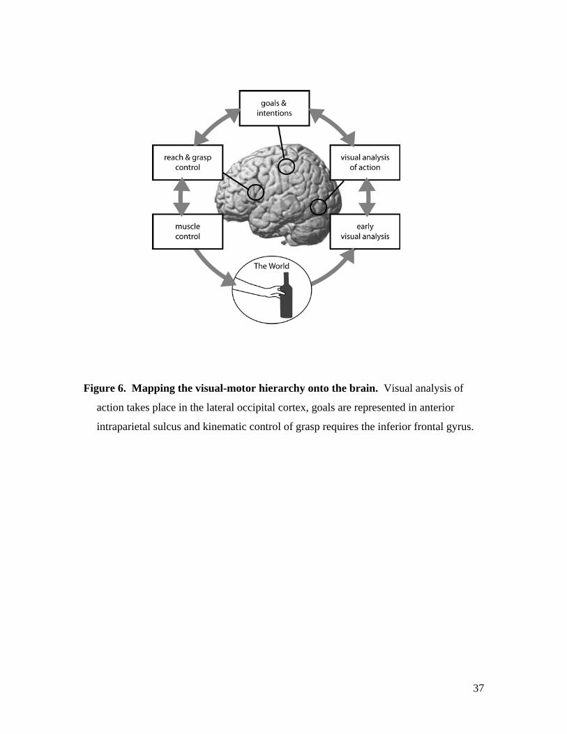
motor hierarchy and the brain, illustrated in Figure 6. We now consider the evidence for
the localisation, function and connectivity of each level in turn.
-------------- FIGURE 6 ABOUT HERE -------------
17
Visual analysis of action
Our data indicate that a representation of observed grasps and observed kinematic
patterns is found in a set of regions in the lateral occipital cortex. Similar regions have
been reported in a variety of studies of action observation (Grezes and Decety, 2001),
biological motion processing (Grossman et al., 2000), and viewing of parts of the human
body (Downing et al., 2001). More recent data in shows that several regions in the
human occipital and temporal cortex have selectivity for body parts, and that middle
temporal regions which are selective for motion also have preferential responses to
images of the human body (Spiridon et al., 2006). Similarly, a recent fMRI study of
motion and body selectivity in the macaque reveals a patchwork of occipito-temporal
regions with selectivity for observed motion and observed actions (Nelissen et al., 2006).
Our results also indicate a set of several lateral occipital regions which contain
representations of observed grasps, rather than a single ‘grasp’ or ‘visual kinematics’
region. Based on the clear visual associations of these lateral occipital regions, we
suggest that this network provides a visual analysis of the kinematic parameters of
observed actions, as indicated on the right of Figure 6.
These data could be taken to imply that the kinematics of an observed action can
be analysed purely by visual mechanisms (Jacob and Jeannerod, 2005), without the need
for a motor simulation (Gallese and Goldman, 1998). However, there is increasing
evidence for close links between visual and motor processing of kinematics. In
particular, activity in EBA is modulated by the performance of a motor task (Astafiev et
al., 2004). Data from our own lab show that lateral occipital cortex has a prominent role
in perceptual weight judgment and in the biasing effect of action on percpetion (Hamilton
et al., 2006). Together, these data implicate the lateral occipital network in visual
processing of human actions with close links to the motor system. Our new results go
further and demonstrate these lateral occipital regions are able to perform a detailed
analysis of the kinematic parameters of action, for both grasp configuration and object
weight.
Somewhat surprisingly, we did not find any involvement of the posterior STS in
the representation of grasp. A number of studies have emphasised the role of STS in
detecting biological motion (Grossman et al., 2000) and even detecting intentions

18
(Pelphrey et al., 2004), and STS has been proposed as the main visual input to the parietal
action representation system (Keysers and Perrett, 2004). In contrast, the regions we
report for grasp in this study, as well as for hand trajectory (Hamilton and Grafton, 2006)
and box weight (Hamilton et al., 2006) in previous studies, all lie below the STS in the
middle or lateral occipital cortex, bordering the middle temporal gyrus. Some of these
discrepancies may be due to lack of homologies between human and monkey brain and
differences in nomenclature, especially in subtraction designs where large activation
clusters can span several regions. The majority of human neuroimaging studies reporting
STS activation use either whole body motion, for example point-light figures (Grossman
et al., 2000) or eye gaze stimuli (Pelphrey et al., 2003), rather than hand actions. Further
work will be needed to differentiate the roles of different occipito-temporal regions in
representing action kinematics. Our own data points to a representation of hand grasp,
hand velocity and hand trajectory in a patchwork of lateral occipital regions.
Action goals
Our analysis of RS for the goal of an action confirmed our previous results
(Hamilton and Grafton, 2006) and demonstrated a central role for the left aIPS / IPL in
the representation of object-goals. More specifically, we suggest that aIPS contains
populations of neurons which encode different possible action goals, and these
populations respond to the observation of a goal directed action by another person. Thus,
we place aIPS at the top of the motor hierarchy (Figure 6), with the most abstract action
representation of those studied. The goal representation in the parietal cortex must also
be qualified by the presence of an interaction with grasp in this region. The interpretation
of this interaction in terms of neural population codes is not clear, and may have to await
further research on the mechanism of RS. However, the presence of the interaction is
indicative of the close links between the different regions of the visuo-motor network,
and the likelihood that information processing overlaps between these regions rather than
being entirely segregated. It is also important to note that the goals we have studied were
defined by the identity of the object taken by the actor, contrasting between a ‘take wine
bottle’ goal and a ‘take dumb bell goal’. It remains to be seen if the same parietal regions
encode other types of goal, for example manipulating the same object in different ways.
Preliminary data from our own lab suggests that this is the case.
19
The idea that aIPS contains an abstract, goal representation is consistent with
recent data showing the TMS over this region impairs the ability to reconfigure one’s
hand or arm to a novel action goal (Tunik et al., 2005). Recordings from the inferior
parietal lobule in macaques, just below the monkey AIP, have provided evidence for
neurons which encode both performed and observed action sequences (Fogassi et al.,
2005), and are sensitive to the end state (goal) of a sequence rather than the initial action.
However, there is also evidence that the anterior portion of the parietal sulcus has
undergone an enlargement in the human relative to the macaque (Orban et al., 2005,
Simon et al., 2002), suggesting that this region has taken on additional functions in the
human brain. The ability to represent and interpret the goals of other people’s actions
might be one of these functions.
Our analysis of RS for goal also revealed robust goal representations in the basal
ganglia and cerebellum. Similar regions were also found in our previous study (Hamilton
and Grafton, 2006), but we did not discuss the result then, as it was not clear if the same
type of repetition suppression behaviour could be expected in subcortical regions
compared to the cortex. However, given the strong replication we find in the present
study, and the recent evidence of RS in the basal ganglia for language (Crinion et al.,
2006), it seems likely that these results are important. There are also plausible reasons to
believe that the cerebellum and basal ganglia might have a role to play in the control and
monitoring of action goals. In particular, the cerebellum has a central role in the
prediction of the sensory consequences of actions by means of forward models
(Blakemore et al., 1998). As suggested in the introduction, a hierarchical system of
forward models (Wolpert et al., 2003) provides a plausible computational mechanism
which could underlie the motor hierarchy. If the lowest level of this system, which deals
with sensory prediction, were located in the cerebellum, it would be necessary for this
system to also provide information to the higher levels, in the aIPS, and for these two
regions to work together in the interpretation of goal directed actions. Direct connectivity
between the cerebellar nuclei and the aIPS has recently been demonstrated (Clower et al.,
2005).
This connectivity study also revealed links between aIPS and the substantia nigra
in the basal ganglia (Clower et al., 2005). The cluster showing RS for goal in the basal
20
ganglia was located primarily in the right head of the caudate but extended to the left
putamen. There are a variety of functions associated with the basal ganglia which might
be relevant to the processing of goals. In particular, the basal ganglia have an important
role in action sequencing (Lehericy et al., 2005, Graybiel, 1998) and in learning about the
rewards associated with actions (Zink et al., 2004, Hollerman et al., 2000). Thus, the
basal ganglia RS for goal might reflect a representation of an action goal as part of a
sequence, or a representation of the value of the goal. Overall, our results imply a
network for goal representation, where aIPS has a clear role at the top of the motor
hierarchy, and BG and cerebellum provide a supporting function.
The motor representation of grasp
As well as the lateral occipital network for grasp, we found RS for grasp in a
region of the inferior frontal gyrus. We suggest that this is evidence of neuronal
populations in this area encoding whole-hand and precision grips, which provide a motor
representation of potential actions. The motor role of IFG has been demonstrated in both
humans and monkeys. In particular, human IFG has been associated with the
performance (Ehrsson et al., 2000), imagination (Grafton et al., 1996) and planning
(Johnson-Frey et al., 2005) of grasping actions in humans. In the macaque, region F5 in
the inferior frontal cortex contains neurons which code for specific hand grasps
(Rizzolatti et al., 1988) but which also have mirror properties and respond to observed
grasps (Gallese et al., 1996, di Pellegrino et al., 1992). The role of IFG in both kinematic
control and the interpretation of observed kinematic patterns has also been demonstrated
in humans. IFG is part of the network of regions active in motor tasks (Stephan et al.,
1995, Rizzolatti et al., 1996), and is also required for judging the weight of a box lifted
by another person (Pobric and Hamilton, 2006), a task which requires interpretation of
kinematics (Hamilton et al., 2005) rather than goals. This region is connected to both
primary motor cortex and the spinal cord (Dum and Strick, 1991, Dum and Strick, 2002,
Shimazu et al., 2004), and thus is ideally placed to provide a motor grasp representation,
closely linked to the performance of an action.
The localisation of motor grasp representations to IFG places this region at a
‘lower’ stage of the motor hierarchy than aIPS, between the muscle output and the goal.
This proposal is at odds with some studies which have attempted to link IFG to more
21
abstract action understanding functions, such as the interpretation of goals (Umilta et al.,
2001) and intentions (Iacoboni et al., 2005). A number of theoretical papers have also
proposed links between the action representations in IFG and language (Rizzolatti and
Arbib, 1998) or mentalising (Gallese and Goldman, 1998) abilities. This proposals may
have arisen partly from the historical accident that mirror neurons were explored in the
IFG (di Pellegrino et al., 1992) before they were studied in the inferior parietal cortex
(Fogassi et al., 2005). Furthermore, the studies which have linked IFG to abstract goals
either did not distinguish between the configuration of the hand and the identity of the
goal object (Umilta et al., 2001), or did not control for context (Iacoboni et al., 2005).
Thus, we suggest that direct evidence for a goal representation in IFG is lacking. Instead,
our own data clearly demonstrate a role for IFG in grasp and in representing the
kinematics of hand actions.
Broader implications
Our model of the visual-motor hierarchy is a step towards defining the neural
systems underlying motor control and action understanding in the human brain. As we
make clear, the underlying cognitive structure we propose is based on previous models
for language (Geschwind, 1965) and hand actions (Tessari and Rumiati, 2004). Our new
data provides a grounding for these models in the brain, and thus makes new predictions
for fMRI, neuropsychological and neurophysiological experiments.
In particular, we provide the first evidence for distinct functions for the IFG and
aIPS regions of the visual-motor loop, arguing that aIPS has more abstract goal
representations while IFG has mainly lower level grasp representations. This conclusion
conflicts with theories which place intention representations in the frontal cortex
(Iacoboni et al., 2005) and which attempt to link sophisticated action understanding
abilities in frontal cortex to language representations in Broca’s area (Rizzolatti and
Arbib, 1998). However, neither language nor action understanding depend on a single
brain region, and it is quite possible that the production of language and hand actions are
intertwined in the inferior frontal cortex, while the meaning of words (Spitsyna et al.,
2006) and goals of actions are represented elsewhere.
There is one potential difference between our model in Figure 6 and the models
previous proposed (Tessari and Rumiati, 2004), which include a direct connection
22
between the visual analysis and motor grasp representations. Psychophysical evidence
supports the existence of a direct connection, with both visual to motor (Kilner et al.,
2003) and motor to visual (Hamilton et al., 2004) effects. However, we do not include
this connection in the figure, because anatomical evidence is weak. Reviews of macaque
neuroanatomy do not report a direct connection from superior and middle temporal
regions to inferior frontal regions (Keysers and Perrett, 2004, Rozzi et al., 2006).
However, a recent study using diffusion tensor imaging suggests that the human arcuate
fasciculus, which is commonly considered to be a language pathway, extends from
posterior middle temporal gyrus to inferior frontal gyrus (Rilling et al., 2006). This fibre
tract therefore has the potential to provide the direct connection between the visual and
motor kinematic representations predicted by many psychophysical studies, and could
easily be incorporated in our model. Further anatomical studies will be needed to
confirm the result.
A second important issue for our model is that we have defined the visuo-motor
hierarchy using action observation rather than motor execution, because this provides
more precise stimulus control and avoids the limitations of the scanner environment. To
draw conclusions about the organisation of these brain systems for the control of one’s
own actions, we rely on the assumption of mirroring between performance and
observation (Rizzolatti and Craighero, 2004). There is independent evidence for the
involvement of aIPS in controlling goals (Tunik et al., 2005), and the localisations we
propose do form a plausible model for the control of visually guided actions, but direct
evidence that mirroring between self and other is specific to each level of the motor
hierarchy remains to be discovered. In particular, it would be important to know if RS is
seen for performed actions, both in the human and in the macaque brain.
Moving up the hierarchy
The model we present in Figure 6 describes the control and understanding of
goal-directed hand actions, but does not itself explain the origin or selection of goals.
The actions we have studied involve taking a single object, which is a very basic form of
goal, but in real life, these actions would be likely to form one step towards achieving a
more complex goal. For example, taking a wine bottle is just one component of pouring
a glass of wine for a friend. Thus, we could postulate a continuing hierarchy of control
23
for more complex action sequences, as suggested by studies of executive function
(Koechlin et al., 2003) and of planning goals in every day tasks (Shallice and Burgess,
1991). However, there remains a gap between the simple action goals studied here and
the long-range or abstract goals examined in motor planning tasks. Further studies of the
cognitive and neural systems for complex actions will be needed to bridge the gap
between motor and executive planning models. In terms of action understanding, there
remains a similar gap between the understanding of action goals in the parietal cortex as
described here, and the understanding of other people’s beliefs, desires and other mental
states in the ‘theory of mind network’ which includes the temporal-parietal junction and
medial prefrontal cortex (Frith and Frith, 2003) but not the action understanding regions
(Saxe, 2005).
Thus, hierarchical control above the goal level described here remains a complex
and unresolved question. Furthermore, all hierarchical models of human cognition are
vulnerable to the problem of a homunculus, who seems to be required at the top level to
control the lower levels, but can neither be localised nor extinguished. A model
composed of multiple overlapping control loops at a variety of levels, as implied here,
has the potential to allow sophisticated motor control without a homunculus (Brooks,
1986), but evidence for such a system in the human brain remains to be found. We
suggest that the repetition suppression approach to segmenting levels of representation, as
outlined in this paper, provides a useful method for interrogating and defining the higher
and more abstract components of the motor hierarchy in future.
Conclusion
We have presented evidence for a hierarchical system for action understanding
and visual motor control in the human brain. This system is composed of a visual
representation of action kinematics in the lateral occipital cortex, a goal / intention
representation in the anterior intraparietal sulcus and a motor representation of kinematics
in the inferior frontal gyrus. The model is compatible with psychophysical and
anatomical data, and provides new framework for interpreting the computational
processes underlying action understanding and simple social interactions.

24
References
Arbib, MA, Billard, A, Iacoboni, M & Oztop, E (2000) Synthetic brain imaging:
grasping, mirror neurons and imitation. Neural Netw, 13, 975-97.
Astafiev, SV, Stanley, CM, Shulman, GL & Corbetta, M (2004) Extrastriate body area in
human occipital cortex responds to the performance of motor actions. Nat
Neurosci, 7, 542-8.
Blakemore, SJ, Wolpert, DM & Frith, CD (1998) Central cancellation of self-produced
tickle sensation. Nat Neurosci, 1, 635-40.
Britten, KH, Shadlen, MN, Newsome, WT & Movshon, JA (1993) Responses of neurons
in macaque MT to stochastic motion signals. Vis Neurosci, 10, 1157-69.
Brooks, RA (1986) A Robust Layered Control System for a Mobile Robot. IEEE Journal
of Robotics and Automation, 2, 14-23.
Buccino, G, Binkofski, F, Fink, GR, Fadiga, L, Fogassi, L, Gallese, V, Seitz, RJ, Zilles,
K, Rizzolatti, G & Freund, HJ (2001) Action observation activates premotor and
parietal areas in a somatotopic manner: an fMRI study. Eur J Neurosci, 13, 400-4.
Buckner, RL, Goodman, J, Burock, M, Rotte, M, Koutstaal, W, Schacter, D, Rosen, B &
Dale, AM (1998) Functional-anatomic correlates of object priming in humans
revealed by rapid presentation event-related fMRI. Neuron, 20, 285-96.
Clower, DM, Dum, RP & Strick, PL (2005) Basal ganglia and cerebellar inputs to 'AIP'.
Cereb Cortex, 15, 913-20.
Crinion, J, Turner, R, Grogan, A, Hanakawa, T, Noppeney, U, Devlin, JT, Aso, T,
Urayama, S, Fukuyama, H, Stockton, K, Usui, K, Green, DW & Price, CJ (2006)
Language control in the bilingual brain. Science, 312, 1537-40.
Culham, JC, Danckert, SL, DeSouza, JF, Gati, JS, Menon, RS & Goodale, MA (2003)
Visually guided grasping produces fMRI activation in dorsal but not ventral
stream brain areas. Exp Brain Res, 153, 180-9.
Dehaene, S & Changeux, JP (1997) A hierarchical neuronal network for planning
behavior. Proc Natl Acad Sci U S A, 94, 13293-8.
di Pellegrino, G, Fadiga, L, Fogassi, L, Gallese, V & Rizzolatti, G (1992) Understanding
motor events: a neurophysiological study. Exp Brain Res, 91, 176-80.
25
Diedrichsen, J & Shadmehr, R (2005) Detecting and adjusting for artifacts in fMRI time
series data. Neuroimage.
Downing, PE, Jiang, Y, Shuman, M & Kanwisher, N (2001) A cortical area selective for
visual processing of the human body. Science, 293, 2470-3.
Dum, RP & Strick, PL (1991) The origin of corticospinal projections from the premotor
areas in the frontal lobe. J Neurosci, 11, 667-89.
Dum, RP & Strick, PL (2002) Motor areas in the frontal lobe of the primate. Physiol
Behav, 77, 677-82.
Ehrsson, HH, Fagergren, A, Jonsson, T, Westling, G, Johansson, RS & Forssberg, H
(2000) Cortical activity in precision- versus power-grip tasks: an fMRI study. J
Neurophysiol, 83, 528-36.
Fogassi, L, Ferrari, PF, Gesierich, B, Rozzi, S, Chersi, F & Rizzolatti, G (2005) Parietal
lobe: from action organization to intention understanding. Science, 308, 662-7.
Frith, U & Frith, CD (2003) Development and neurophysiology of mentalizing. Philos
Trans R Soc Lond B Biol Sci, 358, 459-73.
Gallese, V, Fadiga, L, Fogassi, L & Rizzolatti, G (1996) Action recognition in the
premotor cortex. Brain, 119, 593-609.
Gallese, V & Goldman, A (1998) Mirror neurons and the simulation theory of mind-
reading. Trends in Cognitive Sciences, 2, 493-501.
Georgopoulos, AP, Kalaska, JF, Caminiti, R & Massey, JT (1982) On the relations
between the direction of two-dimensional arm movements and cell discharge in
primate motor cortex. Journal of Neuroscience, 2, 1527-37.
Geschwind, N (1965) Disconnexion syndromes in animals and man. I. Brain, 88, 237-94.
Grafton, ST, Arbib, MA, Fadiga, L & Rizzolatti, G (1996) Localization of grasp
representations in humans by positron emission tomography. 2. Observation
compared with imagination. Exp Brain Res, 112, 103-11.
Grafton, ST, Mazziotta, JC, Woods, RP & Phelps, ME (1992) Human functional anatomy
of visually guided finger movements. Brain, 115 (Pt 2), 565-87.
Graybiel, AM (1998) The basal ganglia and chunking of action repertoires. Neurobiol
Learn Mem, 70, 119-36.
26
Grezes, J & Decety, J (2001) Functional anatomy of execution, mental simulation,
observation, and verb generation of actions: a meta-analysis. Hum Brain Mapp,
12, 1-19.
Grill-Spector, K, Henson, R & Martin, A (2006) Repetition and the brain: neural models
of stimulus-specific effects. Trends Cogn Sci, 10, 14-23.
Grill-Spector, K & Malach, R (2001) fMR-adaptation: a tool for studying the functional
properties of human cortical neurons. Acta Psychol (Amst), 107, 293-321.
Grossman, E, Donnelly, M, Price, R, Pickens, D, Morgan, V, Neighbor, G & Blake, R
(2000) Brain areas involved in perception of biological motion. J Cogn Neurosci,
12, 711-20.
Hamilton, AF & Grafton, ST (2006) Goal representation in human anterior intraparietal
sulcus. J Neurosci, 26, 1133-7.
Hamilton, AF, Joyce, DW, Flanagan, JR, Frith, CD & Wolpert, DM (2005) Kinematic
cues in perceptual weight judgement and their origins in box lifting. Psychol Res,
1-9.
Hamilton, AF, Wolpert, DM & Frith, U (2004) Your own action influences how you
perceive another person's action. Current Biology, 14, 493-498.
Hamilton, AF, Wolpert, DM, Frith, U & Grafton, ST (2006) Where does your own action
influence your perception of another person's action in the brain? Neuroimage, 29,
524-35.
Hamilton, AFdC & Wolpert, DM (2002) Controlling the statistics of action: obstacle
avoidance. J Neurophysiol, 87, 2434-40.
Haruno, M, Wolpert, DM & Kawato, M (2003) Hierarchical MOSAIC for movement
generation. IN Ono, T, Matsumoto, G, Llinas, RR, Berthoz, A, Norgren, H &
Tamura, R (Eds.) Excepta Medica International Coungress Series. Amsterdam,
Elsevier Science.
Hebb, DO (1949) The organization of behavior: A neuropsychological theory, New
York, Wiley.
Henson, RN (2003) Neuroimaging studies of priming. Prog Neurobiol, 70, 53-81.
Hollerman, JR, Tremblay, L & Schultz, W (2000) Involvement of basal ganglia and
orbitofrontal cortex in goal-directed behavior. Prog Brain Res, 126, 193-215.
27
Iacoboni, M, Molnar-Szakacs, I, Gallese, V, Buccino, G, Mazziotta, JC & Rizzolatti, G
(2005) Grasping the Intentions of Others with One's Own Mirror Neuron System.
PLoS Biol, 3, e79.
Jackson, JH (1889) On the comparative
study of diseases of the nervous system. British Medical Journal, 2, 355-362.
Jacob, P & Jeannerod, M (2005) The motor theory of social cognition: a critique. Trends
Cogn Sci, 9, 21-5.
James, W (1890) Principles of Psychology, New York, Holt.
Johnson-Frey, SH, Newman-Norlund, R & Grafton, ST (2005) A distributed left
hemisphere network active during planning of everyday tool use skills. Cereb
Cortex, 15, 681-95.
Keele, SW, Cohen, A & Ivry, R (1990) Motor Programs: Concepts and Issues., chap. 3.
IN Jeannerod, M (Ed.) Attention and Performance 13: Motor Representation and
Control. Hillsdale, NJ, USA: Lawrence Erlbaum Associates, Inc.
Keysers, C & Perrett, DI (2004) Demystifying social cognition: a Hebbian perspective.
Trends Cogn Sci, 8, 501-7.
Kilner, JM, Paulignan, Y & Blakemore, SJ (2003) An interference effect of observed
biological movement on action. Curr Biol, 13, 522-5.
Koechlin, E & Jubault, T (2006) Broca's Area and the Hierarchical Organization of
Human Behavior. Neuron, 50, 963-74.
Koechlin, E, Ody, C & Kouneiher, F (2003) The architecture of cognitive control in the
human prefrontal cortex. Science, 302, 1181-5.
Kourtzi, Z & Kanwisher, N (2000) Cortical regions involved in perceiving object shape. J
Neurosci, 20, 3310-8.
Krekelberg, B, Boynton, GM & van Wezel, RJ (2006) Adaptation: from single cells to
BOLD signals. Trends Neurosci, 29, 250-6.
Lehericy, S, Benali, H, Van de Moortele, PF, Pelegrini-Issac, M, Waechter, T, Ugurbil, K
& Doyon, J (2005) Distinct basal ganglia territories are engaged in early and
advanced motor sequence learning. Proc Natl Acad Sci U S A, 102, 12566-71.

28
Lemon, RN, Baker, SN, Davis, JA, Kirkwood, PA, Maier, MA & Yang, HS (1998) The
importance of the cortico-motoneuronal system for control of grasp. Novartis
Found Symp, 218, 202-15.
Maccotta, L & Buckner, RL (2004) Evidence for neural effects of repetition that directly
correlate with behavioral priming. J Cogn Neurosci, 16, 1625-32.
Manthey, S, Schubotz, RI & von Cramon, DY (2003) Premotor cortex in observing
erroneous action: an fMRI study. Brain Res Cogn Brain Res, 15, 296-307.
Meltzoff, A (1995) Understanding the intentions of others: Re-enactment of intended acts
by 18-month-old children. Dev Psychol, 31, 838-850.
Mushiake, H, Saito, N, Sakamoto, K, Itoyama, Y & Tanji, J (2006) Activity in the lateral
prefrontal cortex reflects multiple steps of future events in action plans. Neuron,
50, 631-41.
Nelissen, K, Vanduffel, W & Orban, GA (2006) Charting the lower superior temporal
region, a new motion-sensitive region in monkey superior temporal sulcus. J
Neurosci, 26, 5929-47.
Noppeney, U & Price, CJ (2004) An FMRI study of syntactic adaptation. J Cogn
Neurosci, 16, 702-13.
Norman, DA & Shallice, T (1986) Attention to action: Willed and automatic control of
behaviour, Plenum Press.
Orban, GA, Claeys, K, Nelissen, K, Smans, R, Sunaert, S, Todd, JT, Wardak, C, Durand,
JB & Vanduffel, W (2005) Mapping the parietal cortex of human and non-human
primates. Neuropsychologia.
Pacherie, E (2006) Towards a dynamic theory of intentions. IN Pocket, S, Banks, WP &
Gallagher, S (Eds.) Does Consciousness Cause Behavior? An Investigation of the
Nature of Volition. Cambridge MA, MIT Press.
Pelphrey, KA, Morris, JP & McCarthy, G (2004) Grasping the intentions of others: the
perceived intentionality of an action influences activity in the superior temporal
sulcus during social perception. J Cogn Neurosci, 16, 1706-16.
Pelphrey, KA, Singerman, JD, Allison, T & McCarthy, G (2003) Brain activation evoked
by perception of gaze shifts: the influence of context. Neuropsychologia, 41, 156-
70.

29
Pinel, P, Dehaene, S, Riviere, D & LeBihan, D (2001) Modulation of parietal activation
by semantic distance in a number comparison task. Neuroimage, 14, 1013-26.
Pobric, G & Hamilton, AF (2006) Action understanding requires the left inferior frontal
cortex. Curr Biol, 16, 524-9.
Rilling, JK, Glasser, MF, Preuss, TM, Ma, X, Zhang, X, Zhao, T, Hu, X & Behrens, T
(2006) A comparative diffusion tensor imaging study of the arcuate fasiculus
pathway in humans, chimpanzees and rhesus macaques. Soc Neurosci Abstr.
Rizzolatti, G & Arbib, MA (1998) Language within our grasp. Trends Neurosci, 21, 188-
94.
Rizzolatti, G, Camarda, R, Fogassi, L, Gentilucci, M, Luppino, G & Matelli, M (1988)
Functional organization of inferior area 6 in the macaque monkey. II. Area F5 and
the control of distal movements. Exp Brain Res, 71, 491-507.
Rizzolatti, G & Craighero, L (2004) The Mirror-Neuron System. Annu Rev Neurosci, 27,
169-192.
Rizzolatti, G, Fadiga, L, Matelli, M, Bettinardi, V, Paulesu, E, Perani, D & Fazio, F
(1996) Localization of grasp representations in humans by PET: 1. Observation
versus execution. Exp Brain Res, 111, 246-52.
Rozzi, S, Calzavara, R, Belmalih, A, Borra, E, Gregoriou, GG, Matelli, M & Luppino, G
(2006) Cortical connections of the inferior parietal cortical convexity of the
macaque monkey. Cereb Cortex, 16, 1389-417.
Saito, N, Mushiake, H, Sakamoto, K, Itoyama, Y & Tanji, J (2005) Representation of
immediate and final behavioral goals in the monkey prefrontal cortex during an
instructed delay period. Cereb Cortex, 15, 1535-46.
Sakata, H, Taira, M, Murata, A & Mine, S (1995) Neural mechanisms of visual guidance
of hand action in the parietal cortex of the monkey. Cereb Cortex, 5, 429-38.
Saxe, R (2005) Against simulation: the argument from error. Trends Cogn Sci, 9, 174-9.
Shallice, T & Burgess, PW (1991) Deficits in strategy application following frontal lobe
damage in man. Brain, 114 (Pt 2), 727-41.
Sherrington, CS (1906) The Integrative Action of the Nervous System, New Haven, CT,
Yale University Press.

30
Shima, K, Mushiake, H, Saito, N & Tanji, J (1996) Role for cells in the presupplementary
motor area in updating motor plans. Proc Natl Acad Sci U S A, 93, 8694-8.
Shimazu, H, Maier, MA, Cerri, G, Kirkwood, PA & Lemon, RN (2004) Macaque ventral
premotor cortex exerts powerful facilitation of motor cortex outputs to upper limb
motoneurons. J Neurosci, 24, 1200-11.
Shmuelof, L & Zohary, E (2005) Dissociation between Ventral and Dorsal fMRI
Activation during Object and Action Recognition. Neuron, 47, 457-70.
Simon, O, Mangin, JF, Cohen, L, Le Bihan, D & Dehaene, S (2002) Topographical
layout of hand, eye, calculation, and language-related areas in the human parietal
lobe. Neuron, 33, 475-87.
Spiridon, M, Fischl, B & Kanwisher, N (2006) Location and spatial profile of category-
specific regions in human extrastriate cortex. Hum Brain Mapp, 27, 77-89.
Spitsyna, G, Warren, JE, Scott, SK, Turkheimer, FE & Wise, RJ (2006) Converging
language streams in the human temporal lobe. J Neurosci, 26, 7328-36.
Stephan, KM, Fink, GR, Passingham, RE, Silbersweig, D, Ceballos-Baumann, AO, Frith,
CD & Frackowiak, RS (1995) Functional anatomy of the mental representation of
upper extremity movements in healthy subjects. J Neurophysiol, 73, 373-86.
Tessari, A & Rumiati, RI (2004) The strategic control of multiple routes in imitation of
actions. J Exp Psychol Hum Percept Perform, 30, 1107-16.
Thompson-Schill, SL, D'Esposito, M & Kan, IP (1999) Effects of repetition and
competition on activity in left prefrontal cortex during word generation. Neuron,
23, 513-22.
Tunik, E, Frey, SH & Grafton, ST (2005) Virtual lesions of the anterior intraparietal area
disrupt goal-dependent on-line adjustments of grasp. Nat Neurosci, 8, 505-11.
Umilta, MA, Kohler, E, Gallese, V, Fogassi, L, Fadiga, L, Keysers, C & Rizzolatti, G
(2001) I know what you are doing. a neurophysiological study. Neuron, 31, 155-
65.
Wig, GS, Grafton, ST, Demos, KE & Kelley, WM (2005) Reductions in neural activity
underlie behavioral components of repetition priming. Nat Neurosci.

31
Wohlschlager, A, Gattis, M & Bekkering, H (2003) Action generation and action
perception in imitation: an instance of the ideomotor principle. Philos Trans R
Soc Lond B Biol Sci, 358, 501-15.
Wolpert, D, Miall, C & Kawato (1998) Internal models in the cerebellum. Trends Cogn
Sci, 2, 338-347.
Wolpert, DM, Doya, K & Kawato, M (2003) A unifying computational framework for
motor control and social interaction. Philos Trans R Soc Lond B Biol Sci, 358,
593-602.
Wolpert, DM & Flanagan, JR (2001) Motor prediction. Curr Biol, 11, R729-32.
Wolpert, DM & Kawato, M (1998) Multiple paired forward and inverse models for motor
control. Neural Netw, 11, 1317-1329.
Zink, CF, Pagnoni, G, Martin-Skurski, ME, Chappelow, JC & Berns, GS (2004) Human
striatal responses to monetary reward depend on saliency. Neuron, 42, 509-17.
32
Figure 1. Components of the visual-motor hierarchy. This simple model assumes a
control loop from visual analysis to an abstract goal representation and then to more
detailed kinematic and muscle control.
33
Figure 2. The likely mechanism of RS. An example of RS is shown for a hypothetical
brain area encoding animals in two populations of neurons, one for dogs and the other
for cats. The top row illustrates the stimulus sequence. The bottom row illustrates
hypothetical firing levels in intermixed populations of neurons representing ‘dog’
(blue) and ‘cat’ (red) with the intensity of the colour indicating the level of firing. See
text for details.
34
Figure 3. Stimulus sequence to obtain RS for goal and grasp in a one-back design.
Top panel shows one frame from each video clip. Lower panels show predicted neural
activity and BOLD in a region encoding goal object and a region encoding hand grasp.

35
Figure 4. Grasp representations in the brain. Repetition suppression for grasp was
found in occipital regions, middle intraparietal sulcus and inferior frontal gyrus. All
regions show stronger responses to novel grasps (red & dark blue) compared to
repeated grasp (orange and light blue). Abbreviations are: n-novel, r-repeated, O-
object goal, G-grasp, new- first stimulus of a block, quest- question trial

36
Figure 5. Goal representations in the brain. RS for the goal object was found in the
cerebellum, basal ganglia and left anterior intraparietal sulcus. All three regions
show stronger responses to novel goals (red and orange) compared to repeated
goals (blue). Abbreviations are: n-novel, r-repeated, O-object goal, G-grasp,
new- first stimulus of a block, quest- question trial
37
Figure 6. Mapping the visual-motor hierarchy onto the brain. Visual analysis of
action takes place in the lateral occipital cortex, goals are represented in anterior
intraparietal sulcus and kinematic control of grasp requires the inferior frontal gyrus.
38
Table 1. Coordinates of regions showing RS in the goal-object and grasp contrasts.
Bold indicates regions meeting the whole brain cluster corrected threshold.
Region No
voxels T
value Corrected probability MNI coords
Uncorrected probability
x y z
Goal - object contrast (wine bottle / dumbell)
superior temporal gyrus 202 5.37 0.102 -50 4 -12 0.00000 Caudate 255 5.24 0.049 6 12 2 0.00001 L IPL 563 4.58 0.001 -52 -20 38 0.00004 -58 -24 30 L aIPS -46 -32 52 SMA 96 4.53 0.457 -10 20 46 0.000044 L Cerebellum 303 4.37 0.027 -54 -60 -26 0.00007 -46 -64 -22 -46 -46 -22 lateral occipital cortex 15 4.19 0.985 -20 -56 10 0.000113 IFG 152 4.19 0.206 -58 10 34 0.000114 IPS / cingulate 33 4.15 0.912 -24 -30 42 0.000125 pSTS 26 4.09 0.948 -66 -40 6 0.000148
Cerebellum 25 4.00 0.953 -32 -50 -26 0.00019 inferior temporal gyrus 38 3.82 0.882 52 -40 -26 0.000315 R aIPS 127 3.79 0.296 54 -26 50 0.00034
Cerebellum 35 3.79 0.901 30 -42 -24 0.00034 calcarine sulcus 10 3.79 0.994 -24 -64 20 0.00034
Caudate 11 3.78 0.992 -10 -12 18 0.00035 middle occipital 13 3.77 0.989 48 -56 -6 0.000361 superior precentral gyrus 13 3.72 0.989 60 6 36 0.00041
Cerebellum 34 3.70 0.907 46 -54 -30 0.00043 thalamus 21 3.68 0.968 4 -22 2 0.00045 Cerebellum 10 3.66 0.994 -42 -86 -18 0.00048 Cerebellum 13 3.60 0.989 -14 -48 -26 0.00057
inferior temporal gyrus 13 3.48 0.989 -32 10 -36 0.000769
Grip contrast (power / precision) middle occipital 67 4.36 0.676 44 -86 -8 0.000071 SMA 51 4.09 0.794 2 14 62 0.00015 middle occipital 40 4.05 0.870 -50 -68 -6 0.000165 middle frontal gyrus 40 4.01 0.870 -40 30 30 0.000184 middle occipital 12 3.91 0.989 36 -96 -14 0.000245 frontal operculum 17 3.87 0.978 -46 14 14 0.000275 mIPS 16 3.70 0.981 -26 -50 58 0.000434 inferior occipital 64 3.65 0.698 42 -68 -16 0.000494
Related Documents