MOLECULAR AND CELLULAR BIOLOGY, Aug. 2003, p. 5409–5420 Vol. 23, No. 15 0270-7306/03/$08.000 DOI: 10.1128/MCB.23.15.5409–5420.2003 Copyright © 2003, American Society for Microbiology. All Rights Reserved. The Mitochondrial Protein hFis1 Regulates Mitochondrial Fission in Mammalian Cells through an Interaction with the Dynamin-Like Protein DLP1 Yisang Yoon, Eugene W. Krueger, Barbara J. Oswald, and Mark A. McNiven* Center for Basic Research in Digestive Diseases and Department of Biochemistry and Molecular Biology, Mayo Clinic and Foundation, Rochester, Minnesota 55905 Received 10 May 2002/Returned for modification 30 July 2002/Accepted 8 May 2003 The yeast protein Fis1p has been shown to participate in mitochondrial fission mediated by the dynamin- related protein Dnm1p. In mammalian cells, the dynamin-like protein DLP1/Drp1 functions as a mitochon- drial fission protein, but the mechanisms by which DLP1/Drp1 and the mitochondrial membrane interact during the fission process are undefined. In this study, we have tested the role of a mammalian homologue of Fis1p, hFis1, and provided new and mechanistic information about the control of mitochondrial fission in mammalian cells. Through differential tagging and deletion experiments, we demonstrate that the intact C-terminal structure of hFis1 is essential for mitochondrial localization, whereas the N-terminal region of hFis1 is necessary for mitochondrial fission. Remarkably, an increased level of cellular hFis1 strongly pro- motes mitochondrial fission, resulting in an accumulation of fragmented mitochondria. Conversely, cell microinjection of hFis1 antibodies or treatment with hFis1 antisense oligonucleotides induces an elongated and collapsed mitochondrial morphology. Further, fluorescence resonance energy transfer and coimmunoprecipi- tation studies demonstrate that hFis1 interacts with DLP1. These results suggest that hFis1 participates in mitochondrial fission through an interaction that recruits DLP1 from the cytosol. We propose that hFis1 is a limiting factor in mitochondrial fission and that the number of hFis1 molecules on the mitochondrial surface determines fission frequency. Mitochondria in mammalian cells are dynamic structures displaying frequent fission, fusion, and translocation (1). These dynamic processes are believed to ensure an appropriate dis- tribution of mitochondria during cell proliferation and provide sufficient energy to a localized cytoplasmic region. It is now known that fusion and fission of mitochondria from yeast to human are balanced to maintain normal tubular mitochondria through the antagonistic action of two distinct large GTPases (6, 9, 16, 17, 19, 20, 24). An imbalance between these two events results in excessive fragmentation or tubulation of mi- tochondria. One of these GTPases is a dynamin-like enzyme called Dnm1p in yeast cells or DLP1/Drp1 in mammalian cells. Studies with yeast and mammalian cells indicated that Dnm1p/ DLP1/Drp1 plays a role in mitochondrial fission (16, 17, 20, 23, 24). In a previous study, DLP1/Drp1 has been found to deform biological membranes in a GTP-dependent manner (32). Although DLP1 is involved in controlling mitochondrial morphology, its distribution to mitochondria in mammalian cells is modest. Both morphological and biochemical data have shown that the majority of DLP1 is cytosolic while subpopu- lations localize to mitochondria and other cellular organelles such as the endoplasmic reticulum, microtubules, and peroxi- somes (10, 13, 14, 22, 31). Although DLP1 is known to function as a mitochondrial fission enzyme, overexpression of DLP1 within cells does not increase mitochondrial fission (17), indi- cating that there are other limiting factors in the mitochondrial fission process. These factors may recruit cytosolic DLP1 to the mitochondrial surface and/or activate the DLP1 GTPase, lead- ing to mitochondrial fission. Yeast genetic screening has iden- tified two proteins, Mdv1p and Fis1p, which are both predicted to function together with Dnm1p in mitochondrial fission (2, 4, 15, 25). Yeast cells defective in these proteins display interconnected net-like mitochondria, which is identical to the Dnm1p-defective phenotype. Morphological studies found that Dnm1p and Mdv1p colocalized on mitochondrial tubules as punctate spots, and in the absence of Fis1p, the size and number of these Dnm1p-containing structures were grossly altered (15, 25). It was also found that the normal distribution of Mdv1p was perturbed in the absence of Dnm1p and/or Fis1p (15, 25). These results suggest that the three proteins interact either directly or indirectly to mediate mitochondrial fission through a multistep pathway (15, 25). Although a cognate ho- mologue of Mdv1p does not exist in higher eukaryotes, it is possible that a structurally similar protein may participate as a functional homologue in the mitochondrial fission process. As mammalian DLP1 distributes predominantly to the cy- tosol, it seemed important to identify a protein that might recruit DLP1 to mitochondria in a regulated manner. Fis1p is a likely candidate for this role based on its membrane topology and proposed function. Fis1p is a small, 17-kDa protein and is predicted to be anchored in the outer mitochondrial mem- brane with its N-terminal region exposed to the cytosol and a short C-terminal tail protruding into the mitochondrial inter- membrane space (15). It has been proposed that Fis1p is re- quired for the proper assembly and activation of the fission- mediating complex for yeast mitochondrial division (15, 25). Furthermore, homologues of Fis1p have been found in many higher eukaryotes, including human (15). Here, we have cloned a * Corresponding author. Mailing address: Mayo Clinic, 200 1st St., S.W., Rochester, MN 55905. Phone: (507) 284-0683. Fax: (507) 284- 0762. E-mail: [email protected]. 5409 on December 30, 2014 by guest http://mcb.asm.org/ Downloaded from

Welcome message from author
This document is posted to help you gain knowledge. Please leave a comment to let me know what you think about it! Share it to your friends and learn new things together.
Transcript
MOLECULAR AND CELLULAR BIOLOGY, Aug. 2003, p. 5409–5420 Vol. 23, No. 150270-7306/03/$08.00�0 DOI: 10.1128/MCB.23.15.5409–5420.2003Copyright © 2003, American Society for Microbiology. All Rights Reserved.
The Mitochondrial Protein hFis1 Regulates Mitochondrial Fission inMammalian Cells through an Interaction with
the Dynamin-Like Protein DLP1Yisang Yoon, Eugene W. Krueger, Barbara J. Oswald, and Mark A. McNiven*
Center for Basic Research in Digestive Diseases and Department of Biochemistry and Molecular Biology,Mayo Clinic and Foundation, Rochester, Minnesota 55905
Received 10 May 2002/Returned for modification 30 July 2002/Accepted 8 May 2003
The yeast protein Fis1p has been shown to participate in mitochondrial fission mediated by the dynamin-related protein Dnm1p. In mammalian cells, the dynamin-like protein DLP1/Drp1 functions as a mitochon-drial fission protein, but the mechanisms by which DLP1/Drp1 and the mitochondrial membrane interactduring the fission process are undefined. In this study, we have tested the role of a mammalian homologue ofFis1p, hFis1, and provided new and mechanistic information about the control of mitochondrial fission inmammalian cells. Through differential tagging and deletion experiments, we demonstrate that the intactC-terminal structure of hFis1 is essential for mitochondrial localization, whereas the N-terminal region ofhFis1 is necessary for mitochondrial fission. Remarkably, an increased level of cellular hFis1 strongly pro-motes mitochondrial fission, resulting in an accumulation of fragmented mitochondria. Conversely, cellmicroinjection of hFis1 antibodies or treatment with hFis1 antisense oligonucleotides induces an elongated andcollapsed mitochondrial morphology. Further, fluorescence resonance energy transfer and coimmunoprecipi-tation studies demonstrate that hFis1 interacts with DLP1. These results suggest that hFis1 participates inmitochondrial fission through an interaction that recruits DLP1 from the cytosol. We propose that hFis1 is alimiting factor in mitochondrial fission and that the number of hFis1 molecules on the mitochondrial surfacedetermines fission frequency.
Mitochondria in mammalian cells are dynamic structuresdisplaying frequent fission, fusion, and translocation (1). Thesedynamic processes are believed to ensure an appropriate dis-tribution of mitochondria during cell proliferation and providesufficient energy to a localized cytoplasmic region. It is nowknown that fusion and fission of mitochondria from yeast tohuman are balanced to maintain normal tubular mitochondriathrough the antagonistic action of two distinct large GTPases(6, 9, 16, 17, 19, 20, 24). An imbalance between these twoevents results in excessive fragmentation or tubulation of mi-tochondria. One of these GTPases is a dynamin-like enzymecalled Dnm1p in yeast cells or DLP1/Drp1 in mammalian cells.Studies with yeast and mammalian cells indicated that Dnm1p/DLP1/Drp1 plays a role in mitochondrial fission (16, 17, 20, 23,24). In a previous study, DLP1/Drp1 has been found to deformbiological membranes in a GTP-dependent manner (32).
Although DLP1 is involved in controlling mitochondrialmorphology, its distribution to mitochondria in mammaliancells is modest. Both morphological and biochemical data haveshown that the majority of DLP1 is cytosolic while subpopu-lations localize to mitochondria and other cellular organellessuch as the endoplasmic reticulum, microtubules, and peroxi-somes (10, 13, 14, 22, 31). Although DLP1 is known to functionas a mitochondrial fission enzyme, overexpression of DLP1within cells does not increase mitochondrial fission (17), indi-cating that there are other limiting factors in the mitochondrialfission process. These factors may recruit cytosolic DLP1 to the
mitochondrial surface and/or activate the DLP1 GTPase, lead-ing to mitochondrial fission. Yeast genetic screening has iden-tified two proteins, Mdv1p and Fis1p, which are both predictedto function together with Dnm1p in mitochondrial fission (2,4, 15, 25). Yeast cells defective in these proteins displayinterconnected net-like mitochondria, which is identical tothe Dnm1p-defective phenotype. Morphological studies foundthat Dnm1p and Mdv1p colocalized on mitochondrial tubulesas punctate spots, and in the absence of Fis1p, the size andnumber of these Dnm1p-containing structures were grosslyaltered (15, 25). It was also found that the normal distributionof Mdv1p was perturbed in the absence of Dnm1p and/or Fis1p(15, 25). These results suggest that the three proteins interacteither directly or indirectly to mediate mitochondrial fissionthrough a multistep pathway (15, 25). Although a cognate ho-mologue of Mdv1p does not exist in higher eukaryotes, it ispossible that a structurally similar protein may participate as afunctional homologue in the mitochondrial fission process.
As mammalian DLP1 distributes predominantly to the cy-tosol, it seemed important to identify a protein that mightrecruit DLP1 to mitochondria in a regulated manner. Fis1p isa likely candidate for this role based on its membrane topologyand proposed function. Fis1p is a small, 17-kDa protein and ispredicted to be anchored in the outer mitochondrial mem-brane with its N-terminal region exposed to the cytosol and ashort C-terminal tail protruding into the mitochondrial inter-membrane space (15). It has been proposed that Fis1p is re-quired for the proper assembly and activation of the fission-mediating complex for yeast mitochondrial division (15, 25).Furthermore, homologues of Fis1p have been found in manyhigher eukaryotes, including human (15). Here, we have cloned a
* Corresponding author. Mailing address: Mayo Clinic, 200 1st St.,S.W., Rochester, MN 55905. Phone: (507) 284-0683. Fax: (507) 284-0762. E-mail: [email protected].
5409
on Decem
ber 30, 2014 by guesthttp://m
cb.asm.org/
Dow
nloaded from
human homologue of Fis1p, hFis1, and obtained mechanisticinformation regarding the role of hFis1 in mitochondrial fis-sion. We demonstrated that the N-terminal region of hFis1 isrequired for mitochondrial fission whereas the C-terminal tailis necessary for mitochondrial localization of hFis1. More im-portantly, we found that an elevated level of cellular hFis1drastically increases mitochondrial fission frequency, resultingin fragmented mitochondria, while disruption of hFis1 functioncauses inhibition of mitochondrial fission. Further, hFis1 andDLP1 form a complex in cells and are able to interact witheach other directly in vitro. These findings both support andexpand the earlier observations made for yeast and provideevidence that hFis1 regulates mitochondrial fission through aprotein-protein interaction that recruits DLP1 from the cytosolto the mitochondrial surface. These results suggest that hFis1is a limiting factor in mitochondrial fission and that the numberof hFis1 molecules on the mitochondrial surface determinesfission frequency.
MATERIALS AND METHODS
Cell culture, DNAs, and transfection. The cell lines Clone 9 (ATCC CRL-1439), HeLa, COS-7, and BHK-21 were used for all experiments. Cells weremaintained at 37°C with 5% CO2 in Ham’s F-12K medium for Clone 9 cells andin Dulbecco’s modified Eagle’s medium for HeLa, COS-7, and BHK-21 cells,supplemented with 10% fetal bovine serum, 100 U of penicillin per ml, and 100�g of streptomycin per ml. The cDNA for hFis1 was cloned by reverse transcrip-tion-PCR with total human RNA as the template. PCR primers were synthesizedaccording to the sequence deposited in GenBank (accession number AF151893).All subcloning into different vectors was carried out by standard molecularbiology techniques with PCR. The absence of mutations in nucleotide sequencesafter PCR was verified by DNA sequencing. Stable cells carrying green fluores-cent protein (GFP) or red fluorescent protein (RFP) in their mitochondrialmatrix were isolated by transfecting pmitoGFP or pmitoRFP and selecting withG418. pmitoGFP and pmitoRFP were constructed by fusing the mitochondrialtransit sequence of human isovaleryl coenzyme A dehydrogenase to the N ter-minus of GFP (pEGFP-N1; Clontech, Inc.) or RFP (pDsRed1-N1; Clontech,Inc.), respectively. Plasmids for recombinant DLP1 and hFis1 were constructedin the pQE-80 series vector (Qiagen, Inc.). For cell transfection, DNA constructswere purified by using plasmid purification columns (Qiagen, Inc.). Cells wereplated for 16 to 24 h before transfection on glass coverslips in 35-mm-diametertissue culture dishes. Transfections were performed by using Lipofectamine(Invitrogen, Inc.) or GeneJammer (Stratagene, Inc.) according to the manufac-turer’s instructions or by microinjecting DNA into the nucleus as describedpreviously (17). Transfected cells were allowed to recover for 16 to 24 h beforebeing processed for either immunofluorescence or electron microscopy.
Immunofluorescence and microscopy. Indirect immunofluorescence was per-formed as described previously (8, 31). Briefly, cells were fixed, permeabilized,and then incubated in a blocking buffer containing 5% goat serum for 1 h at 37°C.Mouse monoclonal anti-Myc antibodies (Zymed, Inc.) and either Alexa 488- or594-conjugated goat anti-mouse immunoglobulin G (Molecular Probes, Inc.)were used for primary and secondary antibodies, respectively. Permeabilization,blocking, and antibody incubation were omitted when the immunodetection wasunnecessary. After appropriate rinsings, coverslips were mounted in ProLongantifade reagent (Molecular Probes, Inc.) on glass slides and cells were viewedwith an Axiovert 35 epifluorescence microscope (Carl Zeiss) equipped with a100� objective (Zeiss Plan-Neofluar; numerical aperture, 1.30). Fluorescenceimages were acquired with an OrcaII cooled charge-coupled device (HamamatsuPhotonics K.K.) driven by IPLab imaging software (Scanalytics, Inc.). For con-focal microscopy, images were acquired in a 0.5-�m optical section with a ZeissLSM510 confocal microscope.
Electron microscopy. Cells were plated on etched grid coverslips with num-bered imprints (Bellco Biotechnology). Cells within one square on the coverslipwere microinjected with Myc-hFis1 or control DNA constructs. After 24 h, cellswere rinsed with 37°C phosphate-buffered saline and submerged sequentially inprimary fixative (100 mM Na-PO4 [pH 7.2], 50 mM sucrose, 3.0% glutaralde-hyde) for 1 h, in 1% osmium tetroxide for 30 min, and in 1% uranyl acetate for30 min. The fixed cells were then dehydrated in a graded series of ethanolsolutions and embedded in Quetol 651 (Ted Pella, Inc.). The block face of the
resin was then trimmed to leave a square box that contained only injected cellsand processed as described previously (7). Samples were viewed and photo-graphed with a JEOL 1200 electron microscope.
Inhibition of endogenous hFis1. Microinjections were performed as describedpreviously (17). Anti-hFis1 antibodies raised against the peptide EDLLKFEKKFQSEKAAGSVSKSTQFEY were affinity purified on a column with the immo-bilized immunogen peptide and then dialyzed against microinjection buffer (10mM KH2PO4 [pH 7.2], 75 mM KCl) and concentrated to a final concentration of5 to 10 mg/ml. Rhodamine-conjugated dextran (molecular weight, 3,000; Molec-ular Probes, Inc.) was added to the antibody solution as an injection marker.Solutions for control injections were microinjection buffer with fluorescent mark-ers. Injected cells were allowed to recover for 2 to 4 h, fixed, and processed forimmunofluorescence. For antisense experiments, a 22-mer antisense oligonucle-otide that spans positions �4 to 18 (5�-GTTCAGCACGGCCTCCATGGCC-3�)was prepared with phosphorothioate bridges. A sense oligonucleotide (5�-ACGAGCTGGTGTCTGTGGAGGA-3�) was used as a control. Oligonucleotideswere introduced into cells by using Oligofectamine reagent (Invitrogen, Inc.)according to the manufacturer’s instructions. After 24- to 72-h treatments, cellswere processed for immunoblotting or immunofluorescence.
FRET technique. COS-7 cells were transfected with Myc-hFis1 and processedfor indirect immunofluorescence. The donor quenching method was used forfluorescence resonance energy transfer (FRET) imaging (12). This method mea-sures release of donor quenching after acceptor photobleaching. For this, sec-ondary antibodies conjugated to the fluorescent cyanine dyes Cy3 and Cy5(Jackson ImmunoResearch Laboratories, Inc.) were used as the donor andacceptor pairs, respectively. An LSM510 confocal microscope (Carl Zeiss) witha 100� oil immersion Plan-Apochromat lens with a numerical aperture of 1.4was used for image acquisition. DLP1-Cy3 was excited with a 543-nm HeNe laserat 10% power, and emission was detected after passage through a 560- to 615-nmband-pass filter. Myc-hFis–Cy5 was excited with a 633-nm HeNe laser at 10%power, and emission was detected after passage through a 650-nm long-passfilter. Images were acquired at 1,024 by 1,024 resolution at a 12-bit pixel depthin a 1.0-�m optical section. Photobleaching of the acceptor dye Cy5 was per-formed with a 633-nm HeNe laser at 100% power for 250 iterations on a box of200 by 50 pixels. Images were acquired in a specific order, i.e., an image of Cy5alone (Cy5pre) and an image of Cy3 alone (Cy3pre), followed by photobleachingof Cy5, another image of Cy3 alone (Cy3post), and an image of Cy5 (Cy5post)to show complete photobleaching of the acceptor molecule. Fluorescence pro-files and numerical data for Cy3pre and Cy3post were obtained by line scanning,and FRET intensity was expressed by the difference in fluorescence (Cy3postminus Cy3pre) as arbitrary units. For a positive control, DLP1 was labeled withboth Cy3 and Cy5 and analyzed for FRET. Either DLP1 spots showing negativeFRET or DLP1 and dynamin labeled with Cy3 and Cy5, respectively, were usedas a negative control.
Purification of recombinant hFis1 and DLP1. hFis1�TM/C and DLP1 cDNAswere cloned into the pQE vector (Qiagen, Inc.) to overexpress His6-taggedproteins. The His6-tagged protein was purified with ProBond resin (Invitrogen,Inc.) according to the manufacturer’s instruction. Briefly, M15 bacteria (Qiagen,Inc.) carrying a plasmid for His6-tagged DLP1 were induced with 0.5 mM IPTG(isopropyl-�-D-thiogalactopyranoside) for 16 to 18 h at room temperature tooverproduce DLP1. Soluble bacterial extracts were obtained by sonication fol-lowed by centrifugation at 15,000 � g for 20 min. Bacterial extracts were incu-bated with Ni�-resin (ProBond; Invitrogen, Inc.) for 30 min at 4°C with gentlerotation to isolate proteins. After the resin was rinsed, His6-protein was elutedwith 500 mM imidazole.
Detection of the interaction between hFis1 and DLP1 by immunoprecipita-tion. To test for a DLP1-hFis1 interaction in intact cells, BHK-21 cells weretransfected with the Myc-hFis1 plasmid and whole cells were subjected to chem-ical cross-linking at 24 h posttransfection. A cleavable, homobifunctional cross-linker [dithiobis(succinimidylpropionate) (DSP)] (Pierce Chemical Co.) was di-luted to a 1.0 mM final concentration in phosphate-buffered saline and added tocultured cells. After incubation for 1 h at room temperature, cross-linking wasstopped by the addition of Tris (pH 7.7) to a 10 mM final concentration. Cellswere lysed with radioimmunoprecipitation assay buffer (50 mM Tris [pH 8.0], 150mM NaCl, 0.1% sodium dodecyl sulfate, 0.5% deoxycholic acid, and 1.0% TritonX-100), and the immunoprecipitation was performed with anti-DLP1 or anti-hFis1 antibodies. To test for a direct interaction between hFis1 and DLP1,purified proteins were concentrated and dialyzed against buffer A (20 mMHEPES [pH 7.4], 100 mM KCl, 2 mM MgCl). A 10 �M concentration of hFis1and the same concentration of purified DLP1 or dynamin were combined inbuffer A and left for 20 min at room temperature. DSP was added to the proteinmixture and left for 30 min. Cross-linking was stopped by the addition of 10 mMTris (pH 7.7). Immunoprecipitation was done with anti-DLP1 (DLP-Mid) or
5410 YOON ET AL. MOL. CELL. BIOL.
on Decem
ber 30, 2014 by guesthttp://m
cb.asm.org/
Dow
nloaded from
antidynamin (Dyn2-II) antibodies and protein A-Sepharose (Sigma ChemicalCo.). Samples were washed three times in buffer A and boiled in sodium dodecylsulfate sample buffer containing 5% 2-mercaptoethanol to cleave the cross-linking. Samples were run on 15% acrylamide gels and subjected to Western blotanalysis with anti-DLP1, anti-Myc (Zymed, Inc.), or anti-RGS–His mouse mono-clonal antibody (Qiagen, Inc.).
GTP hydrolysis assay. GTPase assays were carried out by using thin-layerchromatography. Recombinant DLP1 was diluted to 3 �M in PEM buffer {100mM PIPES [piperazine-N,N�-bis(2-ethanesulfonic acid)] [pH 6.9], 1 mM MgSO4,2 mM EGTA} plus 0.25 mg of bovine serum albumin per ml in the absence orpresence of 3 �M recombinant hFis1 and incubated for 15 min at room tem-perature. The reaction mixtures were then placed on ice while 500 �M cold GTPand 5 �Ci of [�-32P]GTP (ICN Biomedicals) were added. After addition of GTP,the reaction mixtures were incubated at room temperature, and 1-�l aliquotswere removed at 0, 5, 10, 20, 30, 40, and 50 min and spotted on polyethylene-imine cellulose thin-layer chromatography plates (Sigma Chemical Co.). Thethin-layer chromatography plates were resolved in 1.0 M LiCl2, dried, and ex-posed to film. The ratio of GDP to total nucleotide (GDP plus GTP) at each timepoint was determined by densitometry.
RESULTS
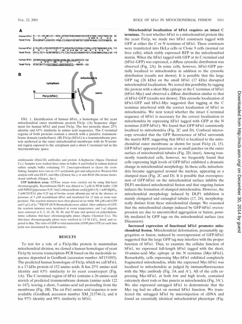
To test for a role of a Fis1p-like protein in mammalianmitochondrial division, we cloned a human homologue of yeastFis1p by reverse transcription-PCR based on the nucleotide se-quence deposited in GenBank (accession number AF151893).The predicted human homologue of Fis1p, which we call hFis1,is a 17-kDa protein of 152 amino acids. It has 25% amino acididentity and 63% similarity to its yeast counterpart (Fig.1A). The C-terminal region of hFis1 contains a 26-amino-acidstretch of predicted transmembrane domain (amino acids 122to 147), leaving a short, 5-amino-acid tail protruding from themembrane (Fig. 1B). The rat Fis1 amino acid sequence is nowavailable (GenBank accession number XM_213746.1), and ithas 97% identity and 99% similarity to hFis1.
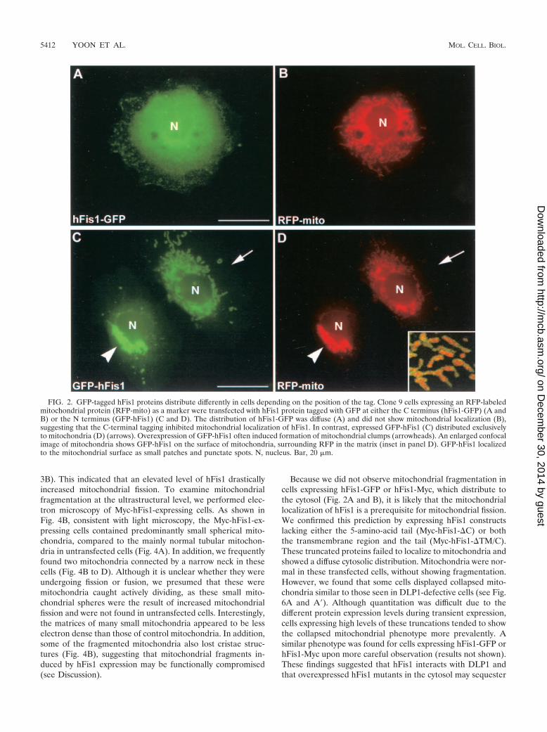
Mitochondrial localization of hFis1 requires an intact Cterminus. To test whether hFis1 is a mitochondrial protein likethe yeast Fis1p, we made two hFis1 constructs tagged withGFP at either the C or N terminus of hFis1. These constructswere transfected into HeLa cells or Clone 9 cells (normal ratliver cells), which stably expressed RFP in the mitochondrialmatrix. When the hFis1 tagged with GFP at its C-terminal end(hFis1-GFP) was expressed, a diffuse cytosolic distribution wasobserved (Fig. 2A). In some cells, however, hFis1-GFP par-tially localized to mitochondria in addition to the cytosolicdistribution (results not shown). It is possible that the largeGFP tag (26 kDa) on the small hFis1 (17 kDa) disruptedmitochondrial localization. We tested this possibility by taggingthe protein with a short Myc epitope at the C terminus of hFis1(hFis1-Myc) and observed a diffuse distribution similar to thatof hFis1-GFP (results not shown). This cytosolic localization ofhFis1-GFP and hFis1-Myc suggested that tagging at the Cterminus interfered with the correct localization of hFis1 tomitochondria. We next tested whether the intact C-terminalsequence of hFis1 is necessary for the correct localization tomitochondria by expressing hFis1 tagged with GFP at the Nterminus (GFP-hFis1). We found that GFP-hFis1 consistentlylocalized to mitochondria (Fig. 2C and D). Confocal micros-copy revealed that the GFP fluorescence of hFis1 surroundsthe matrix RFP, suggesting that hFis1 is localized to the mito-chondrial outer membrane as shown for yeast Fis1p (4, 15).GFP-hFis1 appeared punctate or as small patches on the outersurface of mitochondrial tubules (Fig. 2D, inset). Among tran-siently transfected cells, however, we frequently found thatcells expressing high levels of GFP-hFis1 exhibited a dramaticchange in mitochondrial morphology. In these cells, mitochon-dria became aggregated around the nucleus, appearing as aclumped mass (Fig. 2C and D). It is possible that overexpres-sion of GFP-hFis1 on the mitochondrial surface inhibits theDLP1-mediated mitochondrial fission and that ongoing fusioninduces the formation of clumped mitochondria. However, themitochondrial morphology induced by DLP1 inhibition ismainly elongated and entangled tubules (17, 24), morpholog-ically distinct from these mitochondrial clumps. We reasonedthat the mitochondrial clumps induced by GFP-hFis1 overex-pression are due to uncontrolled aggregation or fusion, possi-bly mediated by GFP tags on the mitochondrial surface (seeDiscussion).
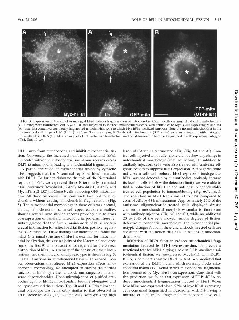
Increased expression of functional hFis1 promotes mito-chondrial fission. Mitochondrial deformation, presumably ag-gregation or fusion, induced by overexpression of GFP-hFis1suggested that the large GFP tag may interfere with the properfunction of hFis1. Thus, to examine the cellular function ofhFis1, we expressed full-length hFis1 tagged with the short,10-amino-acid Myc epitope at the N terminus (Myc-hFis1).Remarkably, cells expressing Myc-hFis1 exhibited completelyfragmented mitochondria, while the expressed Myc-hFis1 waslocalized to mitochondria as judged by immunofluorescencewith the Myc antibody (Fig. 3A and A�). All of the cells ex-pressing Myc-hFis1, at both low and high levels, containedextremely short rods or fine puncta as mitochondria (Fig. 3A�).We also expressed untagged hFis1 to demonstrate that theMyc tag had no effect on normal hFis1 function. We trans-fected the untagged hFis1 by microinjection of cDNA andfound an essentially identical mitochondrial phenotype (Fig.
FIG. 1. Identification of human hFis1, a homologue of the yeastmitochondrial outer membrane protein Fis1p. (A) Sequence align-ment for human hFis1 and yeast Fis1p. The two proteins share 25%identity and 63% similarity in amino acid sequences. The C-terminalregions of both proteins contain a stretch with a putative transmem-brane domain (underlines). (B) Fis1p (hFis1) is a transmembrane pro-tein anchored at the outer mitochondrial membrane with its N-termi-nal region exposed to the cytoplasm and a short C-terminal tail in theintermembrane space.
VOL. 23, 2003 ROLE OF hFis1 IN MITOCHONDRIAL FISSION 5411
on Decem
ber 30, 2014 by guesthttp://m
cb.asm.org/
Dow
nloaded from
3B). This indicated that an elevated level of hFis1 drasticallyincreased mitochondrial fission. To examine mitochondrialfragmentation at the ultrastructural level, we performed elec-tron microscopy of Myc-hFis1-expressing cells. As shown inFig. 4B, consistent with light microscopy, the Myc-hFis1-ex-pressing cells contained predominantly small spherical mito-chondria, compared to the mainly normal tubular mitochon-dria in untransfected cells (Fig. 4A). In addition, we frequentlyfound two mitochondria connected by a narrow neck in thesecells (Fig. 4B to D). Although it is unclear whether they wereundergoing fission or fusion, we presumed that these weremitochondria caught actively dividing, as these small mito-chondrial spheres were the result of increased mitochondrialfission and were not found in untransfected cells. Interestingly,the matrices of many small mitochondria appeared to be lesselectron dense than those of control mitochondria. In addition,some of the fragmented mitochondria also lost cristae struc-tures (Fig. 4B), suggesting that mitochondrial fragments in-duced by hFis1 expression may be functionally compromised(see Discussion).
Because we did not observe mitochondrial fragmentation incells expressing hFis1-GFP or hFis1-Myc, which distribute tothe cytosol (Fig. 2A and B), it is likely that the mitochondriallocalization of hFis1 is a prerequisite for mitochondrial fission.We confirmed this prediction by expressing hFis1 constructslacking either the 5-amino-acid tail (Myc-hFis1-�C) or boththe transmembrane region and the tail (Myc-hFis1-�TM/C).These truncated proteins failed to localize to mitochondria andshowed a diffuse cytosolic distribution. Mitochondria were nor-mal in these transfected cells, without showing fragmentation.However, we found that some cells displayed collapsed mito-chondria similar to those seen in DLP1-defective cells (see Fig.6A and A�). Although quantitation was difficult due to thedifferent protein expression levels during transient expression,cells expressing high levels of these truncations tended to showthe collapsed mitochondrial phenotype more prevalently. Asimilar phenotype was found for cells expressing hFis1-GFP orhFis1-Myc upon more careful observation (results not shown).These findings suggested that hFis1 interacts with DLP1 andthat overexpressed hFis1 mutants in the cytosol may sequester
FIG. 2. GFP-tagged hFis1 proteins distribute differently in cells depending on the position of the tag. Clone 9 cells expressing an RFP-labeledmitochondrial protein (RFP-mito) as a marker were transfected with hFis1 protein tagged with GFP at either the C terminus (hFis1-GFP) (A andB) or the N terminus (GFP-hFis1) (C and D). The distribution of hFis1-GFP was diffuse (A) and did not show mitochondrial localization (B),suggesting that the C-terminal tagging inhibited mitochondrial localization of hFis1. In contrast, expressed GFP-hFis1 (C) distributed exclusivelyto mitochondria (D) (arrows). Overexpression of GFP-hFis1 often induced formation of mitochondrial clumps (arrowheads). An enlarged confocalimage of mitochondria shows GFP-hFis1 on the surface of mitochondria, surrounding RFP in the matrix (inset in panel D). GFP-hFis1 localizedto the mitochondrial surface as small patches and punctate spots. N, nucleus. Bar, 20 �m.
5412 YOON ET AL. MOL. CELL. BIOL.
on Decem
ber 30, 2014 by guesthttp://m
cb.asm.org/
Dow
nloaded from
DLP1 away from mitochondria and inhibit mitochondrial fis-sion. Conversely, the increased number of functional hFis1molecules within the mitochondrial membrane recruits excessDLP1 to mitochondria, leading to mitochondrial fragmentation.
A partial inhibition of mitochondrial fission by cytosolichFis1 suggests that the N-terminal region of hFis1 interactswith DLP1. To further elaborate the role of the N-terminalregion of hFis1, we expressed three N-terminally truncatedhFis1 constructs [Myc-hFis1(32-152), Myc-hFis1(61-152), andMyc-hFis1(92-152)] in Clone 9 cells harboring GFP-mitochon-dria. All three truncated hFis1 constructs localized to mito-chondria without causing mitochondrial fragmentation (Fig.5). The mitochondrial morphology in these cells was normal,although mitochondria in some cells appeared to be unhealthy,showing several large swollen spheres probably due to grossoverexpression of abnormal mitochondrial proteins. These re-sults suggested that the first 31 amino acids of hFis1 containcrucial information for mitochondrial fission, possibly regulat-ing DLP1 function. These findings also indicated that while theintact C-terminal structure of hFis1 is essential for mitochon-drial localization, the vast majority of the N-terminal sequence(up to the first 91 amino acids) is not required for the correctdistribution of hFis1. A summary of the constructs, their local-izations, and their mitochondrial phenotypes is shown in Fig. 5.
hFis1 functions in mitochondrial fission. To expand uponour observations that altered hFis1 expression affects mito-chondrial morphology, we attempted to disrupt the normalfunction of hFis1 by either antibody microinjection or anti-sense oligonucleotides. Upon microinjection of purified anti-bodies against hFis1, mitochondria became elongated andcollapsed around the nucleus (Fig. 6B and B�). This mitochon-drial phenotype was remarkably similar to that observed inDLP1-defective cells (17, 24) and cells overexpressing high
levels of C-terminally truncated hFis1 (Fig. 6A and A�). Con-trol cells injected with buffer alone did not show any change inmitochondrial morphology (data not shown). In addition toantibody injection, cells were also treated with antisense oli-gonucleotides to suppress hFis1 expression. Although we couldnot discern cells with reduced hFis1 expression (endogenoushFis1 was not detectable by our antibodies, probably becauseits level in cells is below the detection limit), we were able tofind a reduction of hFis1 in the antisense oligonucleotide-treated cell population by immunoblotting (Fig. 6C�, inset).The reduction in hFis1 levels was 30 to 50% compared tocontrol cells by 48 h of treatment. Approximately 20% of theantisense oligonucleotide-treated cells displayed drasticchanges in mitochondrial morphology similar to those seenwith antibody injection (Fig. 6C and C�), while an additional20 to 30% of the cells showed various degrees of fission-defective mitochondrial morphology. The mitochondrial phe-notypic changes found in these and antibody-injected cells areconsistent with the notion that hFis1 functions in mitochon-drial fission.
Inhibition of DLP1 function reduces mitochondrial frag-mentation induced by hFis1 overexpression. To provide afunctional test for hFis1 playing a role in DLP1-mediated mi-tochondrial fission, we coexpressed Myc-hFis1 with DLP1-K38A, a dominant-negative DLP1 mutant. We predicted thatexpression of the DLP1 mutant, which normally blocks mito-chondrial fission (17), would inhibit mitochondrial fragmenta-tion promoted by Myc-hFis1 overexpression. Consistent withthis prediction, we found that expression of DLP1-K38A re-duced mitochondrial fragmentation induced by hFis1. WhenMyc-hFis1 was expressed alone, 95% of Myc-hFis1-expressingcells contained fragmented mitochondria, with 5% having amixture of tubular and fragmented mitochondria. No cells
FIG. 3. Expression of Myc-hFis1 or untagged hFis1 induces fragmentation of mitochondria. Clone 9 cells carrying GFP-labeled mitochondria(GFP-mito) were transfected with Myc-hFis1 and subjected to indirect immunofluorescence with antibodies to Myc. Cells expressing Myc-hFis1(A) (asterisk) contained completely fragmented mitochondria (A�) to which Myc-hFis1 localized (arrows). Note the normal mitochondria in theuntransfected cell in panel A� (Un). (B) Clone 9 cells carrying RFP-labeled mitochondria (RFP-mito) were microinjected with untagged,full-length hFis1 DNA (UT-hFis1) along with GFP vector as a transfection marker. Mitochondria became fragmented in cells expressing untaggedhFis1. Bar, 10 �m.
VOL. 23, 2003 ROLE OF hFis1 IN MITOCHONDRIAL FISSION 5413
on Decem
ber 30, 2014 by guesthttp://m
cb.asm.org/
Dow
nloaded from
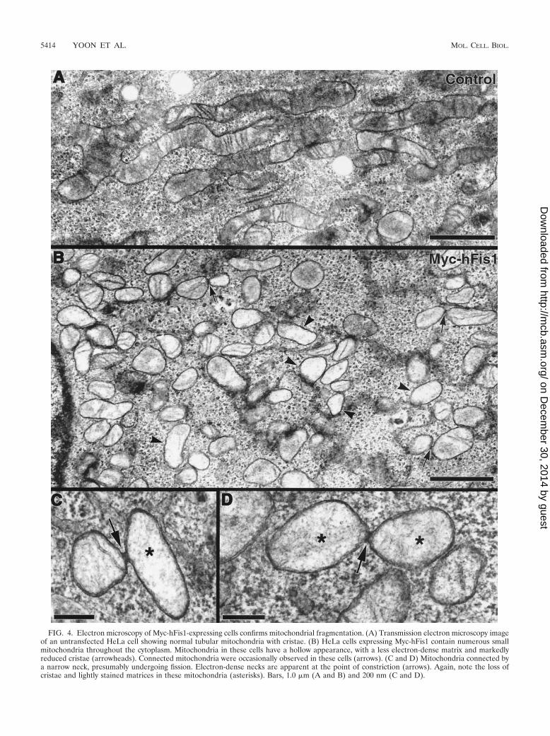
FIG. 4. Electron microscopy of Myc-hFis1-expressing cells confirms mitochondrial fragmentation. (A) Transmission electron microscopy imageof an untransfected HeLa cell showing normal tubular mitochondria with cristae. (B) HeLa cells expressing Myc-hFis1 contain numerous smallmitochondria throughout the cytoplasm. Mitochondria in these cells have a hollow appearance, with a less electron-dense matrix and markedlyreduced cristae (arrowheads). Connected mitochondria were occasionally observed in these cells (arrows). (C and D) Mitochondria connected bya narrow neck, presumably undergoing fission. Electron-dense necks are apparent at the point of constriction (arrows). Again, note the loss ofcristae and lightly stained matrices in these mitochondria (asterisks). Bars, 1.0 �m (A and B) and 200 nm (C and D).
5414 YOON ET AL. MOL. CELL. BIOL.
on Decem
ber 30, 2014 by guesthttp://m
cb.asm.org/
Dow
nloaded from
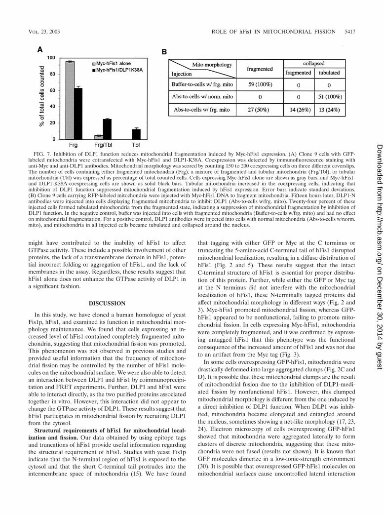
showed tubular mitochondria exclusively (Fig. 7A, gray bars).However, when Myc-hFis1 was coexpressed with DLP1-K38A,cells with fragmented mitochondria were reduced to 63%,while cells containing tubular mitochondria increased by oversevenfold (37% total; 12% with tubular mitochondria and 25%with a mixture of tubular and fragmented mitochondria) (Fig.7A, black bars). These results suggest that mitochondrial frag-mentation induced by hFis1 expression is suppressed throughinhibition of DLP1 function, supporting the finding with yeastthat Fis1p and Dnm1p function together in the same mito-chondrial fission pathway (15). Although mitochondrial frag-mentation in cells coexpressing Myc-hFis1 and DLP1-K38Awas reduced, this suppression was incomplete, showing thatmore than half of the cells contained fragmented mitochon-dria. This partial suppression suggests that the dominant-neg-ative effect of DLP1-K38A may be incomplete in cells coex-pressing Myc-hFis1 and DLP1-K38A. Possibly, the activity ofendogenous wild-type DLP1 in DLP1-K38A-expressing cells issufficient to mediate mitochondrial fragmentation facilitatedby hFis1 overexpression. To test whether we could furtherrestore tubular mitochondria from fragmentation, anti-DLP1antibodies were microinjected into cells expressing Myc-hFis1.A substantial recovery of tubular mitochondria would be ex-pected in the antibody-injected cells, as these DLP1 antibodiesinhibit the endogenous DLP1 function (17). When DLP1 an-tibodies were introduced into Myc-hFis1-expressing cells, 50%of the injected cells exhibited mitochondria collapsed aroundthe nucleus. Among cells with collapsed mitochondria, how-ever, tubular mitochondria were evident in half of the cellswhile the remaining cells appeared to contain fragmented mi-tochondria (Fig. 7B). These results are similar to those ob-tained from coexpression of hFis1 and DLP1-K38A, showing a
partial recovery of tubular mitochondria. The reason for thepartial recovery is not clear, but it is likely that the extensivelyfragmented mitochondria in Myc-hFis1-expressing cells are fu-sion incompetent. Consistent with the fusion incompetence offragmented mitochondria in hFis1-expressing cells, mitochon-drial fragments caused by mutation in yeast Mgm1 have beenshown to be incompetent for fusion (29).
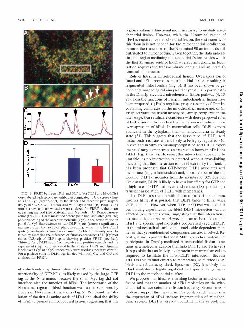
hFis1 and DLP1 form a complex in cells and interact witheach other in vitro. Promotion of DLP1-mediated mitochon-drial fission by expression of Myc-hFis1 suggested that hFis1facilitates targeting of cytosolic DLP1 to the mitochondrialsurface, where DLP1 mediates the fission reaction. Alterna-tively, hFis1 may enhance the GTPase activity of DLP1, thuspromoting the fission reaction. These predictions suggest thathFis1 may interact with DLP1. To test whether these twoproteins interact with each other, we utilized FRET technol-ogy, which is widely used to demonstrate protein-protein in-teractions in the confines of intact cells (12, 27). As we foundthat GFP-tagged hFis1 did not behave normally (Fig. 2), DLP1and Myc-hFis1 were labeled with Cy3 and Cy5 secondary an-tibodies, respectively. Indirect immunofluorescence with sec-ondary antibodies has been successful for FRET techniques (3,11). Myc-hFis1 localized to fragmented mitochondrial punctaas shown earlier in Fig. 3 (red channel) (Fig. 8A and B). Apopulation of DLP1 spots associated with mitochondria dis-playing yellow color, while others distributed to other cytoplas-mic locations. After photobleaching of the acceptor fluoro-phore (Myc-hFis1–Cy5), an increased fluorescence of donormolecules (DLP1-Cy3) was detected, indicating that the twoproteins were in close proximity (Fig. 8C). More than 20 cellstransfected with Myc-hFis1 were analyzed and consistentlyproduced a positive FRET. The FRET intensity between hFis1
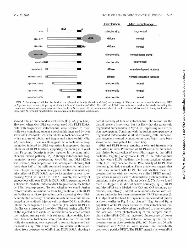
FIG. 5. Summary of cellular distributions and alterations in mitochondrial (Mito.) morphology of different constructs used in this study. GFPor Myc was used as an epitope tag at either the N or C terminus of hFis1. Ten different hFis1 constructs were used in this study, including fivetruncation mutants with mutations in either the C or N terminus. hFis1 proteins modified at the C terminus distributed to the cytosol, whereasthose with N-terminal modifications maintained a mitochondrial localization.
VOL. 23, 2003 ROLE OF hFis1 IN MITOCHONDRIAL FISSION 5415
on Decem
ber 30, 2014 by guesthttp://m
cb.asm.org/
Dow
nloaded from

and DLP1 was significant, showing approximately 70% of thelevel of a positive control in which DLP1 was labeled with bothCy3 and Cy5 (Fig. 8D). However, among the DLP1 punctacolocalized with hFis1, a small number (5 to 12%) of DLP1spots displayed a large positive FRET, while others showedlower or no FRET. This selective FRET provides an importantcontrol for nonspecific FRET and also indicates a transientinteraction between the two proteins. The use of indirect im-munofluorescence methods for FRET experiments has a po-tential limitation because the distance of two primary-second-ary antibody complexes can be extended as far as 68 nm (3).However, FRET drops to the sixth power of the separation,and virtually no FRET occurs beyond a 14-nm distance (12).Our calculated FRET efficiency was 34.7% in the experimentalset compared to 42.5% in the positive control, indicating veryclose proximity between two fluorophores. Considering therandom orientation of the antibodies to each other and thestrong positive FRET in our experiments, hFis1 and DLP1 aremuch closer to each other than the possible 82-nm separation.
Although convincing, these data do not preclude the associa-tion of hFis1 and DLP1 within a complex that includes addi-tional proteins. Regardless of whether they interact directly orindirectly, these data demonstrated that the two proteins are invery close proximity to each other.
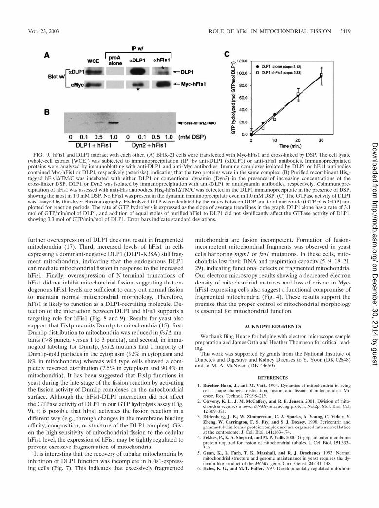
To support the in vivo FRET data, coimmunoprecipitationexperiments were performed from cell lysates. Our initial at-tempt to detect this interaction by coimmunoprecipitation wasinconclusive, suggesting that the interaction might be weakand/or transient as predicted from our FRET results. To detecta potentially transient and weak interaction, we increased thecellular hFis1 level by transfecting BHK-21 cells with Myc-hFis1 and treated them with the membrane-permeant cross-linker DSP. Cells were solubilized, and the homogenates weresubjected to immunoprecipitation. As shown in Fig. 9A, anti-DLP1 antibodies immunoprecipitated Myc-hFis1 from lysatesof cross-linked cells. A reciprocal immunoprecipitation withanti-hFis1 antibodies also pulled down DLP1. As the levels ofMyc-hFis1 and DLP1 coimmunoprecipitated are not extensive,it is likely that this interaction is transient as predicted by theFRET studies. Note that coimmunoprecipitated Myc-hFis1and DLP1 bands in Fig. 9A were overexposed for better dis-play and thus are not quantitative. Along with the FRET data,the coimmunoprecipitation of hFis1 and DLP1 indicates thatthese two proteins are in the same complex. However, it ispossible that this interaction may involve other componentssuch as a mammalian Mdv1p-like protein, as suggested in yeaststudies (26).
To further test the possibility that hFis1 interacts with DLP1directly, we used purified recombinant proteins and again em-ployed the cross-linking reagent to stabilize a possible transientinteraction. His6-tagged DLP1 and hFis1 were expressed andpurified by using a bacterial system. For the ease of purifica-tion, we used a truncated form of hFis1 in which the trans-membrane domain and 5-amino-acid C-terminal tail were de-leted, as the hFis1-DLP1 interaction is likely through theN-terminal region of hFis1. After incubation of the two puri-fied proteins, DLP1 was isolated from the reaction mixture byimmunoprecipitation and examined if hFis1 was coprecipitatedwith DLP1. As shown in Fig. 9B, hFis1 was isolated consis-tently along with DLP1 in the presence of the cross-linker,indicating that hFis1 and DLP1 interact with each other invitro. In a control experiment, purified conventional dynamin(Dyn2) was used to show that random cross-linking betweentwo proteins did not contribute to the hFis1-DLP1 association.Dynamin did not associate with hFis1, indicating that hFis1specifically interacts with DLP1.
Because DLP1 and hFis1 appear to interact with each other,we tested whether this interaction could alter the GTPaseactivity of DLP1. The same recombinant proteins were used inGTP hydrolysis assays to test whether hFis1 functions as anenzymatic cofactor for DLP1. We found that addition of hFis1did not yield any significant effect on GTP hydrolysis by DLP1(Fig. 9C). Without hFis1, DLP1 hydrolyzed GTP at an averagerate of 3.1 mol of GTP/min/mol of DLP1. Addition of an equalmolar concentration of purified hFis1 to the reaction mixtureresulted in no significant change in GTP hydrolysis (Fig. 7B).Further attempts with different molar ratios between DLP1and hFis1 did not influence GTP hydrolysis by DLP1 (resultsnot shown). However, we cannot rule out that other factors
FIG. 6. Disruption of hFis1 function alters mitochondrial shapeand distribution. (A and A�) Clone 9 cells harboring GFP-mitochon-dria were transfected with Myc-hFis1�C. Mitochondria in the celloverexpressing Myc-hFis1�C (arrows) displayed a collapsed pheno-type. (B and B�) Anti-hFis1 antibodies were microinjected into Clone9 cells harboring GFP-mitochondria to inhibit hFis1 function. By 2 h,mitochondria in antibody-injected cells (arrows) became elongatedand collapsed around the nucleus. (C and C�) Antisense oligonucleo-tides were introduced into Clone 9 cells to reduce the expression ofhFis1. At 48 h posttransfection, cells were either fixed for immunoflu-orescence or lysed for immunoblotting. A 50% reduction of hFis1expression was observed when normalized to tubulin by immunoblot-ting (inset). Anti-cytochrome c staining showed that cells (arrows)displayed a fission-defective mitochondrial morphology. N, nucleus; S,sense; AS, antisense.
5416 YOON ET AL. MOL. CELL. BIOL.
on Decem
ber 30, 2014 by guesthttp://m
cb.asm.org/
Dow
nloaded from
might have contributed to the inability of hFis1 to affectGTPase activity. These include a possible involvement of otherproteins, the lack of a transmembrane domain in hFis1, poten-tial incorrect folding or aggregation of hFis1, and the lack ofmembranes in the assay. Regardless, these results suggest thathFis1 alone does not enhance the GTPase activity of DLP1 ina significant fashion.
DISCUSSION
In this study, we have cloned a human homologue of yeastFis1p, hFis1, and examined its function in mitochondrial mor-phology maintenance. We found that cells expressing an in-creased level of hFis1 contained completely fragmented mito-chondria, suggesting that mitochondrial fission was promoted.This phenomenon was not observed in previous studies andprovided useful information that the frequency of mitochon-drial fission may be controlled by the number of hFis1 mole-cules on the mitochondrial surface. We were also able to detectan interaction between DLP1 and hFis1 by coimmunoprecipi-tation and FRET experiments. Further, DLP1 and hFis1 wereable to interact directly, as the two purified proteins associatedtogether in vitro. However, this interaction did not appear tochange the GTPase activity of DLP1. These results suggest thathFis1 participates in mitochondrial fission by recruiting DLP1from the cytosol.
Structural requirements of hFis1 for mitochondrial local-ization and fission. Our data obtained by using epitope tagsand truncations of hFis1 provide useful information regardingthe structural requirement of hFis1. Studies with yeast Fis1pindicate that the N-terminal region of hFis1 is exposed to thecytosol and that the short C-terminal tail protrudes into theintermembrane space of mitochondria (15). We have found
that tagging with either GFP or Myc at the C terminus ortruncating the 5-amino-acid C-terminal tail of hFis1 disruptedmitochondrial localization, resulting in a diffuse distribution ofhFis1 (Fig. 2 and 5). These results suggest that the intactC-terminal structure of hFis1 is essential for proper distribu-tion of this protein. Further, while either the GFP or Myc tagat the N terminus did not interfere with the mitochondriallocalization of hFis1, these N-terminally tagged proteins didaffect mitochondrial morphology in different ways (Fig. 2 and3). Myc-hFis1 promoted mitochondrial fission, whereas GFP-hFis1 appeared to be nonfunctional, failing to promote mito-chondrial fission. In cells expressing Myc-hFis1, mitochondriawere completely fragmented, and it was confirmed by express-ing untagged hFis1 that this phenotype was the functionalconsequence of the increased amount of hFis1 and was not dueto an artifact from the Myc tag (Fig. 3).
In some cells overexpressing GFP-hFis1, mitochondria weredrastically deformed into large aggregated clumps (Fig. 2C andD). It is possible that these mitochondrial clumps are the resultof mitochondrial fusion due to the inhibition of DLP1-medi-ated fission by nonfunctional hFis1. However, this clumpedmitochondrial morphology is different from the one induced bya direct inhibition of DLP1 function. When DLP1 was inhib-ited, mitochondria became elongated and entangled aroundthe nucleus, sometimes showing a net-like morphology (17, 23,24). Electron microscopy of cells overexpressing GFP-hFis1showed that mitochondria were aggregated laterally to formclusters of discrete mitochondria, suggesting that these mito-chondria were not fused (results not shown). It is known thatGFP molecules dimerize in a low-ionic-strength environment(30). It is possible that overexpressed GFP-hFis1 molecules onmitochondrial surfaces cause uncontrolled lateral interaction
FIG. 7. Inhibition of DLP1 function reduces mitochondrial fragmentation induced by Myc-hFis1 expression. (A) Clone 9 cells with GFP-labeled mitochondria were cotransfected with Myc-hFis1 and DLP1-K38A. Coexpression was detected by immunofluorescence staining withanti-Myc and anti-DLP1 antibodies. Mitochondrial morphology was scored by counting 150 to 200 coexpressing cells on three different coverslips.The number of cells containing either fragmented mitochondria (Frg), a mixture of fragmented and tubular mitochondria (Frg/Tbl), or tubularmitochondria (Tbl) was expressed as percentage of total counted cells. Cells expressing Myc-hFis1 alone are shown as gray bars, and Myc-hFis1-and DLP1-K38A-coexpressing cells are shown as solid black bars. Tubular mitochondria increased in the coexpressing cells, indicating thatinhibition of DLP1 function suppressed mitochondrial fragmentation induced by hFis1 expression. Error bars indicate standard deviations.(B) Clone 9 cells carrying RFP-labeled mitochondria were injected with Myc-hFis1 DNA to fragment mitochondria. Fifteen hours later, DLP1-Nantibodies were injected into cells displaying fragmented mitochondria to inhibit DLP1 (Abs-to-cells w/frg. mito). Twenty-four percent of theseinjected cells formed tubulated mitochondria from the fragmented state, indicating a suppression of mitochondrial fragmentation by inhibition ofDLP1 function. In the negative control, buffer was injected into cells with fragmented mitochondria (Buffer-to-cells w/frg. mito) and had no effecton mitochondrial fragmentation. For a positive control, DLP1 antibodies were injected into cells with normal mitochondria (Abs-to-cells w/norm.mito), and mitochondria in all injected cells became tubulated and collapsed around the nucleus.
VOL. 23, 2003 ROLE OF hFis1 IN MITOCHONDRIAL FISSION 5417
on Decem
ber 30, 2014 by guesthttp://m
cb.asm.org/
Dow
nloaded from
of mitochondria by dimerization of GFP moieties. This non-functionality of GFP-hFis1 is likely caused by the large GFPtag at the N terminus, because the small Myc tag did notinterfere with the function of hFis1. The importance of theN-terminal region in hFis1 function was further supported bystudies of N-terminal truncations (Fig. 5). We found that de-letion of the first 31 amino acids of hFis1 abolished the abilityof hFis1 to promote mitochondrial fission, suggesting that this
region contains a functional motif necessary to mediate mito-chondrial fission. However, while the N-terminal region ofhFis1 is required for mitochondrial fission, the vast majority ofthis domain is not needed for the mitochondrial localization,because the truncation of the N-terminal 90 amino acids stilldistributed to mitochondria. Taken together, the data indicatethat the region mediating mitochondrial fission resides withinthe first 31 amino acids of hFis1 whereas mitochondrial local-ization requires the transmembrane domain and an intact C-terminal tail structure.
Role of hFis1 in mitochondrial fission. Overexpression offunctional hFis1 promotes mitochondrial fission, resulting infragmented mitochondria (Fig. 3). It has been shown by ge-netic and morphological analyses that yeast Fis1p participatesin the Dnm1p-mediated mitochondrial fission pathway (4, 15,25). Possible functions of Fis1p in mitochondrial fission havebeen proposed: (i) Fis1p regulates proper assembly of Dnm1p-containing complexes on the mitochondrial membrane, or (ii)Fis1p activates the fission activity of Dnm1p complexes in thelater stage. Our results are consistent with these proposed rolesof Fis1p, since mitochondrial fragmentation was induced uponoverexpression of hFis1. In mammalian cells, DLP1 is moreabundant in the cytoplasm than on mitochondria at steadystate (31). This suggests that the association of DLP1 withmitochondria is transient and likely to be highly regulated. Ourin vivo and in vitro coimmunoprecipitation and FRET exper-iments clearly demonstrate an interaction between hFis1 andDLP1 (Fig. 8 and 9). However, this interaction appears to beunstable, as no interaction is detected without cross-linking,indicating that this interaction is indeed extremely transient. Ithas been proposed that GTP-bound DLP1 associates withmembrane (e.g., mitochondria) and, upon release of the nu-cleotide, DLP1 dissociates from the membrane (32). Further,like dynamin, DLP1 is likely to have a low affinity for GTP anda high rate of GTP hydrolysis and release (28), predicting atransient association of DLP1 with membranes.
If a DLP1 association with the mitochondrial membraneinvolves hFis1, it is possible that DLP1 binds to hFis1 whenGTP is bound. However, when GTP or GTPS was added inour binding experiments, the hFis1-DLP1 interaction was notaffected (results not shown), suggesting that this interaction isnot nucleotide dependent. However, it cannot be ruled out thathFis1 and specific lipid molecules cooperatively recruit DLP1to the mitochondrial surface in a nucleotide-dependent man-ner or that yet-unidentified components are also involved. Re-cently, it was reported that yeast Mdv1p, another protein thatparticipates in Dnm1p-mediated mitochondrial fission, func-tions as a molecular adaptor that links Dnm1p and Fis1p (26).It is possible that an Mdv1p-like protein in mammalian cells isrequired to facilitate the hFis1-DLP1 interaction. BecauseDLP1 is able to bind directly to membranes, as purified DLP1binds and tubulates synthetic liposomes (32), it is likely thathFis1 mediates a highly regulated and specific targeting ofDLP1 to the mitochondrial surface.
We propose that hFis1 is a limiting factor in mitochondrialfission and that the number of hFis1 molecules on the mito-chondrial surface determines fission frequency. Several lines ofevidence support this hypothesis. First, only a slight increase inthe expression of hFis1 induces fragmentation of mitochon-dria. Second, DLP1 is already abundant in the cytosol, and
FIG. 8. FRET between hFis1 and DLP1. (A) DLP1 and Myc-hFis1were labeled with secondary antibodies conjugated to Cy3 (green chan-nel) and Cy5 (red channel) as the donor and acceptor pair, respec-tively, in COS-7 cells transfected with Myc-hFis1. (B) Four DLP1spots (arrows and arrowheads) were analyzed for FRET by the donorquenching method (see Materials and Methods). (C) Donor fluores-cence (Cy3-DLP1) was measured before (blue line) and after (red line)photobleaching of the acceptor molecule (Cy5) in the boxed region inpanel A. Cy3 fluorescence of two DLP1 spots (arrows) significantlyincreased after the acceptor photobleaching, while the other DLP1spots (arrowheads) showed no change. (D) FRET intensity was ob-tained by averaging the difference of fluorescence values (�FI [Cy3postminus Cy3pre]) of DLP1 spots showing positive FRET (red bar).Thirty to forty DLP1 spots from negative and positive controls and theexperiment (Exp) were subjected to the analysis. DLP1 and dynaminlabeled with Cy3 and Cy5, respectively, were used as a negative control.For a positive control, DLP1 was labeled with both Cy3 and Cy5 andanalyzed for FRET.
5418 YOON ET AL. MOL. CELL. BIOL.
on Decem
ber 30, 2014 by guesthttp://m
cb.asm.org/
Dow
nloaded from
further overexpression of DLP1 does not result in fragmentedmitochondria (17). Third, increased levels of hFis1 in cellsexpressing a dominant-negative DLP1 (DLP1-K38A) still frag-ment mitochondria, indicating that the endogenous DLP1can mediate mitochondrial fission in response to the increasedhFis1. Finally, overexpression of N-terminal truncations ofhFis1 did not inhibit mitochondrial fission, suggesting that en-dogenous hFis1 levels are sufficient to carry out normal fissionto maintain normal mitochondrial morphology. Therefore,hFis1 is likely to function as a DLP1-recruiting molecule. De-tection of the interaction between DLP1 and hFis1 supports atargeting role for hFis1 (Fig. 8 and 9). Results for yeast alsosupport that Fis1p recruits Dnm1p to mitochondria (15): first,Dnm1p distribution to mitochondria was reduced in fis1� mu-tants (8 puncta versus 1 to 3 puncta), and second, in immu-nogold labeling for Dnm1p, fis1� mutants had a majority ofDnm1p-gold particles in the cytoplasm (92% in cytoplasm and8% in mitochondria) whereas wild type cells showed a com-pletely reversed distribution (7.5% in cytoplasm and 90.4% inmitochondria). It has been suggested that Fis1p functions inyeast during the late stage of the fission reaction by activatingthe fission activity of Dnm1p complexes on the mitochondrialsurface. Although the hFis1-DLP1 interaction did not affectthe GTPase activity of DLP1 in our GTP hydrolysis assay (Fig.9), it is possible that hFis1 activates the fission reaction in adifferent way (e.g., through changes in the membrane bindingaffinity, composition, or structure of the DLP1 complex). Giv-en the high sensitivity of mitochondrial fission to the cellularhFis1 level, the expression of hFis1 may be tightly regulated toprevent excessive fragmentation of mitochondria.
It is interesting that the recovery of tubular mitochondria byinhibition of DLP1 function was incomplete in hFis1-express-ing cells (Fig. 7). This indicates that excessively fragmented
mitochondria are fusion incompetent. Formation of fusion-incompetent mitochondrial fragments was observed in yeastcells harboring mgm1 or fzo1 mutations. In these cells, mito-chondria lost their DNA and respiration capacity (5, 9, 18, 21,29), indicating functional defects of fragmented mitochondria.Our electron microscopy results showing a decreased electrondensity of mitochondrial matrices and loss of cristae in Myc-hFis1-expressing cells also suggest a functional compromise offragmented mitochondria (Fig. 4). These results support thepremise that the proper control of mitochondrial morphologyis essential for mitochondrial function.
ACKNOWLEDGMENTS
We thank Bing Huang for helping with electron microscope samplepreparation and James Orth and Heather Thompson for critical read-ing.
This work was supported by grants from the National Institute ofDiabetes and Digestive and Kidney Diseases to Y. Yoon (DK 02648)and to M. A. McNiven (DK 44650)
REFERENCES
1. Bereiter-Hahn, J., and M. Voth. 1994. Dynamics of mitochondria in livingcells: shape changes, dislocation, fusion, and fission of mitochondria. Mi-crosc. Res. Technol. 27:198–219.
2. Cerveny, K. L., J. M. McCaffery, and R. E. Jensen. 2001. Division of mito-chondria requires a novel DNM1-interacting protein, Net2p. Mol. Biol. Cell12:309–321.
3. Dictenberg, J. B., W. Zimmerman, C. A. Sparks, A. Young, C. Vidair, Y.Zheng, W. Carrington, F. S. Fay, and S. J. Doxsey. 1998. Pericentrin andgamma-tubulin form a protein complex and are organized into a novel latticeat the centrosome. J. Cell Biol. 141:163–174.
4. Fekkes, P., K. A. Shepard, and M. P. Yaffe. 2000. Gag3p, an outer membraneprotein required for fission of mitochondrial tubules. J. Cell Biol. 151:333–340.
5. Guan, K., L. Farh, T. K. Marshall, and R. J. Deschenes. 1993. Normalmitochondrial structure and genome maintenance in yeast requires the dy-namin-like product of the MGM1 gene. Curr. Genet. 24:141–148.
6. Hales, K. G., and M. T. Fuller. 1997. Developmentally regulated mitochon-
FIG. 9. hFis1 and DLP1 interact with each other. (A) BHK-21 cells were transfected with Myc-hFis1 and cross-linked by DSP. The cell lysate(whole-cell extract [WCE]) was subjected to immunoprecipitation (IP) by anti-DLP1 (�DLP1) or anti-hFis1 antibodies. Immunoprecipitatedproteins were analyzed by immunoblotting with anti-DLP1 and anti-Myc antibodies. Immune complexes isolated by DLP1 or hFis1 antibodiescontained Myc-hFis1 or DLP1, respectively (asterisks), indicating that the two proteins were in the same complex. (B) Purified recombinant His6-tagged hFis1�TM/C was incubated with either DLP1 or conventional dynamin (Dyn2) in the presence of increasing concentrations of thecross-linker DSP. DLP1 or Dyn2 was isolated by immunoprecipitation with anti-DLP1 or antidynamin antibodies, respectively. Coimmunopre-cipitation of hFis1 was assessed with anti-His antibodies. His6-hFis1�TM/C was detected in the DLP1 immunoprecipitate in the presence of DSP,showing the most in 1.0 mM DSP. No hFis1 was present in the dynamin immunoprecipitate even in 1.0 mM DSP. (C) The GTPase activity of DLP1was assayed by thin-layer chromatography. Hydrolyzed GTP was calculated by the ratios between GDP and total nucleotide (GTP plus GDP) andplotted for reaction periods. The rate of GTP hydrolysis is expressed as the slope of average trendlines in the graph. DLP1 alone has a rate of 3.1mol of GTP/min/mol of DLP1, and addition of equal moles of purified hFis1 to DLP1 did not significantly affect the GTPase activity of DLP1,showing 3.3 mol of GTP/min/mol of DLP1. Error bars indicate standard deviations.
VOL. 23, 2003 ROLE OF hFis1 IN MITOCHONDRIAL FISSION 5419
on Decem
ber 30, 2014 by guesthttp://m
cb.asm.org/
Dow
nloaded from
drial fusion mediated by a conserved, novel, predicted GTPase. Cell 90:121–129.
7. Henley, J. R., E. W. A. Krueger, B. J. Oswald, and M. A. McNiven. 1998.Dynamin-mediated internalization of caveolae. J. Cell Biol. 141:85–99.
8. Henley, J. R., and M. A. McNiven. 1996. Association of a dynamin-likeprotein with the Golgi apparatus in mammalian cells. J. Cell Biol. 133:761–775.
9. Hermann, G. J., J. W. Thatcher, J. P. Mills, K. G. Hales, M. T. Fuller, J.Nunnari, and J. M. Shaw. 1998. Mitochondrial fusion in yeast requires thetransmembrane GTPase Fzo1p. J. Cell Biol. 143:359–373.
10. Imoto, M., I. Tachibana, and R. Urrutia. 1998. Identification and functionalcharacterization of a novel human protein highly related to the yeast dy-namin-like GTPase Vps1p. J. Cell Sci. 111:1341–1349.
11. Kam, Z., T. Volberg, and B. Geiger. 1995. Mapping of adherens junctioncomponents using microscopic resonance energy transfer imaging. J. CellSci. 108:1051–1062.
12. Kenworthy, A. K. 2001. Imaging protein-protein interactions using fluores-cence resonance energy transfer microscopy. Methods 24:289–296.
13. Koch, A., M. Thiemann, M. Grabenbauer, Y. Yoon, M. A. McNiven, and M.Schrader. 2003. Dynamin-like protein 1 is involved in peroxisomal fission.J. Biol. Chem. 278:8597–8605.
14. Li, X., and S. J. Gould. 2003. The dynamin-like GTPase DLP1 is essential forperoxisome division and is recruited to peroxisomes in part by PEX11.J. Biol. Chem., 278:17012–17020.
15. Mozdy, A. D., J. M. McCaffery, and J. M. Shaw. 2000. Dnm1p GTPase-mediated mitochondrial fission is a multi-step process requiring the novelintegral membrane component Fis1p. J. Cell Biol. 151:367–380.
16. Otsuga, D., B. R. Keegan, E. Brisch, J. W. Thatcher, G. J. Hermann, W.Bleazard, and J. M. Shaw. 1998. The dynamin-related GTPase, Dnm1p,controls mitochondrial morphology in yeast. J. Cell Biol. 143:333–349.
17. Pitts, K. R., Y. Yoon, E. W. Krueger, and M. A. McNiven. 1999. The dy-namin-like protein DLP1 is essential for normal distribution and morphologyof the endoplasmic reticulum and mitochondria in mammalian cells. Mol.Biol. Cell. 10:4403–4417.
18. Rapaport, D., M. Brunner, W. Neupert, and B. Westermann. 1998. Fzo1p isa mitochondrial outer membrane protein essential for the biogenesis offunctional mitochondria in Saccharomyces cerevisiae. J. Biol. Chem. 273:20150–20155.
19. Santel, A., and M. T. Fuller. 2001. Control of mitochondrial morphology bya human mitofusin. J. Cell Sci. 114:867–874.
20. Sesaki, H., and R. E. Jensen. 1999. Division versus fusion: Dnm1p and Fzo1pantagonistically regulate mitochondrial shape. J. Cell Biol. 147:699–706.
21. Shepard, K. A., and M. P. Yaffe. 1999. The yeast dynamin-like protein,Mgm1p, functions on the mitochondrial outer membrane to mediate mito-chondrial inheritance. J. Cell Biol. 144:711–720.
22. Shin, H. W., C. Shinotsuka, S. Torii, K. Murakami, and K. Nakayama. 1997.Identification and subcellular localization of a novel mammalian dynamin-related protein homologous to yeast Vps1p and Dnm1p. J. Biochem. 122:525–530.
23. Smirnova, E., L. Griparic, D. L. Shurland, and A. M. van der Bliek. 2001.Dynamin-related protein drp1 is required for mitochondrial division in mam-malian cells. Mol. Biol. Cell 12:2245–2256.
24. Smirnova, E., D. L. Shurland, S. N. Ryazantsev, and A. M. van der Bliek.1998. A human dynamin-related protein controls the distribution of mito-chondria. J. Cell Biol. 143:351–358.
25. Tieu, Q., and J. Nunnari. 2000. Mdv1p is a WD repeat protein that interactswith the dynamin-related GTPase, Dnm1p, to trigger mitochondrial division.J. Cell Biol. 151:353–366.
26. Tieu, Q., V. Okreglak, K. Naylor, and J. Nunnari. 2002. The WD repeatprotein, Mdv1p, functions as a molecular adaptor by interacting with Dnm1pand Fis1p during mitochondrial fission. J. Cell Biol. 158:445–452.
27. Truong, K., and M. Ikura. 2001. The use of FRET imaging microscopy todetect protein-protein interactions and protein conformational changes invivo. Curr. Opin. Struct. Biol. 11:573–578.
28. Warnock, D. E., and S. L. Schmid. 1996. Dynamin GTPase, a force-gener-ating molecular switch. Bioessays 18:885–893.
29. Wong, E. D., J. A. Wagner, S. W. Gorsich, J. M. McCaffery, J. M. Shaw, andJ. Nunnari. 2000. The dynamin-related GTPase, Mgm1p, is an intermem-brane space protein required for maintenance of fusion competent mito-chondria. J. Cell Biol. 151:341–352.
30. Yang, F., L. G. Moss, and G. N. Phillips, Jr. 1996. The molecular structureof green fluorescent protein. Nat. Biotechnol. 14:1246–1251.
31. Yoon, Y., K. R. Pitts, S. Dahan, and M. A. McNiven. 1998. A novel dynamin-like protein associates with cytoplasmic vesicles and tubules of the endoplas-mic reticulum in mammalian cells. J. Cell Biol. 140:779–793.
32. Yoon, Y., K. R. Pitts, and M. A. McNiven. 2001. Mammalian dynamin-likeprotein DLP1 tubulates membranes. Mol. Biol. Cell 12:2894–2905.
5420 YOON ET AL. MOL. CELL. BIOL.
on Decem
ber 30, 2014 by guesthttp://m
cb.asm.org/
Dow
nloaded from
Related Documents


