The making of gametes in higher plants LEONOR C. BOAVIDA 1 , JÖRG D. BECKER 1 and JOSÉ A. FEIJÓ* ,1,2 1 Centro de Biologia do Desenvolvimento, Instituto Gulbenkian de Ciência, Oeiras, Portugal and 2 Universidade de Lisboa, Faculdade de Ciências, Dept. Biologia Vegetal, Campo Grande, Lisboa, Portugal ABSTRACT Higher plants have evolved to be one of the predominant life forms on this planet. A great deal of this evolutionary success relies in a very short gametophytic phase which underlies the sexual reproduction cycle. Sexual plant reproduction takes place in special organs of the flower. In most species the processes of gametogenesis, pollination, syngamy and embryogenesis are sequentially coordinated to give rise to a functional seed in a matter of few weeks. Any of these processes is so intricately complex and precisely regulated that it becomes no wonder that each involves more specific genes and cellular processes than any other function in the plant life cycle. While variability generation - the evolutionary output of the sexual cycle - is the same as in any other Kingdom, plants do it using a completely original set of mechanisms, many of which are not yet comprehended. In this paper, we cover the fundamental features of male and female gametoge- nesis. While the physiological and cellular bases of these processes have been continuously described since the early nineteen century, recent usage of Arabidopsis and other species as central models has brought about a great deal of specific information regarding their genetic regulation. Transcriptomics has recently enlarged the repertoire and pollen became the first gametophyte to have a fully described transcriptome in plants. We thus place special emphasis on the way this newly accumulated genetic and transcriptional information impacts our current understanding of the mechanisms of gametogenesis. KEY WORDS: pollen, embryo sac, gametogenesis, microsporogenesis, macrosporogenesis The uniqueness of a life form One of the most remarkable features of life on earth is diversity, a great deal of which is based on the evolutionary output of sexual reproduction. Unlike animals, in which the primordial germ line develops early during embryogenesis, higher plants alternate the growth of the diploid sporophyte organism with a highly reduced growth form on the plant life cycle, the haploid gametophyte. This is a well-suited strategy for selection because plants spend most of their life on the vegetative phase. The gametophytic stage also presents an opportunity for selection at the haploid level (Ottaviano et al., 1990). Plant cells don’t move and positional information instead of lineage is the primary determinant of cell fate in plants. Meiosis triggers the separation between sporophytic and gametophytic generations involving various genes (Caryl et al., 2003). This spatial pleiotropy of the sexual organs has prompted evolution for the appearance of safety mechanisms to prevent the fusion of incompatible genomes while supporting genetic variability. In higher animals genetic mechanisms for sex determination estab- lish striking developmental differences between males and fe- Int. J. Dev. Biol. 49: 595-614 (2005) doi: 10.1387/ijdb.052019lb *Address correspondence to: Dr. José A. Feijó. Instituto Gulbenkian de Ciência, PT-2780-156 Oeiras, Portugal. Fax: +351-21-440-7970. e-mail: [email protected] 0214-6282/2005/$25.00 © UBC Press Printed in Spain www.intjdevbiol.com males. In contrast, most higher plant species develop both male and female structures within the same flower, allowing self- fertilization. Out-crossing is ensured by self-incompatibility mecha- nisms, which evolved under precise genetic control controlling self-recognition and cell-to-cell interaction (Thomas and Franklin- Tong, 2004). The short period that plants spend on their reproductive cycle includes some of the most intriguing and challenging questions in biology, many yet without a mechanistic answer. With the publi- cation of the complete genomic sequence of the flowering plant Arabidopsis thaliana (The Arabidopsis Genome Initiative, 2000), new foundations for functional and comparative genomics emerged to reveal basic genetic differences between plants and other eukaryotes. Although the logic underlying many of the develop- mental processes as pattern formation and cell-cell communica- tion are comparable between plants and animals, the master regulators are unrelated or represent novel protein rearrange- ments which serve identical functions (Meyerowitz, 2002), mak- ing many genes unique to plants. It is therefore of no surprise that we find this prediction mirrored on the various stages of sexual plant reproduction.

Welcome message from author
This document is posted to help you gain knowledge. Please leave a comment to let me know what you think about it! Share it to your friends and learn new things together.
Transcript

The making of gametes in higher plants
LEONOR C. BOAVIDA1, JÖRG D. BECKER1 and JOSÉ A. FEIJÓ*,1,2
1Centro de Biologia do Desenvolvimento, Instituto Gulbenkian de Ciência, Oeiras, Portugal and 2Universidade de Lisboa, Faculdade deCiências, Dept. Biologia Vegetal, Campo Grande, Lisboa, Portugal
ABSTRACT Higher plants have evolved to be one of the predominant life forms on this planet. A
great deal of this evolutionary success relies in a very short gametophytic phase which underlies
the sexual reproduction cycle. Sexual plant reproduction takes place in special organs of the flower.
In most species the processes of gametogenesis, pollination, syngamy and embryogenesis are
sequentially coordinated to give rise to a functional seed in a matter of few weeks. Any of these
processes is so intricately complex and precisely regulated that it becomes no wonder that each
involves more specific genes and cellular processes than any other function in the plant life cycle.
While variability generation - the evolutionary output of the sexual cycle - is the same as in any other
Kingdom, plants do it using a completely original set of mechanisms, many of which are not yet
comprehended. In this paper, we cover the fundamental features of male and female gametoge-
nesis. While the physiological and cellular bases of these processes have been continuously
described since the early nineteen century, recent usage of Arabidopsis and other species as central
models has brought about a great deal of specific information regarding their genetic regulation.
Transcriptomics has recently enlarged the repertoire and pollen became the first gametophyte to
have a fully described transcriptome in plants. We thus place special emphasis on the way this
newly accumulated genetic and transcriptional information impacts our current understanding of
the mechanisms of gametogenesis.
KEY WORDS: pollen, embryo sac, gametogenesis, microsporogenesis, macrosporogenesis
The uniqueness of a life form
One of the most remarkable features of life on earth is diversity,a great deal of which is based on the evolutionary output of sexualreproduction. Unlike animals, in which the primordial germ linedevelops early during embryogenesis, higher plants alternate thegrowth of the diploid sporophyte organism with a highly reducedgrowth form on the plant life cycle, the haploid gametophyte. Thisis a well-suited strategy for selection because plants spend mostof their life on the vegetative phase. The gametophytic stage alsopresents an opportunity for selection at the haploid level (Ottavianoet al., 1990).
Plant cells don’t move and positional information instead oflineage is the primary determinant of cell fate in plants. Meiosistriggers the separation between sporophytic and gametophyticgenerations involving various genes (Caryl et al., 2003). Thisspatial pleiotropy of the sexual organs has prompted evolution forthe appearance of safety mechanisms to prevent the fusion ofincompatible genomes while supporting genetic variability. Inhigher animals genetic mechanisms for sex determination estab-lish striking developmental differences between males and fe-
Int. J. Dev. Biol. 49: 595-614 (2005)doi: 10.1387/ijdb.052019lb
*Address correspondence to: Dr. José A. Feijó. Instituto Gulbenkian de Ciência, PT-2780-156 Oeiras, Portugal. Fax: +351-21-440-7970. e-mail: [email protected]
0214-6282/2005/$25.00© UBC PressPrinted in Spainwww.intjdevbiol.com
males. In contrast, most higher plant species develop both maleand female structures within the same flower, allowing self-fertilization. Out-crossing is ensured by self-incompatibility mecha-nisms, which evolved under precise genetic control controllingself-recognition and cell-to-cell interaction (Thomas and Franklin-Tong, 2004).
The short period that plants spend on their reproductive cycleincludes some of the most intriguing and challenging questions inbiology, many yet without a mechanistic answer. With the publi-cation of the complete genomic sequence of the flowering plantArabidopsis thaliana (The Arabidopsis Genome Initiative, 2000),new foundations for functional and comparative genomics emergedto reveal basic genetic differences between plants and othereukaryotes. Although the logic underlying many of the develop-mental processes as pattern formation and cell-cell communica-tion are comparable between plants and animals, the masterregulators are unrelated or represent novel protein rearrange-ments which serve identical functions (Meyerowitz, 2002), mak-ing many genes unique to plants. It is therefore of no surprise thatwe find this prediction mirrored on the various stages of sexualplant reproduction.
596 L. Boavida et al.
Gametogenesis in the major group of vascular plants:the angiosperms
The phylum Anthophyta, i.e. the Angiosperms (from the Latinword angi, “enclosed” and Greek word sperma, “seed”) or flower-ing plants, is the dominant group of land plants today. They aroseabout 140 million years ago during the late Jurassic and experi-enced rapid diversification during the Cretaceous. There aremore than 250 000 species, grouped into 12 500 genera andabout 300 families and in terms of ecological and nutritionalimportance they surpass all other groups. Numerous traits con-tributed to this rise to dominance, including the ability to reachreproductive maturity rapidly and adaptation to animal pollinationand seed dispersal (Darwin, 1862). Flowers, the reproductiveorgans of angiosperms, are more varied than the equivalentstructures of any other group of organisms (Barrett, 2002).
The life cycle of higher plants includes seed germination,vegetative growth, flowering, fertilization, development of embryoand seed maturation. Induction of flowering is triggered by envi-ronmental cues such as light, temperature and nutrient access, incombination with endogenous signals (e.g. the plant hormonegibberellin and circadian’oscillators), for which many floweringtime genes are described (Mouradov et al., 2002). The integrationof signals directs the activation of meristem identity genes, whichspecify floral identity and the shoot apical meristem is trans-formed into an inflorescence. Finally, the floral organ identitygenes are activated in different regions of the flower, producingfour organ types, sepals, petals, the male stamens, collectivelynamed the androecium and the female carpels, making up thegynoecium, or the pistil, located in the innermost whorl (Smyth etal., 1990). Such are the organs were male and female gametoge-nesis take place respectively.
The male gametophyte - pollen
The development of the male gametophyte involves a series ofevents culminating in the production and release of mature pollengrains from anthers (McCormick, 1993, 2004). Mature pollengrains may be considered as a “cell within a cell”, with veryspecialized functions. Enclosed by a very special cell wall, thevegetative cell harbors an individual cytoplasm with a particulargene expression pattern. Its final fate is to transport the two spermcells through the female tissues and release them in the vicinityof the egg and central cells (see review by Boavida et al., 2005).
It all begins in stamens, the male reproductive organs, whichconsist of an anther, where pollen development takes place anda stalk-like filament which provides support, nutrient transport andpositions anthers to facilitate pollen dispersal (Scott et al., 1991a;Irish, 1999). Stamens are derived from periclinal divisions of theL2 layer of the shoot apical meristem (Jenik and Irish, 2000).
Pollen development takes place within the anther locus, orpollen sac (Fig. 1D), involving perhaps more genes than any othersingle process in plant development and depends on interactionsbetween sporophytic and gametophytic cells (Scott et al., 2004).Both are essential for male fertility as revealed by numerousgenetic screens (van der Meer et al., 1992; Chen and McCormick,1996; Aarts et al., 1997; Hulskamp et al., 1997; Taylor et al., 1998;Ariizumi et al., 2003).
Anther differentiation will lead to the production of severalspecialized cells and tissues involved in reproductive or non-reproductive functions (e.g. support and dehiscence) (Koltunowet al., 1990). The male gametogenesis starts from L2 cells of theshoot apical meristem, that divide to form a primary parietal cell(PPC) and the primary sporogeneous cells (PSC). The PPC laterform several concentric layers on the anther wall, which differen-tiate into the endothecium, middle and the very special innerlayers, known as the tapetum (Fig. 1A). PSC give rise to themicrosporocytes. Coordination of the process is under tight ge-netic control (Goldberg et al., 1993).
The development of viable pollen grains is dependent on thepresence of the tapetum, which has roles in the nourishment ofmicrospores, formation of exine and deposition of tryphine on thepollen wall (Koltunow et al., 1990; Mariani et al., 1990; Goldberget al., 1993). A summary of major developmental events on malegametogenesis with the correspondent anther differentiationstages is represented on Figure 1.
When microspores initiate meiosis, coenocytic masses areestablished by enlarged plasmodesmata, the cytomictic channels(Fig. 1B), which create a cytoplasmic continuum between groupsof cells. This way, there is an effective synchronization duringmicrosporogenesis, by allowing exchange of molecules and ions(Heslop-Harrison, 1966; Mascarenhas, 1975). Microsporocytesare encased in an impermeable β-1,3-glucan wall (callose),isolating the meiocytes from the sporophyte cells (Fig. 1E). Thecallosic wall was proposed to function as a sieve for largemolecules whose influence could impair the commitment to anhaploid genomic expression (Mackenzie et al., 1967; Mascarenhas,1975). However, the apparent ability of large molecules to trans-
Fig. 1 (opposite). A generalized overview of microsporogenesis and pollen development. Schematic representations were adapted from Goldberget al. (1993) and McCormick (1993). C, connective; CCC, circular cell cluster; E, epidermis; En, endothecium; PG, pollen grain; PS, pollen sac; St stomium;T, tapetum; Td tetrads; V, vascular bundle. See text for description of developmental events. Individual steps are illustrated with transmitted electronmicroscopy (TEM) micrographs or TEM-prepared semi-thin sections seen with light microscopy of microsporogenesis of the orchid Ophrys lutea (Feijó,1995). (A) Individualization of a group of sporogeneous cells by a callose envelope. These cells will divide to final size and enter male meiosis. (B)
Meiocytes are connected by large channels, named cytomictic channels (CC), through which a general streaming, including organelles like plastids (Pl)is believed to contribute to the synchronization of meiosis within the whole population. (C) Telophase II, with twin mitotic spindles (f1 and f2) within acallose enclosed tetrad. (D) Mid section of an immature flower showing the relative position of the male gametophyte forming organs, the pollen sacs(Pol1 and 2) and the female gametophyte forming organs, the ovules in the ovary (Ov). (E) Up until the end of meiosis, each microspore is enclosed ona callose wall and groups of microspores are individualized from surrounding tissues by thick callose walls (Cal), here stained with decolorized anilineblue. (F) Formation of individualized microspores within a meiotic tetrad (1,2,3,4). Note the synchronization of one mass of meiocytes in telophase II,while the upper mass is already synchronized in microspore individualization. (G) Polarization and vacuolization of the young microspore. The nucleusis strongly pulled out to one end of the cell, while the plastids are on the opposite end. Small vacuoles are formed to account for the enlargement of thecell and later will coalesce in a single big vacuole. (H) Synchronized pollen mitosis I. The mass of microspores on the right is still on anaphase and onthe left cytokinesis already occurred (arrow), dividing a small generative cell and a big vegetative one. (I-K) Process of pinching-off the generative cell.

The making of gametes in higher plants 597
Initially appressed against the outer cell wall (I) soon the callose wall start to degrade and deform (J) and finally breaks allowing the pinching-off and migrationof the generative cell to a central position on the pollen grain (K). In this last step the cytosol surrounding the generative is impressively active, with numerousdictyosomes and a huge number of secretory vesicles. The generative cell (CG) is individualised within the vegetative cell (CV) cytoplasm by a doublemembrane, a thin polysaccharide extracellular matrix (L) here shown by the Thiéry/PatAg test (see the intense staining of the starch on the amyloplastid -A). (M) Final dehydrated bicellular pollen grain with a typical pleiomorphic vegetative cell nucleus and the round generative cell on a central position.
Flower Induction
SepalCarpel Stamen
Petal Cell SpecificationTissue Differentiation
Meiosis and Spore Formation
Stamen
Cross section
Anther
Filament
Callose wall
Callose wall
MEIOSIS I and II
Pollen Mother Cell
Tetrad
Callose fromtapetum
Immature wall
Nuclearmigration Vacuole
Uninucleate microspore
Free microspores
MICROSPOREMITOSIS I
Vegetative cell nucleusGenerative Cell
Vegetative cell membraneGenerative Cell
Generative Cell MembraneGenerative Cell Nuceus
Immature pollen grain
Immature pollen grain
Germinating pollen grain
Mature pollen grain
Tissue degenerationDehiscence and Pollen Release
DEHYDRATION
POLLEN MITOSIS
Sperm CellsVegetative
Nucleus
Tapetal debrisPore
IntineExine
I
A B C D
E
F
GHJK
L
M
598 L. Boavida et al.
verse this barrier raised some doubts about its actual function(Scott et al., 2004).
Two meiotic divisions transform then each microsporocyte (orpollen mother cell) into a tetrad of haploid microspores, each withits own callose envelop and all encased in the callose wall of thetetrad (Fig. 1 E,F). At this stage the cytomictic channels disap-pear, establishing an individual cytoplasm in each microsporecytoplasm. During meiosis the levels of rRNA and mRNA de-crease dramatically and plastids and mitochondria populationsundergo a cycle of dedifferentiation and replication. These changesat the cytoplasmic level are believed to reset the sporophyticprogram and launch the gametophytic functional program (Bird etal., 1983). Pollen wall deposition also initiates and a prim(exine)layer develops around each microspore which will become visibleat tetrad stage of pollen maturation (Heslop-Harrison, 1963,1971)(Fig. 2B).
STUD (STD) and TETRASPORE (TES) genes were shown tobe essential for cytokinesis. The stud /tes mutants form largetetranucleate microspores with a common cytoplasm, lacking
a role for pectins on tetrad integrity.After release, haploid microspores undergo cytoplasmic re-
organization, small vacuoles coalesce into a single vacuole,which polarizes most of the cytoplasm to one side of the cellperiphery (Fig. 1G) and the nucleus on the other side, a processmediated by the cytoskeleton (Brown and Lemmon, 1991; Zoniaet al., 1999). This polarization leads to an asymmetric divisioncalled pollen mitosis I (PMI) (Fig. 1 G-J), essential for the differ-entiation of a big vegetative cell (VC) and a small generative cell(GC), which later will generate the sperm cells (Fig. 1 H-J). Thisdivision can be viewed as determinative since the two daughtercells will have different cell fates (Horvitz and Herskowitz, 1992).Models have been proposed to explain the different cell fates bypolarised distribution of gametophytic regulatory factors, result-ing in the repression of the vegetative cell specific genes on thegenerative cell as a result of asymmetric division (Eady et al.,1995). These models led to the conventional assumption that thegenerative cell or sperm cell nuclei are transcriptionally repressed(McCormick, 1993).
Fig. 2. Formation of the special pollen wall, the exine on O.lutea. (A) Young microspore stage,with active ameboid tapetum cells (Tap) and a thick isolating callose layer (Cal). (B) Late microsporestage, with degenerating tapetum cells and beginning of deposition of the first layer of sporopollenin,named primexin (Primex). (C) Same stage observed with detail using TEM. The first deposition isbasically organized in small spots, originating from tapetal secretion. Callose is still very thick. (D) Finalstructure of the pollen wall, with the pectocellulosic Intine (In), the sporopollenin Exine (Ex) and laterdeposited membrane like structure which may contain recognition proteins (arrows).
intersporal walls. However, up to four pairs ofsperm cells are observed in stud and tespollen grains (Hulskamp et al., 1997; Spielmanet al., 1997; Yang et al., 2003), suggestingthat meiotic cytokinesis and cytoplasmic iso-lation are not prerequisites to establish cyto-plasmic polarity in each microspore.
In most species, dissolution of the callosewall by callases releases the haploid mi-crospores from the tetrad (McCormick, 1993).The timing of callase secretion appears to becritical for normal pollen development (Frankelet al., 1969; Worrall et al., 1992). TheArabidopsis gene, glucan synthase-like5(AtGsl5), encoding a plasma membrane-lo-calized protein homologous to yeast β-1,3-glucan synthase (callose synthase), has beenshown to partially complement the yeast mu-tant. The gene is developmentally expressedat high levels in flowers and may representthe callose synthase required for depositionof callose in pollen (Ostergaard et al., 2002).In tobacco, the downregulation of TAG1 gene,which encodes a β-1,3-glucanase expressedin the tapetum, was shown not to affect tetraddissolution and pollen development(Bucciaglia et al., 2003). A candidate generequired for tetrad dissolution is QUARTET(QRT ). In quartet (qrt ) mutants pollen grainsare released as tetrads and callose patchesare observed between the fused walls. How-ever, the failure on microspore separationhas been traced to the persistence of pectincomponents on the pollen walls (Preuss etal., 1994; Rhee and Somerville, 1998). QUAR-TET 3 was recently cloned, showing homol-ogy to an endo-polygalacturonase and ex-pression on the tapetum; moreover, whenexpressed in yeast, it shows polygalactur-onase activity (Rhee et al., 2003), confirming
A B
C D

The making of gametes in higher plants 599
Genetics screens in Arabidopsis based on alterations to thetypical tricellular pollen morphology yielded mutants affected oncell division. Sidecar (scp ) was the first gametophytic mutantshown to affect microspore division symmetry: the microsporegoes through a premature symmetrical division, but one of thedaughter cells divides asymmetrically producing a normal VC withtwo sperm cells, supporting the hypothesis that an asymmetriclocalization of a polarity factor exists before PMI (Chen andMcCormick, 1996). In two-in-one pollen (tio ) mutant the maturepollen grains contain two nuclei, due to a failure of cytokinesis atpollen mitosis I (Twell and Howden, 1998; Twell et al., 1998).However, tio microspores undergo maturation and are able toactivate a vegetative cell fate marker, indicating that VC fate is thedefault program in absence of PMI. The gemini pollen 1 (gem1 )mutant is affected in microspore polarity, asymmetric cell divisionand cell fate. The gene was shown to encode a microtubuleassociated protein required for the correct localization of thefragmoplast during cytokinesis (Park et al., 1998; Twell et al.,2002). After PMI the GC cytoplasm becomes isolated from the VCby a thin callose wall that fuses with the inner pollen wall, namedintine (Fig. 1 I,J). This wall is then degraded by the mobilization ofβ-1,3-glucanases, allowing the GC to detach from the intine wall(Fig. 1 J,K). The round GC is then “engulfed” by the VC cytoplasmacquiring a central position on the pollen grain (Fig. 1K). Later theGC undergoes a morphogenetic process, acquiring an elongatedor spindle-like shape, which is stabilized by the microtubulearrays aligned along the GC (Fig. 1 L-M). While not possessing atrue cell wall, the mature GC does possess an external extracel-lular matrix of polysaccharide nature (Fig. 1L). On the gameto-phytic mutant limpet pollen (lip) GC migration is blocked after PMIand generative or sperm cells remain at the periphery of the pollenwall. Since in limpet pollen, the transient wall material thatseparates the GC from the VC persists, LIP is believed to eitherregulate the delivery of β-1,3 –glucanases or to encode generalproducts required for the GC migration (Howden et al., 1998).
In many species the generative cell divides before pollenrelease into two identical sperm cells (tricellular pollen), while inothers this division only occurs after pollen germination (bicellularpollen). At the end of this division (PMII) the vegetative nucleusand the two sperm cells will assume a specific structural arrange-ment, the male germ unit (MGU). Recently, the mud (male germunit displaced) and gum (germ unit malformed) male-specificgametophytic mutants were shown to affect the integrity and/orthe positioning of the MGU in the mature pollen grain. Thesemutants show reduced transmission through the male gameto-phyte (Lalanne and Twell, 2002).
The final steps of pollen maturation are usually coincident withfloral anthesis and anther dehiscence, ending with the release ofmature dehydrated pollen grains. Anther dehiscence is coordi-nated with pollen differentiation and involves three major tissuetypes, the stomium, the endothecium and the circular cell cluster.Several mutants related to jasmonic acid biosynthesis weredemonstrated to be affected in anther dehiscence and causemale sterility (Zhao and Ma, 2000; Ishiguro et al., 2001; Park et al.,2002; Hatakeyama et al., 2003; Rieu et al., 2003). A male sterilemutant in Arabidopsis affecting the AtMYB26 transcription factorshows defects in anther dehiscence because the stamens lackcell wall fortifications in the endothecial cells, required for dehis-cence (Steiner-Lange et al., 2003). Pollen is, however, viable and
can fertilize when released mechanically from the anthers.Mature pollen grains of angiosperms are released dehydrated
(Fig. 1M), with a water content of 15-30%, which is associatedwith an almost inactive metabolism (Heslop-Harrison, 1979;Hoekstra and Bruinsma, 1980). Recently, the ADL1C gene, amember of the a dynamin-like protein family, was shown to beinvolved on the plasma membrane and intine morphology of postmeiotic male gametophytes, with possible involvement on theformation and maintenance of the pollen surface and viabilityduring desiccation (Kang et al., 2003).
The pollen wall
Spores and pollen of higher plants have enormous value intaxonomic studies, due to the species-specific wall structure andsurface pattern. The consistency of the pattern within speciessuggests a high degree of genetic regulation for which there is noobvious explanation. This issue raised an intensive debate whetherthe establishment of the pollen wall pattern is a consequence ofa self-assembly process due to the physical and chemical behav-ior of the components and their interactions, or if it depends onlyon a complex cooperation of cellular components (Sheldon andDickinson, 1983; Fitzgerald and Knox, 1995; Southworth andJernstedt, 1995; Paxson-Sowders et al., 1997; Hemsley et al.,2003).
Pollen wall formation begins soon after meiosis is complete(Fig. 2 A,B) and it continues during tetrad and vacuolated stagesof microspores to almost completion on the first pollen mitosis witha major contribution from the tapetum (Fig. 2C). There are twomajor types of tapetum, the more primitive secretory type, consid-ered to be the prevalent type in the majority of plants and theameboid type that extends to the microspores in the anther loculefor presumable direct delivery of tapetal contents (Fig. 2 A,C)(Furness and Rudall, 2001). The tapetum transfers sporopolleninand tryphine precursors to the pollen wall. Several typical lipidbodies, known as Ubisch bodies or orbicules, found on the fluid ofthe pollen sacs, were shown to accumulate at the site of exineformation on the surface of the developing microspores aftercompletion of meiosis (Fig. 2 B,C). Its function remains elusive,but they are thought to be either a by-product of tapetal metabo-lism, or to have a direct functional role on transport of sporopol-lenin precursors (Heslop-Harrison, 1968a; Staiger and Apel,1993). Recently, a structural protein named RAFTIN isolated inwheat and rice was shown to be essential for pollen developmentand it accumulates in Ubisch bodies targeted to microspore exine(Wang et al., 2003).
Angiosperm pollen walls consist of several layers of chemicallydifferent materials, an outer sporopollenin layer, the exine, whichconsists of two sublayers the sexine and the nexine and apectocellulosic innermost layer, the intine (Fig. 2D).
Intine wall synthesis starts during the free microspore stage(Knox and Heslop-Harrison, 1970) presumably under gameto-phytic control (Fig. 2 B,C). The intine is composed of cellulose-likecomponents, enzymes and other proteins like AGP-related glyco-proteins (Knox and Heslop-Harrison, 1970; Vithanage and Knox,1976; Vaughn, 1982; Dodds et al., 1993; Hiscock et al., 1994; Liet al., 1995; Aouali et al., 2001).
Exine formation begins with the synthesis of a cellulosic matrix,the primexine, which accumulates as a homogeneous layer

600 L. Boavida et al.
between the plasma membrane and the callose wall of themicrospore, except on areas destined to be pollen apertures (Fig.2 B,C). The position of pollen apertures is believed to be set duringmicrosporocyte cytokinesis (Heslop-Harrison, 1968b). Theprimexine precursors are synthesized and secreted by the mi-crosporocytes to the pollen surface (Scott et al., 1991a; Fitzgeraldand Knox, 1995) and may work as a scaffold for polymerization offatty acids and phenolics on the surface (Sheldon and Dickinson,1983; Scott et al., 1991a; Scott, 1994). The chemical compositionof the exine is not exactly known, partly due to the high stabilityand chemical resistant composition of sporopollenin. Variousbiochemical analyses revealed a mixture of biopolymers of lipids,long chain fatty acids, phenylpropanoids, phenolics and caro-tenoids (Brooks and Shaw, 1968; Piffanelli et al., 1997; Piffanelliet al., 1998; Wang et al., 2002; Ahlers et al., 2003). Evans et al.(1992) have shown that a high rate of lipid biosynthesis in pollengrains start after PMI, later corroborated by the high expressionlevels observed in the tapetum for several enzymes related to lipidbiosynthesis in Brassica napus (Piffanelli et al., 1997). TheArabidopsis male sterile (ms2) mutant produces non-viablepollen grains with very thin walls, lacking exine. The MS2 geneis suggested to encode a fatty acyl reductase which converts waxfatty acids to fatty alcohols, with expression observed in thetapetum shortly after the release of microspores from tetrads(Aarts et al., 1997). The mutant faceless pollen-1 (flp1 ), exhibitsconditional male sterility and the FLP1 protein is likely to beinvolved in wax fatty acid biosynthesis required for the synthesisof tryphine, sporopollenin of exine and the wax components ofstems and siliques (Ariizumi et al., 2003).
Enzymes of the phenylpropanoid pathway also show highlevels on tapetal cells (Herdt et al., 1978; Shen and Hsu, 1992).Evidence supports the involvement of phenylpropanoids as com-ponents of sporopollenin (Guilford et al., 1988; Scott, 1994). Inparticular, high activity of chalcone synthase (CHS) and phenyla-lanine ammonia lyase (PAL) have been correlated with pollenfertility (Kishitani et al., 1993; Taylor and Hepler, 1997; Atanassovet al., 1998). Downregulation of PAL and CHS enzymes usingantisense RNA, results in complete male sterility due to abnormalpollen development (van der Meer et al., 1992; Matsuda et al.,1996). An anther-specific transcriptional activator of PAL(NtMYBAS ) was identified in tobacco and likely is a positiveregulator of PAL expression and phenylpropanoid synthesis insporophytic, but not in gametophytic tissues of the anther (Yanget al., 2001). Silencing of the tapetum-specific zinc finger geneTAZ1 (tapetum development zinc finger protein1 ) causes prema-ture degeneration of the tapetum and pollen abortion in Petunia,with reduced flavonol accumulation, defects in pollen wall forma-tion and poor germination (Kapoor et al., 2002). Similarly, the F3Hgene, encoding a flavone-3-hydroxylase gene in Zea mays, wasshown to be correlated with flavonol accumulation in anthers(Deboo et al., 1995). The expression of phenylpropanoid genesextends behind the completion of exine formation, indicating thatthey are also involved in other pollen structures (Shen and Hsu,1992). However, flavonols may not be essential components ofpollen grains in all species, since the flavonol-deficient Arabidopsistt4 (transparent testa4 ) mutant, which disrupts the chalconesynthase gene known to catalyze the first step of flavonoidbiosynthesis, shows normal pollen development (Burbulis et al.,1996; Ylstra et al., 1996). An alternative explanation to account for
the fertility of Arabidopsis is the possibility that phenylpropanoidsother than quercetin and kaempferol can compensate for a lack offlavonoids (Mitchell et al., 1970; Ylstra et al., 1992; Li et al., 1993;Ylstra et al., 1996).
Several mutants have been isolated in recent years, all affect-ing the exine. The Arabidopsis mutant, dex1n (defective inexine1) is blocked in the normal invagination of the plasmamembrane, which disrupts the proper deposition of sporopollenin,providing direct evidence for a critical role of the plasma mem-brane in the pollen wall pattern. DEX1 is predicted to encode amembrane protein that contains several potential calcium-bind-ing domains, which could act as nucleation sites for sporopollenindeposition (Paxson-Sowders et al., 1997; Paxson-Sowders et al.,2001). Two Arabidopsis male sterile mutants, ms9 and ms12,also show defects on exine deposition. In both, the tapetumdegenerates earlier suggesting that the mutation may just affectgeneral functions on the tapetum (Taylor et al., 1998). Lap1 (lessadherent pollen1 ), which shows a disturbed exine pattern, alsopresents defects on pollen adhesion to the stigmatic surface(Zinkl and Preuss, 2000).
In addition to the mechanical protection of the exine, a lipid-richcoat, termed pollen coat, pollenkit or tryphine, fills the spacesbetween the baculae of the exine surface and provides severalimportant functions, such as attachment to pollinators, pollen-stigma interactions, protection of pollen grains from excessivedehydration, UV-radiation or pathogen attack (Pacini, 1997; Zinklet al., 1999; Dickinson et al., 2000).
Unlike exine, the pollen coat is easily extractable by organicsolvents such as cyclohexane (Doughty et al., 1993). Brassicaand Arabidopsis pollen coat has been intensively characterized(Doughty et al., 1993; Preuss et al., 1993; Ross and Murphy,1996; Ruiter et al., 1997c; Murphy and Ross, 1998; Mayfield andPreuss, 2000; Mayfield et al., 2001; Fiebig et al., 2004), showingthe presence of non-polar esters of medium and long-chain fattyacids, very long fatty acids (VLFA), small proteins and glycopro-teins (Preuss et al., 1993; Piffanelli et al., 1997). Non-polar esterssuch as triterpene and sterol esters are thought to maintain thefluidity of the pollen coat in order to enclose and hold togetherproteins and other substances embedded in the pollen coat(Caffrey et al., 1987; Piffanelli et al., 1997). The long chain lipidsof tryphine are known to be important during the initial contact withthe stigma surface either by acting as signaling molecules them-selves or by stabilizing/solubilizing other signal components onthe pollen coat or cell walls (Wolters-Arts et al., 1998; Pruitt et al.,2000). Several eceriferum (cer ) mutants in Arabidopsis, whicheliminate very long chain lipids from the cuticle surface and, insome cases, from the pollen coat, are conditional male sterile.Pollen grains fail to hydrate when placed on the stigma, but areable to hydrate and grow a pollen tube in vitro (Preuss et al., 1993;Aarts et al., 1995; Hulskamp et al., 1995b; Jenks et al., 1995;Fiebig et al., 2000).
Although in species with wind-dispersed pollen the coat ishighly reduced or absent, maize was used to characterize the twopredominant proteins of the pollen coat. One is an endoxylanaseand the other a β-glucanase. They are expressed in the tapetumand are thought to help pollen tube penetration by hydrolyzing thestigma cell wall (Bih et al., 1999; Suen et al., 2003). Some stigmasalso present oleosins, which are thought to stabilize the lipids inthe pollen coat during hydration and re-hydration and assist on

The making of gametes in higher plants 601
adhesion of the pollen coat to stigma and subsequent hydration.In Brassica several glycine-rich oleosins were shown to bespecifically expressed in the tapetum and their expression seemsto be regulated by the water content in anthers (Ruiter et al.,1997a). Finally, mutant pollen from Arabidopsis deficient in oneof the pollen surface glycine-rich proteins (GRP17) could undergogermination and fertilization although hydration on stigma wasdelayed (Mayfield and Preuss, 2000).
Lipases are also a major constituent of the Arabidopsis pollencoat and they were shown to alter lipid composition in vitro,making them putative mediators of pollen coat behavior (Mayfieldet al., 2001). Similarly, non-specific lipid transfer proteins (LTPs),which are small, soluble, basic proteins from plants, are known tostimulate phospholipid transfer between membranes in vitro.They can bind to fatty acids and acyl-CoA esters and are thoughtto secrete or make the deposition of lipophilic substances in cellwalls (Arondel et al., 2000). Several LTP proteins are expressedin the tapetum raising the possibility that these proteins mayparticipate in the transfer of fatty acids and other lipid precursorsfrom the tapetum to the microspores during pollen wall deposition.
The E2 gene encoding a LTP in Brassica napus is exclusivelyexpressed in tapetal cells (Foster et al., 1992). In Arabidopsis theLTP1 protein is highly expressed in the cell walls of stigma andpollen grains. The LTP1 promoter region contains sequenceshomologous to putative regulatory elements of genes in thephenylpropanoid biosynthetic pathway, suggesting that the ex-pression of the LTP1 gene may be regulated by the same orsimilar mechanisms as genes in the phenylpropanoid pathway(Thoma et al., 1994). In Zea mays the MZm3-3 gene wassuggested to be involved in pollen coat formation. It encodes ashort alkaline protein of 10.6 kDa expressed in anthers with aconserved pattern of eight cysteine residues common to lipidtransfer proteins and some male-flower-specific proteins (Laugaet al., 2000). Similarly, the LHM7 gene from Lilium henryi L. isanther specific and also contains the conserved pattern of cys-teine residues present in non-specific lipid transfer proteins fromArabidopsis thaliana, Antirrhinum majus and Lycopersiconesculentum (Crossley et al., 1995).
Despite the new insights into the genetics and function ofpollen coatings, the mechanisms by which these components areformed in the tapetum and translocated to the pollen grain surfaceand how they interact with the stigmatic surface remain far fromclear.
Gene expression on the male gametophyte
The earliest efforts to dissect the molecular basis of pollendevelopment relied on kinetic analysis of transcriptional andtranslational activity of anther-specific genes corresponding todifferent stages of pollen development. The first conclusive evi-dences that transcription and translation of the haploid genomeduring pollen development occur independently of the diploidtissues, were obtained from isoenzymatic profiles (Linskens,1966; Weeden and Gottlieb, 1979; Stinson and Mascarenhas,1985). In addition, detection of different classes of RNA afterincorporation of labelled RNA precursors demonstrated an in-tense RNA synthesis associated with PMI, which resulted in anaccumulation of RNAs upon anther dehiscence. In lily and Tra-descantia the synthesis of ribosomal and transfer RNA was
shown to occur prior to microspore mitosis, followed by a sharpdecrease on synthetic activity leading to inactivation of transcrip-tional activity during the final stages of pollen development(Steffensen, 1966; Mascarenhas and Bell, 1970; Peddada andMascarenhas, 1975). Despite these observations, protein syn-thesis is essential during pollen germination and pollen tubegrowth, suggesting that it is sustained by the presynthetizedRNAs (reviewed by Mascarenhas, 1975). Interestingly, it hasbeen shown in Tradescantia that a substantial amount of smallmolecular weight RNAs, which are not tRNAs, are synthesizedupon pollen germination and tube growth (Mascarenhas andGoralnick, 1971).
The knowledge of pollen specific gene expression increasedconsiderably through the use of differential and subtractive hy-bridization of cDNA libraries from isolated mature pollen grains ofseveral species. Quantitative estimates of the amount of geneexpression in the gametophyte generation and of the extent ofgametophyte-sporophytic overlap were described (Stinson et al.,1987; Hanson et al., 1989; Brown and Crouch, 1990;Theerakulpisut et al., 1991; Weterings et al., 1992a). In maize, amajor switch of gene expression after microspore mitosis wasdemonstrated by differences on the mRNA populations isolatedfrom different developmental stages (Bedinger and Edgerton,1990). Mandaron et al. (1990) used in vivo labelling and two-dimensional gel electrophoresis to show that protein synthesiswas extremely active from tetrad stage to the vacuolated stage ofpollen development, stopped for a short period during starchaccumulation and rapidly increased just before anther dehis-cence, indicating that presumably these proteins were requiredfor pollen germination and tube growth.
All these results established the foundation for the generallyaccepted concept that pollen development is transcriptionallymodulated. RNA populations and protein profiles of severalspecies showed that specific mRNAs could be associated withpre- and post-mitotic stages, sustaining the view proposed byMascarenhas (1990), that two classes of genes were expressedduring pollen development. Transcripts from the “early” genes,which are detected soon after meiosis and are reduced on maturepollen grains and transcripts of “late” genes, which are firstdetected soon after PMI and continue to accumulate as pollenmatures, create a stable pool of mRNA thought to be essential forgermination and early pollen tube growth. Thus, at the time ofanthesis, all the proteins that are required for germination andearly tube growth are either already in the pollen grain, or if new,the messenger RNAs for their synthesis already exist in the un-germinated pollen grain. The genetic program of the latter part ofpollen maturation prior to anthesis should then be the same as theone present during germination and tube growth.
Because most of the research has been done on pollen-specific genes expressed after pollen mitosis, few transcripts ofthe so-called “early” genes were isolated. The undifferentiatedstate of the microspore before mitosis may constitute a reason forthe difficulty in isolating microspore-specific genes. A differentialscreening of cDNA libraries covering the stages of anther devel-opment from pre-meiotic microsporocytes to tri-nucleate pollengrains led to the isolation of several microspore-specific clonesfrom Brassica napus (Scott et al., 1991b). The Bp4 gene inBrassica napus is expressed from the early unicellular mi-crospore stage until the tricellular stage (Albani et al., 1990). In

602 L. Boavida et al.
situ hybridization of the NTM19 transcript of Nicotiana tabacumwas only detected in the unicellular microspore (Oldenhof et al.,1996). However, the expression of the GUS (β-glucuronidase)reporter gene under the control of Bp4 and NTM19 promoters ina heterologous system (Nicotiana tabacum) demonstrated thatthe Bp4 promoter becomes active after the first pollen mitosis, butnot in the microspores. However, the NTM19 promoter turned outto be microspore-specific and to direct very high levels of GUSexpression in unicellular microspores. These results showed theNTM19 promoter as an excellent tool to direct high levels oftransgene expression exclusively to the microspores (Custers etal., 1997).
A good example of the “late” pollen genes, are the LAT (LateAnther Tomato ) genes isolated from Lycopersicon esculetum,which have been intensively characterized in the last years. TheLAT52 and LAT51 genes encode for cysteine-rich proteins andthe LAT56 and LAT59 show sequence similarity to pectate lyaseproteins, all presumably involved on late stages of pollen devel-opment (Twell et al., 1989; Ursin et al., 1989). Conclusive evi-dence of a vegetative cell-specific regulation was obtained forLAT genes by linking the gene promoter sequences to a GUSreporter gene (Twell et al., 1991; Twell, 1992; Eyal et al., 1995;Bate et al., 1996). The LAT52 promoter was able to confer adramatically increased expression of heterologous transcripts ina pollen-specific and strictly developmentally regulated mannerduring the final stages of pollen maturation and thus turned out tobe a suitable promoter to direct transgene expression in maturepollen grains of dicots. Since then it has been widely used innumerous studies (Muschietti et al., 1994; Liu et al., 1999; Gerolaet al., 2000; Cheung, 2001; Faure et al., 2002; Cheung et al.,2003). Several other late genes showing a similar pattern ofexpression were isolated from several species. In some of themthe promoter regions share considerable homology with regula-tory elements of LAT promoters (Hanson et al., 1989; Guerreroet al., 1990; Albani et al., 1991; Hamilton et al., 1992; Lombarderoet al., 1994; Weterings et al., 1995; Hamilton et al., 1998).
Due to their size, sperm RNA is likely to be diluted on the totalpollen RNA and thus poorly represented in EST databases.This called for sperm or generative cell cDNA libraries as a wayof identifying specific transcripts. The approach was first takenon species where the relative large size of the pollen grainwould facilitate generative cell isolation. The LGC1 gene wasisolated from a cDNA library of lily generative cells and wasshown to be expressed exclusively at the surface, suggestinga possible role in sperm-egg interactions (Xu et al., 1999).Moreover, analyses of the LGC1 promoter have shown aregulatory sequence required to direct gametic cell-specificexpression (Singh et al., 2003). Recently thousands of cDNAclones isolated from a sperm-cell cDNA library of Zea maysrevealed the presence of a large diversity of mRNAs (Engel etal., 2003). Most of the sequences are predicted to encodesecreted or plasma membrane localized proteins and a largenumber of transcripts of unknown function, representing poten-tial good candidates to mediate gamete interactions. However,some sperm transcripts were found to be expressed in latemicrospores and hence could be present on the vegetative cellas well. The authors suggest that certain transcripts may betranscribed early in pollen development and later directed intosperm cells.
A transcriptomic definition of pollen
Despite all the approaches described on the previous para-graph, the genetic basis of pollen development and pollen germi-nation and tube growth was restricted to a total of about 150pollen-expressed genes in about 28 species (Twell, 2002). Thedevelopment of technologies allowing true large-scale gene ex-pression profiling has paved the way for the comparison of thetranscriptome of the male gametophyte with the transcriptionalprofiles of sporophytic tissues, which revealed the unique fea-tures of the transcriptome of mature pollen grains. Five recentstudies used Arabidopsis thaliana with two different technolo-gies. While Lee and Lee (2003) used Serial Analysis of GeneExpression (SAGE) technology, two other groups employedGeneChip technology (Becker et al., 2003; Honys and Twell,2003; Honys and Twell, 2004; Pina et al., 2005). The five studiescome to similar general conclusions, but differences arise due tothe technology used and the different experimental approaches.
In a first approach Becker et al. (2003) and Honys and Twell(2003) used Affymetrix AG GeneChip arrays, which representedapproximately 8,200 genes and thus almost one third of theArabidopsis genome (reviewed in da Costa-Nunes, 2003). Bothgroups compared the transcriptional profile of mature pollengrains to those of sporophytic tissues, but there were significantdifferences in the number of pollen-expressed genes (1,584versus 992) and of genes predicted to be selectively expressed inpollen (10% versus 40%). The discrepancy for pollen-selectivelyexpressed genes most probably derived from the differences ofsporophytic tissues chosen for comparison. While we comparedthe transcriptional profile of pollen to those of seedlings, leaves,roots and siliques, Honys and Twell (2003) used developmentalstages instead. The latter approach could cause a dilution effectof individual tissues expression patterns and therefore increasethe proportion of putatively pollen-selective genes. Discrepanciesin pollen-expressed genes and relative expression values be-tween the two studies might also derive from the fact that wedeveloped a protocol for fluorescence activated cell sorting (FACS)of Arabidopsis pollen grains to assure that only highly purified,viable pollen grains were used for the extraction of total RNA andin addition a specialized normalization protocol for the GeneChipraw data sets that accounts for the relatively small number ofgenes expressed in pollen, when compared to the vegetativetissues (Becker et al., 2003). We believe that cell sorting isimportant, not only to avoid that RNA from non-pollen cells isincluded in the pollen sample, but also because autolysis, ob-served in mature Arabidopsis pollen grains (Yamamoto et al.,2003), bears the risk of including non-viable pollen with alteredRNA levels and ongoing RNA degradation in the sample. The factthat different Arabidopsis ecotypes where used in the two studies(Columbia and Landsberg erecta ) might also explain differencesbetween the results obtained. Phenotypic differences betweenecotypes and proteome analysis (Chevalier et al., 2004) raisedoubts on the value of direct comparisons of transcriptionalprofiles derived from different Arabidopsis ecotypes. This notionis supported by recent studies using ATH1 arrays to compare thetranscriptomes of vegetative shoot apices from Col and Ler,which indicates many genuine expression differences betweenthese two ecotypes (Schmid et al., 2003).
Besides the differences in the two studies by Becker et al.

The making of gametes in higher plants 603
(2003) and Honys and Twell (2003), a general picture of theunique characteristics of the pollen transcriptome emerged. Thisincluded a reduced complexity when compared with the sporo-phyte and a relative over-representation of mRNAs in pollenencoding proteins involved in signalling, cell wall metabolism andcytoskeleton dynamics, contrasted by an under-representation ofmRNAs related to energy pathways and translation. These stud-ies comprised an important first step to a better understanding ofpollen tube growth and morphogenesis by providing hundreds ofnew transcripts of potential importance.
The introduction of the ATH1 GeneChip array (Redman et al.,2004) allowed the enlargement of the studies to a nearly full-genome scale, covering more than 80% of the Arabidopsisgenome. Pina et al. (2005) compared the transcriptional profile ofcell-sorted, mature pollen grains with those of seedlings, flowers,leaves and siliques (A. thaliana ecotype Columbia ), while Honysand Twell (2004) have analyzed male gametophyte developmentin A. thaliana ecotype Landsberg erecta from uninucleatemicrospores (UNM) over bicellular (BCP) and tricellular (TCP)pollen to mature non-sorted pollen grains (MPG) in comparisonwith publicly available sporophytic datasets. In another approach,Lee and Lee (2003) used SAGE to profile the transcriptome ofmature pollen (A. thaliana ecotype Columbia ) under normal andchilling conditions and in comparison to leaves. The number ofidentified genes expressed in mature pollen grains ranged from4,211 in the SAGE study over 6,587 genes in cell-sorted pollengrains to 7,235 genes in non-sorted pollen grains. The signifi-cantly lower number obtained in the SAGE study surprises, sincethis technology does not require prior knowledge of the transcriptsand can thus discover previously unknown transcripts, whichwould not be detected on the ATH1 GeneChip. However, apossible explanation for the lower number is that only the limitedamount of 21,237 tags were obtained and sequenced, which inthe authors’ estimate should have allowed them to detect tran-scripts present at a minimum of 5 copies per cell (1/20,000transcripts). In addition, the use of relatively short 10-bp se-quence tags in this study complicates the unique assignment tospecific genes, particularly when members of a gene family havea high degree of similarity (tag-to-gene ambiguity, reviewed inMeyers et al., 2004). The detection sensitivity of AffymetrixGeneChips is approximately 1/100,000 transcripts and with 11probe sets representing each transcript most of the even highlyhomologous sequences can still be distinguished. Thus thenumber of 6587 different transcripts being expressed inArabidopsis pollen based on our study should be more reliable(Pina et al., 2005). An extrapolation to the 28,000 genes encodedin the Arabidopsis genome, would lead to an estimate of about8,200 transcripts stored in mature pollen grains.
The study by Honys and Twell (2004) reveals the remarkabledecline in diversity of mRNA transcripts during the developmentof the male gametophyte, which is most prominent during thetransition from bicellular to tricellular pollen. According to theiranalysis the transcript diversity slightly increases from 11,565 inmicrospores to 11,909 different transcripts in tricellular pollen andthen drops to 8,788 and 7,235 different transcripts expressed intricellular and mature pollen, respectively. However, these num-bers should be used in perspective for two reasons. Firstly thepurity of several stages of the isolated microspores was sub-optimal, ranging from 95% for UNM over 77% for BCP to 88% for
TCP. With the sensitivity of the ATH1 GeneChip estimated at 1transcript per cell (Redman et al., 2004), transcripts with mediumand high expression levels in the impurities will be detected aspresent. In other words, genes expressed at medium or highlevels in bicellular and not in tricellular pollen will have beendetected on the TCP GeneChips, because of the 12% of bicellularpollen contained in the tricellular pollen sample. Thus the declinein diversity of transcripts from bicellular to tricellular pollen is mostprobably underestimated. Secondly, during the data analysis theempirical MAS 4 detection algorithm was used to determinepresent and absent calls for all GeneChip raw data sets (Honysand Twell, 2004), which yields more false positive calls in com-parison to the statistical MAS 5 detection algorithm (Liu et al.,2002). We re-analyzed the data sets for the male gametophyte(J.D. Becker, unpublished) using DNA-Chip Analyzer 1.3, theMAS 5 algorithm for detection calls and the relatively stringentcriterion used by Honys and Twell (2004) of calling only genes asPresent (expressed), when they were called Present in bothreplicates; this stringency seems justified with regard to theimpurities mentioned and the relatively low correlation of thereplicates, especially for BCP and MPG. Based on 22,750 genesbeing represented on the ATH1 GeneChip, our re-analysis iden-tified 9390 different transcripts to be expressed in UNM, 9,602 inBCP, 6,788 in TCP and 5,075 in MPG. 11,405 genes wereexpressed in at least one stage of male gametophyte develop-ment. Since Honys and Twell (2004) identified 13,977 male-gametophyte expressed genes, the difference of 2568 genescould be seen as potential false positives. The comparison of thegametophytic data set (Landsberg erecta ) with sporophytic ATH1data sets derived from the ecotypes Columbia, Wassilewskijaand C24 adds further complications to this study. Yet, this pioneerstudy provides a good general overview of the transcriptomechanges occurring during the development of the male gameto-phyte.
The specialization of the male gametophyte transcriptomeafter pollen mitosis II (PMII) can also be seen in a reduction of thenumber of diverse transcripts comprising the high abundanceclass, while at the same time genes related to cytoskeleton, cell-wall and signaling become over-represented in this class. Thetransition from early to late developmental programs was furtheranalyzed by using cluster analysis, a powerful tool to detect setsof co-regulated genes in time-course data sets. As expected,major clusters containing genes repressed after PMII were iden-tified. Those clusters containing genes up-regulated after BCP/TCP stage should contain genes of importance during pollengermination, tube growth and fertilization. Focusing on genes withpotential regulatory roles in the male gametophyte, Honys andTwell (2004) showed that for most of the core translation factorsexpression is restricted to the early stages. Surprisingly, againstthis trend, 6 of 7 transcripts encoding poly(A)-binding proteins areexpressed in mature pollen grains, some specifically. The analy-sis of 608 transcription factors (412 after our re-analysis), showedclusters containing transcripts with early, constitutive and lateexpression throughout development of the male gametophyte.Those transcription factors showing high expression levels inmature pollen grains are of special interest, because studies inseveral plant species have indicated that the bulk of mRNAneeded for pollen germination and early tube growth is stored inmature pollen grains (Mascarenhas, 1989; Guyon et al., 2000;

604 L. Boavida et al.
Wang”et al., 2004). Honys and Twell (2004) confirm this strictdependence on translation, but not transcription, for Arabidopsisthaliana pollen by using translational and transcriptional inhibi-tors in pollen cultures. However, their criterion to score pollen-tube growth was that tubes had to be longer than two pollen graindiameters. Thus their study does not allow conclusions on whateffect a block of transcription will have on pollen tube growth inlater stages. It is likely that at least some of the transcriptionfactors up-regulated in mature pollen grains are involved in“denovo transcription during pollen tube growth. This idea is sup-ported by differential screens for cDNAs up-regulated or inducedafter pollen germination in Petunia (Guyon et al., 2000), de novosynthesis of mRNAs depleted after cold-storage in lily pollen(Wang et al., 2004) and by an initial study comparing the transcrip-tional profiles of Arabidopsis pollen grains and in vitro grownpollen tubes using ATH1 GeneChips (Miyazaki et al., 2004).
We compared the transcriptomes of pollen and vegetativetissues of the same ecotype (A.t. Col-0 seedlings, leaves, siliquesand flowers) using graphical “Snail-view” representations andPrincipal Component Analysis to underline the distinctiveness ofthe pollen transcriptome (Pina et al., 2005). Based on thesecomparisons we identified 11% of the 6587 genes expressed inmature pollen grains as pollen selectively expressed. Maturepollen grains have not only greater proportions of selectivelyexpressed genes, but also more enriched genes (26%) than anyvegetative tissue, confirming previous conclusions from othergroups (Lee and Lee, 2003; Honys and Twell, 2004).
Can this relative specialization of the pollen transcriptome belinked to the biological functions of pollen, namely germination,pollen tube growth and fertilization of the female gamete? Thethree studies come to similar conclusions: using Gene Ontology(GO) terms and adding an analysis of statistical significance welooked at the frequencies of representation of GO categories inthe different tissues (Pina et al., 2005). Transcripts encodingproteins in the classes signalling (reviewed in Feijó et al., 2004),vesicle trafficking, cytoskeleton and membrane transport wereproportionally over-represented, a tendency also observed inmale-gametophyte specific genes during microsporogenesis(Honys and Twell, 2004). These classes become even moreprominent when analyzing the functions of pollen-enriched genes(Lee and Lee, 2003; Pina et al., 2005). On the contrary, thefunctional classes transcription, protein synthesis (e.g. ribosomes)and general and oxidative metabolism are under-represented inmature pollen grains. Although the protein synthesis class isunder-represented, translation initiation factors are enriched inpollen (Pina et al., 2005), supporting the notion that an mRNA poolready for rapid translation upon re-hydration is stored in maturepollen.
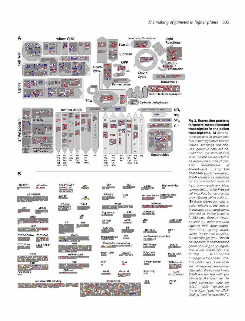
A detailed view on gene families and pathways in Arabidopsispollen has become possible due to the extensive representationof transcripts of gene families and pathways on the ATH1 array.As an example expression data for mature pollen grains com-pared with an average of vegetative tissues are overlaid on a mapof general metabolism in Arabidopsis (Fig. 3A). In accordancewith results of the GO analyses, a high proportion of genesinvolved in cell wall and lipid metabolism are up-regulated and theopposite was found for transcripts involved in energy metabolism.While the absence or down-regulation for transcripts of lightreactions and photorespiration is anticipated, the same tendency
is found in processes like glycolysis and mitochondrial electrontransport. Considering the fast growth rate of pollen tubes, howthe energy needed is provided becomes an imminent question.
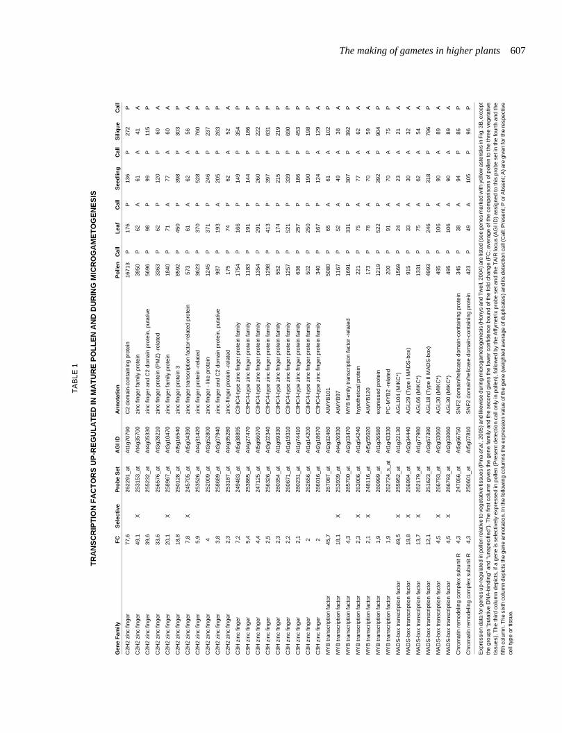
A detailed analysis of transcription factor families hints at animportant role for non-classical MADS-box genes, showing anover-representation of pollen-expressed and enriched genes(Fig. 3B). Interestingly, some of these non-classical type I andMIKC* genes have also been detected as specifically expressedduring reproductive development in a recent study using ATH1GeneChips to profile three stages of Arabidopsis flower and fruitdevelopment (Hennig et al., 2004). As the authors note, their setof identified floral-specific genes is dominated by genes ex-pressed in pollen. The exact functions of the non-classical MADS-box genes are not understood and MADS-box genes are not theonly class of transcription factors with enriched transcripts inpollen. We combined and re-analyzed data sets onmicrogametogenesis with our comparative analysis with the re-striction of different ecotypes being used in these studies. As aresult, we identified transcription factors up-regulated during thedevelopment of the male gametophyte that show at the same timehigher expression levels in mature pollen, when compared withvegetative tissues (Fig. 3B and Table 1). These transcriptionfactors might be the ones controlling de novo transcription inpollen germination and thus should constitute possible primarytargets for reverse-genetic approaches.
Pollen might not only serve as a model to study cell growth andmorphogenesis, but also, by omission, of cell cycle control. Thevegetative nucleus of pollen is thought to be arrested in G1 of thecell cycle. Again, we have used a combination of pollen expres-sion data with a recent schematic overview of the mechanisticregulation of the G1-S and the G2-M transition in plants (Pina etal., 2005), which indicates that the block is achieved by a combi-nation of absence of transcripts encoding essential proteins forthe G1-S transition (CycD, E2F-DP) and an up-regulation ofpotential repressors (DEL3) and of potential factors of increasedcell-cycle duration (CKS2). Surprisingly, pollen seems to featuremost of the transcripts needed for the G2-M transition, thoughprobably kept in their inactive state through a relative downregulation of the CDC25 phosphatase. In support of these results,Hennig et al.(2004) have come to the conclusion that S-Phasegenes were under-represented and G2/M-Phase genes enrichedin the set of genes they identified as specifically expressed duringreproduction. In the light of G2 karyogamy in Arabidopsis thismight indicate a key role for pollen derived transcripts or proteinsduring the first mitosis after fertilization, although it is unclear howthe transcripts or proteins would get to the egg cell unless they aresomehow accumulated in the sperm cells.
A surprising finding was the apparent inactivation of small RNApathways in mature pollen (Pina et al., 2005). The analysis of theexpression of genes involved in small RNA pathways in Arabidopsisrevealed that all of the 15 transcripts analyzed, including thoseencoding Argonaute 1,2,4 and 7, Dicer-like 1-3 and RNA-depen-dent RNA polymerase 1,2 and 6, were called Absent in pollen,while the majority of them were expressed in the vegetativetissues. In addition, a specific down regulation of these transcriptsoccurs during microgametogenesis. The apparent absence ofsmall RNA pathways in Arabidopsis mature pollen would affect allsmall RNA pathways known in plants (Baulcombe, 2004), includ-ing the defence against viruses through siRNAs, the regulation of
The making of gametes in higher plants 605
Fig 3. Expression patterns
for general metabolism and
transcription in the pollen
transcriptome. (A) Gene ex-pression data in pollen rela-tive to the vegetative tissuesleaves, seedlings and siliq-ues (genomic data set de-rived from the study of Pinaet al., (2005) are depicted inan overlay on a map of gen-eral metabolism inArabidopsis using theMAPMAN tool (Thimm et al.,2004). Genes are symbolizedby color-encoded squares(red, down-regulation; blue,up-regulation; white, Presentcall in pollen, but no change;grey, Absent call in pollen).(B) Gene expression data inpollen relative to the vegeta-tive tissues on a map of genesinvolved in transcription inArabidopsis. Genes are sym-bolized by color-encodedsquares (red, down-regula-tion; blue, up-regulation;white, Present call in pollen,but no change; grey, Absentcall in pollen). In addition thosegenes showing an up-regula-tion in this comparison andduring Arabidopsismicrogametogenesis (ma-ture pollen versus uninucle-ate microspores; re-analyzeddata set of (Honys and Twell,2004) are marked with yel-low asterisks and their de-tailed expression data arelisted in table 1 (except forthe groups “putative DNA-binding” and “unspecified”).
A
B
606 L. Boavida et al.
gene expression through miRNAs and the condensation of chro-matin into heterochromatin. This absence of small RNA pathwaysin mature pollen would provide new possible explanations forgenetic and epigenetic phenomena found in the male gametophyteand during/after fertilization.
Though not on a genomic scale, smaller studies in otherspecies, e.g. like those on Petunia (Guyon et al., 2000; Cnudde etal., 2003) and lily (Wang et al., 2004), are complementing theArabidopsis data sets and reveal new aspects of pollen geneticsand physiology. Transcriptomic studies are providing a quantumleap in information available for the male gametophyte.
The female reproductive organ- pistil and ovule devel-opment
Thorough descriptions of the molecular and genetic basis of thefemale organ development Arabidopsis can be found in recentreviews (Gasser et al., 1998; Skinner et al., 2004).
Unlike in other groups of plants, ovules in Angiosperms areenclosed within a carpel and during seed development theyproduce an embryo-nourishing endosperm (Friedman, 2001b).The evolution of a closed carpel (syncarpy) is believed to be oneof the major evolutionary changes, which led to the reproductivesuccess of angiosperms. Carpels are proposed to have evolvedfrom ancestral foliar organs or bract-like structures (Bowman et al.,1999). Carpels may be associated or not in the pistil, or gynoecium,which is the female reproductive unit of the angiosperm flower.Pistils differ widely in form between species (Endress and Igersheim,1999). Despite the differences in the overall morphology, a set ofcommon structures, characterized by cell types with unique prop-erties, can be distinguished. In Arabidopsis, at maturity, mostgynoecia are composed of (1) a basal ovary, usually with fusedcarpels, which encloses the ovules bearing the female gameto-phyte, (2) a short solid style, which places the stigmatic surface inclose contact with the anthers and (3) an apical stigma composedof numerous elongated epidermal cells, where pollen grains ad-here and germinate (Sessions and Zambryski, 1995; Bowman etal., 1999). The style varies in length and appearance in associationwith specific pollination strategies (Barrett, 2002). The transmittingtract is a special layer of cells, which originates in the stigma andspans the entire organ, secreting a mucilaginous extracellularmatrix. After fertilization a prominent elongation of the ovaryprecedes the dispersal of the mature seeds (Ferrandiz et al., 1999).
In Arabidopsis pistil development starts with a ring-shapedprimordium by stage 6 of flower development. The gynoeciumcontinues to elongate and develops as an open cylinder with amedial ridge, where the placental and septum will form (Fig. 4 A,B). At stage 9, rows of ovules arise from the placental tissue alongthe margins of the site of carpel fusion, the septum. By stage 10 theseptum is formed, resulting in a gynoecium with two congenitallyfused carpels. At stage 11, the stigmatic papillae differentiate andovules develop a funiculus and integument. At stage 12 thegynoecium grows, the transmitting tract differentiates and ovulesare completed. Stage 13 defines anthesis, the gynoecium comesto maturity and the flower opens (Smyth et al., 1990; Sessions,1997; Ferrandiz et al., 1999).
Ovules are simple structures, which consist of three elements:at the top, the nucellus harbors the haploid embryo sac or femalegametophyte, a central chalaza, which is characterized by the
integuments initiating at its flanks and at the bottom and thefuniculus or stalk that connects the ovule to the carpel tissue (Fig.4G). The ovules are initiated from divisions occurring in thesubepidermal tissue of the placenta and cell proliferation gives riseto finger-like structures that will differentiate in an ovule (Fig. 4 C-E) (Jenik and Irish, 2000). The integuments are two cell layers thatwill give rise to the seed coat. They cover the nucellus, forming themycropyle, a small aperture through which the pollen tube willpenetrate (Fig. 4G). The outer integument grows asymmetrically,shaping the typical curvature to the ovule (Fig. 4G).
Identically to other floral organs, ovule identity is regulated byMADS-box genes. In tobacco the two mutants Mgr3 and Mgr9have carpelloid structures instead of ovules and in Petunia the co-suppression of the two genes FLORAL BINDING PROTEIN7(FBP7) and FBP11 resulted in a similar phenotype (Evans andMalmberg, 1989; Angenent et al., 1995). Furthermore the ectopicexpression of FBP11 led to ectopic ovuloid expression (Colomboet al., 1995). The identification of these and many other flowerorgan mutants contributed to elucidate much of the molecular andgenetic mechanism of floral organ development, but it should bekept in mind that many of these mutants show pleiotropic effects inearly stages of flower development and thus the elucidation of theirtrue effects may be partially disguised.
The female gametophyte - the embryo sac
The female gametophyte supports pollen tube growth down tothe ovule, directing the sperm cells to the egg cell and central cell,up to seed, embryo and endosperm development (Chaudhury andBerger, 2001; Chaudhury et al., 2001; Higashiyama, 2002; Johnsonand Preuss, 2002; Higashiyama et al., 2003). Sporophytic muta-tions characterized by female sterility, such as megaspore mothercell development and meiosis are developmental processes origi-nated in the surrounding sporophytic tissues (Gasser et al., 1998;Grossniklaus and Schneitz, 1998). On the other hand, systematicscreens based on two essential criteria, abnormal seed set anddistorted Mendelian segregation, identified a number of gameto-phytic mutations disrupting distinct steps of embryo sac ontogen-esis in Arabidopsis and maize, indicating the requirement ofhaploid expressed genes (Christensen et al., 1997; Drews et al.,1998; Siddiqi et al., 2000; Christensen et al., 2002).
The ovule primordium initiates as an outgrowth that emergesfrom the carpel placental tissue (Fig. 4 B-F). Megasporogenesisbegins with the differentiation of an archeosporial cell in thehypodermal cell layer at the distal end of the nucellus (Fig. 4D).Usually only a single cell differentiates. In the mac1 mutant ofmaize several hypodermal cells develop into archeosporial cells,suggesting that this gene may be required to suppress the differ-entiation in neighboring cells and putatively controlling the switchof the hypodermal cells from vegetative to meiotic (Sheridan et al.,1996). In general, megasporogenesis in Arabidopsis involves thedifferentiation of one of these cells in the megaspore mother cell(MMC) in the nucellus, which undergoes meiotic reduction givingrise to four haploid nuclei (Fig. 4 A, C-E). SPOROCYTELESS(SPL) blocks the transition from archeosporial cell into amegasporocyte in ovules (Yang et al., 1999). These defects maybe related with the lack of support and nutrient supply to the femalegametophyte (Reiser et al., 1995; Elliott et al., 1996; Klucher et al.,1996). In agreement with the role of integuments in the control of
The making of gametes in higher plants 607
Gen
e F
amily
FC
Sel
ecti
veP
rob
e S
etA
GI I
DA
nn
ota
tio
nP
olle
nC
all
Lea
fC
all
See
dlin
gC
all
Sili
qu
eC
all
C2H
2 zi
nc fi
nger
77,6
2622
91_a
tA
t1g7
0790
C2
dom
ain-
cont
aini
ng p
rote
in16
713
P17
6P
136
P27
2P
C2H
2 zi
nc fi
nger
49,1
X25
3153
_at
At4
g357
00zi
nc fi
nger
fam
ily p
rote
in39
50P
62A
61A
41A
C2H
2 zi
nc fi
nger
39,6
2552
32_a
tA
t4g0
5330
zinc
fing
er a
nd C
2 do
mai
n pr
otei
n, p
utat
ive
5696
P98
P99
P11
5P
C2H
2 zi
nc fi
nger
33,6
2565
76_a
tA
t3g2
8210
zinc
fing
er p
rote
in (
PM
Z)
-rel
ated
3363
P62
P12
0P
60A
C2H
2 zi
nc fi
nger
20,1
X25
8967
_at
At3
g104
70zi
nc fi
nger
fam
ily p
rote
in18
40P
71A
77A
60A
C2H
2 zi
nc fi
nger
18,8
2501
28_a
tA
t5g1
6540
zinc
fing
er p
rote
in 3
8592
P45
0P
398
P30
3P
C2H
2 zi
nc fi
nger
7,8
X24
5705
_at
At5
g043
90zi
nc fi
nger
tran
scrip
tion
fact
or-r
elat
ed p
rote
in57
3P
61A
62A
56A
C2H
2 zi
nc fi
nger
5,9
2535
26_a
tA
t4g3
1420
zinc
fing
er p
rote
in -
rela
ted
3623
P37
0P
528
P76
0P
C2H
2 zi
nc fi
nger
425
2009
_at
At3
g528
00zi
nc fi
nger
- li
ke p
rote
in12
45P
371
P24
6P
237
P
C2H
2 zi
nc fi
nger
3,8
2586
89_a
tA
t3g0
7940
zinc
fing
er a
nd C
2 do
mai
n pr
otei
n, p
utat
ive
987
P19
3A
205
P26
3P
C2H
2 zi
nc fi
nger
2,3
2531
87_a
tA
t4g3
5280
zinc
-fin
ger
prot
ein
-rel
ated
175
P74
P62
A52
A
C3H
zin
c fin
ger
7,2
2494
83_a
tA
t5g3
8895
C3H
C4-
type
zin
c fin
ger
prot
ein
fam
ily17
54P
166
P14
9P
354
P
C3H
zin
c fin
ger
5,4
2538
65_a
tA
t4g2
7470
C3H
C4-
type
zin
c fin
ger
prot
ein
fam
ily11
83P
191
P14
4P
186
P
C3H
zin
c fin
ger
4,4
2471
25_a
tA
t5g6
6070
C3H
C4-
type
zin
c fin
ger
prot
ein
fam
ily13
54P
291
P26
0P
222
P
C3H
zin
c fin
ger
2,5
2563
26_a
tA
t3g0
2340
C3H
C4-
type
zin
c fin
ger
prot
ein
fam
ily12
98P
413
P39
7P
631
P
C3H
zin
c fin
ger
2,3
2603
54_a
tA
t1g6
9330
C3H
C4-
type
zin
c fin
ger
prot
ein
fam
ily55
2P
174
P21
5P
219
P
C3H
zin
c fin
ger
2,2
2606
71_a
tA
t1g1
9310
C3H
C4-
type
zin
c fin
ger
prot
ein
fam
ily12
57P
521
P33
9P
690
P
C3H
zin
c fin
ger
2,1
2602
31_a
tA
t1g7
4410
C3H
C4-
type
zin
c fin
ger
prot
ein
fam
ily63
6P
257
P18
6P
453
P
C3H
zin
c fin
ger
226
2656
_at
At1
g142
00C
3HC
4-ty
pe z
inc
finge
r pr
otei
n fa
mily
502
P25
0P
190
P19
8P
C3H
zin
c fin
ger
226
6016
_at
At2
g186
70C
3HC
4-ty
pe z
inc
finge
r pr
otei
n fa
mily
340
P16
7P
124
A12
9A
MY
B tr
ansc
riptio
n fa
ctor
45,7
2670
87_a
tA
t2g3
2460
AtM
YB
101
5080
P65
A61
A10
2P
MY
B tr
ansc
riptio
n fa
ctor
18,1
X25
3939
_at
At4
g269
30A
tMY
B97
1167
P52
A49
A38
A
MY
B tr
ansc
riptio
n fa
ctor
4,3
2657
00_a
tA
t2g0
3470
MY
B fa
mily
tran
scrip
tion
fact
or -
rela
ted
1691
P33
1P
307
P39
2P
MY
B tr
ansc
riptio
n fa
ctor
2,3
X26
3006
_at
At1
g542
40hy
poth
etic
al p
rote
in22
1P
75A
77A
62A
MY
B tr
ansc
riptio
n fa
ctor
2,1
X24
8116
_at
At5
g550
20A
tMY
B12
017
3P
78A
70A
59A
MY
B tr
ansc
riptio
n fa
ctor
1,9
2609
99_a
tA
t1g2
6580
expr
esse
d pr
otei
n12
19P
522
P39
2P
904
P
MY
B tr
ansc
riptio
n fa
ctor
1,9
2627
24_s
_at
At1
g433
30P
C-M
YB
2 -r
elat
ed20
0P
91A
70A
75P
MA
DS
-box
tran
scrip
tion
fact
or49
,5X
2559
52_a
tA
t1g2
2130
AG
L104
(M
IKC
*)15
69P
24A
23A
21A
MA
DS
-box
tran
scrip
tion
fact
or19
,8X
2669
94_a
tA
t2g3
4440
AG
L29
(Typ
e I M
AD
S-b
ox)
915
P33
A30
A32
A
MA
DS
-box
tran
scrip
tion
fact
or13
,7X
2621
79_a
tA
t1g7
7980
AG
L66
(MIK
C*)
1331
P75
A62
A54
A
MA
DS
-box
tran
scrip
tion
fact
or12
,125
1623
_at
At3
g573
90A
GL1
8 (T
ype
II M
AD
S-b
ox)
4993
P24
6P
318
P79
6P
MA
DS
-box
tran
scrip
tion
fact
or4,
5X
2667
93_a
tA
t2g0
3060
AG
L30
(MIK
C*)
495
P10
6A
90A
89A
MA
DS
-box
tran
scrip
tion
fact
or4,
5X
2667
93_a
tA
t2g0
3060
AG
L30
(MIK
C*)
495
P10
6A
90A
89A
Chr
omat
in r
emod
elin
g co
mpl
ex s
ubun
it R
4,3
2470
56_a
tA
t5g6
6750
SN
F2
dom
ain/
helic
ase
dom
ain-
cont
aini
ng p
rote
in34
5P
38A
94P
86P
Chr
omat
in r
emod
elin
g co
mpl
ex s
ubun
it R
4,3
2506
01_a
tA
t5g0
7810
SN
F2
dom
ain/
helic
ase
dom
ain-
cont
aini
ng p
rote
in42
3P
49A
105
P96
P
Exp
ress
ion
data
for g
enes
up-
regu
late
d in
pol
len
rela
tive
to v
eget
ativ
e tis
sues
(Pin
a et
al.,
200
5) a
nd li
kew
ise
durin
g m
icro
gam
etog
enes
is (H
onys
and
Tw
ell,
2004
) are
list
ed (s
ee g
enes
mar
ked
with
yel
low
ast
eris
ks in
Fig
. 3B
, exc
ept
the
grou
ps “
puta
tive
DN
A-b
indi
ng”
and
“uns
peci
fied”
). T
he fi
rst c
olum
n gi
ves
the
gene
fam
ily a
nd th
e se
cond
giv
es th
e lo
wer
con
fiden
ce b
ound
of t
he fo
ld c
hang
e (F
C; a
vera
ge o
f the
com
paris
ons
of p
olle
n to
the
thre
e ve
geta
tive
tissu
es).
The
third
col
umn
depi
cts,
if a
gen
e is
sel
ectiv
ely
expr
esse
d in
pol
len
(Pre
sent
det
ectio
n ca
ll on
ly in
pol
len)
, fol
low
ed b
y th
e A
ffym
etrix
pro
be s
et a
nd th
e T
AIR
locu
s (A
GI I
D)
assi
gned
to th
is p
robe
set
in th
e fo
urth
and
the
fifth
col
umn.
The
six
th c
olum
n de
pict
s th
e ge
ne a
nnot
atio
n. In
the
follo
win
g co
lum
ns th
e ex
pres
sion
val
ue o
f the
gen
e (w
eigh
ted
aver
age
of d
uplic
ates
) and
its
dete
ctio
n ca
ll (C
all:
Pre
sent
; P o
r Abs
ent;
A) a
re g
iven
for t
he re
spec
tive
cell
type
or
tissu
e.
TA
BLE
1
TR
AN
SC
RIP
TIO
N F
AC
TO
RS
UP
-RE
GU
LA
TE
D IN
MA
TU
RE
PO
LL
EN
AN
D D
UR
ING
MIC
RO
GA
ME
TO
GE
NE
SIS

608 L. Boavida et al.
meiotic progression is the sterile apetala (sap ) mutant, wheremegasporogenesis is arrested during or just after meiosis. Al-though the SAP gene is expressed initially in the nucellar region,it switches to a prominent integument expression later on (Byzovaet al., 1999). The DYAD gene was reported to be female specificand meiosis is arrested at the dyad stage (Siddiqi et al., 2000). Thefour haploid cells are surrounded by a very thin cell wall aftermeiosis, with plasmodesmata connecting the chalazal megasporeto the neighbouring nucellar cells, suggesting that it may provide apositional signal that promotes the degeneration of the otherspores to undergo cell death. At the beginning of meiosis in
Arabidopsis the MMC shows cytoplasmic polarity, thought to beimportant to determine the functional megaspore (Bajon et al.,1999).
Megagametogenesis begins when the surviving megasporegoes through three rounds of mitosis to form a two-nucleated, four-nucleated (Fig. 4E) and subsequently eight-nucleated embryo sac(Fig. 4A; stage FG1-FG4). Different patterns of gametophytedevelopment have been described in several species, differingfrom each other by variations on cytokinesis during meiosis, on thenumber of mitotic divisions and on the cellularization pattern.Arabidopsis and maize megasporogenesis follows the monosporic
Fig. 4. The development of embryo sac and the ovule. (A) Major events on embryo sac ontogeny. Categories represent checkpoints on embryo sacdevelopment based on mutant analysis. mmc megaspore mother cell; fm female megaspore;CC central cell; EC egg cell; SC synergid cell; pn polar nuclei;ac antipodal cell; sn secondary nucleus; ch chalaza; mp mycropyle; f funiculus. Adapted from Drews et al. (1998) and Yadegari and Drews (2004).(B-E) Embryo sac development in O. lutea. The ovary is divided by 3 parietal placentas (B). The apical cell of the nucellus (N or Nuc) divides longitudinally(C) and then anticlinally to define a central megaspore mother cell (MMC), that enters meiosis (arrow in (C,D) ). This cell gives rise to a linear tetrad (arrowsin (E)), 3 of which will regress and the top one will develop into the mature embryo sac, like depicted in (A) (category 3 on). (F-H) Ovule development.From a capsule like shape (F) ovules (Ov) bend over by differential growth of the integuments to form a pipe-like structure (G). At this stage theinteguments also grow to form the pollen tube entry point, the micropyle (mi). At later stages of mature ovules, the micropyle is sometimes covered withsecretion (H).
A
B C D
E F G H

The making of gametes in higher plants 609
Polygonum type, which is also the most common form in An-giosperms (Maheshwari, 1950; Willemse and van Went, 1984;Huang and Russell, 1992a). At the end of each meiotic stagevacuoles occupy a central location on the cells and microtubulesare thought to mediate nuclear migration to position the nuclei atthe poles of the coenocytic cell (Webb and Gunning, 1994a). Afterthe third mitotic round non-sister nuclei and the embryo sac beginto cellularize to form the final seven-cell structure. The other twonuclei (the polar nuclei) migrate to a central position. In somespecies the nuclei fuse to give rise to the secondary endospermnucleus, in others the polar nuclei just fuse partially prior tofertilization. During cell differentiation the polarity of chalazal-micropyle axis established in the beginning of the female gameto-phyte development will determine that the nuclei positioned at themicropylar end become specified to develop into an egg andsynergids cells and the chalazal nuclei give rise to the antipodalcells. Cell walls are absent in the chalazal end of the egg cell andthe synergids, allowing the direct contact of plasma membranes(Willemse and van Went, 1984; Webb and Gunning, 1994b). Thepolarity of the female gametophyte is believed to be defined by theasymmetry of the surrounding ovule layers and thus to be undersporophytic control, but no factors have yet been identified(Christensen et al., 1997).
Most of the gametophytic mutants, which do not show lesions onthe sporophytic tissues of the ovule, fall in different classescorresponding to key developmental events duringmegagametogenesis, such as mitosis, vacuole formation, nuclearfusion, cellularization and cell death (see categories in Fig. 4)(Feldmann et al., 1997; Schneitz et al., 1997; Christensen et al.,1998; Drews et al., 1998).
Conclusions
Albeit covering most of the recent info, namely the fast accumu-lating genomic and transcriptomic data, this review mostly high-lights the incompleteness of our understanding of many of thefundamental mechanisms underlying the complex puzzle of game-togenesis. Both gametophytes show developmental programs thatprepare these tissues for their future interactions. The events thatlead to gametophyte development are controlled primary by thesporophytic tissues, but are ultimately reassigned to singular cellswith a narrow set of functions to accomplish. When first releasedfrom the sporophyte, the male gametophyte is already committedto a role. The female tissues, on the other hand, form a long andcomplex pathway, branded by different molecular and cellularcontexts. The past years have seen significant advances in ourunderstanding of all these processes, revealing some of the keycellular events, but the identity of many of the molecular compo-nents and signaling pathways in which they are involved is stillmostly unknown. Moreover, the genetic and functional analysishas mostly progressed on a gene-by-gene basis and relying on therelative serendipity of genetic screens, but transcriptomics offersthe promise of major advances through a more deterministicapproach. Near future will prove these to be sound basis to a moreintegrated view of the processes of plant gametogenesis.
AcknowledgementsJAF lab is supported by FCT grants POCTI/BCI/41725/2001, POCTI/
BCI/46453/2002, POCTI/BIA-BCM/60046/2004 and POCTI/BIA-BCM/61270/2004.
References
AARTS, M.G., HODGE, R., KALANTIDIS, K., FLORACK, D., WILSON, Z.A.,MULLIGAN, B.J., STIEKEMA, W.J., SCOTT, R. and PEREIRA, A. (1997). TheArabidopsis MALE STERILITY 2 protein shares similarity with reductases inelongation/condensation complexes. Plant J 12: 615-23.
AARTS, M.G., KEIJZER, C.J., STIEKEMA, W.J. and PEREIRA, A. (1995). Molecularcharacterization of the CER1 gene of Arabidopsis involved in epicuticular waxbiosynthesis and pollen fertility. Plant Cell 7: 2115-27.
AHLERS, F., LAMBERT, J. and WIERMANN, R. (2003). Acetylation and silylationof piperidine solubilized sporopollenin from pollen of Typha angustifolia L. ZNaturforsch [C] 58: 807-11.
ALBANI, D., ALTOSAAR, I., ARNISON, P.G. and FABIJANSKI, S.F. (1991). A geneshowing sequence similarity to pectin esterase is specifically expressed indeveloping pollen of Brassica napus. Sequences in its 5' flanking region areconserved in other pollen-specific promoters. Plant Mol Biol 16: 501-13.
ALBANI, D., ROBERT, L.S., DONALDSON, P.A., ALTOSAAR, I., ARNISON, P.G.and FABIJANSKI, S.F. (1990). Characterization of a pollen-specific gene familyfrom Brassica napus which is activated during early microspore development.Plant Mol Biol 15: 605-22.
ANGENENT, G.C., FRANKEN, J., BUSSCHER, M., VAN DIJKEN, A., VAN WENT,J.L., DONS, H.J. and VAN TUNEN, A.J. (1995). A novel class of MADS boxgenes is involved in ovule development in petunia. Plant Cell 7: 1569-82.
AOUALI, N., LAPORTE, P. and CLEMENT, C. (2001). Pectin secretion anddistribution in the anther during pollen development in Lilium. Planta 213: 71-9.
ARIIZUMI, T., HATAKEYAMA, K., HINATA, K., SATO, S., KATO, T., TABATA, S.and TORIYAMA, K. (2003). A novel male-sterile mutant of Arabidopsis thaliana,faceless pollen-1, produces pollen with a smooth surface and an acetolysis-sensitive exine. Plant Mol Biol 53: 107-16.
ARONDEL, V.V., VERGNOLLE, C., CANTREL, C. and KADER, J. (2000). Lipidtransfer proteins are encoded by a small multigene family in Arabidopsisthaliana. Plant Sci 157: 1-12.
ATANASSOV, I., RUSSINOVA, E., ANTONOV, L. and ATANASSOV, A. (1998).Expression of an anther-specific chalcone synthase-like gene is correlated withuninucleate microspore development in Nicotiana sylvestris. Plant Mol Biol 38:1169-78.
BAJON, C., HORLOW, C., MOTAMAYOR, J.C., SAUVANET, A. and ROBERT, D.(1999). Megasporogenesis in Arabidopsis thaliana L.: and ultrastructural study.Sex Plant Reprod 12: 99-109.
BARRETT, S.C. (2002). The evolution of plant sexual diversity. Nat Rev Genet 3:274-84.
BATE, N., SPURR, C., FOSTER, G.D. and TWELL, D. (1996). Maturation-specifictranslational enhancement mediated by the 5'-UTR of a late pollen transcript.Plant J. 10: 613-23.
BAULCOMBE, D. (2004). RNA silencing in plants. Nature 431: 356-363.
BECKER, J.D., BOAVIDA, L.C., CARNEIRO, J., HAURY, M. and FEIJO, J.A.(2003). Transcriptional profiling of Arabidopsis tissues reveals the uniquecharacteristics of the pollen transcriptome. Plant Physiol 133: 713-725.
BEDINGER, P.A. and EDGERTON, M.D. (1990). Developmental staging of maizemicrospores reveals a transition in developing microspores. Plant Physiol 92:474-479.
BIH, F.Y., WU, S.S., RATNAYAKE, C., WALLING, L.L., NOTHNAGEL, E.A. andHUANG, A.H. (1999). The predominant protein on the surface of maize pollenis an endoxylanase synthesized by a tapetum mRNA with a long 5' leader. J BiolChem 274: 22884-94.
BIRD, J., PORTER, E.K. and DICKINSON, H.G. (1983). Events in the cytoplasmduring male meiosis in Lilium. J Cell Sci 59: 27-42.
BOAVIDA, L.C., VIEIRA, A.M., BECKER, J.D. and FEIJO, J.A. (2005). Gameto-phyte interaction and sexual reproduction: how plants make a zygote. Int. J.Dev. Biol. 49: 615-632.
BOWMAN, J.L., BAUM, S.F., ESHED, Y., PUTTERILL, J. and ALVAREZ, J. (1999).Molecular genetics of gynoecium development in Arabidopsis. Curr Top DevBiol 45: 155-205.
BROOKS, J. and SHAW, G. (1968). Chemical structure of the exine of pollen walls

610 L. Boavida et al.
and a new function for carotenoids in nature. Nature 219: 532-3.
BROWN, R.C. and LEMMON, B.E. (1991). Pollen developement in orchid. 3. Anovel generative pole microtubule system predicts unequal pollen mitosis. JCell Sci 99: 273-281.
BROWN, S.M. and CROUCH, M.L. (1990). Characterization of a gene familyabundantly expressed in Oenothera organensis pollen that shows sequencesimilarity to polygalacturonase. Plant Cell 2: 263-74.
BUCCIAGLIA, P.A., ZIMMERMANN, E. and SMITH, A.G. (2003). Functionalanalysis of a beta-1,3-glucanase gene (Tag1 ) with anther-specific RNA andprotein accumulation using antisense RNA inhibition. J Plant Physiol 160: 1367-73.
BURBULIS, I.E., IACOBUCCI, M. and SHIRLEY, B.W. (1996). A null mutation in thefirst enzyme of flavonoid biosynthesis does not affect male fertility in Arabidopsis.Plant Cell 8: 1013-25.
BYZOVA, M.V., FRANKEN, J., AARTS, M.G., DE ALMEIDA-ENGLER, J., ENGLER,G., MARIANI, C., VAN LOOKEREN CAMPAGNE, M.M. and ANGENENT, G.C.(1999). Arabidopsis STERILE APETALA, a multifunctional gene regulatinginflorescence, flower and ovule development. Genes Dev 13: 1002-14.
CAFFREY, M., WERNER, B.G. and PRIESTLEY, D.A. (1987). A crystalline lipidphase in a dry biological system: evidence from X-ray diffraction analysis ofTypha latifolia pollen. Biochim Biophys Acta 921: 124-34.
CARYL, A.P., JONES, G.H. and FRANKLIN, F.C. (2003). Dissecting plant meiosisusing Arabidopsis thaliana mutants. J Exp Bot 54: 25-38.
CHAUDHURY, A.M. and BERGER, F. (2001). Maternal control of seed develop-ment. Semin Cell Dev Biol 12: 381-6.
CHAUDHURY, A.M., KOLTUNOW, A., PAYNE, T., LUO, M., TUCKER, M.R.,DENNIS, E.S. and PEACOCK, W.J. (2001). Control of early seed development.Annu Rev Cell Dev Biol 17: 677-99.
CHEN, Y.C. and MCCORMICK, S. (1996). Sidecar pollen, an Arabidopsis thalianamale gametophytic mutant with aberrant cell divisions during pollen develop-ment. Development 122: 3243-53.
CHEUNG, A.Y. (2001). Imaging elongating pollen tubes by green fluorescentprotein. Sex. Plant Reprod. 14: 9-14.
CHEUNG, A.Y., CHEN, C.Y.H., TAO, L.Z. andREYEVA, T., TWELL, D. and WU,H.M. (2003). Regulation of pollen tube growth by Rac-like GTPases. J Exp Bot54: 73-81.
CHEVALIER, F., MARTIN, O., ROFIDAL, V., DEVAUCHELLE, A.D., BARTEAU, S.,SOMMERER, N. and ROSSIGNOL, M. (2004). Proteomic investigation ofnatural variation between Arabidopsis ecotypes. Proteomics 4: 1372-1381.
CHRISTENSEN, C.A., GORSICH, S.W., BROWN, R.H., JONES, L.G., BROWN, J.,SHAW, J.M. and DREWS, G.N. (2002). Mitochondrial GFA2 is required forsynergid cell death in Arabidopsis. Plant Cell 14: 2215-2232.
CHRISTENSEN, C.A., KING, E.J., JORDAN, J.R. and DREWS, G.N. (1997).Megagametogenesis in Arabidopsis wild type and the Gf mutant. Sex. PlantReprod. 10: 49-64.
CNUDDE, F., MORETTI, C., PORCEDDU, A., PEZZOTTI, M. and GERATS, T.(2003). Transcript profiling on developing Petunia hybrida floral organs. Sex.Plant Reprod. 16: 77-85.
COLOMBO, L., FRANKEN, J., KOETJE, E., VAN WENT, J., DONS, H.J.,ANGENENT, G.C. and VAN TUNEN, A.J. (1995). The petunia MADS box geneFBP11 determines ovule identity. Plant Cell 7: 1859-68.
CROSSLEY, S.J., GREENLAND, A.J. and DICKINSON, H.G. (1995). Thecharacterisation of tapetum-specific cDNAs isolated from a Lilium henryi L.meiocyte subtractive cDNA library. Planta 196: 523-9.
CUSTERS, J.B., OLDENHOF, M.T., SCHRAUWEN, J.A., CORDEWENER, J.H.,WULLEMS, G.J. and VAN LOOKEREN CAMPAGNE, M.M. (1997). Analysis ofmicrospore-specific promoters in transgenic tobacco. Plant Mol Biol 35: 689-99.
DA COSTA-NUNES, J.A. and GROSSNIKLAUS, U. (2004). Unveiling the gene-expression profile of pollen. Genome Biology 5: 205.
DARWIN, C. (1862). Fertlization of orchids by insects. Murray, J., London.
DEBOO, G.B., ALBERTSEN, M.C. and TAYLOR, L.P. (1995). Flavanone 3-Hydroxylase Transcripts and Flavonol Accumulation Are Temporally Coordi-nate in Maize Anthers. Plant J. 7: 703-713.
DICKINSON, H.G., ELLEMAN, C.J. and DOUGHTY, J. (2000). Pollen coatings -
chimaeric genetics and new functions. Sex Plant Reprod 12: 302-309.
DODDS, P.N., BONIG, I., DU, H., RODIN, J. andERSON, M.A., NEWBIGIN, E. andCLARKE, A.E. (1993). S-RNase gene of Nicotiana alata is expressed indeveloping pollen. Plant Cell 5: 1771-82.
DOUGHTY, J., HEDDERSON, F., MCCUBBIN, A. and DICKINSON, H. (1993).Interaction between a coating-borne peptide of the Brassica pollen grain andstigmatic S (self-incompatibility)-locus-specific glycoproteins. Proc Natl AcadSci USA 90: 467-71.
DREWS, G.N., LEE, D. and CHRISTENSEN, C.A. (1998). Genetic analysis offemale gametophyte development and function. Plant Cell 10: 5-17.
EADY, C., LINDSEY, K. and TWELL, D. (1995). The significance of microsporedivision and division symmetry for vegetative cell-specific transcription andgenerative cell differentiation. Plant Cell 7: 65-74.
ELLIOTT, R.C., BETZNER, A.S., HUTTNER, E., OAKES, M.P., TUCKER, W.Q.,GERENTES, D., PEREZ, P. and SMYTH, D.R. (1996). AINTEGUMENTA, anAPETALA2 -like gene of Arabidopsis with pleiotropic roles in ovule develop-ment and floral organ growth. Plant Cell 8: 155-68.
ENDRESS, P.K. and IGERSHEIM, A. (1999). Gynoecium diversity and systematicsof the basal eudicots. Bot J Linn Soc 130: 305-393.
ENGEL, M.L., CHABOUD, A., DUMAS, C. and MCCORMICK, S. (2003). Spermcells of Zea mays have a complex complement of mRNAs. Plant J. 34: 697-707.
EVANS, D.E., TAYLOR, P.E., SINGH, M.B. and KNOX, R.B. (1992). The interrela-tionship between the accumulation of lipids, protein and the level of acyl carrierprotein during the development of Brassica Napus L. Pollen. Planta 186: 343-354.
EVANS, P.T. and MALMBERG, R.L. (1989). Alternative pathways of tobaccoplacental development: time of commitment and analysis of a mutant. Dev Biol136: 273-83.
EYAL, Y., CURIE, C. and MCCORMICK, S. (1995). Pollen specificity elementsreside in 30 bp of the proximal promoters of two pollen-expressed genes. PlantCell 7: 373-84.
FAURE, J.E., ROTMAN, N., FORTUNE, P. and DUMAS, C. (2002). Fertilization inArabidopsis thaliana wild type: developmental stages and time course. Plant J30: 481-8.
FEIJO, J.A., COSTA, S.S., PRADO, A.M., BECKER, J.D. and CERTAL, A.C.(2004). Signalling by tips. Curr Opin Plant Biol 7: 589-598.
FEIJO, J.A., MALHO, R. and OBERMEYER, G. (1995). Ion dynamics and itspossible role during in vitro pollen germination and tube growth. Protoplasma187: 155-167.
FELDMANN, K.A., COURY, D.A. and CHRISTIANSON, M.L. (1997). Exceptionalsegregation of a selectable marker (KanR ) in Arabidopsis identifies genesimportant for gametophytic growth and development. Genetics 147: 1411-22.
FERRANDIZ, C., PELAZ, S. and YANOFSKY, M.F. (1999). Control of carpel andfruit development in Arabidopsis. Ann Rev Biochem 68: 321-54.
FIEBIG, A., KIMPORT, R. and PREUSS, D. (2004). Comparisons of pollen coatgenes across Brassicaceae species reveal rapid evolution by repeat expansionand diversification. Proc Natl Acad Sci USA 101: 3286-91.
FIEBIG, A., MAYFIELD, J.A., MILEY, N.L., CHAU, S., FISCHER, R.L. and PREUSS,D. (2000). Alterations in CER6, a gene identical to CUT1, differentially affectlong-chain lipid content on the surface of pollen and stems. Plant Cell 12: 2001-8.
FITZGERALD, M.A. and KNOX, R.B. (1995). Initiation of premexine in freeze-substituted microspores of Brassica campestris. Sex. Plant Reprod. 8: 99-104.
FOSTER, G.D., ROBINSON, S.W., BLUNDELL, R.P., ROBERTS, M.R. and HODGE,R. (1992). A Brassica napus mRNA encoding a protein homologous tophospholipid transfer proteins, is expressed especifically in the tapetum anddeveloping microspores. Plant Sci 84: 184-192.
FRANKEL, R., IZHAR, S. and NITSAN, J. (1969). Timing of callase activity andcytoplasmic male sterility in Petunia. Biochem Genet 3: 451-5.
FURNESS, C.A. and RUDALL, P.J. (2001). The tapetum in basal angiosperms:Early diversity. Int J Plant Sci 162: 375-392.
GASSER, C.S., BROADHVEST, J. and HAUSER, B.A. (1998). Genetic analysis ofovule development. Annu Rev Plant Phys 49: 1-24.
GEROLA, P.D., MOL, C.A., NEWBIGIN, E. and LUSH, W.M. (2000). Regulation ofLAT52 promoter activity during pollen tube growth through the pistil of Nicotiana

The making of gametes in higher plants 611
alata. Sex. Plant Reprod. 12: 347-352.
GOLDBERG, R.B., BEALS, T.P. and SANDERS, P.M. (1993). Anther development:basic principles and practical applications. Plant Cell 5: 1217-29.
GROSSNIKLAUS, U. and SCHNEITZ, K. (1998). The molecular and genetic basisof ovule and megagametophyte development. Semin Cell Dev Biol 9: 227-38.
GUERRERO, F.D., CROSSLAND, L., SMUTZER, G.S., HAMILTON, D.A. andMASCARENHAS, J.P. (1990). Promoter sequences from a maize pollen-specific gene direct tissue-specific transcription in tobacco. Mol Gen Genet224: 161-8.
GUILFORD, W.J., SCHENEIDER, D.M., LABOVITZ, J. and OPELLA, S.J. (1988).High resolution solid state 13C NMR spectroscopy of sporopollenins fromdifferent plant taxa. Plant Physiol 86: 851-855.
GUYON, V.N., ASTWOOD, J.D., GARNER, E.C., DUNKER, A.K. and TAYLOR,L.P. (2000). Isolation and characterization of cDNAs expressed in the earlystages of flavonol-induced pollen germination in Petunia. Plant Physiol 123:699-710.
HAMILTON, D.A., ROY, M., RUEDA, J., SINDHU, R.K., SANFORD, J. andMASCARENHAS, J.P. (1992). Dissection of a pollen-specific promoter frommaize by transient transformation assays. Plant Mol Biol 18: 211-8.
HAMILTON, D.A., SCHWARZ, Y.H. and MASCARENHAS, J.P. (1998). A monocotpollen-specific promoter contains separable pollen-specific and quantitativeelements. Plant Mol Biol 38: 663-9.
HANSON, D.D., HAMILTON, D.A., TRAVIS, J.L., BASHE, D.M. andMASCARENHAS, J.P. (1989). Characterization of a pollen-specific cDNA clonefrom Zea mays and its expression. Plant Cell 1: 173-9.
HATAKEYAMA, K., ISHIGURO, S., OKADA, K., TAKASAKI, T. and HINATA, K.(2003). Antisense inhibition of a nuclear gene, BrDAD1, in Brassica causesmale sterility that is restorable with jasmonic acid treatment. Mol. Breed. 11:325-336.
HEMSLEY, A.R., GRIFFITHS, P.C., MATHIAS, R. and MOORE, S.E.M. (2003). Amodel for the role of surfactants in the assembly of exine sculpture. Grana 42:38-42.
HENNIG, L., GRUISSEM, W., GROSSNIKLAUS, U. and KOHLER, C. (2004).Transcriptional programs of early reproductive stages in Arabidopsis. PlantPhysiol 135: 1765-1775.
HERDT, E., SUTFELD, R. and WIERMANN, R. (1978). The occurrence of enzymesinvolved in phenylpropanoid metabolism in the tapetum fraction of anthers.Cytobiologie 17: 433-41.
HESLOP-HARRISON, J. (1963). Ultrastructural aspects of differentiation in sporog-enous tissue. Symp Soc Exp Biol 17: 315-40.
HESLOP-HARRISON, J. (1966). Cytoplasmic connections between angiospermmeiocytes. Ann. Bot. 30: 221-230.
HESLOP-HARRISON, J. (1968a). Pollen wall development. The succession ofevents in the growth of intricately patterned pollen walls is described anddiscussed. Science 161: 230-7.
HESLOP-HARRISON, J. (1968b). Tapetal Origin of Pollen-Coat Substances inLilium. New Phytologist 67: 779-&.
HESLOP-HARRISON, J. (1971). Wall pattern formation in angiosperm microsporo-genesis. Symp Soc Exp Biol 25: 277-300.
HESLOP-HARRISON, J. (1979). An interpretation of the hydrodynamics of pollen.Am. J. Bot. 66: 737-743.
HIGASHIYAMA, T. (2002). The synergid cell: attractor and acceptor of the pollentube for double fertilization. J Plant Res 115: 149-60.
HISCOCK, S.J., DEWEY, F.M., COLEMAN, J.O.D. and DICKINSON, H.G. (1994).Identification and localization of an active cutinase in the pollen of BrassicaNapus L. Planta 193: 377-384.
HOEKSTRA, F.A. and BRUINSMA, J. (1980). Control of respiration of binucleateand trinucleate pollen under humid conditions. Plant Physiol. 48: 71-77.
HONYS, D. and TWELL, D. (2003). Comparative analysis of the Arabidopsis pollentranscriptome. Plant Physiol. 132: 640-652.
HONYS, D. and TWELL, D. (2004). Transcriptome analysis of haploid malegametophyte development in Arabidopsis. Genome Biology 5:R85.
HORVITZ, H.R. and HERSKOWITZ, I. (1992). Mechanims of asymetric cell divi-sion: Two Bs or not to Bs, that is the question. Cell 68: 237-255.
HOWDEN, R., PARK, S.K., MOORE, J.M., ORME, J., GROSSNIKLAUS, U. andTWELL, D. (1998). Selection of T-DNA-tagged male and female gametophyticmutants by segregation distortion in Arabidopsis. Genetics 149: 621-31.
HUANG, B.Q. and RUSSELL, S.D. (1992). Female germ unit: Organization,isolation and function. Int. Rev. Cytol. 140.
HULSKAMP, M., KOPCZAK, S.D., HOREJSI, T.F., KIHL, B.K. and PRUITT, R.E.(1995). Identification of genes required for pollen-stigma recognition inArabidopsis thaliana. Plant J 8: 703-14.
HULSKAMP, M., PAREKH, N.S., GRINI, P., SCHNEITZ, K., ZIMMERMANN, I.,LOLLE, S.J. and PRUITT, R.E. (1997). The STUD gene is required for male-specific cytokinesis after telophase II of meiosis in Arabidopsis thaliana. DevBiol 187: 114-24.
IRISH, V.F. (1999). Petal and stamen development. Curr Top Dev Biol 41: 133-61.
ISHIGURO, S., KAWAI-ODA, A., UEDA, J., NISHIDA, I. and OKADA, K. (2001). TheDEFECTIVE IN ANTHER DEHISCIENCE gene encodes a novel phospholi-pase A1 catalyzing the initial step of jasmonic acid biosynthesis, which synchro-nizes pollen maturation, anther dehiscence and flower opening in Arabidopsis.Plant Cell 13: 2191-209.
JENIK, P.D. and IRISH, V.F. (2000). Regulation of cell proliferation patterns byhomeotic genes during Arabidopsis floral development. Development 127:1267-76.
JENKS, M.A., TUTTLE, H.A., EIGENBRODE, S.D. and FELDMANN, K.A. (1995).Leaf epicuticular waxes of the Eceriferum mutants in Arabidopsis. Plant Physiol108: 369-377.
KANG, B.H., RANCOUR, D.M. and BEDNAREK, S.Y. (2003). The dynamin-likeprotein ADL1C is essential for plasma membrane maintenance during pollenmaturation. Plant J 35: 1-15.
KAPOOR, S., KOBAYASHI, A. and TAKATSUJI, H. (2002). Silencing of thetapetum-specific zinc finger gene TAZ1 causes premature degeneration oftapetum and pollen abortion in petunia. Plant Cell 14: 2353-67.
KISHITANI, S., YOMODA, A., KONNO, N. and TANAKA, Y. (1993). Involvement ofphenylalanine ammonia-lyase in the development of pollen in broccoli (Bras-sica-Oleracea L.). Sex. Plant Reprod. 6: 244-248.
KLUCHER, K.M., CHOW, H., REISER, L. and FISCHER, R.L. (1996). TheAINTEGUMENTA gene of Arabidopsis required for ovule and female gameto-phyte development is related to the floral homeotic gene APETALA2. Plant Cell8: 137-53.
KNOX, R.B. and HESLOP-HARRISON, J. (1970). Pollen-wall proteins: localizationand enzymic activity. J Cell Sci 6: 1-27.
KOLTUNOW, A.M., TRUETTNER, J., COX, K.H., WALLROTH, M. and GOLDBERG,R.B. (1990). Different temporal and spatial gene expression patterns occurduring anther development. Plant Cell 2: 1201-1224.
LALANNE, E. and TWELL, D. (2002). Genetic control of male germ unit organiza-tion in Arabidopsis. Plant Physiol 129: 865-75.
LAUGA, B., CHARBONNEL-CAMPAA, L. and COMBES, D. (2000). Characteriza-tion of MZm3-3, a Zea mays tapetum-specific transcript. Plant Sci 157: 65-75.
LEE, J.Y. and LEE, D.H. (2003). Use of serial analysis of gene expressiontechnology to reveal changes in gene expression in Arabidopsis pollenundergoing cold stress. Plant Physiol. 132: 517-529.
LI, J., OU-LEE, T.M., RABA, R., AMUNDSON, R.G. and LAST, R.L. (1993).Arabidopsis flavonoid mutants are hypersensitive to UV-B irradiation. Plant Cell5: 171-179.
LI, Y.Q., FALERI, C., GEITMANN, A., ZHANG, H.Q. and CRESTI, M. (1995).Immunogold localization of arabinogalactan proteins, unesterified and esteri-fied pectins in pollen grains and pollen tubes of Nicotiana-Tabacum-L.Protoplasma 189: 26-36.
LINSKENS, H.F. (1966). Die Änderung des Protein- und Enzym-Musters währendder Pollenmeiose und Pollenentwicklung. Planta 69: 79-91.
LIU, J.Q., LEGGEWIE, G., VAROTTO, S. and THOMPSON, R.D. (1999). Charac-terization of an anther-expressed protein kinase gene in the potato Solanumberthaultii and its antisense inhibition in transgenic plants. Sex Plant Reprod11: 336-346.
LIU, W.M., MEI, R., DI, X., RYDER, T.B., HUBBELL, E., DEE, S., WEBSTER, T.A.,HARRINGTON, C.A., HO, M.H., BAID, J. et al. (2002). Analysis of high densityexpression microarrays with signed-rank call algorithms. Bioinformatics 18:
612 L. Boavida et al.
1593-9.
LOMBARDERO, M., BARBAS, J.A., MOSCOSO DEL PRADO, J. and CARREIRA,J. (1994). cDNA sequence analysis of the main olive allergen, Ole e I. Clin ExpAllergy 24: 765-70.
MACKENZIE, A., HESLOP-HARRISON, J. and DICKINSON, H.G. (1967). Elimina-tion of ribosomes during meiotic prophase. Nature 215: 997-9.
MAHESHWARI, P. (1950). McGraw-Hill, New York.
MANDARON, P., NIOGRET, M.F., MACHE, R. and MONÉGER, F. (1990). In vitroprotein synthesis in isolated microspores of Zea mays at several stages ofdevelopment. Theor Appl Genet 80: 134-138.
MARIANI, C., DEBEUCKELEER, M., TRUETTNER, J., LEEMANS, J. andGOLDBERG, R.B. (1990). Induction of male-sterility in plants by a chimericribonuclease gene. Nature 347: 737-741.
MASCARENHAS, J.P. (1975). The biochemistry of angiosperm pollen develop-ment. Bot Rev 41: 259-314.
MASCARENHAS, J.P. (1989). The male gametophyte of flowering plants. PlantCell 1: 657-664.
MASCARENHAS, J.P. (1990). Gene activity during pollen development. Annu RevPlant Phys 41: 317-338.
MASCARENHAS, J.P. and BELL, E. (1970). RNA synthesis during development ofthe male gametophyte of Tradescantia. Dev Biol 21: 475-90.
MASCARENHAS, J.P. and GORALNICK, R.D. (1971). Synthesis of small molecu-lar weight RNA in the pollen tube of Tradescantia paludosa. Biochem. Biophys.Acta. 240: 56-61.
MATSUDA, N., TSUCHIYA, T., KISHITANI, S., TANAKA, Y. and TORIYAMA, K.(1996). Partial male sterility in transgenic tobacco carrying antisense and sensePAL cDNA under the control of a tapetum-specific promoter. Plant Cell Physiol.37: 215-222.
MAYFIELD, J.A., FIEBIG, A., JOHNSTONE, S.E. and PREUSS, D. (2001). Genefamilies from the Arabidopsis thaliana pollen coat proteome. Science 292:2482-5.
MAYFIELD, J.A. and PREUSS, D. (2000). Rapid initiation of Arabidopsis pollina-tion requires the oleosin-domain protein GRP17. Nat Cell Biol 2: 128-30.
MCCORMICK, S. (1993). Male gametophyte development. Plant Cell 5: 1265-1275.
MCCORMICK, S. (2004). Control of male gametophyte development. Plant Cell 16Suppl: S142-53.
MEYEROWITZ, E.M. (2002). Plants compared to animals: the broadest compara-tive study of development. Science 295: 1482-5.
MEYERS, B.C., GALBRAITH, D.W., NELSON, T. and AGRAWAL, V. (2004).Methods for transcriptional profiling in plants. Be fruitful and replicate. PlantPhysiol 135: 637-652.
MITCHELL, J.W., MANDAVA, N., WORLEY, J.F., PLIMMER, J.R. and SMITH, M.V.(1970). Brassins - a new family of plant hormones from rape pollen. Nature 225:1065-1066.
MIYAZAKI, S., SUMIKAWA, N., MISHIMA, M., KITANI, M., KUBO, M., DEMURA,T., FUKUDA, H. and HASEBE, M. (2004). Screening of receptor-like kinases ofArabidopsis involved in sexual reproduction mechanism. In Frontiers in SexualPlant Reproduction II, pp. 99. Albany, New York, USA.
MOURADOV, A., CREMER, F. and COUPLAND, G. (2002). Control of floweringtime: interacting pathways as a basis for diversity. Plant Cell 14 Suppl: S111-30.
MURPHY, D.J. and ROSS, J.H. (1998). Biosynthesis, targeting and processing ofoleosin-like proteins, which are major pollen coat components in Brassicanapus. Plant J 13: 1-16.
MUSCHIETTI, J., DIRCKS, L., VANCANNEYT, G. and MCCORMICK, S. (1994).LAT52 protein is essential for tomato pollen development: pollen expressingantisense LAT52 RNA hydrates and germinates abnormally and cannotachieve fertilization. Plant J 6: 321-38.
OLDENHOF, M.T., DEGROOT, P.F.M., VISSER, J.H., SCHRAUWEN, J.A.M. andWULLEMS, G.J. (1996). Isolation and characterization of a microspore-specificgene from tobacco. Plant Mol. Biol. 31: 213-225.
OSTERGAARD, L., PETERSEN, M., MATTSSON, O. and MUNDY, J. (2002). AnArabidopsis callose synthase. Plant Mol. Biol. 49: 559-566.
OTTAVIANO, E., SARI GORLA, M. andnMULCAHY, D.L. (1990). Pollen selection:efficiency and monitoring. Prog Clin Biol Res 344: 575-88.
PACINI, E. (1997). Tapetum character states: analytical keys for tapetum types andactivities. Canadian Journal of Botany-Revue Canadienne De Botanique 75:1448-1459.
PARK, J.H., HALITSCHKE, R., KIM, H.B., BALDWIN, I.T., FELDMANN, K.A. andFEYEREISEN, R. (2002). A knock-out mutation in allene oxide synthase resultsin male sterility and defective wound signal transduction in Arabidopsis due toa block in jasmonic acid biosynthesis. Plant J 31: 1-12.
PARK, S.K., HOWDEN, R. and TWELL, D. (1998). The Arabidopsis thalianagametophytic mutation gemini pollen1 disrupts microspore polarity, divisionasymmetry and pollen cell fate. Development 125: 3789-99.
PAXSON-SOWDERS, D.M., DODRILL, C.H., OWEN, H.A. and MAKAROFF, C.A.(2001). DEX1, a novel plant protein, is required for exine pattern formationduring pollen development in Arabidopsis. Plant Physiol 127: 1739-49.
PAXSON-SOWDERS, D.M., OWEN, H.A. and MAKAROFF, C.A. (1997). A com-parative ultrastructural analysis of exine pattern development in wild-typeArabidopsis and a mutant defective in pattern formation. Protoplasma 198: 53-65.
PEDDADA, L. and MASCARENHAS, J.P. (1975). 5S ribosomal RNA synthesisduring pollen development. Dev Growth Diff 17: 1-8.
PIFFANELLI, P., ROSS, J.H. and MURPHY, D.J. (1997). Intra- and extracellularlipid composition and associated gene expression patterns during pollendevelopment in Brassica napus. Plant J 11: 549-62.
PIFFANELLI, P., ROSS, J.H.E. and MURPHY, D.J. (1998). Biogenesis and functionof the lipidic structures of pollen grains. Sex Plant Reprod. 11: 65-80.
PINA, C., PINTO, F., FEIJÓ, J.A. and BECKER, J.D. (2005). Gene family analysisof the Arabidopsis pollen transcriptome reveals biological implications for cellgrowth, division control and gene expression regulation. Plant Physiol 138:744-756.
PREUSS, D., LEMIEUX, B., YEN, G. and DAVIS, R.W. (1993). A conditional sterilemutation eliminates surface components from Arabidopsis pollen and disruptscell signaling during fertilization. Genes Dev 7: 974-85.
PREUSS, D., RHEE, S.Y. and DAVIS, R.W. (1994). Tetrad analysis possible inArabidopsis with mutation of the QUARTET (QRT) genes. Science 264: 1458-60.
PRUITT, R.E., VIELLE-CALZADA, J.P., PLOENSE, S.E., GROSSNIKLAUS, U.and LOLLE, S.J. (2000). FIDDLEHEAD, a gene required to suppress epidermalcell interactions in Arabidopsis, encodes a putative lipid biosynthetic enzyme.Proc Natl Acad Sci USA 97: 1311-6.
REDMAN, J.C., HAAS, B.J., TANIMOTO, G. and TOWN, C.D. (2004). Develop-ment and evaluation of an Arabidopsis whole genome Affymetrix probe array.Plant J 38: 545-561.
REISER, L., MODRUSAN, Z., MARGOSSIAN, L., SAMACH, A., OHAD, N.,HAUGHN, G.W. and FISCHER, R.L. (1995). The BELL1 gene encodes ahomeodomain protein involved in pattern formation in the Arabidopsis ovuleprimordium. Cell 83: 735-42.
RHEE, S.Y., OSBORNE, E., POINDEXTER, P.D. and SOMERVILLE, C.R. (2003).Microspore separation in the quartet 3 mutants ofnArabidopsis is impaired bya defect in a developmentally regulated polygalacturonase required for pollenmother cell wall degradation. Plant Physiol 133: 1170-80.
RHEE, S.Y. and SOMERVILLE, C.R. (1998). Tetrad pollen formation in quartetmutants of Arabidopsis thaliana is associated with persistence of pecticpolysaccharides of the pollen mother cell wall. Plant J 15: 79-88.
RIEU, I., WOLTERS-ARTS, M., DERKSEN, J., MARIANI, C. and WETERINGS, K.(2003). Ethylene regulates the timing of anther dehiscence in tobacco. Planta217: 131-7.
ROSS, J.H. and MURPHY, D.J. (1996). Characterization of anther-expressedgenes encoding a major class of extracellular oleosin-like proteins in the pollencoat of Brassicaceae. Plant J 9: 625-37.
RUITER, R.K., METTENMEYER, T., VANLAARHOVEN, D., VANELDIK, G.J.,DOUGHTY, J., VANHERPEN, M.M.A., SCHRAWWEN, J.A.M., DICKINSON,H.G. and WULLEMS, G.J. (1997a). Proteins of the pollen coat of Brassicaoleracea. Plant Physiol 150: 85-91.
RUITER, R.K., VAN ELDIK, G.J., VAN HERPEN, R.M., SCHRAUWEN, J.A. andWULLEMS, G.J. (1997b). Characterization of oleosins in the pollen coat of

The making of gametes in higher plants 613
Brassica oleracea. Plant Cell 9: 1621-31.
SCHMID, M., UHLENHAUT, N.H., GODARD, F., DEMAR, M., BRESSAN, R.,WEIGEL, D. and LOHMANN, J.U. (2003). Dissection of floral induction path-ways using global expression analysis. Development 130: 6001-12.
SCHNEITZ, K., HULSKAMP, M., KOPCZAK, S.D. and PRUITT, R.E. (1997).Dissection of sexual organ ontogenesis: a genetic analysis of ovule develop-ment in Arabidopsis thaliana. Development (Cambridge, England) 124: 1367-76.
SCOTT, R., DAGLESS, E., HODGE, R., PAUL, W., SOUFLERI, I. and DRAPER,J. (1991a). Patterns of gene expression in developing anthers of Brassicanapus. Plant Mol Biol 17: 195-207.
SCOTT, R., HODGE, R., PAUL, W. and DRAPER, J. (1991b). The molecularbiology of anther differentiation. Plant Sci. 80: 167-191.
SCOTT, R.J. (1994). Pollen exine: the sporopollenin enigma and the physics ofpattern. Cambridge University Press, Cambridge, UK.
SCOTT, R.J., SPIELMAN, M. and DICKINSON, H.G. (2004). Stamen structure andfunction. Plant Cell 16 Suppl: S46-60.
SESSIONS, R.A. (1997). Arabidopsis (Brassicaceae ) flower development andgynoecium patterning in wild-type and ettin mutants. Am J Bot 84: 1179-1191.
SESSIONS, R.A. and ZAMBRYSKI, P.C. (1995). Arabidopsis gynoecium structurein the wild and in ettin mutants. Development 121: 1519-32.
SHELDON, J.M. and DICKINSON, H.G. (1983). Determination of patterning in thepollen wall of Lilium henryi. J Cell Sci 63: 191-208.
SHEN, J.B. and HSU, F.C. (1992). Brassica anther-specific genes: characteriza-tion and in situ localization of expression. Mol Gen Genet 234: 379-89.
SHERIDAN, W.F., AVALKINA, N.A., SHAMROV, II, BATYGINA, T.B. andGOLUBOVSKAYA, I.N. (1996). The mac1 gene: controlling the commitment tothe meiotic pathway in maize. Genetics 142: 1009-20.
SHIMIZU, K.K. and OKADA, K. (2000). Attractive and repulsive interactionsbetween female and male gametophytes in Arabidopsis pollen tube guidance.Development 127: 4511-8.
SIDDIQI, I., GANESH, G., GROSSNIKLAUS, U. and SUBBIAH, V. (2000). Thedyad gene is required for progression through female meiosis in Arabidopsis.Development 127: 197-207.
SINGH, M., BLIALLA, P.L., XU, H.L. and SINGH, M.B. (2003). Isolation andcharacterization of a flowering plant male gametic cell-specific promoter. FEBSLett. 542: 47-52.
SKINNER, D.J., HILL, T.A. and GASSER, C.S. (2004). Regulation of ovuledevelopment. Plant Cell 16 Suppl: S32-45.
SMYTH, D.R., BOWMAN, J.L. and MEYEROWITZ, E.M. (1990). Early flowerdevelopment in Arabidopsis. Plant Cell 2: 755-67.
SOUTHWORTH, D. and JERNSTEDT, J.A. (1995). Pollen exine developmentprecedes microtubule rearrangement in Vigna-Unguiculata (Fabaceae ) - amodel for pollen wall patterning. Protoplasma 187: 79-87.
SPIELMAN, M., PREUSS, D., LI, F.L., BROWNE, W.E., SCOTT, R.J. andDICKINSON, H.G. (1997). TETRASPORE is required for male meiotic cytoki-nesis in Arabidopsis thaliana. Development 124: 2645-57.
STAIGER, D. and APEL, K. (1993). Molecular characterization of two cDNAs fromSinapis alba L. expressed specifically at an early stage of tapetum development.Plant J 4: 697-703.
STEFFENSEN, D.M. (1966). Synthesis of ribosomal RNA during growth anddivision in Lilium. Exp Cell Res 44: 1-12.
STEINER-LANGE, S., UNTE, U.S., ECKSTEIN, L., YANG, C., WILSON, Z.A.,SCHMELZER, E., DEKKER, K. and SAEDLER, H. (2003). Disruption ofArabidopsis thaliana MYB26 results in male sterility due to non-dehiscentanthers. Plant J 34: 519-28.
STINSON, J.R., EISENBERG, A.J., WILLING, R.P., PE, M.E., HANSON, D.D. andMASCARENHAS, J.P. (1987). Genes expressed in the male gametophyte offlowering plants and their isolation. Plant Physiol 83: 442-447.
STINSON, J.R. and MASCARENHAS, J.P. (1985). Onset of alcohol dehydroge-nase synthesis during microsporogenesis in maize. Plant Physiol 77: 222-224.
SUEN, D.F., WU, S.S.H., CHANG, H.C., DHUGGA, K.S. and HUANG, A.H.C.(2003). Cell wall reactive proteins in the coat and wall of maize pollen - Potentialrole in pollen tube growth on the stigma and through the style. J Biol Chem 278:
43672-43681.
TAYLOR, L.P. and HEPLER, P.K. (1997). Pollen germination and tube growth. AnnRev Plant Phys Plant Molec Biol 48: 461-491.
TAYLOR, P.E., GLOVER, J.A., LAVITHIS, M., CRAIG, S., SINGH, M.B., KNOX,R.B., DENNIS, E.S. and CHAUDHURY, A.M. (1998). Genetic control of malefertility in Arabidopsis thaliana : structural analyses of postmeiotic developmen-tal mutants. Planta 205: 492-505.
THE ARABIDOPSIS GENOME INITIATIVE. (2000). Analysis of the genomesequence of the flowering plant Arabidopsis thaliana. Nature 408: 796-815.
THEERAKULPISUT, P., XU, H., SINGH, M.B., PETTITT, J.M. and KNOX, R.B.(1991). Isolation and developmental expression of Bcp 1, an anther-specificcDNA clone in Brassica campestris. Plant Cell 3: 1073-84.
THIMM, O., BLASING, O., GIBON, Y., NAGEL, A., MEYER, S., KRUGER, P.,SELBIG, J., MULLER, L.A., RHEE, S.Y. and STITT, M. (2004). MAPMAN: auser-driven tool to display genomics data sets onto diagrams of metabolicpathways and other biological processes. Plant J 37: 914-39.
THOMA, S., HECHT, U., KIPPERS, A., BOTELLA, J., DE VRIES, S. andSOMERVILLE, C. (1994). Tissue-specific expression of a gene encoding a cellwall-localized lipid transfer protein from Arabidopsis. Plant Physiol 105: 35-45.
THOMAS, S.G. and FRANKLIN-TONG, V.E. (2004). Self-incompatibility triggersprogrammed cell death in Papaver pollen. Nature 429: 305-9.
TWELL, D. (1992). Use of a nuclear-targeted beta-glucuronidase fusion protein todemonstrate vegetative cell-specific gene-expression in developing pollen.Plant J 2: 887-892.
TWELL, D. (2002). The developmental biology of pollen. In Plant Reproduction, vol.6 (ed. O’NEILL, D. and ROBERTS, J. A.). Sheffield Academic Press Ltd,Sheffield, pp.86-153.
TWELL, D. and HOWDEN, R. (1998). Mechanisms of asymetric division and cellfate determination in developing pollen. INRA-Springer-Verlag, Berlin.
TWELL, D., PARK, S.K. and LALANNE, E. (1998). Asymetric division and cell fatedetermination in developing pollen. Trends Plant Sci 3: 305-310.
TWELL, D., WING, R., YAMAGUCHI, J. and MCCORMICK, S. (1989). Isolation andexpression of an anther-specific gene from tomato. Mol Gen Genet 217: 240-5.
TWELL, D., YAMAGUCHI, J., WING, R.A., USHIBA, J. and MCCORMICK, S.(1991). Promoter analysis of genes that are coordinately expressed duringpollen development reveals pollen-specific enhancer sequences and sharedregulatory elements. Genes Dev 5: 496-507.
URSIN, V.M., YAMAGUCHI, J. and MCCORMICK, S. (1989). Gametophytic andsporophytic expression of anther-specific genes in developing tomato anthers.Plant Cell 1: 727-36.
VAN DER MEER, I.M., STAM, M.E., VAN TUNEN, A.J., MOL, J.N. and STUITJE,A.R. (1992). Antisense inhibition of flavonoid biosynthesis in petunia anthersresults in male sterility. Plant Cell 4: 253-62.
VAUGHN, K.C. (1982). Cytochemical demonstration of DNAse in pollen. His-tochemistry 73: 487-91.
VITHANAGE, H.I. and KNOX, R.B. (1976). Pollen-wall proteins: quantitativecytochemistry of the origins of intine and exine enzymes in Brassica oleracea.J Cell Sci 21: 423-35.
WANG, A., XIA, Q., XIE, W., DATLA, R. and SELVARAJ, G. (2003). The classicalUbisch bodies carry a sporophytically produced structural protein (RAFTIN) thatis essential for pollen development. Proc Natl Acad Sci USA 100: 14487-92.
WANG, A., XIA, Q., XIE, W., DUMONCEAUX, T., ZOU, J., DATLA, R. andSELVARAJ, G. (2002). Male gametophyte development in bread wheat (Triti-cum aestivum L.): molecular, cellular and biochemical analyses of a sporophyticcontribution to pollen wall ontogeny. Plant J. 30: 613-23.
WANG, M.-L., HSU, C.-M., CHANG, L.-C., WANG, C.-S., SU, T.-H., HUANG, Y.-J.J., JIANG, L. and JAUH, G.-Y. (2004). Gene expression profiles of cold-storedand fresh pollen to investigate pollen germination and growth. Plant CellPhysiol. 45: 1519-1528.
WEBB, M.C. and GUNNING, B.E.S. (1994a). Cell biology of embryo sac develop-ment in Arabidopsis. Kluwer Academic Publishers, Dordrecht.
WEBB, M.C. and GUNNING, B.E.S. (1994b). The embryo sac developement inArabidopsis thaliana. II. The cytoskeleton during megagametogenesis. SexPlant Reprod 7: 153-163.

614 L. Boavida et al.
WEEDEN, N.F. and GOTTLIEB, L.D. (1979). Distinguishing allozymes and isozymesof phosphoglucoisomerases by electrophoretic comparisons of pollen andsomatic tissues. Biochem Genet 17: 287-96.
WETERINGS, K., REIJNEN, W., VAN AARSSEN, R., KORTSTEE, A., SPIJKERS,J., VAN HERPEN, M., SCHRAUWEN, J. and WULLEMS, G. (1992). Character-ization of a pollen-specific cDNA clone from Nicotiana tabacum expressedduring microgametogenesis and germination. Plant Mol Biol 18: 1101-11.
WETERINGS, K., SCHRAUWEN, J., WULLEMS, G. and TWELL, D. (1995).Functional dissection of the promoter of the pollen-specific gene NTP303reveals a novel pollen-specific and conserved cis-regulatory element. Plant J.8: 55-63.
WILLEMSE, M.T. and VAN WENT, J.L. (1984). The female gametophyte. Springer-Verlag, Berlin.
WOLTERS-ARTS, M., LUSH, W.M. and MARIANI, C. (1998). Lipids are required fordirectional pollen-tube growth. Nature 392: 818-21.
WORRALL, D., HIRD, D.L., HODGE, R., PAUL, W., DRAPER, J. and SCOTT, R.(1992). Premature dissolution of the microsporocyte callose wall causes malesterility in transgenic tobacco. Plant Cell 4: 759-771.
YADEGARI, R. and DREWS, G.N. (2004). Female gametophyte development.Plant Cell 16 Suppl: S133-41.
XU, H., SWOBODA, I., BHALLA, P.L. and SINGH, M.B. (1999). Male gametic cell-specific gene expression in flowering plants. Proc Natl Acad Sci USA 96: 2554-8.
YAMAMOTO, Y., NISHIMURA, M., HARA-NISHIMURA, I. and NOGUCHI, T.(2003). Behavior of vacuoles during microspore and pollen development inArabidopsis thaliana. Plant Cell Physiol 44: 1192-1201.
YANG, C.Y., SPIELMAN, M., COLES, J.P., LI, Y., GHELANI, S., BOURDON, V.,
BROWN, R.C., LEMMON, B.E., SCOTT, R.J. and DICKINSON, H.G. (2003).TETRASPORE encodes a kinesin required for male meiotic cytokinesis inArabidopsis. Plant J 34: 229-40.
YANG, S.C., SWEETMAN, J.P., AMIRSADEGHI, S., BARGHCHI, M., HUTTLY,A.K., CHUNG, W.I. and TWELL, D. (2001). Novel anther-specific myb genesfrom tobacco as putative regulators of phenylalanine ammonia-lyase expres-sion. Plant Physiol 126: 1738-1753.
YANG, W.C., YE, D., XU, J. and SUNDARESAN, V. (1999). The SPOROCYTELESSgene of Arabidopsis is required for initiation of sporogenesis and encodes anovel nuclear protein. Genes Devel 13: 2108-17.
YLSTRA, B., MUSKENS, M. and VAN TUNEN, A.J. (1996). Flavonols are notessential for fertilization in Arabidopsis thaliana. Plant Mol Biol 32: 1155-8.
YLSTRA, B., TOURAEV, A., MORENO, R.M.B., STOGER, E., VANTUNEN, A.J.,VICENTE, O., MOL, J.N.M. and HEBERLEBORS, E. (1992). Flavonols stimu-late development, germination and tube growth of tobacco pollen. Plant Physiol100: 902-907.
ZHAO, D. and MA, H. (2000). Male fertility: a case of enzyme identity. Curr Biol 10:R904-7.
ZINKL, G.M. and PREUSS, D. (2000). Dissecting Arabidopsis pollen-stigmainteractions reveals novel mechanisms that confer mating specificity. Ann Bot85: 15-21.
ZINKL, G.M., ZWIEBEL, B.I., GRIER, D.G. and PREUSS, D. (1999). Pollen-stigmaadhesion in Arabidopsis : a species-specific interaction mediated by lipophilicmolecules in the pollen exine. Development 126: 5431-40.
ZONIA, L., TUPY, J. and STAIGER, C.J. (1999). Unique actin and microtubulearrays co-ordinate the differentiation of microspores to mature pollen in Nicoti-ana tabacum. J Exp Bot 50: 581-594.
Related Documents