The Lymphatic The Lymphatic System System

The Lymphatic System. Introduction The ability to ward off pathogens that produce disease is called resistance The ability to ward off pathogens that.
Dec 15, 2015
Welcome message from author
This document is posted to help you gain knowledge. Please leave a comment to let me know what you think about it! Share it to your friends and learn new things together.
Transcript

The Lymphatic SystemThe Lymphatic System
IntroductionIntroduction
The ability to ward off pathogens that The ability to ward off pathogens that produce disease is called produce disease is called resistanceresistance
IntroductionIntroduction
Lack of resistance is called Lack of resistance is called susceptibilitysusceptibility

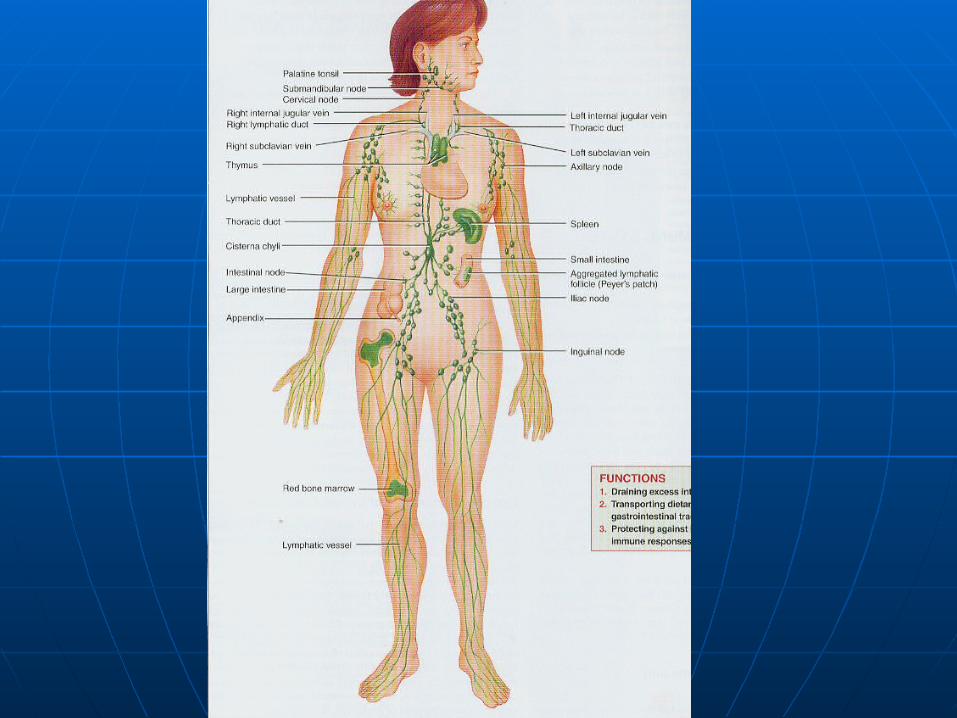
Lymphatic SystemLymphatic System
Consist of;Consist of; lymph (fluid)lymph (fluid) lymphatic vesselslymphatic vessels lymphatic organs and tissueslymphatic organs and tissues bone marrowbone marrow
Lymphatic SystemLymphatic System
Functions;Functions;
1.1. Drain interstitial fluidDrain interstitial fluid

Lymphatic SystemLymphatic System
2. Return leaked plasma proteins to 2. Return leaked plasma proteins to the bloodthe blood
Lymphatic SystemLymphatic System
3. Protects against invasion by 3. Protects against invasion by nonspecific defenses and specific nonspecific defenses and specific immune responsesimmune responses

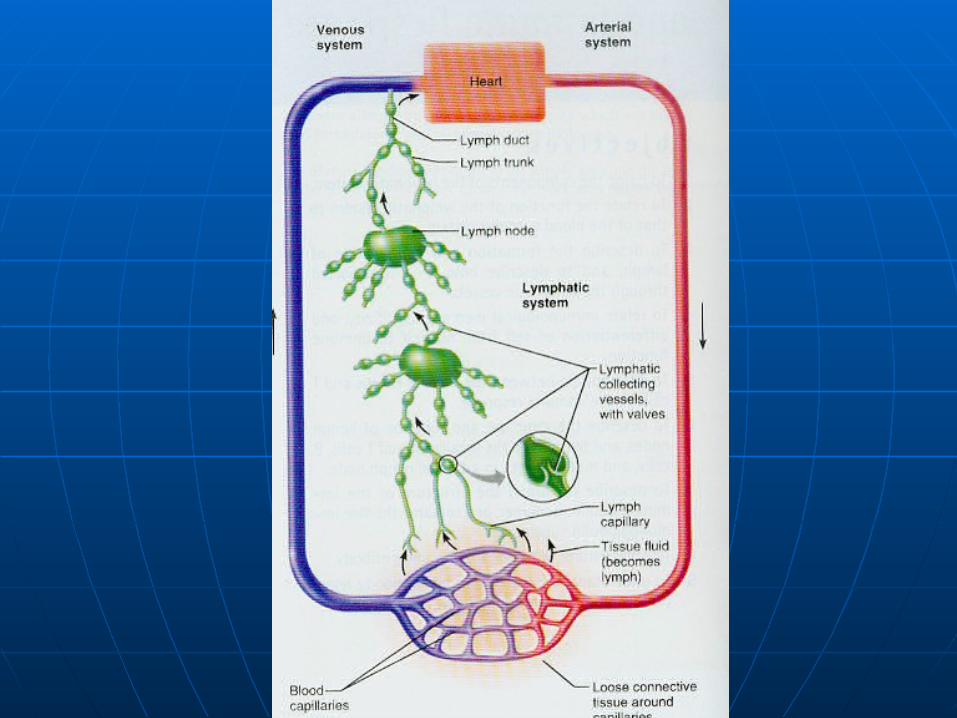
Lymphatic CirculationLymphatic Circulation
Interstitial fluid drains into lymphatic Interstitial fluid drains into lymphatic capillaries thus forming lymphcapillaries thus forming lymph


Lymphatic CirculationLymphatic Circulation
Lymph capillaries merge to form Lymph capillaries merge to form larger vessels, called lymphatic larger vessels, called lymphatic vesselsvessels
Lymphatic CirculationLymphatic Circulation
Convey lymph into and out of Convey lymph into and out of structures called lymph nodesstructures called lymph nodes


Lymphatic CapillariesLymphatic Capillaries
Lymphatic capillaries are found Lymphatic capillaries are found everywhere except;everywhere except;
1. Avascular tissue1. Avascular tissue
Lymphatic CapillariesLymphatic Capillaries
2. CNS2. CNS
Lymphatic CapillariesLymphatic Capillaries
3. Portions of the spleen3. Portions of the spleen
Lymphatic CapillariesLymphatic Capillaries
4. Red bone marrow4. Red bone marrow

Lymphatic CapillariesLymphatic Capillaries
Lacteal – lymphatic capillary in the Lacteal – lymphatic capillary in the villus of the small intestine which villus of the small intestine which transport fatstransport fats
Lymph Trunk and DuctsLymph Trunk and Ducts
1.1. Thoracic DuctThoracic Duct
2.2. Right Lymphatic DuctRight Lymphatic Duct


Thoracic DuctThoracic Duct
Main collecting duct of the lymphatic Main collecting duct of the lymphatic systemsystem

Thoracic DuctThoracic Duct
Receives lymph from the left side of Receives lymph from the left side of the head, neck, and chest, the left the head, neck, and chest, the left upper extremity and the entire body upper extremity and the entire body below the ribsbelow the ribs
Thoracic DuctThoracic Duct
It drains lymph into venous blood via It drains lymph into venous blood via the left subclavian veinthe left subclavian vein
Right Lymphatic DuctRight Lymphatic Duct
Drains lymph from the upper right Drains lymph from the upper right side of the bodyside of the body


Right Lymphatic DuctRight Lymphatic Duct
It drains lymph into venous blood via It drains lymph into venous blood via the right subclavian veinthe right subclavian vein

Flow of LymphFlow of Lymph
1. Fluid flows from arteries and blood 1. Fluid flows from arteries and blood capillaries to interstitial spaces capillaries to interstitial spaces (interstitial fluid)(interstitial fluid)

Flow of LymphFlow of Lymph
2. To lymph capillaries (lymph)2. To lymph capillaries (lymph)

Flow of LymphFlow of Lymph
3. To lymphatic vessels3. To lymphatic vessels


Flow of LymphFlow of Lymph
4. To lymph trunks4. To lymph trunks


Flow of LymphFlow of Lymph
5.To the thoracic duct or right 5.To the thoracic duct or right lymphatic duct lymphatic duct

Flow of LymphFlow of Lymph
6. To the subclavian veins (blood)6. To the subclavian veins (blood)


Lymphatic Organs and TissuesLymphatic Organs and Tissues
Lymphatic organs are classified as Lymphatic organs are classified as primary or secondaryprimary or secondary
Primary Lymphatic OrgansPrimary Lymphatic Organs
Red bone marrowRed bone marrow Thymus glandThymus gland

Secondary Lymphatic OrgansSecondary Lymphatic Organs
Lymph nodesLymph nodes SpleenSpleen Lymphatic nodulesLymphatic nodules

Thymus GlandThymus Gland
Lies between the sternum and the Lies between the sternum and the heartheart
Thymus GlandThymus Gland
Functions in immunity as the site of T Functions in immunity as the site of T cell maturationcell maturation

Thymus GlandThymus Gland
Large in the infant and after puberty Large in the infant and after puberty it is replaced by adipose and areolar it is replaced by adipose and areolar connective tissueconnective tissue

Lymph NodesLymph Nodes
Are encapsulated oval structures Are encapsulated oval structures located along lymphatic vesselslocated along lymphatic vessels
Lymph NodesLymph Nodes
Contain T cells, macrophages, Contain T cells, macrophages, follicular dendritic cells, B cells, and follicular dendritic cells, B cells, and plasma cellsplasma cells
Lymph NodesLymph Nodes
Lymph enters nodes through afferent Lymph enters nodes through afferent lymphatic vessels and is filtered to lymphatic vessels and is filtered to remove damage cells and remove damage cells and microorganismsmicroorganisms
Lymph NodesLymph Nodes
Exits through efferent lymphatic Exits through efferent lymphatic vesselvessel
Lymph NodesLymph Nodes
Foreign substances filtered by the Foreign substances filtered by the lymph nodes are trapped by nodal lymph nodes are trapped by nodal reticular fibersreticular fibers

Lymph NodesLymph Nodes
Macrophages destroys foreign Macrophages destroys foreign substances by phagocytosissubstances by phagocytosis

Lymph NodesLymph Nodes
Lymphocytes bring about the Lymphocytes bring about the destruction of others by immune destruction of others by immune responseresponse
Lymph NodesLymph Nodes
Site of proliferation of plasma cells Site of proliferation of plasma cells (from B cells) and T cells(from B cells) and T cells
Plasma cells make antibodiesPlasma cells make antibodies

SpleenSpleen
Largest mass of lymphatic tissue in Largest mass of lymphatic tissue in the bodythe body

SpleenSpleen
Located between the stomach and Located between the stomach and diaphragmdiaphragm


SpleenSpleen
Made up of Made up of white white and and red pulpred pulp

White PulpWhite Pulp
Lymphatic TissueLymphatic Tissue


White PulpWhite Pulp
Its T lymphocytes directly attack and Its T lymphocytes directly attack and destroy antigens in blooddestroy antigens in blood
White PulpWhite Pulp
Its B lymphocytes develop into Its B lymphocytes develop into antibody producing plasma cells, and antibody producing plasma cells, and the antibodies inactivate antigens in the antibodies inactivate antigens in bloodblood

White PulpWhite Pulp
Macrophages destroy antigens in Macrophages destroy antigens in blood by phagocytosisblood by phagocytosis

Red PulpRed Pulp
Consists of;Consists of;
1.1. Venous sinusesVenous sinuses
2.2. Splenic cordsSplenic cords

Red PulpRed Pulp
Venous sinuses are filled with bloodVenous sinuses are filled with blood
Red PulpRed Pulp
Splenic cords consist of;Splenic cords consist of;
1.1. RBCsRBCs
2.2. MacrophagesMacrophages
3.3. LymphocytesLymphocytes
4.4. Plasma cellsPlasma cells
5.5. granulocytesgranulocytes

Red PulpRed Pulp
Macrophages remove deffective Macrophages remove deffective RBCs, WBCs, and plateletsRBCs, WBCs, and platelets

Red PulpRed Pulp
Stores blood plateletsStores blood platelets

Lymphatic NodulesLymphatic Nodules
Oval-shaped concentrations of Oval-shaped concentrations of lymphatic tissuelymphatic tissue

Lymphatic NodulesLymphatic Nodules
Scattered throughout the mucous Scattered throughout the mucous membranes lining the GI tract, membranes lining the GI tract, respiratory airways, urinary tract, respiratory airways, urinary tract, and reproductive tractand reproductive tract

Peyer’s patchesPeyer’s patches
Lymphatic nodules in the ileum of Lymphatic nodules in the ileum of the small intestinethe small intestine
TonsilsTonsils
Multiple aggregations of large Multiple aggregations of large lymphatic nodules at the junction of lymphatic nodules at the junction of the oral cavity and the pharynxthe oral cavity and the pharynx

TonsilsTonsils
Include;Include;
1.1. PharyngealPharyngeal
2.2. PalatinePalatine
3.3. Lingual tonsilsLingual tonsils

TonsilsTonsils
Participate in immune responses by Participate in immune responses by producing lymphocytes and producing lymphocytes and antibodiesantibodies
Nonspecific Resistance to DiseaseNonspecific Resistance to Disease
Involves a Involves a
1.1. First line of DefenseFirst line of Defense
2.2. Second line of DefenseSecond line of Defense

First Line of DefenseFirst Line of Defense
InvolvesInvolves
1.1. Mechanical ProtectionMechanical Protection
2.2. Chemical ProtectionChemical Protection

Mechanical ProtectionMechanical Protection
IncludeInclude
1. Epidermis layer of the skin1. Epidermis layer of the skin
Mechanical ProtectionMechanical Protection
2. Mucous membranes in the nose 2. Mucous membranes in the nose and tracheaand trachea
Mechanical ProtectionMechanical Protection
3. Lacrimal apparatus3. Lacrimal apparatus

Mechanical ProtectionMechanical Protection
4. Saliva4. Saliva

Mechanical ProtectionMechanical Protection
5. Mucus5. Mucus

Mechanical ProtectionMechanical Protection
6. Cilia6. Cilia
Mechanical ProtectionMechanical Protection
7. Epiglottis7. Epiglottis

Mechanical ProtectionMechanical Protection
8. Flow of urine8. Flow of urine
Mechanical ProtectionMechanical Protection
9. Defecation9. Defecation

Mechanical ProtectionMechanical Protection
10. Vomiting10. Vomiting

Chemical ProtectionChemical Protection
1. The skin produces 1. The skin produces sebumsebum, which , which has a low pHhas a low pH
Chemical ProtectionChemical Protection
2. 2. LysozymeLysozyme in sweat has in sweat has antimicrobial propertiesantimicrobial properties
Chemical ProtectionChemical Protection
3. 3. Gastric juiceGastric juice in the stomach has in the stomach has a low pHa low pH

Chemical ProtectionChemical Protection
4. 4. Vaginal secretionsVaginal secretions are also are also acidicacidic
Second Line of DefenseSecond Line of Defense
InvolvesInvolves
1.1. Antimicrobial proteinsAntimicrobial proteins
2.2. Phagocytic and natural killer cellsPhagocytic and natural killer cells
3.3. InflammationInflammation
4.4. FeverFever
Antimicrobial Proteins Antimicrobial Proteins
1.1. InterferonsInterferons
2.2. Complement SystemComplement System

InterferonsInterferons
Body cells infected with viruses Body cells infected with viruses produce proteins called interferons produce proteins called interferons (IFNs)(IFNs)

InterferonsInterferons
IFN diffuses to uninfected cells and IFN diffuses to uninfected cells and binds to surface receptorsbinds to surface receptors

InterferonsInterferons
This induces uninfected cells to This induces uninfected cells to synthesize antiviral proteins that synthesize antiviral proteins that inhibit viral replicationinhibit viral replication

Complement SystemComplement System
A group of 20 proteins present in A group of 20 proteins present in blood plasma and on cell membranesblood plasma and on cell membranes
Complement SystemComplement System
When activated, these proteins When activated, these proteins enhance immune, allergic, and enhance immune, allergic, and inflammatory reactionsinflammatory reactions

Natural Killer CellsNatural Killer Cells
Lymphocytes that lack the Lymphocytes that lack the membrane molecules that identify T membrane molecules that identify T and B cellsand B cells
Natural Killer CellsNatural Killer Cells
Can kill a variety of infectious Can kill a variety of infectious microbes and some tumor cells microbes and some tumor cells
Natural Killer CellsNatural Killer Cells
Sometime release Sometime release perforins perforins that that insert into the plasma membrane of insert into the plasma membrane of a microbe and make the membrane a microbe and make the membrane leaky so that cytolysis occursleaky so that cytolysis occurs
Natural Killer CellsNatural Killer Cells
Sometimes they bind to a target cell Sometimes they bind to a target cell and inflict damage by direct contactand inflict damage by direct contact

PhagocytesPhagocytes
Neutrophils and macrophagesNeutrophils and macrophages

InflammationInflammation
Four symptomsFour symptoms
1.1. RednessRedness
2.2. PainPain
3.3. HeatHeat
4.4. SwellingSwelling
InflammationInflammation
Three stagesThree stages
1.1. Vasodilation and increased Vasodilation and increased permeabilitypermeability
InflammationInflammation
2. Emigration of phagocytes from the 2. Emigration of phagocytes from the blood into interstitial fluidblood into interstitial fluid

InflammationInflammation
3. Tissue Repair3. Tissue Repair
InflammationInflammation
Vasodilation and increased Vasodilation and increased permeability are responsible for heat, permeability are responsible for heat, redness, and swellingredness, and swelling
InflammationInflammation
Pain results from injury to neurons Pain results from injury to neurons and from toxic chemicals released by and from toxic chemicals released by microbesmicrobes
FeverFever
When macrophages respond to an When macrophages respond to an infection, they release infection, they release interleukin interleukin -1-1

FeverFever
Interleukin-1 stimulates the Interleukin-1 stimulates the hypothalamus to initiate a fever hypothalamus to initiate a fever
FeverFever
Inhibits some microbial growth and Inhibits some microbial growth and speeds up body reactions that aid speeds up body reactions that aid repairrepair
Major Histocompatibility Complex Major Histocompatibility Complex AntigensAntigens
Unique to each person’s body cellsUnique to each person’s body cells
Major Histocompatibility Complex Major Histocompatibility Complex AntigensAntigens
All cells except rbc display All cells except rbc display MHC class MHC class II antigens antigens
Major Histocompatibility Complex Major Histocompatibility Complex AntigensAntigens
Antigen presenting cells Antigen presenting cells (macrophages) also display (macrophages) also display MHC MHC class IIclass II antigens antigens

Pathways of Antigen ProcessingPathways of Antigen Processing
For an immune response to occur, B For an immune response to occur, B and T cells must recognize that a and T cells must recognize that a foreign antigen is presentforeign antigen is present

Pathways of Antigen ProcessingPathways of Antigen Processing
Antigens are chemical substances Antigens are chemical substances that are recognized as foreign by that are recognized as foreign by antigen receptors when introduced antigen receptors when introduced into the bodyinto the body
Pathways of Antigen ProcessingPathways of Antigen Processing
The body contains millions of The body contains millions of different T and B cells each capable different T and B cells each capable of responding to a specific antigenof responding to a specific antigen

Cell-Mediated ImmunityCell-Mediated Immunity
Refers to destruction of antigens by T Refers to destruction of antigens by T cellscells

Cell-Mediated ImmunityCell-Mediated Immunity
Three main stepsThree main steps
Cell-Mediated ImmunityCell-Mediated Immunity
1. T cells recognize antigen fragments 1. T cells recognize antigen fragments associated with MHC II class associated with MHC II class molecules on the surface of an molecules on the surface of an antigen presenting cell antigen presenting cell (macrophage). (macrophage).
Cell-Mediated ImmunityCell-Mediated Immunity
Although CD8 cell receptors bind to Although CD8 cell receptors bind to the antigen associated with MHC the antigen associated with MHC class I molecules on the virus class I molecules on the virus infected cellinfected cell
Cell-Mediated ImmunityCell-Mediated Immunity
2. A small number of T cells proliferate 2. A small number of T cells proliferate and differentiate into a clone of and differentiate into a clone of effector cellseffector cells


Cell-Mediated ImmunityCell-Mediated Immunity
2 continued …2 continued …
Clone of effector cells – a pop. of Clone of effector cells – a pop. of identical cells that can recognize the identical cells that can recognize the same antigensame antigen

Cell-Mediated ImmunityCell-Mediated Immunity
3. Antigen (intruder) is eliminated 3. Antigen (intruder) is eliminated
Types of T CellsTypes of T Cells
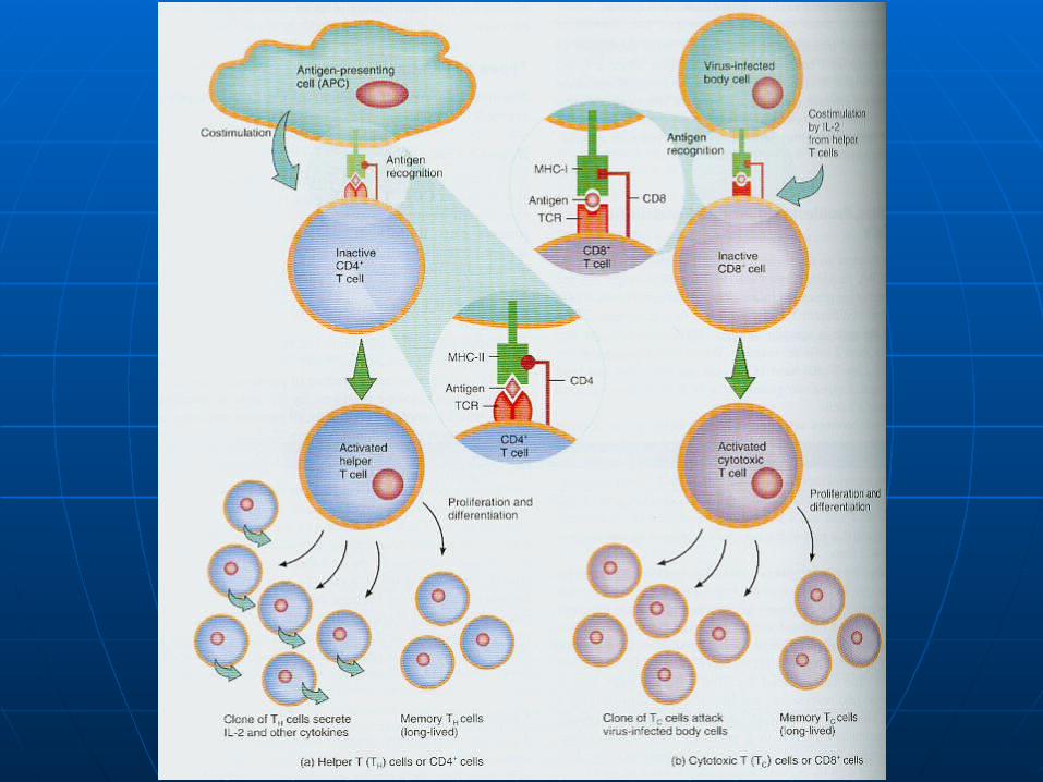
1.1. Helper T (TH) cellsHelper T (TH) cells
2.2. Cytotoxic T (TC) cellsCytotoxic T (TC) cells
3.3. Memory T cellsMemory T cells
Helper T cellsHelper T cells
T4 cellsT4 cells
Helper T cellsHelper T cells
Display CD4 proteinsDisplay CD4 proteins

Helper T cellsHelper T cells
Recognize antigen fragments Recognize antigen fragments associated with MHC-II moleculesassociated with MHC-II molecules
Helper T cellsHelper T cells
Secrete interleukin-2 which as a Secrete interleukin-2 which as a costimulatorcostimulator

Helper T cellsHelper T cells
Proliferation of T cells requires Proliferation of T cells requires costimulationcostimulation

Cytotoxic T cellsCytotoxic T cells
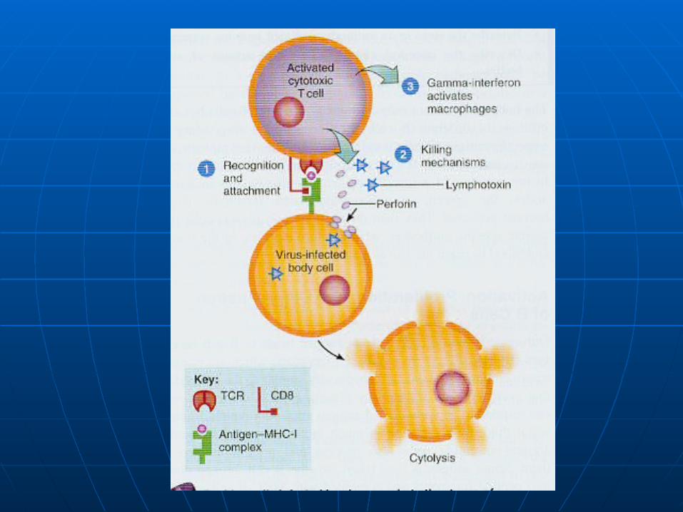
Fight foreign invaders by killing the Fight foreign invaders by killing the target celltarget cell

Cytotoxic T cellsCytotoxic T cells
Target cell – the cell that bears the Target cell – the cell that bears the same antigen that stimulated same antigen that stimulated proliferationproliferation

Cytotoxic T cellsCytotoxic T cells
One killing mechanism uses One killing mechanism uses perforin perforin to cause cytolysis of the target cell.to cause cytolysis of the target cell.

Cytotoxic T cellsCytotoxic T cells
The second mechanism uses The second mechanism uses lymphotoxinlymphotoxin to activate damaging to activate damaging enzymes within the target cellsenzymes within the target cells
Cytotoxic T cellsCytotoxic T cells
Main targets are virus infected cellsMain targets are virus infected cells
Memory T cellsMemory T cells
Programmed to recognize the Programmed to recognize the original invading antigenoriginal invading antigen
Antibody-Mediated ImmunityAntibody-Mediated Immunity
Refers to destruction of antigens by Refers to destruction of antigens by antibodiesantibodies

Antibody-Mediated ImmunityAntibody-Mediated Immunity
There are 4 stepsThere are 4 steps

Antibody-Mediated ImmunityAntibody-Mediated Immunity
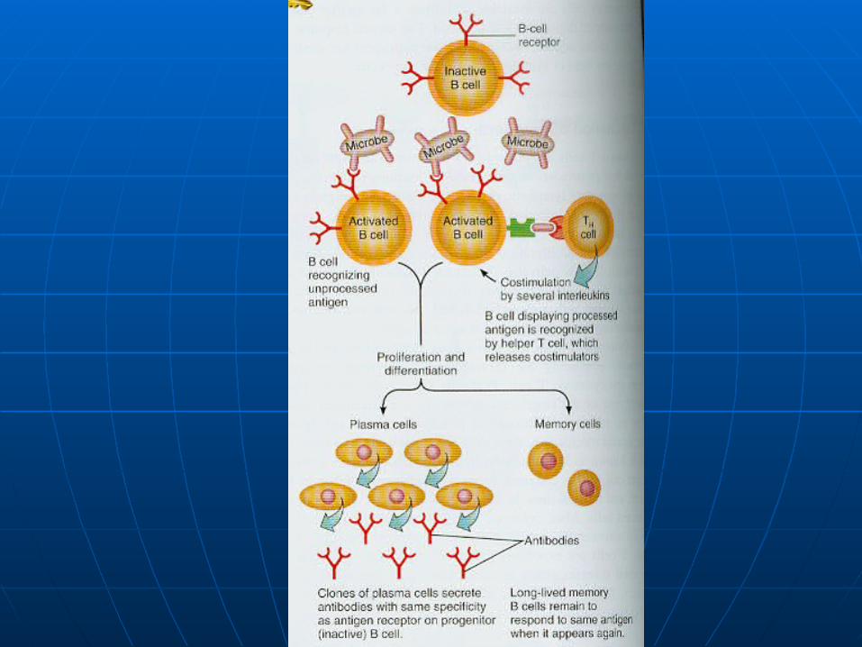
1. An antigen binds to the surface of 1. An antigen binds to the surface of B cellsB cells

Antibody-Mediated ImmunityAntibody-Mediated Immunity
2. Some antigen is taken into the B 2. Some antigen is taken into the B cell, broken down into peptide cell, broken down into peptide fragments and combined with the fragments and combined with the MHC-II self antigen, and moved to MHC-II self antigen, and moved to the B cell surfacethe B cell surface

Antibody-Mediated ImmunityAntibody-Mediated Immunity
3. Hepler T cells recognize the 3. Hepler T cells recognize the antigen-MHC-II combination and antigen-MHC-II combination and secrete interleukinssecrete interleukins

Antibody-Mediated ImmunityAntibody-Mediated Immunity
The interleukins deliver the The interleukins deliver the costimulation needed for B cell costimulation needed for B cell proliferationproliferation

Antibody-Mediated ImmunityAntibody-Mediated Immunity
4. Some activated B cells become 4. Some activated B cells become antibody-secretion plasma cells. antibody-secretion plasma cells. Others become B cellsOthers become B cells

AntibodiesAntibodies
A protein that can combine A protein that can combine specifically with the antigenic specifically with the antigenic determinant on the antigen that determinant on the antigen that triggered its productiontriggered its production
AntibodiesAntibodies
Five classesFive classes
1.1. IgGIgG
2.2. IgAIgA
3.3. IgMIgM
4.4. IgDIgD
5.5. IgEIgE
IgGIgG
It binds to bacteria and viruses It binds to bacteria and viruses

IgGIgG
It crosses the placenta until it can It crosses the placenta until it can begin secreting its own.begin secreting its own.

IgAIgA
It is in blood, breast milk, tears, It is in blood, breast milk, tears, saliva, and intestinal secretions.saliva, and intestinal secretions.
IgAIgA
Levels decrease during stressLevels decrease during stress
IgAIgA
Protects our mucous membranes Protects our mucous membranes against infections with bacteria and against infections with bacteria and virusesviruses
IgMIgM
First antibody to be secreted by First antibody to be secreted by plasma cells after exposure to plasma cells after exposure to antigensantigens
IgMIgM
Antibodies to A and B red cell Antibodies to A and B red cell antigens are IgMantigens are IgM
IgMIgM
Cannot cross the placentaCannot cross the placenta

IgDIgD
Activate B cellsActivate B cells
IgEIgE
On the surface of mast cells and On the surface of mast cells and basophilsbasophils
IgEIgE
Involved in allergic reactionsInvolved in allergic reactions

IgEIgE
When a person with pollen allergies When a person with pollen allergies inhales pollen, it combines with the inhales pollen, it combines with the IgE on their mast cellsIgE on their mast cells
IgEIgE
Causes mast cells to release Causes mast cells to release histaminehistamine
AIDSAIDS
A condition in which a person A condition in which a person experiences infections as a result of experiences infections as a result of the progressive destruction of cells the progressive destruction of cells by the humon immunodeficiency by the humon immunodeficiency virusvirus
HIVHIV
The only documented transmissions The only documented transmissions are by way of blood, semen, vaginal are by way of blood, semen, vaginal secretions, and breast milksecretions, and breast milk
HIVHIV
A recent study in San Fran showed A recent study in San Fran showed that 8% of their HIV-infected patients that 8% of their HIV-infected patients acquired it via oral sexacquired it via oral sex

HIVHIV
Form of a retrovirus with a protein Form of a retrovirus with a protein coatcoat

HIVHIV
HIV enters CD4 positive T HIV enters CD4 positive T lymphocytes and macrophages lymphocytes and macrophages where it sheds its protein coatwhere it sheds its protein coat

HIVHIV
New HIV DNA is produced in the T New HIV DNA is produced in the T cell along with new protein coats and cell along with new protein coats and then releasedthen released

HIVHIV
The T cells are ultimately destroyedThe T cells are ultimately destroyed
HIVHIV
Progression to AIDS occurs because Progression to AIDS occurs because of reduced numbers of T cells and of reduced numbers of T cells and resulting immunodeficiencyresulting immunodeficiency
AIDSAIDS
Person now susceptible to Person now susceptible to opportunistic infectionsopportunistic infections

HIVHIV
Treatment of HIV infection with Treatment of HIV infection with reverse transcriptase inhibitors has reverse transcriptase inhibitors has shown to delay the progression of shown to delay the progression of HIV infection to AIDSHIV infection to AIDS
Related Documents