THE JOURNAL o? BIOLOGICAL CHEMISTRY 0 1992 by The American Society for Biochemistry and Molecular Biology, Inc. Vol. 267, No. 8, Issue of March 15, pp. 5599-5607.1992 Printed in U.S.A. New Inhibitors of Sepiapterin Reductase LACK OF AN EFFECT OF INTRACELLULAR TETRAHYDROBIOPTERINDEPLETIONUPON IN VITRO PROLIFERATION OF TWO HUMAN CELL LINES* (Received for publication, August 22,1991) Gary K. Smith$%, David S. DuchS, Mark P. EdelsteinS, and Eric C. Bighamll From the Divisions of $Cell Biology and llOrganic Chemistry, Wellcome Research Laboratories, Research Triangle Park, North Carolina 27709 N-Acetylserotonin (compound 1) and N-acetyldopa- mine (compound 7) inhibit bovine adrenal medullary sepiapterin reductase in a manner competitive with the pterin substrate and have Ki values of 0.12 and 0.4 PM, respectively. Molecular modeling suggests thatthe phenyl ringsof the two compounds bind in the pyrim- idine pocket of the enzyme with the 3-hydroxyl of dopamine or the 5-hydroxyl of serotonin aligned at the pyrimidine 4-position. Further, the acetyl moieties of the two inhibitors appear to mimic the substrate side chain. Consistent with this analysis, N-acetyl-m-tyra- mine (compound 13) is also an excellent competitive inhibitor (K, = 0.13 PM), whereas N-acetyltryptamine (compound 2), N-acetyl-p-tyramine (compound 14) and N-acetylphenylethylamine (compound 15) all bind poorly. Interestingly, restricted-rotation analogs of N- acetyldopamine and N-acetyl-m-tyramine are noncom- petitive inhibitors of the enzyme. Modification of N- acetyldopamine to N-chloroacetyldopamine (com- pound 10) or of N-acetylserotonin to theN-chloroace- tyl (5) or N-methoxyacetyl (compound 6) analogs re- sults in greatly increased competitive affinity, with K, = 0.014 PM for the dopamine analog and 0.006 and 0.008 cM, respectively, for the serotonin analogs. In MOLT-4 T-cell leukemia and MCF-7 breast adenocar- cinoma in culture, 0.1 mM N-methoxyacetylserotonin depleted tetrahydrobiopterin by 297 and >60%, re- spectively, with no effect upon cell growth. In both cell lines, the GTP cyclohydrolase inhibitor, 2,4-diamino- 6-hydroxypyrimidine at 1-5 m~ also depleted tetra- hydrobiopterin 297%. In this case, however, modest growth inhibition did occur. Since the growth inhibi- tion could not be reversed upon tetrahydrobiopterin repletion, inhibition was due to other effects of the inhibitor rather than to tetrahydrobiopterin depletion. The results show that there is no effect on cell growth when at least 97% of the tetrahydrobiopterin in these cell lines is depleted. Since the sepiapterin reductase inhibitor depleted tetrahydrobiopterin with fewer nonspecific effects than the cyclohydrolase inhibitor, it will be useful for determining metabolic effects of tetrahydrobiopterin depletion. Tetrahydrobiopterin is synthesized from GTP by the cells * The costs of publication of this article were defrayed in part by the payment of page charges. This article must therefore be hereby marked “advertisement” in accordance with 18 U.S.C. Section 1734 solely to indicate this fact. 8 TO whom correspondence should be addressed Division of Cell Biology, Wellcome Research Laboratories, Research Triangle Park, NC 27709. Tel.: 919-248-4179; Fax: 919-248-8375. that use it (1). Minimally, the enzymes on the biosynthetic pathway to this compound include GTP cyclohydrolase, py- ruvoyltetrahydropterin synthase, and sepiapterin reductase (1-5); the relevance of a fourth enzyme, referred to either as pyruvoyketrahydropterin reductase or lactoyltetrahydrop- terin synthase, in the reaction sequence remains controversial (1-9). The overall biosynthetic pathway to tetrahydrobiop- terin is shown in Fig. 1. Tetrahydrobiopterin is the required cofactor for phenylalanine, tyrosine, and tryptophan hydro- xylases and alkylglycerol monooxygenase (1). In allcases, the cofactor is presumed to function by activating molecular oxygen (1, 10-14). One of the goals of current tetrahydrobiopterin research is to determine whether this compound or any of the interme- diates on its biosynthetic pathway have any as yet unrecog- nized functions. In order to explore this further, inhibitorsof sepiapterin reductase have been designed and tested. Sepiap- terin reductase catalyzes the terminal reactions on the bio- synthetic pathway to tetrahydrobiopterin (Fig. l), and intra- cellular inhibition of this enzyme should cause a depletion of tetrahydrobiopterin in cells. In addition, since this enzyme is not required for the biosynthesis of dihydroneopterin triphos- phate and pyruvoyltetrahydropterin, the first two intermedi- ates on the pathway, intracellular sepiapterin reductase in- hibition should not reduce the levels of these intermediates. Thus, sepiapterin reductase inhibition will produce a more specific depletion of tetrahydrobiopterin than would inhibi- tors of earlier reactions in the pathway. Observation of changes occurring in the cells as a result of tetrahydrobiop- terin depletion may lead to discovery of new functions of the cofactor. If the biosynthetic intermediates have specificfunc- tions, this can be determined by comparing results obtained using the sepiapterin reductase inhibitor with those obtained using 2,4-diamino-6-hydroxypyrimidine, an inhibitor of GTP cyclohydrolase (X), the first enzyme on the pathway, which will deplete all of the pathway intermediates. Thus, informa- tion may be gained on potential unrecognized roles of tetra- hydrobiopterin, as well as some of its biosynthetic interme- diates. The previous work of Katoh et al. (16) provided the lead compound, N-acetylserotonin, for the present study. Recently, it was reported that partial depletion of tetrahy- drobiopterin in a murine erythroleukemia cell line caused inhibition of cell growth, suggesting a connection between tetrahydrobiopterin and cell growth in this line (17). In the current work, a structure/activity investigation of inhibitors of sepiapterin reductase is presented. Using one of the tightest binding of these inhibitors, as well as the GTP cyclohydrolase inhibitor, we found that tetrahydrobiopterin can be depleted in both MOLT-4 T-cell leukemia and MCF-7 breast cancer cells with no effect on cell proliferation. This result indicates 5599

Welcome message from author
This document is posted to help you gain knowledge. Please leave a comment to let me know what you think about it! Share it to your friends and learn new things together.
Transcript
THE JOURNAL o? BIOLOGICAL CHEMISTRY 0 1992 by The American Society for Biochemistry and Molecular Biology, Inc.
Vol. 267, No. 8, Issue of March 15, pp. 5599-5607.1992 Printed in U.S.A.
New Inhibitors of Sepiapterin Reductase LACK OF AN EFFECT OF INTRACELLULAR TETRAHYDROBIOPTERIN DEPLETION UPON IN VITRO PROLIFERATION OF TWO HUMAN CELL LINES*
(Received for publication, August 22,1991)
Gary K. Smith$%, David S. DuchS, Mark P. EdelsteinS, and Eric C. Bighamll From the Divisions of $Cell Biology and llOrganic Chemistry, Wellcome Research Laboratories, Research Triangle Park, North Carolina 27709
N-Acetylserotonin (compound 1) and N-acetyldopa- mine (compound 7) inhibit bovine adrenal medullary sepiapterin reductase in a manner competitive with the pterin substrate and have Ki values of 0.12 and 0.4 PM, respectively. Molecular modeling suggests that the phenyl rings of the two compounds bind in the pyrim- idine pocket of the enzyme with the 3-hydroxyl of dopamine or the 5-hydroxyl of serotonin aligned at the pyrimidine 4-position. Further, the acetyl moieties of the two inhibitors appear to mimic the substrate side chain. Consistent with this analysis, N-acetyl-m-tyra- mine (compound 13) is also an excellent competitive inhibitor (K, = 0.13 PM), whereas N-acetyltryptamine (compound 2), N-acetyl-p-tyramine (compound 14) and N-acetylphenylethylamine (compound 15) all bind poorly. Interestingly, restricted-rotation analogs of N- acetyldopamine and N-acetyl-m-tyramine are noncom- petitive inhibitors of the enzyme. Modification of N- acetyldopamine to N-chloroacetyldopamine (com- pound 10) or of N-acetylserotonin to the N-chloroace- tyl (5 ) or N-methoxyacetyl (compound 6) analogs re- sults in greatly increased competitive affinity, with K , = 0.014 PM for the dopamine analog and 0.006 and 0.008 cM, respectively, for the serotonin analogs. In MOLT-4 T-cell leukemia and MCF-7 breast adenocar- cinoma in culture, 0.1 mM N-methoxyacetylserotonin depleted tetrahydrobiopterin by 297 and >60%, re- spectively, with no effect upon cell growth. In both cell lines, the GTP cyclohydrolase inhibitor, 2,4-diamino- 6-hydroxypyrimidine at 1-5 m~ also depleted tetra- hydrobiopterin 297%. In this case, however, modest growth inhibition did occur. Since the growth inhibi- tion could not be reversed upon tetrahydrobiopterin repletion, inhibition was due to other effects of the inhibitor rather than to tetrahydrobiopterin depletion. The results show that there is no effect on cell growth when at least 97% of the tetrahydrobiopterin in these cell lines is depleted. Since the sepiapterin reductase inhibitor depleted tetrahydrobiopterin with fewer nonspecific effects than the cyclohydrolase inhibitor, it will be useful for determining metabolic effects of tetrahydrobiopterin depletion.
Tetrahydrobiopterin is synthesized from GTP by the cells
* The costs of publication of this article were defrayed in part by the payment of page charges. This article must therefore be hereby marked “advertisement” in accordance with 18 U.S.C. Section 1734 solely to indicate this fact.
8 TO whom correspondence should be addressed Division of Cell Biology, Wellcome Research Laboratories, Research Triangle Park, NC 27709. Tel.: 919-248-4179; Fax: 919-248-8375.
that use it (1). Minimally, the enzymes on the biosynthetic pathway to this compound include GTP cyclohydrolase, py- ruvoyltetrahydropterin synthase, and sepiapterin reductase (1-5); the relevance of a fourth enzyme, referred to either as pyruvoyketrahydropterin reductase or lactoyltetrahydrop- terin synthase, in the reaction sequence remains controversial (1-9). The overall biosynthetic pathway to tetrahydrobiop- terin is shown in Fig. 1. Tetrahydrobiopterin is the required cofactor for phenylalanine, tyrosine, and tryptophan hydro- xylases and alkylglycerol monooxygenase (1). In all cases, the cofactor is presumed to function by activating molecular oxygen (1, 10-14).
One of the goals of current tetrahydrobiopterin research is to determine whether this compound or any of the interme- diates on its biosynthetic pathway have any as yet unrecog- nized functions. In order to explore this further, inhibitors of sepiapterin reductase have been designed and tested. Sepiap- terin reductase catalyzes the terminal reactions on the bio- synthetic pathway to tetrahydrobiopterin (Fig. l), and intra- cellular inhibition of this enzyme should cause a depletion of tetrahydrobiopterin in cells. In addition, since this enzyme is not required for the biosynthesis of dihydroneopterin triphos- phate and pyruvoyltetrahydropterin, the first two intermedi- ates on the pathway, intracellular sepiapterin reductase in- hibition should not reduce the levels of these intermediates. Thus, sepiapterin reductase inhibition will produce a more specific depletion of tetrahydrobiopterin than would inhibi- tors of earlier reactions in the pathway. Observation of changes occurring in the cells as a result of tetrahydrobiop- terin depletion may lead to discovery of new functions of the cofactor. If the biosynthetic intermediates have specific func- tions, this can be determined by comparing results obtained using the sepiapterin reductase inhibitor with those obtained using 2,4-diamino-6-hydroxypyrimidine, an inhibitor of GTP cyclohydrolase ( X ) , the first enzyme on the pathway, which will deplete all of the pathway intermediates. Thus, informa- tion may be gained on potential unrecognized roles of tetra- hydrobiopterin, as well as some of its biosynthetic interme- diates. The previous work of Katoh et al. (16) provided the lead compound, N-acetylserotonin, for the present study.
Recently, it was reported that partial depletion of tetrahy- drobiopterin in a murine erythroleukemia cell line caused inhibition of cell growth, suggesting a connection between tetrahydrobiopterin and cell growth in this line (17). In the current work, a structure/activity investigation of inhibitors of sepiapterin reductase is presented. Using one of the tightest binding of these inhibitors, as well as the GTP cyclohydrolase inhibitor, we found that tetrahydrobiopterin can be depleted in both MOLT-4 T-cell leukemia and MCF-7 breast cancer cells with no effect on cell proliferation. This result indicates
5599

5600 Sepiapterin Reductase Inhibitors
that in these two cell lines, tetrahydrobiopterin is not required for cell growth.
RESULTS
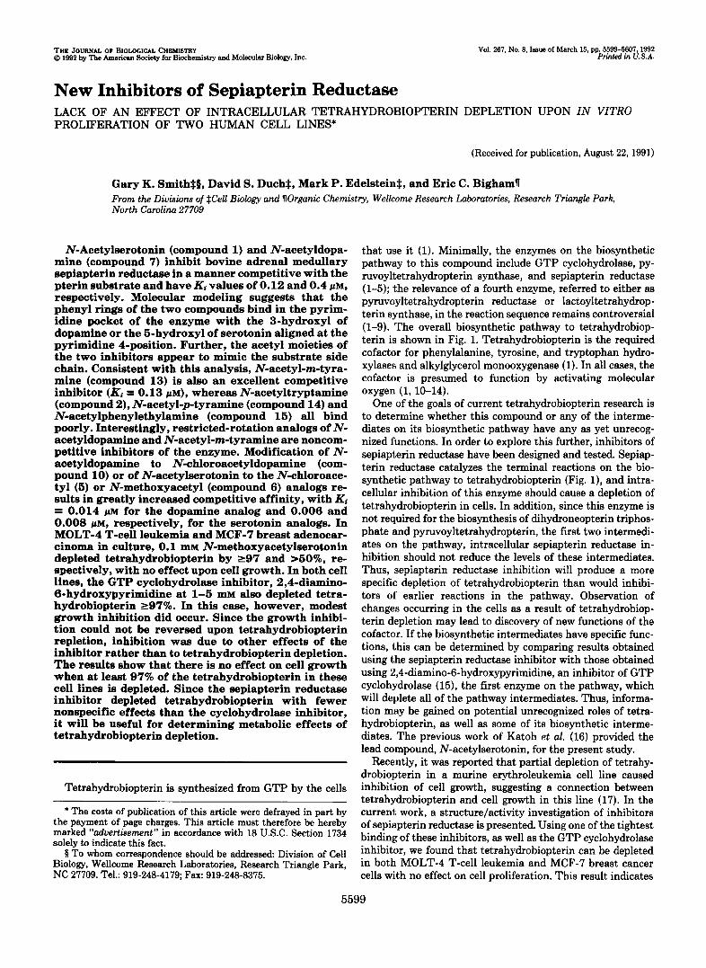
The results of the structure/activity study are shown in Table I. Katoh et al. (16) reported that N-acetylserotonin (l), is a potent competitive inhibitor of partially purified rat brain and homogeneous rat erythrocyte sepiapterin reductase (Ki = 0.2 and 0.17 p ~ , respectively). Our observation of a competi- tive Ki of 0.12 pM support this report (Table I). These data suggest that N-acetylserotonin binds at the substrate site. Katoh et al. (16) also reported that serotonin itself is a poor inhibitor, indicating that the N-acetyl group is critical for binding. In order to determine if the indole hydroxyl moiety is essential for binding, N-acetyltryptamine (2), was synthe- sized and tested. The compound was found to bind almost 100-fold poorer than N-acetylserotonin (Ki = 10 p ~ ) , indi- cating that the aromatic hydroxyl is required for binding. Modeling these observations with MACROMODEL indicates that N-acetylserotonin binding may mimic the binding of sepiapterin or pyruvoyltetrahydropterin (one of the natural sepiapterin reductase substrates) to the enzyme. The relevant comparisons are shown in Fig. 2. In this model, the 5-hydroxyl group mimics the pterin 4-one and the acetyl group of N- acetylserotonin mimics the pterin side chain.
These modeling studies suggested that N-acetyldopamine might also mimic the substrate in a similar fashion (Fig. 2) and bind to the sepiapterin-binding site. However, the model does not predict as good a fit for the side chain. It was found that N-acetyldopamine (7), does bind to the enzyme in a manner competitive with the substrate. The Ki for this inter- action is 0.4 p ~ . The high affinity of the interaction and its competitive nature support the model. Both compounds bind significantly better than the substrate sepiapterin, which has a Km of 5 p ~ . These results led to the synthesis of two other dopamine analogs, N-pyruvoyl- (8), and N-lactoyldopamine (D), which would be expected to further mimic the sepiapterin and pyruvoyltetrahydropterin side chains. However, the re- sults in Table I show that no further enhancement of binding was realized by the introduction of the new side chains. The
SEPIAPTERIN REDUCTASE
H
H# H”NzCH3 N N 6-(L-i’-HVDROXY-2”OXOP~OPVL]~M~PTERIN 1 H O
SEPIAPTERIN REDUCTASE 1
J FIG. 1. Biosynthesis of tetrahydrobiopterin.
model predicts that, like the 5-hydroxyl of N-acetylserotonin, the meta-hydroxyl of N-acetyldopamine mimics the 4-oxygen of sepiapterin and that removal of this hydroxyl should di- minish binding. Indeed, similarly to N-acetyltryptamine, both N-acetylphenethylamine (15) and N-acetyl-p-tyramine (14) are poor inhibitors of sepiapterin reductase (Ki > 100 pM). By contrast, and as expected from Fig. 2, removal of the para-hydroxyl from N-acetyldopamine to form N-acetyl-m- tyramine (13), does not decrease affinity; rather binding is enhanced approximately 3-fold over the parent (Ki = 0.13 pM). Apparently, there is an unfavorable interaction between the pura-hydroxyl of N-acetyldopamine and the enzyme at the position of the pterin N-3-binding site.
Katoh et al. (16) reported that serotonin is a weak inhibitor of sepiapterin reductase, whereas the amino acid, 5-hydroxy- tryptophan is not an inhibitor at all. Similarly, the results of the present study show that N-acetyltryptamine is a weak inhibitor, and N-acetyltryptophan also does not bind to the enzyme. These data suggest that the presence of the amino acid a-carboxyl group destroys binding. Similarly, since N- acetyl-L-noradrenaline (11, Ki = 26 p ~ ) binds 65-fold poorer than N-acetyldopamine, the @-hydroxyl group of noradrena- line also decreases binding. Interestingly, methylation of N- acetyl-L-noradrenaline to form N-acetyl-L-adrenaline (12), abolishes binding. Poor binding by a-carboxyl, @-hydroxyl, and N-methylated compounds may be due to either confor- mational restriction or unfavorable interactions with the en- zyme.
In order to more closely mimic the pyrazine ring moiety of the pterin, the flexible meta-tyramine active conformation shown in Fig. 26 was “frozen” into the inhibitor molecules. This was accomplished by bridging the side chain from the phenyl 2-position of 13 to produce compounds 16-18. It was expected that the reduced degrees of freedom resulting from this modification might increase affinity by lowering the energy cost of assuming the active conformation. However, in all three cases the modifications resulted in decreased affinity of the compounds for the enzyme and changed the inhibition from strictly competitive to mixed or noncompetitive. These results suggested that the bridging methylene in 16 was a poor mimic for the pterin N-5 and resulted in alternate binding. Nonetheless, the model predicts the relative order of binding even for these mixed inhibitors. Compound 16 is an excellent match for the substrate, but compound 17 has the hydroxyls in the wrong locations and compound 18 is locked into the undesirable extended dopamine conformation. As predicted, the relative affinities are 16 > 17 > 18. To pursue this further, three additional compounds that model the N-5 portion of the pterin were synthesized. These compounds, 19-21, possess the tyramine phenol ring moiety and the “C- 5” portion of the restricted rotation molecule 16, but they lack the remaining two methylene groups. With these com- pounds, the effect of substitution at the pterin N-5 moiety could be explored more directly. As in the cases of 16-18, these compounds exhibit mixed noncompetitive inhibition and lower affinity binding compared with 13. This is partic- ularly obvious with compound 19, the direct analog of N- acetyl-m-tyramine (13). This compound binds almost 400- fold poorer than N-acetyl-m-tyramine and 49-fold poorer than 16, and the mode of inhibition is mixed. Presumably, the hydrogen-bonded conformer locks this compound in the inactive position. The ketone analog (ZO), which cannot hydrogen bond was more active. Apparently, the introduction of C-5 not only decreases affinity but perturbs the binding of these compounds, causing them to bind in part outside of the sepiapterin site. Whether this is due to an unfavorable inter-

Sepiupterin Reductase Inhibitors 5601 TABLE I
Sepiapterin reductase inhibitors Assays were performed using bovine adrenal medullary sepiapterin reductase as described in the text. K; and
CYK; are defined as previously described (38), where K, = the dissociation constant for E1 and aK; = the constant for the dissociation of I from ESI ( E = enzyme, I = inhibitor, S = substrate). Inhibition patterns were analyzed for best fit to competitive, noncompetitive, or uncompetitive inhibition.
Class Compound no. Structure aKP Ki
1 2 3 4 5 6
7 8 9 10
11 12
13 14 15
16 17
18
19 20 21
R R’ R*
HO H Me H H Me Me0 H Me HO C02H Me HO H CH2CI HO H CH20Me
R R’ R* R3 R4
HO HO H H Me HO HO H H -COMe HO HO H H -CH(OH)Me HO HO H H -CH&I
HO HO HO H Me HO HO HO Me Me
H HO H H Me HO H H H Me H H H H Me
R R’ R*
H H HO HO HO H
X Y
P M
0.12 10 30
>loo 0.006 0.008
0.4 0.66 0.62 0.014
26 >loo
0.13 >loo >lo0
0.7 1 27 16
350 20
77 49 59 6 83 12
Compounds for which CYK; is reported were found to best tit the noncompetitive inhibition model. All other compounds for which absolute K, values are reported were found to be strictly competitive with the substrate sepiapterin. In these cases, CY would be infinite. Mixed inhibition is indicated for CY greater than 1 and less than m.
action of the C-5 moiety with the pterin N-5-binding site causing misalignment of the entire molecule or due to their inability to adopt the correct side chain binding conformation is not known.
Since it was clear from the structural analysis that the N- acetyl group is critical for binding and that additional bulk could also be tolerated at this position (as in compounds 8 and D), modifications were explored to enhance affinity or to produce an irreversible inhibitor. Unpublished data from our laboratory and the report of sepiapterin reductase inhibition by mercury (35) suggested that the enzyme has an active site cysteine that might be inactivated by a haloacetyl group.
Thus, N-chloroacetyldopamine and N-chloroacetylserotonin were synthesized (compounds 10 and 5 ) and found to be excellent inhibitors of the enzyme (Table I). However, for time periods up to 60 min, no time-dependent inactivation could be seen. Subsequent kinetic analysis demonstrated that both compounds are strict competitive inhibitors of sepiap- terin reductase, with Ki values 20 and 30 times lower than the corresponding N-acetyl derivatives of serotonin and dopa- mine, respectively. Thus, noncovalent effects, rather than covalent coupling, appeared to cause the enhanced binding. To explore this further, N-methoxyacetylserotonin (com- pound e) , which should have similar electronic effects without
Chloroacetyl rather than bromoacetyl was chosen, because its covalent reactivity or instability, was synthesized and tested. lower reactivity in solution might provide greater specificity. This compound was found to inhibit sepiapterin reductase
5602 Sepiapterin Reductase Inhibitors
A
FIG. 2. Comparison of computed stereo models of the Sepiapterin re- ductase substrate, pyruvoyltetrah- ydropterin (solid bonds), with in- hibitors (open bonds). A, 1, B, 13. Color scheme: hydrogen, black; carbon, green; nitrogen, blue, and oxygen, red.
B
TABLE II Znhibition of crude human cell seDbterin reductase
Commund MOLT-4 cell MCF-7 cell
Sepiapterin Kt (pM)
N-Acetylserotonin (1) 0.65 0.5 N-Chloroacetylserotonin (5) 0.026 0.027 N-Methoxyacetylserotonin (6) 0.048 0.049
0.06 0.05
with a competitive binding constant indistinguishable from N-chloroacetylserotonin. Since this compound should be inca- pable of irreversible modification of the enzyme, these results indicate simple noncovalent tight binding of these com- pounds. These compounds are the tightest binding inhibitors of sepiapterin reductase reported to date.
Human tissue culture experiments were initiated to assess the usefulness of these new inhibitors in the study of biopterin biosynthesis and possible novel roles for the cofactor. The efficacy of the compounds was first tested on crude enzyme from the human cell sources to ensure that the results ob- tained from the bovine enzyme could be carried over into the human system. The results of enzyme kinetics obtained with MOLT-4 and MCF-7 cells are shown in Table 11. It can be seen that kinetic constants for the two cells lines are identical and that both enzymes bind substrate and all inhibitors 4-6- fold more poorly than the bovine enzyme. The relative binding order was maintained, demonstrating that compounds 5, 6, and 10 are indeed very potent inhibitors of the crude human enzyme and should be active in depleting tetrahydrobiopterin in these human cells.
Neopterin, pterin, and tetrahydrobiopterin levels and ef- fects on cell growth were determined in cells grown for 72 h in the presence of varying concentrations of the compounds.
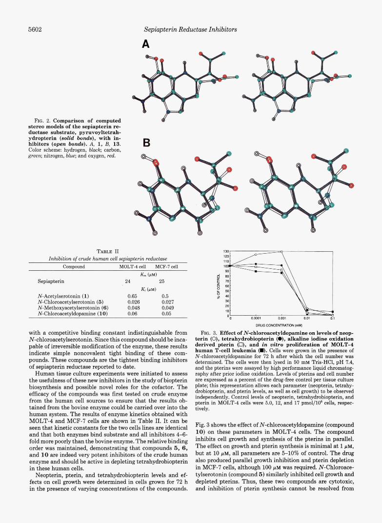
"0 0.m1 0.001 0.01
DRuacoNcENlRAm(mMl FIG. 3. Effect of N-chloroacetyldopamine on levels of neop-
terin (0), tetrahydrobiopterin (O), alkaline iodine oxidation derived pterin 0, and in vitro proliferation of MOLT-4 human T-cell leukemia m. Cells were grown in the presence of N-chloroacetyldopamine for 72 h after which the cell number was determined. The cells were then lysed in 50 m~ Tris-HC1, pH 7.4, and the pterins were assayed by high performance liquid chromatog- raphy after prior iodine oxidation. Levels of pterins and cell number are expressed as a percent of the drug-free control per tissue culture plate; thie representation allows each parameter (neopterin, tetrahy- drobiopterin, and pterin levels, as well as cell growth) to be obsewed independently. Control levels of neopterin, tetrahydrobiopterin, and pterin in MOLT-4 cella were 5.0, 12, and 17 pmol/106 cells, respec- tively.
Fig. 3 shows the effect of N-chloroacetyldopamine (compound 10) on these parameters in MOLT-4 cells. The compound inhibits cell growth and synthesis of the pterins in parallel. The effect on growth and pterin synthesis is minimal at 1 pM, but at 10 PM, all parameters are &lo% of control. The drug also produced parallel growth inhibition and pterin depletion in MCF-7 cella, although 100 PM was required. N-Chloroace- tylserotonin (compound 5 ) similarly inhibited cell growth and depleted pterins. Thus, these two compounds are cytotoxic, and inhibition of pterin synthesis cannot be resolved from

Sepiapterin Reductase Inhibitors 5603
inhibition of growth. Fig. 4A shows the effect of N-methoxy- acetylserotonin (6), on MOLT-4 cells. The compound pro- duced no significant inhibition of cell growth at concentra- tions as high as 100 p ~ . However, the compound decreased tetrahydrobiopterin and total pterin levels 50% at 10 p~ and 97% at 100 p ~ . Neopterin levels were not affected by the drug. Since neopterin is derived from the product of GTP cyclohydrolase, the first enzyme of the biosynthetic pathway, the sepiapterin reductase inhibitor would not be expected to deplete this pterin.
The GTP cyclohydrolase inhibitor 2,4-diamino-6-hydroxy- pyrimidine also inhibits tetrahydrobiopterin and pterin syn- thesis in these cells (Fig. 4 3 shows a 98% depletion of tetra- hydrobiopterin and 100% depletion of pterin levels), but as expected, neopterin synthesis is inhibited in parallel. Similar to N-methoxyacetylserotonin, this compound is not cytotoxic over 72 h at concentrations up to 5 mM. Since there is no correlation between inhibition of biopterin biosynthesis and cell growth with these compounds, the cytotoxic effects of N- chloroacetyldopamine and N-chloroacetylserotonin are ap- parently due to other effects of the haloacetyl compound.
MCF-7 cells also synthesize tetrahydrobiopterin, but se- piapterin reductase activity in this cell line is 20 times higher than in MOLT-4 cells (4.5 uersus 0.19 nmol/min/mg of pro- tein). Thus, it might be expected that the enzyme would have less control over the pathway in MCF-7 cells than in MOLT- 4 cells and that higher levels of N-methoxyacetylserotonin would be required to inhibit tetrahydrobiopterin synthesis in this line (36). N-Methoxyacetylserotonin had no effect on MCF-7 pterins or cell growth (data not shown); thus, tetra- hydrobiopterin synthesis was not significantly influenced by the compound. The effects of 2,4-diamino-6-hydroxypyrimi- dine are shown in Fig. 5. It can be seen that the GTP cyclohydrolase inhibitor had very similar effects in MOLT-4 and MCF-7 cells. Tetrahydrobiopterin and pterin were de- pleted 98 and loo%, respectively, by l mM 2,4-diamino-6- hydroxypyrimidine, but cell growth was not significantly af-
0' Y
0 .m1 a.m1 0.01 0.1
ORUG CONCENTRATION (mM)
0.01 0.1 1
DRUQ CONCENTRATION (mM)
ORUG CONCENTRATION (mM)
0.01 0.1 1
DRUQ CONCENTRATION (mM)
FIG. 4. Effect of N-methoxyacetylserotonin (compound 6) (A) and 2.4-diamino-6-hydroxypyrimidine ( B ) on levels of neopterin (O), tetrahydrobiopterin (O), and alkaline iodine oxidation-derived pterin (O), and in vitro proliferation of MOLT-4 human T-cell leukemia 0. Experimental conditions were as described in the legend to Fig. 3.
1101 1
0.01 0.1
DRUG CONCENTRATION (mM)
FIG. 5. Effect of 2,4-diamino-6-hydroxypyrimidine on lev- els of tetrahydrobiopterin (0) and alkaline iodine oxidation- derived pterin (0) and in vitro proliferation of MCF-7 cells m). Experimental conditions were as described in the legend to Fig. 3. Neopterin could not be assayed in MCF-7 cells due to interfering high performance liquid chromatography peaks; tetrahydrobiopterin and pterin levels mere 5.3 and 7.7 pmol/lOs cells, respectively.
fected at concentrations up to 5 mM, which indicates that tetrahydrobiopterin is also not required for growth of MCF-7 cells. Since the sepiapterin reductase inhibitor did not inhibit biopterin synthesis at concentrations up to 100 pM, it is likely that sepiapterin reductase does have less control over biop- terin synthesis in MCF-7 cells. These results demonstrate that comparison of the effects of the sepiapterin reductase and GTP cyclohydrolase inhibitors provides significantly more insight into tetrahydrobiopterin synthesis by MOLT-4 and MCF-7 cells than either compound alone.
Figs. 4 and 5 represent short term experiments. During the 72-h period of the experiment, control MOLT-4 cells under- went 3.5 doublings and MCF-7,2 doublings. It is possible that tetrahydrobiopterin or its biosynthetic intermediates have an effect on growth or viability that does not appear in this short time period. Thus, the experiment was repeated by exposing the MOLT-4 cells to the drugs for 264 h (13.5 doublings, 11,600-fold increase in cell number) and the MCF-7 cells for 336 h (7.4 doublings, 170-fold increase in cell number).
The results of this experiment are shown in Tables I11 and IV. In this study, MOLT-4 cells were split with fresh drug and medium at 96 and 192 h; the MCF-7 cells were split at 168 h, and fresh drug and medium were given at 72, 168, and 240 h. Growth, tetrahydrobiopterin, and neopterin levels were determined at the times of splitting and at the end of the experiment. Consistent with Fig. 4A, 100 pM N-methoxyace- tylserotonin depleted MOLT-4 cellular biopterin 95-98% by 96 h. There was little change with longer exposure up to 264 h and no significant effect on neopterin at any time. No level of drug had any effect on MOLT-4 growth. This indicates that 95-9875 of cellular tetrahydrobiopterin in MOLT-4 is not required for growth. When 2,4-diamino-6-hydroxypyrim- idine was used to deplete pterins, both tetrahydrobiopterin and neopterin were decreased by at least 98% within 96 h by 1-5 mM drug, which is consistent with the results of Fig. 4B. Short term treatment with 1 mM drug had no significant effect on growth, but 5 mM produced modest growth inhibi- tion. Upon longer exposure (192 and 264 h), some growth inhibition was also apparent at 1 mM.
In Table IV, the effects of these drugs on MCF-7 cells show that at long times 100 p~ N-methoxyacetylserotonin depleted tetrahydrobiopterin over 50% with no effect on growth. The effect of long term treatment with 2,4-diamino-6-hydroxypy- rimidine on tetrahydrobiopterin levels in MCF-7 cells was similar to that shown in Fig. 5. As with long term exposure of MOLT-4 cells to this drug, 1 and especially 5 mM had a modest effect on growth.

5604 Sepiapterin Reductase Inhibitors TABLE I11
Effect of long term exposure of MOLT-4 cells to methoxyacetylserotonin (6) or 2,4-diamirw-6-hydro~pyrimidim Prior to lysis for pterin analysis, cells were washed once with ice-cold Dulbecco's phosphate-buffered saline to
remove extracellular pterins. This did not affect pterin levels in control cells.
[el 96 h 192 h 264 h
% cells % BH, % neop" % cells % BH, % neop % cells % BH, % neop
Compound 6 0 mM 0.001 mM 0.01 mM 0.1 mM
2,4-Diamino-6-hydroxypyr- imidine
0 mM 0.1 mM 1.0 mM
0 mM plus 10 jtM sepiap-
1.0 mM plus 10 pM sepiap-
5.0 mM plus 10 p~ sepiap-
5.0 mM
terin
terin
terin
100 120 101 90
100 95 50 3.2
100 101 96 98
100 91 101 115
100 100 62 5
100 100 110 108 122 109 122 100
100 95 30 <2
100 108 105 111
100 100 103 51 91
73
100 99
10 62 <5 <5
100 103 69 55 100
85
100 115
<2 900
980
2.5
100 100 68 105 2 88
<5 73
100 130
<2 2.7
100 95 <5 <5
2 2
1230
730
535 48 51 700
Neop, neopterin.
TABLE IV Effect of long term exposure of MCF-7 ceUs to methoxyacetylserotonin
(6) or 2,4-diamino-6-hydroxypyrimidine Prior to lysis for pterin analysis, cells were washed once with ice-
cold Dulbecco's phosphate-buffered saline to remove extracellular pterins. This did not affect pterin levels in control cells.
DISCUSSION
The data in Table I show that tight binding inhibitors of sepiapterin reductase can be obtained from analogs of both serotonin and dopamine. Modeling studies with the inhibitors strongly suggest that these compounds bind in the pterin pocket, with the indole 5-hydroxyl and catechol 3-hydroxyl taking the place of the pterin 4-oxygen. The alternative ori- entation having the catechol 3-OH mimicking the pterin 2- amino is possible for the dopamine analogues, but is not possible for serotonin derivatives, which makes this orienta- tion unlikely. The modeling further suggests that acetyl moie- ties interact at the binding site(s) of the side chain ketones in sepiapterin and pyruvoyltetrahydropterin. Interestingly, introduction of a chloro or methoxy group onto the methyl carbon of the N-acetyl group greatly enhances binding over the parent N-acetyl compounds, yielding the tightest binding sepiapterin reductase inhibitors reported to date. These com- pounds have been used to explore the role of tetrahydrobiop- terin in the regulation of growth of two cell lines, the MCF-7 human breast adenocarcinoma and MOLT-4 human T-cell leukemia. The results show that in both cell lines tetrahydro- biopterin does not appear to be involved in growth regulation. This can be seen in the lack of any effect on cell proliferation when a sepiapterin reductase inhibitor depleted tetrahydro- biopterin 97% in MOLT-4 cells and 50% in MCF-7 cells. This can also be seen by the >97% depletion of tetrahydrobiopterin in both cell lines by the GTP cyclohydrolase inhibitor with only modest effects upon cell growth that could not be re- versed by tetrahydrobiopterin repletion. The data, however, cannot rule out that a biosynthetic intermediate may be required for growth, since it was not possible to replace the intermediates depleted by the GTP cyclohydrolase inhibitor.
It was recently reported by Tanaka and et aE. (17) that a 2- h exposure to 1 mM N-acetylserotonin causes a 50% depletion of tetrahydrobiopterin in Friend virus transformed murine erythroleukemia. This depletion was found to be associated with a similar inhibition of both cell growth, determined 22 h after addition of drug, and DNA synthesis determined 2 h after addition. Since the addition of sepiapterin to the cul- tures, which repleted tetrahydrobiopterin levels, reversed both the inhibition of growth and DNA synthesis, the authors concluded that tetrahydrobiopterin plays a role in the prolif-
168 h 336 h
?6 cells % BH, ?6 cells % BHA
Compound 6 0 mM 0.001 mM 0.01 mM 0.1 mM
2,4-Diamino-6-hydroxypyr- imidine
0 mM 0.1 mM 1.0 mM
0 mM plus 100 j t ~ tetra-
1.0 mM plus 100 p~ tet-
5.0 mM
hydrobiopterin
rahydrobiopterin
rahvdrobiouterin 5.0 mM plus 100 pM k t -
100 100 116 78 100 95 100 48
100 89 95 95
100 108 85 39
100 99 77 52 100 1
100 33
100 98 70 58 133
106
100 40 <3 <3
1500
630
<3 <3 150
110 1110
59 750 41 300
To determine whether the effect of 2,4-diamino-6-hydro- xypyrimidine on MOLT-4 and MCF-7 growth was due to biopterin depletion or to collateral effects of the drug, cells were treated with the drug under conditions that prevented the cellular depletion of tetrahydrobiopterin (Tables I11 and IV). MCF-7 cells were grown in the presence of 100 p~ tetrahydrobiopterin, and MOLT-4 cells were grown in the presence of 10 PM sepiapterin, which was converted to tetra- hydrobiopterin inside the cells. (Tetrahydrobiopterin itself could not be used with MOLT-4 cells due to growth inhibition by the compound in this line; data not shown.) The addition of tetrahydrobiopterin (MCF-7) or sepiapterin (MOLT-4) to the cultures resulted in a 5-12-fold elevation of tetrahydro- biopterin levels above control. This tetrahydrobiopterin re- pletion had no effect on cell growth in the absence of drug or on the growth inhibition due to 5 mM 2,4-diamino-6-hydro- xypyrimidine. Thus, the growth inhibition of MOLT-4 and MCF-7 cells by this drug was not due to tetrahydrobiopterin depletion.

Sepiapterin Reductase Inhibitors 5605
eration of these cells. These results are in contrast to the results of the current report, which demonstrate no growth inhibition by a long term 97% depletion of tetrahydrobiop- terin in MOLT-4 and a 50% depletion in MCF-7 cells by another sepiapterin reductase inhibitor, methoxyacetylsero- tonin. The data of Tanaka et al. (17) are also in contrast with previous results from our laboratory (37), which demonstrated that tetrahydrobiopterin levels in several other cell lines were below the levels of detection and suggested that the cells grew without the cofactor. In summary, the data from our labora- tory indicate that control of cell growth by tetrahydrobiop- terin cannot be a general phenomenon. Since only a 50% depletion of tetrahydrobiopterin was obtained in the murine erythroleukemia, compared with greater than 95% depletion reported here, the effect of the more potent inhibitor N- methoxyacetylserotonin, 6, and the GTP cyclohydrolase in- hibitor, 2,4-diamino-6-hydroxypyrimidine, on the murine sys- tem would be interesting to obtain. Recently, two clones of Friend murine erythroleukemia with low GTP cyclohydrolase and tetrahydrobiopterin levels were reported (39). These cells divided normally, and upon differentiation with dimethyl sulfoxide, tetrahydrobiopterin increased in one cell line and decreased still further in the other. These results suggest that in these clones, as in MOLT-4 and MCF-7 cells, biopterin levels are not linked to cell growth.
A comparison of the effects of chloroacetyldopamine, chlo- roacetylserotonin, N-methoxyacetylserotonin, and 2,4-dia- mino-6-hydroxypyrimidine demonstrates the importance of using multiple inhibitors of the pathway to explore the effects of tetrahydrobiopterin depletion on cell proliferation. The depletion of tetrahydrobiopterin by N-methoxyacetylsero- tonin without any associated cell toxicity indicates that the toxic effect of chloroacetyldopamine and chloroacetylsero- tonin are not due to a depletion of tetrahydrobiopterin levels. Further, since the GTP cyclohydrolase inhibitor, 2,4-diamino- 6-hydroxypyrimidine, depleted tetrahydrobiopterin in both MCF-7 and MOLT-4, whereas N-methoxyacetylserotonin, the sepiapterin reductase inhibitor, was only effective in the MOLT-4 line, the importance of exploration of more than one site of inhibition is clear. Last, since GTP cyclohydrolase inhibitors deplete both tetrahydrobiopterin and neopterin, whereas sepiapterin reductase inhibitors affect only tetrahy- drobiopterin, it is necessary to use both types of inhibitors to separate effects of intermediates from effects of tetrahydro- biopterin itself. As indicated above, by this analysis a role of tetrahydrobiopterin on growth in these lines can be ruled out, but a role for biosynthetic intermediates can not.
In conclusion, structure/activity relationships for inhibitor binding to sepiapterin reductase have been examined. This analysis has led to the production of the tightest binding inhibitors of sepiapterin reductase reported to date. One of these compounds, N-methoxyacetylserotonin (6), inhibits biopterin synthesis in both MOLT-4 and MCF-7 cells, but does not affect neopterin levels and is not cytotoxic at active concentrations. Thus, it will now be possible to explore the effects of biopterin depletion in cells in the absence of neop- terin depletion and cytotoxicity. Further studies with these compounds, which are not generally toxic, will now allow detailed analysis of the metabolic effects of tetrahydrobiop- terin depletion and lead to a clearer understanding of the roles of tetrahydrobiopterin in various cell types, such as MOLT-4 and MCF-7, in which the pterins’ functions are unknown.
REFERENCES 1. Nichol, C. A., Smith, G. K., and Duch, D. S. (1985) Annu. Rev.
Biochem. 6 4 , 729-764
2. Brown, G. M., Primus, J. P., and Switchenko, A. C. (1986) in Chemistry and Biology of Pteridines 1986 (Cooper, B. A., and Whitehead, V. M., eds) pp. 125-140, Walter De Gruyter, Berlin
3. Curtius, H. C., Takikawa, S., and Niederwieser, A. (1986) in Chemistry and Biology of Pteridines 1986 (Cooper, B. A., and Whitehead, V. M., eds) pp. 141-150, Walter De Gruyter, Berlin
4. Smith, G. K., Duch, D. S., and Nichol, C. A. (1986) in Chemistry and Biology of Pteridines 1986 (Cooper, B. A., and Whitehead, V. M., eds) pp. 151-168, Walter De Gruyter, Berlin
5. Milstien, S., and Kaufman, S. (1986) in Chemistry and Biology of Pteridines 1986 (Cooper, B. A., and Whitehead, V. M., eds) pp. 169-182, Walter De Gruyter, Berlin
6. Smith, G. K. (1987) Arch. Biochem. Biophys. 266,254-266 7. Takikawa, S.-I., Curtius, H.-C., Redweik, U., Leimbacher, W.,
and Ghisla, S. (1986) Eur. J. Biochem. 161,295-302 8. Milstien, S., and Kaufman, S. (1989) J. Biol. Chem. 264,8066-
8073 9. Steinerstauch, P., Leimbacher, W., Ghisla, S., and Curtius, H. C.
(1990) in Chemistry and Biology of Pteridines I989 (Curtius, H. C., Ghisla, S., and Blau, N., eds) pp. 238-242, Walter de Gruyter, Berlin
10. Kaufman, S., and Fisher, D. B. (1974) in Molecular Mechanisms of Oxygen Activation (Hayaishi, O., ed) pp. 285-369, Academic Press, New York
11. Kaufman, S. (1967) Annu. Rev. Biochem. 36,171-184 12. Benkovic, S. J. (1980) Annu. Reu. Biochem. 49 , 227-251 13. Ayling, J. E., and Bailey, S. W. (1982) in Oxygenuses and Oxygen
Metabolism: A Symposium in Honor of Osamu Hayaishi (No- zaki, M., Yamamoto, s., Ishimura, Y., Coon, M. J., Ernster, L., and Estabrook, R. W., eds) pp. 267-279, Academic Press, New York
14. Tietz, A., Lindberg, M., and Kennedy, E. P. (1964) J. Biol. Chem.
15. Brautigam, M., Dreesen, R., and Herken, H. (1984) J. Neurochem.
16. Katoh, S., Sueoka, T., and Yamada, S. (1982) Biochem. Biophys.
17. Tanaka, K., Kaufman, S., and Milstien, S. (1989) Proc. Natl.
18. Smith, G. K., and Nichol, C. A. (1986) J. Biol. Chem. 261,2725-
19. Spath, E., and Lederer, E. (1930) Ber. Dtsch. Chem. Ges. 63B,
20. Eloriaga, C., Fernandez-Alvarez, E., Gonzalez-Munoz, F., Mon- tuenga, C., and Nieto-Lopez, 0. (1976) An. Quim. 7 2 , 1018- 1025
21. Pratesi, P., La Manna, A., Campiglio, O., and Ghislandi, V. (1959) J. Chem. SOC. (Lond.) 4062-4065
22. Bretschneider, H. (1948) Monutsh. Chem. 78, 71-81 23. Naruto, S., and Yonemitsu, 0. (1973) Chem. Pharm. Bull. (Tokyo)
24. Foster, B. C., Coutts, R. T., and Pasutto, F. M. (1989) Xenobiotica
25. Cacchi, S., and Misiti, D. (1982) J. Org. Chem. 4 7 , 2995-2999 26. Harries, C. (1891) Ber. Dtsch. Chem. Ges. 24,3180-3184 27. Ottenheijm, H. C. J., and DeMan, J. H. M. (1975) Synthesis,
28. Schenker, F., Schmidt, R. A., Williams, T., and Brossi, A. (1971) J. Heterocycl. Chem. 8,665-668
29. Smissman, E. E., Reid, J . R., and Walsh, D. A. (1976) J. Med. Chem. 19 , 127-131
30. Hirabayashi, S., and Kurita, Y. (Konishiroku Photo Industry Co., Ltd., assignee) June 25, 1987,62, 141,553; Cl.G03C7/36; Appl. 85/284,038
31. Still, W. C., Richards, H. G., Guida, W. C., Lipton, M., Liskamp, R., Chang, G., Hendrickson, T. (1989) MACROMODEL V2.5, Department of Chemistry, Columbia University, New York
32. Nair, M. G., Murthy, B. R., Patil, S. D., Kisliuk, R. L., Thorndike, J., Gaumont, Y., Ferone, R., Duch, D. S., and Edelstein, M. P. (1989) J. Med. Chem. 3 2 , 1277-1283
33. Woolf, J. H., Nichol, C. A., and Duch, D. S. (1983) J. Chromatogr.
34. Smith, G. K., Cichetti, J. A., Chandrasurin, P., and Nichol, C. A.
35. Sueoka, T., and Katoh, S. (1985) Biochim. Biophys. Acta 8 4 3 ,
36. Groen, A. K., Van Der Meer, R., Westerhoff, H. V., Wanders, R. J. A., Akerboom, T. P. M., and Tager, J. M. (1982) in Metabolic
239,4081-4090
42,390-396
Res. Commun. 106, 75-81
Acad. Sci. U. S. A. 86,5864-5867
2737
120-125
2 1,629-633
19,531-538
163-164
274,398-402
(1985) J. Biol. Chem. 260,5221-5224
193-198

5606 Sepiapterin Reductase Inhibitors Compartmentation (Sies, H., ed) pp. 9-37, Academic Press, 38. Segel, I. H. (1975) Enzyme Kinetics: Behavior and Amlysis of London Rapid Equilibrium and Steady-State Enzyme Systems, pp. 161-
37. Nichol, C. A., Smith, G. K., and Duch, D. S. (1984) in BiochmicaE and Clinical Aspects of Pteridines (Pfleiderer, w., Wachter, H.9 39. Kerler, F., Ziegler, I., Schmid, C., and Bacher, A. (1990) Exp. Cell and Curtius, H. C., eds) Vol. 3, pp. 61-75, Walter de Gruyter, Berlin
226, John Wiley and Sons, New York
Res. 189,151-156
Supplementary Materialto
New Inh~b~torrofSep~apterln Reductase Lack of an Effect of
Prollferation o f Two Human Cell Lmel
Gary K Smtth'? , Davld 5. Ducht. Mark P Edelrte8nt and E n < c Blghamx
Runnmg Tnle' Sepvapterm Redunaselnh+b8toir
Wellmme Rerearch labaratorler. Research Trlangle Park, NC 27709 USA D~vs~onso f Cell Bdogy'and OrgancChem!rtry #
Rerearch Trtangle Park. NC 27709
3030 Cornwallis Rd
Phone (919) 248-4179
FAX(919) 248-8375
Intracellular Tetrahydrob8opterln Depletton Upon
Matemi l and Methods:
Allreagentrfo~en2ymepreparat~onandarraywereobta~nedardes~r~bedprev~ousIy(6,18). RPMl 1640wara product of G~bro Laboratorler.Grand Island, NY. Fetal bovmererum way
purchased from Hazelton Bdogi~s . Inc. Lenexa. KS. Wellcovor~n 1(6R,S)-leucovorln) wasa product of Burroughr-Wellmme Co. Research Trlangle Park, NC Campounds 1.3. and %were purchased
ChemlcalCompany. St L o u 6 MO
from the Aldrlch C h e m d Co , Milwaukee. WI. Compound ?war purchased from the Sqma
The followtng rampaundr were rynthewed analogourly to those shown below but were
previously reported. 2_1!91. (012Ql lJ1211, p 2 1 . 13(231. ~ 1 2 4 1 . a I 2 S l . and U l Z S l
I~Chlom-N-L2-(5~hydr~xy~ndole-3-yllethyllatet~m~d~, 5 I~hroroa~et~lrerotonIn)
tr~ethylamtne m CHzC12 at 0°C for 5 hr. The O_,N-dmylated mtermedmte w a l trolated by
Serotonm hydrochlor8de (Aldrlch) wartreated wtth 4 eq of thloroacetyl chloride and 4 eq of
ertractlon. The O ~ a c y l group was removed by KzCOJIMeOH at room temperature. The crude
produ~twar~hromatographed onril~cagel wlth44( MeOH 8nCH2Clzandthen recryrtallmd from
EtOAt-hexanes Ywld 3'3% of fluffywh$tecryrtalr M P.83-86oC NMR(CDCI4S: 2 8911.
1 = 6 73 Hz. Ar~CHz). 2.55 iq, J = 6 05 Uz. NCHzl. 3 98 (I, -CHzCI). 6 7 (br. NHI. 6 77 ldd. I = 2 421 and
8 73 Hz. H-61.6 97 (2s. A6 = 1 6 Hz. H-4 and H-2). 7 1) id, 1 E 8 64 Hz, H-7). 8 4 lbr. NHI Anal Calcd
C. 57 04. H. 5 19 N. 11 09, CI. 14 03. Found C, 57 01, H . 5 37, N. 10 55. CI, 15 28 El-Ms mle cakd
252 0666 Found 252 0660(16%)
2-Melhoxy-~-[2-i5-hyd,oxy~ndole-3-yl)ethyliacetam~de.6(methoxy acetylrerotonin).
Thrscompomd was prepared by a procedure analogoustocompound 5 M P. 157.159'C NMR
(DMSO-ds) 8 2 73 (1.1 = 7 5 Hz. Ar-CH2). 3 27 (I, OMe). 3 35 (m, NCHl). 3 77 11. OCH2). 6 57 (dd.
1 = 2 ~ a n d ~ 6 H ~ . H - 6 ) . 6 8 3 ( d . 1 = 2 2 H ~ , H - 4 ~ , 7 0 2 ( d , J ~ 2 1 5 H ~ , H - 2 ) , 7 0 9 I d r l = 8 7 5 H ~ , H - 7 ) ,
i24.400) .278~6210~.210rh123000~.3001h(4920~,313rh~3280~. Anal ralcd.C.6244.H.6.53;
7 78 It. I = 5 8 Hz, CONHI. 8 53 (5, NH), 10 4 lbr, OH) UV (pH 7 phosphate buffer) hma, (cl 222
N. 11 20 Found C, 62.49. H, 6 58, N. 1 I 02.
N - ~ 2 - ( 3 , 4 - O ~ h y d r o x y p h ~ " ~ l j ~ t h ~ l l - 2 - o x o p r ~ ~ ~ " ~ m ~ d ~ , 8
TO a 5 0 l ~ t i o n of dopamlne hydrochloride (30 mmol. Srgma). in dry DMF a t 0°C was added
fr~ethylam~ne (4 25 mll and then pyrwoyl chloride (30.5 mmol) droDwlre (27) This Iolut8on war stirred1 hrandthenpduiedintoIcewater[300mll TheaqueourIayerwarraturatedwlthNaCl and extracted twice with 200 ml pon,ons of EtOAc. The extractswere drted over Mg504, ftltered.
and evaporated to a gum. This gum W ~ I redlrrolved ~n EtOAc, evaporated, and placed underhrgh
vacuum three tmes to remove DMF. The product was heated ~n 2% MeOH in CHzCIz for 2 hr aod
suspended ,n 1 2CH2CIz. EtzOand fjltered. Y d d 1.29 M P = 118.122'C. NMR(DMSO-dJ8. filtered Thefdtrate war evaporated to an 0 4 1 that eventuallycrystall~zed. Thesolld was
2 32 (I, Me). 2 55 tm. ArCH2). 3 26 (M. NCH?). 6.45-6 69 (m. AM). 8.62 1I.OH). 8 72 ( 5 . OH). IR (KBi) 1710 (c = 0 ) and 1660 (amlde) cm-1 Anal calcd C. 59 18, H. 5 86. N. 6 28. Found C. 59 00; H. 5.89; N.6 27
( S ) ~ - ) . N - [ 2 - ( 3 . 4 - D ~ h y d r o r y p h e n y l ) e t h q l ~ . 2 ~ h q d ~ ~ ~ y ~ ~ ~ p ~ ~ ~ ~ m ~ d e , 9
Dopam~ne-HCI (1 9. Sigma) wardlrsolved 10 dry MeOH and treated w t h 1 eq of NaOMe. TO thts solution war added ethyl L~(~)-lactate (25 ml). The methanol wardlst~lled off, and the rerultlng brown m l m o n was heated at 8o'c for 3 days. The e x e s ethyl ladate wardlstolled off under high
product was then treated wlth 40 ml of 2N NaOH to hydrolyze any erterr The pH war adlusted to 7 5, NaCI waradded. andtheproduRextracted with EtOAc The emactrweredrled iMgSOn). filtered. and evaporated. The rerultmggum could not be uyrtallned Thegum war chromatographed on a 1 27 II 51 cm column of C-8 modlfted Ili8ca (E Merck) wlth 10". MeOH In
robd Y8eldG4g.M~ =131-134T (a120 = 105 K.01.95:0EtOHl NMRiDMSO~d616 1.18
( h u m p . O H ) . 6 . 4 1 ~ d d . J , ~ 8 H ~ . 1 , = 2 t H ~ . H - 6 ' ) . 6 . 5 8 ~ d . l = 2 H ~ , H - 2 ' ) . 6 6 4 i d . 1 . = 8 H ~ . H - S ) .
/ d . 1 ~ 6 . 8 H r . M e ~ . 2 5 ( m . A r C H ~ ~ . 3 2 5 ( m , l = 5 6 a n d 2 1 H r . N C H ~ ~ , 3 9 2 i q . l = 6 8 H ~ , O C H ) , S 4
7.57(brt,1=56H~.NHI.8.6ihump.OH'r) IR(K8rlcm-I: 350O-J000~OH).1500-t600(vbr.amidel.
1175(~rOH). l l lOIC-OH).875iAr) .795(Ar) Anal.calcd C,5866;H,671,N.6.22 FoundC,
58 45. H. 6 76, N. 6 16
va<uum The ferult<ng 011 W ~ T flarh chromatographed on v l l l d gel wtth 10% MeOH 8" CtOAc The
H20. The productwar recrystallized from EtOAc'petraleum ether to obtaln a yellowish-whole
'TO Wh~mCorrerponden~eShould be Addressed
Acetnr anhydrlde (40 ml) and 1,2.3.4-tet~ahydr~-8-hydroiylroqulnollne (28) 13 0 g) were refluxed
N-Acetyl - l .2 .3 .4- tetrahydro-8-hyd~o~y~~q"~"~l~ne. 16
under NI far 2.5 hrr The m&xtUre warevaporated under vacuum and drted under hlgh vacuum for 18 hr to abtaln ~,g~d~a~etyl -1,2,3,4- tetrahqdro-8-hydro~y~roqu~nal~ne NMR (CDC138. 2 1 3
is, NAd. 2 30 is. OAd. 2 9 (brm. H-4). 3 61 it. J = 6 Hz. 3-H of malor rotamerl. 3 78 (1, I = 5 5 Hz, 3-H
of manor rotamer), 4 41 (I, 1 -H mmor). 4 55 (I, 1 .H malor). 6 95 im. ArH)
fhediacelyl derivatwe wardisolved I" a solution containing 25 mlo195H EtOH and 14.3 rnl of 1~ N ~ O H at room temperature under N2. After 3 hr. the sOlut#On was neutralized with HOAc.
Therolut~onwarthen~ondenredtolSml. Awh~tesolidwarfilteredanddrledat50'Cfor3dayr ThirIOlidwaSrecryrtalllled from EtoAclhelanertoobtaln16. Yleld 2.09. M.P. = 167.169. UV (EtOH)hma.(r): 274nml1900l.281(1900l. NMR(DM5O-dd6: 208i1.AO.2.78(b~.H-4) .3 .61(1. l=6.0H~.H-31,4.M(~.H-l~.4.6S(r.H-tofminorrotamer~.6.65i~rt.H-5~6l.6.99lt,H-71. IR(K8r) cm-1 1620. 1580 (amlde) Anal. calcd. C. 69 09; H. 6 85; N. 7 32. Found C. 69 14; H. 6 87.
N, 7 29
2 - A ~ e t y l - 1 . 2 . 3 . 4 - t e t r a h y d r o - 6 . 7 - d ~ h y d ~ ~ ~ y ~ s o ~ ~ ~ ~ ~ l ~ ~ ~ , l l
Theacetylatmn of 1.2.3.4- t~tr~h~dro-6, l -d~hydr~-6,7-d~hydlo~y~.oqulnol lne (29) arabove gave the~O.O-trlacetate. wh~Chwarpan,ally hydrolyzed wlth 1N NaOH and EtOH (1 1). The crude
product warrecryrtallized from EtOAdhexaner toabtam Car ran crystalr Yceld 84% M P =
~ . 5 9 ~ ~ . H - 4 ~ . 3 . 5 7 ( t , 1 ~ 5 . 9 ~ ~ . ~ - 3 ) . 4 4 0 l 2 r . ~ - l o f t w a r o t o m e r r l , 6 5 1 ~ ~ . H - 5 a n d 8 ~ . 8 6 8 ~ b r ~ . 177.179'. IR(KBr) cm-I: 1620 and 1580 (amide). NMR (DMSO-ds) 6 2 04 (I. Ad, 2.63 ibrt.
2 OH'$ Anal. Calcd. C. 63 76. H. 6.32; N. 6 76. Found C. 63 89: N. 6.36. N. 6.74.
~-(1.2.3.4-Tetrahydro-6.7-d~hydroxy-2-"aphthyl)~~~t~m~de, 18.
Acetylation and hydr6lyrlraf (+-)-2.am~no-6.7-d1hydroiy-1.2,3.4-tetrahydraoaphthalene
iK8i) m - 1 1630and 1520 (amide) UV (95% EtOH) A,,, 1 4 289 (4000) NMR (DMSO-dd 6 1.53
(Calb8ochcm)gave ~ar~olorlerrcryrtalrfromEtOAclheraner. Yteld 50% M P. = 220-221Y IR
(m. H-3). 1 82 is. Me). I 84im. H.03). 2 42 idd. 1 = 15.75 and 9 52 Hz. H-1). 2 62 (m. H-4). 2 73
i d d . l = 1 5 7 5 a n d 5 3 H ~ . H - l l . 3 0 3 ( m . H - 2 ) . 6 . 3 9 ~ r . A r H ) . 7 . 9 i d , l = 7 H Z . N H ~ . ~ . 6 i 2 I , O H ' ~ ) . A ~ a l d c d C. 65 14. H. 6 83; N. 6 33 Found C. 65.14; H. 6 87. N. 6 31
N-12-H~droxyben~y I )a~~t~m~de. 19130).
2-Hydroxyben2ylaminewara~etylated wlth acetlcanhydr8dc.and thedlacctyl mtermedcatewar
hydrolyzed wnh 1 N NaOHIEtOH. The product was rerryrtalllred from EtOAr ta obtaln rS as off-whale crystais. M P i 140-141°C lR (KBr )cm. f 1650.1600.1540(amidel. NMR(CDCId6. 202(r.Ad.432(d,
N C H ~ I . 6 5 1 ( b r r . N H ) , 6 8 2 ~ t o f d . 1 ~ , ~ = 7 . 4 5 H ~ . 1 ~ , ~ = 1 2 2 H z . H - 5 l . 6 9 S ( d d . I 3 , ~ = 8 0 6 H ~ . J ~ , ~ = 1 2 2 H ~ . H - 3 ) , 7 . 0 8 l d d . J ~ , ~ ~ 7 3 2 H ~ . 1 ~ , s s 1 7 1 . H - 6 ~ , 7 2 1 i t 0 f d . l ~ , s = 7 4 5 , l d 6 = 1 7 1 H z . H-4) Anal Calcd C, 65 44: H. 6.71, N. 8 48 Found C, 65 24, H, 6 83. N, 8 28 M ~ I ~ ~ ~ I ~ ~ Modellng srurturerwere constructed and mmtmlredw?th the MM2-hke force beid ~n MACROMODEL (31) ~orf lemble moletules. mult8ple rtadlng conformerr weregenerated with
fhe Multlc funrtlon Thele Itdrtlng confarmerr were m8n8m8zed and sorted 10 obtal" a ret Q f
e n e r g e t l d y favorable conformerr In the case of N-acetvlreroton8n. 1. and N-acetyl-m-tyramlne. the cgnforlnerfhat best matched therubrtrate 6-py,uvoyl-5,6.7,8-tetrahydropter~n WalnOt
the lowert energy conformer For compound 1. the prerumed a m v e m f o r m e r wai 13 9 k J mol
higher ~n energy than the lowelt conformer found. and far L1. the dCtlVe COnfOrmel W d l
12 klimol hlgherthan the lowest energy ?anformer found The lowe51 energy Conformer O f
benzyla<etam#de "war theItrongly hydrogen bonded conformer. ahtch war calculated tO be 1 , klimol lower I" energy than the alternate trans-planar ItrULture whlch malrher fhe Pterin rubrtrate
Seplapterm reductare was purlfled from b o w e adrenal medulla 70 to 150-fold over homogenate
Sepharel Ion exchange chromatography (18). Eared upon the spcufIc a c t w y of 6 X 1G 3
unttrimg. the preparatton tr 0 5% pure (61. The enzyme preparatmn was allquoted and stored at . ZO'C Nochange~nad~v~tywasdetectedoveroneyearofrtorageunderthere~ond~t~anr. Thtr
preparation Prowded a veryrel~ableenzvmerource iorrornpar~ron of Nnhlbmrsover many months In contrart,the homogeneour rat erythrocyteenzyme ~runrtable 16) HPLC analyr,rof
the enzyme Preparation "red here demanrlrated the absence of any detectable pter~nr rn the
determrned by mearurlng the NADPH dependent mnversmn ofreplapterin todrhydrob1opterv preparation. thurcompetltlon bqcontamlnatlng IQandr~runl~keiy Enzymeactlv,tywar
urlng 40 10 60% saturated ammomum sulfate prec~pbtat~on. ACA-34 gel flltrallon and DEAE-

Sepiapterin Reductase Inhibitors 5607
The product was oxldtzed to b#opter#n wlth actdic rodme and determmed by HPLC ar Reacttonswere performed at 370C I" 50 mM Trls-HCI pH 7.4 contalnlng 5 mM d#th#othre,tol (6)
dercrlbed (6.18) For klnetlc rtudler, NADPH war held constant at 200 pM, and replapterln was varied from S to 50 Y M
Seplapterln reductare attiwty levels ~n MOLT-4 and MCF 7 cell5 were determmed on whole cell homogenaterarwellar20.000grupernatantflu~drandpelletr Forthearray,whlchwarcatrled
The amount of homogenate used rn the asayswdrvar~ed to matntaln assay senr8trvlty and linearity durlng the 30~m1nute Nncubation ~ ~ n a l d#lut,on of homogenate ~n the array was
O ~ t a ~ d e i r r ~ b e d a b o v e , 1 x 1 0 7 c e l l r w e i e l y r e d b y u l t r a r o n ~ ~ a t ~ o n ~ n t m l a f 5 0 m M T ~ ~ r ~ H C I , p H 7 4
generally 20~10 100-fold In both cell Ihney.4 dCt#wty war found 881 therupernatdnrrolut,onr For
enzyme k8netirr on crude human cell homogenatercelli were lysed and centr#fuged asabove and
assayed directly. or, prior to m a y . the homogenate supernatant was pdrred through a 5 mi Blo
Gel P-6 d u m n equllibrated wlth 50 mM Tris-HCI pH 7 4 Alternatlvely cellrat a denrlty of
107 rellr per ml of 0 3 M sucrore. 10 mM HEPEI. pH? 2 were lysed by Dounce homogenlzatmn. 20
rtroker on ~ t e Then the homogenate was centrtfuged a1 20,000 x 9 for 20 man and deralted as above. Prellmlnary eiperlmentrdemonrtrated thatthlr procedure removed cell membrane. nuclear. m8tochondrlal and lysosomal enzymes. Mlthaeh conrtantr (Km for replapterln and KI
for Inh#b#torr) obtamed from all preparattonswere #dentical
Tlrrue Culture. MOLT-4 human T~cell leukernla and MCF-7 human breartadenocarclnoma were g ~ O w n a ~ d e ~ ~ r ~ b e d ~ 3 2 l ~ n f o l a t e f r e e R P M l 1 6 4 0 ~ o n t a ~ n ~ n g l O n M l e u ~ o v o r ~ n a r t h e f o l a t e r o u r ~ e and 10% charcoal-d#alyzed fetalcalf serum Fordrug treatmentr.~ellrweregrown ~n rhe
prerenre of the drug. harvested. counted and homogemzed by soncal~on ~n 50 mM T~II-HCI. pH
7 4 Cell number was determlntd wlth a Coulter Counter (Coulter Electronlcr, Inc , Hlaleah. FI).
per ml. 8 " both cases 35 mm Petrj dishes were reeded wtth 4 ml of cell5 MOLT-4 cellr underwent
MOLT-4~el lrwerereededat3?Sx104rel l rpermlandMCF?rel l rwere1eededat75rlO~cel lr
3 5 . 5 1 . 1 0 4 d n d 1 3 S d o u b l ~ n g r o v e r 7 2 . 9 6 . 1 9 2 a n d 2 6 4 h r . r e r p e ~ t ~ v e l y . M C F 7 ~ e l l r u n d e r w e n t
2 3 . 4 1 . a n d 7 4 d o u b l ~ n g r a v e r 7 2 . 1 6 8 a n d 3 3 6 h r . respert8vely Levelrofneopter8n. tetrahydroblopterln, and pterln I" truue culture extrattrweredetermlned by HPLC following artd#coralkal<ne lodmeoxldatmn (18.33). Neopterinwardeterm~nedfollaw~ng acldwodtne
or#dat#an. tetrahydrob,opter#n wardetermlned from thedlfference m blopterlo produced
follow#ngac#d#cand a l k a l ~ n e ~ o d ~ n e a x ~ d a l ~ o n . a n d ptem wardeterm~ned fo l low~ngaikal~ne
oxldatlon. thelatterreprerentrthe total of pterln,letrahydrOblopterlnand tetrahydropterln
Nodlne oxsdatlon Since a variety of tet,ahydrapterlnrwlll produce p t e m upon alkal#ne Nodme
tetrahydropte,inr(l.33.34) mtermedmterfrom the btorynthetlc pathway ar well arpotentlally unknown cellular
Related Documents