The Italian Consensus Conference on Diagnostic Criteria for Myelofibrosis with Myeloid Metaplasia G IOVANNI B AROSI , 1 ACHILLE A MBROSETTI , 2 C ARLO F INELLI , 3 A LBERTO G ROSSI , 4 P IETRO L EONI , 5 N ICOLA L. L IBERATO, 1 MARIA C. P ETTI , 6 E NRICO P OGLIANI , 7 MARILENA R ICETTI , 2 S ERENA RUPOLI , 5 G IUSEPPE V ISANI 3 AND S ANTE T URA 3 for the ICSG on MMM (Italian Cooperative Study Group on Myelofibrosis with Myeloid Metaplasia) 1 Laboratorio di Informatica Medica, IRCCS Policlinico S. Matteo, Pavia, 2 Cattedra di Ematologia, Policlinico Borgo Roma, Verona, 3 Istituto di Ematologia ‘Seragnoli’, Bologna, 4 Divisione di Ematologia, Policlinico di Carreggi, Firenze, 5 Clinica di Ematologia, Ospedale Generale Regionale, Torrette di Ancona, 6 Dipartimento di Biopatologia Umana, Universita ` ‘La Sapienza’, Roma, and 7 Cattedra di Medicina Interna II, Nuovo Ospedale S. Gerardo, Monza, Italy Received 10 August 1998; accepted for publication 9 December 1998 Summary. The purpose of this work was to develop a definition of myelofibrosis with myeloid metaplasia (MMM) using diagnostic criteria that would remain valid within the set of patients with chronic myeloproliferative disorders or myelodysplastic syndromes. A list of 12 names for the disease and 37 diagnostic criteria were proposed to a Consensus Panel of 12 Italian experts who ranked them in order so as to identify a core set of criteria. The Panel was then asked to score the diagnosis of 46 patient profiles as appropriate or not appropriate for MMM. Using the experts’ consensus as the gold standard, the performance of 90 possible definitions of the disease obtained through the core set was evaluated. ‘Myelofibrosis with myeloid metaplasia’ ranked as the preferred name of the disease. Necessary criteria consisted of ‘diffuse bone marrow fibrosis’ and ‘absence of Philadelphia chromosome or BCR-ABL rearrangement in peripheral blood cells’. The six optional criteria in the core set consisted of: splenomegaly of any grade; anisopoikilocytosis with tear- drop erythrocytes; the presence of circulating immature myeloid cells; the presence of circulating erythroblasts; the presence of clusters of megakaryoblasts and anomalous megakaryocytes in bone marrow sections; myeloid meta- plasia. The definition of the disease with the highest final score was as follows: necessary criteria plus any other two criteria when splenomegaly is present or any four when splenomegaly is absent. The use of this definition will help to standardize the conduct and reporting of clinical studies and should help practitioners in clinical practice. Keywords: myelofibrosis with myeloid metaplasia, consensus conference, diagnostic criteria, chronic myeloproliferative disorders, myelodysplastic syndromes. The disorder formerly referred to by various names but which we will, as a result of the present work, call myelofibrosis with myeloid metaplasia (MMM), belongs to the clonal proliferations of haemopoiesis categorized among the spectrum of chronic myeloproliferative disorders (CMD). The need to unequivocally distinguish individual clinical entities among CMD led to ad hoc committees which developed diagnostic criteria, and the literature now reports parameters designed to identify certain ones such as chronic myeloid leukaemia (CML) (Bennett et al, 1994), polycythae- mia vera (PV) (Berlin, 1975) and essential thrombocythae- mia (ET) (Murphy et al, 1986). Criteria for the diagnosis of MMM were first proposed by Laszlo (1975) of the Poly- cythemia Vera Study Group (PVSG). The features agreed upon included fibrosis involving more than one-third the sectional area of a bone marrow biopsy, splenomegaly, leucoerythroblastic blood reaction, the absence of an increased red blood cell mass and the absence of the Philadelphia chromosome. However, over the last 20 years a wide range of clinical and pathologic phenotypes of the disease (Bentley et al, 1977; Barosi et al, 1983, 1991; Polino et al, 1986) and mixed or transitional CMD with features resembling MMM (Krauss, 1966; Pettit et al, 1976) have been reported. Moreover, among the disorders categorized under the heading of myelodysplastic syndromes (MDS) (Bennett et al, 1982; Kouides & Bennet, 1996), the possibility of true myeloproliferative characteristics as in chronic myelomonocytic leukaemia (CMMoL) (Fenaux et al, British Journal of Haematology , 1999, 104, 730–737 730 q 1999 Blackwell Science Ltd Correspondence: Dr Giovanni Barosi, Laboratorio di Informatica Medica, IRCCS Policlinico S. Matteo, Piazzale Golgi 2, 27100 Pavia, Italy. e-mail: [email protected].

Welcome message from author
This document is posted to help you gain knowledge. Please leave a comment to let me know what you think about it! Share it to your friends and learn new things together.
Transcript
The Italian Consensus Conference on Diagnostic Criteriafor Myelofibrosis with Myeloid Metaplasia
GIOVANNI BAROSI,1 ACHILLE AMBROS ETTI,2 CARLO FINE LLI,3 ALBERTO GROSS I,4 PIE TRO LEONI,5 NIC OLA L. LIB ERATO,1
MARIA C. PE TTI,6 ENRICO POGLIANI,7 MARILENA RI CETTI,2 SERENA RUP OLI,5 GIUSEP PE VISANI3
AND SANTE TURA3
for the ICSG on MMM (Italian Cooperative Study Group on Myelofibrosis with Myeloid Metaplasia)1Laboratorio di Informatica Medica, IRCCS Policlinico S. Matteo, Pavia, 2Cattedra di Ematologia, Policlinico Borgo Roma,Verona, 3Istituto di Ematologia ‘Seragnoli’, Bologna, 4Divisione di Ematologia, Policlinico di Carreggi, Firenze,5Clinica di Ematologia, Ospedale Generale Regionale, Torrette di Ancona, 6Dipartimento di Biopatologia Umana,Universita ‘La Sapienza’, Roma, and 7Cattedra di Medicina Interna II, Nuovo Ospedale S. Gerardo, Monza, Italy
Received 10 August 1998; accepted for publication 9 December 1998
Summary. The purpose of this work was to develop adefinition of myelofibrosis with myeloid metaplasia (MMM)using diagnostic criteria that would remain valid within theset of patients with chronic myeloproliferative disorders ormyelodysplastic syndromes. A list of 12 names for the diseaseand 37 diagnostic criteria were proposed to a ConsensusPanel of 12 Italian experts who ranked them in order so as toidentify a core set of criteria. The Panel was then asked toscore the diagnosis of 46 patient profiles as appropriate ornot appropriate for MMM. Using the experts’ consensus asthe gold standard, the performance of 90 possible definitionsof the disease obtained through the core set was evaluated.‘Myelofibrosis with myeloid metaplasia’ ranked as thepreferred name of the disease. Necessary criteria consistedof ‘diffuse bone marrow fibrosis’ and ‘absence of Philadelphiachromosome or BCR-ABL rearrangement in peripheral blood
cells’. The six optional criteria in the core set consisted of:splenomegaly of any grade; anisopoikilocytosis with tear-drop erythrocytes; the presence of circulating immaturemyeloid cells; the presence of circulating erythroblasts; thepresence of clusters of megakaryoblasts and anomalousmegakaryocytes in bone marrow sections; myeloid meta-plasia. The definition of the disease with the highest finalscore was as follows: necessary criteria plus any other twocriteria when splenomegaly is present or any four whensplenomegaly is absent. The use of this definition will help tostandardize the conduct and reporting of clinical studies andshould help practitioners in clinical practice.
Keywords: myelofibrosis with myeloid metaplasia, consensusconference, diagnostic criteria, chronic myeloproliferativedisorders, myelodysplastic syndromes.
The disorder formerly referred to by various names butwhich we will, as a result of the present work, callmyelofibrosis with myeloid metaplasia (MMM), belongs tothe clonal proliferations of haemopoiesis categorized amongthe spectrum of chronic myeloproliferative disorders (CMD).The need to unequivocally distinguish individual clinicalentities among CMD led to ad hoc committees whichdeveloped diagnostic criteria, and the literature now reportsparameters designed to identify certain ones such as chronicmyeloid leukaemia (CML) (Bennett et al, 1994), polycythae-mia vera (PV) (Berlin, 1975) and essential thrombocythae-mia (ET) (Murphy et al, 1986). Criteria for the diagnosis of
MMM were first proposed by Laszlo (1975) of the Poly-cythemia Vera Study Group (PVSG). The features agreedupon included fibrosis involving more than one-third thesectional area of a bone marrow biopsy, splenomegaly,leucoerythroblastic blood reaction, the absence of anincreased red blood cell mass and the absence of thePhiladelphia chromosome. However, over the last 20 yearsa wide range of clinical and pathologic phenotypes of thedisease (Bentley et al, 1977; Barosi et al, 1983, 1991; Polinoet al, 1986) and mixed or transitional CMD with featuresresembling MMM (Krauss, 1966; Pettit et al, 1976) havebeen reported. Moreover, among the disorders categorizedunder the heading of myelodysplastic syndromes (MDS)(Bennett et al, 1982; Kouides & Bennet, 1996), thepossibility of true myeloproliferative characteristics as inchronic myelomonocytic leukaemia (CMMoL) (Fenaux et al,
British Journal of Haematology, 1999, 104, 730–737
730 q 1999 Blackwell Science Ltd
Correspondence: Dr Giovanni Barosi, Laboratorio di InformaticaMedica, IRCCS Policlinico S. Matteo, Piazzale Golgi 2, 27100 Pavia,Italy. e-mail: [email protected].
731Consensus Conference on Diagnostic Criteria for MMM
q 1999 Blackwell Science Ltd, British Journal of Haematology 104: 730–737
1987; Storniolo et al, 1990; Bennett et al, 1994; Greenberget al, 1997; Niemeyer et al, 1997), presentation with bonemarrow fibrosis (Pagliuca et al, 1989; Verhoef et al, 1991;Maschek et al, 1992; Krishnan & Seldon, 1996) and mixedmyelodysplastic and myeloproliferative features (Neuwirtovaet al, 1996) make the distinction between MMM difficult insome cases. These reasons led most of the published MMMseries to report patient populations which were less strictlydefined than that required by the originally proposed criteria.In particular, the parameters of normal or decreased red cellmass (Barosi et al, 1988; Visani et al, 1990; Dupriez et al,1996), and the presence of splenomegaly at diagnosis of thedisease (Cheng, 1979; Rupoli et al, 1994; Barosi et al, 1998),do not always seem to have been met in all patients.Moreover, others did not consider bone marrow fibrosis to bea necessary diagnostic criterion in early hypercellular stageswhen haemopoiesis is characterized by an abnormalcytologic appearance of megakaryocytes (Georgii et al,1990; Thiele et al, 1996) and the term ‘chronic megakar-yocytic-granulocytic myelosis’ was coined to describe theearly nonfibrotic stage of myelofibrosis (Georgii et al, 1990).
The lack of accepted standardization of criteria fordistinguishing among the spectrum of similar forms maylead to nonhomogenous inclusion of patients in clinical trialswith the risk of reporting bias, ambiguous interpretation ofresults, and the inability to compare therapies. Perceivingthe need for rigorous, consistent and feasible criteria for thediagnosis of MMM, we undertook this project. Our purposewas to identify a core set of criteria and to develop adefinition of MMM that would aid in the classification ofindividual patients as well as be applicable to future clinicalstudies. We envisioned that the definition of MMM might alsobe useful for physicians assessing patients in routine practice.
METHODS
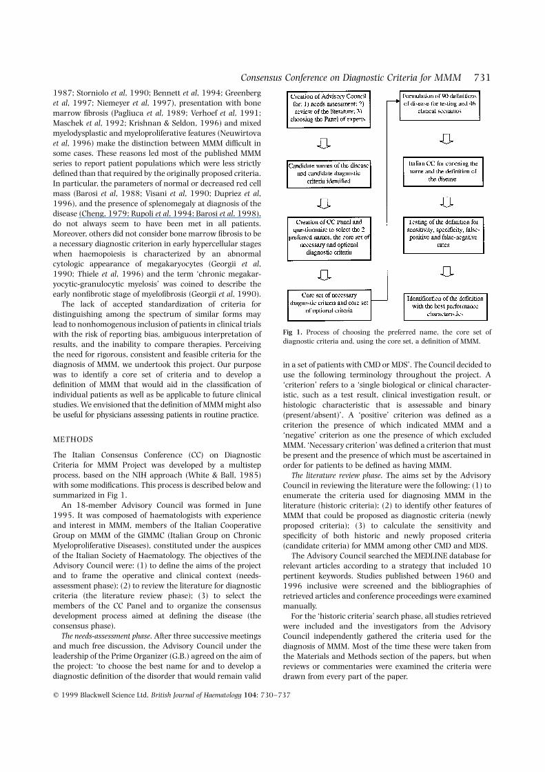
The Italian Consensus Conference (CC) on DiagnosticCriteria for MMM Project was developed by a multistepprocess, based on the NIH approach (White & Ball, 1985)with some modifications. This process is described below andsummarized in Fig 1.
An 18-member Advisory Council was formed in June1995. It was composed of haematologists with experienceand interest in MMM, members of the Italian CooperativeGroup on MMM of the GIMMC (Italian Group on ChronicMyeloproliferative Diseases), constituted under the auspicesof the Italian Society of Haematology. The objectives of theAdvisory Council were: (1) to define the aims of the projectand to frame the operative and clinical context (needs-assessment phase); (2) to review the literature for diagnosticcriteria (the literature review phase); (3) to select themembers of the CC Panel and to organize the consensusdevelopment process aimed at defining the disease (theconsensus phase).
The needs-assessment phase. After three successive meetingsand much free discussion, the Advisory Council under theleadership of the Prime Organizer (G.B.) agreed on the aim ofthe project: ‘to choose the best name for and to develop adiagnostic definition of the disorder that would remain valid
in a set of patients with CMD or MDS’. The Council decided touse the following terminology throughout the project. A‘criterion’ refers to a ‘single biological or clinical character-istic, such as a test result, clinical investigation result, orhistologic characteristic that is assessable and binary(present/absent)’. A ‘positive’ criterion was defined as acriterion the presence of which indicated MMM and a‘negative’ criterion as one the presence of which excludedMMM. ‘Necessary criterion’ was defined a criterion that mustbe present and the presence of which must be ascertained inorder for patients to be defined as having MMM.
The literature review phase. The aims set by the AdvisoryCouncil in reviewing the literature were the following: (1) toenumerate the criteria used for diagnosing MMM in theliterature (historic criteria); (2) to identify other features ofMMM that could be proposed as diagnostic criteria (newlyproposed criteria); (3) to calculate the sensitivity andspecificity of both historic and newly proposed criteria(candidate criteria) for MMM among other CMD and MDS.
The Advisory Council searched the MEDLINE database forrelevant articles according to a strategy that included 10pertinent keywords. Studies published between 1960 and1996 inclusive were screened and the bibliographies ofretrieved articles and conference proceedings were examinedmanually.
For the ‘historic criteria’ search phase, all studies retrievedwere included and the investigators from the AdvisoryCouncil independently gathered the criteria used for thediagnosis of MMM. Most of the time these were taken fromthe Materials and Methods section of the papers, but whenreviews or commentaries were examined the criteria weredrawn from every part of the paper.
Fig 1. Process of choosing the preferred name, the core set ofdiagnostic criteria and, using the core set, a definition of MMM.
For the ‘newly proposed criteria’ search phase, onlyoriginal papers were retrieved and the members of theCouncil were independently assigned to peruse literaturedealing with one of the following topics: clinics, pathology,cytogenetics, immunology, imaging and isotopic studies andin vitro cultures. The reviewers were to search for newcandidate criteria for the diagnosis of MMM.
For the calculation of the specificity and sensitivity phase,only studies dealing with Philadelphia-negative CMD or MDSwith bone marrow fibrosis (the diagnostic context) in whichmore than 10 subjects were enrolled and in which datanecessary to calculate both the sensitivity and specificitywere reported were included. The Advisory Council assumedthat the diagnostic characteristic of any criterion drawnfrom the literature represented an index of diagnosticconfidence which the scientific community had given tothat criterion. The performance of any criterion wascharacterized by its sensitivity and specificity and by itsdiagnostic efficiency (true positive plus true negative rates),i.e. the probability that a criterion correctly identified MMMpatients within the diagnostic context. Diagnostic efficiencywas obtained after correction for disease prevalence, derivedfrom the incidence and mean duration as reported in theliterature (Bilgrami & Greenberg, 1995; Tefferi et al, 1995a,b; Maynadie et al, 1996). The Advisory Council assumed thatthe disutility or cost of false-positive criteria and the disutilityof false-negative criteria were equal.
The consensus phase. Twelve Italian scientists were asked tojoin the Panel for the CC Project. The Panel was composed ofexperts in clinical medicine, clinical research, pathology,outcomes/health services research and medical decisionmaking. The clinical experts were from the fields ofhaematology and medical oncology, and both academicand hospital representatives were included; they werechosen for their representativeness and prominent positionin Italian medical societies.
A pre-consensus conference questionnaire was mailed toeach Panel member asking them: (1) to rank the top choiceamong the disease designations used by the medicalliterature; (2) to select the necessary criteria, that is thecriteria from the list of candidate criteria which shouldnecessarily be ascertained and be present to diagnose MMM;(3) to rank the top six choices among candidate criteria,excluding the necessary ones (optional criteria). An ‘other’category was provided to add names and criteria notincluded in the list. The panelists were also provided with abooklet that summarized the aims of the Project and theresults of the literature search phase. For all candidatecriteria the statistical performance, when available, wasincluded. All the questionnaires were returned and thenames of the disease, the necessary criteria and thecandidate criteria were ranked according to their priorityvotes. The two necessary and the six optional criteria whichranked highest formed the core set of criteria.
A CC was held in Bologna, Italy, in May 1997. Themeeting was attended by the 12 members of the CC Panelwith the assistance of two members of the Advisory Council(G.B. and N.L.L.). The overall aim of the meeting was todecide upon the final name of the disease and a definition of
the disease based on a core set of criteria, using acombination of statistical and consensus formation techni-ques (Delbecq et al, 1975). In order to achieve this there werethree objectives, which are described in consecutive orderbelow.
1. Rate the two names of the disease receiving the highestscores from the questionnaire using the nominal group technique.Participants were asked to silently choose the preferredname. If an 80% consensus on the preferred name was notachieved, the choices were discussed in round-robin fashionand a second vote taken. If an 80% consensus was still notattained, the name was declared undecidable and no furtherattempt was made.
2. Rate each of 46 paper patient profiles as appropriate or notappropriate for a diagnosis of MMM using the nominal grouptechnique. Existing data bases were exploited to build 46patient profiles to be presented to conference attendees. TheGIMMC had conducted a retrospective trial to evaluate theincidence of blast transformation in MMM (n ¼ 560) (Barosiet al, 1998) and the Institute of Internal Medicine andMedical Oncology of the University Hospital of Pavia hadconducted a retrospective trial to evaluate prognostic factorsfor MDS (n ¼ 148) (Ascari et al, 1997). These data bases wereselected as a source of patients. The profiles selected werethose of patients with doubts regarding the diagnosticclassification raised by their clinical records. Absolutevalues at diagnosis were shown for each criterion. Partici-pants at the CC were asked to silently rate each of the 46patient profiles as MMM or not. The moderator then askedeach member how he had voted on each profile. If an 80%consensus about whether a patient had MMM was notachieved, the case was discussed in round-robin fashion anda second vote taken. If an 80% consensus was still notattained, the patient profile was declared uninterpretableand was not considered further in the nominal group.
3. Using the physicians’ consensus judgement as the goldstandard, calculate the percent false-positive and false-negativerates, chi-square, sensitivity and specificity for each of the 90definitions of MMM. By using combinations of the variablesin the core set selected on the questionnaire, the AdvisoryCouncil developed for testing a set of 90 sound definitions ofMMM. Then the ability of the 90 candidate definitions of thedisease to classify individual patients as having MMM or notwas evaluated, and the agreement between the decisionbased on the criteria and the consensus of the physicians wasassessed. Only patient profiles for which physician consensuswas achieved were used. For each definition, we calculatedthe chi-square (1 degree of freedom) and the correspondingP value, sensitivity (ability of the definition to identify ashaving MMM a patient who had been classified as havingMMM by the physicians), specificity (ability of the definitionto identify as not having MMM a patient who had beenclassified as not having MMM by the physicians), rate offalse-positivity ([number falsely identified as having MMM bycriteria/all patients identified as having MMM] × 100), andrate of false-negativity ([number falsely identified as nothaving MMM by the criteria/all patients identified as nothaving MMM] × 100). Two-sided 95% confidence intervals(one-sided 97·5% for values of 100%) for both sensitivity and
q 1999 Blackwell Science Ltd, British Journal of Haematology 104: 730–737
732 Giovanni Barosi et al
733Consensus Conference on Diagnostic Criteria for MMM
q 1999 Blackwell Science Ltd, British Journal of Haematology 104: 730–737
specificity were calculated by the exact binomial method.Those definitions of the disease showing either a sensitivityor specificity of <80% were eliminated from furtherconsideration. We used the kappa statistic as an additionalmeasure of agreement between the physicians’ evaluationand the definitions: k values > 0·7 were considered to beevidence of agreement.
RESULTS
Preferred name of the diseaseAmong the 12 names for the disease found in the literature,the questionnaires returned showed a preference for:‘myelofibrosis with myeloid metaplasia’ and ‘idiopathicmyelofibrosis’. Using the consensus formation technique,the CC Panel agreed upon the designation ‘myelofibrosiswith myeloid metaplasia’.
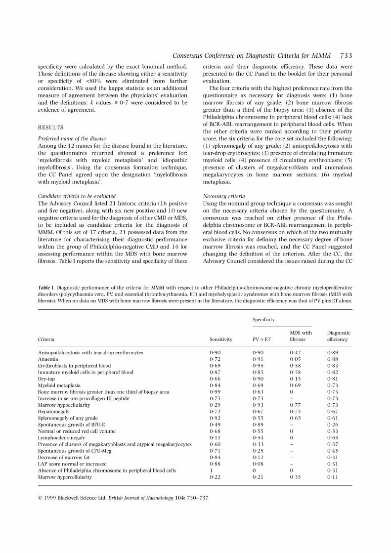
Candidate criteria to be evaluatedThe Advisory Council listed 21 historic criteria (16 positiveand five negative), along with six new positive and 10 newnegative criteria used for the diagnosis of other CMD or MDS,to be included as candidate criteria for the diagnosis ofMMM. Of this set of 37 criteria, 21 possessed data from theliterature for characterizing their diagnostic performancewithin the group of Philadelphia-negative CMD and 14 forassessing performance within the MDS with bone marrowfibrosis. Table I reports the sensitivity and specificity of these
criteria and their diagnostic efficiency. These data werepresented to the CC Panel in the booklet for their personalevaluation.
The four criteria with the highest preference rate from thequestionnaire as necessary for diagnosis were: (1) bonemarrow fibrosis of any grade; (2) bone marrow fibrosisgreater than a third of the biopsy area; (3) absence of thePhiladelphia chromosome in peripheral blood cells; (4) lackof BCR-ABL rearrangement in peripheral blood cells. Whenthe other criteria were ranked according to their priorityscore, the six criteria for the core set included the following:(1) splenomegaly of any grade; (2) anisopoikilocytosis withtear-drop erythrocytes; (3) presence of circulating immaturemyeloid cells; (4) presence of circulating erythroblasts; (5)presence of clusters of megakaryoblasts and anomalousmegakaryocytes in bone marrow sections; (6) myeloidmetaplasia.
Necessary criteriaUsing the nominal group technique a consensus was soughton the necessary criteria chosen by the questionnaire. Aconsensus was reached on either presence of the Phila-delphia chromosome or BCR-ABL rearrangement in periph-eral blood cells. No consensus on which of the two mutuallyexclusive criteria for defining the necessary degree of bonemarrow fibrosis was reached, and the CC Panel suggestedchanging the definition of the criterion. After the CC, theAdvisory Council considered the issues raised during the CC
Table I. Diagnostic performance of the criteria for MMM with respect to other Philadelphia-chromosome-negative chronic myeloproliferativedisorders (polycythaemia vera, PV, and essential thrombocythaemia, ET) and myelodysplastic syndromes with bone marrow fibrosis (MDS withfibrosis). When no data on MDS with bone marrow fibrosis were present in the literature, the diagnostic efficiency was that of PV plus ET alone.
Specificity
MDS with DiagnosticCriteria Sensitivity PV þ ET fibrosis efficiency
Anisopoikilocytosis with tear-drop erythrocytes 0·90 0·90 0·47 0·89Anaemia 0·72 0·91 0·05 0·88Erythroblasts in peripheral blood 0·69 0·95 0·58 0·83Immature myeloid cells in peripheral blood 0·87 0·85 0·58 0·82Dry-tap 0·66 0·90 0·15 0·81Myeloid metaplasia 0·84 0·69 0·69 0·73Bone marrow fibrosis greater than one third of biopsy area 0·99 0·63 0 0·73Increase in serum procollagen III peptide 0·75 0·75 – 0·73Marrow hypocellularity 0·29 0·93 0·77 0·73Hepatomegaly 0·72 0·67 0·73 0·67Splenomegaly of any grade 0·92 0·55 0·65 0·61Spontaneous growth of BFU-E 0·49 0·89 – 0·26Normal or reduced red cell volume 0·68 0·55 0 0·53Lymphoadenomegaly 0·11 0·54 0 0·65Presence of clusters of megakaryoblasts and atypical megakaryocytes 0·60 0·33 – 0·37Spontaneous growth of CFU-Meg 0·71 0·25 – 0·45Decrease of marrow fat 0·84 0·12 – 0·31LAP score normal or increased 0·88 0·08 – 0·31Absence of Philadelphia chromosome in peripheral blood cells 1 0 0 0·31Marrow hypercellularity 0·22 0·21 0·35 0·11
q 1999 Blackwell Science Ltd, British Journal of Haematology 104: 730–737
734 Giovanni Barosi et al
Tabl
eII
.Fin
alre
sult
sfo
rth
eei
ght
best
defin
itio
ns
ofM
MM
.
Sen
sitiv
ity
Spec
ifici
tyan
d9
5%
and
95
%D
efin
itio
n(c
rite
ria
oth
erth
ann
eces
sary
ones
,i.e
.bon
eco
nfid
ence
con
fiden
ceFa
lse-
posi
tive
Fals
e-n
egat
ive
Km
arro
wfib
rosi
san
dla
ckof
the
gen
etic
mar
ker
ofC
ML)
Ch
i-sq
uar
e*in
terv
als
inte
rval
sra
tera
test
atis
tics
Any
two
ofth
eot
her
spr
esen
tw
hen
sple
nom
egal
yis
pres
ent;
21
·10
·88
(0·6
9–0
·97
)1
(0·6
3–1
)†0
0·2
70
·83
any
fou
rof
the
oth
ers
pres
ent
wh
ensp
len
omeg
aly
isab
sen
t
Any
two
ofth
eot
her
spr
esen
tw
hen
sple
nom
egal
yor
14
·80
·92
(0·7
4–0
·99
)0
·75
(0·3
5–0
·97
)0
·08
0·2
50
·67
anis
opoi
kilo
cyto
sis
wit
hte
ar-d
rop
eryt
hro
cyte
sis
pres
ent;
any
fou
rof
the
oth
ers
pres
ent
wh
ensp
len
omeg
aly
and
anis
opoi
kilo
cyto
sis
wit
hte
ar-d
rop
eryt
hro
cyte
sar
eab
sen
t
Any
two
ofth
eot
her
spr
esen
tw
hen
sple
nom
egal
yis
pres
ent
14
·30
·76
(0·5
5–0
·91
)1
(0·6
3–1
)†0
0·4
30
·66
Any
two
ofth
eot
her
spr
esen
tw
hen
sple
nom
egal
yan
d1
2·7
0·7
20
·51
–0·8
8)
1(0
·63
–1)†
00
·47
0·6
2an
isop
oiki
locy
tosi
sw
ith
tear
-dro
per
yth
rocy
tes
are
pres
ent;
the
fou
rot
her
spr
esen
tw
hen
sple
nom
egal
yan
dan
isop
oiki
locy
tosi
sw
ith
tear
-dro
per
yth
rocy
tes
are
abse
nt
Any
two
ofth
eot
her
spr
esen
tw
hen
sple
nom
egal
yis
pres
ent;
12
·70
·72
(0·5
1–0
·88
)1
(0·6
3–1
)†0
0·4
70
·62
the
oth
erfiv
epr
esen
tw
hen
sple
nom
egal
yis
abse
nt
Any
two
ofth
eot
her
spr
esen
tw
hen
sple
nom
egal
yor
12
·70
·72
(0·5
1–0
·88
)1
(0·6
3–1
)†0
0·4
70
·62
anis
opoi
kilo
cyto
sis
wit
hte
ar-d
rop
eryt
hro
cyte
sis
pres
ent
Any
oth
eron
epr
esen
tw
hen
sple
nom
egal
yis
pres
ent;
10
·31
(0·8
6–1
)†0
·38
(0·0
8–0
·75
)0
·17
00
·56
any
thre
eof
the
oth
ers
pres
ent
wh
ensp
len
omeg
aly
isab
sen
t
*Pva
lues
<0
·00
1.†
On
e-si
ded
97
·5%
con
fiden
cein
terv
als.
735Consensus Conference on Diagnostic Criteria for MMM
q 1999 Blackwell Science Ltd, British Journal of Haematology 104: 730–737
discussion, reconvened and framed the following alternativedefinition of bone marrow fibrosis: ‘microscopic evidence ofreticulin fibres at 100× in any area of bone marrow sectionsin biopsies no smaller than 3 × 15 mm’. The question wasmailed and 80% of the replies agreed with this definition ofthe bone marrow fibrosis necessary for diagnosis.
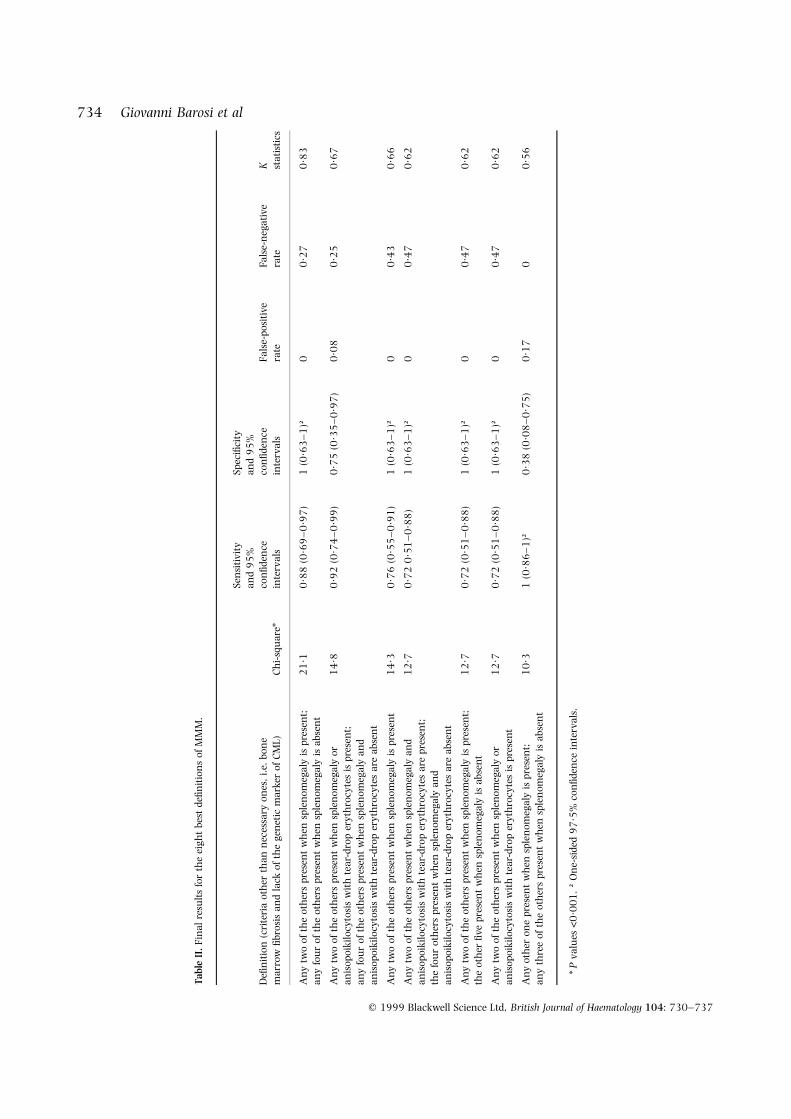
Identification of the best performing definitionThe 12 Panel members scored 25 of the 46 patient profiles ashaving MMM, eight as not having MMM, and 13 asuninterpretable. Eight of the 90 definitions of MMMshowed chi-square >10. These eight definitions, theircorresponding chi-square values, sensitivity and specificity,confidence intervals, P values, false-positive and false-negative rates are shown in Table II. The definition ofMMM that scored highest was as follows (Table III): diffusebone marrow fibrosis necessarily present and Philadelphiachromosome or BCR-ABL rearrangement in peripheral bloodcells necessarily absent; any two of the other core set criteriapresent when splenomegaly is present; any four of the othercore set criteria present when splenomegaly is absent.
DISCUSSION
In this work we report a consensus on the diagnosticdefinition of myelofibrosis with myeloid metaplasia. In theabsence of a specific biological marker for the disease, wewere aware that searching for a definition of MMM raisedboth a true diagnostic issue, i.e. which diagnostic criteriaand how to use them for the diagnosis, and a classificatoryone, i.e. how to distinguish this disease among a spectrum ofdisorders of the same nature and with similar features. Tofocus the problem, the Advisory Council clearly stated thatthe aim of this project was to arrive at a definition of MMMwithin the limited scenario of CMD or MDS.
The task of finding a consensus for diagnostic criteria wascomplicated by the fact that the area is usually characterizedby few ad hoc studies reporting the statistical information
needed for summing up the evidence. Actually, of the 37candidate criteria retrieved from the literature, only 21 hadstudies that were useful for evaluating how specific andsensitive they were in classifying MMM among other chronicPhiladelphia-negative CMD, and only 14 showed suchstudies for a similar evaluation among MDS with bonemarrow fibrosis, i.e. our diagnostic context. The Panel putthe highest value on criteria sensitivity when it had to choosethe criteria that must be present for diagnosis, i.e. absence ofthe genetic marker of CML and evidence of bone marrowfibrosis, and put value on the overall measure of diagnosticefficiency, i.e. a summary index of positive and negativepredictive value, when it had to identify the optional criteria.
Using consensus formation and a statistical approach, theresults of this project suggest that MMM can be defined asfollows (Table III): bone marrow fibrosis and lack of thegenetic marker of CML necessarily present; any two of the sixoptional criteria of the core set present if splenomegaly ispresent, or any four of the optional core set criteria present ifsplenomegaly is absent. As most of the formulations of thecriteria were drawn from the literature, one could noteambiguity in these formulations. For example, pathologistsmight feel uncertain about how to define exactly ‘clusters ofmegakaryoblasts and anomalous megakaryocytes in thebone marrow sections’ (Rupoli et al, 1994; Thiele et al,1996). ‘Myeloid metaplasia’ is also a criterion for which thebiological meaning is clear but for which the methods ofdetection vary. However, investigators in future trials on thedisease should feel free to replace this definition of thecriterion with other more specific definitions or to betterspecify which method of assessment should be fulfilled for theinclusion of patients. The only source of disagreement abouta criterion definition among the consensus panelists was onbone marrow fibrosis. As a result, the new formulationpresented by the Advisory Committee and accepted by thepanelists is as follows: ‘microscopic evidence of reticulinfibres at 100× in any area of bone marrow sections inbiopsies no smaller than 3 × 15 mm’.
The definition of MMM formulated in this work is one thatwas constructed with features of the disease that are notuniversally agreed upon for enrolling cases in clinical trialsor used by clinicians to diagnose the disease in individualpatients. The panelists agreed on the necessary presence ofbone marrow fibrosis for defining MMM. As a consequence,in CMD without myelofibrosis but with megakaryocytematuration defects and atypical histotopography, classifiedby some pathologists as stage 0 myelofibrosis (Georgii et al,1990; Thiele et al, 1996), a diagnosis of MMM shouldavoided. The unnecessary red blood cell volume measure-ment means that both transitional disorders with increasedred cell volume and bone marrow fibrosis (Pettit et al, 1976),as well as post-polycythaemia myeloid metaplasia withpersistently increased red cell volume (Ellis et al, 1986),should be not strictly excluded. It is thus the intention of thepanelists that the term MMM encompass patients with bothidiopathic disease and one that transforms from other CMD.
The results of this work derived from a structuredconsensus process and a statistical analysis on the experts’reactions to 42 real cases. The performing characteristics of
Table III. The Italian criteria for the diagnosis of myelofibrosis withmyeloid metaplasia.
Necessary criteriaA. Diffuse bone marrow fibrosisB. Absence of Philadelphia chromosome or BCR-ABL rearrange-
ment in peripheral blood cells
Optional criteria1. Splenomegaly of any grade2. Anisopoikilocytosis with tear-drop erythrocytes3. Presence of circulating immature myeloid cells4. Presence of circulating erythroblasts5. Presence of cluster of megakaryoblasts and anomalous mega-
karyocytes in bone marrow sections6. Myeloid metaplasia
Diagnosis of MMM is acceptable if the following combinations arepresent: the two necessary criteria plus any other two optionalcriteria when splenomegaly is present; the two necessary criteriaplus any other four optional criteria when splenomegaly is absent.
the resulting disease definition, i.e. its specificity andsensitivity, should be interpreted as a result of uncertaintyinherent both to the consensus process and to the panelists’idea of the disease. The former depends on the numberschosen for the sample of experts and cases used during theconsensus process; the latter captures the absence of clearmarkers for defining the disease. The resulting definition had100% specificity and 88% sensitivity, which thus assured nofalse positives, but some false negatives. These criteriatherefore seemed to be appropriate when used for enrollingpatients in clinical trials.
A consequence of the results of this Project is that patientsto be included in studies on MMM will have to be analysed forthe absence of the CML genetic marker, even in cases inwhich a diagnosis of MMM seems clear. Performing achromosomal or molecular study in MMM patients is notthe rule today (Kvasnicka et al, 1997; Visani et al, 1990;Dupriez et al, 1996). Another important consequence of theresults is that a pivotal role in the diagnosis is in the hands ofpathologists. They have to make a judgment on bonemarrow fibrosis and on the presence of pathological clustersof megakaryoblasts or anomalous megakaryocytes.
In conclusion, this Project proposes a definition of MMMthat has >80% sensitivity and specificity. Use of the criteriaforming the definition should be required for communicationwithin the scientific community and for the inclusion ofpatients in any scientific trials.
ACKNOWLEDGMENTS
The Panel of the Bologna Consensus Conference ondiagnostic criteria for myelofibrosis with myeloid metaplasiawas composed of Professor Sante Tura, Chairman (Bologna),Professor Massimo Aglietta (Turin), Professor Tiziano Barbui(Bergamo), Professor Mario Cazzola (Pavia), Dr AlbertoGrossi (Florence), Dr Luigi Gugliotta (Bologna), ProfessorMario Lazzarino (Pavia), Professor Umberto Magrini (Pavia),Professor Stefano A. Pileri (Bologna), Professor P. Luigi RossiFerrini (Florence), Professor Bruno Rotoli (Naples) andProfessor Giuseppe Saglio (Novara). The authors thank DrEdward H. Giannini and Dr Nicolino Ruperto for their adviceconcerning the methodology used in this project, Dr AngelaPistorio for statistical assistance. Dr Edoardo Ascari andDr Roberta Guarnone provided the data of the patients withMDS used in this work.
REFERENCES
Ascari, E., Balduini, C., Cazzola, M. & Invernizzi, R. (1997) LeSindromi Mielodisplastiche. Luigi Pozzi Ed. Roma.
Barosi, G., Ambrosetti, A., Centra, A., Falcone, A., Finelli, C., Foa, P.,Grossi, A., Guarnone, R., Leoni, P., Luciano, L., Petti, M.C.,Pogliani, E., Russo, D., Ruggeri, M. & Quaglini, S., for the ICSG onMMM (Italian Cooperative Study Group on Myelofibrosis withMyeloid Metaplasia) (1998) Splenectomy and risk of blasttransformation in myelofibrosis with myeloid metaplasia. Blood,91, 3630–3636.
Barosi, G., Baraldi, A., Cazzola, M., Spriano, P. & Magrini, U. (1983)Red cell aplasia in myelofibrosis with myeloid metaplasia: adistinct functional and clinical entity. Cancer, 52, 1290–1296.
Barosi, G., Berzuini, C., Liberato, L.N., Costa, A., Polino, G. &Ascari, E. (1988) A prognostic classification of myelofibrosis withmyeloid metaplasia. British Journal of Haematology, 70, 397–401.
Barosi, G., Buratti, A., Costa, A., Liberato, N.L., Balduini, C., Cazzola,M., Rosti, V., Magrini, U. & Ascari, E. (1991) An atypicalmyeloproliferative disorder with high thrombotic risk and slowdisease progression. Cancer, 68, 2310–2318.
Bennett, J.M., Catovsky, D., Daniel, M.T., Flandrin, G., Galton, D.A.G.,Gralnick, H.R. & Sultan, C. (1982) Proposal for the classificationof the myelodysplastic syndromes. British Journal of Haematology,51, 189–199.
Bennett, J.M., Catovsky, D., Daniel, M.T., Flandrin, G., Galton, D.A.G.,Gralnick, H., Sultan, C. & Cox, C. (1994) The chronic myeloidleukaemias: guidelines for distinguishing chronic granulocytic,atypical chronic myeloid, and chronic myelomonocytic leukae-mia: proposal by the French–American–British CooperativeLeukaemia Group. British Journal of Haematology, 87, 746–754.
Bentley, S.A., Murray, K.H., Lewis, S.M. & Roberts, P.D. (1977)Erythroid hypoplasia in myelofibrosis: a feature associated withblastic transformation. British Journal of Haematology, 36, 41–47.
Berlin, M.I. (1975) Diagnosis and classification of the polycythemias.Seminars in Hematology, 12, 339–351.
Bilgrami, S. & Greenberg, B.R. (1995) Polycythemia rubra vera.Seminars in Oncology, 22, 307–326.
Cheng, D.S. (1979) Idiopathic myelofibrosis without splenomegaly.Cancer, 43, 1761–1765.
Delbecq, A.L., van de Ven, A.H. & Gustafson, D.H. (1975) GroupTechniques for Program Planning: a Guide to Nominal Group andDelphi Processes. Scott, Foresman and Co., Glenview, Ill.
Dupriez, B., Morel, P., Demory, J.L., Lai, J.L., Simon, M., Plantier, I. &Bauters, F. (1996) Prognostic factors in agnogenic myeloidmetaplasia: a report on 195 cases with a new scoring system.Blood, 88, 1013–1018.
Ellis, J.T., Peterson, P., Geller, S.A. & Rappaport, H. (1986) Studies ofthe bone marrow in polycythemia vera and the evolution ofmyelofibrosis and second hematologic malignancies. Seminars inHematology, 23, 144–155.
Fenaux, P., Jouet, J.P., Zandecki, M., Lai, J.L., Simon, M., Pollet, J.P. &Bauters, F. (1987) Chronic and subacute myelomonocyticleukemia in the adult: a report of 60 cases with special referenceto prognostic factors. British Journal of Haematology, 65, 101–106.
Georgii, A., Vykoupil, K.F., Buhr, T., Chortitz, H., Doler, U., Kaloutsi, V.& Werner, M. (1990) Chronic myeloproliferative disorders in bonemarrow biopsies. Pathology Research and Practice, 186, 3–27.
Greenberg, P., Cox, C., LeBeau, M.M., Fenaux, P., Morel, P., Sanz, G.,Sanz, M., Vallespi, T., Hamblin, T., Oscier, D., Ohyashoki, K.,Toyama, K., Aul, C., Mufti, G. & Bennet, J. (1997) Internationalscoring system for evaluating prognosis in myelodysplsticsyndromes. Blood, 89, 2079–2088.
Kouides, P.A. & Bennet, J.M. (1996) Morphology and classification ofthe myelodysplastic syndromes and their pathologic variants.Seminars in Hematology, 33, 95–110.
Krauss, S. (1966) Chronic myelocytic leukemia with featuressimulating myelofibrosis with myeloid metaplasia. Cancer, 19,1321–1332.
Krishnan, K. & Seldon, S. (1996) A new translocation,t(3;6)(q12;24) associated with chronic myelomonocytic leuke-mia and marrow fibrosis. Clinical and Laboratory Haematology,18, 47–49.
Kvasnicka, H.M., Thiele, J., Werden, C., Zankovich, R., Diehl, V. &Fischer, R. (1997) Prognostic factors in idiopathic (primary)osteomyelofibrosis. Cancer, 80, 708–719.
Laszlo, J. (1975) Myeloproliferative disorders (MPD): myelofibrosis,myelosclerosis, extramedullary hematopoiesis, undifferentiated
q 1999 Blackwell Science Ltd, British Journal of Haematology 104: 730–737
736 Giovanni Barosi et al
737Consensus Conference on Diagnostic Criteria for MMM
q 1999 Blackwell Science Ltd, British Journal of Haematology 104: 730–737
MPD, and hemorrhagic thrombocythemia. Seminars in Hematol-ogy, 2, 409–432.
Maschek, H., Georgii, A., Kaloutsi, V., Werner, M., Bandecar, K.,Kressel, M.G., Choritz, H., Freund, M. & Hufnagl, D. (1992)Myelofibrosis in primary myelodysplastic syndromes: a retro-spective study of 352 patients. European Journal of Haematology, 48,208–214.
Maynadie, M., Verret, C., Moskovtchenko, P., Mueneret, F.,Petrella, T., Caillot, D. & Carli, P.M. (1996) Epidemiologicalcharacteristics of myelodysplastic syndrome in a well-definedFrench population. British Journal of Cancer, 74, 288–290.
Murphy, S., Iland, H., Rosenthal, D. & Laszlo, J. (1986) Essentialthrombocythemia: an interim report from the Polycythemia VeraStudy Group. Seminars in Hematology, 23, 177–182.
Neuwirtova, R., Mocikova, K., Musilova, J., Jelinek, J., Havlicek, F.,Michalova, K. & Adamkov, M. (1996) Mixed myelodysplastic andmyeloproliferative syndromes. Leukemia Research, 20, 717–726.
Niemeyer, C.M., Arico, M., Basso, G., Biondi, A., Cantu Rajnoldi, A.,Creutzig, U., Haas, O., Harbott, J., Hasle, H., Kerndrup, G.,Locatelli, F., Mann, G., Stollmann-Gibbels, B., van’t Veer-Korthof, E.T., van Wering, E., Zimmermann, M., and Members ofthe European Working Group on Myelodysplastic Syndromes inChildhood (EWOG-MDS) (1997) Chronic myelomonocytic leuke-mia in childhood: a retrospective analysis of 110 cases. Blood, 10,3534–3543.
Pagliuca, A., Layton, D.M., Manoharan, A., Gordon, S., Green, P.J. &Mufti, G.J. (1989) Myelofibrosis in primary myelodysplasticsyndromes: a clinico-morphological study in 10 cases. BritishJournal of Haematology, 71, 499–504.
Pettit, J.E., Lewis, S.M. & Nicholas, A.W. (1976) Transitionalmyeloproliferative disorders. British Journal of Haematology, 43,167–184.
Polino, G., Barosi, G., Berzuini, A., Castelli, G., Isernia, P., Palestra, P.,Riccardi, A., Rossi, F. & Magrini, U. (1986) Fatty bone marrowwith severe myeloid hypoplasia in idiopathic myelofibrosis.Haematologica, 71, 117–121.
Rupoli, S., Da Lio, L., Sisiti, S., Campanati, G., Salvi, A.,
Brianzoni, M.F., D’Amico, S., Ciciripini, A. & Leoni, P. (1994)Primary myelofibrosis: a detailed statistical analysis of theclinicopathological variables influencing survival. Annals ofHematology, 68, 205–212.
Storniolo, A.M., Moloney, W.C., Rosenthal, D.S., Cox, C. & Bennet,J.M. (1990) Chronic myelomonocytic leukemia. Leukemia, 4,766–770.
Tefferi, A., Silverstein, M.N. & Hoagland, H.C. (1995a) Primarythrombocythemia. Seminars in Oncology, 22, 334–340.
Tefferi, A., Silverstein, M.N. & Noel, P. (1995b) Agnogenic myeloidmetaplasia. Seminars in Oncology, 22, 327–333.
Thiele, J., Kvasnicka, H.M., Werden, C., Zankovich, R., Diehl, V. &Fisher, R. (1996) Idiopathic primary osteo-myelofibrosis: a clinico-pathological study on 208 patients with special emphasis onevolution of disease features, differentiation from essentialthrombocythemia and variables of prognostic impact. Leukemiaand Lymphoma, 22, 303–317.
Thiele, J., Wagner, S., Degel, C., Dienemann, D., Wienhold, S.,Zankovich, R., Fischer, R. & Stein, H. (1990) Megakaryocyteprecursors (pro- and megakaryoblasts) in bone marrow tissuefrom patients with reactive thrombocytosis, polycythemia veraand primary (essential) thrombocythemia;. Virchows Archiv B, CellPathology including Molecular Pathology, 58, 295–302.
Verhoef, G.E., De Wolf-Peeters, C., Ferrant, A., Deprez, S., Meeus, P.,Stul, M., Zachee, P., Cassiman, J.J., Van den Berghe, H. &Boogaerts, M.A. (1991) Myelodysplastic syndromes with bonemarrow fibrosis: a myelodysplastic disorder with proliferativefeatures. Annals of Hematology, 63, 235–241.
Visani, G., Finelli, C., Castelli, U., Petti, M.C., Ricci, P., Vianelli, N.,Gianni, L., Zuffa, E., Aloe Spiriti, M.A., Latagliata, R., Pileri, S.,Magrini, U., Gugliotta, L., Morra, E., Bernasconi, C., Mandelli, F. &Tura, S. (1990) Myelofibrosis with myeloid metaplasia: clinicaland haematological parameters predicting survival in a series of133 patients. British Journal of Haematology, 75, 4–9.
White, L.J. & Ball, J.R. (1985) The clinical efficacy assessment projectof the American College of Physicians. International Journal ofTechnology Assessment in Health Care, 1, 169–185.
Related Documents