The Integumentary System

Welcome message from author
This document is posted to help you gain knowledge. Please leave a comment to let me know what you think about it! Share it to your friends and learn new things together.
Transcript

The Integumentary System

5 2
Integumentary System - General
Skin & its derivatives (sweat glands, oil glands, hair & nails)Skin is largest organ (7% of body by wt.), 1.5 - 4 mm. thickFlexion (flexure) creases – on surface where skin folds during movement (palmar surface)

5 3

5 4
Integumentary System - General
Flexion lines – on surface where skin stretches during movement (elbow)2 Major regions of skin:
Epidermis – superficial, stratified squamous epitheliumDermis – dense irregular C.T.
Skin sits upon hypodermis (superficial fascia, or subcutaneous) – loose C.T.

5 5

5 6
Epidermis
EctodermalUsually < 0.12 mm thick
Thicker at friction sitesKeratinocytes – most abundant cell type
Derived from deepest part of epidermis by mitosis & cytokinesisProduce keratinInterconnected by desmosomes
Other cells present

5 7
5 8
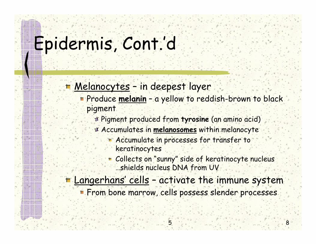
Epidermis, Cont.’d
Melanocytes – in deepest layerProduce melanin – a yellow to reddish-brown to black pigment
Pigment produced from tyrosine (an amino acid)Accumulates in melanosomes within melanocyte
Accumulate in processes for transfer to keratinocytesCollects on “sunny” side of keratinocyte nucleus …shields nucleus DNA from UV
Langerhans’ cells – activate the immune system From bone marrow, cells possess slender processes

5 9

5 10
Epidermis, Cont.’d
Merkel cells – at epidermal-dermal junction
Associated with disc like sensory nerve endingMerkel disc = Merkel cell + nerve ending
Function: touch receptor
5 11
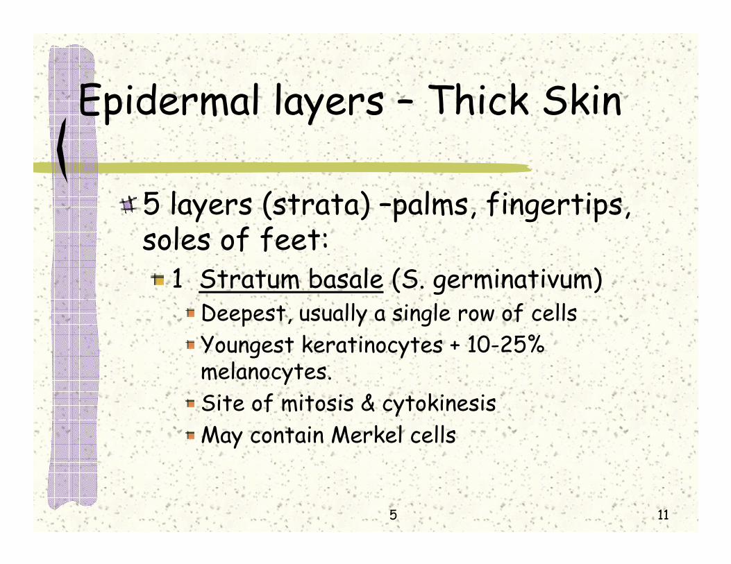
Epidermal layers – Thick Skin
5 layers (strata) –palms, fingertips, soles of feet:
1 Stratum basale (S. germinativum)Deepest, usually a single row of cellsYoungest keratinocytes + 10-25% melanocytes.Site of mitosis & cytokinesisMay contain Merkel cells

5 12

5 13
Epidermal layers – Thick Skin
2 Stratum spinosum – 8-10 cell layersCells may appear flattened & spinyMainly keratinocytes with melanin granules & Langerhans’ cells
3 Stratum granulosum – 3-5 cell layersCells more flattenedNuclei & organelles begin to disintegrate (distance from blood vessels)Accumulate a gummy keratohyaline granules & lamellated granules (contain waterproofing glycolipid)
5 14
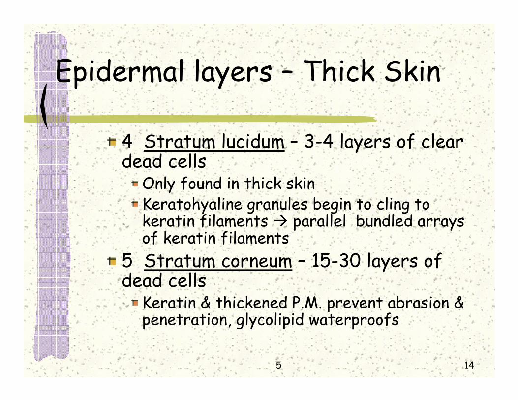
Epidermal layers – Thick Skin
4 Stratum lucidum – 3-4 layers of clear dead cells
Only found in thick skinKeratohyaline granules begin to cling to keratin filaments parallel bundled arrays of keratin filaments
5 Stratum corneum – 15-30 layers of dead cells
Keratin & thickened P.M. prevent abrasion & penetration, glycolipid waterproofs

5 15

5 16
Epidermal layers
Shingle-like cells called cornified cells
Thin skin – Covers most of bodyStratum lucidum absent & other strata may be thinner
Rete pegs – The projections of the epidermis extending between upward projections of dermis (papillary pegs)

5 17

5 18
Dermis – Averages 2 mm. thick
Deeper region of skinVascularContains sensory receptors and nerve fibersContains glandsContains major portion of hair follicles

5 19
Dermis – 2 layers
Papillary layer – thin & superficial 1/5 Loose c. t.Papillary pegs (dermal papillae)
May contain capillary loops, free nerve endings (pain receptors), & Meissner’s corpuscles (touch receptors)
Dermal ridges on palms & solesElevate overlying epidermis to form epidermal ridges …fingerprints

5 20

5 21
Dermis – 2 layers
Reticular layer – deep 4/5 of dermisDense irregular c.t.
Separations or less dense regions called cleavage lines
On limbs…longitudinal, on trunk…circularSignificance ………
Striae (stretch marks) – dermal tearingBlister – separation of epidermis from dermis (from burn or other trauma)

5 22
Skin Color
Melanin – skin coloration reflects the relative kind and amount produced
In humans melanocyte number is same in same region of bodyLocal accumulations: freckles, pig. MolesTan – skin darkening with local accumulation, due to UV exposureExcessive sun: leathery skin, skin cancer, premature aging of skin, depresses immunity

5 23
Skin Color
Photosensitivity – increased skin sensitivity to UV
Antibiotics, perfumes, detergents, other chemicalsBlisterlike lesions and skin peeling
Carotene – yellow to orange pigmentProduced by plantsAccumulates in S. corneum & hypodermis

5 24
Skin Color
Degree of oxygenation of hemoglobinCyanosis - blue appearing skin
Mucous membranes & nail bedsErythema (redness) – blushing(b.v. diameter), fever, hypertension, polycythemia, inflammation or allergyPallor (blanching) – emotional stress (fear, anger) or anemia
5 25
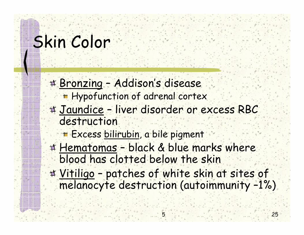
Skin Color
Bronzing – Addison’s diseaseHypofunction of adrenal cortex
Jaundice – liver disorder or excess RBC destruction
Excess bilirubin, a bile pigmentHematomas – black & blue marks where blood has clotted below the skinVitiligo – patches of white skin at sites of melanocyte destruction (autoimmunity –1%)

5 26
Appendages of the skin
Sweat glands (sudoriferous glands)Absent from lips Eccrine – merocrine, general distribution
Simple coiled tubular gland, pore at skin surfaceSweat released by exocytosis, it contains:
99% H2O, NaCl, vitamin C, antibodies, wastes (urea, ammonia, uric acid), lactic acid & some drugspH = 4-6Prevents overheating

5 27

5 28
Appendages of the skin
Apocrine – anal, genital, axillary, & areola & nipple
Duct opens into hair follicle, myoepithelial cellsSweat same as eccrine plus fatty substances and proteins present …body odorDevelop at puberty in response to androgensRespond to emotional stress, pain, sexual foreplay, not temperature
Ceruminous glands – produce cerumin (earwax) in external ear canal

5 29

5 30
Appendages of the skin
Mammary gland – secrete milkSebaceous gland – sebum (oil) product
Holocrine, hormone stimulationUsually develop from & secrete into hair follicleUsually simple, sometimes branched alveolarAbsent from palmar & plantar surfaces

5 31

5 32
Appendages of the skin
Whitehead – sebum blocked sebaceous gl. duct
Blackhead – oxidized & dried sebumAcne – an active inflammation of sebaceous gland with skin eruption
Proprionibacterium acnesSeborrhea –common skin condition with overproduction of sebum
Ex.: Cradle cap of infants – washing to remove excessive oils

5 33

5 34
Hairs & Hair Follicles
Absent from palms, soles, lips, nipples, & parts of ext. genitaliaScalp hair guards against physical trauma, heat loss & sunlightEyelashes shield eyes

5 35
5 36
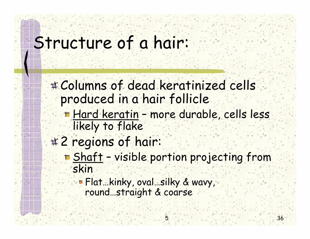
Structure of a hair:
Columns of dead keratinized cells produced in a hair follicle
Hard keratin – more durable, cells less likely to flake
2 regions of hair:Shaft – visible portion projecting from skin
Flat…kinky, oval…silky & wavy, round…straight & coarse

5 37
Structure of a hair:
Root – part embedded in skin3 concentric layers of hair(c.s.):
Medulla – central coreAbsent from fine hairsLarge cells & air spaces
Cortex – surrounds medullaSeveral layers, receives pigment from melanocytes at the base of hair follicle
5 38

5 39
Structure of a hair:
Cuticle – single layer of overlapping cellsProvides strength and compaction“split ends” – when cuticle wears awayHair color – various proportions of melanins
Red hair – iron-containing melanin (trichosiderin)Gray hair – decreased melanin production & air bubbles in medulla

5 40
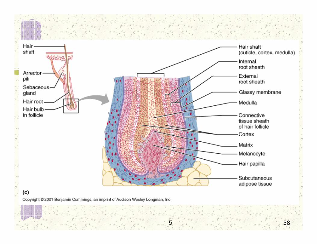
Structure of hair follicle:
Bulb – the expanded base of follicleContains the hair papilla
Indentation containing loose c.t. & blood vessels
Concentric hair follicle regions:Connective tissue sheath (outer)Glassy membrane (b.m. of next region)External root sheath – epithelial

5 41

5 42
Structure of hair follicle:
Internal root sheath – epithelialDoes not extend superficially past sebaceous gland duct3 single-celled layers (Henle’s, Huxley’s & cuticle layer of inner root sheath)Narrows more deeply to single layer, the hair matrix
Actively carry on mitosis & cytokinesisHair matrix cells originate & migrate from a slightly more superficial region, the hair bulge

5 43
Arrector pili
Bundle of smooth muscle running diagonally from c.t. sheath at base of follicle to papillary layer of dermisContraction erection of hair (goosepimples)Increases sebum secretion, other??

5 44

5 45
Types of hair
Vellus hair – a pale, fine hair varietyBody hair of children & adult females
Terminal hair – coarser, longer, often darker hair of eyebrows, scalp, and after puberty axillary & pubic regions
Grow in response to androgens, such as testosterone

5 46
Growth of terminal hair
Influenced by nutrition & hormonesPoor nutrition…poor hair growthIncreased blood flow…enhance hair growthLarge amount of testosterone…luxuriant hair growth
Removal of unwanted hair by electrolysis & laser treatments

5 47
Growth of terminal hair, Cont.’d
Alternate grow state & resting stageResting stage – hair matrix inactive & hair base and bulb atrophiesEye lashes – grow 30 days, rest 105 daysScalp hair – averages 4 year growth, rests a few months

5 48
Hair Thinning & Baldness
After 40’s hair growth slowsThinning and some degree of baldness (alopecia) in both sexes
Usually begins at ant. hair lineTerminal hairs replaced by villus hairs
Male pattern baldnessSex-influenced conditionGene switches on in adulthood & changes response to testosterone
Growth cycle shortens with replacement by villus hair Minoxidil (high B.P. treat.) & life-long finasteride

5 49
Hair Thinning & Baldness
Hair thinning induced by:High fever, surgery, severe emotional trauma, excess Vit. A, some antidepressants & chemotherapy drugsProtein deficient diets
Inadequate protein for keratin synthesisLactation
Protein used for milk production, temporary

5 50
Nails
Plates of tightly packed, hard, keratinized epidermal cells over dorsal distal portions of finger & toes Picking up small objects & scratchingParts: Free edge, body & rootNail bed – surface to which nail body root are attached

5 51

5 52

5 53
Nails
Nail matrix – thickened proximal portion of nail bed (stratum germinativum)
Responsible for nail growthLunula – small portion of nail matrix visible through nail body, crescent shape
Because of thickness – appears white
Eponychium (cuticle) – stratum corneum that occupies the proximal border of nail

5 54

5 55
Nails, Cont.’d
Hyponychium – thickened layer of stratum corneum below free edge of nailNail folds: proximal, lateral & medial
Skin folds overlapping borders of nail

5 56
Functions of the Integumentary System:
Chemical barrier –secretions & melaninAcid mantle (low pH) retards bacterial growthSebum may kill some bacteriaHuman defensin – natural antibioticMelanin helps prevent UV damage

5 57
Functions of the Integumentary System:
Physical (mechanical barrier):Due to continuity of skin & hardness of keratin…bacterial barrierGlycolipids from lamellated granules –prevent the loss from and entry intobody through the skin of H2O & H2O soluble substances
Not blocked are fat soluble : O2, CO2, fat soluble vitamins (A, D, E, K), & steroids

5 58
Functions of the Integumentary System:
Not blocked plant resins : of poison ivy & poison sumacNot blocked organic solvents : acetone, dry cleaner solvent, paint thinner (they dissolve cell lipids) – kidney failure, brain damageNot blocked salts of heavy metals : lead (Pb), mercury (Hg), nickel (Ni)
Pb – anemia & neurological defectsNot blocked penetration enhancing drugs : dialkylamino acetates

5 59
Functions of the Integumentary System:
Biological barriers :Langerhans’ Cells of epidermis & macrophage cells of dermis : phagocytize & present antigen to cells of immune system (T-lymphocytes) –activate immune systemMacrophages of dermis –directly phagocytize & destroy some attackers

5 60
Functions of the Integumentary System:
Body temperature regulation : Cooling factors (heat loss center*):
Sweat glandsDilation of dermal vessels
Warming factors (heat gain center*) :Constriction of dermal vesselsContraction of skeletal muscles
*located in hypothalamusof brain

5 61
Functions of the Integumentary System:
Cutaneous sensation :Meissner’s corpuscles – light touch (clothing)Merkel discs – samePacinian corpuscles – heavy pressureRoot hair plexuses – bending of hairFree nerve endings – painHeat & cold receptors

5 62
Functions of the Integumentary System:
Production of Vitamin D :
7-dehydrocholesterolVit. D3
(Cholecalciferol)
-liver-kidney
Calcitriol
Increased Intestinal absorption of Ca++ + PO4
---
UVlight

5 63
Functions of the Integumentary System:
Keratinocytes:Convert topically applied cortisone to hydrocortisone (anti-inflammatory)Produce hormone that stimulates T-cellsDisarm some cancer causing chemicals
Skin cells :produce collagenase for collagen fiber turn-over (deters wrinkling)

5 64
Functions of the Integumentary System:
Blood reservoirCan hold up to 5% of blood volumeConstriction of dermal vessels diverts blood to “needy site”
Excretion of ammonia, urea, uric acid, some medications, NaCl, excess H2O

5 65
Skin Cancer
Tumors (neoplasms) can be benign or malignant
Most benign – ex.: wartRisk factors:
UV of sunlight – disables tumor suppressor gene (p53) or patched gene (ptc)Frequent irritation: infection, chemicals, traumafas protein – reduces risk in sunburned skin by skin suicide method & peeling of skin

5 66
Skin Cancer
Basal cell carcinoma – 75% of skin cancers, least likely to spread, stratum basale proliferates
Invades dermis & hypodermis, slow growingShiny dome shaped, develop a central ulcer with pearly beaded edge99% curable by surgery

5 67
Skin Cancer
Squamous Cell Carcinoma – about 20%Scaly reddened papule (small rounded elevation) Most often on head (scalp, ears, lower lip) and handsGrows rapidly & metastasizesSurgical removal &/or radiation, if early good results

5 68
Skin Cancer
Melanoma – about 5%Wherever melanin present, 1/3 from molesSpreading brown to black patchMetastasizes rapidly to surrounding lymph & blood vesselsChance of survival if over 4 mm. thick is poorWide surgical incision & immunotherapy

5 69
Skin Cancer
ABCD(E) Rule (American Cancer Society) for melanoma
Asymmetry – 2 sides do not matchBorder – exhibits indentationsColor – contains several colors (black, brown, tan, blue, red)Diameter – 6 mm. or more diameter (pencil eraser)Elevation

5 70
Burns
Loss of tissue fluid & plasmaDehydration, kidney damage, shock, infectionDegree:
1st : Only epidermal layers damagedPain, redness, swelling (as in mild sunburn)
2nd : Damage of epidermis & some dermisBlisters (vesicles)
3rd : No epidermis at site, extends subdermally

5 71
Burns, Cont.’d
May call for graftingSplit skin grafting – epidermis & ½ of dermisArtificial skin grafts – silicone(epid.) + collagen & cartilage(dermis), + cultured patient’s cellsRule of nines – estimating burn extent
H 9, SL9, Trunk36, IL18, Genetalia1

5 72

5 73
Clinical Terms:
Boils -Milia- small white spotsCarbuncles on forehead & noseCold soresDecubitus ulcer (bedsore)DermatologyImpetigoVernix caseosa – white cheesy sebum of newborn
Related Documents