J. Zool., Lond. (2005) 266, 385–394 C 2005 The Zoological Society of London Printed in the United Kingdom doi:10.1017/S095283690500703X The inguinal macroglands of the frog Physalaemus nattereri (Leptodactylidae): structure, toxic secretion and relationship with deimatic behaviour R. Lenzi-Mattos 1 , M. M. Antoniazzi 1 , C. F. B. Haddad 2 , D. V. Tambourgi 3 , M. T. Rodrigues 4 and C. Jared 1,4∗ 1 Laborat´ orio de Biologia Celular, Instituto Butantan, Av. Vital Brazil 1500, CEP 05503-900, S˜ ao Paulo, Brazil 2 Departamento de Zoologia, Instituto de Biociˆ encias, Universidade Estadual Paulista, Rio Claro (SP), Brazil 3 Laborat´ orio de Imunoqu´ ımica, Instituto Butantan, S˜ ao Paulo, Brazil 4 Departamento de Zoologia, Instituto de Biociˆ encias, Universidade de S˜ ao Paulo, S˜ ao Paulo, Brazil (Accepted 5 January 2005) Abstract Amphibian skin is characterized by the presence of mucous glands, related to cutaneous breathing, reproduction and water balance, and granular glands, related to the production of toxins used in defence. In some species the granular glands can form accumulations in certain regions of the body. This is the case for inguinal macroglands of the leptodactylid frog Physalaemus nattereri, where these structures form a pair of black discs associated with deimatic behaviour. The morphology of the inguinal macroglands and their secretion were studied in this species and correlated to deimatic behaviour. The inguinal macroglands are formed from elongated granular glands that, in contrast with the granular glands of the rest of the skin, have small spherical granules with a proteinic content. In the dermis of the whole body, except for the inguinal macroglands and the inguinal region, a well-developed calcified dermal layer is observed. During deimatic behaviour these macroglands discourage a potential predator from attacking, but if visual cues are insufficient and the predator persists in the attack, a toxic secretion is eliminated in its mouth. This elimination is favoured by the absence of a calcified dermal layer in the macroglands, which makes the dermal region softer than the rest of the dorsal skin. Key words: Amphibia, integument, inguinal macroglands, skin secretion, Physalaemus nattereri, defence INTRODUCTION Amphibian integument plays an important role in the defence against predators and microorganisms (Zug, 1993), which is directly associated to the presence in the dermis of mucous and granular glands responsible for the secretion of mucus and toxins, respectively (Lutz, 1966; Stebbins & Cohen, 1995; Toledo & Jared, 1995). In anurans, the mucous glands are basically formed by simple acini, and the granular glands are formed by secretory syncytia without lumen. Both types of glands are involved by a myoepithelial monolayer (Delfino, Nosi, Brizzi et al., 2001; Delfino, Nosi & Giaci, 2001) generally associated with the expulsion of the secretion. A great variety of chemical compounds is present in the secretion of the granular glands. For this reason, amphibian skin has been considered as a valuable source for the prospecting of new bioactive compounds (Toledo & Jared, 1995; Clarke, 1997). *All correspondence to: C. Jared, Laborat´ orio de Biologia Celular, Instituto Butantan, Av. Vital Brazil 1500, CEP 05503-900, S˜ ao Paulo, Brazil. E-mail: [email protected] In many species the granular glands form clusters, or macroglands (Toledo & Jared, 1995), which are strategi- cally located on the body surface. In toads (bufonids) macroglands are common, forming the parotoids in the post-orbital region (Lutz, 1966; Hostetler & Cannon, 1974; Duellman & Trueb, 1986; Pough et al., 2001). In certain frogs, as in the lepto- dactylids, lumbar and inguinal macroglands are present in the dorsal skin (Toledo & Jared, 1989a, 1995; Toledo, Jared & Brunner, 1996). This is the case for Physalaemus nattereri, a frog occurring in mid-west and south- east Brazilian savanna (Cei, 1980). One of the most conspicuous characteristics of this species is the presence, both in males and females, of a pair of black and round inguinal glands, emarginated by a white contour (Vizotto, 1964; Lynch, 1970; Sazima & Caramaschi, 1986), giving the impression of two large black eyes. In this paper, the morphology and histochemistry of the inguinal macroglands of Physalaemus nattereri are studied, and a preliminary analysis made of some biochemical and toxinological parameters of the bulk secretion extracted from these macroglands and their correlation with defensive behaviour. The morphological data revealed a specialized glandular structure, with

Welcome message from author
This document is posted to help you gain knowledge. Please leave a comment to let me know what you think about it! Share it to your friends and learn new things together.
Transcript
J. Zool., Lond. (2005) 266, 385–394 C© 2005 The Zoological Society of London Printed in the United Kingdom doi:10.1017/S095283690500703X
The inguinal macroglands of the frog Physalaemus nattereri(Leptodactylidae): structure, toxic secretion and relationshipwith deimatic behaviour
R. Lenzi-Mattos1, M. M. Antoniazzi1, C. F. B. Haddad2, D. V. Tambourgi3, M. T. Rodrigues4 and C. Jared1,4∗1 Laboratorio de Biologia Celular, Instituto Butantan, Av. Vital Brazil 1500, CEP 05503-900, Sao Paulo, Brazil2 Departamento de Zoologia, Instituto de Biociencias, Universidade Estadual Paulista, Rio Claro (SP), Brazil3 Laboratorio de Imunoquımica, Instituto Butantan, Sao Paulo, Brazil4 Departamento de Zoologia, Instituto de Biociencias, Universidade de Sao Paulo, Sao Paulo, Brazil
(Accepted 5 January 2005)
AbstractAmphibian skin is characterized by the presence of mucous glands, related to cutaneous breathing, reproductionand water balance, and granular glands, related to the production of toxins used in defence. In some species thegranular glands can form accumulations in certain regions of the body. This is the case for inguinal macroglandsof the leptodactylid frog Physalaemus nattereri, where these structures form a pair of black discs associated withdeimatic behaviour. The morphology of the inguinal macroglands and their secretion were studied in this speciesand correlated to deimatic behaviour. The inguinal macroglands are formed from elongated granular glands that,in contrast with the granular glands of the rest of the skin, have small spherical granules with a proteinic content.In the dermis of the whole body, except for the inguinal macroglands and the inguinal region, a well-developedcalcified dermal layer is observed. During deimatic behaviour these macroglands discourage a potential predatorfrom attacking, but if visual cues are insufficient and the predator persists in the attack, a toxic secretion is eliminatedin its mouth. This elimination is favoured by the absence of a calcified dermal layer in the macroglands, whichmakes the dermal region softer than the rest of the dorsal skin.
Key words: Amphibia, integument, inguinal macroglands, skin secretion, Physalaemus nattereri, defence
INTRODUCTION
Amphibian integument plays an important role in thedefence against predators and microorganisms (Zug,1993), which is directly associated to the presence inthe dermis of mucous and granular glands responsiblefor the secretion of mucus and toxins, respectively (Lutz,1966; Stebbins & Cohen, 1995; Toledo & Jared, 1995). Inanurans, the mucous glands are basically formed by simpleacini, and the granular glands are formed by secretorysyncytia without lumen. Both types of glands are involvedby a myoepithelial monolayer (Delfino, Nosi, Brizzi et al.,2001; Delfino, Nosi & Giaci, 2001) generally associatedwith the expulsion of the secretion. A great variety ofchemical compounds is present in the secretion of thegranular glands. For this reason, amphibian skin has beenconsidered as a valuable source for the prospecting ofnew bioactive compounds (Toledo & Jared, 1995; Clarke,1997).
*All correspondence to: C. Jared, Laboratorio de Biologia Celular, InstitutoButantan, Av. Vital Brazil 1500, CEP 05503-900, Sao Paulo, Brazil.E-mail: [email protected]
In many species the granular glands form clusters, ormacroglands (Toledo & Jared, 1995), which are strategi-cally located on the body surface. In toads(bufonids) macroglands are common, forming theparotoids in the post-orbital region (Lutz, 1966;Hostetler & Cannon, 1974; Duellman & Trueb, 1986;Pough et al., 2001). In certain frogs, as in the lepto-dactylids, lumbar and inguinal macroglands are presentin the dorsal skin (Toledo & Jared, 1989a, 1995; Toledo,Jared & Brunner, 1996). This is the case for Physalaemusnattereri, a frog occurring in mid-west and south-east Brazilian savanna (Cei, 1980). One of the mostconspicuous characteristics of this species is the presence,both in males and females, of a pair of black and roundinguinal glands, emarginated by a white contour (Vizotto,1964; Lynch, 1970; Sazima & Caramaschi, 1986), givingthe impression of two large black eyes.
In this paper, the morphology and histochemistryof the inguinal macroglands of Physalaemus nattereriare studied, and a preliminary analysis made of somebiochemical and toxinological parameters of the bulksecretion extracted from these macroglands and theircorrelation with defensive behaviour. The morphologicaldata revealed a specialized glandular structure, with
386 R. LENZI-MATTOS ET AL.
secretion granules totally different from those observedin the rest of the skin. The analysis of the secretiondemonstrated a high toxic potential.
Our results lead to the conclusion that the defence ofP. nattereri may occur in two successive phases. First,the frog may try to intimidate a possible predator byexhibiting the large and false ‘black eyes’ when harassed.If this display is not effective and the predator persists intrying to bite the anuran, it may then be affected by thetoxic secretion expulsed from the macroglands inside itsmouth.
MATERIAL AND METHODS
Animals
Ten adult specimens of Physalaemus nattereriSteindachner, 1863 (snout–vent length 3.5–4.0 cm), werecollected in the district of Rio Claro, State of Sao Paulo(Brazil). The animals were maintained for 4 months inthe vivarium of the Laboratory of Cellular Biology of theInstituto Butantan in terraria containing humid earth atthe bottom and dry branches. Food was supplied everyother day and was composed of beetles Tenebrio molitor,crickets Gryllus sp. and cockroaches Pycnocellussurinamensis. During this period, behavioural observa-tions were made and the secretion of the inguinal glandsof 5 individuals was extracted monthly. The other non-extracted animals were then killed with an intraperitonealoverdose of Thionembutal, and samples of the skin andinguinal glands were removed for morphological studies.Voucher specimens were deposited in the collectionof the Museum of Zoology, University of Sao Paulo(MZUSP).
Behavioural observations
The observations were made when the frogs weremanipulated during the maintenance of the terraria (every3 days). To stimulate defensive behaviour, the frogs wereteased with the observer’s forefinger. The defensivedisplay was photographically registered and notes weretaken.
Histology
Fragments of the dorsal, ventral and inguinal skin andboth inguinal macroglands were removed from the frogs.For histological preparation the skin was cut in strips ofc. 3 × 5 mm using a razor blade. Skin samples and theentire glands were fixed in 4% paraformaldehyde in PBS0.1 M, pH 7.2 for 24 h. The material was embedded eitherin Leica historesin or paraffin.
For general study of the tissues, 2 µm historesin sectionswere stained with toluidine-fuchsin (Junqueira, 1995) and5–7 µm paraffin sections were stained with haematoxylin-eosin. Collagen fibres were analysed using picro-Siriusstaining followed by polarized microscopy (Junqueira,Bignolas & Brentani, 1979).
Histochemistry
Historesin and paraffin sections were submitted to thefollowing histochemical methods: periodic-acid Schiff(PAS) and alcian blue pH 2.5 for identification of neutraland acidic mucosubstances, respectively; von Kossa foridentification of calcium phosphate; bromophenol bluefor identification of proteins (Bancroft & Stevens, 1996).
Photomicrography
Photographs were obtained in a Leica DMLB microscopeequipped with a Leica MPS60 photographic system.Sections stained with picro-Sirius were photographedunder polarized light in an Olympus BX-60 microscopeequipped with a PM-C35DX photographic system.
Transmission electron microscopy
Skin and inguinal macrogland fragments (c. 1 mm3) werefixed in Karnovsky solution, pH 7.2 (Karnovsky, 1965)for 24 h, post-fixed in 1% osmium tetroxide, contrasted in2% uranyl acetate, dehydrated in ethanol and embedded inepoxy resin. Ultrathin sections were examined in a LEO906E, operating at 80 kV.
Collection of secretion from the inguinal macroglands
The secretion of the inguinal glands of 5 frogs wascollected by covering the macroglands with 2-cm2 filterpaper and squeezing them laterally between the thumb andthe forefinger. The paper and resulting secretion was thensoaked in 1 ml of PBS and crushed with a micropipettetip. After centrifugation, the resulting milky solution wasstored at − 20 ◦C and then lyophilized.
Lethality and LD50 calculation of the macroglandsecretion
The tests were made in BALB/c mice aged 2 months andweighing 18–20 g, obtained from Bioterio de Criacao deAnimais Isogenicos do Instituto Butantan, SP, Brazil. Themice were treated and maintained under strict ethical con-ditions according to animal welfare international recom-mendation (Committee Members, International Societyon Toxinology, 1991). The lethal toxicity of P. nattererimacrogland secretion was assessed in BALB/c miceby intraperitoneal injection of different amounts of thesecretion (5, 10, 15, 25, 40 and 80 µg per animal) dilutedin 200 µl of PBS. Five mice were used for each toxindose. The LD50 was calculated by probity analysis of deathoccurring within 72 h of sample injection.
Electrophoresis
Samples of P. nattereri macrogland secretion weresolubilized in non-reducing sample buffer, run on 12%SDS-PAGE (Laemmli, 1970) and stained with silver(Morrissey, 1980).
Inguinal macroglands of the frog Physalaemus nattereri 387
Zymography
Gelatinase activity of P. nattereri secretion was analysedby zymography (Kleiner & Stetler-Stewensow, 1994).Samples (3 µg) were run under non-reducing conditionson a 10% polyacrylamide gel containing 1% gelatine. Thegels were washed twice for 30 min at room temperaturein 2.5% Triton X-100, and incubated overnight at 37 ◦Cin zymography buffer (50 mm Tris-HCl, 200 mm NaCl,10 mm CaCl2, 0.05% Brij-35; pH 8.3). Gels were stainedin Coomassie brilliant blue solution (40% methanol, 10%acetic acid and 0.1% Coomassie brilliant blue).
RESULTS
Defensive behaviour of Physalaemus nattereri in captivity
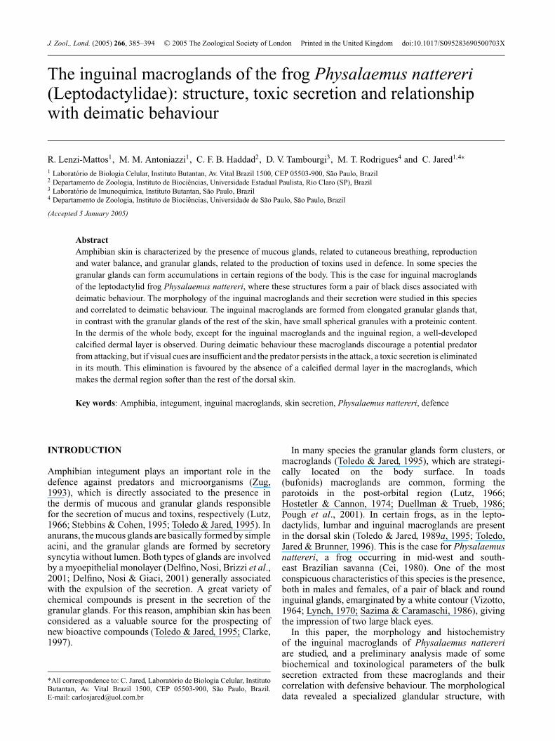
When threatened, P. nattereri (Fig. 1a), usually presentsa stereotyped set of postures and behaviours, consistingof puffing up the body laterally by inflation of the lungs,turning the back to the threatening agent and elevatingthe hind parts (Fig. 1b, c). In this posture the blackinguinal glands are exhibited together with the prominentcoccyx, giving the observer the impression of a large facewith a pair of black eyes (Fig. 1b). The more the frogis mechanically stimulated, the more conspicuous thisdeimatic display is observed. When intensively harassedduring the deimatic displays, the black skin covering themacroglands may turn whitish as a consequence of a milkysecretion.
General morphological and histochemicalcharacteristics of the skin
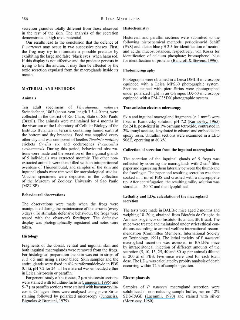
The skin is characterized by a large number of mucousand granular glands in the more external region of thedermis, the stratum spongiosum, just below the epidermis.The dorsal skin, when compared to the ventral skin,has a larger number of pigment cells (Fig. 2a), mainlychromatophores, with their characteristic black granules(Fig. 2c). The epidermis is composed of at least four layersof cells. The more superficial stratum corneum is formedby a single layer of flat nucleated cells (Fig. 2a).
The dermis is composed of the stratum spongiosum,filled by loose connective tissue, and the stratumcompactum, characterized by the dense connective tissueconsisting mainly of collagen fibres. Chromatophoresand cutaneous glands are located within the stratumspongiosum (Fig. 2a).
The mucous glands are acinar, with a simple duct andlarger diameter measuring c. 45 µm (Fig. 2a). The aciniare formed by cells whose secretion is positive to PAS andalcian blue pH 2.5 methods.
The granular glands (G1) are syncytial, without alumen (Fig. 2a), and have a sole excretory duct. Theyare larger than the mucous glands, with a maximumdiameter of c. 80 µm. The secretory syncytium ischaracterized by peripheral nuclei and by many large
(a)
(b)
(c)
5 mm
5 mm
5 mm
Fig. 1. Physalaemus nattereri. (a) Normal lateral view: (b, c)during defensive (deimatic) behaviour, exhibiting the inguinalmacroglands: (b) back view; (c) lateral view.
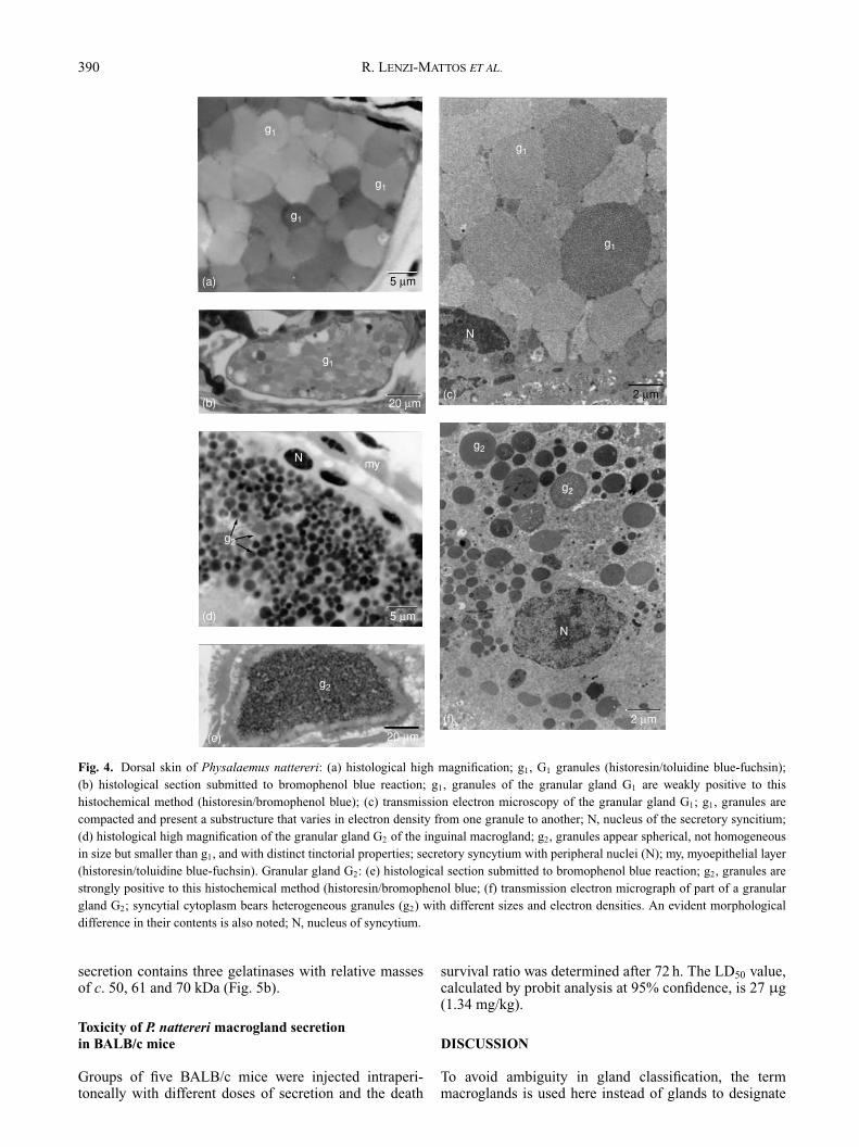
compacted granules. When stained with toluidine-fuchsin(Fig. 4a) or haematoxylin-eosin, these granules seemto be non-homogeneous both in size and stain affinity.Histochemistry reveals that these granules are negative
388 R. LENZI-MATTOS ET AL.
(a)
sc
e
MG1
SC
N
G1
MV
e
(b)
(c) 1 µm
20 µm
20 µm
Fig. 2. Physalaemus nattereri. Histological section of dorsal skin:(a) where the two types of glands are observed; M, mucous gland;G1, granular gland; arrowhead, stratum corneum; e, epidermis;*, chromatophores; sc, stratum compactum; arrow, subcutaneoustissue; v, blood vessel (historesin/toluidine blue-fuchsin staining);(b) where the calcified dermal layer is evidenced as a black line(arrows) basally contouring the cutaneous glands (historesin/vonKossa reaction), abbreviations as (a). (c) Transmission electronmicrograph of the calcified dermal layer, consisting of globularstructures (arrows) immersed in an electron-transparent matrix;upper right region shows a melanophore with melanosomes (*).N, Melanophore nucleus; sc, stratum compactum.
to PAS and alcian blue pH 2.5 and moderately positiveto bromophenol blue (Fig. 4b). When observed at atransmission electron microscope (Fig. 4c), the granulesare shown to be enclosed by a membrane and are differentsizes and electron densities, despite all being made up ofmany circular subunits.
Both mucous and granular glands are enclosed by amonolayer of myoepithelial cells. These glands presentthe same morphological and histochemical characteristicsin both dorsal and ventral skin.
Between the stratum spongiosum and the stratumcompactum, a well-defined, calcified dermal layer(Eberth–Kastschenko layer) is present, contouring themucous and granular glands. This layer is evidencedby the method of von Kossa (Fig. 2b), which indicatesthe presence of calcium phosphate. The calcified layeris well developed and continuous in the dorsal skin(Fig. 2b) but less developed and discontinuous in theventral skin. Transmission electron microscopy shows thatit is composed of irregular globules, with an homogeneouscontent of medium electron density, immersed in anelectron transparent matrix (Fig. 2c).
The skin is internally delimited by a thin layer ofsubcutaneous tissue rich in blood vessels (Fig. 2a).
Histology and histochemistry of the inguinalmacroglands
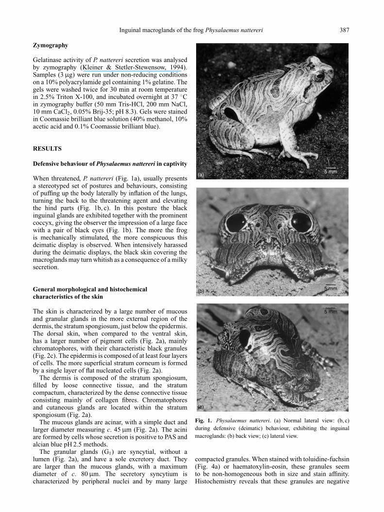
The inguinal macroglands comprise a pair of well-definedcutaneous structures. They are characterized by a clusterof granular glands (Fig. 3a, b), that are here referred to asG2 to differentiate them from those present in the rest ofdorsal and ventral skin (G1). G2 are elongated and muchlarger than G1, measuring c. 340 µm high and 140 µmwide. Similarly to G1, they are connected to the exteriorby a sole duct (Fig. 3b, insert), do not have lumen and areenclosed by a myoepithelial monolayer (Fig. 4d). Theyare located side by side in the dermis in a honeycomb-like arrangement, each alveolus corresponding to onesyncytium (Fig. 3a, b). The alveoli are immersed in denseconnective tissue, forming a framework evidenced bythe picro-Sirius method followed by polarized lightmicroscopy (Fig. 3a). Under these conditions, it is possibleto distinguish at least two types of collagen fibres inthis framework, appearing in green and orange. At thetransmission electron microscope, the epidermis lining themacroglands shows a microrugosity at the surface (Fig. 3c,insert). Among the keratinocytes, it bears melanophoresand many sparse melanin granules, which are found evenwithin the cornified layer (Fig. 3c). In the same way, thedermis in this region is highly pigmented, exhibiting anexceptional number of melanophores (Fig. 3c). This heavypigmentation is responsible for the black colour observedin the inguinal macroglands.
Granules present in G2 are much smaller than thegranules in G1 (compare Fig. 4a & 4d). They are sphericaland very eosinophilic when submitted to haematoxylin-eosin staining, and have an intense affinity to toluidine blue(Figs 3b & 4d). They are strongly positive to bromophenol
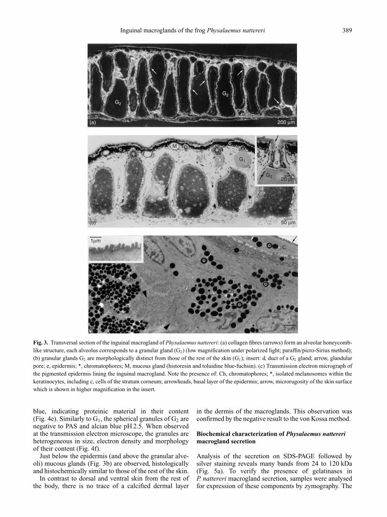
Inguinal macroglands of the frog Physalaemus nattereri 389
(a)
(b)
G2
G2
G1
G2
G2
M
Ch
Ch
C
ee
d
G2
200 µm
(c)
20 µm
50 µm
1µm
2 µm
Fig. 3. Transversal section of the inguinal macrogland of Physalaemus nattereri: (a) collagen fibres (arrows) form an alveolar honeycomb-like structure, each alveolus corresponds to a granular gland (G2) (low magnification under polarized light; paraffin/picro-Sirius method);(b) granular glands G2 are morphologically distinct from those of the rest of the skin (G1); insert: d, duct of a G2 gland; arrow, glandularpore; e, epidermis; *, chromatophores; M, mucous gland (historesin and toluidine blue-fuchsin). (c) Transmission electron micrograph ofthe pigmented epidermis lining the inguinal macrogland. Note the presence of: Ch, chromatophores; *, isolated melanosomes within thekeratinocytes, including c, cells of the stratum corneum; arrowheads, basal layer of the epidermis; arrow, microrugosity of the skin surfacewhich is shown in higher magnification in the insert.
blue, indicating proteinic material in their content(Fig. 4e). Similarly to G1, the spherical granules of G2 arenegative to PAS and alcian blue pH 2.5. When observedat the transmission electron microscope, the granules areheterogeneous in size, electron density and morphologyof their content (Fig. 4f).
Just below the epidermis (and above the granular alve-oli) mucous glands (Fig. 3b) are observed, histologicallyand histochemically similar to those of the rest of the skin.
In contrast to dorsal and ventral skin from the rest ofthe body, there is no trace of a calcified dermal layer
in the dermis of the macroglands. This observation wasconfirmed by the negative result to the von Kossa method.
Biochemical characterization of Physalaemus nattererimacrogland secretion
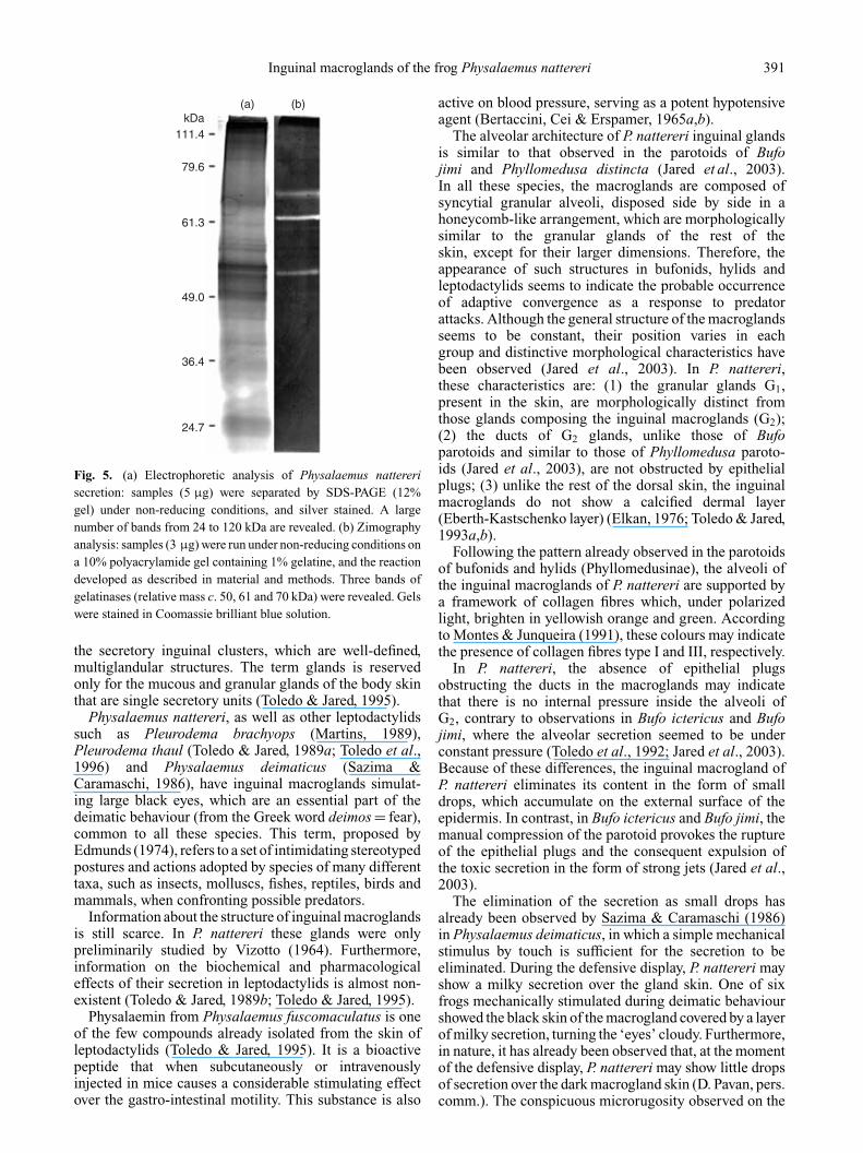
Analysis of the secretion on SDS-PAGE followed bysilver staining reveals many bands from 24 to 120 kDa(Fig. 5a). To verify the presence of gelatinases inP. nattereri macrogland secretion, samples were analysedfor expression of these components by zymography. The
390 R. LENZI-MATTOS ET AL.
(a)
g1
g2
g2
g2N my
g2
g1
g1
g1
g1
g1
(b)
(d)
(e)
(f)
(c)
5 µm
20 µm
5 µm
20 µm
2 µm
2 µm
N
N
Fig. 4. Dorsal skin of Physalaemus nattereri: (a) histological high magnification; g1, G1 granules (historesin/toluidine blue-fuchsin);(b) histological section submitted to bromophenol blue reaction; g1, granules of the granular gland G1 are weakly positive to thishistochemical method (historesin/bromophenol blue); (c) transmission electron microscopy of the granular gland G1; g1, granules arecompacted and present a substructure that varies in electron density from one granule to another; N, nucleus of the secretory syncitium;(d) histological high magnification of the granular gland G2 of the inguinal macrogland; g2, granules appear spherical, not homogeneousin size but smaller than g1, and with distinct tinctorial properties; secretory syncytium with peripheral nuclei (N); my, myoepithelial layer(historesin/toluidine blue-fuchsin). Granular gland G2: (e) histological section submitted to bromophenol blue reaction; g2, granules arestrongly positive to this histochemical method (historesin/bromophenol blue; (f) transmission electron micrograph of part of a granulargland G2; syncytial cytoplasm bears heterogeneous granules (g2) with different sizes and electron densities. An evident morphologicaldifference in their contents is also noted; N, nucleus of syncytium.
secretion contains three gelatinases with relative massesof c. 50, 61 and 70 kDa (Fig. 5b).
Toxicity of P. nattereri macrogland secretionin BALB/c mice
Groups of five BALB/c mice were injected intraperi-toneally with different doses of secretion and the death
survival ratio was determined after 72 h. The LD50 value,calculated by probit analysis at 95% confidence, is 27 µg(1.34 mg/kg).
DISCUSSION
To avoid ambiguity in gland classification, the termmacroglands is used here instead of glands to designate
Inguinal macroglands of the frog Physalaemus nattereri 391
24.7
36.4
49.0
61.3
79.6
111.4kDa
(a) (b)
Fig. 5. (a) Electrophoretic analysis of Physalaemus nattererisecretion: samples (5 µg) were separated by SDS-PAGE (12%gel) under non-reducing conditions, and silver stained. A largenumber of bands from 24 to 120 kDa are revealed. (b) Zimographyanalysis: samples (3 µg) were run under non-reducing conditions ona 10% polyacrylamide gel containing 1% gelatine, and the reactiondeveloped as described in material and methods. Three bands ofgelatinases (relative mass c. 50, 61 and 70 kDa) were revealed. Gelswere stained in Coomassie brilliant blue solution.
the secretory inguinal clusters, which are well-defined,multiglandular structures. The term glands is reservedonly for the mucous and granular glands of the body skinthat are single secretory units (Toledo & Jared, 1995).
Physalaemus nattereri, as well as other leptodactylidssuch as Pleurodema brachyops (Martins, 1989),Pleurodema thaul (Toledo & Jared, 1989a; Toledo et al.,1996) and Physalaemus deimaticus (Sazima &Caramaschi, 1986), have inguinal macroglands simulat-ing large black eyes, which are an essential part of thedeimatic behaviour (from the Greek word deimos = fear),common to all these species. This term, proposed byEdmunds (1974), refers to a set of intimidating stereotypedpostures and actions adopted by species of many differenttaxa, such as insects, molluscs, fishes, reptiles, birds andmammals, when confronting possible predators.
Information about the structure of inguinal macroglandsis still scarce. In P. nattereri these glands were onlypreliminarily studied by Vizotto (1964). Furthermore,information on the biochemical and pharmacologicaleffects of their secretion in leptodactylids is almost non-existent (Toledo & Jared, 1989b; Toledo & Jared, 1995).
Physalaemin from Physalaemus fuscomaculatus is oneof the few compounds already isolated from the skin ofleptodactylids (Toledo & Jared, 1995). It is a bioactivepeptide that when subcutaneously or intravenouslyinjected in mice causes a considerable stimulating effectover the gastro-intestinal motility. This substance is also
active on blood pressure, serving as a potent hypotensiveagent (Bertaccini, Cei & Erspamer, 1965a,b).
The alveolar architecture of P. nattereri inguinal glandsis similar to that observed in the parotoids of Bufojimi and Phyllomedusa distincta (Jared et al., 2003).In all these species, the macroglands are composed ofsyncytial granular alveoli, disposed side by side in ahoneycomb-like arrangement, which are morphologicallysimilar to the granular glands of the rest of theskin, except for their larger dimensions. Therefore, theappearance of such structures in bufonids, hylids andleptodactylids seems to indicate the probable occurrenceof adaptive convergence as a response to predatorattacks. Although the general structure of the macroglandsseems to be constant, their position varies in eachgroup and distinctive morphological characteristics havebeen observed (Jared et al., 2003). In P. nattereri,these characteristics are: (1) the granular glands G1,present in the skin, are morphologically distinct fromthose glands composing the inguinal macroglands (G2);(2) the ducts of G2 glands, unlike those of Bufoparotoids and similar to those of Phyllomedusa paroto-ids (Jared et al., 2003), are not obstructed by epithelialplugs; (3) unlike the rest of the dorsal skin, the inguinalmacroglands do not show a calcified dermal layer(Eberth-Kastschenko layer) (Elkan, 1976; Toledo & Jared,1993a,b).
Following the pattern already observed in the parotoidsof bufonids and hylids (Phyllomedusinae), the alveoli ofthe inguinal macroglands of P. nattereri are supported bya framework of collagen fibres which, under polarizedlight, brighten in yellowish orange and green. Accordingto Montes & Junqueira (1991), these colours may indicatethe presence of collagen fibres type I and III, respectively.
In P. nattereri, the absence of epithelial plugsobstructing the ducts in the macroglands may indicatethat there is no internal pressure inside the alveoli ofG2, contrary to observations in Bufo ictericus and Bufojimi, where the alveolar secretion seemed to be underconstant pressure (Toledo et al., 1992; Jared et al., 2003).Because of these differences, the inguinal macrogland ofP. nattereri eliminates its content in the form of smalldrops, which accumulate on the external surface of theepidermis. In contrast, in Bufo ictericus and Bufo jimi, themanual compression of the parotoid provokes the ruptureof the epithelial plugs and the consequent expulsion ofthe toxic secretion in the form of strong jets (Jared et al.,2003).
The elimination of the secretion as small drops hasalready been observed by Sazima & Caramaschi (1986)in Physalaemus deimaticus, in which a simple mechanicalstimulus by touch is sufficient for the secretion to beeliminated. During the defensive display, P. nattereri mayshow a milky secretion over the gland skin. One of sixfrogs mechanically stimulated during deimatic behaviourshowed the black skin of the macrogland covered by a layerof milky secretion, turning the ‘eyes’ cloudy. Furthermore,in nature, it has already been observed that, at the momentof the defensive display, P. nattereri may show little dropsof secretion over the dark macrogland skin (D. Pavan, pers.comm.). The conspicuous microrugosity observed on the

392 R. LENZI-MATTOS ET AL.
surface of the macrogland epidermis may facilitate theretention of glandular secretion over the skin surface. Onthe other hand, the elimination of glandular secretion inmany species of anurans is controlled by the myoepitheliallayer, which involves the granular glands (Delfino, Nosi,Brizzi et al., 2001; Delfino, Nosi & Giachi, 2001). Inthe pipid Xenopus laevis, it is known that the eliminationof the cutaneous secretion is controlled by an adrenergicaction over the myoepithelium and follows a holocrinemechanism of secretion (Barthalmus, 1994). The samemechanism may occur in P. nattereri when harassed, asVizotto (1964) has already suggested.
In P. nattereri, the black colour of the inguinal glands iscertainly related to the great quantity of melanin granulesobserved in the dermal and epidermal strata. The ‘blackeyes’ that are formed in this way, together with the whitecontour which stands them out even more, create anefficient visual warning which, in many cases, must besufficient to discourage the attack of potential predators,visually oriented. In the case the predator insists in theattack, trying to bite the frog, its oral mucosa will be incontact with the gland secretion, causing its poisoning.
On the other hand, in Physalaemus fuscomaculatus, theinguinal glands are not black and consequently do notstand out from the cryptic pattern of the dorsal skin. Eventhough these glands are exhibited in postures similar tothose observed in P. nattereri, they probably do not havethe same visual impact (Sazima & Caramaschi, 1986). InP. fuscomaculatus, therefore, the defensive display maybe less efficient. As the natural history of both P. nattereriand P. fuscomaculatus is still poorly known, differencesregarding pigmentation of macroglands may be related topredators or predation strategies of these species.
One of the most important results of our analysis ofP. nattereri skin is the histological and ultrastructuraldifferences observed between the granular glands G1 andG2. This finding seems unreported for anurans and suggesta high degree of integument specialization, in whichdifferent glandular secretions were developed in specificregions of the body, possibly associated with deimaticbehaviour.
The inguinal macroglands release secretion with hightoxicity, capable of envenoming and killing a potentialpredator. An LD50 of c. 27 µg is very high when comparedwith, for example, the viperid snake Bothrops jararaca(LD50 = 45 µg; Sanchez, Freitas et al., 1992), responsiblefor the largest number of snakebites in Brazil (Ministerioda Saude, Fundacao Nacional da Saude, 1998). As anaverage of 8 mg of protein is obtained from each indivi-dual, the toxic compounds in the macroglands of one frogwould have the potential to kill c. 150 mice. Furthermore,the gelatinases in the inguinal secretion detected byzimography may contribute to its toxicity, as proteasescapable of digesting gelatine are abundant in the venomof snakes and arachnids and play an important role inthe pathology of envenomation (Sanchez, Cordeiro et al.,1995; Almeida et al., 2002).
Toxins from the inguinal macroglands seems to beefficient in the chemical defence of P. nattereri. Sazima &Caramaschi (1986) reported observations, both in nature
and in captivity, of predation (or predation attempts) onP. nattereri by potential vertebrate predators. Theseauthors report that in nature, under the perch of an owlTyto alba, four dead but intact specimens of P. nattereriwere found together with regurgitated pellets, indicatingan immediate toxic effect of the skin secretions on the bird.Additionally an experiment in captivity where a specimenof P. nattereri was offered to the procyonid Nasua nasua(coati) showed that the frog was mouthed and immediatelyrejected, fleeing alive. Just after, the coati repeatedlyrubbed its snout with the forelimbs, demonstrating thatit was disturbed. A second specimen of the frog offeredto the same animal was vigorously rolled and rubbedagainst the soil before ingestion. Another possible naturalpredator of P. nattereri is the colubrid snake Waglerophismerremii (formerly Xenodon merremii), known for itsspecialization in preying on toads (Brazil & Vellard, 1926;Fonseca, 1949). This assertion was supported by Sazima(1973) who observed this snake preying on P. nattereri incaptivity.
The toxic granules of the inguinal macroglands G2showed a strong positivity to the bromophenol bluemethod, indicating a high proteinic content in the secre-tion, which coincides with our electrophoretic results. Thesame histochemical method, however, when applied toG1, indicates a weak positivity of its granules. Theseresults reinforce the evident morphological differencesobserved between the G1 and G2 granular glands. Thesehistochemical findings lead to the conclusion that thesedifferences are probably not only morphological but alsochemical. On the other hand, in the same way thecompound physalaemin was identified in the skin ofPhysalaemus fuscomaculatus (Bertaccini et al., 1965a,b),it is possible that analogue peptides may also occur in theskin of P. nattereri.
Another important fact in this context is the absenceof the calcified dermal layer in the inguinal macrogland,contrasting with the rest of the body skin. Without acalcified layer, the integument of this region must bemore malleable, facilitating glandular compression at themoment of the predator’s bite and enabling a more efficientelimination of the toxic secretion inside its mouth.
In contrast to the prominent post-orbital parotoids ofbufonids, hylids and urodelans, the position of the inguinalglands of P. nattereri suggests that they were not selectedto avoid the same predator strategy expulsing the secretionat the moment of the predator’s bite. Their position, theirsimilarity with eyes and the fact they are not able to expulsethe glandular content in the form of jets, indicate thattheir evolution, probably was initially more due to theselection of visual displays capable of intimidating the pre-dator than to the efficiency of secretion expulsion. Thebackwards posture and the spontaneous elimination ofsecretion in the form of small drops deposited on the skinsurface, however, may favour the contact of toxins withthe predator’s oral mucosa.
Sazima & Charamaschi (1986) emphasize a few reportsof people walking in the field that confused Physalaemusdeimaticus (a species that has the same associationposture/eyes as those observed in P. nattereri) with a snake
Inguinal macroglands of the frog Physalaemus nattereri 393
head. Similar situations with P. nattereri have also beenexperienced during field expeditions. In fact, the ‘face’exhibited by P. nattereri during deimatic display is veryconvincing. It is composed of the pair of ‘eyes’, whichare enhanced by the white contours, a ‘nose’ formedby the coccyx, and a ‘mouth’ formed by the hindlimbsjuxtaposed to the body. Furthermore, the appearance ofthe face is enhanced by the lateral inflation of the lungsand the elevation of the hind parts out of the substrate.
This paper was about a leptodactylid presenting deima-tic behaviour and having eye-like inguinal macroglands.To contribute to the knowledge of these structures, this lineof investigation could be continued with a comparativestudy of other leptodactylids with inguinal macroglands,such as P. fuscomaculatus (deimatic behaviour but noblack pigmentation) and species of Ciclorhamphus(neither deimatic display nor pigmentation).
Acknowledgements
The authors are thankful to Dr Ida S. Sano-Martinsand Dr Jose Roberto M. C. Silva for the use of thephotomicroscopes, and to Tatiana L. Fernandes, LaurindaA. Soares, Alaıde M. Marques and Maria Helena Ferreirafor their kind technical assistance. Thanks are also dueto Brazilian National Research Council CNPq, FAPESPand Fundacao Butantan. Specimens were collected underIBAMA permits Procs. nos 02001.000636/01-86 and02001.002792/98-03. Cynthia P. A. Prado, Luıs F. Toledoand Juliana Zina helped with the fieldwork.
REFERENCES
Almeida, F. M., Pimenta, A. M., De Figueiredo, S. G., Santoro,M. M., Martin-Eauclaire, M. F., Diniz, C. R. & De Lima, M. M.(2002). Enzymes with gelatinolytic activity can be found in Tityusbahiensis and Tityus serrulatus venoms. Toxicon 40: 1041–1045.
Bancroft, J. D. & Stevens, A. (1996). Theory and practice ofhistological techniques. 4th edn. New York: ChurchillLivingstone.
Barthalmus, G. T. (1994). Biological roles of amphibian skinsecretions. In Amphibian biology. I. The integument: 382–410.Heatwole, H. (Ed.). Australia: Surrey Beatty.
Bertaccini, G., Cei, J. M. & Erspamer, V. (1965a). Occurrenceof physalaemin in extracts of the skin of Physalaemusfuscomaculatus and its pharmacological actions on extravascularsmooth muscle. Br. J. Pharmacol. 25: 363–379.
Bertaccini, G., Cei, J. M. & Erspamer, V. (1965b). The action ofphysalaemin on the systemic arterial blood pressure of someexperimental animals. Br. J. Pharmacol. 25: 380–391.
Brazil, V. & Vellard, J. (1926). Contribuicao ao estudo dosbatrachios. Mem. Inst. Butantan 3: 7–70.
Cei, J. M. (1980). Amphibians of Argentina. Monit. Zool. Ital. (NS)Monogr. 2: 1–690.
Clarke, B. T. (1997). The natural history of amphibian skinsecretions, their normal functioning and potential medicalapplications. Biol. Rev. 72: 365–379.
Delfino, G., Nosi, D. & Giachi, F. (2001). Secretory granule-cytoplasm relationships in serous glands of anurans:ultrastructural evidence and possible functional role. Toxicon 39:1161–1171.
Delfino, G., Nosi, D., Brizzi, R. & Alvarez, B. B. (2001).Serous cutaneous glands in the paludiculine frog Physalaemusbiligonigerus (Anura, Leptodactylidae): patterns of cytodiffer-entation and secretory activity in premetamorphic specimens.Acta Zool. (Stockh.) 82: 149–158.
Duellman, W. E. & Trueb, L. (1986). Biology of amphibians. NewYork: MacGraw-Hill.
Edmunds, M. (1974). Defense in animals. New York: Longman.Elkan, E. (1976). Ground substance: anuran defense against desic-
cation. In Physiology of the amphibia: 101–110. Lofts, B. (Ed.).New York: Academic Press.
Fonseca, F. (1949). Animais Peconhentos. Sao Paulo: InstitutoButantan.
Hostetler, J. R. & Cannon, M. S. (1974). The anatomy of parotoidgland in Bufonidae with some histochemical findings. I. Bufomarinus. J. Morphol. 142: 225–240.
Jared, C., Antoniazzi, M. M., Neves, P. R., Silva, J. R. M. C. &Carneiro, S. M. (2003). Cutaneous glands in anuran amphibians:comparative morphology in Bufo paracnemis (Bufonidae) andPhyllomedusa distincta (Hilidae) parotoid glands. Acta Microsc.12: 561–562.
Junqueira, L. C. U., Bignolas, G. & Brentani, R. (1979). Picrosiriusstaining plus polarization microscopy, a specific method forcollagen detection in tissue sections. Histochem. J. 11: 447–455.
Junqueira, L. C. U. (1995). Histology revisited. Technicalimprovement promoted by the use of hydrophilic resinembedding. Cienc. Cult. (Sao Paulo) 47: 92–95.
Karnovsky, M. J. (1965). A formaldehyde–glutaraldehyde fixativeof high osmolality for use in electron microscopy. J. Cell Biol.27: 137A–138A.
Kleiner, D. E. & Stetler-Stewensow, W. G. (1994). Quantitativezymography: detection of pictogram quantities of gelatinases.Anal. Biochem. 218: 325–329.
Laemmli, U. K. (1970). Cleavage of structural proteins during theassembly of the head of bacteriophage T4. Nature (Lond.) 227:680–685.
Lutz, B. (1966). Biological significance of cutaneous secretions intoads and frogs. Mem. Inst. Butantan 33: 55–59.
Lynch, J. D. (1970). Systematic status of the American leptodactylidfrog genere Engystomops, Eupemphix and Physalaemus. Copeia3: 488–496.
Martins, M. (1989). Deimatic behavior in Pleurodema brachyops.J. Herpetol. 23: 305–307.
Ministerio da Saude, Fundacao Nacional de Saude (1998).Manual de diagnostico e tratamento de acidentespor animais peconhentos. Brasılea: Governo FederalBrasileiro.
Montes, G. S. & Junqueira, L. C. U. (1991). The use of the picro-sirius – polarization method for the study of the biopathology ofcollagen. Mem. Inst. Oswaldo Cruz 86: 1–11.
Morrissey, J. H. (1980). Silver stain for proteins in polyacrylamidegels: a modified procedure with enhanced uniform sensitivity.Anal. Biochem. 117: 307–310.
Pough, F. H., Andrews, R. M., Cadle, J. E., Crump, M. L., Savitzky,A. H. & Wells, K. D. (2001). Herpetology. 2nd edn. New Jersey:Prentice Hall.
Sanchez, E. F., Cordeiro, M. N., De Oliveira, E. B., Juliano, L.,Prado, E. S. & Diniz, C. R. (1995). Proteolytic specificity oftwo hemorrhagic factors, LHF-I and LHF-II, isolated from thevenom of the bushmaster snake (Lachesis muta muta). Toxicon33: 1061–1069.
Sanchez, E. F., Freitas, T. V., Ferreira-Alves, D. L., Velarde, D. T.,Diniz, M. R., Cordeiro, M. N., Agostini-Cotta, G. & Diniz, C. R.(1992). Biological activities of venoms from South Americansnakes. Toxicon 30: 95–103.
Sazima, I. (1973). A exibicao de ‘ocelos’ inguinais em Physalaemusnattereri (Steindachner). Cienc. Cult. (Sao Paulo) 25(Suppl):390.
394 R. LENZI-MATTOS ET AL.
Sazima, I. & Caramaschi, U. (1986). Descricao de Physalaemusdeimaticus, sp. n., e observacoes sobre comportamentodeimatico em Physalaemus nattereri (Steindachner) – Anura,Leptodactylidae. Rev. Biol. 13: 91–101.
Stebbins, R. C. & Cohen, N. W. (1995). A natural history ofamphibians. New Jersey: Princeton University Press.
Toledo, R. C., Jared, C. & Brunner, A. (1992). Morphology of thelarge granular alveoli of toad (Bufo ictericus) parotoid glandsbefore and after compression. Comp. Biochem. Physiol. 105:593–608.
Toledo, R. C. & Jared, C. (1989a). Estudo histologico dasglandulas lombares de Pleurodema thaul (Amphibia, Anura,Leptodactylidae). Rev. Bras. Biol. 49: 421–428.
Toledo, R. C. & Jared, C. (1989b). Consideracoes sobre o venenodos anfıbios. Cienc. Cult. (Sao Paulo) 41: 250–258.
Toledo, R. C. & Jared, C. (1993a). The calcified dermal layer inanurans. Comp. Biochem. Physiol. 104: 443–448.
Toledo, R. C. & Jared, C. (1993b). Cutaneous adaptations to waterbalance in amphibians. Comp. Biochem. Physiol. 105: 593–603.
Toledo, R. C. & Jared, C. (1995). Cutaneous granular glands andamphibian venoms. Comp. Biochem. Physiol. 111: 1–29.
Toledo, R. C., Jared, C. & Brunner, A. (1996). The lumbar glands ofthe frog Pleurodema thaul (Amphibia, Anura, Leptodactylidae):an ultrastructural study. Rev. Bras. Biol. 52: 451–457.
Vizotto, L. D. (1964). Observacoes preliminares sobre glandulasinguinais de Eupemphix nattereri Steindachner, 1863 (Amphibia,Salienta). Cienc. Cult. (Sao Paulo) 16: 168.
Zug, G. R. (1993). Herpetology: an introductory biology ofamphibians and reptiles. London: Academic Press.
Related Documents


