RESEARCH Open Access The impact of laser ablation on optical soft tissue differentiation for tissue specific laser surgery–an experimental ex vivo study Florian Stelzle 1,4* , Ingo Terwey 1 , Christian Knipfer 1 , Werner Adler 5 , Katja Tangermann-Gerk 2,4 , Emeka Nkenke 1,4 and Michael Schmidt 2,3,4 Abstract Background: Optical diffuse reflectance can remotely differentiate various bio tissues. To implement this technique in an optical feedback system to guide laser surgery in a tissue-specific way, the alteration of optical tissue properties by laser ablation has to be taken into account. It was the aim of this study to evaluate the general feasibility of optical soft tissue differentiation by diffuse reflectance spectroscopy under the influence of laser ablation, comparing the tissue differentiation results before and after laser intervention. Methods: A total of 70 ex vivo tissue samples (5 tissue types) were taken from 14 bisected pig heads. Diffuse reflectance spectra were recorded before and after Er:YAG-laser ablation. The spectra were analyzed and differentiated using principal component analysis (PCA), followed by linear discriminant analysis (LDA). To assess the potential of tissue differentiation, area under the curve (AUC), sensitivity and specificity was computed for each pair of tissue types before and after laser ablation, and compared to each other. Results: Optical tissue differentiation showed good results before laser exposure (total classification error 13.51%). However, the tissue pair nerve and fat yielded lower AUC results of only 0.75. After laser ablation slightly reduced differentiation results were found with a total classification error of 16.83%. The tissue pair nerve and fat showed enhanced differentiation (AUC: 0.85). Laser ablation reduced the sensitivity in 50% and specificity in 80% of the cases of tissue pair comparison. The sensitivity of nerve–fat differentiation was enhanced by 35%. Conclusions: The observed results show the general feasibility of tissue differentiation by diffuse reflectance spectroscopy even under conditions of tissue alteration by laser ablation. The contrast enhancement for the differentiation between nerve and fat tissue after ablation is assumed to be due to laser removal of the surrounding lipid-rich nerve sheath. The results create the basis for a guidance system to control laser ablation in a tissue- specific way. Keywords: Laser ablation, Laser surgery guidance, Remote optical measurement, Remote surgical methods, Spectra analysis * Correspondence: [email protected] 1 Department of Oral and Maxillofacial Surgery, Friedrich-Alexander University of Erlangen-Nuremberg, Glückstrasse 11, 91054, Erlangen, Germany 4 SAOT—Graduate School in Advanced Optical Technologies, Friedrich-Alexander University of Erlangen-Nuremberg, 91054, Erlangen, Germany Full list of author information is available at the end of the article © 2012 Stelzle et al.; licensee BioMed Central Ltd. This is an Open Access article distributed under the terms of the Creative Commons Attribution License (http://creativecommons.org/licenses/by/2.0), which permits unrestricted use, distribution, and reproduction in any medium, provided the original work is properly cited. Stelzle et al. Journal of Translational Medicine 2012, 10:123 http://www.translational-medicine.com/content/10/1/123

Welcome message from author
This document is posted to help you gain knowledge. Please leave a comment to let me know what you think about it! Share it to your friends and learn new things together.
Transcript
RESEARCH Open Access
The impact of laser ablation on optical soft tissuedifferentiation for tissue specific laser surgery–anexperimental ex vivo studyFlorian Stelzle1,4*, Ingo Terwey1, Christian Knipfer1, Werner Adler5, Katja Tangermann-Gerk2,4, Emeka Nkenke1,4 andMichael Schmidt2,3,4
Abstract
Background: Optical diffuse reflectance can remotely differentiate various bio tissues. To implement this techniquein an optical feedback system to guide laser surgery in a tissue-specific way, the alteration of optical tissueproperties by laser ablation has to be taken into account. It was the aim of this study to evaluate the generalfeasibility of optical soft tissue differentiation by diffuse reflectance spectroscopy under the influence of laserablation, comparing the tissue differentiation results before and after laser intervention.
Methods: A total of 70 ex vivo tissue samples (5 tissue types) were taken from 14 bisected pig heads. Diffusereflectance spectra were recorded before and after Er:YAG-laser ablation. The spectra were analyzed anddifferentiated using principal component analysis (PCA), followed by linear discriminant analysis (LDA). To assess thepotential of tissue differentiation, area under the curve (AUC), sensitivity and specificity was computed for each pairof tissue types before and after laser ablation, and compared to each other.
Results: Optical tissue differentiation showed good results before laser exposure (total classification error 13.51%).However, the tissue pair nerve and fat yielded lower AUC results of only 0.75. After laser ablation slightly reduceddifferentiation results were found with a total classification error of 16.83%. The tissue pair nerve and fat showedenhanced differentiation (AUC: 0.85). Laser ablation reduced the sensitivity in 50% and specificity in 80% of thecases of tissue pair comparison. The sensitivity of nerve–fat differentiation was enhanced by 35%.
Conclusions: The observed results show the general feasibility of tissue differentiation by diffuse reflectancespectroscopy even under conditions of tissue alteration by laser ablation. The contrast enhancement for thedifferentiation between nerve and fat tissue after ablation is assumed to be due to laser removal of the surroundinglipid-rich nerve sheath. The results create the basis for a guidance system to control laser ablation in a tissue-specific way.
Keywords: Laser ablation, Laser surgery guidance, Remote optical measurement, Remote surgical methods, Spectraanalysis
* Correspondence: [email protected] of Oral and Maxillofacial Surgery, Friedrich-Alexander Universityof Erlangen-Nuremberg, Glückstrasse 11, 91054, Erlangen, Germany4SAOT—Graduate School in Advanced Optical Technologies,Friedrich-Alexander University of Erlangen-Nuremberg, 91054, Erlangen,GermanyFull list of author information is available at the end of the article
© 2012 Stelzle et al.; licensee BioMed Central Ltd. This is an Open Access article distributed under the terms of the CreativeCommons Attribution License (http://creativecommons.org/licenses/by/2.0), which permits unrestricted use, distribution, andreproduction in any medium, provided the original work is properly cited.
Stelzle et al. Journal of Translational Medicine 2012, 10:123http://www.translational-medicine.com/content/10/1/123
BackgroundLaser surgery has emerged as an established method inadvanced medicine. Laser-induced remote tissue treat-ment provides a number of advantages: controllable co-agulation and cutting of surgical tissues with wavelengthand tissue-specific cutting efficiencies [1,2]. Further-more, laser surgery allows for a high level of sterility andprecision when ablating superficial tissue [3-5]. However,the facial area in particular inherits a wealth of criticallyimportant structures and organs like nerves, salivaryglands and a high number of blood vessels and laser ab-lation is still mainly controlled by visual feedback andtherefore subjectively dependent on the surgeon. Duringa pulse range, it is virtually impossible for the surgeon toestimate the depth of the laser cut and identify whichstructure is currently being ablated. Thus, the risk of iat-rogenic damage to sensitive structures like blood vesselsor adjacent nerves increases dramatically [6-9]. For thatreason, the application of surgical lasers is mainly lim-ited to superficial tissue ablation. Thus, when consider-ing profound tissue-ablation, the surgeon has to resortto a specific feedback mechanism that provides informa-tion about which structures are being affected by thelaser light at the subsurface. To precisely ablate subsur-face tissue and minimize the risk of iatrogenic injury, atissue-specific feedback system based on optical tissuedifferentiation could provide an essential prospect. Todate, various approaches have been employed for tissuedifferentiation by optical methods [10-13]. Optical spec-troscopic techniques provide noninvasive and real-timeinformation about the bio-morphological tissue para-meters by measuring light scattering and absorptionproperties. In this context, diffuse reflectance spectros-copy (DRS) has proven to be a straightforward, easy-to-use and effective method for optical tissue differentiationregarding premalignant and malignant tissue differenti-ation [14-16]. Recently, our workgroup was able to dem-onstrate the prospects of diffuse reflectance spectroscopyfor optical differentiation of several soft and hard tissuetypes [17,18].However, when performing laser surgery, high
amounts of energy are deposited in the tissue. Hence,various tissue alterations, primarily photochemical, ther-mal and non-linear processes, are known to occur [19-22], which may change the optical properties of tissue.Investigating the specific tissue alterations, the subse-quent changes of optical properties and its impact onoptical tissue differentiation present a crucial step to-wards implementing a remote optical feedback mechan-ism for laser ablation in a clinical setting. Fluorescenceemission parameters were found to be altered underconditions of laser ablation [23]. The dynamic changesto tissue that occur during laser ablation were shown byoptical coherence tomography [2]. Further studies were
able to visualize the thermal alterations occurring duringthe laser ablation of cartilage, aortic and prostate tissue[24-26]. For the implementation of a remote opticalfeedback system for tissue-specific laser surgery, it is amajor issue that even after performing laser ablation,various tissue types can be differentiated. However, thereis only little information about the differentiation ofphysiological tissue types under laser ablation conditionsusing diffuse reflectance spectroscopy.The objective of this ex vivo study was to evaluate the
viability of optical tissue differentiation of physiologicaltissue by diffuse reflectance spectroscopy under the con-dition of laser surgical intervention. Additionally, thestudy focused on the comparison between the differenti-ation performance before and after laser ablation. Thestudy placed special emphasis on the identification ofnervous tissue, as preservation of these structures is es-sential for any surgical intervention.
Materials and methodsTissue samples5 types of tissue were obtained from bisected ex vivo pigheads (domestic pig). Types of tissue and the regions oftissue sample dissection are specified in Table 1. A totalof 70 tissue samples were taken from 14 bisected pigheads – one sample each of the 5 tissue types, from eachbisected head. The tissue samples had an average thick-ness of 5–7 mm and a dimension of 4X4 cm; besides thenervous tissue sample, which could not be obtained inthis dimension due to its anatomical characteristics:nerve tissue was prepared with a length of 5 cm in totaland an average diameter of 1 cm. The tissue sampleswere prepared with a scalpel. After dissection, the tissuesamples were carefully rinsed with a sterile saline solu-tion to remove all superficial contamination, includingclotted blood particles. This step was performed verycarefully in order not to mechanically alter the tissuesurface with any instruments, avoiding an iatrogenicchange of optical properties.The optical measurements took place on the day of
slaughter with a maximum ex vivo time of 6 h. To main-tain the tissue-specific properties, desiccation wasavoided by moistening the samples with a sterile salinesolution and storing the tissue samples in an opaque
Table 1 Tissue samples
tissue sample region
skin regio buccalis
fat regio buccalis, subcutaneal
muscle musculus masseter
nerve nervus infraorbitalis
mucosa regio vestibularis
Stelzle et al. Journal of Translational Medicine 2012, 10:123 Page 2 of 10http://www.translational-medicine.com/content/10/1/123
box. All processing steps including measurements wereconducted under a constant room temperature of 22°C.The animals were free of local or systemic diseases thatcould cause any pathological tissue alteration prior tosample extraction.
Experimental setupEach tissue sample was optically measured at 6 differentmeasurement spots with a pre-defined distance betweenthe borders of the single measurement spots of 5 mm,to avoid any bias by spot overlapping. All points ofmeasurement were marked in the given distance to pro-vide a standardized localization protocol, using a geo-metric grid that was laid over the tissue. Before and afterthe ablation, the same measurement points wereinvestigated.For diffuse reflectance spectroscopy of the tissues, the
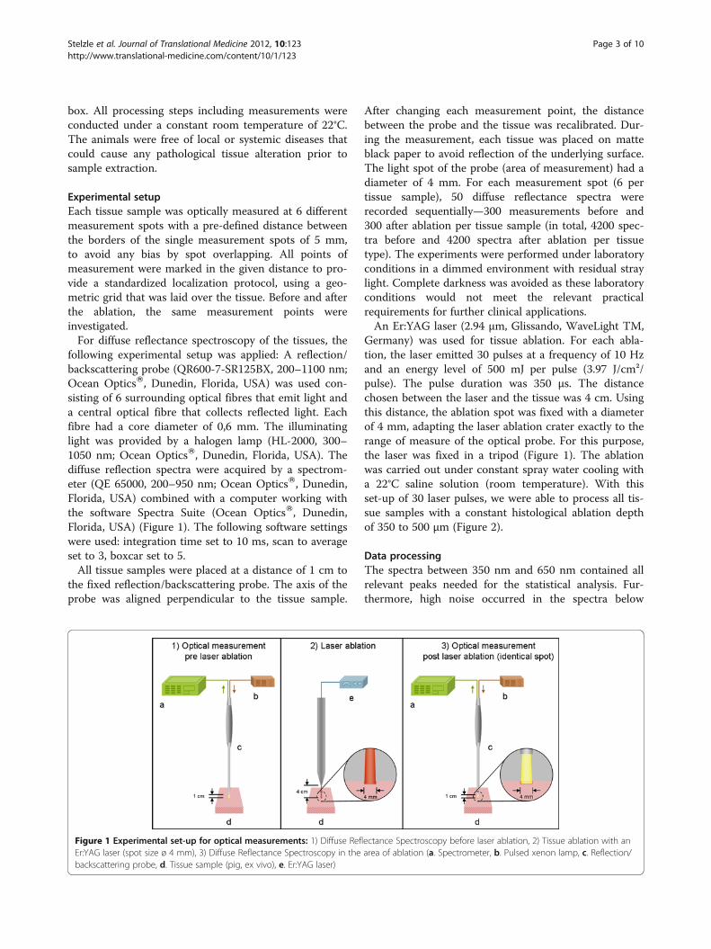
following experimental setup was applied: A reflection/backscattering probe (QR600-7-SR125BX, 200–1100 nm;Ocean OpticsW, Dunedin, Florida, USA) was used con-sisting of 6 surrounding optical fibres that emit light anda central optical fibre that collects reflected light. Eachfibre had a core diameter of 0,6 mm. The illuminatinglight was provided by a halogen lamp (HL-2000, 300–1050 nm; Ocean OpticsW, Dunedin, Florida, USA). Thediffuse reflection spectra were acquired by a spectrom-eter (QE 65000, 200–950 nm; Ocean OpticsW, Dunedin,Florida, USA) combined with a computer working withthe software Spectra Suite (Ocean OpticsW, Dunedin,Florida, USA) (Figure 1). The following software settingswere used: integration time set to 10 ms, scan to averageset to 3, boxcar set to 5.All tissue samples were placed at a distance of 1 cm to
the fixed reflection/backscattering probe. The axis of theprobe was aligned perpendicular to the tissue sample.
After changing each measurement point, the distancebetween the probe and the tissue was recalibrated. Dur-ing the measurement, each tissue was placed on matteblack paper to avoid reflection of the underlying surface.The light spot of the probe (area of measurement) had adiameter of 4 mm. For each measurement spot (6 pertissue sample), 50 diffuse reflectance spectra wererecorded sequentially—300 measurements before and300 after ablation per tissue sample (in total, 4200 spec-tra before and 4200 spectra after ablation per tissuetype). The experiments were performed under laboratoryconditions in a dimmed environment with residual straylight. Complete darkness was avoided as these laboratoryconditions would not meet the relevant practicalrequirements for further clinical applications.An Er:YAG laser (2.94 μm, Glissando, WaveLight TM,
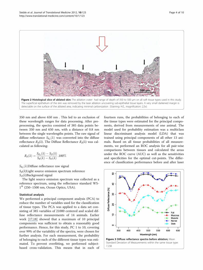
Germany) was used for tissue ablation. For each abla-tion, the laser emitted 30 pulses at a frequency of 10 Hzand an energy level of 500 mJ per pulse (3.97 J/cm²/pulse). The pulse duration was 350 μs. The distancechosen between the laser and the tissue was 4 cm. Usingthis distance, the ablation spot was fixed with a diameterof 4 mm, adapting the laser ablation crater exactly to therange of measure of the optical probe. For this purpose,the laser was fixed in a tripod (Figure 1). The ablationwas carried out under constant spray water cooling witha 22°C saline solution (room temperature). With thisset-up of 30 laser pulses, we were able to process all tis-sue samples with a constant histological ablation depthof 350 to 500 μm (Figure 2).
Data processingThe spectra between 350 nm and 650 nm contained allrelevant peaks needed for the statistical analysis. Fur-thermore, high noise occurred in the spectra below
Figure 1 Experimental set-up for optical measurements: 1) Diffuse Reflectance Spectroscopy before laser ablation, 2) Tissue ablation with anEr:YAG laser (spot size ø 4 mm), 3) Diffuse Reflectance Spectroscopy in the area of ablation (a. Spectrometer, b. Pulsed xenon lamp, c. Reflection/backscattering probe, d. Tissue sample (pig, ex vivo), e. Er:YAG laser)
Stelzle et al. Journal of Translational Medicine 2012, 10:123 Page 3 of 10http://www.translational-medicine.com/content/10/1/123
350 nm and above 650 nm . This led to an exclusion ofthese wavelength ranges for data processing. After pre-processing, the spectra consisted of 385 data points be-tween 350 nm and 650 nm, with a distance of 0.8 nmbetween the single wavelengths points. The raw signal ofdiffuse reflectance SRd λð Þ was converted into the diffusereflectance Rd(λ). The Diffuse Reflectance Rd(λ) was cal-culated as following:
Rd λð Þ ¼ SRd λð Þ � SD λð ÞSR λð Þ � SD λð Þ :100%
SRd λð ÞDiffuse reflectance raw signal
SR(λ)Light source emission spectrum referenceSD(λ)Background signalThe light source emission spectrum was collected as a
reference spectrum, using the reflectance standard WS-1W (250–1500 nm, Ocean Optics, USA).
Statistical analysisWe performed a principal component analysis (PCA) toreduce the number of variables used for the classificationof tissue types. The PCA was applied to a data set con-sisting of 385 variables of 21000 centered and scaled dif-fuse reflectance measurements of 14 animals. Earlierwork [17,18] showed that a maximum of 10 principalcomponents was sufficient to obtain a reasonably goodperformance. Hence, for this study, PC 1 to 10, coveringover 99% of the variability of the spectra, were chosen forfurther analysis. For each measurement, the probabilityof belonging to each of the different tissue types was esti-mated. To prevent overfitting, we performed subject-based cross-validation. This means that in each of
fourteen runs, the probabilities of belonging to each ofthe tissue types were estimated for the principal compo-nents, derived from measurements of one animal. Themodel used for probability estimation was a multiclasslinear discriminant analysis model (LDA) that wastrained using principal components of all other 13 ani-mals. Based on all tissue probabilities of all measure-ments, we performed an ROC analysis for all pair-wisecomparisons between tissues and calculated the areasunder the ROC curve (AUC) as well as the sensitivitiesand specificities for the optimal cut-points. The differ-ence of classification performance before and after laser
Figure 2 Histological slice of ablated skin: The ablation crater had range of depth of 350 to 500 μm on all soft tissue types used in this study.The superficial epithelium of the skin was removed by the laser ablation uncovering sub-epithelial tissue layers. A very small darkened margin isdetectable on the surface of the ablated area, indicating minimal carbonization (Staining: H.E., magnification: 2,5x)
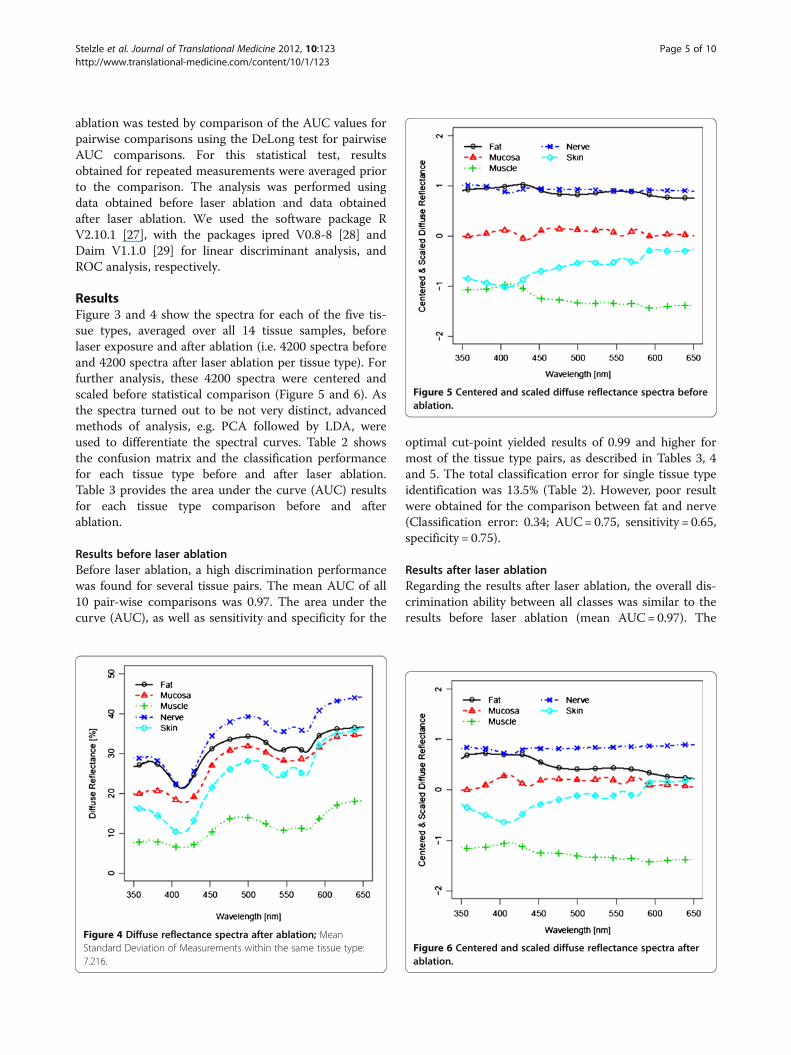
Figure 3 Diffuse reflectance spectra before ablation; MeanStandard Deviation of Measurements within the same tissue type:7.238
Stelzle et al. Journal of Translational Medicine 2012, 10:123 Page 4 of 10http://www.translational-medicine.com/content/10/1/123
ablation was tested by comparison of the AUC values forpairwise comparisons using the DeLong test for pairwiseAUC comparisons. For this statistical test, resultsobtained for repeated measurements were averaged priorto the comparison. The analysis was performed usingdata obtained before laser ablation and data obtainedafter laser ablation. We used the software package RV2.10.1 [27], with the packages ipred V0.8-8 [28] andDaim V1.1.0 [29] for linear discriminant analysis, andROC analysis, respectively.
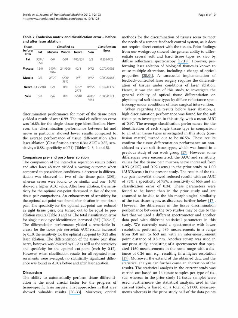
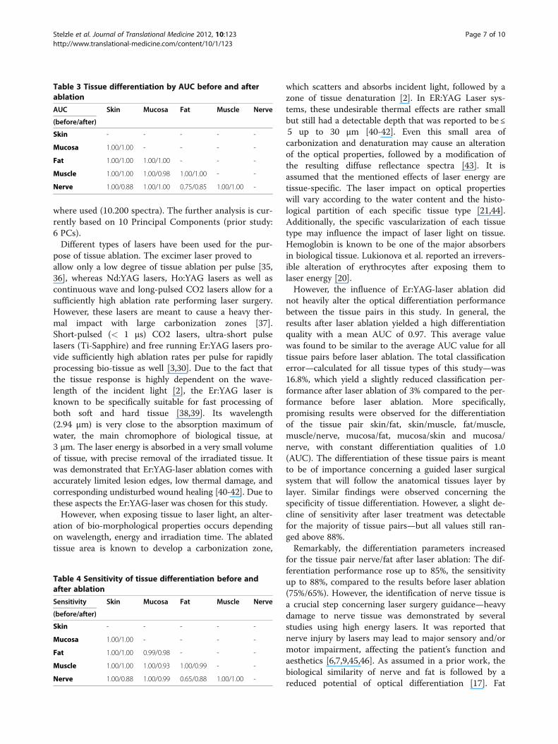
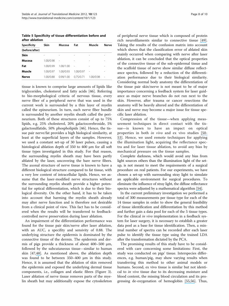
ResultsFigure 3 and 4 show the spectra for each of the five tis-sue types, averaged over all 14 tissue samples, beforelaser exposure and after ablation (i.e. 4200 spectra beforeand 4200 spectra after laser ablation per tissue type). Forfurther analysis, these 4200 spectra were centered andscaled before statistical comparison (Figure 5 and 6). Asthe spectra turned out to be not very distinct, advancedmethods of analysis, e.g. PCA followed by LDA, wereused to differentiate the spectral curves. Table 2 showsthe confusion matrix and the classification performancefor each tissue type before and after laser ablation.Table 3 provides the area under the curve (AUC) resultsfor each tissue type comparison before and afterablation.
Results before laser ablationBefore laser ablation, a high discrimination performancewas found for several tissue pairs. The mean AUC of all10 pair-wise comparisons was 0.97. The area under thecurve (AUC), as well as sensitivity and specificity for the
optimal cut-point yielded results of 0.99 and higher formost of the tissue type pairs, as described in Tables 3, 4and 5. The total classification error for single tissue typeidentification was 13.5% (Table 2). However, poor resultwere obtained for the comparison between fat and nerve(Classification error: 0.34; AUC= 0.75, sensitivity = 0.65,specificity = 0.75).
Results after laser ablationRegarding the results after laser ablation, the overall dis-crimination ability between all classes was similar to theresults before laser ablation (mean AUC=0.97). The
Figure 4 Diffuse reflectance spectra after ablation; MeanStandard Deviation of Measurements within the same tissue type:7.216.
Figure 5 Centered and scaled diffuse reflectance spectra beforeablation.
Figure 6 Centered and scaled diffuse reflectance spectra afterablation.
Stelzle et al. Journal of Translational Medicine 2012, 10:123 Page 5 of 10http://www.translational-medicine.com/content/10/1/123
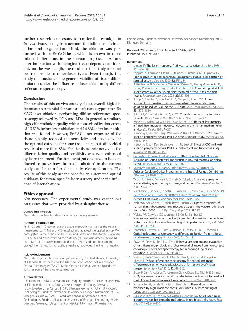
discrimination performance for most of the tissue pairsyielded a result of over 0.99. The total classification errorwas 16.8% for the single tissue type identification. How-ever, the discrimination performance between fat andnerve in particular showed lower results compared tothe average performance of tissue differentiation afterlaser ablation (Classification error: 0.34; AUC= 0.85, sen-sitivity = 0.88, specificity = 0.71) (Tables 2, 3, 4 and 5).
Comparison pre- and post- laser ablationThe comparison of the inter-class separation results beforeand after laser ablation yielded a varying outcome: whencompared to pre-ablation conditions, a decrease in differen-tiation was observed in two of the tissue pairs (20%),whereas seven were found to be similar and one (10%)showed a higher AUC value. After laser ablation, the sensi-tivity for the optimal cut-point decreased in five of the tentissue pair comparisons. An enhancement of sensitivity forthe optimal cut-point was found after ablation in one tissuepair. The specificity for the optimal cut-point was reducedin eight tissue pairs, one turned out to be equal to pre-ablation results (Table 3 and 4). The total classification errorfor single tissue type identification increased (3%) (Table 2).The differentiation performance yielded a remarkable in-crease for the tissue pair nerve/fat: AUC results increasedby 0.10, the sensitivity for the optimal cut-point by 0.23 afterlaser ablation. The differentiation of the tissue pair skin/nerve, however, was lowered by 0.12 as well as the sensitivityand specificity for the optimal cut-point (each by 0.12).However, when classification results for all repeated mea-surements were averaged, no statistically significant differ-ence was found in AUCs before and after laser ablation.
DiscussionThe ability to automatically perform tissue differenti-ation is the most crucial factor for the progress oftissue-specific laser surgery. First approaches in that areashowed valuable results [30-33]. Moreover, optical
methods for the discrimination of tissues seem to meetthe needs of a remote feedback control system, as it doesnot require direct contact with the tissues. Prior findingsfrom our workgroup showed the general ability to differ-entiate several soft and hard tissue types ex vivo bydiffuse reflectance spectroscopy [17,18]. However, per-forming laser ablation of biological tissues is known tocause multiple alterations, including a change of opticalproperties [20,34]. A successful implementation offeedback-controlled laser surgery requires the differenti-ation of tissues under conditions of laser ablation.Hence, it was the aim of this study to investigate thegeneral viability of optical tissue differentiation onphysiological soft tissue types by diffuse reflectance spec-troscopy under conditions of laser surgical intervention.When regarding the results before laser ablation, a
high discrimination performance was found for the softtissue pairs investigated in this study, with a mean AUCof 0.97. The average classification performance for theidentification of each single tissue type in comparisonto all other tissue types investigated in this study (con-fusion matrix) turned out to be 86.5%. These resultsconfirm the tissue differentiation performance on non-ablated ex vivo soft tissue types, which was found in aprevious study of our work group [17]. However, somedifferences were encountered: the AUC and sensitivityvalues for the tissue pair mucosa/nerve increased from0.93 (AUC) and 0.92 (sens.) in the prior study to 1.00(AUC&sens.) in the present study. The results of the tis-sue pair nerve/fat showed reduced results with an AUCof 75%, a specificity of 75%, a sensitivity of 65% and anclassification error of 0.34. These parameters werefound to be lower than in the prior study and areassumed to be due to the bio-morphological similarityof the two tissue types, as discussed further below [17].However, the differences in the tissue discriminationperformance between the two studies may be due to thefact that we used a different spectrometer and anotherdata pool with different statistical parameters in thisstudy. We currently used a spectrometer with lowerresolution, performing 385 measurements in a rangefrom 350 nm to 650 nm with an inter-measurementpoint distance of 0.8 nm. Another set-up was used inour prior study, consisting of a spectrometer that oper-ated 1150 measurements in the same range with a dis-tance of 0.26 nm, e.g., resulting in a higher resolution[17]. Moreover, the extend of the obtained data and thestatistical analysis can further cause an aberration of theresults. The statistical analysis in the current study wascarried out based on 14 tissue samples per type of tis-sue, whereas in the prior study 12 tissue samples wereused. Furthermore the statistical analysis, used in thecurrent study, is based on a total of 21.000 measure-ments whereas in the prior study half of the data points
Table 2 Confusion matrix and classification error – beforeand after laser ablation
Tissuebefore/after
Classified as ClassificationErrorFat Mucosa Muscle Nerve Skin
Fat 3094/3276
0/0 0/91 1106/831 0/2 0.263/0.22
Mucosa 12/0 3907/3814
241/306 40/8 0/72 0.070/0.092
Muscle 0/0 0/222 4200/3913
0/3 0/62 0.000/0.068
Nerve 1438/918 0/9 0/0 2762/2778
0/495 0.342/0.339
Skin 0/0 0/6 0/0 0/510 4200/3684
0.000/0.123
Stelzle et al. Journal of Translational Medicine 2012, 10:123 Page 6 of 10http://www.translational-medicine.com/content/10/1/123
where used (10.200 spectra). The further analysis is cur-rently based on 10 Principal Components (prior study:6 PCs).Different types of lasers have been used for the pur-
pose of tissue ablation. The excimer laser proved toallow only a low degree of tissue ablation per pulse [35,36], whereas Nd:YAG lasers, Ho:YAG lasers as well ascontinuous wave and long-pulsed CO2 lasers allow for asufficiently high ablation rate performing laser surgery.However, these lasers are meant to cause a heavy ther-mal impact with large carbonization zones [37].Short-pulsed (< 1 μs) CO2 lasers, ultra-short pulselasers (Ti-Sapphire) and free running Er:YAG lasers pro-vide sufficiently high ablation rates per pulse for rapidlyprocessing bio-tissue as well [3,30]. Due to the fact thatthe tissue response is highly dependent on the wave-length of the incident light [2], the Er:YAG laser isknown to be specifically suitable for fast processing ofboth soft and hard tissue [38,39]. Its wavelength(2.94 μm) is very close to the absorption maximum ofwater, the main chromophore of biological tissue, at3 μm. The laser energy is absorbed in a very small volumeof tissue, with precise removal of the irradiated tissue. Itwas demonstrated that Er:YAG-laser ablation comes withaccurately limited lesion edges, low thermal damage, andcorresponding undisturbed wound healing [40-42]. Due tothese aspects the Er:YAG-laser was chosen for this study.However, when exposing tissue to laser light, an alter-
ation of bio-morphological properties occurs dependingon wavelength, energy and irradiation time. The ablatedtissue area is known to develop a carbonization zone,
which scatters and absorbs incident light, followed by azone of tissue denaturation [2]. In ER:YAG Laser sys-tems, these undesirable thermal effects are rather smallbut still had a detectable depth that was reported to be ≤5 up to 30 μm [40-42]. Even this small area ofcarbonization and denaturation may cause an alterationof the optical properties, followed by a modification ofthe resulting diffuse reflectance spectra [43]. It isassumed that the mentioned effects of laser energy aretissue-specific. The laser impact on optical propertieswill vary according to the water content and the histo-logical partition of each specific tissue type [21,44].Additionally, the specific vascularization of each tissuetype may influence the impact of laser light on tissue.Hemoglobin is known to be one of the major absorbersin biological tissue. Lukionova et al. reported an irrevers-ible alteration of erythrocytes after exposing them tolaser energy [20].However, the influence of Er:YAG-laser ablation did
not heavily alter the optical differentiation performancebetween the tissue pairs in this study. In general, theresults after laser ablation yielded a high differentiationquality with a mean AUC of 0.97. This average valuewas found to be similar to the average AUC value for alltissue pairs before laser ablation. The total classificationerror—calculated for all tissue types of this study—was16.8%, which yield a slightly reduced classification per-formance after laser ablation of 3% compared to the per-formance before laser ablation. More specifically,promising results were observed for the differentiationof the tissue pair skin/fat, skin/muscle, fat/muscle,muscle/nerve, mucosa/fat, mucosa/skin and mucosa/nerve, with constant differentiation qualities of 1.0(AUC). The differentiation of these tissue pairs is meantto be of importance concerning a guided laser surgicalsystem that will follow the anatomical tissues layer bylayer. Similar findings were observed concerning thespecificity of tissue differentiation. However, a slight de-cline of sensitivity after laser treatment was detectablefor the majority of tissue pairs—but all values still ran-ged above 88%.Remarkably, the differentiation parameters increased
for the tissue pair nerve/fat after laser ablation: The dif-ferentiation performance rose up to 85%, the sensitivityup to 88%, compared to the results before laser ablation(75%/65%). However, the identification of nerve tissue isa crucial step concerning laser surgery guidance—heavydamage to nerve tissue was demonstrated by severalstudies using high energy lasers. It was reported thatnerve injury by lasers may lead to major sensory and/ormotor impairment, affecting the patient’s function andaesthetics [6,7,9,45,46]. As assumed in a prior work, thebiological similarity of nerve and fat is followed by areduced potential of optical differentiation [17]. Fat
Table 3 Tissue differentiation by AUC before and afterablation
AUC Skin Mucosa Fat Muscle Nerve
(before/after)
Skin - - - - -
Mucosa 1.00/1.00 - - - -
Fat 1.00/1.00 1.00/1.00 - - -
Muscle 1.00/1.00 1.00/0.98 1.00/1.00 - -
Nerve 1.00/0.88 1.00/1.00 0.75/0.85 1.00/1.00 -
Table 4 Sensitivity of tissue differentiation before andafter ablation
Sensitivity Skin Mucosa Fat Muscle Nerve
(before/after)
Skin - - - - -
Mucosa 1.00/1.00 - - - -
Fat 1.00/1.00 0.99/0.98 - - -
Muscle 1.00/1.00 1.00/0.93 1.00/0.99 - -
Nerve 1.00/0.88 1.00/0.99 0.65/0.88 1.00/1.00 -
Stelzle et al. Journal of Translational Medicine 2012, 10:123 Page 7 of 10http://www.translational-medicine.com/content/10/1/123
tissue is known to comprise large amounts of lipids liketriglycerides, cholesterol and fatty acids [46]. Referringto bio-morphological criteria of nervous tissue, everynerve fiber of a peripheral nerve that was used in thecurrent work is surrounded by a thin layer of myelincalled the epineurium. In turn, each nerve fiber bundleis surrounded by another myelin sheath called the peri-neurium. Both of these structures consist of up to 75%lipids, e.g. 25% cholesterol, 20% galactocerebroside, 5%galactosulfatide, 50% phospholipids [46]. Hence, the tis-sue pair nerve/fat provides a high biological similarity, atleast at the superficial layers of the samples. However,we used a constant set-up of 30 laser pulses, causing ahistological ablation depth of 350 to 400 μm for all softtissue types investigated in this study. For that reason,the surrounding myelin sheath may have been partlyablated by the laser, uncovering the bare nerve fibers.The axonal structure of nerve tissue is known to have adifferent biological structure compared to fat tissue, witha very low content of intracellular lipids. Hence, we as-sume that the laser-modified nerve structures withoutthe surrounding myelin sheath provide a higher poten-tial for optical differentiation, which is due to their bio-logical diversity. On the other hand, it has to be takeninto account that harming the myelin sheath alreadymay alter nerve function and is therefore not desirablefrom a clinical point of view. This fact has to be consid-ered when the results will be transferred to feedback-controlled nerve preservation during laser ablation.An impairment of the differentiation performance was
found for the tissue pair skin/nerve after laser ablation,with an AUC, a specifity and sensivity of 0.88. Theunderlying structure the epidermis is dominated by theconnective tissue of the dermis. The epidermis and der-mis of pigs provide a thickness of about 400–500 μm,followed by the subcutaneous tissue—similar to humanskin [47,48]. As mentioned above, the ablation depthwas found to be between 350–400 μm in this study.Hence, it is assumed that the ablation of skin removedthe epidermis and exposed the underlying dermal tissuecomponents, i.e., collagen and elastic fibres (Figure 3).Laser ablation of nerve tissue removes parts of the mye-lin sheath but may additionally expose the cytoskeleton
of peripheral nerve tissue which is composed of proteinrich neurofilaments similar to connective tissue [49].Taking the results of the confusion matrix into accountwhich shows that the classification error of ablated skinmainly occurred when comparing with nerve after laserablation, it can be concluded that the optical propertiesof the connective tissue of the sub-epidermal tissue andthe scaffold tissue of nerve show similar diffuse reflect-ance spectra, followed by a reduction of the differenti-ation performance due to their biological similarity.Considering normal body anatomy the differentiation ofthe tissue pair skin/nerve is not meant to be of majorimportance concerning a feedback system for laser guid-ance as major nerve branches do not run next to theskin. However, after trauma or cancer resections theanatomy will be heavily altered and the differentiation ofskin and nerve may become a major issue for tissue spe-cific laser ablation.Compression of the tissue—when applying meas-
urement techniques in direct contact with the tis-sue—is known to have an impact on opticalproperties in both in vivo and ex vivo studies [50-53]. Hence, we used remote techniques for applyingthe illumination light, acquiring the reflectance spec-tra and for laser tissue ablation, to avoid any bias bymechanical pressure on the tissue samples.Complete darkness, which would avoid any bias from
light sources others than the illumination light of the set-up, is not meant to meet the requirements of a surgicalprocedure on real patients. For our experiments, we havechosen a set-up with surrounding stray light to simulatean applicable environment for surgical procedures. Toeliminate the influence of stray light, the diffuse reflectancespectra were adjusted by a mathematical algorithm [54].In the current preliminary investigation we performed a
total of 300 measurements per tissue type for each of the14 tissue samples in order to show the general feasibilityof tissue identification and differentiation by this methodand further gain a data pool for each of the 5 tissue types.For the clinical in vivo implementation in a feedback sys-tem for laser surgery, it is necessary to establish a greaterdata pool as a base for tissue identification. Then, a min-imal number of spectra can be recorded after each laserpulse to identify the tissue type using the trained LDAafter the transformation dictated by the PCA.The promising results of this study have to be consid-
ered with care concerning some limitations: First, thestudy was conducted on pigs’ tissue. Interspecies differ-ences, e.g. human/pig, may show varying results whentransferring this method to other animal models orhumans. Second, ex vivo tissue is similar but not identi-cal to in vivo tissue due to its decreasing moisture andblood content, the missing blood circulation and its pro-gressing de-oxygenation of hemoglobin [55,56]. Thus,
Table 5 Specificity of tissue differentiation before andafter ablation
Specificity Skin Mucosa Fat Muscle Nerve
(before/after)
Skin - - - - -
Mucosa 1.00/0.98 - - - -
Fat 1.00/0.99 1.00/1.00 - - -
Muscle 1.00/0.97 1.00/0.93 1.00/0.97 - -
Nerve 1.00/0.88 0.99/1.00 0.75/0.71 1.00/0.98 -
Stelzle et al. Journal of Translational Medicine 2012, 10:123 Page 8 of 10http://www.translational-medicine.com/content/10/1/123
further research is necessary to transfer the technique toin vivo tissue, taking into account the influence of circu-lation and oxygenation. Third, the ablation was per-formed with an Er:YAG-laser, which is known to causeminimal alterations to the surrounding tissue. As anylaser interaction with biological tissue depends consider-ably on the wavelength, the results of this study may notbe transferable to other laser types. Even though, thisstudy demonstrated the general viability of tissue differ-entiation under the influence of laser ablation by diffusereflectance spectroscopy.
ConclusionThe results of this ex vivo study yield an overall high dif-ferentiation potential for various soft tissue types after Er:YAG laser ablation, performing diffuse reflectance spec-troscopy followed by PCA and LDA. In general, a similarlyhigh differentiation quality with a total classification errorsof 13.51% before laser ablation and 16.83% after laser abla-tion was found. However, Er:YAG laser exposure of thetissue slightly reduced the sensitivity and specificity forthe optimal cutpoint for some tissue pairs, but still yieldedresults of more than 85%. For the tissue pair nerve/fat, thedifferentiation quality and sensitivity was even enhancedby laser treatment. Further investigations have to be con-ducted to prove how the results obtained in the currentstudy can be transferred to an in vivo application. Theresults of this study set the base for an automated opticalguidance for tissue-specific laser surgery under the influ-ence of laser ablation.
Ethics approvalNot necessary. The experimental study was carried outon tissues that were provided by a slaughterhouse.
Competing interestsThe authors declare that they have no competing interests
Authors’ contributionsFS, IT, CK and KTG carried out the tissue preparation as well as the opticalmeasurements. IT, MS and KTG installed and adapted the optical set-up. WAparticipated in the design of the study and performed the statistical analysis.FS, CK, EN and MS performed the data analysis and assessment. FS and MSconceived of the study, participated in its design and coordination anddrafted the manuscript. All authors read and approved the final manuscript.
AcknowledgementsThe authors gratefully acknowledge funding by the ELAN-Funds, Universityof Erlangen-Nuremberg and the Erlangen Graduate School in AdvancedOptical Technologies (SAOT) by the German National Science Foundation(DFG) as part of the Excellence Initiative.
Author details1Department of Oral and Maxillofacial Surgery, Friedrich-Alexander Universityof Erlangen-Nuremberg, Glückstrasse 11, 91054, Erlangen, Germany.2blz—Bavarian Laser Center, 91054, Erlangen, Germany. 3Chair of PhotonicTechnologies, Friedrich-Alexander-University of Erlangen-Nuremberg, 91054,Erlangen, Germany. 4SAOT—Graduate School in Advanced OpticalTechnologies, Friedrich-Alexander University of Erlangen-Nuremberg, 91054,Erlangen, Germany. 5Department of Medical Informatics, Biometry and
Epidemiology, Friedrich-Alexander University of Erlangen-Nuremberg, 91054,Erlangen, Germany.
Received: 20 February 2012 Accepted: 16 May 2012Published: 15 June 2012
References1. Minton JP: The laser in surgery. A 23 year perspective. Am J Surg 1986,
151:725–729.2. Boppart SA, Herrmann J, Pitris C, Stamper DL, Brezinski ME, Fujimoto JG:
High-resolution optical coherence tomography-guided laser ablation ofsurgical tissue. J Surg Res 1999, 82:275–284.
3. Kuttenberger JJ, Stubinger S, Waibel A, Werner M, Klasing M, Ivanenko M,Hering P, Von Rechenberg B, Sader R, Zeilhofer HF: Computer-guided CO2-laser osteotomy of the sheep tibia: technical prerequisites and firstresults. Photomed Laser Surg 2008, 26:129–136.
4. Stopp S, Svejdar D, von Kienlin E, Deppe H, Lueth TC: A newapproach for creating defined geometries by navigated laserablation based on volumetric 3-D data. IEEE Trans Biomed Eng 2008,55:1872–1880.
5. Spinelli P, Calarco G, Mancini A, Ni XG: Operative colonoscopy in cancerpatients. Minim Invasive Ther Allied Technol 2006, 15:339–347.
6. Baxter GD, Walsh DM, Allen JM, Lowe AS, Bell AJ: Effects of low intensityinfrared laser irradiation upon conduction in the human median nervein vivo. Exp Physiol 1994, 79:227.
7. Menovsky T, van den Bergh Weerman M, Beek JF: Effect of CO2 milliwattlaser on peripheral nerves: Part I. A dose–response study. Microsurg 1996,17:562–567.
8. Menovsky T, Van Den Bergh Weerman M, Beek JF: Effect of CO2 milliwattlaser on peripheral nerves: Part II. A histological and functional study.Microsurg 2000, 20:150–155.
9. Orchardson R, Peacock JM, Whitters CJ: Effect of pulsed Nd: YAG laserradiation on action potential conduction in isolated mammalian spinalnerves. Lasers Surg Med 1997, 21:142–148.
10. Ebert DW, Roberts C, Farrar SK, Johnston WM, Litsky AS, Bertone AL:Articular Cartilage Optical Properties in the Spectral Range 300–850 nm.J Biomed Opt 1998, 3:326.
11. Taroni P, Pifferi A, Torricelli A, Comelli D, Cubeddu R: In vivo absorptionand scattering spectroscopy of biological tissues. Photochem Photobiol Sci2003, 2:124–129.
12. Marchesini R, Pignoli E, Tomatis S, Fumagalli S, Sichirollo AE, Di Palma S, DalFante M, Spinelli P, Croce AC, Bottiroli G: Ex vivo optical properties ofhuman colon tissue. Lasers Surg Med 1994, 15:351–357.
13. Bashkatov AN, Genina EA, Kochubey VI, Tuchin VV: Optical properties ofhuman skin, subcutaneous and mucous tissues in the wavelength rangefrom 400 to 2000 nm. J Phys D Appl Phys 2005, 38:2543.
14. Wallace VP, Crawford DC, Mortimer PS, Ott RJ, Bamber JC:Spectrophotometric assessment of pigmented skin lesions: methods andfeature selection for evaluation of diagnostic performance. Phys Med Biol2000, 45:735–751.
15. Bensalah K, Peswani D, Tuncel A, Raman JD, Zeltser I, Liu H, Cadeddu J:Optical reflectance spectroscopy to differentiate benign from malignantrenal tumors at surgery. Urology 2009, 73:178–181.
16. Fawzy YS, Petek M, Tercelj M, Zeng H: In vivo assessment and evaluationof lung tissue morphologic and physiological changes from non-contactendoscopic reflectance spectroscopy for improving lung cancerdetection. J Biomed Opt 2006, 11:044003.
17. Stelzle F, Tangermann-Gerk K, Adler W, Zam A, Schmidt M, Douplik A,Nkenke E: Diffuse reflectance spectroscopy for optical soft tissuedifferentiation as remote feedback control for tissue-specific lasersurgery. Lasers Surg Med 2010, 42:319–325.
18. Stelzle F, Zam A, Adler W, Tangermann-Gerk K, Douplik A, Nkenke E, SchmidtM: Optical nerve detection by diffuse reflectance spectroscopy for feedbackcontrolled oral and maxillofacial laser surgery. J Transl Med 2011, 9:20.
19. Schomacker KT, Walsh JT, Flotte TJ, Deutsch TF: Thermal damageproduced by high-lrradiance continuous wave CO2 laser cutting oftissue. Lasers Surg Med 1990, 10:74–84.
20. Lukianova-Hleb EY, Oginsky AO, Olson JS, Lapotko DO: Short laser pulse-induced irreversible photothermal effects in red blood cells. Lasers SurgMed 2011, 43:249–260.
Stelzle et al. Journal of Translational Medicine 2012, 10:123 Page 9 of 10http://www.translational-medicine.com/content/10/1/123
21. Ritz JP, Roggan A, Germer CT, Isbert C, Muller G, Buhr HJ: Continuouschanges in the optical properties of liver tissue during laser-inducedinterstitial thermotherapy. Lasers Surg Med 2001, 28:307–312.
22. Ross EV, McKinlay JR, Sajben FP, Miller CH, Barnette DJ, Meehan KJ, ChhiengNP, Deavers MJ, Zelickson BD: Use of a novel erbium laser in a Yucatanminipig: A study of residual thermal damage, ablation, and woundhealing as a function of pulse duration. Lasers Surg Med 2002, 30:93–100.
23. Douplik A, Zam A, Hohenstein R, Kalitzeos A, Nkenke E, Stelzle F: Limitations ofcancer margin delineation by means of autofluorescence imaging underconditions of laser surgery. J Innov Opt Health Sci 2010, 3:45–51.
24. Patel NA, Li X, Stamper DL, Fujimoto JG, Brezinski ME: Guidance of aorticablation using optical coherence tomography. Int J Cardiovasc Imaging2003, 19:171–178.
25. Patel NA, Li X, Stamper DL, Fujimoto JG, Brezinski ME: Using opticalcoherence tomography to guide articular cartilage ablation. Am J Orthop2005, 34:111–115.
26. Boppart SA, Herrmann JM, Pitris C, Stamper DL, Brezinski ME, Fujimoto JG:Real-time optical coherence tomography for minimally invasive imagingof prostate ablation. Comput Aided Surg 2001, 6:94–103.
27. Team RDC: R: A language and environment for statistical computing. Vienna,Austria: R Foundation for Statistical Computing; 2008. http://wwwR-projectorg.
28. Peters A, Hothorn T: ipred: Improved Predictors; R package version 0.8-8. 2000.http://CRAN.R-project.org/package=ipred.
29. Potapov S, Adler W, Lausen B: Daim: Diagnostic accuracy of classificationmodels, R package version 1.1.0. 2009. http://CRAN.R-project.org/package=Daim.
30. Kim BM, Feit MD, Rubenchik AM, Mammini BM, Da Silva LB: Opticalfeedback signal for ultrashort laser pulse ablation of tissue. Appl Surf Sci1998, 127:857–862.
31. Rupprecht S, Tangermann K, Kessler P, Neukam FW, Wiltfang J: Er: YAGlaser osteotomy directed by sensor controlled systems. J Cranio-Maxillofac Surg 2003, 31:337–342.
32. Rupprecht S, Tangermann-Gerk K, Wiltfang J, Neukam FW, Schlegel A:Sensor-based laser ablation for tissue specific cutting: an experimentalstudy. Lasers Med Sci 2004, 19:81–88.
33. Tangermann K, Roth S, Muller D, Tragler H, Uller J, Rupprecht S: Sensor-controlled laser processes for medical applications. Proc SPIE 2003,5287:24.
34. Strauss RA: Lasers in oral and maxillofacial surgery. Dent Clin N Am 2000,44:851–873.
35. Hohla K: Vorrichtung zur Laserbehandlung von Gewebe. InOffenlegungsschrift DE3813918A1. 1988.
36. Lane PM, Gilhuly T, Whitehead P, Zeng H, Poh CF, Ng S, Williams PM, ZhangL, Rosin MP, MacAulay CE: Simple device for the direct visualization oforal-cavity tissue fluorescence. J Biomed Opt 2006, 11:024006.
37. Walsh JT Jr, Deutsch TF: Er:YAG laser ablation of tissue: measurement ofablation rates. Lasers Surg Med 1989, 9:327–337.
38. Stanislawki M, Meister J, Mitra T, Ivanenko MM, Zanger K, Hering P: Hardtissue ablation with a free running Er :YAG and a Q-switched CO2 laser:a comparative study. Applied Physics B: Lasers and Optics 2001, 72:115–120.
39. Jahn R, Bleckmann A, Duczynski E, Huber G, Lierse W, Struve B, JungbluthKH: Thermal side effects after use of the pulsed IR laser on meniscus andbone tissue. Unfallchirurgie 1994, 20:1–10.
40. Horch H: Laser in der Zahnärztlichen- und Mund-Kiefer-Gesichtschirurgie.:Landsberg-München- Zürich: ecomed Verlag; 1993.
41. Keller U, Hibst R: Lasersysteme für die orale Hart- und Weichgewebschirurgie–Gewebewirkungen und Indikationen. Stuttgart: Gustav Fischer Verlag; 1994.
42. Romanos G, Ko HH, Froum S, Tarnow D: The use of CO(2) laser in thetreatment of peri-implantitis. Photomed Laser Surg 2009, 3:381–386.
43. Luerssen K, Lubatschowski H, Ptok M: Erbium:YAG laser surgery on vocalfold tissue. HNO 2007, 55:443–446.
44. Choi JY, Tanenbaum BS, Milner TE, Dao XV, Nelson JS, Sobol EN, Wong BJ:Theramal, mechanical, optical, and morphologic changes in bovinenucleus pulposus induced by Nd:YAG (lambda = 1.32 microm) laserirradiation. Lasers Surg Med 2001, 28:248–254.
45. Mack KF, Leinung M, Stieve M, Lenarz T, Schwab B: Clinical feasibility teston a minimally invasive laser therapy system in microsurgery of nerves.Minim Invasive Ther Allied Technol 2008, 17:292–299.
46. Ross MH, Pawlina W: Histology: a text and atlas: with correlated cell andmolecular biology. Lippincott Williams & Wilkins; 2006.
47. Bronaugh RL, Stewart RF, Congdon ER: Methods for in vitro percutaneousabsorption studies II. Animal models for human skin. Toxicol ApplPharmacol 1982, 62:481–488.
48. Jacobi U, Kaiser M, Toll R, Mangelsdorf S, Audring H, Otberg N, Sterry W,Lademann J: Porcine ear skin: an in vitro model for human skin. Skin ResTechnol 2007, 13:19–24.
49. Wais-Steider C, Eagles PA, Gilbert DS, Hopkins JM: Structural similaritiesand differences amongst neurofilaments. J Mol Biol 1983, 165:393–400.
50. Chan EK, Sorg B, Protsenko D, O’Neil M, Motamedi M, Welch AJ: Effects ofCompression on Soft Tissue Optical Properties. IEEE J Sel Top QuantElectron 1996, 2:943.
51. Nath A, Rivoire K, Chang S, Cox D, Atkinson EN, Follen M, Richards-KortumR: Effect of probe pressure on cervical fluorescence spectroscopymeasurements. J Biomed Opt 2004, 9:523–533.
52. Reif R, Amorosino MS, Calabro KW, A’Amar O, Singh SK, Bigio IJ: Analysis ofchanges in reflectance measurements on biological tissues subjected todifferent probe pressures. J Biomed Opt 2008, 13:010502.
53. Ti Y, Lin WC: Effects of probe contact pressure on in vivo opticalspectroscopy. Opt Express 2008, 16:4250–4262.
54. Ye Z, Auner G: Principal component analysis approach for biomedicalsample identification. IEEE Int Conf Syst Man Cybern 2004, 2:1348–1353.
55. Salomatina E, Yaroslavsky AN: Evaluation of the in vivo and ex vivo opticalproperties in a mouse ear model. Phys Med Biol 2008, 53:2797–2808.
56. Palmer GM, Marshek CL, Vrotsos KM, Ramanujam N: Optimal methods forfluorescence and diffuse reflectance measurements of tissue biopsysamples. Lasers Surg Med 2002, 30:191–200.
doi:10.1186/1479-5876-10-123Cite this article as: Stelzle et al.: The impact of laser ablation on opticalsoft tissue differentiation for tissue specific laser surgery–anexperimental ex vivo study. Journal of Translational Medicine 2012 10:123.
Submit your next manuscript to BioMed Centraland take full advantage of:
• Convenient online submission
• Thorough peer review
• No space constraints or color figure charges
• Immediate publication on acceptance
• Inclusion in PubMed, CAS, Scopus and Google Scholar
• Research which is freely available for redistribution
Submit your manuscript at www.biomedcentral.com/submit
Stelzle et al. Journal of Translational Medicine 2012, 10:123 Page 10 of 10http://www.translational-medicine.com/content/10/1/123
Related Documents