REVIEW The immunomodulatory properties of probiotic microorganisms beyond their viability (ghost probiotics: proposal of paraprobiotic concept) Valentina Taverniti • Simone Guglielmetti Received: 13 February 2011 / Accepted: 24 March 2011 / Published online: 16 April 2011 Ó Springer-Verlag 2011 Abstract The probiotic approach represents a potentially effective and mild alternative strategy for the prevention and treatment of either inflammatory or allergic diseases. Several studies have shown that different bacterial strains can exert their probiotic abilities by influencing the host’s immune system, thereby modulating immune responses. However, the emerging concern regarding safety problems arising from the extensive use of live microbial cells is enhancing the interest in non-viable microorganisms or microbial cell extracts, as they could eliminate shelf-life problems and reduce the risks of microbial translocation and infection. The purpose of this review is to provide an overview of the scientific literature concerning studies in which dead microbial cells or crude microbial cell fractions have been used as health-promoting agents. Particular attention will be given to the modulation of host immune responses. Possible mechanisms determining the effect on the immune system will also be discussed. Finally, in the light of the FAO/WHO definition of probiotics, indicating that the word ‘probiotic’ should be restricted to products that contain live microorganisms, and considering the sci- entific evidence indicating that inactivated microbes can positively affect human health, we propose the new term ‘paraprobiotic’ to indicate the use of inactivated microbial cells or cell fractions to confer a health benefit to the consumer. Keywords Paraprobiotic Á Probiotic Á Immunomodulation Á Lactobacillus Á Bifidobacterium Introduction A basic Google Internet search (executed on 21 January 2011) yielded approximately 6 million results for the word ‘probiotic’. About 7500 scientific references were listed for the same word by PubMed, more than 20% of which appeared in 2010. These simple data indicate the growing interest in the field of probiotic microorganisms and products, which supports a global market that generated $15.9 billion in 2008 and is expected to be worth $19.6 billion in 2013 (BCC Research 2008). The majority of the scientific reports define probiotics according to the definition recommended by an FAO/WHO workshop conducted in 2002, which describes probiotics as ‘live microorganisms which when administered in ade- quate amounts confer a health benefit on the host’ (FAO/ WHO 2002). This definition specifies that probiotic microorganisms must be ‘live’, and this stipulation is supported by an extensive number of studies suggesting that to provide health benefits, probiotic microorganisms must be viable (Gobbetti et al. 2010). Nevertheless, sci- entific evidence indicating that inactivated microbes posi- tively affect human health can also be found in the literature (Kataria et al. 2009). Accordingly, products intentionally containing non-viable microbial cells are already present in the market (e.g. Lacte´ol Fort from PUMC Pharmaceutical Co., Ltd and Fermenti Lattici Tin- dalizzati from Frau, AF United S.p.a.). The mechanisms underlying probiotic effects are gen- erally attributed to the interaction of probiotics with other microorganisms (members of the microbiota or pathogens) or to the cross-talk of probiotics with host cells. The former type of interaction is typically (though not exclusively) dependent on the viability of probiotic cells, since it is exerted by competitive exclusion (competition for nutrients V. Taverniti Á S. Guglielmetti (&) Department of Food Science and Microbiology (DiSTAM), Universita ` degli Studi di Milano, Via Celoria 2, 20133 Milan, Italy e-mail: [email protected] 123 Genes Nutr (2011) 6:261–274 DOI 10.1007/s12263-011-0218-x

Welcome message from author
This document is posted to help you gain knowledge. Please leave a comment to let me know what you think about it! Share it to your friends and learn new things together.
Transcript
REVIEW
The immunomodulatory properties of probiotic microorganismsbeyond their viability (ghost probiotics: proposal of paraprobioticconcept)
Valentina Taverniti • Simone Guglielmetti
Received: 13 February 2011 / Accepted: 24 March 2011 / Published online: 16 April 2011
� Springer-Verlag 2011
Abstract The probiotic approach represents a potentially
effective and mild alternative strategy for the prevention
and treatment of either inflammatory or allergic diseases.
Several studies have shown that different bacterial strains
can exert their probiotic abilities by influencing the host’s
immune system, thereby modulating immune responses.
However, the emerging concern regarding safety problems
arising from the extensive use of live microbial cells is
enhancing the interest in non-viable microorganisms or
microbial cell extracts, as they could eliminate shelf-life
problems and reduce the risks of microbial translocation
and infection. The purpose of this review is to provide an
overview of the scientific literature concerning studies in
which dead microbial cells or crude microbial cell fractions
have been used as health-promoting agents. Particular
attention will be given to the modulation of host immune
responses. Possible mechanisms determining the effect on
the immune system will also be discussed. Finally, in the
light of the FAO/WHO definition of probiotics, indicating
that the word ‘probiotic’ should be restricted to products
that contain live microorganisms, and considering the sci-
entific evidence indicating that inactivated microbes can
positively affect human health, we propose the new term
‘paraprobiotic’ to indicate the use of inactivated microbial
cells or cell fractions to confer a health benefit to the
consumer.
Keywords Paraprobiotic � Probiotic �Immunomodulation � Lactobacillus � Bifidobacterium
Introduction
A basic Google Internet search (executed on 21 January
2011) yielded approximately 6 million results for the word
‘probiotic’. About 7500 scientific references were listed for
the same word by PubMed, more than 20% of which
appeared in 2010. These simple data indicate the growing
interest in the field of probiotic microorganisms and
products, which supports a global market that generated
$15.9 billion in 2008 and is expected to be worth $19.6
billion in 2013 (BCC Research 2008).
The majority of the scientific reports define probiotics
according to the definition recommended by an FAO/WHO
workshop conducted in 2002, which describes probiotics as
‘live microorganisms which when administered in ade-
quate amounts confer a health benefit on the host’ (FAO/
WHO 2002). This definition specifies that probiotic
microorganisms must be ‘live’, and this stipulation is
supported by an extensive number of studies suggesting
that to provide health benefits, probiotic microorganisms
must be viable (Gobbetti et al. 2010). Nevertheless, sci-
entific evidence indicating that inactivated microbes posi-
tively affect human health can also be found in the
literature (Kataria et al. 2009). Accordingly, products
intentionally containing non-viable microbial cells are
already present in the market (e.g. Lacteol Fort from
PUMC Pharmaceutical Co., Ltd and Fermenti Lattici Tin-
dalizzati from Frau, AF United S.p.a.).
The mechanisms underlying probiotic effects are gen-
erally attributed to the interaction of probiotics with other
microorganisms (members of the microbiota or pathogens)
or to the cross-talk of probiotics with host cells. The former
type of interaction is typically (though not exclusively)
dependent on the viability of probiotic cells, since it is
exerted by competitive exclusion (competition for nutrients
V. Taverniti � S. Guglielmetti (&)
Department of Food Science and Microbiology (DiSTAM),
Universita degli Studi di Milano, Via Celoria 2,
20133 Milan, Italy
e-mail: [email protected]
123
Genes Nutr (2011) 6:261–274
DOI 10.1007/s12263-011-0218-x
or adhesion sites), direct inhibition of certain microorgan-
isms (production of antimicrobial molecules) or increased
growth of healthy components of the microbiota (nutri-
tional or environmental proto-cooperation). In contrast,
direct interaction with the host can be mediated by bacte-
rial cells independent of their viability and is based on the
capacity of human cells to recognise specific bacterial
components or products, giving rise to responses that
commonly involve the mucosa-associated lymphoid tissue
(MALT) and, therefore, the immune system (Adams 2010).
The purpose of this review is to provide an overview of
the scientific literature concerning studies in which non-
viable microbial cells or crude microbial cell fractions have
been investigated as health-promoting agents. In particular,
attention will focus on the modulation of host immune
responses, as this modulation is the primary means by
which components of dead cells are believed to exert their
bioactivities (Adams 2010). Possible mechanisms deter-
mining the effect on the immune system will be discussed
based on studies that have demonstrated particular bacterial
structural molecules or components to activate specific
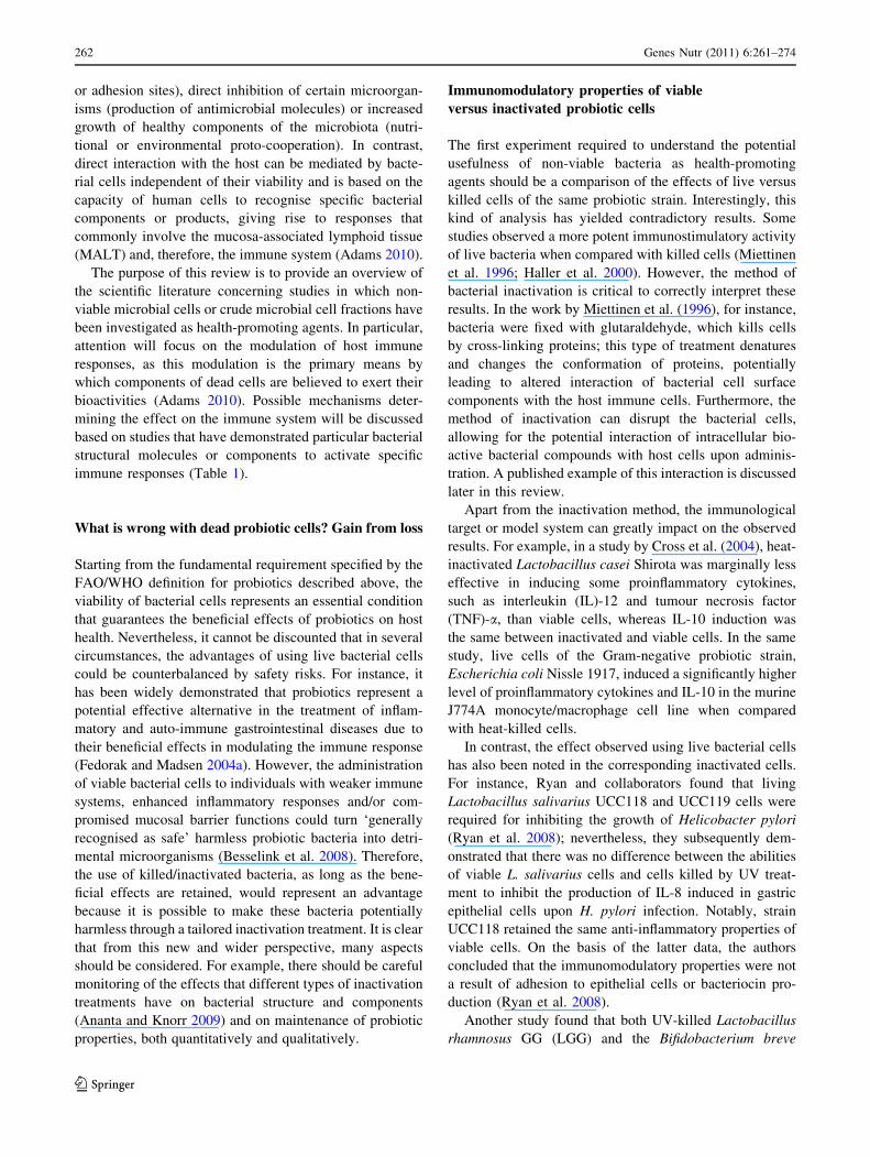
immune responses (Table 1).
What is wrong with dead probiotic cells? Gain from loss
Starting from the fundamental requirement specified by the
FAO/WHO definition for probiotics described above, the
viability of bacterial cells represents an essential condition
that guarantees the beneficial effects of probiotics on host
health. Nevertheless, it cannot be discounted that in several
circumstances, the advantages of using live bacterial cells
could be counterbalanced by safety risks. For instance, it
has been widely demonstrated that probiotics represent a
potential effective alternative in the treatment of inflam-
matory and auto-immune gastrointestinal diseases due to
their beneficial effects in modulating the immune response
(Fedorak and Madsen 2004a). However, the administration
of viable bacterial cells to individuals with weaker immune
systems, enhanced inflammatory responses and/or com-
promised mucosal barrier functions could turn ‘generally
recognised as safe’ harmless probiotic bacteria into detri-
mental microorganisms (Besselink et al. 2008). Therefore,
the use of killed/inactivated bacteria, as long as the bene-
ficial effects are retained, would represent an advantage
because it is possible to make these bacteria potentially
harmless through a tailored inactivation treatment. It is clear
that from this new and wider perspective, many aspects
should be considered. For example, there should be careful
monitoring of the effects that different types of inactivation
treatments have on bacterial structure and components
(Ananta and Knorr 2009) and on maintenance of probiotic
properties, both quantitatively and qualitatively.
Immunomodulatory properties of viable
versus inactivated probiotic cells
The first experiment required to understand the potential
usefulness of non-viable bacteria as health-promoting
agents should be a comparison of the effects of live versus
killed cells of the same probiotic strain. Interestingly, this
kind of analysis has yielded contradictory results. Some
studies observed a more potent immunostimulatory activity
of live bacteria when compared with killed cells (Miettinen
et al. 1996; Haller et al. 2000). However, the method of
bacterial inactivation is critical to correctly interpret these
results. In the work by Miettinen et al. (1996), for instance,
bacteria were fixed with glutaraldehyde, which kills cells
by cross-linking proteins; this type of treatment denatures
and changes the conformation of proteins, potentially
leading to altered interaction of bacterial cell surface
components with the host immune cells. Furthermore, the
method of inactivation can disrupt the bacterial cells,
allowing for the potential interaction of intracellular bio-
active bacterial compounds with host cells upon adminis-
tration. A published example of this interaction is discussed
later in this review.
Apart from the inactivation method, the immunological
target or model system can greatly impact on the observed
results. For example, in a study by Cross et al. (2004), heat-
inactivated Lactobacillus casei Shirota was marginally less
effective in inducing some proinflammatory cytokines,
such as interleukin (IL)-12 and tumour necrosis factor
(TNF)-a, than viable cells, whereas IL-10 induction was
the same between inactivated and viable cells. In the same
study, live cells of the Gram-negative probiotic strain,
Escherichia coli Nissle 1917, induced a significantly higher
level of proinflammatory cytokines and IL-10 in the murine
J774A monocyte/macrophage cell line when compared
with heat-killed cells.
In contrast, the effect observed using live bacterial cells
has also been noted in the corresponding inactivated cells.
For instance, Ryan and collaborators found that living
Lactobacillus salivarius UCC118 and UCC119 cells were
required for inhibiting the growth of Helicobacter pylori
(Ryan et al. 2008); nevertheless, they subsequently dem-
onstrated that there was no difference between the abilities
of viable L. salivarius cells and cells killed by UV treat-
ment to inhibit the production of IL-8 induced in gastric
epithelial cells upon H. pylori infection. Notably, strain
UCC118 retained the same anti-inflammatory properties of
viable cells. On the basis of the latter data, the authors
concluded that the immunomodulatory properties were not
a result of adhesion to epithelial cells or bacteriocin pro-
duction (Ryan et al. 2008).
Another study found that both UV-killed Lactobacillus
rhamnosus GG (LGG) and the Bifidobacterium breve
262 Genes Nutr (2011) 6:261–274
123
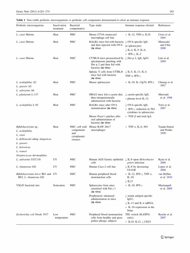
Table 1 Non-viable probiotic microorganisms or probiotic cell components demonstrated to elicit an immune response
Probiotic microorganism Inactivation
treatment
Bacterial
component(s)
Type study Immune response elicited References
L. casei Shirota Heat WBC Mouse J774A monocyte/
macrophage cell line
� IL-12, TNF-a, IL10 Cross et al.
2004
L. casei Shirota Heat WBC BALB/c mice fed with bacteria
and then injected with OVA
(in vivo)
; OVA-specific IgE;
in splenocytes:
; IL-4, IL-5, IL-6;
� IFN-c, IL-2
Matsuzaki
and Chin
2000
L. casei Shirota Heat WBC C57BL/6 mice presensitised by
epicutaneous patching with
Der p 2 and then fed with
bacteria (in vivo)
; Der p 2, IgE, IgG1 Lim et al.
2009
Splenic T cells from C57BL/6
mice fed with bacteria
(in vivo)
; IL-5, IL-13, IL-4
TNF-a, IFN-c
L. acidophilus A2
L. gasseri A5
L. salivarius A6
Heat WBC Mouse splenocytes � IL-10, IL-12p70, IFN-c Chuang et al.
2007
L. plantarum L-137 Heat WBC DBA/2 mice fed a casein diet,
then intraperitoneally
administered with bacteria
; casein-specific IgE;
�plasma level IL-12
Murosaki
et al. 1998
L. acidophilus L-92 Heat WBC BALB/c mice after OVA
immunisation (in vivo)
; OVA-specific IgE;
� IFN-c, reduction in Th2
cytokines in splenocytes
Torii et al.
2007
Mouse Peyer’s patches after
oral administration of
bacteria (in vivo)
� TGF-b and total IgA
Bifidobacterium sp.,
L. acidophilus
L. casei
L. delbrueckii subsp. bulgaricus
L. gasseri
L. helveticus
L. reuteri
Streptococcus thermophilus
Heat WBC, cell wall
components
and
cytoplasmic
extracts
Mouse RAW 264.7
macrophages
� TNF-a, IL-6, NO Tejada-Simon
and Pestka
1999
L. salivarius UCC118 UV WBC Human AGS Gastric epithelial
cells
; IL-8 upon Helicobacterpylori infection
Ryan et al.
2008
L. rhamnosus GG UV WBC Human Caco-2 cell line ; IL-8 by decreasing
Ub-IjB
Lopez et al.
2008
Bifidobacterium breve Bb1 and
Bb2; L. rhamnosus GG
UV WBC Human peripheral blood
mononuclear cells
� IL-12, IFN-c, TNF-a,
IL-10;
; IL13
van Hoffen
et al. 2010
VSL#3 bacterial mix Sonication WBC Splenocytes from mice
sensitised with Par j 1
(in vivo)
� IL-10, IFN-c Mastrangeli
et al. 2009
Prophylactic intranasal
administration in mice
(in vivo)
; serum antigen-specific
IgG1;
; IL-13 and IL-4 mRNA;
� IL-10 expression in the
lungs
Escherichia coli Nissle 1917 Low
temperature
WBC Peripheral blood mononuclear
cells from healthy and grass
pollen allergic subjects
TH1 switch (IL4/IFNcratio);
� IL10, IL12; ; CD23
Rasche et al.
2007
Genes Nutr (2011) 6:261–274 263
123
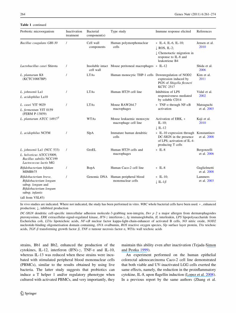
strains, Bb1 and Bb2, enhanced the production of the
cytokines, IL-12, interferon (IFN)-c, TNF-a and IL-10,
whereas IL-13 was reduced when these strains were incu-
bated with stimulated peripheral blood mononuclear cells
(PBMCs), similar to the results obtained by using live
bacteria. The latter study suggests that probiotics can
induce a T helper 1 and/or regulatory phenotype when
cultured with activated PBMCs, and very importantly, they
maintain this ability even after inactivation (Tejada-Simon
and Pestka 1999).
An experiment performed on the human epithelial
colorectal adenocarcinoma Caco-2 cell line demonstrated
that both viable and UV-inactivated LGG cells exerted the
same effects, namely, the reduction in the proinflammatory
cytokine, IL-8, upon flagellin induction (Lopez et al. 2008).
In a previous report by the same authors (Zhang et al.
Table 1 continued
Probiotic microorganism Inactivation
treatment
Bacterial
component(s)
Type study Immune response elicited References
Bacillus coagulans GBI-30 / Cell wall
components
Human polymorphonuclear
cells
� IL-4, IL-6, IL-10;
; ROS, IL-2;
; Chemotactic migration in
response to IL-8 and
leukotriene B4
Jensen et al.
2010
Lactobacillus casei Shirota / Insoluble intact
cell wall
Mouse peritoneal macrophages � IL-12 Shida et al.
2006
L. plantarum K8
(KCTC10887BP)
/ LTAs Human monocytic THP-1 cells Downregulation of NOD2
expression induced by
PGN of Shigella flexneriKCTC 2517
Kim et al.
2011
L. johnsonii La1
L. acidophilus La10
/ LTAs Human HT29 cell line Inhibition of LPS
responsiveness mediated
by soluble CD14
Vidal et al.
2002
L. casei YIT 9029
L. fermentum YIT 0159
(FERM P-13859)
/ LTAs Mouse RAW264.7
macrophages
� TNF-a through NF-jB
activation
Matsuguchi
et al. 2003
L. plantarum ATCC 14917T WTAs Mouse leukaemic monocyte
macrophage cell line
Activation of ERK, �IL-10;
; IL-12
Kaji et al.
2010
L. acidophilus NCFM / SlpA Immature human dendritic
cells
� IL-10 expression through
DC-SIGN in the presence
of LPS; activation of IL-4-
producing T cells
Konstantinov
et al. 2008
L. johnsonii La1 (NCC 533)
L. helveticus ATCC15009,
Bacillus subtilis NCC199
Lactococcus lactis MG
/ GroEL Human HT29 cells and
macrophages
� IL-8 Bergonzelli
et al. 2006
Bifidobacterium bifidumMIMBb75
/ BopA Human Caco-2 cell line � IL-8 Guglielmetti
et al. 2008
Bifidobacterium breve,Bifidobacterium longumsubsp. longum and
Bifidobacterium longumsubsp. infantis
(all from VSL#3)
/ Genomic DNA Human peripheral blood
mononuclear cells
� IL-10;
; IL-1b
Lammers
et al. 2003
In vivo studies are indicated. Where not indicated, the study has been performed in vitro. WBC whole bacterial cells have been used. �, enhanced
production; ;, inhibited production
DC-SIGN dendritic cell-specific intercellular adhesion molecule-3-grabbing non-integrin, Der p 2 a major allergen from dermatophagoides
pteronyssinus, ERK extracellular-signal-regulated kinase, IFN-c interferon-c, Ig immunoglobulin, IL interleukin, LPS lipopolysaccharide from
Escherichia coli, LTAs lipoteichoic acids, NF-jB nuclear factor kappa-light-chain-enhancer of activated B cells, NO nitric oxide, NOD2nucleotide-binding oligomerisation domain containing, OVA ovalbumin, ROS reactive oxygen species, Slp surface layer protein, TAs teichoic
acids, TGF-b transforming growth factor b, TNF-a tumour necrosis factor-a, WTAs wall teichoic acids
264 Genes Nutr (2011) 6:261–274
123
2005), an identical LGG strain was inactivated with heat
treatment and used in experiments with Caco-2 cells. They
observed that although pretreatment with both viable and
inactivated LGG cells was effective in downregulating the
inflammatory response induced by TNF-a, the highest
tested dose of the live agent without pre-existing inflam-
matory stimulation actually caused a large increase in IL-8
production, whereas this effect was minimal with the heat-
killed form. In the light of these data, it could be suggested
that under certain conditions, heat-killed bacteria may
represent a safer alternative (Kataria et al. 2009).
It is plausible to propose that when different responses
are triggered by viable and inactivated cells, the mecha-
nism of action is different, and different signalling cascades
may be activated. However, it has also been observed that
even when viable and inactivated cells trigger the same
immune response, there could be different underlying
mechanisms of action. Accordingly, Lopez et al. (2008)
demonstrated that UV-inactivated and live LGG were
equally effective at decreasing IL-8 production in Caco-2
cells by altering the inhibitor protein of nuclear factor
(NF)-jB namely IjB, thereby impeding NF-jB transloca-
tion. Surprisingly, they found that only UV-inactivated
LGG decreased the levels of the ubiquitinated inhibitor-jB
(Ub-IjB), whereas live LGG did not affect Ub-IjB.
Therefore, although both live and UV-inactivated LGG
altered cytoplasmic IjB, thereby inhibiting NF-jB nuclear
translocation, the mechanism proceeded along different
pathways (Lopez et al. 2008).
The scientific literature presented thus far suggests that
loss of viability of probiotic microorganisms can induce
further and more complex effects than expected, in terms
of immunomodulation.
The ability of inactivated probiotics to mediate the Th1/
Th2 switch: potential role in allergy management
The balance of T helper (Th) cell populations is believed to
be important for the maintenance of homeostasis in the
host. Th1 cytokines, such as IL-2 and TNF-a, augment
cellular immunity, whereas Th2 cytokines, such as IL-4
and IL-13, enhance humoral immunity (Fedorak and
Madsen 2004b; Sartor 2005). Once this balance becomes
disturbed, various immunological diseases, such as aller-
gies and infections, can occur through the evasion of host
defence mechanisms.
In recent years, the beneficial effects of probiotics on
immune-mediated diseases, such as allergies and asthma,
have been documented (Di Giacinto et al. 2005). One
explanation for the beneficial effect of probiotics on
allergic responses is their inhibition of the production of
IgE (Majamaa and Isolauri 1997). Furthermore, many
studies have proposed that probiotics, even as inactivated
cells, are able to turn a Th2 response into a Th1 or Th0/
Treg response. A brief review of in vivo and ex vivo
studies concerning the usefulness of inactivated probiotics
in allergy management is presented below.
Recently, an in vivo study revealed the immunomod-
ulatory activity of inactivated probiotic cells from VSL#3,
a commercial probiotic mixture that contains eight dif-
ferent bacterial strains (four lactobacilli, three bifidobac-
teria and one Streptococcus thermophilus strain), which
was demonstrated to be beneficial in treating inflammatory
bowel diseases (Bibiloni et al. 2005). In the study by
Mastrangeli et al. (2009), both live and sonicated VSL#3
preparations were shown to modulate cytokine production
by splenocytes in mice sensitised with Par j 1 (the pre-
dominant allergen protein from the pollen of Parietaria
judaica) towards a Treg/Th0 profile characterised by
increased IL-10 and IFN-c production. In addition, pro-
phylactic treatment of mice by intranasal administration of
sonicated VSL#3 cells before immunisation with recom-
binant Par j induced a significant reduction in serum
antigen-specific IgG1, markedly reduced IL-13 and IL-4
mRNA and increased IL-10 expression in the lungs.
Therefore, inactivated VSL#3 preparations not only had
the capacity to bias primary immune responses towards a
Treg/Th0-type profile but also to modulate the develop-
ment of Th2-biased responses. It is interesting that the
predetermined properties of a probiotic product that is
already on the market are maintained after inactivation of
its probiotic cells.
In the case of a mixture of different bacterial strains
from diverse genera, such as VSL#3, it is possible that
different microbial strains exert diverse effects, as sug-
gested by Hart et al. (2004). This study found that cell wall
fractions from the individual strains of VSL#3 had distinct
immunomodulatory effects on dendritic cells. The most
marked anti-inflammatory effects were produced by
bifidobacteria, which upregulated IL-10 production by
dendritic cells in a dose-dependent manner and decreased
IFN-c production by T cells. Bifidobacteria also reduced
the expression of the costimulatory molecule, cluster of
differentiation 80 (CD80), a protein present on activated B
cells and monocytes that provides a costimulatory signal
necessary for T-cell activation and survival. Moreover, cell
envelope components of B. breve and B. infantis reduced
the level of CD40 expression on dendritic cells (DCs).
Signalling through CD40 increases IL-12 production by
DCs and enhances their survival (Bjorck et al. 1997; Cella
et al. 1996); in addition, increased levels of CD40
expression have been demonstrated to be important in
mucosal inflammation (Stagg et al. 2000). In contrast, cell
wall preparations from VSL#3 lactobacilli decreased or
failed to affect the production of IL-10 by DCs. Thus,
Genes Nutr (2011) 6:261–274 265
123
unlike bifidobacteria, lactobacilli appear to generate
‘‘semi-mature’’ DCs (Lutz and Schuler 2002), a phenotype
characterised by increased costimulatory marker expres-
sion but low production of proinflammatory cytokines. As
described previously, ‘‘semi-mature’’ DCs have been
shown to contribute to the induction of regulatory T cells
and subsequent tolerance in vivo (Lutz and Schuler 2002).
These data support the idea that synergistic effects between
different probiotic strains can enhance or modify immu-
nomodulatory effects compared with a single bacterial
strain.
Several other groups have reported that inactivated
lactobacilli can induce a switch of T helper response
towards a Th1 phenotype. For instance, in a study by
Chuang et al. (2007), three different strains of Lactoba-
cillus, including L. acidophilus A2, L. gasseri A5 and
L. salivarius A6, were tested on mouse splenocytes after
heat inactivation to evaluate proliferation and cytokine
profiles. The bacterial preparations induced the prolifera-
tion of splenocytes and the production of IL-10, IL-12p70
and IFN-c. The three heat-killed Lactobacillus strains were
also tested on bone marrow-derived dendritic cells from
BALB/c mice, and these strains were found to induce high
levels of IL-12p70, but the levels of IL-4 and IL-10 were
lower and unchanged, respectively, compared with controls
(Chuang et al. 2007).
In other studies, inactivated Lactobacillus cells induced
a decrease in the production of IgE. As an example, when
BALB/c mice were fed heat-killed L. casei Shirota and
subsequently immunised by intraperitoneal injection of
ovalbumin (OVA), Matsuzaki and Chin found that mice
had significantly reduced levels of OVA-specific IgE
compared to the untreated controls (Matsuzaki and Chin
2000). In addition, in mice that had been fed inactivated
Shirota cells, the levels of Th1-associated cytokines, such
as IFN-c and IL-2, produced by splenocytes were higher
than in the control group, whereas the production of Th2-
associated cytokines, such as IL-4, IL-5 and IL-6, was
lower. These results indicate that inactivated L. casei Shi-
rota cells inhibited Th2 cells, thereby reducing the pro-
duction of IgE.
In another mouse allergy model, the properties of
L. casei Shirota were tested under Th2 differentiation
conditions. C57BL/6 mice were presensitised by epicuta-
neous patching with recombinant Der p 2, a major allergen
from Dermatophagoides pteronyssinus (the main species of
house dust mite and a major inducer of asthma), and orally
alimented with heat-killed wild-type Shirota cells (Lim
et al. 2009). Der p 2-sensitised mice fed with inactivated
Shirota showed significantly lower Der p 2-specific IgE and
IgG1 after subcutaneous immunisation and airway chal-
lenge with Der p 2 compared with the untreated group.
Moreover, splenic T cells of Shirota-fed mice showed
suppression of Th-2 (IL-5, IL-13 and IL-4) and proin-
flammatory (TNF-a, IFN-c) cytokines, whereas in the case
of airway allergen challenge, Shirota-fed mice had histo-
logical evidence of reduced lung inflammation as well as
decreased proinflammatory cytokines in bronchoalveolar
lavage fluid. Consequently, the authors proposed the use of
inactivated L. casei Shirota cells as an intrinsic adjuvant for
secondary prevention or treatment of allergic respiratory
diseases (Lim et al. 2009).
Lactobacilli have also been shown to be ‘protective’
against a Th2 response in the context of food allergy. Heat-
killed Lactobacillus plantarum L-137, for instance, was
administered intraperitoneally to DBA/2 mice fed a casein
diet (Murosaki et al. 1998). It was previously observed
from in vitro analyses that inactivated L-137 directly
induced IL-12 production by peritoneal macrophages and
stimulated spleen cells to produce both IL-12 and IFN-c.
Furthermore, in vivo experiments demonstrated an increase
in plasma levels of IL-12 and significant suppression of
casein-specific IgE in DBA/2 mice fed a casein diet and
injected with killed L-137. These data suggest that inacti-
vated L. plantarum L-137 cells are potent inducers of IL-12
in macrophages both in vitro and in vivo and could be
useful as a therapeutic or prophylactic agent to control food
allergies.
The ability of certain lactobacilli strains to induce IL-12
is of particular interest, since this cytokine has been shown
to inhibit IL-4-induced IgE synthesis in vitro (Kiniwa et al.
1992) and suppress anti-IgD-induced IgE production in
vivo (Morris et al. 1994); furthermore, the presence of IL-
12 during initial antigen presentation has been shown to
inhibit Th2 responses (Manetti et al. 1994). It has also been
suggested that IL-12 released from macrophages is the
primary cause of the biological effects of lactobacilli on
IFN-c production and augmentation of natural killer cell
activity (de Simone et al. 1986; Muscettola et al. 1994).
The effects of oral administration of heat-killed Lacto-
bacillus acidophilus L-92 on BALB/c mice following OVA
immunisation revealed a significant reduction in OVA-
specific IgE. In addition, analysis of splenocytes collected
from immunised mice confirmed that inactivated L-92 cells
increased IFN-c and reduced Th2 cytokines (Torii et al.
2007). Further analyses were then performed to investigate
how oral administration of inactivated L-92 cells affected
gut immunity in mice using cells from Peyer’s patches
(PPs). It has been proposed that orally administered lactic
acid bacteria (LAB) are initially incorporated into M cells,
likely interacting via Toll-like receptor (TLR) 2 (TLR2 has
been suggested to mediate transcytosis and transport of
LAB; Tohno et al. 2005). LAB are then transferred to PPs,
which play a leading role in gut immunity by stimulating
DCs. Experiments on killed L-92 cells showed a significant
increase in TGF-b and total IgA in PP cells. On the basis of
266 Genes Nutr (2011) 6:261–274
123
these results, it seems possible that the suppression of IgE
production by this Lactobacillus strain proceeds through a
mechanism that does not involve a shift to Th1-dominant
immunity. TGF-b is associated with activation of regula-
tory T (Treg) cells. Therefore, L-92 might induce Th3
regulatory cells, which subsequently migrate throughout
the body and modulate the Th1/Th2 balance. It has also
been shown that TGF-b, secreted from DCs within PPs,
induces oral tolerance through induction of Treg cells
(Weiner 2001a; Weiner 2001b) as already proposed for
lactobacilli by Smits et al., who assessed the mechanism of
Treg cell activation in human DCs using lactobacilli of
different species (Smits et al. 2005).
In 2007, Rasche and collaborators published the results
of an ex vivo study where they described the effects of a
probiotic Lactobacillus acidophilus strain and E. coli
Nissle 1917 strain on the phenotype and function of T and
B cells from allergic and healthy subjects. PBMCs were
extracted from sensitised allergic individuals and healthy
controls, and it was found that costimulation with grass
pollen allergen in the presence of low-temperature-inacti-
vated bacterial cells from both strains switched the immune
response from a Th2-dominated response to a Th1-domi-
nated response as determined by the IL-4/IFNc ratio. This
result was true especially for E. coli, which, unlike
Lactobacillus, also enhanced the production of IL-10 and
IL-12. Moreover, allergic patients showed decreased expres-
sion of CD23 in the presence of bacteria and especially in
the presence of E. coli Nissle. CD23 is an important
receptor involved in the regulation of IgE and plays a
crucial role in regulating T- and B-cell interactions during
the allergic response (Corominas et al. 1998). Accordingly,
the same authors observed an increase in expression of
CD23 in patients suffering from atopic dermatitis and
allergic rhinitis (Frotscher et al. 2002). Once activated,
CD23 becomes a soluble factor and migrates into the blood
where it induces the recruitment of non-sensitised B cells
and the presentation of peptides to allergen-specific B cells,
thereby increasing the production of allergen-specific IgE.
These observations, as suggested by the authors, are closely
related to the modulating effects of IL-4 and IFNc on CD23
expression. A previous study described an increased level
of CD23 expression that was related to IL-4 production and
a reduction in both CD23 mRNA and sCD23 production in
human B cells as a result of IFNc expression (Corominas
et al. 1998). These studies indicate that the bioactive
compounds from non-viable L. acidophilus and E. coli
modulate the Th2-like response characterised by enhanced
IL-4 production and upregulation of CD23 on B lympho-
cytes in allergic individuals. The shift in the Th1/Th2
balance as assessed by IL-4 and IFNc supports the idea of a
therapeutic potential for inactivated probiotic cells in the
management of allergic disease. On the contrary, the pro-
motion of Th1-dominated immune responses by long-term
administration of bacteria, especially E. coli, may be
problematic, as autoimmune diseases are characterised by
high levels of Th1 cytokines (Rasche et al. 2007). In the
light of the possible impact on host immunity, it would be
preferable to select potential probiotic strains to promote a
Th0/Treg profile.
What is keeping bacteria immunologically effective
after inactivation? Possible mechanisms of action
The discovery of a clear potential to exert beneficial
immunological effects using inactivated bacteria suggests
that further investigation into their mechanisms of action is
required. Several mechanistic studies have demonstrated
that specific chemical compounds isolated from bacteria
can induce specific immune responses (Fig. 1). These
investigations provide the scientific basis for a molecular
explanation of the immunological effects observed in vivo
after administration of inactivated probiotic bacteria or
probiotic cell extracts. Excluding extracellular bacterial
products, a major role in immunomodulatory activity
should be mediated by the structural components of the
cell, particularly the cell envelope, the outermost structure
that immune system cells come into contact with first,
which includes cell wall constituents or, if they are present,
S-layer proteins, capsule and pellicle (Chapot-Chartier
et al. 2010). Nevertheless, attention should be given to
every possible bacterial cell component as suggested by
Tejada-Simon and Pestka (1999). In this study, RAW 264.7
macrophages were exposed to heat-killed Bifidobacterium
sp., Lactobacillus acidophilus, Lactobacillus bulgaricus,
Lactobacillus casei, Lactobacillus gasseri, Lactobacillus
helveticus, Lactobacillus reuteri and Streptococcus ther-
mophilus as well as the cell envelope components and
cytoplasmic extracts of these bacteria. Whole inactivated
cells, the cell envelope components and cytoplasmic frac-
tions from probiotic bacteria stimulated macrophages to
produce TNF-a, IL-6 and nitric oxide, suggesting that
bioactive compounds are potentially located everywhere in
the bacterial cells. More recently, it was shown that cell
wall components isolated from the probiotic spore-forming
bacterium, Bacillus coagulans GBI-30, had inhibitory
activity in several bioassays involving pro-inflammatory
immune responses (Jensen et al. 2010). These responses
included the inhibition of reactive oxygen species (ROS),
reduced polymorphonuclear (PMN) cell chemotactic
migration in response to IL-8 and leukotriene B4, the
production of the Th2 cytokines (IL-4, IL-6 and IL-10) and
the inhibition of IL-2 (Jensen et al. 2010).
Genes Nutr (2011) 6:261–274 267
123
Bacterial cell wall components
The immunological effects of bacterial cell envelope
components are not surprising, considering the literature
briefly discussed below and the immunomodulatory prop-
erties attributed to specific molecular cell wall components.
Peptidoglycan (PGN) and lipopolysaccharide (LPS, also
known as bacterial endotoxin) are well-known potent acti-
vators of immune responses. PGN is the main constituent of
Gram-positive bacterial cell walls, accounting for up to
90% of their weight, whereas it constitutes only 15-20% of
the cell wall in Gram-negative bacteria (Warshakoon et al.
2009). A thick PGN layer is generally the outermost
structure covering Gram-positive cells, whereas in Gram-
negative bacteria, there is an outer biological membrane
that contains around 13% LPS and exposes the LPS-core
polysaccharides and LPS-O-antigens to the external envi-
ronment. Numerous studies conducted on PGN and LPS
isolated from pathogenic bacteria have demonstrated that
both types of molecules stimulate the immune system
through a receptor-dependent process involving the host
cell–surface protein, CD14. Specialised conserved pattern
recognition receptors (PRRs) on host cell membranes, such
as TLRs and the nucleotide-binding domain (NOD) proteins
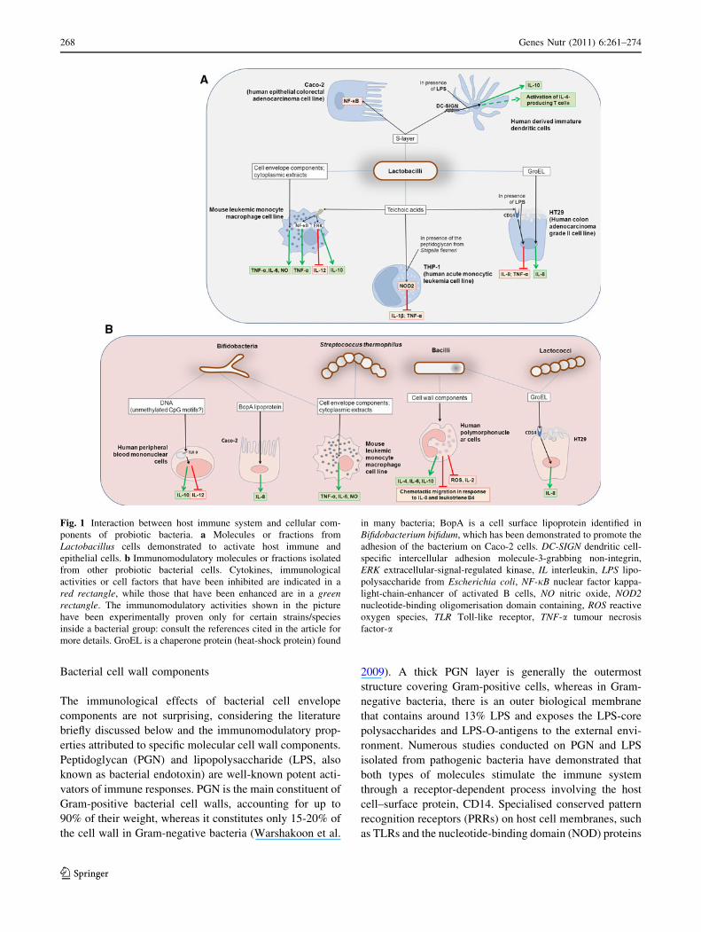
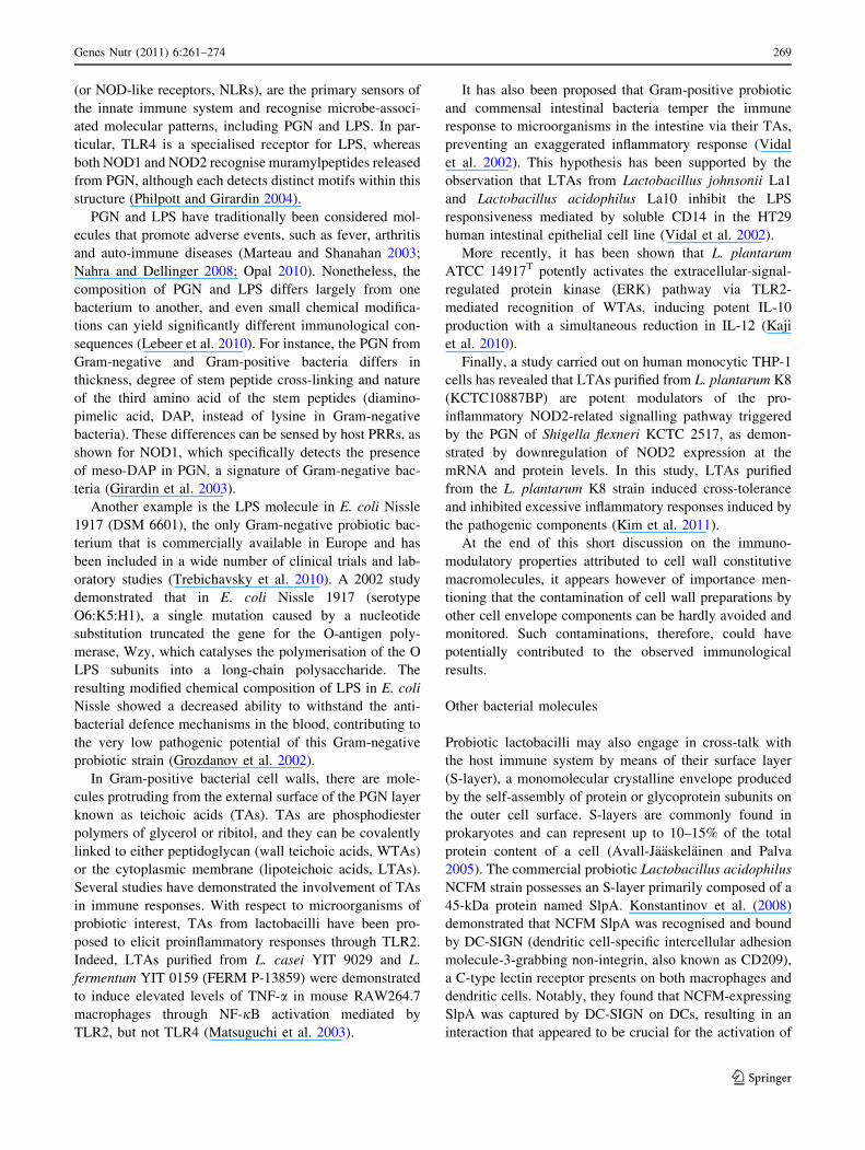
Fig. 1 Interaction between host immune system and cellular com-
ponents of probiotic bacteria. a Molecules or fractions from
Lactobacillus cells demonstrated to activate host immune and
epithelial cells. b Immunomodulatory molecules or fractions isolated
from other probiotic bacterial cells. Cytokines, immunological
activities or cell factors that have been inhibited are indicated in a
red rectangle, while those that have been enhanced are in a greenrectangle. The immunomodulatory activities shown in the picture
have been experimentally proven only for certain strains/species
inside a bacterial group: consult the references cited in the article for
more details. GroEL is a chaperone protein (heat-shock protein) found
in many bacteria; BopA is a cell surface lipoprotein identified in
Bifidobacterium bifidum, which has been demonstrated to promote the
adhesion of the bacterium on Caco-2 cells. DC-SIGN dendritic cell-
specific intercellular adhesion molecule-3-grabbing non-integrin,
ERK extracellular-signal-regulated kinase, IL interleukin, LPS lipo-
polysaccharide from Escherichia coli, NF-jB nuclear factor kappa-
light-chain-enhancer of activated B cells, NO nitric oxide, NOD2nucleotide-binding oligomerisation domain containing, ROS reactive
oxygen species, TLR Toll-like receptor, TNF-a tumour necrosis
factor-a
268 Genes Nutr (2011) 6:261–274
123
(or NOD-like receptors, NLRs), are the primary sensors of
the innate immune system and recognise microbe-associ-
ated molecular patterns, including PGN and LPS. In par-
ticular, TLR4 is a specialised receptor for LPS, whereas
both NOD1 and NOD2 recognise muramylpeptides released
from PGN, although each detects distinct motifs within this
structure (Philpott and Girardin 2004).
PGN and LPS have traditionally been considered mol-
ecules that promote adverse events, such as fever, arthritis
and auto-immune diseases (Marteau and Shanahan 2003;
Nahra and Dellinger 2008; Opal 2010). Nonetheless, the
composition of PGN and LPS differs largely from one
bacterium to another, and even small chemical modifica-
tions can yield significantly different immunological con-
sequences (Lebeer et al. 2010). For instance, the PGN from
Gram-negative and Gram-positive bacteria differs in
thickness, degree of stem peptide cross-linking and nature
of the third amino acid of the stem peptides (diamino-
pimelic acid, DAP, instead of lysine in Gram-negative
bacteria). These differences can be sensed by host PRRs, as
shown for NOD1, which specifically detects the presence
of meso-DAP in PGN, a signature of Gram-negative bac-
teria (Girardin et al. 2003).
Another example is the LPS molecule in E. coli Nissle
1917 (DSM 6601), the only Gram-negative probiotic bac-
terium that is commercially available in Europe and has
been included in a wide number of clinical trials and lab-
oratory studies (Trebichavsky et al. 2010). A 2002 study
demonstrated that in E. coli Nissle 1917 (serotype
O6:K5:H1), a single mutation caused by a nucleotide
substitution truncated the gene for the O-antigen poly-
merase, Wzy, which catalyses the polymerisation of the O
LPS subunits into a long-chain polysaccharide. The
resulting modified chemical composition of LPS in E. coli
Nissle showed a decreased ability to withstand the anti-
bacterial defence mechanisms in the blood, contributing to
the very low pathogenic potential of this Gram-negative
probiotic strain (Grozdanov et al. 2002).
In Gram-positive bacterial cell walls, there are mole-
cules protruding from the external surface of the PGN layer
known as teichoic acids (TAs). TAs are phosphodiester
polymers of glycerol or ribitol, and they can be covalently
linked to either peptidoglycan (wall teichoic acids, WTAs)
or the cytoplasmic membrane (lipoteichoic acids, LTAs).
Several studies have demonstrated the involvement of TAs
in immune responses. With respect to microorganisms of
probiotic interest, TAs from lactobacilli have been pro-
posed to elicit proinflammatory responses through TLR2.
Indeed, LTAs purified from L. casei YIT 9029 and L.
fermentum YIT 0159 (FERM P-13859) were demonstrated
to induce elevated levels of TNF-a in mouse RAW264.7
macrophages through NF-jB activation mediated by
TLR2, but not TLR4 (Matsuguchi et al. 2003).
It has also been proposed that Gram-positive probiotic
and commensal intestinal bacteria temper the immune
response to microorganisms in the intestine via their TAs,
preventing an exaggerated inflammatory response (Vidal
et al. 2002). This hypothesis has been supported by the
observation that LTAs from Lactobacillus johnsonii La1
and Lactobacillus acidophilus La10 inhibit the LPS
responsiveness mediated by soluble CD14 in the HT29
human intestinal epithelial cell line (Vidal et al. 2002).
More recently, it has been shown that L. plantarum
ATCC 14917T potently activates the extracellular-signal-
regulated protein kinase (ERK) pathway via TLR2-
mediated recognition of WTAs, inducing potent IL-10
production with a simultaneous reduction in IL-12 (Kaji
et al. 2010).
Finally, a study carried out on human monocytic THP-1
cells has revealed that LTAs purified from L. plantarum K8
(KCTC10887BP) are potent modulators of the pro-
inflammatory NOD2-related signalling pathway triggered
by the PGN of Shigella flexneri KCTC 2517, as demon-
strated by downregulation of NOD2 expression at the
mRNA and protein levels. In this study, LTAs purified
from the L. plantarum K8 strain induced cross-tolerance
and inhibited excessive inflammatory responses induced by
the pathogenic components (Kim et al. 2011).
At the end of this short discussion on the immuno-
modulatory properties attributed to cell wall constitutive
macromolecules, it appears however of importance men-
tioning that the contamination of cell wall preparations by
other cell envelope components can be hardly avoided and
monitored. Such contaminations, therefore, could have
potentially contributed to the observed immunological
results.
Other bacterial molecules
Probiotic lactobacilli may also engage in cross-talk with
the host immune system by means of their surface layer
(S-layer), a monomolecular crystalline envelope produced
by the self-assembly of protein or glycoprotein subunits on
the outer cell surface. S-layers are commonly found in
prokaryotes and can represent up to 10–15% of the total
protein content of a cell (Avall-Jaaskelainen and Palva
2005). The commercial probiotic Lactobacillus acidophilus
NCFM strain possesses an S-layer primarily composed of a
45-kDa protein named SlpA. Konstantinov et al. (2008)
demonstrated that NCFM SlpA was recognised and bound
by DC-SIGN (dendritic cell-specific intercellular adhesion
molecule-3-grabbing non-integrin, also known as CD209),
a C-type lectin receptor presents on both macrophages and
dendritic cells. Notably, they found that NCFM-expressing
SlpA was captured by DC-SIGN on DCs, resulting in an
interaction that appeared to be crucial for the activation of
Genes Nutr (2011) 6:261–274 269
123
IL-4-producing T cells; in contrast, a knockout mutant of
L. acidophilus NCFM lacking SlpA demonstrated signifi-
cantly impaired binding to DC-SIGN (Konstantinov et al.
2008). These data were confirmed by experiments per-
formed with purified SlpA protein, which ligated to
DC-SIGN and induced IL-10 expression by DCs in the
presence of LPS (Konstantinov et al. 2008).
A homologous Slp protein is also present in Lactoba-
cillus helveticus strains, and it appears to mediate an
immunological effect for members of this species as well.
For instance, SlpA from L. helveticus MIMLh5, a bacte-
rium demonstrated to interact with host cells and modulate
immune responses (Guglielmetti et al. 2010), markedly
altered cytokine production through the inhibition of NF-
jB activation in human intestinal epithelial cells (Taverniti
and Guglielmetti, manuscript in preparation).
Other proteins associated with probiotic bacteria have
been shown to elicit immune responses. For example, the
heat-shock protein GroEL (Hsp60 class), of L. johnsonii
La1 (NCC 533), which is also present at the cell surface,
was expressed in E. coli, and its purified recombinant form
(rGroEL) was shown to bind to mucins and epithelial cells,
stimulating IL-8 secretion in macrophages and HT29 cells
in a CD14-dependent manner (Bergonzelli et al. 2006).
This property was also observed in rGroEL from three
other Gram-positive bacteria, including L. helveticus
ATCC15009, Bacillus subtilis NCC199 and Lactococcus
lactis MG, but not in the rGroEL of the gastric pathogen,
H. pylori P1 (Bergonzelli et al. 2006).
Another example is BopA, a cell surface-associated
lipoprotein of Bifidobacterium bifidum MIMBb75 that
mediates adhesion to human Caco-2 intestinal epithelial
cells. Upon purification from strain MIMBb75, BopA has
been demonstrated to induce the production of IL-8 by
Caco-2 cells in a dose-dependent manner (Guglielmetti
et al. 2008).
Finally, probiotics can interact with the host immune
system by means of their genomic DNA. Convincing sci-
entific evidence has shown that prokaryotic DNA contains
unmethylated CpG motifs that can activate immune
responses in vitro and in vivo (Agrawal and Kandimalla
2002; Rachmilewitz et al. 2002). Lammers et al. (2003)
observed that bacterial genomic DNA extracted from pure
bifidobacterial cultures of the probiotic commercial prod-
uct, VSL#3 (including B. breve, B. longum subsp. longum
and B. longum subsp. infantis), influenced cytokine pro-
duction by peripheral blood mononuclear cells (PBMCs),
decreasing IL-1b and increasing IL-10. The anti-inflam-
matory effect of genomic DNA from VSL#3 bacteria was
also confirmed in an in vivo murine study, which demon-
strated that TLR9 signalling was essential in mediating this
anti-inflammatory effect (Rachmilewitz et al. 2004). It has
been suggested that the immunological effect observed
with bifidobacterial genomic DNA is favoured by the high
guanine–cytosine (GC) content of the Bifidobacterium
genus (58-61%), which explains the redundancy of differ-
ent CpG motifs in the genomes of these bacteria (Lammers
et al. 2003).
In a more recent study, Medina et al. (2007) detected a
large variation in the ability of seven different strains of the
same Bifidobacterium species (B. longum) to modulate the
in vitro production of cytokines by PBMCs, suggesting the
importance of careful strain selection before any specific
use. The authors also proposed that the differential effects
exerted by B. longum strains could be due to differences in
the presence or redundancy of CpG motifs in their gen-
omes. These variations, in fact, can induce a more or less
pronounced immunomodulatory effect (Yi et al. 2002). In
addition, genome comparison is revealing that Bifidobac-
terium strains may encode different restriction/modification
systems (O’Connell Motherway et al. 2009; Lee and
O’Sullivan 2010), which in turn lead to differences in their
DNA methylation profiles and, possibly, immune respon-
ses. It should be, finally, taken into consideration that the
purification of DNA from bacterial cultures is a challeng-
ing task, particularly due to the contamination by exo-
polysaccharides (EPS), such as those known to be often
produced by bifidobacteria (Ruas-Madiedo et al. 2007).
Since variation in such EPS molecules is substantial and
strain dependent, the actual immunological differences
may be potentially due to EPS rather than DNA.
Despite the high-quality research performed up to now,
we are still far from an exhaustive explanation of the
biological effects observed after administration of non-
viable probiotics. Mechanistic reductionist approaches,
such as those employed in the studies described here, are
not sufficient to unveil all the potential properties of
inactivated bacteria or bacterial cell extracts. A single
molecule, in fact, can display different effects if studied
alone or in a complex multicomponent context (Kaji et al.
2010). The potential bioactivity of a specific bacterial
compound can be masked by other cell structures, and the
effects of a single molecule can be influenced by the
presence of additional bioactive substances. In support of
this concept, a recent study by Kaji and collaborators (Kaji
et al. 2010) identified TAs as key factors for triggering the
synergistic induction of IL-10 production; they demon-
strated that TAs alone only weakly induced IL-10 pro-
duction, but when macrophages sensed WTAs or LTAs in
the presence of L. casei strain Shirota, these stimuli
cooperatively induced potent production of IL-10 (Kaji
et al. 2010).
Another investigation showed that the whole bacterial
cell wall was necessary to trigger an immunological
response. Shida and colleagues observed that the insoluble
intact cell wall of the probiotic strain, L. casei Shirota, was
270 Genes Nutr (2011) 6:261–274
123
necessary in stimulating macrophages to produce IL-12,
since this ability was lost when only the soluble polysac-
charide–peptidoglycan complex released from the cell wall
was tested (Shida et al. 2006).
In a different study, Ryan et al. (2009) separated the cell
envelope from the cytoplasmic fraction of L. salivarius
UCC118, a strain that was demonstrated independently of
its viability to decrease IL-8 production by gastric epithe-
lial cells upon exposure to H. pylori cells. However, when
they tested the two fractions separately, neither was able to
retain any statistically significant anti-IL-8 activity, sug-
gesting that intact cells, either alive or killed, were required
(Ryan et al. 2009).
Conclusions
A considerable amount of published data, some of which
have been reported here and in other review articles
(Adams 2010; Kataria et al. 2009), indicates that the use of
non-viable microbial cells or cell components can influence
host’s immune system. The emerging concern regarding
potential safety problems arising from the extensive use of
live microbial cells has been dramatically validated by
Besselink’s communication on the effects of probiotics in
acute pancreatitis (Besselink et al. 2008). These concerns
have enhanced the interest in non-viable microorganisms
or microbial cell extracts, as they could drastically reduce
shelf-life problems and eliminate the risks of microbial
translocation and infection for the consumer (Cross et al.
2004).
In the last fifteen years, several definitions of probiotic
have been proposed, some of them even comprising non-
viable microbial cells. For instance, Reuter (1997) descri-
bed probiotics as ‘a microbial preparation which contains
live and/or dead cells including their metabolites which is
intended to improve the microbial or enzymatic balance at
mucosal surfaces or to stimulate immune mechanisms’.
Similarly, Salminen et al. (1999) spoke about probiotics as
‘microbial cell preparations or components of microbial
cells that have a beneficial effect on the health and well-
being of the host’. Despite the potential legitimacy of these
definitions, nowadays, at least in Western Countries, the
definition of probiotics nearly unanimously accepted is that
by FAO/WHO (‘live microorganisms which when admin-
istered in adequate amounts confer a health benefit on the
host’). Accordingly, national/government institutions are
introducing the FAO/WHO definition in their guidelines
for probiotics. For instance, this definition is proposed in
the guidelines of Italian Ministry of Health (Ministero della
Salute 2005), in a dossier from French Agency for Food
Safety, (Agence Francaise de Securite Sanitaire des Ali-
ments 2005) and in the guidance document on the use of
probiotics in food by the Department for Public Health of
Canada (Health Canada 2009). Thereby, use of the word
‘probiotic’ should be restricted to products that contain live
microorganisms; consequently, we now require new ter-
minology to unambiguously define the use of non-viable
microorganisms or microbial fractions to positively affect
health. To this end, we propose the use of the term
‘paraprobiotic’ (or ‘ghost probiotics’), to be defined as
‘non-viable microbial cells (intact or broken) or crude cell
extracts (i.e. with complex chemical composition), which,
when administered (orally or topically) in adequate
amounts, confer a benefit on the human or animal con-
sumer’. The prefix ‘para’ (from the ancient Greek, paqa9)has been chosen because of its meaning of ‘alongside of’ or
‘atypical’, which can simultaneously indicate similarity to
and difference from the traditional probiotic definition.
Purified molecules of microbial origin or pure microbial cell
products are omitted from the concept of paraprobiotics,
since their use should be included in conventional pharma-
ceutical methodologies. In addition, once a health benefit is
demonstrated, the assignation of a product into the parap-
robiotic category should not be influenced by the methods
used for microbial cell inactivation, which may be achieved
using physical or chemical strategies, including heat treat-
ment, c or UV ray deactivation, chemical or mechanical
disruption, pressure, lyophilisation or acid deactivation.
In conclusion, the preparations included in the new
paraprobiotic definition, namely, non-viable material of
microbial origin, have been demonstrated to positively
affect human/animal health, and they have the noticeable
advantage over probiotics of allowing for the generation of
safer and more stable products. Consequently, paraprobi-
otics are gaining in popularity and will be widely used in
food, supplements, medicine and feed in the future.
References
Adams CA (2010) The probiotic paradox: live and dead cells are
biological response modifiers. Nutr Res Rev 23:37–46
Agence Francaise de Securite Sanitaire des Aliments (2005) Effets
des probiotiques et prebiotiques sur la flore et l’immunite de
l’homme adulte. US Probiotics Web Site. http://www.
usprobiotics.org/docs/AFFSA%20probiotic%20prebiotic%20
flora%20immunity%2005.pdf. Accessed 12 March 2011
Agrawal S, Kandimalla ER (2002) Medicinal chemistry and thera-
peutic potential of CpG DNA. Trends Mol Med 8:114–121
Ananta E, Knorr D (2009) Comparison of inactivation pathways of
thermal or high pressure inactivated Lactobacillus rhamnosusATCC 53103 by flow cytometry analysis. Food Microbiol
26:542–546
Avall-Jaaskelainen S, Palva A (2005) Lactobacillus surface layers
and their applications. FEMS Microbiol Rev 29:511–529
BCC Research (2008) The probiotic market: ingredients, supple-
ments, foods (FOD035B). BCC Research web site. http://www.
Genes Nutr (2011) 6:261–274 271
123
bccresearch.com/pressroom/FOD035B.html. Accessed 13 March
2011
Bergonzelli GE, Granato D, Pridmore RD, Marvin-Guy LF, Donni-
cola D, Corthesy-Theulaz IE (2006) GroEL of Lactobacillusjohnsonii La1 (NCC 533) is cell surface associated: potential
role in interactions with the host and the gastric pathogen
Helicobacter pylori. Infect Immun 74:425–434
Besselink MG, van Santvoort HC, Buskens E et al (2008) Probiotic
prophylaxis in predicted severe acute pancreatitis: a randomised,
double-blind, placebo-controlled trial. Lancet 371:651–659
Bibiloni R, Fedorak RN, Tannock GW, Madsen KL, Gionchetti P,
Campieri M, De Simone C, Sartor RB (2005) VSL#3 probiotic-
mixture induces remission in patients with active ulcerative
colitis. Am J Gastroenterol 100:1539–1546
Bjorck P, Banchereau J, Flores-Romo L (1997) CD40 ligation
counteracts Fas-induced apoptosis of human dendritic cells. Int
Immunol 9:365–372
Cella M, Scheidegger D, Palmer-Lehmann K, Lane P, Lanzavecchia
A, Alber G (1996) Ligation of CD40 on dendritic cells triggers
production of high levels of interleukin-12 and enhances T cell
stimulatory capacity: T-T help via APC activation. J Exp Med
184:747–752
Chapot-Chartier MP, Vinogradov E, Sadovskaya I, Andre G, Mistou
MY, Trieu-Cuot P, Furlan S, Bidnenko E, Courtin P, Pechoux C,
Hols P, Dufrene YF, Kulakauskas S (2010) Cell surface of
Lactococcus lactis is covered by a protective polysaccharide
pellicle. J Biol Chem 285(14):10464–10471
Chuang L, Wu KG, Pai C, Hsieh PS, Tsai JJ, Yen JH, Lin MY (2007)
Heat-killed cells of lactobacilli skew the immune response
toward T helper 1 polarization in mouse splenocytes and
dendritic cell-treated T cells. J Agric Food Chem 55:11080–
11086
Corominas M, Mestre M, Bas J, Buendia E (1998) Distinct
modulation by interferon-gamma (IFN-gamma) of CD23 expres-
sion on B and T lymphocytes of atopic subjects. Clin Exp
Immunol 112:276–280
Cross ML, Ganner A, Teilab D, Fray LM (2004) Patterns of cytokine
induction by gram-positive and gram-negative probiotic bacteria.
FEMS Immunol Med Microbiol 42:173–180
De Simone C, Bianchi Salvadori B, Negri R, Ferrazzi M, Baldinelli L,
Vesely R (1986) The adjuvant effect of yogurt on production of
gamma-interferon by Con A-stimulated human peripheral blood
lymphocytes. Nutr Rep Int 33:419–433
Di Giacinto C, Marinaro M, Sanchez M, Strober W, Boirivant M
(2005) Probiotics meliorate recurrent Th1-mediated murine
colitis by inducing IL-10 and IL-10-dependent TGF-beta-bearing
regulatory cells. J Immunol 174(6):3237–3246
FAO/WHO (2002) Report of a joint FAO/WHO expert consultation
on guidelines for the evaluation of probiotics in food. World
Health Organization and Food and Agriculture Organization of
the United Nations, London Ontario, Canada
Fedorak RN, Madsen KL (2004a) Probiotics and prebiotics in
gastrointestinal disorders. Curr Opin Gastroenterol 20:146–155
Fedorak RN, Madsen KL (2004b) Probiotics and the management of
inflammatory bowel disease. Inflamm Bowel Dis 10:286–299
Frotscher B, Anton K, Worm M (2002) Inhibition of IgE production
by the imidazoquinoline resiquimod in nonallergic and allergic
donors. J Invest Dermatol 119:1059–1064
Girardin SE, Boneca IG, Carneiro LA, Antignac A, Jehanno M, Viala
J, Tedin K, Taha MK, Labigne A, Zahringer U, Coyle AJ,
DiStefano PS, Bertin J, Sansonetti PJ, Philpott DJ (2003) Nod1
detects a unique muropeptide from Gram-negative bacterial
peptidoglycan. Science 300:1584–1587
Gobbetti M, Cagno RD, De Angelis M (2010) Functional microor-
ganisms for functional food quality. Crit Rev Food Sci Nutr
50:716–727
Grozdanov L, Zahringer U, Blum-Oehler G, Brade L, Henne A,
Knirel YA, Schombel U, Schulze J, Sonnenborn U, Gottschalk
G, Hacker J, Rietschel ET, Dobrindt U (2002) A single
nucleotide exchange in the wzy gene is responsible for the
semirough O6 lipopolysaccharide phenotype and serum sensi-
tivity of Escherichia coli strain Nissle 1917. J Bacteriol
184:5912–5925
Guglielmetti S, Tamagnini I, Mora D, Minuzzo M, Scarafoni A,
Arioli S, Hellman J, Karp M, Parini C (2008) Implication of an
outer surface lipoprotein in adhesion of Bifidobacterium bifidumto Caco-2 cells. Appl Environ Microbiol 74:4695–4702
Guglielmetti S, Taverniti V, Minuzzo M, Arioli S, Zanoni I, Stuknyte
M, Granucci F, Karp M, Mora D (2010) A dairy bacterium
displays in vitro probiotic properties for the pharyngeal mucosa
by antagonizing group A streptococci and modulating the
immune response. Infect Immun 78:4734–4743
Haller D, Bode C, Hammes WP, Pfeifer AM, Schiffrin EJ, Blum S
(2000) Non-pathogenic bacteria elicit a differential cytokine
response by intestinal epithelial cell/leucocyte co-cultures. Gut
47:79–87
Hart AL, Lammers K, Brigidi P, Vitali B, Rizzello F, Gionchetti P,
Campieri M, Kamm MA, Knight SC, Stagg AJ (2004) Modu-
lation of human dendritic cell phenotype and function by
probiotic bacteria. Gut 53:1602–1609
Health Canada (2009). Guidance Document: The Use of Probiotic
Microorganisms in Food. Health Canada web site.
http://www.hc-sc.gc.ca/fn-an/alt_formats/hpfb-dgpsa/pdf/
legislation/probiotics_guidance-orientation_probiotiques-
eng.pdf. Accessed 12 March 2011
Jensen GS, Benson KF, Carter SG, Endres JR (2010) GanedenBC30cell wall and metabolites: anti-inflammatory and immune
modulating effects in vitro. BMC Immunol 11:1–15
Kaji R, Kiyoshima-Shibata J, Nagaoka M, Nanno M, Shida K (2010)
Bacterial teichoic acids reverse predominant IL-12 production
induced by certain lactobacillus strains into predominant IL-10
production via TLR2-dependent ERK activation in macro-
phages. J Immunol 184:3505–3513
Kataria J, Li N, Wynn JL, Neu J (2009) Probiotic microbes: do they
need to be alive to be beneficial? Nutr Rev 67:546–550
Kim HG, Lee SY, Kim NR, Lee HY, Ko MY, Jung BJ, Kim CM, Lee
JM, Park JH, Han SH, Chung DK (2011) Lactobacillusplantarum lipoteichoic acid down-regulated Shigella flexneripeptidoglycan-induced inflammation. Mol Immunol 48:382–391
Kiniwa M, Gately M, Gubler U, Chizzonite R, Fargeas C, Delespesse
G (1992) Recombinant interleukin-12 suppresses the synthesis of
immunoglobulin E by interleukin-4 stimulated human lympho-
cytes. J Clin Invest 90:262–966
Konstantinov SR, Smidt H, de Vos WM, Bruijns SC, Singh SK,
Valence F, Molle D, Lortal S, Altermann E, Klaenhammer TR,
van Kooyk Y (2008) S layer protein A of Lactobacillusacidophilus NCFM regulates immature dendritic cell and T cell
functions. Proc Natl Acad Sci USA 105:19474–19479
Lammers KM, Brigidi P, Vitali B, Gionchetti P, Rizzello F, Caramelli
E, Matteuzzi D, Campieri M (2003) Immunomodulatory effects
of probiotic bacteria DNA: IL-1 and IL-10 response in human
peripheral blood mononuclear cells. FEMS Immunol Med
Microbiol 38:165–172
Lebeer S, Vanderleyden J, De Keersmaecker SC (2010) Host
interactions of probiotic bacterial surface molecules: comparison
with commensals and pathogens. Nat Rev Microbiol 8:171–184
Lee JH, O’Sullivan DJ (2010) Genomic insights into bifidobacteria.
Microbiol Mol Biol Rev 74:378–416
Lim LH, Li HY, Huang CH, Lee BW, Lee YK, Chua KY (2009) The
effects of heat-killed wild-type Lactobacillus casei Shirota on
allergic immune responses in an allergy mouse model. Int Arch
Allergy Immunol 148:297–304
272 Genes Nutr (2011) 6:261–274
123
Lopez M, Li N, Kataria J, Russell M, Neu J (2008) Live and
ultraviolet-inactivated Lactobacillus rhamnosus GG decrease
flagellin-induced interleukin-8 production in Caco-2 cells. J Nutr
138:2264–2268
Lutz MB, Schuler G (2002) Immature, semi-mature and fully mature
dendritic cells: which signals induce tolerance or immunity?
Trends Immunol 23:445–449
Majamaa H, Isolauri E (1997) Probiotics: a novel approach in the
management of food allergy. J Allergy Clin Immunol
99:179–185
Manetti R, Gerosa F, Giudizi MG, Biagiotti R, Parronchi P, Piccinni
MP, Sampognaro S, Maggi E, Romagnani S, Trinchieri G et al
(1994) Interleukin 12 induces stable priming for interferon
gamma (IFN-gamma) production during differentiation of
human T helper (Th) cells and transient IFN-gamma production
in established Th2 cell clones. J Exp Med 179:1273–1283
Marteau P, Shanahan F (2003) Basic aspects and pharmacology of
probiotics: an overview of pharmacokinetics, mechanisms of
action and side-effects. Best Pract Res Clin Gastroenterol
17:725–740
Mastrangeli G, Corinti S, Butteroni C, Afferni C, Bonura A, Boirivant
M, Colombo P, Di Felice G (2009) Effects of live and
inactivated VSL#3 probiotic preparations in the modulation of
in vitro and in vivo allergen-induced Th2 responses. Int Arch
Allergy Immunol 150:133–143
Matsuguchi T, Takagi A, Matsuzaki T, Nagaoka M, Ishikawa K,
Yokokura T, Yoshikai Y (2003) Lipoteichoic acids from
Lactobacillus strains elicit strong tumor necrosis factor alpha-
inducing activities in macrophages through Toll-like receptor 2.
Clin Diagn Lab Immunol 10:259–266
Matsuzaki T, Chin J (2000) Modulating immune responses with
probiotic bacteria. Immunol Cell Biol 78:67–73
Medina M, Izquierdo E, Ennahar S, Sanz Y (2007) Differential
immunomodulatory properties of Bifidobacterium longumstrains: relevance to probiotic selection and clinical applications.
Clin Exp Immunol 150:531–538
Miettinen M, Vuopio-Varkila J, Varkila K (1996) Production of
human tumor necrosis factor alpha, interleukin-6, and interleu-
kin-10 is induced by lactic acid bacteria. Infect Immun
64:5403–5405
Ministero della Salute (2005) Linee guida probiotici e probiotici.
Ministero della Salute Web Site. http://www.salute.gov.it/
imgs/C_17_pubblicazioni_1016_allegato.pdf. Accessed 12
March 2011
Morris SC, Madden KB, Adamovicz JJ, Gause WC, Hubbard BR,
Gately MK, Finkelman FD (1994) Effects of IL-12 on in vivo
cytokine gene expression and Ig isotype selection. J Immunol
152:1047–1056
Murosaki S, Yamamoto Y, Ito K, Inokuchi T, Kusaka H, Ikeda H,
Yoshikai Y (1998) Heat-killed Lactobacillus plantarum L-137
suppresses naturally fed antigen-specific IgE production by
stimulation of IL-12 production in mice. J Allergy Clin Immunol
102:57–64
Muscettola M, Massai L, Tanganelli C, Grasso G (1994) Effects of
lactobacilli on interferon production in young and aged mice.
Ann N Y Acad Sci 717:226–232
Nahra R, Dellinger RP (2008) Targeting the lipopolysaccharides: still
a matter of debate? Curr Opin Anaesthesiol 21:98–104
O’Connell Motherway M, O’Driscoll J, Fitzgerald GF, Van Sinderen
D (2009) Overcoming the restriction barrier to plasmid trans-
formation and targeted mutagenesis in Bifidobacterium breve
UCC2003. Microb Biotechnol 2(3):321–332
Opal SM (2010) Endotoxins and other sepsis triggers. Contrib
Nephrol 167:14–24
Philpott DJ, Girardin SE (2004) The role of Toll-like receptors and
Nod proteins in bacterial infection. Mol Immunol 41:1099–1108
Rachmilewitz D, Karmeli F, Takabayashi K, Hayashi T, Leider-Trejo
L, Lee J, Leoni LM, Raz E (2002) Immunostimulatory DNA
ameliorates experimental and spontaneous murine colitis. Gas-
troenterology 122:1428–1441
Rachmilewitz D, Katakura K, Karmeli F, Hayashi T, Reinus C,
Rudensky B, Akira S, Takeda K, Lee J, Takabayashi K, Raz E
(2004) Toll-like receptor 9 signaling mediates the anti-inflam-
matory effects of probiotics in murine experimental colitis.
Gastroenterology 126:520–528
Rasche C, Wolfram C, Wahls M, Worm M (2007) Differential
immunomodulating effects of inactivated probiotic bacteria on
the allergic immune response. Acta Derm Venereol 87:305–311
Reuter G (1997) Present and future probiotics in Germany and in
Center Europe. Biosci Microflora 16:43–51
Ruas-Madiedo P, Moreno JA, Salazar N, Delgado S, Mayo B,
Margolles A, de Los Reyes-Gavilan CG (2007) Screening of
exopolysaccharide-producing Lactobacillus and Bifidobacterium
strains isolated from the human intestinal microbiota. Appl
Environ Microbiol 73(13):4385–4388
Ryan KA, Daly P, Li Y, Hooton C, O’Toole PW (2008) Strain-
specific inhibition of Helicobacter pylori by Lactobacillussalivarius and other lactobacilli. J Antimicrob Chemother
61:831–834
Ryan KA, O’Hara AM, van Pijkeren JP, Douillard FP, O’Toole PW
(2009) Lactobacillus salivarius modulates cytokine induction
and virulence factor gene expression in Helicobacter pylori.J Med Microbiol 58:996–1005
Salminen S, Ouwehand A, Benno Y, Lee YK (1999) Probiotics: how
should they be defined. Trends Food Sci Technol 10:107–110
Sartor RB (2005) Probiotic therapy of intestinal inflammation and
infections. Curr Opin Gastroenterol 21:44–50
Shida K, Kiyoshima-Shibata J, Nagaoka M, Watanabe K, Nanno M
(2006) Induction of interleukin-12 by Lactobacillus strains
having a rigid cell wall resistant to intracellular digestion.
J Dairy Sci 89:3306–3317
Smits HH, Engering A, de Jong EC, Schipper K, van Capel TM, Zaat
BA, Yazdanbakhsh M, Wierenga EA, van Kooyk Y, Kapsenberg
ML (2005) Selective probiotic bacteria induce IL-10-producing
regulatory T cells in vitro by modulating dendritic cell function
through dendritic cell-specific intercellular adhesion molecule
3-grabbing nonintegrin. J Allergy Clin Immunol 115:1260–1267
Stagg AJ, Bell SJ, Rigby R et al (2000) Treatment with anti-
TNFalpha antibody reduces expression of CD40 on lamina
propria dendritic cells. Gastroenterology 118:A353
Tejada-Simon MV, Pestka JJ (1999) Proinflammatory cytokine and
nitric oxide induction in murine macrophages by cell wall and
cytoplasmic extracts of lactic acid bacteria. J Food Prot
62:1435–1444
Tohno M, Shimosato T, Kitazawa H, Katoh S, Iliev ID, Kimura T,
Kawai Y, Watanabe K, Aso H, Yamaguchi T, Saito T (2005)
Toll-like receptor 2 is expressed on the intestinal M cells in
swine. Biochem Biophys Res Commun 330:547–554
Torii A, Torii S, Fujiwara S, Tanaka H, Inagaki N, Nagai H (2007)
Lactobacillus acidophilus strain L-92 regulates the production of
Th1 cytokine as well as Th2 cytokines. Allergol Int 56:293–301
Trebichavsky I, Splichal I, Rada V, Splichalova A (2010) Modulation
of natural immunity in the gut by Escherichia coli strain Nissle
1917. Nutr Rev 68:459–464
van Hoffen E, Korthagen NM, de Kivit S, Schouten B, Bardoel B,
Duivelshof A, Knol J, Garssen J, Willemsen LE (2010) Exposure
of intestinal epithelial cells to UV-killed Lactobacillus GG but
not Bifidobacterium breve enhances the effector immune
response in vitro. Int Arch Allergy Immunol 152:159–168
Vidal K, Donnet-Hughes A, Granato D (2002) Lipoteichoic acids
from Lactobacillus johnsonii strain La1 and Lactobacillusacidophilus strain La10 antagonize the responsiveness of human
Genes Nutr (2011) 6:261–274 273
123

intestinal epithelial HT29 cells to lipopolysaccharide and Gram-
negative bacteria. Infect Immun 70:2057–2064
Warshakoon HJ, Hood JD, Kimbrell MR, Malladi S, Wu WY, Shukla
NM, Agnihotri G, Sil D, David SA (2009) Potential adjuvantic
properties of innate immune stimuli. Hum Vaccin 5:381–394
Weiner HL (2001a) Oral tolerance: immune mechanisms and the
generation of Th3-type TGF-beta-secreting regulatory cells.
Microbes Infect 3:947–954
Weiner HL (2001b) The mucosal milieu creates tolerogenic dendritic
cells and T(R)1 and T(H)3 regulatory cells. Nat Immunol
2:671–672
Yi AK, Yoon JG, Yeo SJ, Hong SC, English BK, Krieg AM (2002)
Role of mitogen-activated protein kinases in CpG DNA-medi-
ated IL-10 and IL-12 production: central role of extracellular
signal-regulated kinase in the negative feedback loop of the CpG
DNA-mediated Th1 response. J Immunol 168:4711–4720
Zhang L, Li N, Caicedo R, Neu J (2005) Alive and dead Lactobacillusrhamnosus GG decrease TNF-a-induced interleukin-8 produc-
tion in Caco-2 cells. J Nutr 135:1752–1756
274 Genes Nutr (2011) 6:261–274
123
Related Documents