ORIGINAL ARTICLE The identification of irreversible rituximab-resistant lymphoma caused by CD20 gene mutations Y Mishima 1,2 , Y Terui 1 , K Takeuchi 3 , Y Matsumoto-Mishima 1 , S Matsusaka 1 , R Utsubo-Kuniyoshi 1,2 and K Hatake 1 1 Department of Clinical Chemotherapy, Cancer Chemotherapy Center, Japanese Foundation for Cancer Research, Tokyo, Japan; 2 Olympas Bio-Imaging Lab, Cancer Chemotherapy Center, Japanese Foundation for Cancer Research, Tokyo, Japan and 3 Division of Pathology, Cancer Institute, Japanese Foundation for Cancer Research, Tokyo, Japan C-terminal mutations of CD20 constitute part of the mechan- isms that resist rituximab therapy. Most CD20 having a C-terminal mutation was not recognized by L26 antibody. As the exact epitope of L26 has not been determined, expression and localization of mutated CD20 have not been completely elucidated. In this study, we revealed that the binding site of L26 monoclonal antibody is located in the C-terminal cytoplas- mic region of CD20 molecule, which was often lost in mutated CD20 molecules. This indicates that it is difficult to distinguish the mutation of CD20 from under expression of the CD20 protein. To detect comprehensive CD20 molecules including the resistant mutants, we developed a novel monoclonal antibody that recognizes the N-terminal cytoplasm region of CD20 molecule. We screened L26-negative cases with our antibody and found several mutations. A rituximab-binding analysis using the cryopreserved specimen that mutation was identified in CD20 molecules indicated that the C-terminal region of CD20 undertakes a critical role in presentation of the large loop in which the rituximab-binding site locates. Thus, combination of antibodies of two kinds of epitope permits the identification of C-terminal CD20 mutations associated with irreversible resistance to rituximab and may help the decision of the treatment strategy. Blood Cancer Journal (2011) 1, e15; doi:10.1038/bcj.2011.11; published online 8 April 2011 Keywords: B-cell lymphomas; mutations; antibody therapy; CD20; rituximab Introduction Previously, we reported that gene mutations of CD20 were somehow involved in resistance to rituximab therapy, and we proposed that C-terminal deletion mutations of CD20 might be related to relapse/resistance after rituximab therapy. 1 Many of these C-terminal truncated CD20 molecules were not recog- nized by the L26 monoclonal antibody used routinely in most clinical laboratories. Therefore, expression of CD20 seemed to have been completely lost for these lymphomas. However, an immunohistochemical study using a polyclonal antibody showed that some kind of C-terminal truncated CD20 was present in cytoplasm, so it was possible that the epitope of L26 was lost by gene mutations. 1 L26 recognizes the cytoplasmic region of CD20 molecules, but no more detailed information about its epitope had been reported. 2,3 In this study, we determine a recognition site of L26 by using a series of deletion mutants of CD20 molecules. In addition, to detect every one of the mutated CD20 molecules, we developed new antibodies that recognize the N-terminal region of CD20 molecules. We used these antibodies to identify cells that have CD20 molecules with abnormalities in the C-terminal cytoplasmic region. We characterized these mutated CD20 molecules using living primary lymphoma cells. Materials and methods Cells, viruses and DNA constructs The coding region of the CD20 gene was amplified by reverse transcription PCR (RT-PCR) from RNA extracted from a Burkitt’s lymphoma cell line, Raji, and was cloned into a pDON-AI retroviral vector (Takara, Ohtsu, Japan). A series of deletion mutants of CD20 in the C-terminal cytoplasmic region was constructed by inserting stop codons after nucleotides encoding E281, E263, E245, V228 and G210. Retroviruses carrying wild type and deletion mutants of CD20, together with mock construct, were produced with the transient retrovirus packaging cell line G3T-hi (Takara) according to the manufacturer’s protocol. Packaged retrovirus vectors were then used to infect a myeloma cell line, KMS12PE, 4 with subsequent selection using 500 mg/ml G418. For the transfection of mutant CD20 gene, whole coding region of CD20 complementary DNA (cDNA) prepared by RT-PCR of total RNA isolated from the patient cells was cloned into first cassette of a bicistronic retrovirus vector carrying a green fluorescent protein (Takara). Bicistronic expression of ZsGreen is facilitated by internal ribosomal entry site only when CD20 mutant gene was translated, enabling the efficient selection of transformed cells. Generation of monoclonal antibody secreting hybridomas A synthetic peptide corresponding to residues 23–36 of human CD20 with one additional cysteine at the N-terminus (CMQSGPKPLFRRMSS) was synthesized. The peptide was coupled with keyhole limpet hemocyanin. BALB/c mice were primed with a subcutaneous injection of the keyhole limpet hemocyanin-conjugated synthetic peptide emulsified in Freund’s complete adjuvant. Mice were boosted four times at two-week intervals with the same antigen. Mice that developed antibodies as measured by enzyme-linked immunosorbent assay with the immunizing peptide were boosted intravenously with the same peptide 4 days before splenocytes were harvested and fused to mouse myeloma cells. Hybridization and cloning were performed according to standard procedures. 5 CD20 gene sequencing Pleural effusion mononuclear cells were obtained by density gradient centrifugation using Ficoll-Hypaque 1.077 (Sigma, Received 28 October 2010; revised 7 January 2011; accepted 1 February 2011 Correspondence: Dr K Hatake, Department of Clinical Oncology and Hematology, Cancer Institute Hospital, Japanese Foundation for Cancer Research, 3-8-31, Ariake Koto-ku, Tokyo 135-8550, Japan. E-mail: [email protected] Citation: Blood Cancer Journal (2011) 1, e15; doi:10.1038/bcj.2011.11 & 2011 Macmillan Publishers Limited All rights reserved 2044-5385/11 www.nature.com/bcj

Welcome message from author
This document is posted to help you gain knowledge. Please leave a comment to let me know what you think about it! Share it to your friends and learn new things together.
Transcript
ORIGINAL ARTICLE
The identification of irreversible rituximab-resistant lymphoma caused by CD20 genemutations
Y Mishima1,2, Y Terui1, K Takeuchi3, Y Matsumoto-Mishima1, S Matsusaka1, R Utsubo-Kuniyoshi1,2 and K Hatake1
1Department of Clinical Chemotherapy, Cancer Chemotherapy Center, Japanese Foundation for Cancer Research, Tokyo, Japan;2Olympas Bio-Imaging Lab, Cancer Chemotherapy Center, Japanese Foundation for Cancer Research, Tokyo, Japan and 3Divisionof Pathology, Cancer Institute, Japanese Foundation for Cancer Research, Tokyo, Japan
C-terminal mutations of CD20 constitute part of the mechan-isms that resist rituximab therapy. Most CD20 having aC-terminal mutation was not recognized by L26 antibody. Asthe exact epitope of L26 has not been determined, expressionand localization of mutated CD20 have not been completelyelucidated. In this study, we revealed that the binding site ofL26 monoclonal antibody is located in the C-terminal cytoplas-mic region of CD20 molecule, which was often lost in mutatedCD20 molecules. This indicates that it is difficult to distinguishthe mutation of CD20 from under expression of the CD20protein. To detect comprehensive CD20 molecules includingthe resistant mutants, we developed a novel monoclonalantibody that recognizes the N-terminal cytoplasm region ofCD20 molecule. We screened L26-negative cases with ourantibody and found several mutations. A rituximab-bindinganalysis using the cryopreserved specimen that mutation wasidentified in CD20 molecules indicated that the C-terminalregion of CD20 undertakes a critical role in presentation of thelarge loop in which the rituximab-binding site locates. Thus,combination of antibodies of two kinds of epitope permits theidentification of C-terminal CD20 mutations associated withirreversible resistance to rituximab and may help the decisionof the treatment strategy.Blood Cancer Journal (2011) 1, e15; doi:10.1038/bcj.2011.11;published online 8 April 2011Keywords: B-cell lymphomas; mutations; antibody therapy; CD20;rituximab
Introduction
Previously, we reported that gene mutations of CD20 weresomehow involved in resistance to rituximab therapy, and weproposed that C-terminal deletion mutations of CD20 might berelated to relapse/resistance after rituximab therapy.1 Many ofthese C-terminal truncated CD20 molecules were not recog-nized by the L26 monoclonal antibody used routinely in mostclinical laboratories. Therefore, expression of CD20 seemed tohave been completely lost for these lymphomas. However, animmunohistochemical study using a polyclonal antibodyshowed that some kind of C-terminal truncated CD20 waspresent in cytoplasm, so it was possible that the epitope of L26was lost by gene mutations.1 L26 recognizes the cytoplasmicregion of CD20 molecules, but no more detailed informationabout its epitope had been reported.2,3 In this study, wedetermine a recognition site of L26 by using a series of deletionmutants of CD20 molecules. In addition, to detect every one ofthe mutated CD20 molecules, we developed new antibodies
that recognize the N-terminal region of CD20 molecules. Weused these antibodies to identify cells that have CD20 moleculeswith abnormalities in the C-terminal cytoplasmic region. Wecharacterized these mutated CD20 molecules using livingprimary lymphoma cells.
Materials and methods
Cells, viruses and DNA constructsThe coding region of the CD20 gene was amplified by reversetranscription PCR (RT-PCR) from RNA extracted from a Burkitt’slymphoma cell line, Raji, and was cloned into a pDON-AIretroviral vector (Takara, Ohtsu, Japan). A series of deletionmutants of CD20 in the C-terminal cytoplasmic region wasconstructed by inserting stop codons after nucleotides encodingE281, E263, E245, V228 and G210. Retroviruses carrying wildtype and deletion mutants of CD20, together with mockconstruct, were produced with the transient retrovirus packagingcell line G3T-hi (Takara) according to the manufacturer’sprotocol. Packaged retrovirus vectors were then used to infecta myeloma cell line, KMS12PE,4 with subsequent selectionusing 500mg/ml G418.
For the transfection of mutant CD20 gene, whole codingregion of CD20 complementary DNA (cDNA) prepared byRT-PCR of total RNA isolated from the patient cells was clonedinto first cassette of a bicistronic retrovirus vector carrying agreen fluorescent protein (Takara). Bicistronic expression ofZsGreen is facilitated by internal ribosomal entry site only whenCD20 mutant gene was translated, enabling the efficientselection of transformed cells.
Generation of monoclonal antibody secretinghybridomasA synthetic peptide corresponding to residues 23–36 of humanCD20 with one additional cysteine at the N-terminus(CMQSGPKPLFRRMSS) was synthesized. The peptide wascoupled with keyhole limpet hemocyanin. BALB/c mice wereprimed with a subcutaneous injection of the keyhole limpethemocyanin-conjugated synthetic peptide emulsified inFreund’s complete adjuvant. Mice were boosted four times attwo-week intervals with the same antigen. Mice that developedantibodies as measured by enzyme-linked immunosorbent assaywith the immunizing peptide were boosted intravenously withthe same peptide 4 days before splenocytes were harvested andfused to mouse myeloma cells. Hybridization and cloning wereperformed according to standard procedures.5
CD20 gene sequencingPleural effusion mononuclear cells were obtained by densitygradient centrifugation using Ficoll-Hypaque 1.077 (Sigma,
Received 28 October 2010; revised 7 January 2011; accepted1 February 2011
Correspondence: Dr K Hatake, Department of Clinical Oncology andHematology, Cancer Institute Hospital, Japanese Foundation forCancer Research, 3-8-31, Ariake Koto-ku, Tokyo 135-8550, Japan.E-mail: [email protected]
Citation: Blood Cancer Journal (2011) 1, e15; doi:10.1038/bcj.2011.11& 2011 Macmillan Publishers Limited All rights reserved 2044-5385/11
www.nature.com/bcj
St Louis, MO, USA). The isolated mononuclear cells thenunderwent negative immunomagnetic selection using a B CellIsolation Kit II (Miltenyi Biotec, Bergisch Gladbach, Germany)for purifying B-linage cells. Total RNA was prepared usingTRIzol reagent according to the instructions of the manufacturer(Invitrogen, Carlsbad, CA, USA) and 1 mg was subjected toreverse transcription under MMLV reverse transcriptase(Takara). To prepare cDNA-containing whole coding region ofCD20, PCR amplification was performed from 2 ml of cDNAusing primers outside of the start and the stop codon: hCD20-50-FW (50-GCAGCTAGCATCCAAATCAG-30) and hCD20-30-RV(50-TGGTGCGTATGTGCAGAGTA-30). To determine the se-quence of the CD20, we performed cycle sequencing on a3130 DNA analyzer (Applied Biosystems, Foster City, CA, USA),directly from PCR-purified products, in both directions using aBigDye Terminator Cycle Sequencing Kit v3.1 (Applied Biosystems).
ImmunocytochemistryCells were stained by rituximab and L26 antibody (DAKO,Carpentaria, CA, USA) sequentially. Briefly, the cells werelabeled by incubating in 10 mg/ml of rituximab conjugated withAlexa Fluor 647 (Invitrogen) according to previously describedprocedure6 and washed twice with phosphate-buffered saline.Rituximab-labeled cells were swelled by treatment with 75 mM
KCl and then were made to adhere onto collagen I-coated coverglass by brief centrifugation. After fixing and permeabilization,the specimens were exposed to L26 antibody. Subsequently, thespecimens were washed and were incubated in Alexa488-conjugated goat anti-mouse IgG. Nuclear staining was per-formed using 5 mg/ml of 4’-6-diamidino-2-phenylindole. Thepreparations were screened for fluorescence with a confocalmicroscope (FV1000, OLYMPUS, Tokyo, Japan) using excitationwavelengths of 405, 488 and 633 nm to detect emission bynuclear staining (4’-6-diamidino-2-phenylindole), rituximab orL26 staining, respectively.
ImmunohistochemistryThe tissues had been routinely fixed in 10% neutral formalin andembedded in paraffin. L26 antibody and monoclonal antibodiesraised against the N-terminal cytoplasmic region of CD20 wereused. The sections were deparaffinized and rehydrated in gradedalcohol. For heat-induced epitope retrieval, the sections weresubjected to Target Retrieval Solution, pH 9 (DAKO) at 97 1C for40 min. The sections then were brought to an automated stainer(DAKO) by following the vendor’s protocol. EnVision Plus(DAKO) and peroxidase detection methods were used.
Rituximab-binding analysisRituximab-binding analysis was performed according to ourpreviously developed imaging-based procedure, with somemodifications.6 Briefly, approximately ten thousands cells ofpurified living lymphoma cells using a Dead cell removal kit(Miltenyi Biotec) were incubated with 10mg/ml of anti-CD19 mAb labeled with Alexa Fluor 488 (Invitrogen) andrituximab labeled with Alexa Fluor 647 (Invitrogen) for 30 minat 4 1C. The cells were washed with phosphate-buffered salinetwo times and were then suspended in 4ml of RPMI1640medium supplemented with 10% fetal bovine serum and 5 mg/ml of Hoechist 33 342. The cell suspension was pipetted into awell made of silicon, 2.5 mm in diameter and 2 mm in depth, ona piece of cover glass. Images were collected by means of anOLYMPUS PlanApo � 60 oil objective in an OLYMPUSFV-1000 confocal microscope (OLYMPUS). The fluorophore
was excited by laser at 405, 488 or 633 nm. The cells wereoptically sectioned along the z-axis, and the images werecollected at 640� 640 pixel resolution in a sequential mode tominimize the crossover between channels. The step size in thez-axis was 0.5mm. For three-dimensional reconstruction, two-dimensional confocal stacks were saved in an Olympus.oibformat and three-dimensional images were generated usingFluoview software (OLYMPUS).
Cell sorting and genetic analysisThe cryopreserved B-linage cells of the pleural effusion atrelapse were further sorted into two fractions of the cells withhigh affinity to rituximab (R-high) and those of low (R-low) byusing flow cytometry (FACSvantage, Becton Dickinson, FranklinLakes, NJ, USA). The cells were labeled with rituximabconjugated with AlexaFluor488 (Invitrogen) and anti-CD19-PE(Becton Dickinson), then resuspended in phosphate-bufferedsaline containing 2 mg/ml of 7-AAD (Sigma) to exclude deadcells. The sorting gate used for ‘R-high’ was rituximabhigh/CD19þ /7-AAD�, and for ‘R-low’ was rituximablow/CD19þ /7-AAD�. The sorted cells were immediately used for extraction ofboth genomic DNA and total RNA using QIAamp DNA microkit and RNeasy micro kit, respectively, (QIAGEN, Hilden,Germany). For amplifying the DNA-containing exon 8 of CD20from genomic DNA, PCR was carried out using the forwardprimer (50-TTCTGTTTTZGAACATAGTTCTCCCTGTCCA-30) andthe reverse primer (50-CAGAAAACAGAAATCACTTAAGGAGAG-30).RT-PCR to amplify the CD20 cDNA were performed accordingto the method described above. The PCR and RT-PCR productswere used to determine the CD20 gene sequence by directsequencing method. In addition, 1 ml of RT-PCR products werecloned into a TA-cloning vector (pCR2.1, Invitrogen) and CD20DNA sequences of 16 clones of each RT-PCR product weredetermined for estimating the ratio of different sequences.
Results
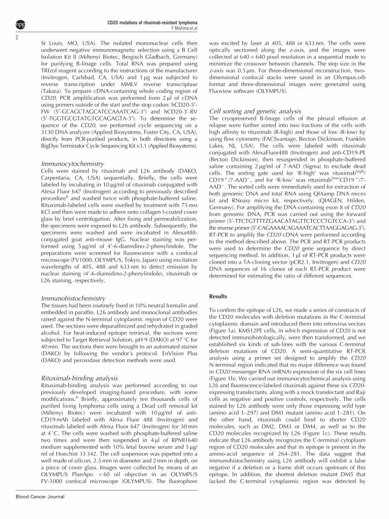
To confirm the epitope of L26, we made a series of constructs ofthe CD20 molecules with deletion mutations in the C-terminalcytoplasmic domain and introduced them into retrovirus vectors(Figure 1a). KMS12PE cells, in which expression of CD20 is notdetected immunohistologically, were then transformed, and weestablished six kinds of sub-lines with the various C-terminaldeletion mutations of CD20. A semi-quantitative RT-PCRanalysis using a primer set designed to amplify the CD20N-terminal region indicated that no major difference was foundin CD20 messenger RNA (mRNA) expression of the six cell lines(Figure 1b). We carried out immunocytochemical analysis usingL26 and fluorescence-labeled rituximab against these six CD20-expressing transfectants, along with a mock transfectant and Rajicells as negative and positive controls, respectively. The cellsstained by L26 antibody were only those expressing wild type(amino acid 1–297) and DM1 mutant (amino acid 1–281). Onthe other hand, rituximab could bind to shorter CD20molecules, such as DM2, DM3 or DM4, as well as to theCD20 molecules recognized by L26 (Figure 1c). These resultsindicate that L26 antibody recognizes the C-terminal cytoplasmregion of CD20 molecules and that its epitope is present in theamino-acid sequence of 264–281. The data suggest thatimmunohistochemistry using L26 antibody will exhibit a falsenegative if a deletion or a frame shift occurs upstream of thisepitope. In addition, the shortest deletion mutant DM5 thatlacked the C-terminal cytoplasmic region was detected by
CD20 mutations of rituximab-resistant lymphomaY Mishima et al
2
Blood Cancer Journal
neither L26 antibody nor rituximab. Possible reasons mightinclude the low membrane localization of DM5 mutant and/orlow stability of the post-translational product. To clarify this, wecarried out western blot analysis using total cell lysate of theseries of deletion mutants that were tagged by FLAG peptide atthe N-terminal region of CD20 constructs. We found that thecellular protein level of the DM5 deletion mutant of CD20 wasremarkably lower than that of others, suggesting that low post-translational stability may be the reason for the low antigenicityof DM5 mutant (data not shown).
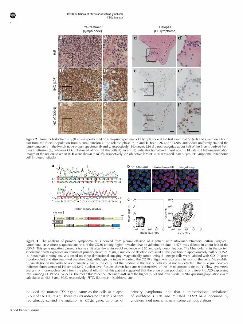
To detect CD20 in the clinical specimens that acquiredrituximab resistance mutations, we developed novel anti-CD20antibodies and screened them in paraffin sections. We isolated anantibody specific for the CD20 N-terminus and named it CD20N.Using this antibody, we carried out immunohistochemistryconducted on the CD20-negative cases by L26 antibody-basedanalysis and found several cases with mutations in the C-terminalcytoplasm region. Among them, a cell specimen, a part of whichhad been cryopreserved as living cells, was included. This waspleural effusion from a patient with relapsed diffuse large cellB-cell lymphoma after complete remission following rituximabcontaining chemotherapy (rituximab, cyclophosphamide, adria-mycin, vincristine and prednisone). This patient did not respond tofurther treatment with salvage therapies containing rituximab. Theresults of immunochemical analysis of the fibrin clot of the pleuraleffusion at relapse, as well as the biopsy of lymph nodes at the firstexamination, are shown in Figure 2. The lymph node biopsybefore rituximab treatment was stained in the same way by bothL26 and CD20N antibodies. However, L26 antibody did not stainat the population having mutated CD20 in the pleural effusion.On the other hand, the CD20N antibody stained almost all of theB-cell population. It should be noted that most of the cells werestained at the region of plasma membrane by CD20N antibody.
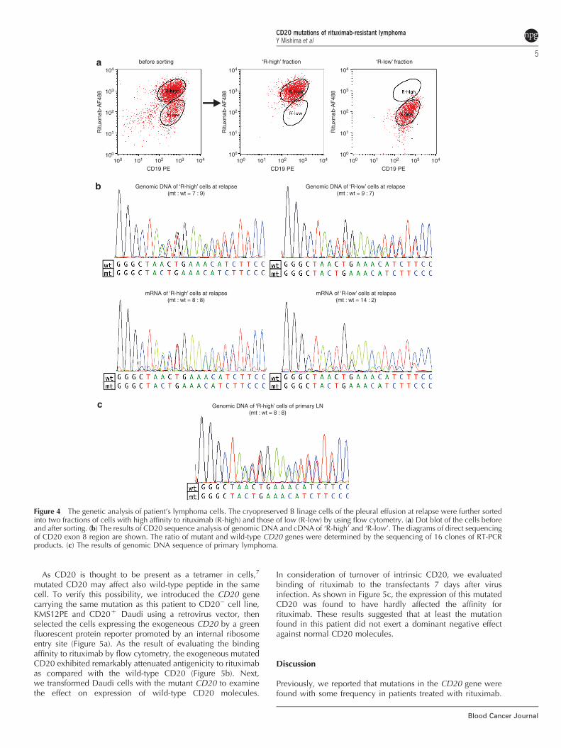
As a result of DNA sequencing of purified B cells, we foundone nucleotide deletion mutation, which caused a frameshift after the amino-acid residue 251, in half of the DNA
(Figure 3a). We conducted a binding analysis of rituximab using theviable cells of B-lineage purified from cryopreserved mononuclearcells. Most of these cells were CD19 positive. However, these cellsconsisted of two kinds of cell populations that were quite differentin their affinity for rituximab (Figure 3b). Furthermore, flowcytometry analysis revealed that the fluorescent intensity offluoro-labeled monoclonal antibody of CD20 decreased to lessthan one-tenth in about half of the B-cell population (Figure 3c).These results indicate that the C-terminal mutation of the CD20gene in this case caused abnormality of extracellular-antigenpresentation, even though the protein expression and the cellmembrane localization seemed to be normal.
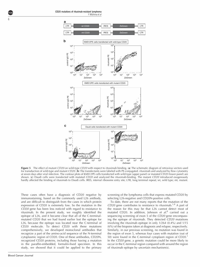
To reveal the genetic mechanisms of the reduced antigenicityto rituximab, we conducted gene-sequencing analysis aboutboth population of cells that differ in affinity to rituximab. Thecryopreserved B lymphocytes of the pleural effusion at relapsewere sorted into two fractions of cells of high affinity torituximab (R-high) and those of low (R-low) by using flowcytometry (Figure 4a). Then genomic DNA and total RNA fromeach fraction were extracted. The results of DNA sequencing ofexon 8 of CD20 in which the mutation was found demonstratedthat, surprisingly, both cell populations had both normal andthe monobasic deletion mutant CD20 genes. To determinethe proportion of the genes, the PCR-amplified DNA fragmentswere inserted into a TA-cloning vector and 16 clones from both‘R-high’ and ‘R-low’ fractions were determined the sequence ofCD20. As a result, the ratio of mutant DNA accounted forapproximately half of genomic DNA of both ‘R-high’ (7 out of 16)and ‘R-low’ (9 out of 16). On the other hand, the resultsof sequence analysis about the cDNA revealed that the ratio ofmutant mRNA in ‘R-low’ was remarkably increased (14 out of 16),whereas that of ‘R-high’ was as the same of the resultsof genomic DNA (8 out of 16; Figure 4b). We also carried outa sequence analysis about genomic DNA from a slice of paraffin-embedded tissue of lymph node at the first diagnosis of thispatient, and found that approximately half of the genomic DNA
WT Cytoplasmic Cytoplasmic Cytoplasmic
297
281
263
245
228
210
TM TM TM TMExtracellular
Cytoplasmic Cytoplasmic CytoplasmicTM TM TM TMExtracellular
Cytoplasmic Cytoplasmic CytoplasmicTM TM TM TMExtracellular
Cytoplasmic CytoplasmicTM TM TM TMExtracellular
Cytoplasmic CytoplasmicTM TM TM TMExtracellular
Cytoplasmic CytoplasmicTM TM TM TMExtracellular
DM1
DM2
DM3
DM4
DM5
CD20
Moc
kCD20
WT
CD20DM
1
CD20DM
2
CD20DM
3
CD20DM
4
CD20DM
5
G3PDH
DIC
KMS12PE/mock
CD20 [WT](aa 1-297)
CD20 [DM1](aa 1-281)
CD20 [DM2](aa 1-263)
CD20 [DM3](aa 1-245)
CD20 [DM4](aa 1-228)
CD20 [DM5](aa 1-210)
Raji
DAPI Rituximab L26 Merge
Figure 1 Search for epitopic site of L26 antibody and analysis of the extracellular exposure of CD20 molecules having C-terminal deletionmutation. (a) To explore the binding site of the L26 antibody, we constructed various lengths of the CD20 deletion mutant at their C-terminalcytoplasmic domains. TM: transmembrane domain. (b) KMS12PE cells retrovirally transduced with C-terminal truncated CD20 showedapproximately similar levels of mRNA expression. (c) Immunocytochemical studies suggested that L26 recognizes amino-acid sequence between264 and 281. An objective lens of � 60 was used with a �10 digital zoom, bar: 5 mm. DAPI, 4’-6-diamidino-2-phenylindole; DIC, differentialinterference contrast; G3PDH, glyceraldehyde-3-phosphate dehydrogenase; WT, wild type.
CD20 mutations of rituximab-resistant lymphomaY Mishima et al
3
Blood Cancer Journal
included the mutant CD20 gene same as the cells at relapse(8 out of 16; Figure 4c). These results indicated that this patienthad already carried the mutation in CD20 gene, as onset of
primary lymphoma, and that a transcriptional imbalanceof wild-type CD20 and mutated CD20 have occurred byundetermined mechanisms in some cell populations.
5 ’ -5 ’ -
3 ’ -3 ’ -
Protein primary structure
CD19-Alexa488
104
104
103
103
102
102
101
101100
100
Mou
se lg
G1
PE
Mouse lgG1 FITC
104
104
103
103
102
102
101
101100
100
CD
20 P
E
CD19 FITC
rituximab-Alexa647 Merged image
wild type1
- 3 ’
*
*
- 3 ’
- 5 ’- 5 ’
mutated
Cytoplasmic Cytoplasmic
Cytoplasmic
Cytoplasmic
297
2902501
CytoplasmicCytoplasmic
TM TM
TM
TM Extracellular
ExtracellularTM
TM
TMTM
Figure 3 The analysis of primary lymphoma cells derived from pleural effusion of a patient with rituximab-refractory, diffuse large-celllymphoma. (a) A direct sequence analysis of the CD20-coding region revealed that an adenine residue (þ478) was deleted in about half of thecDNA. This gene mutation caused a frame shift after the amino-acid sequence of 250 and early denomination. The blue column in the proteinschematic charts expresses an abnormal primary structure. *Single nucleotide deletion occurred at this position in approximately half of cDNA.(b) Rituximab-binding analysis based on three-dimensional imaging. Magnetically sorted living B-lineage cells were labeled with CD19 (greenpseudo-color) and rituximab (red pseudo-color). Although the intensity varied, the CD19 antigen was expressed in most of the cells. Meanwhile,rituximab bound markedly to approximately half of the cells, but the binding to the rest of cells could not be detected. The blue pseudo-colorindicates fluorescence of Hoechist22242 nuclear dye. Results shown here are representative of the 19 microscopic fields. (c) Flow cytometricanalysis of mononuclear cells from the pleural effusion of this patient suggested that there were two populations of different CD20-expressinglevels among CD19 positive cells. The mean fluorescence intensities (MFIs) of the higher (blue) and lower (red) CD20-expressing populations werecalculated as 486.8 and 30.5, respectively. FITC, fluorescein isothiocyanate.
Pre-treatment(lymph node)
H/E
IHC
(L2
6)IH
C (
CD
20N
)
Relapse(PE lymphoma)
Figure 2 Immunohistochemistry (IHC) was performed on a biopsied specimen of a lymph node at the first examination (a, b and c) and on a fibrinclot from the B-cell population from pleural effusion at the relapse phase (d, e and f). Both L26 and CD20N antibodies uniformly stained thelymphoma cells in the lymph node biopsy specimen (b and c, respectively). However, L26 did not recognize about half of the B cells derived frompleural effusion (e), whereas CD20N stained almost all the cells (f). (a and d) indicates hematoxylin and eosin (H/E) stain. High-magnificationimages of the region boxed in (a–f) were shown in (a0–f0), respectively. An objective lens of �60 was used, bar: 50mm. PE lymphoma, lymphomacell in pleural effusion.
CD20 mutations of rituximab-resistant lymphomaY Mishima et al
4
Blood Cancer Journal
As CD20 is thought to be present as a tetramer in cells,7
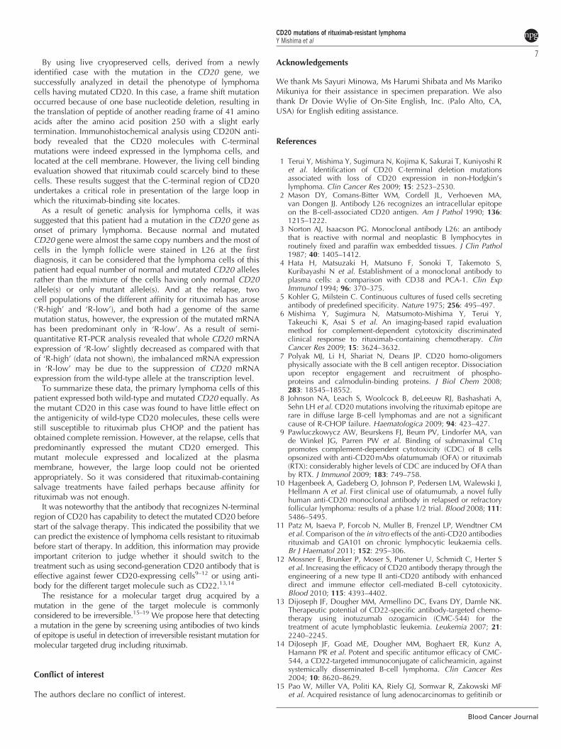
mutated CD20 may affect also wild-type peptide in the samecell. To verify this possibility, we introduced the CD20 genecarrying the same mutation as this patient to CD20� cell line,KMS12PE and CD20þ Daudi using a retrovirus vector, thenselected the cells expressing the exogeneous CD20 by a greenfluorescent protein reporter promoted by an internal ribosomeentry site (Figure 5a). As the result of evaluating the bindingaffinity to rituximab by flow cytometry, the exogeneous mutatedCD20 exhibited remarkably attenuated antigenicity to rituximabas compared with the wild-type CD20 (Figure 5b). Next,we transformed Daudi cells with the mutant CD20 to examinethe effect on expression of wild-type CD20 molecules.
In consideration of turnover of intrinsic CD20, we evaluatedbinding of rituximab to the transfectants 7 days after virusinfection. As shown in Figure 5c, the expression of this mutatedCD20 was found to have hardly affected the affinity forrituximab. These results suggested that at least the mutationfound in this patient did not exert a dominant negative effectagainst normal CD20 molecules.
Discussion
Previously, we reported that mutations in the CD20 gene werefound with some frequency in patients treated with rituximab.
before sorting ‘R-high’ fraction ‘R-low’ fraction
104
104
103
103
102
102
CD19 PE
Genomic DNA of ‘R-high’ cells at relapse(mt : wt = 7 : 9)
Genomic DNA of ‘R-low’ cells at relapse(mt : wt = 9 : 7)
mRNA of ‘R-high’ cells at relapse(mt : wt = 8 : 8)
Genomic DNA of ‘R-high’ cells of primary LN(mt : wt = 8 : 8)
mRNA of ‘R-low’ cells at relapse(mt : wt = 14 : 2)
101
101100
100 104103102
CD19 PE101100 104103102
CD19 PE101100
Ritu
xmab
-AF
488
104
103
102
101
100
Ritu
xmab
-AF
488
104
103
102
101
100
Ritu
xmab
-AF
488
Figure 4 The genetic analysis of patient’s lymphoma cells. The cryopreserved B linage cells of the pleural effusion at relapse were further sortedinto two fractions of cells with high affinity to rituximab (R-high) and those of low (R-low) by using flow cytometry. (a) Dot blot of the cells beforeand after sorting. (b) The results of CD20 sequence analysis of genomic DNA and cDNA of ‘R-high’ and ‘R-low’. The diagrams of direct sequencingof CD20 exon 8 region are shown. The ratio of mutant and wild-type CD20 genes were determined by the sequencing of 16 clones of RT-PCRproducts. (c) The results of genomic DNA sequence of primary lymphoma.
CD20 mutations of rituximab-resistant lymphomaY Mishima et al
5
Blood Cancer Journal
These cases often have a diagnosis of CD20 negative byimmunostaining, based on the commonly used L26 antibody,and are difficult to distinguish from the cases in which proteinexpression of CD20 is extremely low. So the mutation in theCD20 gene has been less noticed with regard to resistance torituximab. In the present study, we roughly identified theepitope of L26, and it became clear that all of the C-terminal-mutated CD20 that we had found earlier lost the epitope forL26, because the epitope was located near the C-terminal ofCD20 molecule. To detect CD20 with these mutationscomprehensively, we developed monoclonal antibodies thatrecognize a part of the amino-acid sequence of the N-terminalcytoplasmic region of CD20. One of these antibodies, CD20N,recognized CD20 proteins, including those having a mutationin the paraffin-embedded, formalin-fixed specimen. In thisstudy, we showed that it could be applied to the primary
screening of the lymphoma cells that express mutated CD20 byselecting L26-negative and CD20N-positive cells.
To date, there are not many reports that the mutation of theCD20 gene contributes to resistance to rituximab.1,8 A part ofthe reason for this may be that L26 cannot detect most ofmutated CD20. In addition, Johnson et al.8 carried out asequencing screening of exon 5 of the CD20 gene encompass-ing the epitope of rituximab. They detected CD20 mutationsinvolving the rituximab epitope in only 1/264 (0.4%) and 1/15(6%) of the biopsies taken at diagnosis and relapse, respectively.Similarly, in our previous screening, no mutation was found inthe region of exon 5, whereas four cases with mutation (out of50) were found in the C-terminal cytoplasm region of CD20.1
In the CD20 gene, a genetic mutation could be more likely tooccur in the C-terminal region compared with around the regionof rituximab epitope by uncertain mechanism(s).
LTR wt CD20 IRES
IRES
ZsGreen LTR
LTRZsGreenmt CD20
KMS12PE cells transfected with wild-type CD20
KMS12PE cells transfected with mutant CD20
Daudi cells transfected with mutant CD20
104
104
103
103
102
102
ZsGreen ZsGreen
ZsGreen ZsGreen
ZsGreen ZsGreen
cont
rol l
gG P
Eco
ntro
l lgG
PE
cont
rol l
gG P
E
ritux
imab
PE
ritux
imab
PE
ritux
imab
PE
101
101100
104
103
102
101
100
104
103
102
101
100
104
103
102
101
100
104
103
102
101
100
104
103
102
101
100
100 104103102101100
104103102101100104103102101100
104103102101100 104103102101100
LTR
Figure 5 The effect of mutant CD20 on wild-type CD20 with respect to rituximab binding. (a) The schematic diagram of retrovirus vectors usedfor transduction of wild-type and mutant CD20. (b) The transfectants were labeled with PE-conjugated. rituximab and analyzed by flow cytometryat seven days after viral infection. The contour plots of KMS12PE cells transfected with wild-type (upper panel) or mutated CD20 (lower panel) areshown. (c) Daudi cells were transfected with mutated CD20 and analyzed the rituximab-binding. The mutant CD20 introduced exogenouslyhardly affected the binding of rituximab to Daudi cells. IRES, internal ribosome entry site; LTR, long terminal repeat; wt, wild type; mt, mutant.
CD20 mutations of rituximab-resistant lymphomaY Mishima et al
6
Blood Cancer Journal
By using live cryopreserved cells, derived from a newlyidentified case with the mutation in the CD20 gene, wesuccessfully analyzed in detail the phenotype of lymphomacells having mutated CD20. In this case, a frame shift mutationoccurred because of one base nucleotide deletion, resulting inthe translation of peptide of another reading frame of 41 aminoacids after the amino acid position 250 with a slight earlytermination. Immunohistochemical analysis using CD20N anti-body revealed that the CD20 molecules with C-terminalmutations were indeed expressed in the lymphoma cells, andlocated at the cell membrane. However, the living cell bindingevaluation showed that rituximab could scarcely bind to thesecells. These results suggest that the C-terminal region of CD20undertakes a critical role in presentation of the large loop inwhich the rituximab-binding site locates.
As a result of genetic analysis for lymphoma cells, it wassuggested that this patient had a mutation in the CD20 gene asonset of primary lymphoma. Because normal and mutatedCD20 gene were almost the same copy numbers and the most ofcells in the lymph follicle were stained in L26 at the firstdiagnosis, it can be considered that the lymphoma cells of thispatient had equal number of normal and mutated CD20 allelesrather than the mixture of the cells having only normal CD20allele(s) or only mutant allele(s). And at the relapse, twocell populations of the different affinity for rituximab has arose(‘R-high’ and ‘R-low’), and both had a genome of the samemutation status, however, the expression of the mutated mRNAhas been predominant only in ‘R-low’. As a result of semi-quantitative RT-PCR analysis revealed that whole CD20 mRNAexpression of ‘R-low’ slightly decreased as compared with thatof ‘R-high’ (data not shown), the imbalanced mRNA expressionin ‘R-low’ may be due to the suppression of CD20 mRNAexpression from the wild-type allele at the transcription level.
To summarize these data, the primary lymphoma cells of thispatient expressed both wild-type and mutated CD20 equally. Asthe mutant CD20 in this case was found to have little effect onthe antigenicity of wild-type CD20 molecules, these cells werestill susceptible to rituximab plus CHOP and the patient hasobtained complete remission. However, at the relapse, cells thatpredominantly expressed the mutant CD20 emerged. Thismutant molecule expressed and localized at the plasmamembrane, however, the large loop could not be orientedappropriately. So it was considered that rituximab-containingsalvage treatments have failed perhaps because affinity forrituximab was not enough.
It was noteworthy that the antibody that recognizes N-terminalregion of CD20 has capability to detect the mutated CD20 beforestart of the salvage therapy. This indicated the possibility that wecan predict the existence of lymphoma cells resistant to rituximabbefore start of therapy. In addition, this information may provideimportant criterion to judge whether it should switch to thetreatment such as using second-generation CD20 antibody that iseffective against fewer CD20-expressing cells9–12 or using anti-body for the different target molecule such as CD22.13,14
The resistance for a molecular target drug acquired by amutation in the gene of the target molecule is commonlyconsidered to be irreversible.15–19 We propose here that detectinga mutation in the gene by screening using antibodies of two kindsof epitope is useful in detection of irreversible resistant mutation formolecular targeted drug including rituximab.
Conflict of interest
The authors declare no conflict of interest.
Acknowledgements
We thank Ms Sayuri Minowa, Ms Harumi Shibata and Ms MarikoMikuniya for their assistance in specimen preparation. We alsothank Dr Dovie Wylie of On-Site English, Inc. (Palo Alto, CA,USA) for English editing assistance.
References
1 Terui Y, Mishima Y, Sugimura N, Kojima K, Sakurai T, Kuniyoshi Ret al. Identification of CD20 C-terminal deletion mutationsassociated with loss of CD20 expression in non-Hodgkin’slymphoma. Clin Cancer Res 2009; 15: 2523–2530.
2 Mason DY, Comans-Bitter WM, Cordell JL, Verhoeven MA,van Dongen JJ. Antibody L26 recognizes an intracellular epitopeon the B-cell-associated CD20 antigen. Am J Pathol 1990; 136:1215–1222.
3 Norton AJ, Isaacson PG. Monoclonal antibody L26: an antibodythat is reactive with normal and neoplastic B lymphocytes inroutinely fixed and paraffin wax embedded tissues. J Clin Pathol1987; 40: 1405–1412.
4 Hata H, Matsuzaki H, Matsuno F, Sonoki T, Takemoto S,Kuribayashi N et al. Establishment of a monoclonal antibody toplasma cells: a comparison with CD38 and PCA-1. Clin ExpImmunol 1994; 96: 370–375.
5 Kohler G, Milstein C. Continuous cultures of fused cells secretingantibody of predefined specificity. Nature 1975; 256: 495–497.
6 Mishima Y, Sugimura N, Matsumoto-Mishima Y, Terui Y,Takeuchi K, Asai S et al. An imaging-based rapid evaluationmethod for complement-dependent cytotoxicity discriminatedclinical response to rituximab-containing chemotherapy. ClinCancer Res 2009; 15: 3624–3632.
7 Polyak MJ, Li H, Shariat N, Deans JP. CD20 homo-oligomersphysically associate with the B cell antigen receptor. Dissociationupon receptor engagement and recruitment of phospho-proteins and calmodulin-binding proteins. J Biol Chem 2008;283: 18545–18552.
8 Johnson NA, Leach S, Woolcock B, deLeeuw RJ, Bashashati A,Sehn LH et al. CD20 mutations involving the rituximab epitope arerare in diffuse large B-cell lymphomas and are not a significantcause of R-CHOP failure. Haematologica 2009; 94: 423–427.
9 Pawluczkowycz AW, Beurskens FJ, Beum PV, Lindorfer MA, vande Winkel JG, Parren PW et al. Binding of submaximal C1qpromotes complement-dependent cytotoxicity (CDC) of B cellsopsonized with anti-CD20 mAbs ofatumumab (OFA) or rituximab(RTX): considerably higher levels of CDC are induced by OFA thanby RTX. J Immunol 2009; 183: 749–758.
10 Hagenbeek A, Gadeberg O, Johnson P, Pedersen LM, Walewski J,Hellmann A et al. First clinical use of ofatumumab, a novel fullyhuman anti-CD20 monoclonal antibody in relapsed or refractoryfollicular lymphoma: results of a phase 1/2 trial. Blood 2008; 111:5486–5495.
11 Patz M, Isaeva P, Forcob N, Muller B, Frenzel LP, Wendtner CMet al. Comparison of the in vitro effects of the anti-CD20 antibodiesrituximab and GA101 on chronic lymphocytic leukaemia cells.Br J Haematol 2011; 152: 295–306.
12 Mossner E, Brunker P, Moser S, Puntener U, Schmidt C, Herter Set al. Increasing the efficacy of CD20 antibody therapy through theengineering of a new type II anti-CD20 antibody with enhanceddirect and immune effector cell-mediated B-cell cytotoxicity.Blood 2010; 115: 4393–4402.
13 Dijoseph JF, Dougher MM, Armellino DC, Evans DY, Damle NK.Therapeutic potential of CD22-specific antibody-targeted chemo-therapy using inotuzumab ozogamicin (CMC-544) for thetreatment of acute lymphoblastic leukemia. Leukemia 2007; 21:2240–2245.
14 DiJoseph JF, Goad ME, Dougher MM, Boghaert ER, Kunz A,Hamann PR et al. Potent and specific antitumor efficacy of CMC-544, a CD22-targeted immunoconjugate of calicheamicin, againstsystemically disseminated B-cell lymphoma. Clin Cancer Res2004; 10: 8620–8629.
15 Pao W, Miller VA, Politi KA, Riely GJ, Somwar R, Zakowski MFet al. Acquired resistance of lung adenocarcinomas to gefitinib or
CD20 mutations of rituximab-resistant lymphomaY Mishima et al
7
Blood Cancer Journal
erlotinib is associated with a second mutation in the EGFR kinasedomain. PLoS Med 2005; 2: e73.
16 Engelman JA, Janne PA. Mechanisms of acquired resistanceto epidermal growth factor receptor tyrosine kinase inhibitorsin non-small cell lung cancer. Clin Cancer Res 2008; 14:2895–2899.
17 Shah NP, Sawyers CL. Mechanisms of resistance to STI571 inPhiladelphia chromosome-associated leukemias. Oncogene 2003;22: 7389–7395.
18 Shah NP, Nicoll JM, Nagar B, Gorre ME, Paquette RL, Kuriyan Jet al. Multiple BCR-ABL kinase domain mutations conferpolyclonal resistance to the tyrosine kinase inhibitor imatinib
(STI571) in chronic phase and blast crisis chronic myeloidleukemia. Cancer Cell 2002; 2: 117–125.
19 Wang SE, Narasanna A, Perez-Torres M, Xiang B, Wu FY, Yang Set al. HER2 kinase domain mutation results in constitutivephosphorylation and activation of HER2 and EGFR and resistanceto EGFR tyrosine kinase inhibitors. Cancer Cell 2006; 10: 25–38.
This work is licensed under the Creative CommonsAttribution-NonCommercial-No Derivative Works
3.0 Unported License. To view a copy of this license, visit http://creativecommons.org/licenses/by-nc-nd/3.0/
CD20 mutations of rituximab-resistant lymphomaY Mishima et al
8
Blood Cancer Journal
Related Documents