The Human Trithorax Protein hASH2 Functions as an Oncoprotein Juliane Lu ¨scher-Firzlaff, 1 Isabella Gawlista, 1 Jo ¨rg Vervoorts, 1 Karsten Kapelle, 1 Till Braunschweig, 2 Gesa Walsemann, 1 Chantal Rodgarkia-Schamberger, 3 Henning Schuchlautz, 1 Stephan Dreschers, 1 Elisabeth Kremmer, 4 Richard Lilischkis, 1 Christa Cerni, 3 Axel Wellmann, 2 and Bernhard Lu ¨scher 1 1 Institut fu ¨r Biochemie and 2 Institut fu ¨r Pathologie, Klinikum, RWTH Aachen University, Aachen, Germany; 3 Institut fu ¨r Krebsforschung, Klinik fu ¨r Innere Medizin I, Medizinische Universita¨t Wien, Wien, Austria; and 4 GSF-Institut fu ¨r Molekulare Immunologie, Munich, Germany Abstract Regulation of chromatin is an important aspect of controlling promoter activity and gene expression. Posttranslational modifications of core histones allow proteins associated with gene transcription to access chromatin. Closely associated with promoters of actively transcribed genes, trimethylation of histone H3 at lysine 4 (H3K4me3) is a core histone mark set by several protein complexes. Some of these protein com- plexes contain the trithorax protein ASH2 combined with the MLL oncoproteins. We identified human ASH2 in a complex with the oncoprotein MYC. This finding, together with the observation that hASH2 interacts with MLL, led us to test whether hASH2 itself is involved in transformation. We observed that hASH2 cooperates with Ha-RAS to transform primary rat embryo fibroblasts (REF). Furthermore, transfor- mation of REFs by MYC and Ha-RAS required the presence of rAsh2. In an animal model, the hASH2/Ha-RAS–transformed REFs formed rapidly growing tumors characteristic of fibrosarcomas that, compared with tumors derived from MYC/Ha-RAS transformed cells, were poorly differentiated. This finding suggests that ASH2 functions as an oncoprotein. Although hASH2 expression at the mRNA level was generally not deregulated, hASH2 protein expression was increased in most human tumors and tumor cell lines. In addition, knockdown of hASH2 inhibited tumor cell proliferation. Taken together, these observations define hASH2 as a novel oncoprotein. [Cancer Res 2008;68(3):749–58] Introduction The development of a tumorigenic cell is a complex multistep genetic process that results in the deregulation of fundamental aspects of cell physiology, including proliferation, differentiation, and apoptosis (1). In recent years, RNA expression profiling was instrumental to identify genes that are deregulated in tumor cells and thus potentially relevant for tumor development and progression (2). In addition, comparative genomic hybridization on a global scale allowed the identification of DNA regions that are either underrepresented or overrepresented in distinct tumors (3). These studies revealed novel oncogenes and tumor suppressor genes that are deregulated or genetically altered in human malignancies. However, some oncogenes and tumor suppressor genes cannot be depicted by these methods. This is true for cases in which proteins are deregulated as a result of small mutations or because of deregulated cellular signaling that affects the function of the respective proteins (4, 5). Linking such proteins with tumor development and/or progression requires, in general, functional assays. The polycomb group (PcG) and trithorax group (TrxG) genes were originally identified in Drosophila , where the products of these genes are intimately involved in the segmentation of the embryo (6). The expression pattern of hox genes, initially defined by gap and pair-rule genes, is maintained after gastrulation by the activities of PcG and TrxG gene products. In general, although the PcG proteins are responsible to maintain the repressed status of distinct hox genes, the TrxG proteins perpetuate the transcribed status of these genes (7, 8). PcG and TrxG proteins assemble into large protein complexes that control gene transcription, at least in part, by modulating chromatin. The TrxG gene ash2 ( absent, small , or homeotic discs 2 ) was discovered in a screen for lethality during the late larval/early pupal stage of Drosophila development with imaginal disc defects (9). Mutations of Ash2 cause homeotic transformations in many segments of Drosophila and, additionally, result in a variety of pattern formation defects (10). Loss of Ash2 inhibits hox gene expression, consistent with a function of Ash2 as a TrxG protein associated with maintaining the expression of hox genes (11). Furthermore, array analyses revealed many additional targets controlled by Ash2, including genes involved in cell cycle and cell proliferation control (12, 13). These findings suggest that Ash2 is not only involved in regulating aspects of tissue development but also controls functions associated with cell proliferation in the adult. The molecular analysis showed that mammalian ASH2 is a component of protein complexes of various composition, some including mixed-lineage leukemia (MLL) oncoproteins, that contain histone methyltransferase (HMT) activity (14–20). Recent findings have shown that these complexes trimethylate (me3) histone H3 at lysine 4 (H3K4; refs. 21–23). H3K4me3 is one of a large number of distinct modifications that have been identified on core histones and are part of elaborate mechanisms that evolved to control the structure and the chemical surface of chromatin (24, 25). These modifications control the access of proteins to nucleosomes that, in turn, regulate various aspects associated with gene transcription, including chromatin remodeling and the activities of transcriptional regulators and polymerase complexes (26). H3K4me3 is a histone mark that is closely associated with transcribed genes (22, 25, 27). Several recent studies have shown Note: J. Lu ¨scher-Firzlaff and I. Gawlista contributed equally to this work. Current address for G. Walsemann: PAREXEL International GmbH, Am Bahnhof Westend 11, 14059 Berlin, Germany. Current address for R. Lilischkis: BTF Precise Microbiology, 35-41 Waterloo Road, North Ryde Sydney, NSW 2113, Australia. Current address for A. Wellman: Institut fu ¨r Pathologie, Siemensplatz 4, 29223 Celle, Germany. C. Cerni is deceased. Requests for reprints: Bernhard Lu ¨scher, Institut fu ¨r Biochemie, Klinikum, RWTH Aachen University, Pauwelsstrasse 30, 52057 Aachen, Germany. Phone: 49-241-8088850; E-mail: [email protected]. I2008 American Association for Cancer Research. doi:10.1158/0008-5472.CAN-07-3158 www.aacrjournals.org 749 Cancer Res 2008; 68: (3). February 1, 2008 Research Article Research. on January 7, 2016. © 2008 American Association for Cancer cancerres.aacrjournals.org Downloaded from

Welcome message from author
This document is posted to help you gain knowledge. Please leave a comment to let me know what you think about it! Share it to your friends and learn new things together.
Transcript

The Human Trithorax Protein hASH2 Functions as an Oncoprotein
Juliane Luscher-Firzlaff,1Isabella Gawlista,
1Jorg Vervoorts,
1Karsten Kapelle,
1Till Braunschweig,
2
Gesa Walsemann,1Chantal Rodgarkia-Schamberger,
3Henning Schuchlautz,
1
Stephan Dreschers,1Elisabeth Kremmer,
4Richard Lilischkis,
1Christa Cerni,
3
Axel Wellmann,2and Bernhard Luscher
1
1Institut fur Biochemie and 2Institut fur Pathologie, Klinikum, RWTH Aachen University, Aachen, Germany; 3Institut fur Krebsforschung,Klinik fur Innere Medizin I, Medizinische Universitat Wien, Wien, Austria; and 4GSF-Institut fur Molekulare Immunologie,Munich, Germany
Abstract
Regulation of chromatin is an important aspect of controllingpromoter activity and gene expression. Posttranslationalmodifications of core histones allow proteins associated withgene transcription to access chromatin. Closely associatedwith promoters of actively transcribed genes, trimethylationof histone H3 at lysine 4 (H3K4me3) is a core histone mark setby several protein complexes. Some of these protein com-plexes contain the trithorax protein ASH2 combined with theMLL oncoproteins. We identified human ASH2 in a complexwith the oncoprotein MYC. This finding, together with theobservation that hASH2 interacts with MLL, led us to testwhether hASH2 itself is involved in transformation. Weobserved that hASH2 cooperates with Ha-RAS to transformprimary rat embryo fibroblasts (REF). Furthermore, transfor-mation of REFs by MYC and Ha-RAS required the presence ofrAsh2. In an animal model, the hASH2/Ha-RAS–transformedREFs formed rapidly growing tumors characteristic offibrosarcomas that, compared with tumors derived fromMYC/Ha-RAS transformed cells, were poorly differentiated.This finding suggests that ASH2 functions as an oncoprotein.Although hASH2 expression at the mRNA level was generallynot deregulated, hASH2 protein expression was increased inmost human tumors and tumor cell lines. In addition,knockdown of hASH2 inhibited tumor cell proliferation.Taken together, these observations define hASH2 as a noveloncoprotein. [Cancer Res 2008;68(3):749–58]
Introduction
The development of a tumorigenic cell is a complex multistepgenetic process that results in the deregulation of fundamentalaspects of cell physiology, including proliferation, differentiation,and apoptosis (1). In recent years, RNA expression profiling wasinstrumental to identify genes that are deregulated in tumor cellsand thus potentially relevant for tumor development andprogression (2). In addition, comparative genomic hybridizationon a global scale allowed the identification of DNA regions that areeither underrepresented or overrepresented in distinct tumors (3).
These studies revealed novel oncogenes and tumor suppressorgenes that are deregulated or genetically altered in humanmalignancies. However, some oncogenes and tumor suppressorgenes cannot be depicted by these methods. This is true for casesin which proteins are deregulated as a result of small mutations orbecause of deregulated cellular signaling that affects the functionof the respective proteins (4, 5). Linking such proteins with tumordevelopment and/or progression requires, in general, functionalassays.
The polycomb group (PcG) and trithorax group (TrxG) geneswere originally identified in Drosophila , where the products ofthese genes are intimately involved in the segmentation of theembryo (6). The expression pattern of hox genes, initially definedby gap and pair-rule genes, is maintained after gastrulation by theactivities of PcG and TrxG gene products. In general, although thePcG proteins are responsible to maintain the repressed status ofdistinct hox genes, the TrxG proteins perpetuate the transcribedstatus of these genes (7, 8). PcG and TrxG proteins assemble intolarge protein complexes that control gene transcription, at least inpart, by modulating chromatin. The TrxG gene ash2 (absent, small ,or homeotic discs 2) was discovered in a screen for lethality duringthe late larval/early pupal stage of Drosophila development withimaginal disc defects (9). Mutations of Ash2 cause homeotictransformations in many segments of Drosophila and, additionally,result in a variety of pattern formation defects (10). Loss of Ash2inhibits hox gene expression, consistent with a function of Ash2 asa TrxG protein associated with maintaining the expression of hoxgenes (11). Furthermore, array analyses revealed many additionaltargets controlled by Ash2, including genes involved in cell cycleand cell proliferation control (12, 13). These findings suggest thatAsh2 is not only involved in regulating aspects of tissuedevelopment but also controls functions associated with cellproliferation in the adult.
The molecular analysis showed that mammalian ASH2 is acomponent of protein complexes of various composition, someincluding mixed-lineage leukemia (MLL) oncoproteins, thatcontain histone methyltransferase (HMT) activity (14–20). Recentfindings have shown that these complexes trimethylate (me3)histone H3 at lysine 4 (H3K4; refs. 21–23). H3K4me3 is one of alarge number of distinct modifications that have been identified oncore histones and are part of elaborate mechanisms that evolvedto control the structure and the chemical surface of chromatin(24, 25). These modifications control the access of proteins tonucleosomes that, in turn, regulate various aspects associated withgene transcription, including chromatin remodeling and theactivities of transcriptional regulators and polymerase complexes(26). H3K4me3 is a histone mark that is closely associated withtranscribed genes (22, 25, 27). Several recent studies have shown
Note: J. Luscher-Firzlaff and I. Gawlista contributed equally to this work.Current address for G. Walsemann: PAREXEL International GmbH, Am Bahnhof
Westend 11, 14059 Berlin, Germany. Current address for R. Lilischkis: BTF PreciseMicrobiology, 35-41 Waterloo Road, North Ryde Sydney, NSW 2113, Australia. Currentaddress for A. Wellman: Institut fur Pathologie, Siemensplatz 4, 29223 Celle, Germany.
C. Cerni is deceased.Requests for reprints: Bernhard Luscher, Institut fur Biochemie, Klinikum, RWTH
Aachen University, Pauwelsstrasse 30, 52057 Aachen, Germany. Phone: 49-241-8088850;E-mail: [email protected].
I2008 American Association for Cancer Research.doi:10.1158/0008-5472.CAN-07-3158
www.aacrjournals.org 749 Cancer Res 2008; 68: (3). February 1, 2008
Research Article
Research. on January 7, 2016. © 2008 American Association for Cancercancerres.aacrjournals.org Downloaded from
that H3K4me3 is found within the 5¶ region (i.e., the proximalpromoter) of virtually all active genes. In addition, this modifica-tion correlates strongly with the presence of active polymerase II(28–32). The proteins that can read H3K4me3 include members ofthe ING family that associate with chromatin remodeling activityand with histone acetyl transferases (33). Furthermore, the TAF3subunit of the basal transcription factor complex transcriptionfactor IID interacts with H3K4me3, thereby enhancing polymeraseII recruitment (34). In addition, the JMJD2A demethylase alsoassociates with H3K4me3 (35). Together, these findings suggest thattrimethylation of H3K4 is an important modification of activechromatin that is strongly associated with gene transcription.
Recently, we identified several proteins associated with theoncoprotein MYC in a human T-cell tumor line (36). Micro-sequencing of the protein species with an apparent molecularweight of 86 kDa revealed its identity as human ASH2. This findingsuggested that ASH2 might be involved in transformation.Therefore, we tested for the transforming activity of ASH2 usingthe rat embryo fibroblast (REF) cotransformation assay (37). Wefound that ASH2 cooperates efficiently with an activated Ha-RAS.Furthermore, knockdown of rAsh2 inhibited transformation byMYC and Ha-RAS. The ASH2/Ha-RAS–transformed REFs formedrapidly growing, poorly differentiated tumors in animals. Interest-ingly, the analysis of ASH2 in human tumors revealed that theASH2 gene is not overexpressed, but that, in general, the protein ispresent at increased levels. Knockdown of ASH2 interfered with theproliferation of tumor cell lines. Together, these findings suggestedstrongly that ASH2 possesses oncogenic activities and that itsexpression is relevant for tumor cell proliferation.
Materials and Methods
Cells and transformation assays. All human cell lines were cultured
as recommended by the American Tissue Culture Collection. Human diploidfibroblasts (HDF; obtained from J. Baron, Department of Dermatology,
RWTH, Aachen, Germany) were grown in DMEM supplemented with 10%
FCS and human mammary epithelial cells (HMEC) in MEGM CC-3051
medium (Clonetics). Peripheral blood mononuclear cells were obtained fromthe blood bank of the RWTH Aachen University Medical School. Human
tumor cell lines were transfected using ExGen500 as recommended by the
manufacturer (MBI-Fermentas). REFs were obtained from 15.5-gestation-day-old Fischer rat embryos by fractionated trypsinization as described
previously (38). REFs were grown in phenol red–free DMEM (Life
Technologies) with 10% low-estrogen FCS (Bioconcept) and transfected
with 2.5 Ag of expression plasmids encoding the respective oncoproteins,5 Ag of pSUPER plasmids, and 1 Ag of pRSVneo per 60-mm Petri dish using
the calcium phosphate technique. Foci were stained and counted 14 days
later. Transformed hASH2/Ha-RAS clones were selected in 200 Ag/mL G418
(Geneticin, Life Technologies). MYC/Ha-RAS, E1a/Ha-RAS, and p53m/Ha-RAS clones were established previously (38). For proliferation assays,
cells were cotransfected with pSUPER constructs and pBABE-puro and
selected in the presence of puromycin (2 Ag/mL) for 24 h. The cells were then
washed and cultivated for an additional 4 to 5 days before staining withmethylene blue (2 mg/mL methanol). Bromodeoxyuridine (BrdUrd)
incorporation was measured in cells cotransfected with pSUPER constructs
and pEGFP (ratio 15:1). The cells were pulse labeled with BrdUrd for1 h before harvesting 72 h after transfection. The cells were then fixed,
treated with DNaseI, and the incorporated BrdUrd was detected by indirect
immunofluorescence (anti-bromodeoxyuridine antibody; Boehringer
Mannheim). The number of BrdUrd-positive cells was determined relativeto the number of transfected cells.
Plasmids. A PCR-amplified cDNA encoding hASH2 was cloned into an
expression vector by standard procedures and sequenced. DNA oligonu-
cleotides containing sequences corresponding to short interfering RNAs
(siRNA) were cloned into the pSUPER vector (39). The following sequenceswere used: siRNA-hASH2, 5¶-GGATCTCACTTACCGCCCT-3¶; siRNA-rAsh2,
5¶-CCCTAGCAGATCCATGCTT-3¶; siRNA-rAsh2m, 5¶-CCCTGCAGATC-
CATGCTT-3¶; siRNA-rc-Myc, 5¶-ATCCTGTACCTCGTCCGAT-3¶; siRNA-
rc-Mycm, 5¶-ATCCTGTATCGTCCGAT-3¶; siRNA-hSKP2, NT8 (see ref. 40).The different specific siRNA constructs reduced the relevant mRNA levels to
15% or less (data not shown). Expression vectors encoding human MYC,
Ha-RAS, adenoviral E1a, and mutant p53 have been described previously
(38). pBABE-puro and the expression vector for p27KIP1 were obtained fromG. Evan (University of California San Francisco, San Francisco, CA) and
R. Bernards (Netherlands Cancer Institute, Amsterdam, The Netherlands),
respectively.
Animal experiments. Seven-day-old male Fisher rats received s.c.injections of 5 � 104 transformed cells in PBS. Tumor size was monitored
regularly, and the animals were killed by cervical dislocation when tumor
diameters were f20 mm for younger and 40 mm for older rats or before anunfavorable tumor localization would have restricted the rats’ comfort. The
animal experiment was performed according to the animal experiment
approval GZ 66.009/98-Pr/4/00 given by the Austrian Ministry of Culture
and Education. Similarly, BALB/c nude mice were injected s.c. with tumorcells, and tumor progression was monitored as above (Bezirksregierung
Koln, 50.203.2-Ac36, 14/06).
Protein analysis. Western blot analyses were performed on whole-cell
lysates in Frackelton buffer of exponentially growing cells (41). Monoclonalantibodies (mAb) 4C5 and 6A9 were generated by immunizing a LOU/C rat
with bacterially expressed glutathione S-transferase–hASH2(1–444) fusion
protein.RNA analysis. Northern blot analyses were performed as described
previously (41). A commercially available multitissue Northern blot was used
(Clontech). The cancer profiling array (BD Bioscience) was obtained from
E. Dahl (Institute of Pathology, RWTH, Aachen, Germany). For sequencingand real-time PCR analysis, RNA isolated by Trizol Reagent (Invitrogen) or
RNeasy (Qiagen; tissue culture cells) was transcribed into cDNA using the
Transcriptor First Strand cDNA Synthesis kit (Roche) as recommended.
ASH2-specific cDNA was amplified ( forward primer, 5¶-GATGGCGGCGG-CAGGAGC-3¶ and reverse primer, 5¶-GTGGGGAGAAAATGGTGAAAGG-
CAGG-3¶), purified, and sequenced. Quantitative PCR was performed with
the LightCycler FastStart DNA MasterSYBR Green I (Roche) or theQuantiTect SYBR Green PCR kit with a QuantiTect Primer Assay for hASH2
(Qiagen). The expression of b-glucuronidase was used for internal control in
all experiments ( forward primer, 5¶-CTCATTTGGAATTTTGCCGATT-3¶ andreverse primer, 5¶-CCGAGTGAAGATCCCCTTTTTA-3¶).Microdissection. Frozen sections of tumors were processed for H&E
staining. Normal and tumor tissue was identified microscopically. Ten
adjacent 16-Am-thick frozen sections were mounted; the different regions of
the tissue were marked, scraped off with a scalpel, and collected for Trizolreagent extraction (Invitrogen) according to the manufacturer’s recommen-
dations. The extracted tissue corresponded to roughly 50,000 cells per sample.
Immunohistochemistry. Sections of archived tumor samples as
formalin-fixed, paraffin-embedded tissue were deparaffinized in xylene andrehydrated over ethanol and deionized water. Endogenous peroxidases were
blocked by treating with 3% H2O2 in PBS for 15 min. Antigen demasking was
performed in citrate buffer (pH 6.0) at 130jC for 10 min. Then, the sampleswere blocked in 1% fat-free milk powder and 10% horse serum in PBS. The
specific staining was done using culture supernatant of mAb 4C5 and 6A9
diluted 1:200 in 1% fat-free milk powder in PBS at 37jC for 1 h. Biotinylated
secondary anti-rat antibodies (1:500; Dianova) were incubated on thewashed sample at 37jC for 30 min. Further incubation with avidin-biotin-
peroxidase complex was performed as indicated by the manufacturer
(Vectastain kit PK6100 Standard; Vector Laboratories). The staining of
sections was performed with 3,3¶-diaminobenzidine followed by counter-staining and coverslipping.
Results
Human ASH2 cooperates with HaRAS in transforming REFs.Previously, we identified novel interaction partners of the
Cancer Research
Cancer Res 2008; 68: (3). February 1, 2008 750 www.aacrjournals.org
Research. on January 7, 2016. © 2008 American Association for Cancercancerres.aacrjournals.org Downloaded from
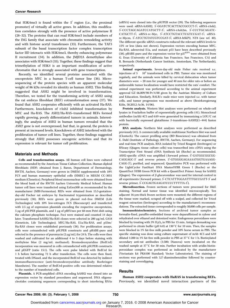
oncoprotein MYC, including PARP10, nucleolin, and a protein withan apparent molecular mass of 86 kDa (36). Sequencing of trypticpeptides revealed that the p86 protein is the human orthologue ofthe Drosophila Ash2 protein (data not shown). A detailed accountof the interaction of ASH2 with MYC will be described elsewhere.The finding that ASH2 interacts with MYC and the identification ofASH2 in protein complexes that contain the oncoprotein MLL1 orMLL4 prompted us to test whether hASH2 affects MYC/Ha-RAS–dependent transformation of REFs. Coexpression of hASH2 led to amild increase in the number of foci (Fig. 1A). In contrast, knock-down of rat Ash2 (rAsh2) significantly inhibited foci formation(Fig. 1A). This effect was specific because siRNA constructsexpressing a mutated version of the rAsh2-specific oligonucleotidehad no effect (Fig. 1A). Furthermore, knockdown of rAsh2 did notinterfere with E1a/HaRAS and mutant p53/HaRAS transformationof REFs (Fig. 1C and D). These findings suggested that rAsh2 waspivotal for cMYC/HaRAS-dependent transformation.
We also cotransfected expression constructs for hASH2 andHa-RAS. Surprisingly, hASH2 was capable to cooperate with Ha-RASin transforming REFs (Fig. 1A). The number of foci obtained withhASH2/Ha-RAS was f70% of that observed for MYC/Ha-RAS–transformed cells. Thus, hASH2 behaved as an oncoprotein in theREF assay. To evaluate whether rat Myc was required for hASH2/Ha-RAS transformation, knockdown experiments were performed.Repression of rat Myc strongly inhibited hASH2/Ha-RAS but alsoE1a/Ha-RAS and mutant p53/Ha-RAS transformation (Fig. 1B–D).Together, these findings suggested that hASH2 has oncogenicproperties in the REF cotransformation assay.hASH2/HaRAS transformed REFs form tumors in animals.
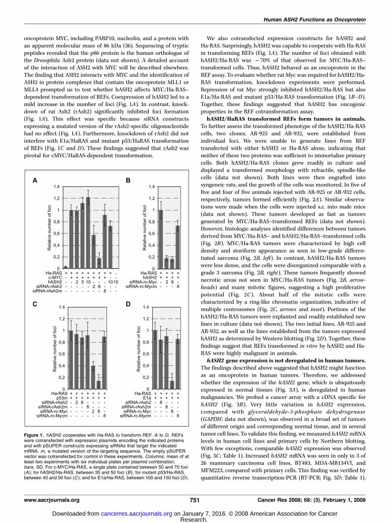
To further assess the transformed phenotype of the hASH2/Ha-RAScells, two clones, AR-925 and AR-932, were established fromindividual foci. We were unable to generate lines from REFtransfected with either hASH2 or Ha-RAS alone, indicating thatneither of these two proteins was sufficient to immortalize primarycells. Both hASH2/Ha-RAS clones grew readily in culture anddisplayed a transformed morphology with refractile, spindle-likecells (data not shown). Both lines were then engrafted intosyngeneic rats, and the growth of the cells was monitored. In five offive and four of five animals injected with AR-925 or AR-932 cells,respectively, tumors formed efficiently (Fig. 2A). Similar observa-tions were made when the cells were injected s.c. into nude mice(data not shown). These tumors developed as fast as tumorsgenerated by MYC/Ha-RAS–transformed REFs (data not shown).However, histologic analyses identified differences between tumorsderived from MYC/Ha-RAS– and hASH2/Ha-RAS–transformed cells(Fig. 2B). MYC/Ha-RAS tumors were characterized by high celldensity and storiform appearance as seen in low-grade differen-tiated sarcoma (Fig. 2B, left). In contrast, hASH2/Ha-RAS tumorswere less dense, and the cells were disorganized comparable with agrade 3 sarcoma (Fig. 2B, right). These tumors frequently showednecrotic areas not seen in MYC/Ha-RAS tumors (Fig. 2B, arrow-heads) and many mitotic figures, suggesting a high proliferativepotential (Fig. 2C ). About half of the mitotic cells werecharacterized by a ring-like chromatin organization, indicative ofmultiple centrosomes (Fig. 2C, arrows and inset). Portions of thehASH2/Ha-RAS tumors were explanted and readily established newlines in culture (data not shown). The two initial lines, AR-925 andAR-932, as well as the lines established from the tumors expressedhASH2 as determined by Western blotting (Fig. 2D). Together, thesefindings suggest that REFs transformed in vitro by hASH2 and Ha-RAS were highly malignant in animals.hASH2 gene expression is not deregulated in human tumors.
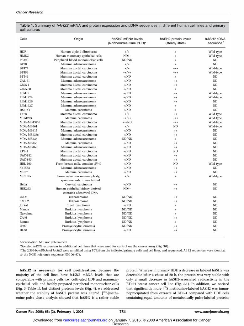
The findings described above suggested that hASH2 might functionas an oncoprotein in human tumors. Therefore, we addressedwhether the expression of the hASH2 gene, which is ubiquitouslyexpressed in normal tissues (Fig. 3A), is deregulated in humanmalignancies. We probed a cancer array with a cDNA specific forhASH2 (Fig. 3B). Very little variation in hASH2 expression,compared with glyceraldehyde-3-phosphate dehydrogenase(GAPDH ; data not shown), was observed in a broad set of tumorsof different origin and corresponding normal tissue, and in severaltumor cell lines. To validate this finding, we measured hASH2 mRNAlevels in human cell lines and primary cells by Northern blotting.With few exceptions, comparable hASH2 expression was observed(Fig. 3C ; Table 1). Increased hASH2 mRNA was seen in only in 3 of26 mammary carcinoma cell lines, BT483, MDA-MB134VI, andMFM223, compared with primary cells. This finding was verified byquantitative reverse transcription-PCR (RT-PCR; Fig. 3D ; Table 1).
Figure 1. hASH2 cooperates with Ha-RAS to transform REF. A to D, REFswere cotransfected with expression plasmids encoding the indicated proteinsand with pSUPER constructs expressing siRNAs that target the indicatedmRNA. m, a mutated version of the targeting sequence. The empty pSUPERvector was cotransfected for control in these experiments. Columns, mean of atleast two experiments with six individual plates per plasmid combination;bars, SD. For c-MYC/Ha-RAS, a single plate contained between 50 and 70 foci(A); for hASH2/Ha-RAS, between 35 and 50 foci (B); for mutant p53/Ha-RAS,between 40 and 50 foci (C ); and for E1a/Ha-RAS, between 100 and 150 foci (D ).
Human ASH2 Functions as Oncoprotein
www.aacrjournals.org 751 Cancer Res 2008; 68: (3). February 1, 2008
Research. on January 7, 2016. © 2008 American Association for Cancercancerres.aacrjournals.org Downloaded from
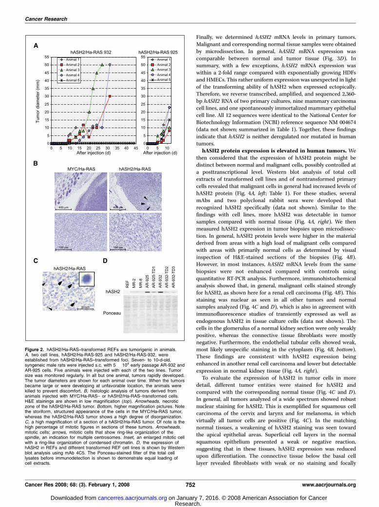
Finally, we determined hASH2 mRNA levels in primary tumors.Malignant and corresponding normal tissue samples were obtainedby microdissection. In general, hASH2 mRNA expression wascomparable between normal and tumor tissue (Fig. 3D). Insummary, with a few exceptions, hASH2 mRNA expression waswithin a 2-fold range compared with exponentially growing HDFsand HMECs. This rather uniform expression was unexpected in lightof the transforming ability of hASH2 when expressed ectopically.Therefore, we reverse transcribed, amplified, and sequenced 2,360-bp hASH2 RNA of two primary cultures, nine mammary carcinomacell lines, and one spontaneously immortalized mammary epithelialcell line. All 12 sequences were identical to the National Center forBiotechnology Information (NCBI) reference sequence NM 004674(data not shown; summarized in Table 1). Together, these findingsindicate that hASH2 is neither deregulated nor mutated in humantumors.hASH2 protein expression is elevated in human tumors. We
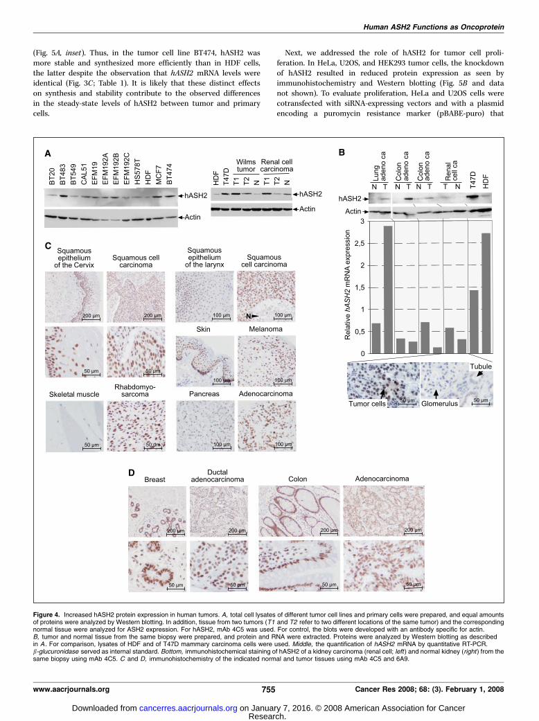
then considered that the expression of hASH2 protein might bedistinct between normal and malignant cells, possibly controlled ata posttranscriptional level. Western blot analysis of total cellextracts of transformed cell lines and of nontransformed primarycells revealed that malignant cells in general had increased levels ofhASH2 protein (Fig. 4A, left ; Table 1). For these studies, severalmAbs and two polyclonal rabbit sera were developed thatrecognized hASH2 specifically (data not shown). Similar to thefindings with cell lines, more hASH2 was detectable in tumorsamples compared with normal tissue (Fig. 4A, right). We thenmeasured hASH2 expression in tumor biopsies upon microdissec-tion. In general, hASH2 protein levels were higher in the materialderived from areas with a high load of malignant cells comparedwith areas with primarily normal cells as determined by visualinspection of H&E-stained sections of the biopsies (Fig. 4B).However, in most instances, hASH2 mRNA levels from the samebiopsies were not enhanced compared with controls usingquantitative RT-PCR analysis. Furthermore, immunohistochemicalanalysis showed that, in general, malignant cells stained stronglyfor hASH2, as shown here for a renal cell carcinoma (Fig. 4B). Thisstaining was nuclear as seen in all other tumors and normalsamples analyzed (Fig. 4C and D), which is also in agreement withimmunofluorescence studies of transiently expressed as well asendogenous hASH2 in tissue culture cells (data not shown). Thecells in the glomerulus of a normal kidney section were only weaklypositive, whereas the connective tissue fibroblasts were mostlynegative. Furthermore, the endothelial tubular cells showed weak,most likely unspecific staining in the cytoplasm (Fig. 4B, bottom).These findings are consistent with hASH2 expression beingenhanced in another renal cell carcinoma and lower but detectableexpression in normal kidney tissue (Fig. 4A, right).
To evaluate the expression of hASH2 in tumor cells in moredetail, different tumor entities were stained for hASH2 andcompared with the corresponding normal tissue (Fig. 4C and D).In general, all tumors analyzed of a wide spectrum showed robustnuclear staining for hASH2. This is exemplified for squamous cellcarcinoma of the cervix and larynx and for melanoma, in whichvirtually all tumor cells are positive (Fig. 4C). In the matchingnormal tissues, a weakening of hASH2 staining was seen towardthe apical epithelial areas. Superficial cell layers in the normalsquamous epithelium presented a weak or negative reaction,suggesting that in these tissues, hASH2 expression was reducedupon differentiation. The connective tissue below the basal celllayer revealed fibroblasts with weak or no staining and focally
Figure 2. hASH2/Ha-RAS–transformed REFs are tumorigenic in animals.A, two cell lines, hASH2/Ha-RAS-925 and hASH2/Ha-RAS-932, wereestablished from hASH2/Ha-RAS–transformed foci. Seven- to 10-d-old,syngeneic male rats were injected s.c. with 5 � 104 early passage AR-932 andAR-925 cells. Five animals were injected with each of the two lines. Tumorsize was monitored regularly. In all but one animal, tumors rapidly developed.The tumor diameters are shown for each animal over time. When the tumorsbecame large or were developing at unfavorable location, the animals werekilled to prevent discomfort. B, histologic analysis of tumors derived fromanimals injected with MYC/Ha-RAS– or hASH2/Ha-RAS–transformed cells.H&E stainings are shown in low magnification (top ). Arrowheads, necroticzone of the hASH2/Ha-RAS tumor. Bottom, higher magnification pictures. Notethe storiform, structured appearance of the cells in the MYC/Ha-RAS tumor,whereas the hASH2/Ha-RAS tumor shows a high degree of disorganization.C, a high magnification of a section of a hASH2/Ha-RAS tumor. Of note is thehigh percentage of mitotic figures in sections of these tumors. Arrowheads,mitotic cells; arrows, mitotic cells that show ring-like organization of thespindle, an indication for multiple centrosomes. Inset, an enlarged mitotic cellwith a ring-like organization of condensed chromatin. D, the expression ofhASH2 in REFs and different transformed REF cell lines is shown by Westernblot analysis using mAb 4C5. The Ponceau-stained filter of the total celllysates before immunodetection is shown to demonstrate equal loading ofcell extracts.
Cancer Research
Cancer Res 2008; 68: (3). February 1, 2008 752 www.aacrjournals.org
Research. on January 7, 2016. © 2008 American Association for Cancercancerres.aacrjournals.org Downloaded from
strongly stained lymphocytes (Fig. 4C). In addition, the tumor cellsof a rhabdomyosarcoma showed strong nuclear staining, whereasthe matching normal skeletal muscle was almost negative (Fig. 4C).Robust staining of tumor cell nuclei was also detected in anadenocarcinoma of the pancreas (Fig. 4C). Normal pancreas showedintermediate reactivity in all acinar cells, whereas ductal cellswere less positive (Fig. 4C). Furthermore, strong nuclear stainingfor hASH2 was present in cells of breast and colon carcinomas(Fig. 4D). Normal breast tissue also exhibited strong nuclearstaining in the acini. Normal colon epithelium was generally
strongly nuclear positive in goblet cells but showed a slightweakening in nuclei of cells located more apically in the crypts. Thesurrounding connective tissue was weakly positive with theexception of substantially stained lymphocytes (Fig. 4D). Theseexamples are from a selection of >200 different tumor samplesanalyzed. In all cases, tumor cells were positive for hASH2. It isimportant to note that many normal cells also stained positive,indicating that the expression of hASH2 is not exclusive to tumorcells. Nevertheless, the expression of hASH2 was frequently elevatedin tumor cells compared with the corresponding normal cells.
Figure 3. hASH2 mRNA expression is not deregulated in human tumors. A, a commercially available multitissue Northern blot was probed with an hASH2 cDNA.A single mRNA species of about 2.36 kb was detected (top ). For control, the blot was developed with a GAPDH probe (bottom ). B, a commercially availablecancer profiling array was probed with an hASH2 cDNA. The tissues analyzed and the cDNAs of tumors (T ) and of the corresponding normal tissues (N ) are indicated.For some samples, in addition to cDNAs of tumor and normal tissue, cDNA of a metastasis (M ) is spotted as indicated (bottom right hand corner ). For control, cDNAs ofdifferent cell lines and RNA or DNA of different origin are spotted as indicated (to the right of the filter ). C, total RNA of the indicated cell lines was analyzed byNorthern blotting using an hASH2 cDNA. For control, the weak cross-reactivity to 18S and 28S rRNA is shown. D, quantitative RT-PCR analysis of hASH2 expressionin different cell lines and in tumor, and corresponding normal tissue. The reactions were standardized for h-glucuronidase expression. The signal from primary HDFwas artificially set as 1. BT483 and MFM223 were selected from a total of 26 breast carcinoma (ca ) cell lines because Northern blot analysis revealed increasedhASH2 mRNA levels in both lines.
Human ASH2 Functions as Oncoprotein
www.aacrjournals.org 753 Cancer Res 2008; 68: (3). February 1, 2008
Research. on January 7, 2016. © 2008 American Association for Cancercancerres.aacrjournals.org Downloaded from
hASH2 is necessary for cell proliferation. Because themajority of the cell lines have hASH2 mRNA levels that arecomparable with primary cells, i.e., cultivated HDF and mammaryepithelial cells and freshly prepared peripheral mononuclear cells(Fig. 3; Table 1), but distinct proteins levels (Fig. 4), we addressedwhether the stability of hASH2 protein was altered. [35S]methi-onine pulse chase analysis showed that hASH2 is a rather stable
protein. Whereas in primary HDF, a decrease in labeled hASH2 wasdetectable after a chase of 20 h, the protein was very stable withonly a small decrease in hASH2-associated radioactivity in theBT474 breast cancer cell line (Fig. 5A). In addition, we noticedthat significantly more [35S]methionine-labeled hASH2 was immu-noprecipitated from extracts of BT474 compared with HDF cellscontaining equal amounts of metabolically pulse-labeled proteins
Table 1. Summary of hAHS2 mRNA and protein expression and cDNA sequences in different human cell lines and primarycell cultures
Cells Origin hASH2 mRNA levels(Northern/real-time PCR)*
hASH2 protein levels(steady state)
hASH2 cDNAsequence
c
HDF Human diploid fibroblasts +/+ + Wild-typeHMEC Human mammary epithelial cells ND/+ + Wild-type
PBMC Peripheral blood mononuclear cells ND/ND + ND
BT20 Mamma adenocarcinoma +/+ + NDBT474 Mamma ductal carcinoma +/+ +++ Wild-type
BT483 Mamma ductal carcinoma ++/++ +++ Wild-type
BT549 Mamma ductal carcinoma +/ND + ND
CAL-51 Mamma adenocarcinoma +/ND ++ NDZR75-1 Mamma ductal carcinoma +/ND ++ ND
ZR75-30 Mamma ductal carcinoma +/ND + ND
EFM19 Mamma adenocarcinoma +/ND ++ Wild-type
EFM192A Mamma adenocarcinoma +/ND ++ Wild-typeEFM192B Mamma adenocarcinoma +/ND ++ ND
EFM192C Mamma adenocarcinoma +/ND + ND
HS578T Mamma carcinoma +/ND + ND
T47D Mamma ductal carcinoma +/+ ++ Wild-typeMFM223 Mamma carcinoma ++/++ +++ Wild-type
MDA-MB134VI Mamma ductal carcinoma ++/ND +++ Wild-type
MDA-MB361 Mamma ductal carcinoma +/+ ND Wild-typeMDA-MB415 Mamma adenocarcinoma +/ND ++ ND
MDA-MB435s Mamma ductal carcinoma +/ND ++ ND
MDA-MB436 Mamma adenocarcinoma ND/ND + ND
MDA-MB453 Mamma carcinoma +/ND ++ NDMDA-MB468 Mamma adenocarcinoma +/ND ++ ND
KPL-1 Mamma ductal carcinoma +/ND ND ND
UAC-812 Mamma ductal carcinoma +/ND ++ ND
UAC-893 Mamma ductal carcinoma +/ND ++ NDHBL-100 From breast milk, contains SV40 +/ND ND Wild-type
SK-BR3 Mamma adenocarcinoma +/ND ++ ND
MCF7 Mamma carcinoma +/ND ++ NDMCF12a From reduction mammoplasty,
spontaneously immortalized
+/+ + Wild-type
HeLa Cervical carcinoma +/ND ++ ND
HEK293 Human epithelial kidney derived,contains adenoviral DNA
ND/+ ++ ND
U2OS Osteosarcoma ND/ND ++ ND
SAOS2 Osteosarcoma ND/ND ++ ND
Jurkat T cell lymphoma +/ND + NDManca Burkitt’s lymphoma ND/ND + ND
Nawalma Burkitt’s lymphoma ND/ND + ND
CA46 Burkitt’s lymphoma ND/ND ++ NDRamos Burkitt’s lymphoma ND/ND + ND
U937 Promyelocytic leukemia ND/ND ++ ND
HL60 Promyelocytic leukemia +/ND ++ ND
Abbreviation: ND, not determined.*See also hASH2 expression in additional cell lines that were used for control on the cancer array (Fig. 3B).cThe 2,360-bp cDNAs of hASH2 were amplified using PCR from the indicated primary cells and cell lines, and sequenced. All 12 sequences were identical
to the NCBI reference sequence NM 004674.
Cancer Research
Cancer Res 2008; 68: (3). February 1, 2008 754 www.aacrjournals.org
Research. on January 7, 2016. © 2008 American Association for Cancercancerres.aacrjournals.org Downloaded from
(Fig. 5A, inset). Thus, in the tumor cell line BT474, hASH2 wasmore stable and synthesized more efficiently than in HDF cells,the latter despite the observation that hASH2 mRNA levels wereidentical (Fig. 3C ; Table 1). It is likely that these distinct effectson synthesis and stability contribute to the observed differencesin the steady-state levels of hASH2 between tumor and primarycells.
Next, we addressed the role of hASH2 for tumor cell proli-feration. In HeLa, U2OS, and HEK293 tumor cells, the knockdownof hASH2 resulted in reduced protein expression as seen byimmunohistochemistry and Western blotting (Fig. 5B and datanot shown). To evaluate proliferation, HeLa and U2OS cells werecotransfected with siRNA-expressing vectors and with a plasmidencoding a puromycin resistance marker (pBABE-puro) that
Figure 4. Increased hASH2 protein expression in human tumors. A, total cell lysates of different tumor cell lines and primary cells were prepared, and equal amountsof proteins were analyzed by Western blotting. In addition, tissue from two tumors (T1 and T2 refer to two different locations of the same tumor) and the correspondingnormal tissue were analyzed for ASH2 expression. For hASH2, mAb 4C5 was used. For control, the blots were developed with an antibody specific for actin.B, tumor and normal tissue from the same biopsy were prepared, and protein and RNA were extracted. Proteins were analyzed by Western blotting as describedin A . For comparison, lysates of HDF and of T47D mammary carcinoma cells were used. Middle, the quantification of hASH2 mRNA by quantitative RT-PCR.b-glucuronidase served as internal standard. Bottom, immunohistochemical staining of hASH2 of a kidney carcinoma (renal cell; left ) and normal kidney (right ) from thesame biopsy using mAb 4C5. C and D, immunohistochemistry of the indicated normal and tumor tissues using mAb 4C5 and 6A9.
Human ASH2 Functions as Oncoprotein
www.aacrjournals.org 755 Cancer Res 2008; 68: (3). February 1, 2008
Research. on January 7, 2016. © 2008 American Association for Cancercancerres.aacrjournals.org Downloaded from

allowed selecting the transfected cells with puromycin. ReducinghASH2 levels inhibited proliferation of the transfected cells inboth lines, whereas si-rASH2m or an siRNA specific for luciferasehad no effect (Fig. 5C). These effects were at least as strong asknockdown of hSKP2. This protein mediates degradation of thecyclin-dependent kinase inhibitor p27KIP1 at the G1-S phasetransition, and its knockdown inhibits proliferation (40). In theabsence of pBABE-puro, no cells survived the puromycinselection. Furthermore, we determined the effect of the hASH2knockdown on the ability of cells to enter S phase. Repression ofhASH2 reduced S-phase progression in HeLa and U2OS cells asdetermined by measuring the incorporation of BrdUrd (Fig. 5D).In this assay, the effect was comparable with the one elicited bythe knockdown of hSKP2. As for the proliferation assay, si-rASH2m was ineffective. We concluded from these studies thathASH2 overexpression provides tumor cells with a growthadvantage.
Discussion
In this report, we provide evidence that the trithorax proteinASH2 functions as an oncoprotein. This conclusion is based on thefindings that hASH2 cooperates with Ha-RAS in the transformationof REFs, that hASH2 is overexpressed in human tumors, and thatthe knockdown of hASH2 results in the inhibition of proliferationof tumor cell lines. Importantly, the overexpression of hASH2 is dueto increased protein synthesis and stability in tumor cells and notdue to deregulated expression of the gene.
hASH2 has been found in at least four human complexes thatpossess HMT activity. These complexes have distinct proteincompositions with different catalytic subunits that can specificallytrimethylate H3K4 (21, 23). This histone mark is closely associatedwith promoters of actively transcribed genes (25, 27). Of particularinterest are the findings that two of these HMT complexes containthe tumor suppressor Menin and either MLL1 or MLL4 as thecatalytic subunits (15, 18, 20, 42, 43). MLL proteins function as
Figure 5. Regulation of hASH2 protein expression and relevance for tumor cell proliferation. A, HDF and BT474 cells were metabolically labeled with [35S]methioninefor 30 min and then chased with excess unlabeled methionine for the indicated times. The incorporated radioactivity was determined for each cell lysate, andaliquots of these lysates with equal amounts of radioactivity were used for hASH2-specific immunoprecipitations. The radioactivity associated with hASH2 was thenmeasured, and the relative amounts are blotted. Inset, [35S]methionine pulse-labeled, immunoprecipitated hASH2 is shown. The relative labeling of hASH2 in HDF andBT474 cells is given from four independent experiments. B, pSUPER constructs expressing the indicated siRNAs were transfected into HeLa cells together witha pEGFP control plasmid. Transfection efficiency was about 80% as determined by green fluorescent protein fluorescence (data not shown). The transfected cellswere embedded in paraffin, and then hASH2 expression was analyzed immunohistochemically using mAb 4C5 (top ). In addition, the transfected cells were lysed inFrackelton buffer, and the expression of hASH2 was examined by Western blotting using mAb 4C5. For control, the Ponceau-stained blot is shown. C, HeLa andU2OS cells were transfected with pSUPER constructs expressing the indicated siRNAs. In addition, pBABE-puro was cotransfected, except for the plates shownon the right [si-LUC(�)]. The cells were selected on puromycin for 24 h, replated, and stained after 4 d. D, HeLa and U2OS cells were transfected with pSUPERconstructs expressing the indicated siRNAs and with pEGFP. After 72 h, the cells were pulse-labeled with BrdUrd for 1 h. The number of BrdUrd-positive cells of thetransfected cells was determined. Columns, mean values of three independent experiments with >100 cells analyzed per experiment; bars, SD.
Cancer Research
Cancer Res 2008; 68: (3). February 1, 2008 756 www.aacrjournals.org
Research. on January 7, 2016. © 2008 American Association for Cancercancerres.aacrjournals.org Downloaded from

oncoproteins in a number of distinct human tumors. The MLL1gene is translocated in and associated with aggressive acuteleukemias, whereas MLL4 is amplified in some solid tumors (44).Menin is the product of the tumor suppressor gene MEN1 (43).Loss of function mutations of MEN1 are observed in sporadic andheritable endocrine tumors (42). Thus, with MLL and Menin, thesecomplexes contain both a tumor suppressor protein and anoncoprotein. Our data now provide evidence for an additionalsubunit, ASH2, associated with these complexes that is involved intumorigenesis. Although MLL1, MLL4, and Menin are associatedwith specific tumors, our data suggest that increased hASH2expression is very broadly associated with tumor formation.Together, these findings strongly indicate that the ASH2/MLL/Menin complexes are tied closely to tumor formation in manydifferent cell types.
Because these complexes are involved in chromatin regulation, itseems likely that the deregulation of any of these proteins isassociated with altered gene transcription. Indeed, MLLs have beenlinked to the expression of HOX and CDK inhibitor genes.Deregulation of these seems to be relevant for tumor formation(18, 45–47). The molecular function of Menin is less wellunderstood. It has been suggested that the HMT activity of MLL4
is dependent on Menin (43). The molecular role of AHS2 is poorlyunderstood. Knockdown of ASH2 results in a reduction oftrimethylation of H3K4 (21),5 suggesting that this protein isimportant for the activity of the HMT complexes. This might resultin the deregulation of many genes and possibly explains thereduced proliferation in response to the ASH2 knockdown (Fig. 5).It is conceivable that ASH2 is important to allow complexformation and/or to stabilize the MLL complex (21). Additionally,ASH2 might target the MLL complexes to specific promoters byinteracting with transcriptional regulators or other chromatin-bound proteins. In support, ASH2 directly binds to the oncoproteinMYC.5 It will now be important to define the molecular functions ofASH2 in more detail. Together, the findings presented here and thepublished observations strongly suggest that the ASH2/MLL/Menincomplexes affect cell transformation by controlling gene expression.
Acknowledgments
Received 8/16/2007; revised 10/22/2007; accepted 11/26/2007.Grant support: Interdisciplinary Centre for Clinical Research ‘‘BIOMAT’’ within
the faculty of Medicine at the RWTH Aachen University (TV M1) and the DeutscheForschungsgemeinschaft DFG (B. Luscher).
The costs of publication of this article were defrayed in part by the payment of pagecharges. This article must therefore be hereby marked advertisement in accordancewith 18 U.S.C. Section 1734 solely to indicate this fact.
We thank R. Kraft and M. Austen for microsequencing the p86 protein band,E. Dahl for providing the Cancer Profiling Array filter, G. Evan and R. Bernards for theexpression vectors, J. Baron for the human diploid fibroblasts, and D. Smeets andJ. Stahl for expert technical assistance.5 Our own unpublished findings.
Human ASH2 Functions as Oncoprotein
www.aacrjournals.org 757 Cancer Res 2008; 68: (3). February 1, 2008
References1. Hanahan D, Weinberg RA. The hallmarks of cancer.Cell 2000;100:57–70.2. Guo QM. DNA microarray and cancer. Curr OpinOncol 2003;15:36–43.3. Pinkel D, Albertson DG. Array comparative genomichybridization and its applications in cancer. Nat Genet2005;37 Suppl:S11–7.4. Downward J. Targeting RAS signalling pathways incancer therapy. Nat Rev Cancer 2003;3:11–22.5. Koff A. How to decrease p27Kip1 levels during tumordevelopment. Cancer Cell 2006;9:75–6.6. Orlando V, Paro R. Chromatin multiprotein complexesinvolved in the maintenance of transcription patterns.Curr Opin Genet Dev 1995;5:174–9.7. Ringrose L, Paro R. Epigenetic regulation of cellularmemory by the Polycomb and Trithorax group proteins.Annu Rev Genet 2004;38:413–43.8. Schwartz YB, Pirrotta V. Polycomb silencing mecha-nisms and the management of genomic programmes.Nat Rev Genet 2007;8:9–22.9. Shearn A, Rice T, Garen A, Gehring W. Imaginal discabnormalities in lethal mutants of Drosophila . Proc NatlAcad Sci U S A 1971;68:2594–8.10. Adamson AL, Shearn A. Molecular genetic analysis of
Drosophila ash2, a member of the trithorax grouprequired for imaginal disc pattern formation. Genetics1996;144:621–33.11. LaJeunesse D, Shearn A. Trans-regulation of thoracichomeotic selector genes of the Antennapedia andbithorax complexes by the trithorax group genes:absent, small, and homeotic discs 1 and 2. Mech Dev1995;53:123–39.12. Beltran S, Angulo M, Pignatelli M, Serras F,Corominas M. Functional dissection of the ash2and ash1 transcriptomes provides insights into thetranscriptional basis of wing phenotypes andreveals conserved protein interactions. Genome Biol2007;8:R67.13. Beltran S, Blanco E, Serras F, et al. Transcriptional
network controlled by the trithorax-group gene ash2 inDrosophila melanogaster . Proc Natl Acad Sci U S A 2003;100:3293–8.14. Goo YH, Sohn YC, Kim DH, et al. Activating signalcointegrator 2 belongs to a novel steady-state complexthat contains a subset of trithorax group proteins. MolCell Biol 2003;23:140–9.15. Hughes CM, Rozenblatt-Rosen O, Milne TA, et al.Menin associates with a trithorax family histonemethyltransferase complex and with the hoxc8 locus.Mol Cell 2004;13:587–97.16. Lee JH, Skalnik DG. CpG-binding protein (CXXCfinger protein 1) is a component of the mammalian Set1histone H3–4 methyltransferase complex, the analogueof the yeast Set1/COMPASS complex. J Biol Chem 2005;280:41725–31.17. Miller T, Krogan NJ, Dover J, et al. COMPASS: acomplex of proteins associated with a trithorax-relatedSET domain protein. Proc Natl Acad Sci U S A 2001;98:12902–7.18. Milne TA, Hughes CM, Lloyd R, et al. Menin and MLLcooperatively regulate expression of cyclin-dependentkinase inhibitors. Proc Natl Acad Sci U S A 2005;102:749–54.19. Wysocka J, Myers MP, Laherty CD, Eisenman RN,Herr W. Human Sin3 deacetylase and trithorax-relatedSet1/Ash2 histone H3-4 methyltransferase are tetheredtogether selectively by the cell-proliferation factor HCF-1. Genes Dev 2003;17:896–911.20. Yokoyama A, Wang Z, Wysocka J, et al. Leukemiaproto-oncoprotein MLL forms a SET1-like histonemethyltransferase complex with menin to regulate Hoxgene expression. Mol Cell Biol 2004;24:5639–49.21. Dou Y, Milne TA, Ruthenburg AJ, et al. Regulation ofMLL1 H3K4 methyltransferase activity by its corecomponents. Nat Struct Mol Biol 2006;13:713–9.22. Guccione E, Bassi C, Casadio F, et al. Methylation ofhistone H3R2 by PRMT6 and H3K4 by an MLL complexare mutually exclusive. Nature 2007;449:933–7.23. Steward MM, Lee JS, O’Donovan A, et al. Molecularregulation of H3K4 trimethylation by ASH2L, a shared
subunit of MLL complexes. Nat Struct Mol Biol 2006;13:852–4.24. Berger SL. The complex language of chromatinregulation during transcription. Nature 2007;447:407–12.25. Li B, Carey M, Workman JL. The role of chromatinduring transcription. Cell 2007;128:707–19.26. Cosgrove MS, Wolberger C. How does the histonecode work? Biochem Cell Biol 2005;83:468–76.27. Ruthenburg AJ, Allis CD, Wysocka J. Methylationof lysine 4 on histone H3: intricacy of writing andreading a single epigenetic mark. Mol Cell 2007;25:15–30.28. Bernstein BE, Kamal M, Lindblad-Toh K, et al.Genomic maps and comparative analysis of histonemodifications in human and mouse. Cell 2005;120:169–81.29. Ng HH, Robert F, Young RA, Struhl K. Targetedrecruitment of Set1 histone methylase by elongating PolII provides a localized mark and memory of recenttranscriptional activity. Mol Cell 2003;11:709–19.30. Santos-Rosa H, Schneider R, Bannister AJ, et al.Active genes are tri-methylated at K4 of histone H3.Nature 2002;419:407–11.31. Schneider R, Bannister AJ, Myers FA, et al. HistoneH3 lysine 4 methylation patterns in higher eukaryoticgenes. Nat Cell Biol 2004;6:73–7.32. Schubeler D, MacAlpine DM, Scalzo D, et al. Thehistone modification pattern of active genes revealedthrough genome-wide chromatin analysis of a highereukaryote. Genes Dev 2004;18:1263–71.33. Shi X, Gozani O. The fellowships of the INGs. J CellBiochem 2005;96:1127–36.34. Vermeulen M, Mulder KW, Denissov S, et al. Selectiveanchoring of TFIID to nucleosomes by trimethylation ofhistone H3 lysine 4. Cell 2007;131:58–69.35. Shi Y, Whetstine JR. Dynamic regulation ofhistone lysine methylation by demethylases. Mol Cell2007;25:1–14.36. Yu M, Schreek S, Cerni C, et al. PARP-10, a novel Myc-interacting protein with poly(ADP-ribose) polymerase
Research. on January 7, 2016. © 2008 American Association for Cancercancerres.aacrjournals.org Downloaded from

Cancer Research
Cancer Res 2008; 68: (3). February 1, 2008 758 www.aacrjournals.org
activity, inhibits transformation. Oncogene 2005;24:1982–93.37. Land H, Parada LF, Weinberg RA. Tumorigenic con-version of primary embryo fibroblasts requires at leasttwo cooperating oncogenes. Nature 1983;304:596–602.38. Cerni C, Bousset K, Seelos C, et al. Differential effectsby Mad and Max on transformation by cellular and viraloncoproteins. Oncogene 1995;11:587–96.39. Brummelkamp TR, Bernards R, Agami R. A systemfor stable expression of short interfering RNAs inmammalian cells. Science 2002;296:550–3.40. Garriga J, Bhattacharya S, Calbo J, et al. CDK9 isconstitutively expressed throughout the cell cycle, and
its steady-state expression is independent of SKP2. MolCell Biol 2003;23:5165–73.41. Sommer A, Bousset K, Kremmer E, Austen M,Luscher B. Identification and characterization ofspecific DNA-binding complexes containing membersof the Myc/Max/Mad network of transcriptional regu-lators. J Biol Chem 1998;273:6632–42.42. Chandrasekharappa SC, Teh BT. Functional studiesof the MEN1 gene. J Intern Med 2003;253:606–15.43. Yokoyama A, Somervaille TC, Smith KS, et al. Themenin tumor suppressor protein is an essentialoncogenic cofactor for MLL-associated leukemogenesis.Cell 2005;123:207–18.
44. Hess JL. Mechanisms of transformation by MLL. CritRev Eukaryot Gene Expr 2004;14:235–54.45. Ayton PM, Cleary ML. Transformation of myeloidprogenitors by MLL oncoproteins is dependent onHoxa7 and Hoxa9. Genes Dev 2003;17:2298–307.46. Karnik SK, Hughes CM, Gu X, et al. Meninregulates pancreatic islet growth by promoting histonemethylation and expression of genes encodingp27Kip1 and p18INK4c. Proc Natl Acad Sci U S A2005;102:14659–64.47. Zeisig BB, Milne T, Garcia-Cuellar MP, et al. Hoxa9and Meis1 are key targets for MLL-ENL-mediatedcellular immortalization. Mol Cell Biol 2004;24:617–28.
Research. on January 7, 2016. © 2008 American Association for Cancercancerres.aacrjournals.org Downloaded from
2008;68:749-758. Cancer Res Juliane Lüscher-Firzlaff, Isabella Gawlista, Jörg Vervoorts, et al. OncoproteinThe Human Trithorax Protein hASH2 Functions as an
Updated version
http://cancerres.aacrjournals.org/content/68/3/749
Access the most recent version of this article at:
Cited articles
http://cancerres.aacrjournals.org/content/68/3/749.full.html#ref-list-1
This article cites 47 articles, 16 of which you can access for free at:
Citing articles
http://cancerres.aacrjournals.org/content/68/3/749.full.html#related-urls
This article has been cited by 19 HighWire-hosted articles. Access the articles at:
E-mail alerts related to this article or journal.Sign up to receive free email-alerts
Subscriptions
Reprints and
To order reprints of this article or to subscribe to the journal, contact the AACR Publications
Permissions
To request permission to re-use all or part of this article, contact the AACR Publications
Research. on January 7, 2016. © 2008 American Association for Cancercancerres.aacrjournals.org Downloaded from
Related Documents


![Human Papillomavirus Type 16 E7 Oncoprotein-induced ... · [CANCER RESEARCH 61, 2356–2360, March 15, 2001] Advances in Brief Human Papillomavirus Type 16 E7 Oncoprotein-induced](https://static.cupdf.com/doc/110x72/605dd1c1b72c9c6f905bfd49/human-papillomavirus-type-16-e7-oncoprotein-induced-cancer-research-61-2356a2360.jpg)