r Human Brain Mapping 33:419–430 (2012) r The Human Dorsal Premotor Cortex Facilitates the Excitability of Ipsilateral Primary Motor Cortex Via a Short Latency Cortico-Cortical Route Sergiu Groppa, 1 * Boris H. Schlaak, 1 Alexander Mu ¨ nchau, 2 Nicole Werner-Petroll, 1 Janin Du ¨nnweber, 1 Tobias Ba ¨umer, 2 Bart F.L. van Nuenen, 1,3 and Hartwig R. Siebner 1,4,5 1 Department of Neurology, Christian Albrechts University, Kiel, Germany 2 Department of Neurology, University Medical Center Hamburg-Eppendorf, Hamburg, Germany 3 Department of Neurology, Radboud University Nijmegen Medical Centre, Donders Institute for Brain, Cognition and Behaviour, Nijmegen, The Netherlands 4 MR-Department, Danish Research Center for Magnetic Resonance, Copenhagen University Hospital Hvidovre, Hvidovre, Denmark 5 Institute of Neurology, Psychiatry and Senses, University of Copenhagen, Denmark r r Abstract: In non-human primates, invasive tracing and electrostimulation studies have identified strong ipsilateral cortico-cortical connections between dorsal premotor- (PMd) and the primary motor cortex (M1 HAND ). Here, we applied dual-site transcranial magnetic stimulation (dsTMS) to left PMd and M1 HAND through specifically designed minicoils to selectively probe ipsilateral PMd-to-M1 HAND connectivity in humans. A suprathreshold test stimulus (TS) was applied to M1 HAND producing a motor evoked potential (MEP) of about 0.5 mV in the relaxed right first dorsal interosseus muscle (FDI). A subthreshold conditioning stimulus (CS) was given to PMd 2.0–5.2 ms after the TS at intensities of 50-, 70-, or 90% of TS. The CS to PMd facilitated the MEP evoked by TS over M1 HAND at interstimulus intervals (ISI) of 2.4 or 2.8 ms. There was a second facilitatory peak at ISI of 4.4 ms. PMd-to-M1 HAND facilitation did not change as a function of CS intensity. Even at higher intensities, the CS alone failed to elicit a MEP or a cortical silent period in the pre-activated FDI, excluding a direct spread of excitation from PMd to M1 HAND . No MEP facilitation was present while CS was applied rostrally over lateral prefrontal cortex. Together our results indicate that our dsTMS paradigm probes a short-latency facilitatory PMd-to-M1 HAND pathway. The temporal pattern of MEP facilitation suggests a PMd-to-M1 HAND route that targets intracortical M1 HAND circuits involved in the generation of indirect corticospinal volleys. This paradigm opens up new possibilities to study context-dependent intrahemispheric PMd-to-M1 HAND interactions in the intact human brain. Hum Brain Mapp 33:419–430, 2012. V C 2011 Wiley Periodicals, Inc. Key words: connectivity; motor cortex; I-waves; dorsal premotor cortex; transcranial magnetic stimulation r r Contract grant sponsor: Volkswagenstiftung; Contract grant number: I/79-932. *Correspondence to: Sergiu Groppa, Department of Neurology, Christian Albrechts University, Schittenhelmstr 10, Kiel 24105, Germany. E-mail: [email protected] Received for publication 17 May 2010; Revised 2 November 2010; Accepted 8 November 2010 DOI: 10.1002/hbm.21221 Published online 9 March 2011 in Wiley Online Library (wileyonlinelibrary.com). V C 2011 Wiley Periodicals, Inc.

Welcome message from author
This document is posted to help you gain knowledge. Please leave a comment to let me know what you think about it! Share it to your friends and learn new things together.
Transcript

r Human Brain Mapping 33:419–430 (2012) r
The Human Dorsal Premotor Cortex Facilitatesthe Excitability of Ipsilateral Primary Motor Cortex
Via a Short Latency Cortico-Cortical Route
Sergiu Groppa,1* Boris H. Schlaak,1 Alexander Munchau,2
Nicole Werner-Petroll,1 Janin Dunnweber,1 Tobias Baumer,2
Bart F.L. van Nuenen,1,3 and Hartwig R. Siebner1,4,5
1Department of Neurology, Christian Albrechts University, Kiel, Germany2Department of Neurology, University Medical Center Hamburg-Eppendorf, Hamburg, Germany3Department of Neurology, Radboud University Nijmegen Medical Centre, Donders Institute for
Brain, Cognition and Behaviour, Nijmegen, The Netherlands4MR-Department, Danish Research Center for Magnetic Resonance, Copenhagen University Hospital
Hvidovre, Hvidovre, Denmark5Institute of Neurology, Psychiatry and Senses, University of Copenhagen, Denmark
r r
Abstract: In non-human primates, invasive tracing and electrostimulation studies have identifiedstrong ipsilateral cortico-cortical connections between dorsal premotor- (PMd) and the primary motorcortex (M1HAND). Here, we applied dual-site transcranial magnetic stimulation (dsTMS) to left PMdand M1HAND through specifically designed minicoils to selectively probe ipsilateral PMd-to-M1HAND
connectivity in humans. A suprathreshold test stimulus (TS) was applied to M1HAND producing amotor evoked potential (MEP) of about 0.5 mV in the relaxed right first dorsal interosseus muscle(FDI). A subthreshold conditioning stimulus (CS) was given to PMd 2.0–5.2 ms after the TS atintensities of 50-, 70-, or 90% of TS. The CS to PMd facilitated the MEP evoked by TS over M1HAND atinterstimulus intervals (ISI) of 2.4 or 2.8 ms. There was a second facilitatory peak at ISI of 4.4 ms.PMd-to-M1HAND facilitation did not change as a function of CS intensity. Even at higher intensities,the CS alone failed to elicit a MEP or a cortical silent period in the pre-activated FDI, excluding adirect spread of excitation from PMd to M1HAND. No MEP facilitation was present while CS wasapplied rostrally over lateral prefrontal cortex. Together our results indicate that our dsTMS paradigmprobes a short-latency facilitatory PMd-to-M1HAND pathway. The temporal pattern of MEP facilitationsuggests a PMd-to-M1HAND route that targets intracortical M1HAND circuits involved in the generationof indirect corticospinal volleys. This paradigm opens up new possibilities to study context-dependentintrahemispheric PMd-to-M1HAND interactions in the intact human brain. Hum Brain Mapp 33:419–430,2012. VC 2011 Wiley Periodicals, Inc.
Keywords: connectivity; motor cortex; I-waves; dorsal premotor cortex; transcranial magneticstimulation
r r
Contract grant sponsor: Volkswagenstiftung; Contract grantnumber: I/79-932.
*Correspondence to: Sergiu Groppa, Department of Neurology,Christian Albrechts University, Schittenhelmstr 10, Kiel 24105,Germany. E-mail: [email protected]
Received for publication 17 May 2010; Revised 2 November 2010;Accepted 8 November 2010
DOI: 10.1002/hbm.21221Published online 9 March 2011 in Wiley Online Library(wileyonlinelibrary.com).
VC 2011 Wiley Periodicals, Inc.
INTRODUCTION
The human primary motor hand area (M1HAND) is animportant executive area for manual motor control.M1HAND is located in the anterior bank of the central sul-cus having a characteristic knob-like structure with anomega shaped surface in the axial plane and a hook-likesurface in the sagittal plane [Yousry et al., 1997]. Theanatomical connectivity pattern of M1HAND has been thor-oughly studied in non-human primates and underscoresthe pivotal role of this region in manual motor control[Lemon, 2008]. The M1HAND is a major source of fast-conducting monosynaptic projections to the cervical motorneurons [He et al., 1993; Martino and Strick, 1987], whichare thought to be critically involved in the generation andcontrol of highly skilled hand movements [Lemon, 2008].In addition, the M1HAND receives direct cortico-corticalinputs from a set of premotor areas, including the dorsaland ventral premotor cortex and supplementary motorarea [Dum and Strick, 1991; He et al., 1995; Kermadi et al.,1998; Luppino et al., 1993]. Although these premotor areassend descending projections to the cervical spinal cord,they exert substantial influence on descending motor con-trol indirectly via their cortico-cortical inputs into M1HAND
[Schmidlin et al., 2008].In humans, transcranial magnetic stimulation (TMS) is a
well established means of studying the excitability of fastmonosynaptic corticospinal projections originating inM1HAND by recording the motor evoked potentials (MEPs)in contralateral hand muscles [Barker et al., 1985; Hallett,2000]. Epidural invasive recordings at the level of thecervical spinal cord have consistently shown that a singlesuprathreshold TMS pulse gives rise to a series ofdescending volleys in the corticospinal pathway [Amassianet al., 1987; Di Lazzaro et al., 2010]. When the pulse indu-ces a posterior-to-anterior current in M1HAND, TMS primar-ily elicits so-called indirect waves (I-waves) [Di Lazzaroet al., 1999a; Patton and Amassian, 1954]. I-waves aregenerated trans-synaptically by indirect excitation of thecorticospinal neurons via intracortical circuits [Di Lazzaroet al., 1999a; Patton and Amassian, 1954]. The interactionsamong circuits within motor cortex involved in the produc-tion of I-waves can be studied non-invasively using paired-pulse TMS [Tokimura et al., 1996; Ziemann and Rothwell,2000]. Facilitatory I-wave interaction can be demonstratedin the human primary motor cortex by applying pairsof magnetic stimuli to M1HAND through the same coil.The motor response to paired-pulse TMS is consistentlyfacilitated, when the interval between the stimuli is around1.0–1.5 ms, 2.5–3.0 ms, or 4.5 ms [Di Lazzaro et al., 1999b;Tokimura et al., 1996; Ziemann et al., 1998].
Invasive recordings in animals have shown that corticalelectrical stimulation of the premotor or somatosensorycortex can also elicit multiple I-waves in the pyramidaltract, but only if the primary motor cortex is intact[Amassian et al., 1987]. This finding indicates that premo-tor-to-motor and sensory-to-motor cortico-cortical inputs
influence intracortical circuits of M1HAND that generatemotor commands in fast-conducting corticospinal outputneurons. The activation of cortico-cortical inputs to M1
HAND are thought to underlie the modulatory effects of aconditioning stimulus (CS) of a connected cortical area oncorticospinal excitability in M1HAND [Mochizuki et al.,2004]. Accordingly, microstimulation studies in monkeysshowed short-latency facilitation of intracortically evokedtest responses in M1HAND that was present less than 3 msafter the excitation of premotor-to-motor projections [Cerriet al., 2003; Prabhu et al., 2009; Shimazu et al., 2004;Tokuno and Nambu, 2000].
Using a conditioning-test approach with a conditioningcoil placed over a non-primary motor area and a test coilover M1HAND, dual-site TMS (dsTMS) has been recentlyestablished as a valuable tool to probe the excitability ofdistinct cortico-cortical inputs from ipsilateral and contra-lateral frontal and parietal areas to the M1HAND [Baumeret al., 2006; Civardi et al., 2001; Ferbert et al., 1992; Kochet al., 2007a; Mochizuki et al., 2004]. These studies foundinhibitory or facilitatory interactions or both depending onthe dsTMS protocol [Davare et al., 2008; Koch et al.,2007b]. In general, the conditioning effects on corticospinalexcitability in M1HAND critically depended on the timingof the CS relative to the test stimulus (TS) given toM1HAND. Usually, the CS had to be given several millisec-onds (4–12 ms) before the TS to impact on corticospinalexcitability.
Several studies have successfully applied the dsTMSapproach to study the inputs from contralateral dorsal pre-motor cortex (PMd) to M1HAND [Baumer et al., 2006; Kochet al., 2006; Mochizuki et al., 2004; O’Shea et al., 2007]. Thereason for this is that commercially available coils are rela-tive large precluding the concurrent stimulation of ipsilat-eral PMd and M1HAND. According to a meta-analysis offunctional imaging studies the rostral and caudal part ofthe PMd are located on average 8 and 23 mm anterior toM1HAND [Picard and Strick, 2001]. In this study, we usedspecifically designed minicoils to perform dsTMS of leftPMd and M1HAND. We hypothesized that ipsilateral PMd-to-M1HAND inputs might show a facilitatory interaction withintracortical circuits in M1HAND generating late I-waves.Given that the late I-waves leave the M1HAND severalmilliseconds after a magnetic stimulus is applied toM1HAND, we hypothesized that an ipsilateral CS shouldstill be able to modulate the motor response evoked inM1HAND via a short latency pathway when the premotorCS is given after the TS.
METHODS
Subjects
Eighteen male healthy subjects aged between 21 and 40years (mean 27.4 years � 5.2) took part in the study. Allparticipants were consistently right-handed according tothe Edinburgh handedness inventory. Informed consent
r Groppa et al. r
r 420 r
was obtained from all subjects prior to their participationin the study. The experiments were approved by theEthics Committee of University of Kiel, Germany. Duringthe experiment, participants were seated in a recliningchair with a head rest. Participants were asked to keep theeyes open and relax.
Set-Up for Dual-Site Transcranial
Magnetic Stimulation
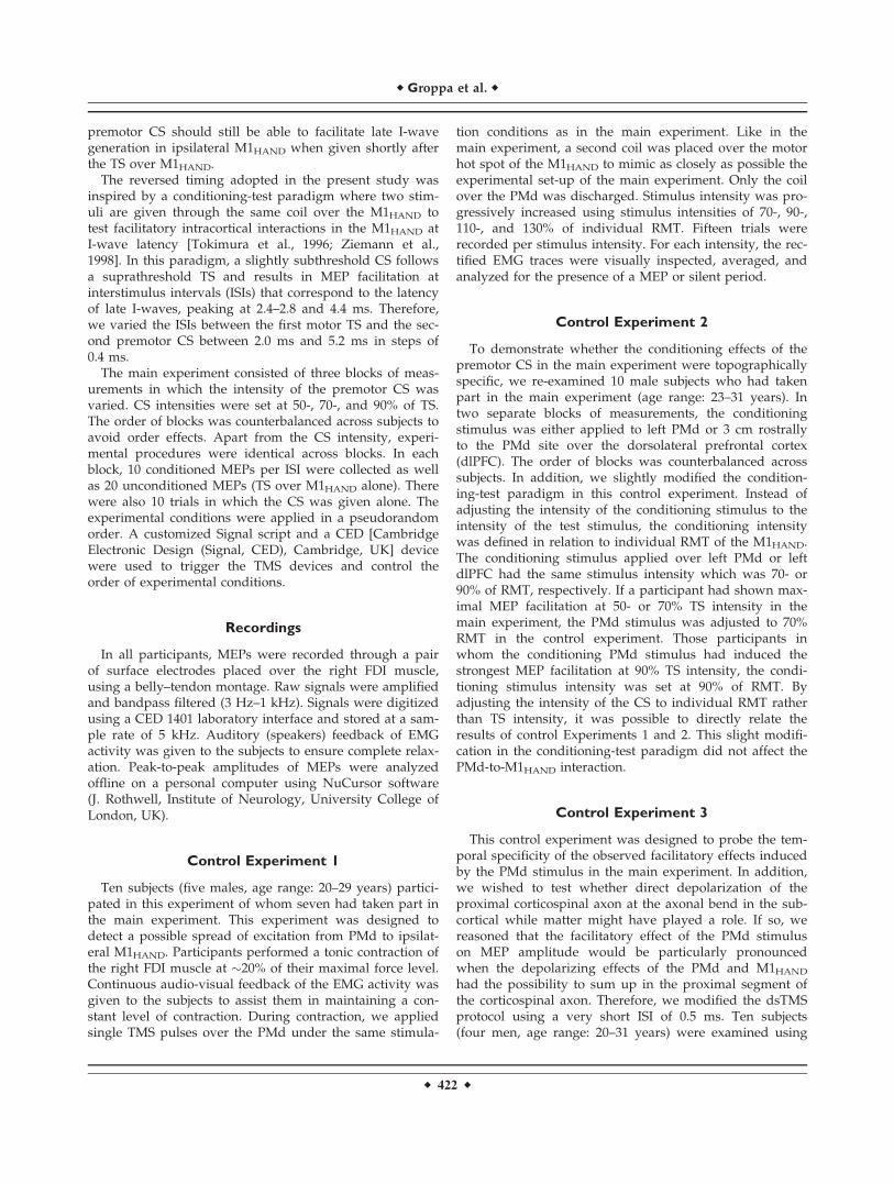
TMS of left M1HAND and PMd was performed with twocustom-made figure-of eight minicoils attached to a P-Stim160 stimulator (Mag&More, Munich, Germany). The geom-etry of the minicoils used a modified figure-of-eightdesign. The in-plane dimensions of the coil were 56 mm(x-axis) and 104 mm (y-axis). The handle of the coil wasmounted on the coil perpendicularly to the plane of thecoil (z-axis). The modified figure-of-eight coil containedtwo layers of wires with identical geometry which wereserially connected. Each layer consisted of two wings witheight windings per wing. The windings of each wing werearranged eccentrically having a drop-like geometry withan angular component. This geometry shifted maximalstimulation in the x-axis away from the geometric centerof the coil towards one edge of the coil (Fig. 1).
The first minicoil was centered over the M1HAND. TheM1HAND was functionally localized, placing the coil overthe site where a single suprathreshold pulse elicited amaximal MEP in the right first dorsal interosseous muscle(FDI). The coil was placed tangentially on the scalp withthe x-axis being approximately in parallel to the centralsulcus (45–60 degree relative to the mid-sagittal line). Thecoil was positioned over the M1HAND such that the site ofM1HAND stimulation was located in the anterior part ofthe coil.
The second coil targeting the adjacent left PMd wasplaced immediately anterior to and with the x-axis ori-ented in parallel to the coil targeting left M1HAND. To min-imize the distance between the primary motor and dorsalpremotor sites of stimulation, the anterior coil was placedover the PMd in a mirrored fashion with the site of maxi-mal stimulation being located in the posterior part of thecoil. The coils were aligned to each other so that the twohot spots were as close as possible. This allowed us toreduce the distance between the sites of effective stimula-tion to �3–4 cm. A detailed drawing of the coil ispresented in Figure 1C.
Conditioning-Test Design
We used dsTMS to probe cortico-cortical connectivitybetween left PMd and M1HAND. Using a conditioning-testapproach, the CS was given to left PMd and TS to leftM1HAND (Fig. 1). Single pulses with biphasic pulse config-uration were applied with the second phase of the pulseinducing a posterior–anterior (PA) current in the cortex.The pulse direction was changed in the PMd coil to stimu-late in the same current direction.
After functional localization of the M1HAND stimulationsite, we determined the active motor threshold (AMT) andresting motor threshold (RMT). The RMT was defined asthe stimulus intensity at which a single biphasic pulseelicited an MEP of at least 50 lV peak-to-peak amplitudein five out of ten trials [Rossini et al., 1994]. The TS wasused to adjust the intensity of the premotor TMS pulse.Since we had no specific predictions regarding the optimalintensity of the premotor CS, we used three differentintensities for premotor TMS (50-, 70-, and 90% of TS-Intensity). The TS was given at an intensity which evokedMEPs with peak-to-peak amplitudes of about 0.5 mV.
In contrast to previous studies, the premotor CS wasgiven after the TS to M1HAND. The inversion in temporalorder with the TS given over M1HAND preceding the CSover left PMd was motivated by the following considera-tions: a suprathreshold TMS pulse over the M1HAND
causes multiple descending volleys in the corticospinaltract with the latest volleys (i.e., late I-waves) leaving thecortex several milliseconds after the TMS pulse is applied.Assuming that the premotor-to-motor pathway is mono-synaptic, we reasoned that ipsilateral premotor-to-motorconduction time should be less than 2 ms. If so, the
Figure 1.
Experimental setup. A: Stimulation paradigm. Conditional stimu-
lus (S2) was applied after the test stimulus (S1) over M1HAND.
ISI (interstimulus interval) was randomly varied in one session.
Three sessions with different S2 (50-, 70-, 90%) intensities were
performed. B: View of the two mini-coils aligned to each other.
The stimulation current was switched in one coil and achieve
same current direction in both coils. C: Schematic drawing of
the decentral coil windings.
r Probing Ipsilateral Premotor-to-Motor Connectivity with Highly Focal TMS r
r 421 r
premotor CS should still be able to facilitate late I-wavegeneration in ipsilateral M1HAND when given shortly afterthe TS over M1HAND.
The reversed timing adopted in the present study wasinspired by a conditioning-test paradigm where two stim-uli are given through the same coil over the M1HAND totest facilitatory intracortical interactions in the M1HAND atI-wave latency [Tokimura et al., 1996; Ziemann et al.,1998]. In this paradigm, a slightly subthreshold CS followsa suprathreshold TS and results in MEP facilitation atinterstimulus intervals (ISIs) that correspond to the latencyof late I-waves, peaking at 2.4–2.8 and 4.4 ms. Therefore,we varied the ISIs between the first motor TS and the sec-ond premotor CS between 2.0 ms and 5.2 ms in steps of0.4 ms.
The main experiment consisted of three blocks of meas-urements in which the intensity of the premotor CS wasvaried. CS intensities were set at 50-, 70-, and 90% of TS.The order of blocks was counterbalanced across subjects toavoid order effects. Apart from the CS intensity, experi-mental procedures were identical across blocks. In eachblock, 10 conditioned MEPs per ISI were collected as wellas 20 unconditioned MEPs (TS over M1HAND alone). Therewere also 10 trials in which the CS was given alone. Theexperimental conditions were applied in a pseudorandomorder. A customized Signal script and a CED [CambridgeElectronic Design (Signal, CED), Cambridge, UK] devicewere used to trigger the TMS devices and control theorder of experimental conditions.
Recordings
In all participants, MEPs were recorded through a pairof surface electrodes placed over the right FDI muscle,using a belly–tendon montage. Raw signals were amplifiedand bandpass filtered (3 Hz–1 kHz). Signals were digitizedusing a CED 1401 laboratory interface and stored at a sam-ple rate of 5 kHz. Auditory (speakers) feedback of EMGactivity was given to the subjects to ensure complete relax-ation. Peak-to-peak amplitudes of MEPs were analyzedoffline on a personal computer using NuCursor software(J. Rothwell, Institute of Neurology, University College ofLondon, UK).
Control Experiment 1
Ten subjects (five males, age range: 20–29 years) partici-pated in this experiment of whom seven had taken part inthe main experiment. This experiment was designed todetect a possible spread of excitation from PMd to ipsilat-eral M1HAND. Participants performed a tonic contraction ofthe right FDI muscle at �20% of their maximal force level.Continuous audio-visual feedback of the EMG activity wasgiven to the subjects to assist them in maintaining a con-stant level of contraction. During contraction, we appliedsingle TMS pulses over the PMd under the same stimula-
tion conditions as in the main experiment. Like in themain experiment, a second coil was placed over the motorhot spot of the M1HAND to mimic as closely as possible theexperimental set-up of the main experiment. Only the coilover the PMd was discharged. Stimulus intensity was pro-gressively increased using stimulus intensities of 70-, 90-,110-, and 130% of individual RMT. Fifteen trials wererecorded per stimulus intensity. For each intensity, the rec-tified EMG traces were visually inspected, averaged, andanalyzed for the presence of a MEP or silent period.
Control Experiment 2
To demonstrate whether the conditioning effects of thepremotor CS in the main experiment were topographicallyspecific, we re-examined 10 male subjects who had takenpart in the main experiment (age range: 23–31 years). Intwo separate blocks of measurements, the conditioningstimulus was either applied to left PMd or 3 cm rostrallyto the PMd site over the dorsolateral prefrontal cortex(dlPFC). The order of blocks was counterbalanced acrosssubjects. In addition, we slightly modified the condition-ing-test paradigm in this control experiment. Instead ofadjusting the intensity of the conditioning stimulus to theintensity of the test stimulus, the conditioning intensitywas defined in relation to individual RMT of the M1HAND.The conditioning stimulus applied over left PMd or leftdlPFC had the same stimulus intensity which was 70- or90% of RMT, respectively. If a participant had shown max-imal MEP facilitation at 50- or 70% TS intensity in themain experiment, the PMd stimulus was adjusted to 70%RMT in the control experiment. Those participants inwhom the conditioning PMd stimulus had induced thestrongest MEP facilitation at 90% TS intensity, the condi-tioning stimulus intensity was set at 90% of RMT. Byadjusting the intensity of the CS to individual RMT ratherthan TS intensity, it was possible to directly relate theresults of control Experiments 1 and 2. This slight modifi-cation in the conditioning-test paradigm did not affect thePMd-to-M1HAND interaction.
Control Experiment 3
This control experiment was designed to probe the tem-poral specificity of the observed facilitatory effects inducedby the PMd stimulus in the main experiment. In addition,we wished to test whether direct depolarization of theproximal corticospinal axon at the axonal bend in the sub-cortical while matter might have played a role. If so, wereasoned that the facilitatory effect of the PMd stimuluson MEP amplitude would be particularly pronouncedwhen the depolarizing effects of the PMd and M1HAND
had the possibility to sum up in the proximal segment ofthe corticospinal axon. Therefore, we modified the dsTMSprotocol using a very short ISI of 0.5 ms. Ten subjects(four men, age range: 20–31 years) were examined using
r Groppa et al. r
r 422 r
the same experimental setup described in the main experi-ment. The conditioning stimulus was given to left PMdalready 0.5 ms after the TS over M1HAND. Fifteen condi-tioned and 15 unconditioned stimuli (M1HAND alone) wereapplied in pseudorandom order.
Control Experiment 4
This control experiment was designed to directly com-pare the facilitatory effects of dsTMS of left PMd andM1HAND and the standard paired-pulse TMS protocol thathas been introduced to probe facilitatory I-wave interac-tion in the M1HAND [Tokimura et al., 1996; Ziemann andRothwell, 2000].
Ten subjects participated in this experiment (five men,age range: 24–33 years, five subjects had participated inthe main experiment). In the first part of the experiment,short-latency intracortical facilitation was examined usinga figure-of eight coil (MC-B70) charged by a MagPro stim-ulator (MagVenture, Farum, Denmark). Three stimulusconditions were applied in pseudorandom order (10 repe-titions per condition). A biphasic TS was either givenalone or followed by a conditioning pulse at ISIs of 2.4 or2.8 ms. The intensity of the CS was set at 90% of the RMT,while the TS was adjusted to elicit a mean MEP amplitudeof 0.5 mV in the relaxed right FDI muscle when givenalone. In the second part of the experiment, we used theidentical dual-site TMS setup as in the main experiment.A TS was either given alone to left M1HAND or togetherwith a CS over left PMd at ISIs of 2.4- or 2.8 ms. PMdintensity was set here at 90% of the RMT. The order of thetwo experiments was counterbalanced across subjects.
Data Analysis
Data processing and analysis were performed with SPSSsoftware (Release 16.0, Copyright SPSS). We hypothesizedthat TMS over left PMd activates facilitatory PMd-to-M1HAND monosynaptic connections that project on intra-cortical circuits in M1HAND involved in the generation ofI-waves. Therefore, we expected that the premotor CSwould produce its facilitatory effect at an ISI of 2.4 or2.8 ms corresponding to the latency of the second I-wave[Ziemann et al., 1998].
We expected the optimal intensity at which the premotorCS produces premotor-to-motor facilitation to be uncoupledfrom the threshold to evoke MEPs over the M1HAND. There-fore, we examined in each subject at which CS intensity(i.e., 50-, 70-, and 90% of TS) the premotor CS producedmaximal premotor-to-motor facilitation at an ISI of 2.4 or2.8 ms. The individual paired-pulse excitability curve show-ing maximal paired-pulse facilitation was selected for groupanalysis and paired-pulse MEPs were normalized to themean MEP amplitude elicited by the TS over left M1HAND.
We further predicted that the ISI producing maximalpremotor-to-motor facilitation would slightly vary in time
from subject to subject with some subjects showing peakfacilitation at an ISI of 2.4 ms and others at 2.8 ms. There-fore, temporal realignment was performed to improvecomparability across subjects and to better reveal I-waveperiodicity. At the group level, individual excitabilitycurves were aligned to the peak of MEP facilitation at theISI of interest (2.4 or 2.8 ms). Hence, the peak of maximalpaired-pulse facilitation was identical for all subjects afterrealignment. The normalized and realigned paired-pulseexcitability curves were analyzed using repeated-measuresanalysis of variance (ANOVA) with the within-subjectfactor ISI (10 levels: 2.0, 2.4, 2.8, 3.2, 3.6, 4.0, 4.4, 4.8, 5.2,and 5.6 ms). Conditional on a significant F value, weperformed Fisher’s LSD post hoc pair-wise comparisons(P < 0.05; two-tailed; no adjustment for multiple compari-sons) comparing mean MEP amplitudes of consecutiveISIs. The same statistical approach was applied for controlExperiment 2.
In control Experiment 3, the one-way ANOVA includedthe main factor stimulation (M1HAND-PMd, unconditioned)to asses the excitability changes. The data acquired in con-trol Experiment 4 we analyzed using a two-way ANOVAwith the within-subject factors stimulation (2 levels: F8-Coil, PMd-M1) and states (3 levels: Test, ISI of 2.4, ISI of2.8 ms). We performed simple linear regression analysis totest whether the relative magnitude of MEP facilitationachieved with standard paired-pulse TMS of M1HAND
predicts the relative MEP facilitation induced by dsTMS ofM1HAND and PMd.
RESULTS
Main Experiment
Mean AMT and RMT were 53.15% � 10.89% and65.07% � 9.78% of maximum stimulator output, respec-tively. The amplitude of unconditioned MEP was matchedamong the three sessions in which premotor CS was givenat 50% (0.58 � 0.32 mV), 70% (0.49 � 0.22 mV), or 90% TS(0.51 � 0.26 mV). The premotor CS never elicited MEPswhen given alone.
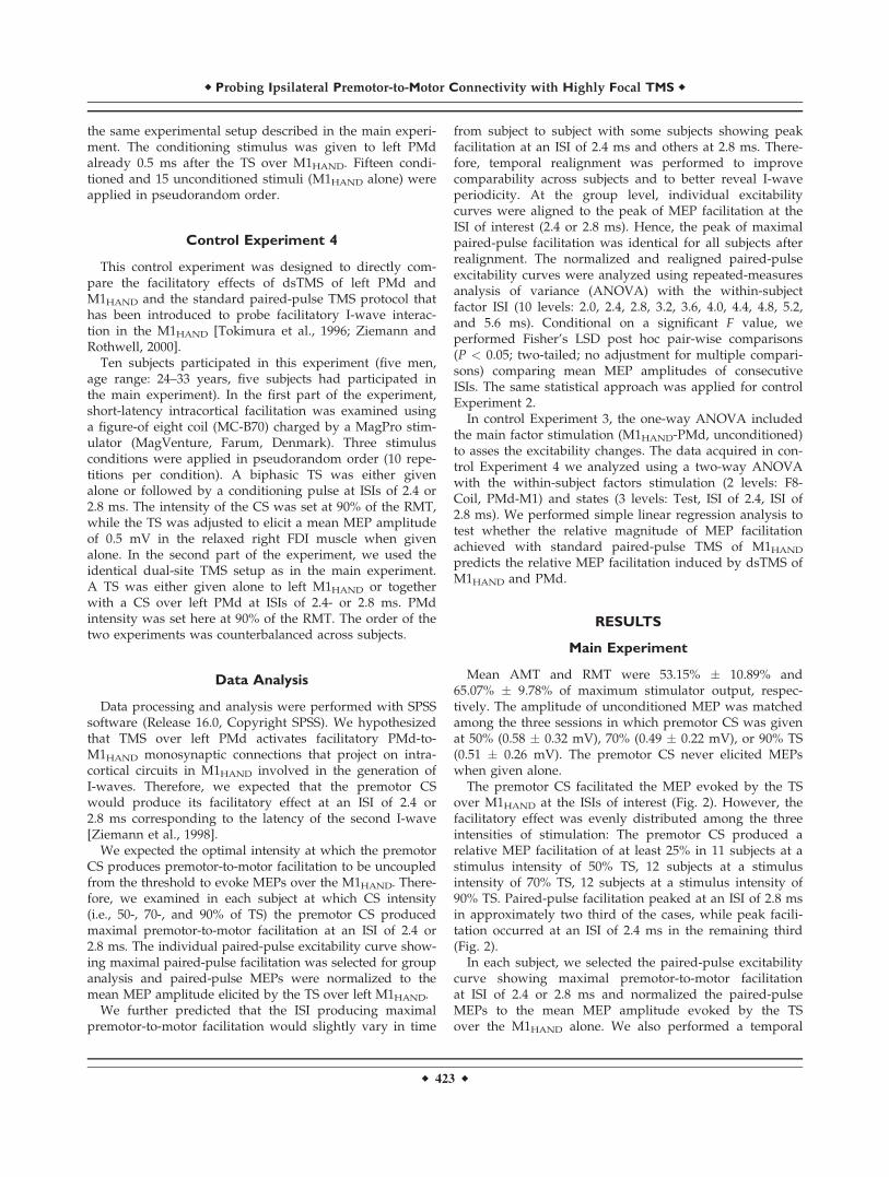
The premotor CS facilitated the MEP evoked by the TSover M1HAND at the ISIs of interest (Fig. 2). However, thefacilitatory effect was evenly distributed among the threeintensities of stimulation: The premotor CS produced arelative MEP facilitation of at least 25% in 11 subjects at astimulus intensity of 50% TS, 12 subjects at a stimulusintensity of 70% TS, 12 subjects at a stimulus intensity of90% TS. Paired-pulse facilitation peaked at an ISI of 2.8 msin approximately two third of the cases, while peak facili-tation occurred at an ISI of 2.4 ms in the remaining third(Fig. 2).
In each subject, we selected the paired-pulse excitabilitycurve showing maximal premotor-to-motor facilitationat ISI of 2.4 or 2.8 ms and normalized the paired-pulseMEPs to the mean MEP amplitude evoked by the TSover the M1HAND alone. We also performed a temporal
r Probing Ipsilateral Premotor-to-Motor Connectivity with Highly Focal TMS r
r 423 r
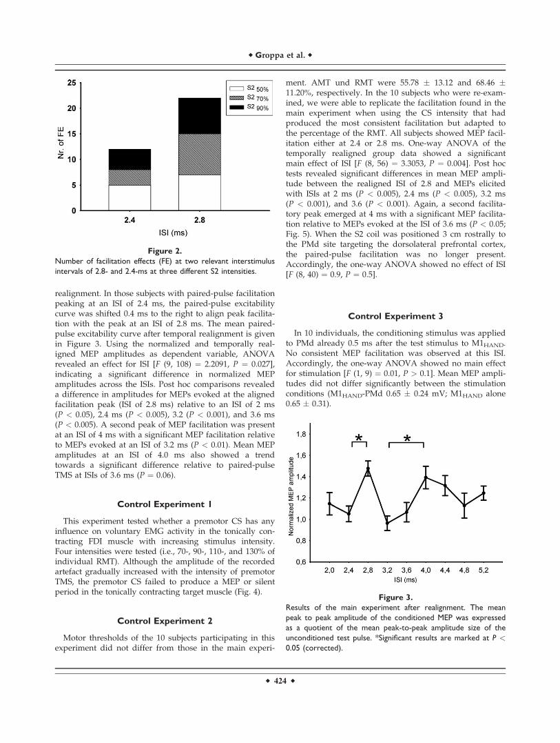
realignment. In those subjects with paired-pulse facilitationpeaking at an ISI of 2.4 ms, the paired-pulse excitabilitycurve was shifted 0.4 ms to the right to align peak facilita-tion with the peak at an ISI of 2.8 ms. The mean paired-pulse excitability curve after temporal realignment is givenin Figure 3. Using the normalized and temporally real-igned MEP amplitudes as dependent variable, ANOVArevealed an effect for ISI [F (9, 108) ¼ 2.2091, P ¼ 0.027],indicating a significant difference in normalized MEPamplitudes across the ISIs. Post hoc comparisons revealeda difference in amplitudes for MEPs evoked at the alignedfacilitation peak (ISI of 2.8 ms) relative to an ISI of 2 ms(P < 0.05), 2.4 ms (P < 0.005), 3.2 (P < 0.001), and 3.6 ms(P < 0.005). A second peak of MEP facilitation was presentat an ISI of 4 ms with a significant MEP facilitation relativeto MEPs evoked at an ISI of 3.2 ms (P < 0.01). Mean MEPamplitudes at an ISI of 4.0 ms also showed a trendtowards a significant difference relative to paired-pulseTMS at ISIs of 3.6 ms (P ¼ 0.06).
Control Experiment 1
This experiment tested whether a premotor CS has anyinfluence on voluntary EMG activity in the tonically con-tracting FDI muscle with increasing stimulus intensity.Four intensities were tested (i.e., 70-, 90-, 110-, and 130% ofindividual RMT). Although the amplitude of the recordedartefact gradually increased with the intensity of premotorTMS, the premotor CS failed to produce a MEP or silentperiod in the tonically contracting target muscle (Fig. 4).
Control Experiment 2
Motor thresholds of the 10 subjects participating in thisexperiment did not differ from those in the main experi-
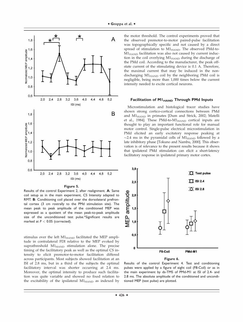
ment. AMT und RMT were 55.78 � 13.12 and 68.46 �11.20%, respectively. In the 10 subjects who were re-exam-ined, we were able to replicate the facilitation found in themain experiment when using the CS intensity that hadproduced the most consistent facilitation but adapted tothe percentage of the RMT. All subjects showed MEP facil-itation either at 2.4 or 2.8 ms. One-way ANOVA of thetemporally realigned group data showed a significantmain effect of ISI [F (8, 56) ¼ 3.3053, P ¼ 0.004]. Post hoctests revealed significant differences in mean MEP ampli-tude between the realigned ISI of 2.8 and MEPs elicitedwith ISIs at 2 ms (P < 0.005), 2.4 ms (P < 0.005), 3.2 ms(P < 0.001), and 3.6 (P < 0.001). Again, a second facilita-tory peak emerged at 4 ms with a significant MEP facilita-tion relative to MEPs evoked at the ISI of 3.6 ms (P < 0.05;Fig. 5). When the S2 coil was positioned 3 cm rostrally tothe PMd site targeting the dorsolateral prefrontal cortex,the paired-pulse facilitation was no longer present.Accordingly, the one-way ANOVA showed no effect of ISI[F (8, 40) ¼ 0.9, P ¼ 0.5].
Control Experiment 3
In 10 individuals, the conditioning stimulus was appliedto PMd already 0.5 ms after the test stimulus to M1HAND.No consistent MEP facilitation was observed at this ISI.Accordingly, the one-way ANOVA showed no main effectfor stimulation [F (1, 9) ¼ 0.01, P > 0.1]. Mean MEP ampli-tudes did not differ significantly between the stimulationconditions (M1HAND-PMd 0.65 � 0.24 mV; M1HAND alone0.65 � 0.31).
Figure 2.
Number of facilitation effects (FE) at two relevant interstimulus
intervals of 2.8- and 2.4-ms at three different S2 intensities.
Figure 3.
Results of the main experiment after realignment. The mean
peak to peak amplitude of the conditioned MEP was expressed
as a quotient of the mean peak-to-peak amplitude size of the
unconditioned test pulse. *Significant results are marked at P <0.05 (corrected).
r Groppa et al. r
r 424 r

Control Experiment 4
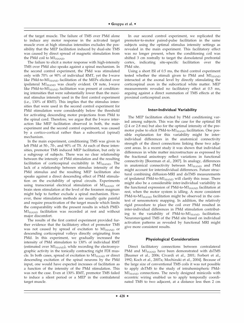
Figure 6 illustrates the results of control Experiment 4which includes the data of 10 subjects. A two-factorialrepeated-measures ANOVA revealed a statistical trend forthe factor stimulation paradigm [F (1, 9) ¼ 4.67, P < 0.1],and a significant main effect for the factor stimulus condi-tion [F (2, 18) ¼ 8.72, P < 0.005]. The interaction betweenthe two factors was not significant [F (1, 9) ¼ 4.67, P >0.05]. Accordingly, there were no differences in the uncon-ditioned MEPs evoked by single-pulse TMS over M1HAND
in both stimulation paradigms (M1HAND-PMd dsTMS:0.76 � 0.33 mV; Paired-pulse TMS of M1HAND: 0.78 � 0.38mV). Overall, the conditioning pulses given 2.4 or 2.8 msafter the TS induced MEP facilitation in both paradigms,but paired-pulse TMS of M1HAND induced a more pro-
nounced MEP facilitation compared to dsTMS of M1HAND
and PMd (Fig. 6). For both ISIs, simple regression analysisshowed that the relative magnitude of MEP facilitationachieved with paired-pulse TMS of M1HAND did not pre-dict the relative MEP facilitation induced by dsTMS ofM1HAND and PMd (for the ISI of 2.4 ms: r ¼ 0.40, P > 0.1;for ISI of 2.8 ms: r ¼ 0.43, P > 0.1).
DISCUSSION
Here, we used a novel dsTMS approach with specificallydesigned minicoils to probe non-invasively the ipsilateralcortico-cortical connectivity between the left PMd andM1HAND in healthy human subjects. A subthreshold pulsegiven to left PMd 2.4 or 2.8 ms after a suprathreshold
Figure 4.
Control Experiment 1. No silent period activity or MEPs were monitored at increased stimula-
tion intensities at PMd site in the periphery tonically activated muscles.
r Probing Ipsilateral Premotor-to-Motor Connectivity with Highly Focal TMS r
r 425 r
stimulus over the left M1HAND facilitated the MEP ampli-tude in contralateral FDI relative to the MEP evoked bysuprathreshold M1HAND stimulation alone. The precisetiming of the facilitatory peak as well as the optimal CS in-tensity to elicit premotor-to-motor facilitation differedacross participants. Most subjects showed facilitation at anISI of 2.8 ms, but in a third of the subjects the optimalfacilitatory interval was shorter occurring at 2.4 ms.Moreover, the optimal intensity to produce such facilita-tion was quite variable and showed no fixed relation tothe excitability of the ipsilateral M1HAND as indexed by
the motor threshold. The control experiments proved thatthe observed premotor-to-motor paired-pulse facilitationwas topographically specific and not caused by a directspread of stimulation to M1HAND. The observed PMd-to-M1HAND facilitation was also not caused by current induc-tion in the coil overlying M1HAND during the discharge ofthe PMd coil. According to the manufacturer, the peak off-state current of the stimulating device is 0.1 A. Therefore,the maximal current that may be induced in the non-discharging M1HAND coil by the neighboring PMd coil isnegligible, being more than 1,000 times below the currentintensity needed to excite cortical neurons.
Facilitation of M1HAND Through PMd Inputs
Microstimulation and histological tracer studies haveshown strong cortico-cortical connections between PMdand M1HAND in primates [Dum and Strick, 2002; Matelliet al., 1984]. These PMd-to-M1HAND cortical inputs arethought to play an important functional role for manualmotor control. Single-pulse electrical microstimulation inPMd elicited an early excitatory response peaking at�2.4 ms in the pyramidal cells of M1HAND followed by alate inhibitory phase [Tokuno and Nambu, 2000]. This obser-vation is of relevance to the present results because it showsthat ipsilateral PMd stimulation can elicit a short-latencyfacilitatory response in ipsilateral primary motor cortex.
Figure 5.
Results of the control Experiment 2, after realignment. A: Same
coil setup as in the main experiment, CS Intensity adapted to
RMT. B: Conditioning coil placed over the dorsolateral prefron-
tal cortex (3 cm rostrally to the PMd stimulation site). The
mean peak to peak amplitude of the conditioned MEP was
expressed as a quotient of the mean peak-to-peak amplitude
size of the unconditioned test pulse.*Significant results are
marked at P < 0.05 (corrected).
Figure 6.
Results of the control Experiment 4. Test and conditioning
pulses were applied by a figure of eight coil (F8-Coil) or as in
the main experiment by ds-TMS of PMd-M1 at ISI of 2.4- and
2.8 ms. The absolute amplitude of the conditioned and uncondi-
tioned MEP (test pulse) are plotted.
r Groppa et al. r
r 426 r

Little is known about the functional connections of thepremotor motor areas in humans. Double pulse TMS para-digms have probed interactions between different corticalareas [Baumer et al., 2006, 2009; Civardi et al., 2001; Kochet al., 2006, 2007a]. Using two small figure-of-eight coils, atranscranial stimulus given 5 cm anterior to the M1HAND
hot-spot had an inhibitory effect on corticospinal excitabil-ity of ipsilateral M1HAND when a subthreshold condition-ing stimulus was given 6 ms before a transcranial teststimulus [Civardi et al., 2001]. This inhibitory conditioningeffect gradually turned from inhibition into facilitationwhen the intensity of the conditioning stimulus was grad-ually increased from 90- to 120% of active motor threshold[Civardi et al., 2001]. Interestingly, the facilitatory effect ofa suprathreshold conditioning stimulus on ipsilateral corti-cospinal excitability was suppressed by an additionalsubthreshold conditioning stimulus given 5 ms before theconditioning stimulus [Koch et al., 2007b].
So far, the large size of standard TMS coils has limitedbifocal TMS of the ipsilateral PMd and M1HAND. Althoughthe studies by Civardi et al. [2001] and Koch et al. [2007b]used dsTMS to probe intrahemispheric connectivitybetween rostral frontal areas and M1HAND, the condition-ing pulse was applied rostrally from the PMd in thedorsolateral prefrontal cortex [Baumer et al., 2009]. Thismay explain why the temporal pattern of cortico-corticalinteraction in the studies by Civardi et al. [2001] and Kochet al. [2007b] clearly differed from the pattern revealed bycortical microstimulation in primates [Tokuno and Nambu,2000]. Since the dorsolateral prefrontal cortex has no directcortico-cortical connections with M1HAND [Dum andStrick, 2005], we hypothesize that the inhibitory M1HAND
response 6 ms after prefrontal conditioning represents‘‘late inhibition’’ produced by polysynaptic (e.g., prefron-tal-premotor-motor) projections [Civardi et al., 2001]. Insummary, a direct comparison between the present studyand the study by Civardi et al. [2001] is difficult becauseof the substantial differences with respect to the temporalsequence of the applied pulses over PMd and M1HAND
and the positioning of the conditioning coil over PMd inthe present study and prefrontal cortex in the study byCivardi et al.
The increase of the conditioning pulse intensity changedthe inhibition in a slight facilitation, by possibly involvingadjacent cortical areas. And indeed another recent workpresented a similar facilitation of the M1 after a supra-threshold PMd pulse at 6 ms [Koch et al., 2007b]. Differentstimulation current directions in the conditioning- and testcoil and a CS-intensity might account for this differentpattern of PMd-M1 interaction. Furthermore, due to spaceconsiderations the exact stimulation site remains notcompletely defined.
The results of our study support the long held view thatdirect fast connections from PMd to the ipsilateralM1HAND are involved in the generation of I-waves[Ziemann and Rothwell, 2000]. Indeed, surface stimulationof the dorsal premotor area in primates elicited repetitive
descending I-waves that were abolished after ablation ofthe ipsilateral primary motor cortex [Patton and Amassian,1954]. When setting out to probe this premotor-to-motorroute, we modified the paired-pulse TMS paradigm origi-nally developed for probing facilitatory I-wave interactionsin the premotor motor cortex at short ISI [Tokimura et al.,1996; Ziemann et al., 1998]. In that paradigm, two transcra-nial stimuli are applied through the same coil centeredover the motor hot spot to target circuits in the M1HAND
that generate this short-latency intracortical facilitation.While analyzing the same range of ISIs, we used two mag-netic coils rather than a single coil in the present studyand applied the first suprathreshold pulse to M1HAND andthe subsequent low-intensity pulse over PMd. Using thisdsTMS paradigm, the low-intensity premotor stimulusconditioned the corticomotor response evoked by thesuprathreshold stimulus in ipsilateral M1HAND. Premotor-to-motor facilitation occurred when the conditioning stim-ulus given to the PMd 2.4 or 2.8 ms after TMS of theM1HAND. This implies that premotor-to-motor facilitationoccurred via a short latency pathway that reached ipsilat-eral M1HAND quickly enough to interact with the intracort-ical excitation evoked by direct TMS of the M1HAND. Ourresults suggest an interaction between the PMd input toM1HAND and some intracortical circuits in the M1HAND
involved in I-wave generation. Yet we can not make anyinferences about the true latency of the premotor-to-motorinteraction or comment on which I-wave circuits were spe-cifically facilitated by the premotor input. Here invasiverecordings of the induced corticospinal descending volleysat the level of the spinal cord might help to tackle thisquestion [Di Lazzaro et al., 2010].
Temporal realignment of the paired-pulse excitabilitycurves across subjects revealed a distinct ISI-specific peakof MEP facilitation with no consistent facilitation at the ISIbefore or after the facilitatory peak. However, a secondfacilitatory peak emerged at an ISI of 4.0 ms suggesting anundulatory pattern of premotor-to-motor facilitation. Onthe basis of the timing and undulatory character of theobserved premotor-to-motor MEP facilitation, it is conceiv-able that the conditioning premotor stimulus activated ashort latencyPMd-to-M1HAND cortico-cortical pathwaywhich interacted with intracortical circuits in the M1HAND
implicated in the generation of corticospinal descendingI-waves.
Site of Facilitatory Interaction
In our first control experiment, the PMd stimulus alonefailed to induce a MEP or silent period in the pre-activatedcontralateral FDI muscle, even when stimulus intensitywas increased to 130% of RMT of the left M1HAND. Inother words, a premotor stimulus intensity of 130% RMTwas still subthreshold for producing significant descend-ing corticospinal excitation even though the corticospinalsystem was more excitable due to voluntary preactivation
r Probing Ipsilateral Premotor-to-Motor Connectivity with Highly Focal TMS r
r 427 r
of the target muscle. The failure of TMS over PMd aloneto induce any motor response in the activated targetmuscle even at high stimulus intensities excludes the pos-sibility that the MEP facilitation induced by dual-site TMSwas caused by direct spread of magnetic stimulation fromthe PMd coil to M1HAND.
The failure to elicit a motor response with high-intensityTMS over PMd also speaks against a spinal mechanism. Inthe second control experiment, the PMd was stimulatedonly with 70% or 90% of individual RMT, yet the I-wavelike PMd-to-M1HAND facilitation of the MEPs elicited overipsilateral M1HAND was clearly evident. Of note, I-wavelike PMd-to-M1HAND facilitation was present at condition-ing intensities that were substantially lower than the maxi-mal stimulus intensity used in the first control experiment(i.e., 130% of RMT). This implies that the stimulus inten-sities that were used in the second control experiment forPMd stimulation were considerably below the thresholdfor activating descending motor projections from PMd tothe spinal cord. Therefore, we argue that the I-wave inter-action like MEP facilitation observed in both, the mainexperiment and the second control experiment, was causedby a cortico-cortical rather than a subcortical (spinal)mechanism.
In the main experiment, conditioning TMS was given toleft PMd at 50-, 70-, and 90% of TS. At each of these inten-sities, premotor TMS induced MEP facilitation, but only ina subgroup of subjects. There was no clear relationshipbetween the intensity of PMd stimulation and the resultingfacilitation of corticospinal excitability in M1HAND. Thelack of a relationship between stimulus intensity of thePMd stimulus and the resulting MEP facilitation alsospeaks against a direct descending effect of PMd stimula-tion on the excitability of spinal circuits. Experimentsusing transcranial electrical stimulation of M1HAND orbrain stem stimulation at the level of the foramen magnummight help to further exclude a spinal mechanism. How-ever, these stimulation methods are usually quite painfuland require preactivation of the target muscle which limitsthe comparability with the present results in which PMD-M1HAND facilitation was recorded at rest and withoutmajor discomfort.
The results of the first control experiment provided fur-ther evidence that the facilitatory effect of premotor TMSwas not caused by spread of excitation to M1HAND ordescending corticospinal volleys directly originating fromPMd. In this experiment, we gradually increased theintensity of PMd stimulation to 130% of individual RMT(estimated over M1HAND), while recording the electromyo-graphic activity in the tonically contracting right FDI mus-cle. In both cases, spread of excitation to M1HAND or directdescending excitation of the spinal neurons by the PMdinput, one would have expected increases of facilitation asa function of the intensity of the PMd stimulation. Thiswas not the case. Even at 130% RMT, premotor TMS failedto induce a silent period or a MEP in the contralateraltarget muscle.
In our second control experiment, we replicated thepremotor-to-motor paired-pulse facilitation in the samesubjects using the optimal stimulus intensity settings asrevealed in the main experiment. This facilitatory effectwas no longer present, when the conditioning coil wasshifted 3 cm rostrally to target the dorsolateral prefrontalcortex, indicating site-specific facilitation over thePMd site.
Using a short ISI of 0.5 ms, the third control experimenttested whether the stimuli given to PMd and M1HAND
interacted at the axonal level by directly stimulating thecorticospinal axon in the subcortical white matter. MEPmeasurements revealed no facilitatory effect at 0.5 ms,arguing against a direct summation of TMS effects at theproximal corticospinal axon.
Inter-Individual Variability
The MEP facilitation elicited by PMd conditioning var-ied among subjects. This was the case for the optimal ISI(2.4 or 2.8 ms) but also for the optimal intensity of the pre-motor pulse to elicit PMd-to-M1HAND facilitation. One pos-sible explanation for this variability might be inter-individual differences in the anatomical connectivitystrength of the direct connections linking these two adja-cent areas. In a recent study it was shown that individualdifferences in white matter microstructure as expressed bythe fractional anisotropy reflect variations in functionalconnectivity [Boorman et al., 2007]. In analogy, differencesin anatomical connectivity between M1HAND and PMdmight account for interindividual differences. Future struc-tural combining diffusion-MRI and dsTMS measurementsof ipsilateral PMd-to-M1HAND will clarify this issue. Theremight also be a considerable inter-individual variability inthe functional expression of PMd-to-M1HAND facilitation atrest, when the motor system is idling. A more consistentPMd-to-M1HAND facilitation might be observed in the con-text of sensomotoric mapping. In addition, the relativelyrigid procedure to place the coil over PMd resulted ininter-individual differences in PMd stimulation contribut-ing to the variability of PMd-to-M1HAND facilitation.Neuronavigated TMS of the PMd site based on individualactivation patterns as revealed by functional MRI mightgive more consistent results.
Physiological Considerations
Direct facilitatory connections between contralateralPMd and M1HAND have been demonstrated with dsTMS[Baumer et al., 2006; Civardi et al., 2001; Ferbert et al.,1992; Koch et al., 2007a; Mochizuki et al., 2004]. Because ofthe large size of conventional TMS coils it was not possibleto apply dsTMS to the study of intrahemispheric PMd-M1HAND connections. The newly designed minicoils witheccentric wiring enabled us to apply temporally coordi-nated TMS to two adjacent, at a distance less then 2 cm
r Groppa et al. r
r 428 r

close cortical areas. This inspired us to modulate the activ-ity of the ipsilateral M1HAND and PMd and to analyze theshort latency facilitatory interaction between these motorareas. The latencies of the observed facilitatory peaks atISIs of 2.4 ms or 2.8 and 4.0 ms are in concordance withthe timing of the descending I-waves in epidural record-ings after TMS stimulation of the motor cortex [Burkeet al., 1993]. Studies which involved electrical cortical stim-ulation and data from epidural recordings as well as ani-mal studies confirmed the cortical origin of thesedescending volleys [Di Lazzaro et al., 1999a; Patton andAmassian, 1954; Tokimura et al., 1996]. The excitability ofthe facilitatory circuits involved in I-wave generation havebeen studied in the intact human brain with paired-pulseTMS using one figure-of-eight coil [Tokimura et al., 1996;Ziemann et al., 1998]. The exact physiological substrate forthis short-latency cortical facilitation at I-wave latencies isnot fully clarified. Although the same coil is used to probefacilitatory I-wave interactions in M1HAND, this does notnecessarily imply that both pulses excite the same interac-tion by stimulating two segregated but functionally inter-acting neural populations. It might be possible thatI-waves are generated by excitation of different chains ofinterneurons in M1HAND that receive cortico-cortical inputsfrom connected premotor or sensory cortical areas [Sakaiet al., 1997]. Our results are compatible with this view andsuggest that premotor inputs into the M1HAND interactwith the intracortical circuits in M1HAND that generatedescending I-waves in the corticospinal tract.
At present, it is not possible to directly address thequestion whether the pathway mediating the PMd-to-M1HAND interactions projects on the same I-wave circuitsthat are tested with standard paired pulse protocols inM1HAND. The currently available TMS hardware used toprobe PMD-to-M1 interactions and standard I-wave inter-actions in M1 differ substantially. Therefore, differentneural populations are probed in the stimulated M1 withthe two protocols. This notion is supported by the controlexperiment in which we compared the facilitatory effectsinduced by dsTMS of left PMd and M1HAND and thestandard paired-pulse TMS protocol applied over theM1HAND [Tokimura et al., 1996; Ziemann and Rothwell,2000]. Paired-pulse TMS of M1HAND with a large figure-of-eight coil induced a more pronounced MEP facilitationrelative to dsTMS of M1HAND and PMd with highly focalminicoils (Fig. 6). In addition, the inter-individual varia-tions in I-wave facilitation induced by paired-pulse TMSof M1HAND did not predict the relative MEP facilitationinduced by dsTMS of M1HAND and PMd. How much thesedifferences reflect true neurobiological differences is hardto tell given the marked differences in the TMS hardware.We hypothesize that in both paradigms, the facilitation ofcorticospinal excitability relies on intracortical neural cir-cuits in the M1HAND, but that the paradigms probe theexcitability of different neural circuits in the M1HAND.Future studies have to tackle this question, once both para-digms can be performed with the same TMS hardware.
Relevance of the Study
Premotor cortical areas and especially PMd play an im-portant role in the preparation and sensory guidance ofhand movements [Picard and Strick, 2001; Schluter et al.,1998]. Direct connections from premotor areas to M1HAND
have been demonstrated in animal studies and especially inprimates, yet little is know about these pathways inhumans. Our study demonstrates, for the first time, with theaid of highly focal TMS a direct short latency facilitatorypremotor to motor route. The observed temporal pattern ofthe interaction resembles classical I-wave interaction thatcan be demonstrated with paired-pulse stimulation over theM1HAND. This route was highly specific for the stimulatedregion of PMd and no interaction occurred if the coil wasmoved further rostrally. As PMd plays an important role forimplementing sensory or visual information for movementpreparation and fine motor tuning [Petrides, 1985] it islikely that this short latency route might play a crucial rolein context-dependent modulation of M1HAND excitabilityvia the PMd during manual motor control.
REFERENCES
Amassian VE, Stewart M, Quirk GJ, Rosenthal JL (1987): Physio-logical basis of motor effects of a transient stimulus to cerebralcortex. Neurosurgery 20:74–93.
Barker AT, Jalinous R, Freeston IL (1985): Non-invasive magneticstimulation of human motor cortex. Lancet 1:1106–1107.
Baumer T, Bock F, Koch G, Lange R, Rothwell JC, Siebner HR,Munchau A (2006): Magnetic stimulation of human premotoror motor cortex produces interhemispheric facilitation throughdistinct pathways. J Physiol 572 (Part 3):857–868.
Baumer T, Schippling S, Kroeger J, Zittel S, Koch G, Thomalla G,Rothwell JC, Siebner HR, Orth M, Munchau A (2009): Inhibi-tory and facilitatory connectivity from ventral premotor to pri-mary motor cortex in healthy humans at rest: A bifocal TMSstudy. Clin Neurophysiol 120:1724–1731.
Boorman ED, O’Shea J, Sebastian C, Rushworth MFS, Johansen-Berg H (2007): Individual differences in white-matter micro-structure reflect variation in functional connectivity duringchoice. Curr Biol 17:1426–1431.
Burke D, Hicks R, Gandevia SC, Stephen J, Woodforth I, CrawfordM (1993): Direct comparison of corticospinal volleys in humansubjects to transcranial magnetic and electrical stimulation.J Physiol 470:383–393.
Cerri G, Shimazu H, Maier MA, Lemon RN (2003): Facilitationfrom ventral premotor cortex of primary motor cortex outputsto macaque hand muscles. J Neurophysiol 90:832–842.
Civardi C, Cantello R, Asselman P, Rothwell JC (2001): Transcra-nial magnetic stimulation can be used to test connectionsto primary motor areas from frontal and medial cortex inhumans. Neuroimage 14:1444–1453.
Davare M, Lemon RN, Olivier E (2008): Selective modulations ofinteractions between ventral premotor cortex and primarymotor cortex during precision grasping in humans. J Physiol586:2735–2742.
Di Lazzaro V, Oliviero A, Profice P, Insola A, Mazzone P, Tonali P,Rothwell JC (1999a): Direct recordings of descending volleysafter transcranial magnetic and electric motor cortex stimulation
r Probing Ipsilateral Premotor-to-Motor Connectivity with Highly Focal TMS r
r 429 r

in conscious humans. Electroencephalogr Clin NeurophysiolSuppl 51:120–126.
Di Lazzaro V, Rothwell JC, Oliviero A, Profice P, Insola A, MazzoneP, Tonali P (1999b): Intracortical origin of the short latency facili-tation produced by pairs of threshold magnetic stimuli appliedto human motor cortex. Exp Brain Res 129:494–499.
Di Lazzaro V, Profice P, Pilato F, Dileone M, Oliviero A, ZiemannU (2010): The effects of motor cortex rTMS on corticospinaldescending activity. Clin Neurophysiol 121:464–473.
Dum RP, Strick PL (1991): The origin of corticospinal projectionsfrom the premotor areas in the frontal lobe. J Neurosci 11:667–689.
Dum RP, Strick PL (2002): Motor areas in the frontal lobe of theprimate. Physiol Behav 77:677–682.
Dum RP, Strick PL (2005): Frontal lobe inputs to the digit repre-sentations of the motor areas on the lateral surface of thehemisphere. J Neurosci 25:1375–1386.
Ferbert A, Priori A, Rothwell JC, Day BL, Colebatch JG, MarsdenCD (1992): Interhemispheric inhibition of the human motorcortex. J Physiol 453:525–546.
Hallett M (2000): Transcranial magnetic stimulation and thehuman brain. Nature 406:147–150.
He SQ, Dum RP, Strick PL (1993): Topographic organization ofcorticospinal projections from the frontal lobe: Motor areas onthe lateral surface of the hemisphere. J Neurosci 13:952–980.
He SQ, Dum RP, Strick PL (1995): Topographic organization of cortico-spinal projections from the frontal lobe: Motor areas on the medialsurface of the hemisphere. J Neurosci 15 (5 Part 1):3284–3306.
Kermadi I, Liu Y, Tempini A, Calciati E, Rouiller EM (1998):Neuronal activity in the primate supplementary motor areaand the primary motor cortex in relation to spatio-temporalbimanual coordination. Somatosens Motor Res 15:287–308.
Koch G, Franca M, Del Olmo MF, Cheeran B, Milton R, AlvarezSauco M, Rothwell JC (2006): Time course of functionalconnectivity between dorsal premotor and contralateral motorcortex during movement selection. J Neurosci 26:7452–7459.
Koch G, Fernandez Del Olmo M, Cheeran B, Ruge D, SchipplingS, Caltagirone C, Rothwell JC (2007a): Focal stimulation of theposterior parietal cortex increases the excitability of the ipsilat-eral motor cortex. J Neurosci 27:6815–6822.
Koch G, Franca M, Mochizuki H, Marconi B, Caltagirone C, RothwellJC (2007b): Interactions between pairs of transcranial magneticstimuli over the human left dorsal premotor cortex differ fromthose seen in primary motor cortex. J Physiology 578:551–562.
Lemon RN (2008): Descending pathways in motor control. AnnuRev Neurosci 31:195–218.
Luppino G, Matelli M, Camarda RM, Rizzolatti G (1993):Corticocortical connections of area F3 (SMA-proper) and areaF6 (pre-SMA) in the macaque monkey. J Compar Neurol338:114–140.
Martino AM, Strick PL (1987): Corticospinal projections originatefrom the arcuate premotor area. Brain Res 404:307–312.
Matelli M, Camarda R, Glickstein M, Rizzolatti G (1984): Intercon-nections within the postarcuate cortex (area 6) of the macaquemonkey. Brain Res 310:388–392.
Mochizuki H, Huang YZ, Rothwell JC (2004): Interhemisphericinteraction between human dorsal premotor and contralateralprimary motor cortex. J Physiol 561 (Part 1):331–338.
O’Shea J, Sebastian C, Boorman ED, Johansen-Berg H, RushworthMF (2007): Functional specificity of human premotor-motorcortical interactions during action selection. Eur J Neurosci26:2085–2095.
Patton HD, Amassian VE (1954): Single and multiple-unit analysisof cortical stage of pyramidal tract activation. J Neurophysiol17:345–363.
Petrides M (1985): Deficits on conditional associative-learningtasks after frontal- and temporal-lobe lesions in man. Neuro-psychologia 23:601–614.
Picard N, Strick PL (2001): Imaging the premotor areas. CurrOpin Neurobiol 11:663–672.
Prabhu G, Shimazu H, Cerri G, Brochier T, Spinks RL, Maier MA,Lemon RN (2009): Modulation of primary motor cortex outputsfrom ventral premotor cortex during visually-guided grasp inthe macaque monkey. J Physiology 587:1057–1069.
Rossini PM, Barker AT, Berardelli A, Caramia MD, Caruso G,Cracco RQ, Dimitrijevic MR, Hallett M, Katayama Y,Lucking CH (1994): Non-invasive electrical and magneticstimulation of the brain, spinal cord and roots: Basic princi-ples and procedures for routine clinical application. Reportof an IFCN committee. Electroencephalogr Clin Neurophy-siol 91:79–92.
Sakai K, Ugawa Y, Terao Y, Hanajima R, Furubayashi T,Kanazawa I (1997): Preferential activation of different I wavesby transcranial magnetic stimulation with a figure-of-eight-shaped coil. Exp Brain Res 113:24–32.
Schluter ND, Rushworth MF, Passingham RE, Mills KR (1998): Tem-porary interference in human lateral premotor cortex suggestsdominance for the selection of movements. A study using trans-cranial magnetic stimulation. Brain 121 (Part 5):785–799.
Schmidlin E, Brochier T, Maier MA, Kirkwood PA, Lemon RN(2008): Pronounced reduction of digit motor responses evokedfrom macaque ventral premotor cortex after reversible inacti-vation of the primary motor cortex hand area. J Neurosci28:5772–5783.
Shimazu H, Maier MA, Cerri G, Kirkwood PA, Lemon RN (2004):Macaque ventral premotor cortex exerts powerful facilitationof motor cortex outputs to upper limb motoneurons. J Neuro-sci 24:1200–1211.
Tokimura H, Ridding MC, Tokimura Y, Amassian VE, RothwellJC (1996): Short latency facilitation between pairs of thresholdmagnetic stimuli applied to human motor cortex. Electroence-phalogr Clin Neurophysiol 101:263–272.
Tokuno H, Nambu A (2000): Organization of nonprimary motorcortical inputs on pyramidal and nonpyramidal tract neuronsof primary motor cortex: An electrophysiological study in themacaque monkey. Cereb Cortex 10:58–68.
Yousry TA, Schmid UD, Alkadhi H, Schmidt D, Peraud A, BuettnerA, Winkler P (1997): Localization of the motor hand area to aknob on the precentral gyrus. A new landmark. Brain 120:141–157.
Ziemann U, Rothwell JC (2000): I-waves in motor cortex. J ClinNeurophysiol 17:397–405.
Ziemann U, Tergau F, Wassermann EM, Wischer S, Hildebrandt J,Paulus W (1998): Demonstration of facilitatory I wave interac-tion in the human motor cortex by paired transcranial magneticstimulation. J Physiol 511 (Part 1): 181–190.
r Groppa et al. r
r 430 r
Related Documents