The HIV-1 Envelope Transmembrane Domain Binds TLR2 through a Distinct Dimerization Motif and Inhibits TLR2- Mediated Responses Eliran Moshe Reuven 1. , Mohammad Ali 1. , Etai Rotem 1 , Roland Schwarzter 2 , Andrea Gramatica 2 , Anthony H. Futerman 1 , Yechiel Shai 1 * 1 Department of Biological Chemistry, The Weizmann Institute of Science, Rehovot, Israel, 2 Institut fu ¨ r Biologie, AG Molekulare Biophysik, Humboldt-Universita ¨t zu Berlin, Berlin, Germany Abstract HIV-1 uses a number of means to manipulate the immune system, to avoid recognition and to highjack signaling pathways. HIV-1 infected cells show limited Toll-Like Receptor (TLR) responsiveness via as yet unknown mechanisms. Using biochemical and biophysical approaches, we demonstrate that the trans-membrane domain (TMD) of the HIV-1 envelope (ENV) directly interacts with TLR2 TMD within the membrane milieu. This interaction attenuates TNFa, IL-6 and MCP-1 secretion in macrophages, induced by natural ligands of TLR2 both in in vitro and in vivo models. This was associated with decreased levels of ERK phosphorylation. Furthermore, mutagenesis demonstrated the importance of a conserved GxxxG motif in driving this interaction within the membrane milieu. The administration of the ENV TMD in vivo to lipotechoic acid (LTA)/Galactosamine-mediated septic mice resulted in a significant decrease in mortality and in tissue damage, due to the weakening of systemic macrophage activation. Our findings suggest that the TMD of ENV is involved in modulation of the innate immune response during HIV infection. Furthermore, due to the high functional homology of viral ENV proteins this function may be a general character of viral-induced immune modulation. Citation: Reuven EM, Ali M, Rotem E, Schwarzter R, Gramatica A, et al. (2014) The HIV-1 Envelope Transmembrane Domain Binds TLR2 through a Distinct Dimerization Motif and Inhibits TLR2-Mediated Responses. PLoS Pathog 10(8): e1004248. doi:10.1371/journal.ppat.1004248 Editor: Daniel C. Douek, Vaccine Research Center, United States of America Received November 11, 2013; Accepted May 29, 2014; Published August 14, 2014 Copyright: ß 2014 Reuven et al. This is an open-access article distributed under the terms of the Creative Commons Attribution License, which permits unrestricted use, distribution, and reproduction in any medium, provided the original author and source are credited. Funding: This study was funded by the Israel Science Foundation. YS is the incumbent of the Harold S. and Harriet B. Brady Professorial Chair in Cancer Research. AHF is the Joseph Meyerhoff Professor of Biochemistry at the Weizmann Institute of Science. The funders had no role in study design, data collection and analysis, decision to publish, or preparation of the manuscript. Competing Interests: The authors have declared that no competing interests exist. * Email: [email protected] . These authors contributed equally to this work. Introduction The ongoing race between pathogens and their hosts’ responses to eliminate them led to the development of many mechanisms driven by the invading pathogen to impair immune responses. The cell populations that are mainly targeted by the virus include mononuclear phagocytes (e.g. macrophages and monocytes) and T cells [1]. Several mechanisms of immune evasion and suppression have been described for the pathology of the human immunode- ficiency virus type 1 (HIV-1) [2,3]. Regarding mononuclear phagocytes, studies implicated the importance of early genes, expressed by HIV-1 in advanced phases of infection, for immune manipulation [4,5]. However, as these cells are hallmarks of innate immunity, there is also a requirement for immune manipulation at stages of viral entry and latency. Little is known about the ability of HIV-1 to modulate innate immune responses of these cells during its entry and latent stages, particularly against members of the Toll-Like receptor (TLR) family. TLRs are critical in the immediate innate immune response against bacterial and viral pathogens [6,7]. TLRs are conserved membrane receptors that recognize a wide variety of pathogen- associated molecular patterns (PAMPs), such as lipopolysaccharide (LPS) from Gram-negative bacteria, lipoteichoic acid (LTA) from Gram-positive bacteria, flagellin, in addition to intracellular molecules such as single-stranded DNA and RNA [8,9]. To induce ligand recognition and subsequent signaling, the hetero- dimerization of TLR2 with TLR6 or TLR1 is required. This is coordinated through ligand binding to the extracellular regions of the proteins and conformational changes throughout the proteins [10,11,12]. The significance of the TLR2 and TLR6 TMDs in the regulation and activation of formation of the receptor complex and in downstream signaling has been recently described [13], revealing that activation of TLR2 increases resistance of macro- phages to HIV-1 infection [14]. Interestingly, dendritic cells (DCs) infected with HIV-1 were reported to be less responsive via TLR upon expression of ENV on the membrane [15]. These emerging studies link the manipulation of TLR2 responses and HIV-1 pathogenesis through as yet unknown mechanisms. HIV-1 infects cells via the HIV-1 ENV protein which mediates viral entry to host cells that express CD4 together with an additional co-receptor such as monocytes and dendritic cells, through membrane fusion. In addition to its fusogenic activity, the ENV protein binds proteins localized to membrane microdomains on macrophages and DCs. In addition, TLRs expressed on the cell membrane are recruited to cholesterol-enriched membrane microdomains upon their ligand recognition, initiating signal PLOS Pathogens | www.plospathogens.org 1 August 2014 | Volume 10 | Issue 8 | e1004248

Welcome message from author
This document is posted to help you gain knowledge. Please leave a comment to let me know what you think about it! Share it to your friends and learn new things together.
Transcript
The HIV-1 Envelope Transmembrane Domain Binds TLR2through a Distinct Dimerization Motif and Inhibits TLR2-Mediated ResponsesEliran Moshe Reuven1., Mohammad Ali1., Etai Rotem1, Roland Schwarzter2, Andrea Gramatica2,
Anthony H. Futerman1, Yechiel Shai1*
1 Department of Biological Chemistry, The Weizmann Institute of Science, Rehovot, Israel, 2 Institut fur Biologie, AG Molekulare Biophysik, Humboldt-Universitat zu Berlin,
Berlin, Germany
Abstract
HIV-1 uses a number of means to manipulate the immune system, to avoid recognition and to highjack signaling pathways.HIV-1 infected cells show limited Toll-Like Receptor (TLR) responsiveness via as yet unknown mechanisms. Usingbiochemical and biophysical approaches, we demonstrate that the trans-membrane domain (TMD) of the HIV-1 envelope(ENV) directly interacts with TLR2 TMD within the membrane milieu. This interaction attenuates TNFa, IL-6 and MCP-1secretion in macrophages, induced by natural ligands of TLR2 both in in vitro and in vivo models. This was associated withdecreased levels of ERK phosphorylation. Furthermore, mutagenesis demonstrated the importance of a conserved GxxxGmotif in driving this interaction within the membrane milieu. The administration of the ENV TMD in vivo to lipotechoic acid(LTA)/Galactosamine-mediated septic mice resulted in a significant decrease in mortality and in tissue damage, due to theweakening of systemic macrophage activation. Our findings suggest that the TMD of ENV is involved in modulation of theinnate immune response during HIV infection. Furthermore, due to the high functional homology of viral ENV proteins thisfunction may be a general character of viral-induced immune modulation.
Citation: Reuven EM, Ali M, Rotem E, Schwarzter R, Gramatica A, et al. (2014) The HIV-1 Envelope Transmembrane Domain Binds TLR2 through a DistinctDimerization Motif and Inhibits TLR2-Mediated Responses. PLoS Pathog 10(8): e1004248. doi:10.1371/journal.ppat.1004248
Editor: Daniel C. Douek, Vaccine Research Center, United States of America
Received November 11, 2013; Accepted May 29, 2014; Published August 14, 2014
Copyright: � 2014 Reuven et al. This is an open-access article distributed under the terms of the Creative Commons Attribution License, which permitsunrestricted use, distribution, and reproduction in any medium, provided the original author and source are credited.
Funding: This study was funded by the Israel Science Foundation. YS is the incumbent of the Harold S. and Harriet B. Brady Professorial Chair in Cancer Research.AHF is the Joseph Meyerhoff Professor of Biochemistry at the Weizmann Institute of Science. The funders had no role in study design, data collection and analysis,decision to publish, or preparation of the manuscript.
Competing Interests: The authors have declared that no competing interests exist.
* Email: [email protected]
. These authors contributed equally to this work.
Introduction
The ongoing race between pathogens and their hosts’ responses
to eliminate them led to the development of many mechanisms
driven by the invading pathogen to impair immune responses. The
cell populations that are mainly targeted by the virus include
mononuclear phagocytes (e.g. macrophages and monocytes) and T
cells [1]. Several mechanisms of immune evasion and suppression
have been described for the pathology of the human immunode-
ficiency virus type 1 (HIV-1) [2,3]. Regarding mononuclear
phagocytes, studies implicated the importance of early genes,
expressed by HIV-1 in advanced phases of infection, for immune
manipulation [4,5]. However, as these cells are hallmarks of innate
immunity, there is also a requirement for immune manipulation at
stages of viral entry and latency. Little is known about the ability of
HIV-1 to modulate innate immune responses of these cells during
its entry and latent stages, particularly against members of the
Toll-Like receptor (TLR) family.
TLRs are critical in the immediate innate immune response
against bacterial and viral pathogens [6,7]. TLRs are conserved
membrane receptors that recognize a wide variety of pathogen-
associated molecular patterns (PAMPs), such as lipopolysaccharide
(LPS) from Gram-negative bacteria, lipoteichoic acid (LTA) from
Gram-positive bacteria, flagellin, in addition to intracellular
molecules such as single-stranded DNA and RNA [8,9]. To
induce ligand recognition and subsequent signaling, the hetero-
dimerization of TLR2 with TLR6 or TLR1 is required. This is
coordinated through ligand binding to the extracellular regions of
the proteins and conformational changes throughout the proteins
[10,11,12]. The significance of the TLR2 and TLR6 TMDs in the
regulation and activation of formation of the receptor complex
and in downstream signaling has been recently described [13],
revealing that activation of TLR2 increases resistance of macro-
phages to HIV-1 infection [14]. Interestingly, dendritic cells (DCs)
infected with HIV-1 were reported to be less responsive via TLR
upon expression of ENV on the membrane [15]. These emerging
studies link the manipulation of TLR2 responses and HIV-1
pathogenesis through as yet unknown mechanisms.
HIV-1 infects cells via the HIV-1 ENV protein which mediates
viral entry to host cells that express CD4 together with an
additional co-receptor such as monocytes and dendritic cells,
through membrane fusion. In addition to its fusogenic activity, the
ENV protein binds proteins localized to membrane microdomains
on macrophages and DCs. In addition, TLRs expressed on the cell
membrane are recruited to cholesterol-enriched membrane
microdomains upon their ligand recognition, initiating signal
PLOS Pathogens | www.plospathogens.org 1 August 2014 | Volume 10 | Issue 8 | e1004248
transduction [16,17]. ENV is also targeted to cholesterol-enriched
membrane microdomains through a well-defined localization
signal located adjacent to its TMD. The TMD of ENV and its
adjacent regions are extremely conserved among all clades
currently reported of HIV-1, and are similar to the TMD of
HIV-2 gp41 [18]. Interestingly, recent studies showed that TLR2
recognition of several viral related glycoproteins (gp) induces
activation of an antiviral response [19]. Taken together, we
hypothesized that the ENV plays a role in impairment of TLR-
induced responses, contributing both to its evasion from recogni-
tion during the fusion process and to expropriate native TLR
responses at latent stages, thus assisting in viral replication upon its
initiation.
Here, we report that the gp41 TMD associates with TLR2
TMD in the membrane. As a result, cultured macrophages treated
with peptides derived from the gp41 TMD displayed reduced
TLR2-mediated signaling and decreased pro-inflammatory cyto-
kine secretion. Moreover, the ectopic expression of the intact gp41
and gp160 inhibited LTA mediated cytokine secretion with gp41
having the highest potency. To demonstrate the TLR2 inhibitory
effect in vivo, we show that the gp41 TMD protected mice against
LTA and D-galactosamine (LTA/GLN)-mediated acute sepsis,
concomitant with a decrease in tissue damage.
Results
Gp41 TMD Inhibits TLR2/6 Signaling and Pro-inflammatory Gene Expression in RAW264.7 Cells
We synthesized a peptide derived from the N9 terminus of the
ENV TMD region (gp41 TMD) previously reported to be involved
in cellular responses to viral infection [20]. Since this region
harbors the well-defined GxxxG motif known to drive the
assembly of TMDs [21], we also synthesized a peptide lacking
this motif (gp41 mutant) (Table S1 in Text S1). We then utilized
the macrophage cell line RAW264.7 to study the functionality of
TLR2. We activated TLR2 by purified LTA that was recently
shown to be regulated, at least in part, through interactions
between the TMDs of TLR2 and TLR6 [13]. We measured the
level of phosphorylated ERK1,2 upon treatment with LTA, as
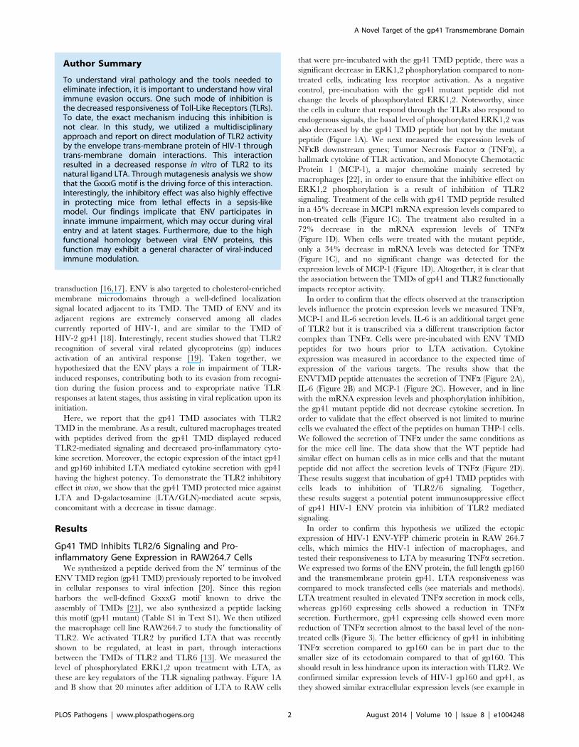
these are key regulators of the TLR signaling pathway. Figure 1A
and B show that 20 minutes after addition of LTA to RAW cells
that were pre-incubated with the gp41 TMD peptide, there was a
significant decrease in ERK1,2 phosphorylation compared to non-
treated cells, indicating less receptor activation. As a negative
control, pre-incubation with the gp41 mutant peptide did not
change the levels of phosphorylated ERK1,2. Noteworthy, since
the cells in culture that respond through the TLRs also respond to
endogenous signals, the basal level of phosphorylated ERK1,2 was
also decreased by the gp41 TMD peptide but not by the mutant
peptide (Figure 1A). We next measured the expression levels of
NFkB downstream genes; Tumor Necrosis Factor a (TNFa), a
hallmark cytokine of TLR activation, and Monocyte Chemotactic
Protein 1 (MCP-1), a major chemokine mainly secreted by
macrophages [22], in order to ensure that the inhibitive effect on
ERK1,2 phosphorylation is a result of inhibition of TLR2
signaling. Treatment of the cells with gp41 TMD peptide resulted
in a 45% decrease in MCP1 mRNA expression levels compared to
non-treated cells (Figure 1C). The treatment also resulted in a
72% decrease in the mRNA expression levels of TNFa(Figure 1D). When cells were treated with the mutant peptide,
only a 34% decrease in mRNA levels was detected for TNFa(Figure 1C), and no significant change was detected for the
expression levels of MCP-1 (Figure 1D). Altogether, it is clear that
the association between the TMDs of gp41 and TLR2 functionally
impacts receptor activity.
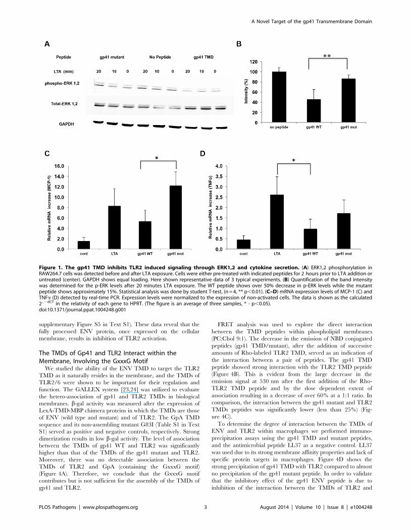
In order to confirm that the effects observed at the transcription
levels influence the protein expression levels we measured TNFa,
MCP-1 and IL-6 secretion levels. IL-6 is an additional target gene
of TLR2 but it is transcribed via a different transcription factor
complex than TNFa. Cells were pre-incubated with ENV TMD
peptides for two hours prior to LTA activation. Cytokine
expression was measured in accordance to the expected time of
expression of the various targets. The results show that the
ENVTMD peptide attenuates the secretion of TNFa (Figure 2A),
IL-6 (Figure 2B) and MCP-1 (Figure 2C). However, and in line
with the mRNA expression levels and phosphorylation inhibition,
the gp41 mutant peptide did not decrease cytokine secretion. In
order to validate that the effect observed is not limited to murine
cells we evaluated the effect of the peptides on human THP-1 cells.
We followed the secretion of TNFa under the same conditions as
for the mice cell line. The data show that the WT peptide had
similar effect on human cells as in mice cells and that the mutant
peptide did not affect the secretion levels of TNFa (Figure 2D).
These results suggest that incubation of gp41 TMD peptides with
cells leads to inhibition of TLR2/6 signaling. Together,
these results suggest a potential potent immunosuppressive effect
of gp41 HIV-1 ENV protein via inhibition of TLR2 mediated
signaling.
In order to confirm this hypothesis we utilized the ectopic
expression of HIV-1 ENV-YFP chimeric protein in RAW 264.7
cells, which mimics the HIV-1 infection of macrophages, and
tested their responsiveness to LTA by measuring TNFa secretion.
We expressed two forms of the ENV protein, the full length gp160
and the transmembrane protein gp41. LTA responsiveness was
compared to mock transfected cells (see materials and methods).
LTA treatment resulted in elevated TNFa secretion in mock cells,
whereas gp160 expressing cells showed a reduction in TNFasecretion. Furthermore, gp41 expressing cells showed even more
reduction of TNFa secretion almost to the basal level of the non-
treated cells (Figure 3). The better efficiency of gp41 in inhibiting
TNFa secretion compared to gp160 can be in part due to the
smaller size of its ectodomain compared to that of gp160. This
should result in less hindrance upon its interaction with TLR2. We
confirmed similar expression levels of HIV-1 gp160 and gp41, as
they showed similar extracellular expression levels (see example in
Author Summary
To understand viral pathology and the tools needed toeliminate infection, it is important to understand how viralimmune evasion occurs. One such mode of inhibition isthe decreased responsiveness of Toll-Like Receptors (TLRs).To date, the exact mechanism inducing this inhibition isnot clear. In this study, we utilized a multidisciplinaryapproach and report on direct modulation of TLR2 activityby the envelope trans-membrane protein of HIV-1 throughtrans-membrane domain interactions. This interactionresulted in a decreased response in vitro of TLR2 to itsnatural ligand LTA. Through mutagenesis analysis we showthat the GxxxG motif is the driving force of this interaction.Interestingly, the inhibitory effect was also highly effectivein protecting mice from lethal effects in a sepsis-likemodel. Our findings implicate that ENV participates ininnate immune impairment, which may occur during viralentry and at latent stages. Furthermore, due to the highfunctional homology between viral ENV proteins, thisfunction may exhibit a general character of viral-inducedimmune modulation.
A Novel Target of the gp41 Transmembrane Domain
PLOS Pathogens | www.plospathogens.org 2 August 2014 | Volume 10 | Issue 8 | e1004248
supplementary Figure S5 in Text S1). These data reveal that the
fully processed ENV protein, once expressed on the cellular
membrane, results in inhibition of TLR2 activation.
The TMDs of Gp41 and TLR2 Interact within theMembrane, Involving the GxxxG Motif
We studied the ability of the ENV TMD to target the TLR2
TMD as it naturally resides in the membrane, and the TMDs of
TLR2/6 were shown to be important for their regulation and
function. The GALLEX system [23,24] was utilized to evaluate
the hetero-association of gp41 and TLR2 TMDs in biological
membranes. b-gal activity was measured after the expression of
LexA-TMD-MBP chimera proteins in which the TMDs are those
of ENV (wild type and mutant) and of TLR2. The GpA TMD
sequence and its non-assembling mutant G83I (Table S1 in Text
S1) served as positive and negative controls, respectively. Strong
dimerization results in low b-gal activity. The level of association
between the TMDs of gp41 WT and TLR2 was significantly
higher than that of the TMDs of the gp41 mutant and TLR2.
Moreover, there was no detectable association between the
TMDs of TLR2 and GpA (containing the GxxxG motif)
(Figure 4A). Therefore, we conclude that the GxxxG motif
contributes but is not sufficient for the assembly of the TMDs of
gp41 and TLR2.
FRET analysis was used to explore the direct interaction
between the TMD peptides within phospholipid membranes
(PC:Chol 9:1). The decrease in the emission of NBD conjugated
peptides (gp41 TMD/mutant), after the addition of successive
amounts of Rho-labeled TLR2 TMD, served as an indication of
the interaction between a pair of peptides. The gp41 TMD
peptide showed strong interaction with the TLR2 TMD peptide
(Figure 4B). This is evident from the large decrease in the
emission signal at 530 nm after the first addition of the Rho-
TLR2 TMD peptide and by the dose dependent extent of
association resulting in a decrease of over 60% at a 1:1 ratio. In
comparison, the interaction between the gp41 mutant and TLR2
TMDs peptides was significantly lower (less than 25%) (Fig-
ure 4C).
To determine the degree of interaction between the TMDs of
ENV and TLR2 within macrophages we performed immuno-
precipitation assays using the gp41 TMD and mutant peptides,
and the antimicrobial peptide LL37 as a negative control. LL37
was used due to its strong membrane affinity properties and lack of
specific protein targets in macrophages. Figure 4D shows the
strong precipitation of gp41 TMD with TLR2 compared to almost
no precipitation of the gp41 mutant peptide. In order to validate
that the inhibitory effect of the gp41 ENV peptide is due to
inhibition of the interaction between the TMDs of TLR2 and
Figure 1. The gp41 TMD inhibits TLR2 induced signaling through ERK1,2 and cytokine secretion. (A) ERK1,2 phosphorylation inRAW264.7 cells was detected before and after LTA exposure. Cells were either pre-treated with indicated peptides for 2 hours prior to LTA addition oruntreated (center). GAPDH shows equal loading. Here shown representative data of 3 typical experiments. (B) Quantification of the band intensitywas determined for the p-ERK levels after 20 minutes LTA exposure. The WT peptide shows over 50% decrease in p-ERK levels while the mutantpeptide shows approximately 15%. Statistical analysis was done by student T-test, (n = 4, ** p,0.01). (C–D) mRNA expression levels of MCP-1 (C) andTNFa (D) detected by real-time PCR. Expression levels were normalized to the expression of non-activated cells. The data is shown as the calculated2‘2dCT in the relativity of each gene to HPRT. (The figure is an average of three samples, * - p,0.05).doi:10.1371/journal.ppat.1004248.g001
A Novel Target of the gp41 Transmembrane Domain
PLOS Pathogens | www.plospathogens.org 3 August 2014 | Volume 10 | Issue 8 | e1004248
TLR6, we performed a FRET competition assay between TLR2
and 6 in the absence (Figure S3 a in Text S1) or presence (Figure
S3 b in Text S1) of the gp41 WT peptide. The data show that
when the ENV peptide is present, the degree of association
between the TLR peptides decreased from 40% FRET to 16%.
This inhibition was further corroborated by immunoprecipitation
of TLR6-TMD peptides with the TLR2 protein in the absence
(Figure S1 c, left lane in Text S1) or the presence (Figure S1 c,
right lane in Text S1) of the gp41 WT peptide.
The Secondary Structures of the TMDsIn order to verify that the TMD peptides preserved helical
structures, we used circular dichorism (CD) spectroscopy in a
membrane mimicking solution (1% LPC). All three peptides
exhibited a-helical secondary structures, typical of a TMD peptide
(Figure S2 in Text S1). It is clear from the CD results that the gp41
mutant peptide (loss of the GxxxG motif) preserves a helical
structure suggesting that any functional discrepancies are not as a
result of major structural deformation.
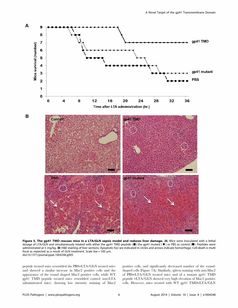
The ENV TMD Peptide Rescues Mice from LTA/GLNMediated Sepsis
In order to corroborate the effect of the ENV TMD on TLR2
activation in vivo we utilized a murine model for acute sepsis
caused by hyper-activation of TLR2 [13]. Mice were injected
intraperitoneally (i.p) with 100 mg of LTA and 700 mg/kg D-
galactosamine (GLN), and were either treated intravenous (i.v.)
with gp41 TMD peptide (5 mg/kg), mutated peptide (5 mg/kg) or
PBS. Upon injection of LTA/GLN, 7/9 PBS-treated mice and 6/
9 of the gp41 mutant treated mice died while only 2/9 of the gp41
TMD treated mice died (Figure 5A). Histological analysis of the
liver indicated that LTA/GLN treatment resulted in massive
parenchyma damage associated with hemorrhage in both PBS and
gp41 mutant treated mice (Figure 5B). However in the gp41 TMD
treated samples, the tissue resembled that of control mice
(Figure 5B) with no observable tissue damage or hemorrhage.
This was also evident from the external feature of the tissues
themselves, showing that mice not treated with the ENV TMD
peptides had enlarged and inflamed livers and spleens (Figure S3
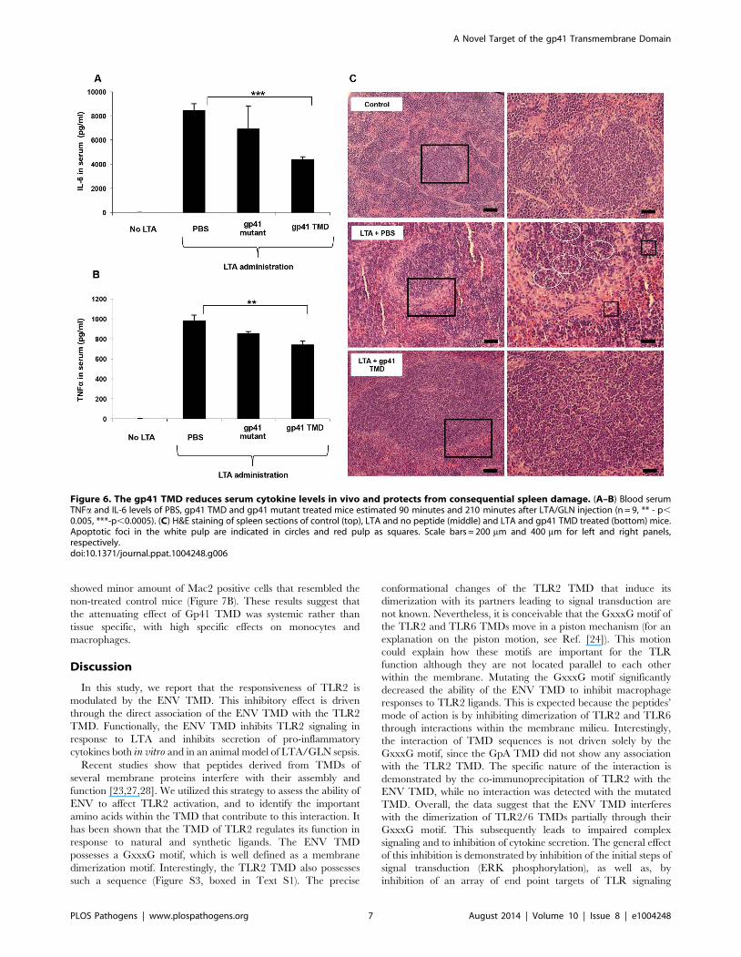
in Text S1). We analyzed serum levels of TNFa and IL-6 as these
cytokines are secreted by resident macrophages and blood
monocytes in the LTA/GLN model [13]. In accordance with
the survival rates, our results indicate that the blood serum levels of
TNFa and IL-6 were significantly lower for the gp41 TMD
peptide-treated group compared to the untreated group (Fig-
ure 6A and B, respectively). We therefore examined spleen, in
order to test activation of the immune system. The effect of an
uncontrolled inflammatory response may lead to secretion of high
levels of steroids, leading to massive cell death in the lymphocyte
zones [25]. LTA/GLN injection resulted in a large number of
apoptotic cell foci in the white pulp and congestion throughout the
tissue in comparison to control mice spleens (Figure 6C middle
and top panels, respectively). Comparably, gp41 TMD treatment
decreased apoptosis to levels similar to the naıve samples
(Figure 6C lower panel). Cell death was also detected in the red
pulp of untreated mice and was absent from gp41 TMD treated
mice. This result indicates that gp41 TMD decreased monocyte
activation, resulting in less apoptosis in the spleen.
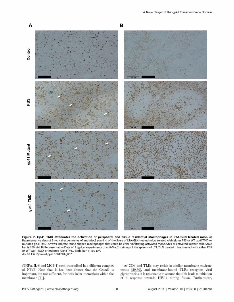
In order to test the specific attenuating effect of gp41 TMD on
LTA/GLN mediated sepsis, we tested whether the treatment
resulted in an inhibitive effect on macrophages, as macrophages
are the main immune cells that orchestrate this type of
inflammation. We investigated whether the inhibitory effect of
gp41 TMD peptide was tissue specific or systemic by staining both
liver (tissue specific) and spleen (systemic) for the monocyte
activation marker, Mac2 [26]. Liver staining showed that the
administration of LTA/GLN resulted in increase in Mac2 positive
cells, in addition to the appearance of round-shaped Mac2 positive
cells that could be either activated residential macrophages
(kupffer cells) that phagocysed dead hepatocytes, or infiltrated
activated monocytes (Figure 7A, white arrows). The mutant gp41
Figure 2. ENV TMD inhibits secretion of cytokines andchemokines induced by TLR2. (A–C) RAW cells were incubatedwith/without the indicated peptides for 2 hours, then washed twiceand activated with 500 ng/ml of purified LTA. Media was collected after5 hours (TNFa) or 24 hours (MCP-1 and IL-6). ELISA was performedusing standard kits. (n = 3, 6 represent SD) (D) THP-1 humanmacrophages were incubated using similar conditions as described inA, and TNFa levels were measured (* - p,0.05).doi:10.1371/journal.ppat.1004248.g002
A Novel Target of the gp41 Transmembrane Domain
PLOS Pathogens | www.plospathogens.org 4 August 2014 | Volume 10 | Issue 8 | e1004248
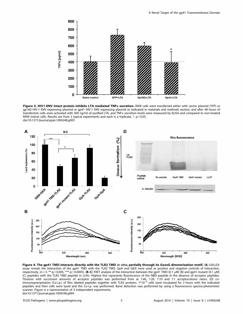
Figure 4. The gp41 TMD interacts directly with the TLR2 TMD in vitro, partially through its GxxxG dimmerization motif. (A) GALLEXassay reveals the interaction of the gp41 TMD with the TLR2 TMD. GpA and G83I were used as positive and negative controls of interaction,respectively. (n = 3, **-p,0.005, ***-p,0.0005). (B–C) FRET analysis of the interaction between the gp41 TMD (0.1 mM) (B) and gp41 mutant (0.1 mM)(C) peptides with the TLR2 TMD peptide in LUVs. Highest line represents fluorescence of the NBD peptide in the absence of acceptor peptides.Titration with successive amounts of acceptor peptides was performed from at 1:40, 1:20, 1:10 and 1:1 acceptor:donor ratios. (D) co-immunoprecipitation (Co-i.p.) of Rho labeled peptides together with TLR2 proteins. 1*10‘6 cells were incubated for 2 hours with the indicatedpeptides and then cells were lysed and the Co-i.p. was performed. Band detection was performed by using a fluorescence spectra-photometerscanner. Figure is a representative of 3 independent experiments.doi:10.1371/journal.ppat.1004248.g004
Figure 3. HIV1-ENV intact protein inhibits LTA mediated TNFa secretion. RAW cells were transfected either with vector plasmid (YFP) orgp160 HIV-1 ENV expressing plasmid or gp41 HIV-1 ENV expressing plasmid as indicated in materials and methods section, and after 48 hours oftransfection cells were activated with 500 ng/ml of purified LTA, and TNFa secretion levels were measured by ELISA and compared to non-treatedRAW (naıve) cells. Results are from 3 typical experiments and each is a triplicate, *, p,0.05.doi:10.1371/journal.ppat.1004248.g003
A Novel Target of the gp41 Transmembrane Domain
PLOS Pathogens | www.plospathogens.org 5 August 2014 | Volume 10 | Issue 8 | e1004248
peptide treated mice resembled the PBS+LTA/GLN treated mice
and showed a similar increase in Mac2 positive cells and the
appearance of the round shaped Mac2 positive cells, while WT
gp41 TMD peptide treated mice resembled control non-LTA
administrated mice, showing low intensity staining of Mac2
positive cells, and significantly decreased number of the round-
shaped cells (Figure 7A). Similarly, spleen staining with anti-Mac2
of PBS+LTA/GLN treated mice and of a mutant gp41 TMD
peptide +LTA/GLN showed very high elevation of Mac2 positive
cells. However, mice treated with WT gp41 TMD+LTA/GLN
Figure 5. The gp41 TMD rescues mice in a LTA/GLN sepsis model and reduces liver damage. (A) Mice were inoculated with a lethaldosage of LTA/GLN and simultaneously treated with either the gp41 TMD peptide (&) the gp41 mutant (¤) or PBS as control (N). Peptides wereadministrated at 5 mg/kg. (B) H&E staining of liver sections. Apoptotic foci are indicated in circles and arrows indicate hemorrhage. Cell death is multifocal as expected as a result of GLN treatment. Scale bar = 100 mm.doi:10.1371/journal.ppat.1004248.g005
A Novel Target of the gp41 Transmembrane Domain
PLOS Pathogens | www.plospathogens.org 6 August 2014 | Volume 10 | Issue 8 | e1004248
showed minor amount of Mac2 positive cells that resembled the
non-treated control mice (Figure 7B). These results suggest that
the attenuating effect of Gp41 TMD was systemic rather than
tissue specific, with high specific effects on monocytes and
macrophages.
Discussion
In this study, we report that the responsiveness of TLR2 is
modulated by the ENV TMD. This inhibitory effect is driven
through the direct association of the ENV TMD with the TLR2
TMD. Functionally, the ENV TMD inhibits TLR2 signaling in
response to LTA and inhibits secretion of pro-inflammatory
cytokines both in vitro and in an animal model of LTA/GLN sepsis.
Recent studies show that peptides derived from TMDs of
several membrane proteins interfere with their assembly and
function [23,27,28]. We utilized this strategy to assess the ability of
ENV to affect TLR2 activation, and to identify the important
amino acids within the TMD that contribute to this interaction. It
has been shown that the TMD of TLR2 regulates its function in
response to natural and synthetic ligands. The ENV TMD
possesses a GxxxG motif, which is well defined as a membrane
dimerization motif. Interestingly, the TLR2 TMD also possesses
such a sequence (Figure S3, boxed in Text S1). The precise
conformational changes of the TLR2 TMD that induce its
dimerization with its partners leading to signal transduction are
not known. Nevertheless, it is conceivable that the GxxxG motif of
the TLR2 and TLR6 TMDs move in a piston mechanism (for an
explanation on the piston motion, see Ref. [24]). This motion
could explain how these motifs are important for the TLR
function although they are not located parallel to each other
within the membrane. Mutating the GxxxG motif significantly
decreased the ability of the ENV TMD to inhibit macrophage
responses to TLR2 ligands. This is expected because the peptides’
mode of action is by inhibiting dimerization of TLR2 and TLR6
through interactions within the membrane milieu. Interestingly,
the interaction of TMD sequences is not driven solely by the
GxxxG motif, since the GpA TMD did not show any association
with the TLR2 TMD. The specific nature of the interaction is
demonstrated by the co-immunoprecipitation of TLR2 with the
ENV TMD, while no interaction was detected with the mutated
TMD. Overall, the data suggest that the ENV TMD interferes
with the dimerization of TLR2/6 TMDs partially through their
GxxxG motif. This subsequently leads to impaired complex
signaling and to inhibition of cytokine secretion. The general effect
of this inhibition is demonstrated by inhibition of the initial steps of
signal transduction (ERK phosphorylation), as well as, by
inhibition of an array of end point targets of TLR signaling
Figure 6. The gp41 TMD reduces serum cytokine levels in vivo and protects from consequential spleen damage. (A–B) Blood serumTNFa and IL-6 levels of PBS, gp41 TMD and gp41 mutant treated mice estimated 90 minutes and 210 minutes after LTA/GLN injection (n = 9, ** - p,0.005, ***-p,0.0005). (C) H&E staining of spleen sections of control (top), LTA and no peptide (middle) and LTA and gp41 TMD treated (bottom) mice.Apoptotic foci in the white pulp are indicated in circles and red pulp as squares. Scale bars = 200 mm and 400 mm for left and right panels,respectively.doi:10.1371/journal.ppat.1004248.g006
A Novel Target of the gp41 Transmembrane Domain
PLOS Pathogens | www.plospathogens.org 7 August 2014 | Volume 10 | Issue 8 | e1004248
(TNFa, IL-6 and MCP-1) each transcribed in a different complex
of NFkB. Note that it has been shown that the GxxxG is
important, but not sufficient, for helix-helix interactions within the
membrane [21].
As CD4 and TLRs may reside in similar membrane environ-
ments [29,30], and membrane-bound TLRs recognize viral
glycoproteins, it is reasonable to assume that this leads to initiation
of a response towards HIV-1 during fusion. Furthermore,
Figure 7. Gp41 TMD attenuates the activation of peripheral and tissue residential Macrophages in LTA/GLN treated mice. A)Representative data of 3 typical experiments of anti-Mac2 staining of the livers of LTA/GLN treated mice, treated with either PBS or WT gp41TMD ormutated gp41TMD. Arrows indicate round shaped macrophages that could be either infiltrating activated monocytes or activated kupffer cells. Scalebar is 100 mM. B) Representative Data of 3 typical experiments of anti-Mac2 staining of the spleens of LTA/GLN treated mice, treated with either PBSor WT Gp41TMD or mutated Gp41TMD. Scale bar is 100 mM.doi:10.1371/journal.ppat.1004248.g007
A Novel Target of the gp41 Transmembrane Domain
PLOS Pathogens | www.plospathogens.org 8 August 2014 | Volume 10 | Issue 8 | e1004248
activation of TLR signaling prior to HIV infection causes dendritic
cells to be less permissive to infection [31]. Therefore, inhibiting
the initial response towards the invading pathogen is a powerful
tool to allow virus entry into the cell. An additional possible need
for this inhibitory activity is at latent stages of viral infection. HIV-
1 ENV and TLR2 both possess a membrane localization signal
motif adjacent to their TMD (Figure S3 in Text S1). This signal
motif determines the precise environment within the membrane in
which the proteins reside. Although for T cells, ENV is expressed
at low levels during latency, ENV expression levels on mononu-
clear phagocytes are high [32]. Several studies indicated the
impairment of TLR signaling after viral infection and incorpora-
tion [5] which may assist in the establishment of infection by
opportunistic bacteria and fungi, in addition to the loss of CD4+ T
cells [33]. Together with our findings about the modulatory effect
of ENV on TLR responses, this impairment may occur as a result
of the interactions of the TMDs of ENV and TLRs, leading to the
prevention of TLR activation upon ligand recognition. Notewor-
thy, several studies have linked the activation of TLR cascades to
viral replication and burst. These studies focus on cells with
established infection. This means that the virus successfully
transmitted its genome into the cells and initiated the expression
of its early genes [34]. In view of the small number of proteins
expressed by the HIV genome, this immunosuppressive activity
might exist with its fusogenic activity to help the virus to escape
immune recognition and response towards it, prior to the activity
of the viral early genes.
In summary, we demonstrate that the ENV TMD inhibits the
activation of TLR2 signaling by interacting with its TMD. This
property could be of great importance during fusion and serves as
an additional mean of regulation of the activity of TLR at stages of
Env expression. Our findings further demonstrate the complex
strategies used by HIV-1 to manipulate the immune system.
Interestingly, the ENV TMD is also involved in the late steps of
the membrane fusion reaction. Such dual activity further
demonstrates how viruses although expressing a limited number
of proteins have evolved to alter so many cellular processes.
Considering the strong ability of other gp41-derived segments to
inhibit T-cell activation by various mechanisms, our study may
indicate that these effects are also applicable to mononuclear
phagocytes and additional targets should be further investigated.
Apart from HIV pathogenesis, specific gp41 derived peptides may
provide potential tools for treating uncontrolled immune respons-
es, manifested here by sepsis.
Materials and Methods
Cell CultureAll in vitro assays were performed on RAW264.7 murine
macrophages (ATCC- TIB71). Cells were grown at 37uC in the
presence of 5% CO2 in DMEM supplemented with 10% FBS, L-
glutamine, sodium pyruvate, non-essential amino acids, and
antibiotics (Biological Industries, Beit-Haemek, Israel). For cyto-
kine secretion measurements 26105 cells per well were cultured
overnight in a 96-well plate. At the following day, media were
replaced by fresh DMEM, including all supplements. Gp41 TMD
peptides were dissolved in DMSO and added to the cells in
different concentrations. The final concentration of DMSO was
1% for all groups. Cells were incubated with the peptide for
2 hours, washed and incubated with fresh media containing LTA.
Cells were incubated with LTA for 5 hrs (TNFa) or 12 hours (IL-
6, MCP-1) at 37uC, after which samples of the media were
collected and stored at 220uC. Cytokine levels were evaluated
using a mouse TNFa/IL-6 enzyme-linked immunosorbent assay
kit (ELISAmax, Biolegend) according to the manufacturer’s
protocol. All experiments were done in triplicates.
Gp41 and gp160 Intact Proteins ExpressionRaw264.7 macrophages were transfected twice, 48 h and again
24 h prior to the experiment, either with p96ZM651gp160-CD5-opt
plasmid for the expression of HIV envelope protein gp160, or with
gp41-YFP expressing plasmid for the expression of gp41 HIV fusion
protein [35]. To increase the expression rate we utilized TurboFect
reagent (Thermo Scientific, Waltham, MA, USA) according to the
manufacturer protocol. Surface expression of untagged gp160 was
analyzed using immunofluorescence. Briefly, cells were washed twice
with PBS and fixed with 4% paraformaldehyde for 15 min at room
temperature. Antibody staining was conducted using a goat
polyclonal anti-HIV-1 gp120-biotin conjugated antibody and a
ATTO 488 conjugated, goat IgG (H&L) antibody. Fluorescence
intensities were obtained using an inverted FluoView 1000 confocal
microscope (Olympus, Tokyo, Japan) with a 606 oil immersion
objective (numerical aperture 1.35) at 25uC with a frame size of
5126512 pixels. ATTO 488 was excited with a laser diode at 488 nm
and detected between 500 and 600 nm.
Fluorescence Resonance Energy Transfer (FRET) betweenNBD and Rhodamine (TAMRA)-Labeled Peptides
Large Unilamellar Vesicles (LUVs) composed of phosphatidyl-
choline (PC) and cholesterol (Chol) (9:1, w/w) were prepared using
the extrusion method as described previously [36]. The fluores-
cence experiments were performed with pairs of peptides using 4-
Fluoro-7-nitrobenzofurazan (NBD, BioChemika) -labeled peptides
as donors and rhodamine-labeled peptides as acceptors and as
described previously [13]. Maximum FRET values were obtained
by calculating the changes in the emission of the donor peptide at
530 nm.
Secondary Structure Determination Utilizing CircularDichroism (CD) Spectroscopy
CD measurements were performed by using an Applied
Photophysics spectropolarimeter. The spectra were scanned using
a thermostatic quartz cuvette with a path length of 1 mm.
Wavelength scans were performed at 25uC; the average recording
time was 15 s, in 1 nm steps, through the wavelength range of
195–260 nm. Peptides were scanned at a concentration of 50 mM
in buffer only (HEPES 5 mM) or in a membrane mimetic
environment of 1% Lysophophatidilcholine (LPC) in buffer.
Construction of the GALLEX ChimeraThe hetero-interactions of the TMDs within a natural
membrane environment were studied using the GALLEX assay
[37]. The expression vectors and E. coli strains required for the
assay were kindly provided by Dr. Dirk Schneider. Briefly, Two
plasmids are used in the assay containing either the wild-type
LexA sequence (pBLM, encoding for resistance to Tetracycline) or
a mutated form of LexA (pALM, encoding for resistance to
Ampicillin). The desired TM sequences are inserted between these
two protein sequences using standard cloning techniques. The
resulting plasmids were both transformed into an SU202 E. coli
strain, which then expressed the LexA-TM-MalE fusion proteins
(after induction with 1 mM IPTG). Hetero-association of TM
domains leads to the formation of LexAWT/LexAmut heterodimers
that bind to a hybrid LexA promoter/operator in the reporter
strain SU202 (that recognizes the mutant LexA domain), and
repress the expression of b-gal. Furthermore, as the hybrid
promoter is designed to bind only the wt and mutant heterodi-
A Novel Target of the gp41 Transmembrane Domain
PLOS Pathogens | www.plospathogens.org 9 August 2014 | Volume 10 | Issue 8 | e1004248
mers, it will not recognize wild-type LexA homodimers. Thus, TM
homo-oligomer formation does not interfere with the measure-
ment of hetero-association. The TMDs of GlycophorinA (GpA)
and its G83I mutant, used as positive and negative controls,
respectively, are ITLIIFGVMAGVIGTILLI and ITLIIFGVMAI-
VIGTILLI. All the constructs were confirmed by DNA sequenc-
ing.
Measurements of Dimerization (b-Galactosidase Assay)The dimerization propensity of the two constructs was
measured by transforming an E. coli SU202 indicator strain with
the relevant pALM and pBLM plasmids (each harboring a
different TM sequence) simultaneously. The bacteria were grown
overnight in the presence of 1 mM IPTG in LB medium with
ampicillin and tetracycline in a 24-well plate. 10 mL of bacteria
were resuspended in 100 mL of Z-buffer (60 mM Na2HPO4,
40 mM NaH2PO4, 10 mM KCl, 1 mM MgSO4, pH = 7.0, 10%
chloroform, 1% b-mercaptoethanol), and the absorbance (OD600)
was measured. Bacteria were lysed with 50 mL of 2% SDS in Z-
buffer. O-nitrophenyl-galactopyranose (ONPG) was added and a
kinetic reading of absorbance at OD405 was performed for 20 min
to measure the activity of b-gal. Vmax was calculated and divided
by the absorbance at OD600 to correct for bacterial growth. Strong
dimerization results in strong inhibition of b-gal expression and
less color formation.
Western Blot AnalysisCell lysates were prepared in radioimmunoprecipitation (RIPA)
assay buffer (50 mM Tris HCl pH 7.5, 150 mM NaCl, 1% Nonidet
P-40, 0.5% sodium deoxycholate, 0.1% SDS) containing 50 mM
NaF, 2 mM Na3VO4, protease and phosphatase inhibitors (Sigma-
Aldrich). Protein concentration was measured using the BCA
Protein Assay Kit (Pierce Chemical Co.). Fifty mg of protein was
loaded and separated on 8–15% SDS-PAGE and transferred to a
nitrocellulose membrane. The membrane was blocked using 5%
bovine serum albumin (BSA) in phosphate-buffered saline contain-
ing 0.1% Tween-20 (PBST) for 1 h at room temperature. The
primary antibody was diluted in PBST containing 1% BSA and
incubated with the membrane at 4uC overnight. After 3 washes with
PBST, membranes were incubated with the secondary antibody in
PBST containing 1% BSA at room temperature for 1 h.
qPCR Gene ExpressionThe following sequences were used for quantitative mRNA
analysis:
TNFa Forward: CTTGTGGCAGGGGCCACCAC
Reverse: CCATGCCGTTGGCCAGGAGG
MCP-1 Forward: TCACCTGCTGCTACTCATTCACCA
Reverse: AGCACAGACCTCTCTCTTGAGCTT
Antibodies and ReagentsAnti phospho-ERK1, 2 and anti-total ERK1, 2 were purchased
from Sigma-Aldrich and anti GAPDH was purchased from
Millipore. Goat polyclonal anti-HIV-1 gp120-biotin conjugated
antibody was purchased from Abcam (Cambridge, United
Kingdom). ATTO 488 Conjugated Goat IgG (H&L) antibody
was purchased from Rockland (Gilbertsville, PA, USA). S. aureusLTA was purchased from Sigma-Aldrich and D-galactosamine
was purchased from Calbiochem. TNFa and IL-6 ELISA kits
were purchased from Biolegend. The plasmid p96ZM651gp160-
CD5-opt was obtained through the NIH AIDS Reagent Program,
Division of AIDS, NIAID, NIH: from Drs. Yingying Li, Feng
Gao, and Beatrice H. Hahn. The plasmid gp41-YFP, provided by
Roland Schwarzer encodes a HIV-1 gp41 fusion protein with the
c-terminal external parts of the protein replaced by a Yellow
Fluorescent Protein.
LTA Induced Sepsis-Like ModelAnimal studies were carried out in strict accordance with the
Israeli law and the National Research Council guidelines. All
animal experiments were conducted at the Weizmann Institute of
Science and approved by the Weizmann Institutional Animal
Care and Use Committee (IACUC permit no. 01190107-4). 12-
week-old C57 Black female mice were injected IP with 50 mg/kg
of S.aureus LTA and 800 mg/kgof D-galactosamine in PBS, as
previously described [13], in combination with 5 mg/kg of gp41-
TMD or mutated gp41-TMD or PBS, and monitored for survival
for 36 hours. Blood samples were collected for TNFa and IL-6
serum levels measurements. Serum was extracted by blood
coagulation in RT followed by spin down centrifugation. Serum
samples were analyzed by ELISA assays.
Histological AnalysisSpleen and liver samples were collected either post mortem or
from sacrificed mice 36 hours after the injection of LTA/GLN
and kept in formaldehyde (4%) for 18 hours and embedded in
paraffin blocks. 4 mm sections were then prepared and stained
with hematoxylin and eosin by standard protocols.
Peptide Synthesis and PurificationPeptides were synthesized by a 9-fluorenylmethoxylcarbonyl
(Fmoc) solid-phase method on Rink amide MBHA resin
(Calbiochem-novabiochem, San Diego, California) by using an
ABI 433A automatic peptide synthesizer (Applied Biosystems,
Foster City, CA). Peptide synthesis was followed by cleavage from
the resin by incubation for 3 h with 95% TFA, 2.5% H2O, and
2.5% triethylsilane, followed by purification by RP-HPLC (.98%)
on a Vydac C4 column (Grace Discovery Sciences, Deerfield, Il),
and then identification by electro-spray mass spectroscopy. The
following fluorophores were used for fluorescent labeling: 4-
Fluoro-7-nitrobenzofurazan (NBD, BioChemika) and 5(6)-Car-
boxytetramethylrhodamine N-succinimidyl ester (TAMRA, Bio-
Chemika). Peptides used for in vivo experiments were treated
twice with 20% acetic acid in order to replace the trifluoroacetate
anion added during HPLC purification.
Supporting Information
Text S1 This file contains Table S1 and Figures S1–S5. FigureS1. This figure shows that gp41 TMD disrupts the interaction
between TLR2 and TLR6 as revealed by Fluorescence Resonance
Energy Transfer (FRET) measurements. In this experiment the
NBD-labeled TLR2 TMD peptide was added first from a stock
solution in DMSO (final concentration 0.1 mM and a maximum of
0.25% (v/v) DMSO) to a dispersion of PC:Chol LUVs (100 mM) in
PBS. This was followed by the addition of the following: A.
Rhodamine labeled TLR 6 TMD or in addition with un labeled
WT gp41 (B), in sequential doses ranging from 0.01 mM to 0.1 mM
(stock in DMSO), generating a ratio of 1:10, 1:5, 1:2 and 1:1
Rhodamine:NBD presented from top to bottom. In both graphs,
the upper spectrum represents the emission of the NBD-labeled
peptide alone. (C) IP of TLR2 from raw264.7 cells in the presence of
the indicated Rho-labeled peptides. The result of the last lane from
the marker shows that in the presence of WT gp41 there is a
decrease in the binding of the TLR6-TMD peptide to the TLR2
protein. Figure S2. This figure shows the secondary structure of
the peptides used in this study as measured by CD spectroscopy.
A Novel Target of the gp41 Transmembrane Domain
PLOS Pathogens | www.plospathogens.org 10 August 2014 | Volume 10 | Issue 8 | e1004248
Spectra were measured at 10 mM in a 1% LPC solution. Graphs are
the mean of 3 measurements. Figure S3. This figure shows that
treatment with gp41 WT peptide rescues mice organs form
inflammatory related damage. In this experiment tissue samples of
liver (A) and spleen (B) from mice taken at the experimental end
point. In both tissues inflammation is evident by the tissue color.
Figure S4. This figure shows sequence alignment of the TMDs of
gp41 (top) and TLR2 (bottom). Arrows indicate the predicted
TMD. Underline indicates the localization motif to cholesterol
enriched regions within the membrane. Figure S5. This figure
demonstrates that gp160 and gp41 infected RAW cells show a
membranous expression of gp160 and gp41. (A) Representative
results of cells infected with gp160 plasmid (see materials and
methods) that were stained with anti-gp160 antibody without
permeabilization. Green indicates membranous gp160. Scale bar is
20 mm. (B) Representative data of RAW cells expressing GFP-gp41
chimera protein (see materials and methods). Scale bar is 40 mm. (C)
A representative image of confocal microscopy analysis of GFP-
gp41 expressing RAW cells showing membranous expression of the
chimeric protein. Scale bar is 10 mm. (D) Representative data of
FACS analysis of GFP-gp41 RAW transfected cells.
(DOCX)
Acknowledgments
We thank Dr. Ori Brenner of the Weizmann Institute of Science for his
help in analysis of the histological specimens and their interpretation. We
also thank Dr. Avraham Ashkenazi and Mr. Yoel Klug for their critical
revision of the manuscript and fruitful inputs.
Author Contributions
Conceived and designed the experiments: EMR MA RS AG ER YS AHF.
Performed the experiments: EMR MA ER RS AG. Analyzed the data:
EMR MA ER RS. Contributed reagents/materials/analysis tools: EMR
MA. Wrote the paper: EMR MA RS YS AHF.
References
1. Cassol E, Alfano M, Biswas P, Poli G (2006) Monocyte-derived macrophages
and myeloid cell lines as targets of HIV-1 replication and persistence. J Leukoc
Biol 80: 1018–1030.2. Kirchhoff F (2010) Immune evasion and counteraction of restriction factors by
HIV-1 and other primate lentiviruses. Cell Host Microbe 8: 55–67.3. Manel N, Littman DR (2011) Hiding in plain sight: how HIV evades innate
immune responses. Cell 147: 271–274.4. Ahmed Z, Czubala M, Blanchet F, Piguet V (2012) HIV impairment of immune
responses in dendritic cells. Adv Exp Med Biol 762: 201–238.
5. Doehle BP, Hladik F, McNevin JP, McElrath MJ, Gale M, Jr. (2009) Humanimmunodeficiency virus type 1 mediates global disruption of innate antiviral
signaling and immune defenses within infected cells. J Virol 83: 10395–10405.6. Sasai M, Yamamoto M (2013) Pathogen recognition receptors: ligands and
signaling pathways by Toll-like receptors. Int Rev Immunol 32: 116–133.
7. Song DH, Lee JO (2012) Sensing of microbial molecular patterns by Toll-likereceptors. Immunol Rev 250: 216–229.
8. Kirschning CJ, Schumann RR (2002) TLR2: cellular sensor for microbial andendogenous molecular patterns. Curr Top Microbiol Immunol 270: 121–144.
9. Werling D, Jungi TW (2003) TOLL-like receptors linking innate and adaptiveimmune response. Vet Immunol Immunopathol 91: 1–12.
10. Akira S, Takeda K (2004) Toll-like receptor signalling. Nat Rev Immunol 4:
499–511.11. Jin MS, Kim SE, Heo JY, Lee ME, Kim HM, et al. (2007) Crystal structure of
the TLR1-TLR2 heterodimer induced by binding of a tri-acylated lipopeptide.Cell 130: 1071–1082.
12. Monie TP, Bryant CE, Gay NJ (2009) Activating immunity: lessons from the
TLRs and NLRs. Trends Biochem Sci 34: 553–561.13. Fink A, Reuven EM, Arnusch CJ, Shmuel-Galia L, Antonovsky N, et al. (2013)
Assembly of the TLR2/6 Transmembrane Domains Is Essential for Activationand Is a Target for Prevention of Sepsis. J Immunol 190: 6410–6422.
14. Victoria S, Temerozo JR, Gobbo L, Pimenta-Inada HK, Bou-Habib DC (2013)Activation of Toll-like receptor 2 increases macrophage resistance to HIV-1
infection. Immunobiology.
15. Martinson JA, Roman-Gonzalez A, Tenorio AR, Montoya CJ, Gichinga CN,et al. (2007) Dendritic cells from HIV-1 infected individuals are less responsive to
toll-like receptor (TLR) ligands. Cell Immunol 250: 75–84.16. Triantafilou M, Miyake K, Golenbock DT, Triantafilou K (2002) Mediators of
innate immune recognition of bacteria concentrate in lipid rafts and facilitate
lipopolysaccharide-induced cell activation. J Cell Sci 115: 2603–2611.17. Fessler MB, Parks JS (2011) Intracellular lipid flux and membrane microdomains
as organizing principles in inflammatory cell signaling. J Immunol 187: 1529–1535.
18. Sun ZY, Oh KJ, Kim M, Yu J, Brusic V, et al. (2008) HIV-1 broadly
neutralizing antibody extracts its epitope from a kinked gp41 ectodomain regionon the viral membrane. Immunity 28: 52–63.
19. Barbalat R, Lau L, Locksley RM, Barton GM (2009) Toll-like receptor 2 oninflammatory monocytes induces type I interferon in response to viral but not
bacterial ligands. Nat Immunol 10: 1200–1207.20. Cohen T, Cohen SJ, Antonovsky N, Cohen IR, Shai Y (2011) HIV-1 gp41 and
TCRalpha trans-membrane domains share a motif exploited by the HIV virus to
modulate T-cell proliferation. PLoS Pathog 6: e1001085.
21. Schneider D, Engelman DM (2004) Motifs of two small residues can assist but
are not sufficient to mediate transmembrane helix interactions. J Mol Biol 343:
799–804.
22. Ghosh TK, Mickelson DJ, Fink J, Solberg JC, Inglefield JR, et al. (2006) Toll-
like receptor (TLR) 2–9 agonists-induced cytokines and chemokines: I.
Comparison with T cell receptor-induced responses. Cell Immunol 243: 48–57.
23. Fink A, Sal-Man N, Gerber D, Shai Y (2012) Transmembrane domains
interactions within the membrane milieu: principles, advances and challenges.
Biochim Biophys Acta 1818: 974–983.
24. Cymer F, Sanders CR, Schneider D (2013) Analyzing oligomerization of
individual transmembrane helices and of entire membrane proteins in E. coli: A
hitchhiker’s guide to GALLEX. Methods Mol Biol 932: 259–276.
25. Zhan J, Deng R, Tang J, Zhang B, Tang Y, et al. (2006) The spleen as a target in
severe acute respiratory syndrome. Faseb J 20: 2321–2328.
26. Farfel-Becker T, Vitner EB, Kelly SL, Bame JR, Duan J, et al. (2013) Neuronal
accumulation of glucosylceramide in a mouse model of neuronopathic Gaucher
disease leads to neurodegeneration. Hum Mol Genet 23: 843–854.
27. He L, Shobnam N, Hristova K (2011) Specific inhibition of a pathogenic
receptor tyrosine kinase by its transmembrane domain. Biochim Biophys Acta
1808: 253–259.
28. Yin H, Slusky JS, Berger BW, Walters RS, Vilaire G, et al. (2007)
Computational design of peptides that target transmembrane helices. Science
315: 1817–1822.
29. Fragoso R, Ren D, Zhang X, Su MW, Burakoff SJ, et al. (2003) Lipid raft
distribution of CD4 depends on its palmitoylation and association with Lck, and
evidence for CD4-induced lipid raft aggregation as an additional mechanism to
enhance CD3 signaling. J Immunol 170: 913–921.
30. Triantafilou M, Lepper PM, Olden R, Dias IS, Triantafilou K (2011) Location,
location, location: is membrane partitioning everything when it comes to innate
immune activation? Mediators Inflamm 2011: 186093.
31. Ogawa Y, Kawamura T, Kimura T, Ito M, Blauvelt A, et al. (2009) Gram-
positive bacteria enhance HIV-1 susceptibility in Langerhans cells, but not in
dendritic cells, via Toll-like receptor activation. Blood 113: 5157–5166.
32. Willey RL, Bonifacino JS, Potts BJ, Martin MA, Klausner RD (1988)
Biosynthesis, cleavage, and degradation of the human immunodeficiency virus
1 envelope glycoprotein gp160. Proc Natl Acad Sci U S A 85: 9580–9584.
33. Okoye AA, Picker LJ (2013) CD4(+) T-cell depletion in HIV infection:
mechanisms of immunological failure. Immunol Rev 254: 54–64.
34. Sundstrom JB, Little DM, Villinger F, Ellis JE, Ansari AA (2004) Signaling
through Toll-like receptors triggers HIV-1 replication in latently infected mast
cells. J Immunol 172: 4391–4401.
35. Schwarzer R, Levental I, Gramatica A, Scolari S, Buschmann V, et al. (2014)
The cholesterol-binding motif of the HIV-1 glycoprotein gp41 regulates lateral
sorting and oligomerization. Cell Microbiol: in press.
36. Cohen T, Pevsner-Fischer M, Cohen N, Cohen IR, Shai Y (2008)
Characterization of the interacting domain of the HIV-1 fusion peptide with
the transmembrane domain of the T-cell receptor. Biochemistry 47: 4826–4833.
37. Schneider D, Engelman DM (2003) GALLEX, a measurement of heterologous
association of transmembrane helices in a biological membrane. J Biol Chem
278: 3105–3111.
A Novel Target of the gp41 Transmembrane Domain
PLOS Pathogens | www.plospathogens.org 11 August 2014 | Volume 10 | Issue 8 | e1004248
Related Documents


