Mol Genet Genomics (2009) 282:417–435 DOI 10.1007/s00438-009-0475-1 123 ORIGINAL PAPER The highly attenuated oncolytic recombinant vaccinia virus GLV-1h68: comparative genomic features and the contribution of F14.5L inactivation Qian Zhang · Chunguang Liang · Yong A. Yu · Nanhai Chen · Thomas Dandekar · Aladar A. Szalay Received: 9 May 2009 / Accepted: 31 July 2009 / Published online: 22 August 2009 © The Author(s) 2009. This article is published with open access at Springerlink.com Abstract As a new anticancer treatment option, vaccinia virus (VACV) has shown remarkable antitumor activities (oncolysis) in preclinical studies, but potential infection of other organs remains a safety concern. We present here genome comparisons between the de novo sequence of GLV- 1h68, a recombinant VACV, and other VACVs. The identi- Wed diVerences in open reading frames (ORFs) include genes encoding host-range selection, virulence and immune modu- lation proteins, e.g., ankyrin-like proteins, serine proteinase inhibitor SPI-2/CrmA, tumor necrosis factor (TNF) receptor homolog CrmC, semaphorin-like and interleukin-1 receptor homolog proteins. Phylogenetic analyses indicate that GLV- 1h68 is closest to Lister strains but has lost several ORFs present in its parental LIVP strain, including genes encoding CrmE and a viral Golgi anti-apoptotic protein, v-GAAP. The reduced pathogenicity of GLV-1h68 is conWrmed in male mice bearing C6 rat glioma and in immunocompetent mice bearing B16-F10 murine melanoma. The contribution of for- eign gene expression cassettes in the F14.5L, J2R and A56R loci is analyzed, in particular the contribution of F14.5L inac- tivation to the reduced virulence is demonstrated by compar- ing the virulence of GLV-1h68 with its F14.5L-null and revertant viruses. GLV-1h68 is a promising engineered VACV variant for anticancer therapy with tumor-speciWc replication, reduced pathogenicity and benign tissue tropism. Keywords Virulence · Immune modulation · Tissue tropism · Foreign expression cassette · Phylogeny Abbreviations VACV Vaccinia virus CPV Cowpox virus OPV Orthopoxvirus GLV-1h68 Vaccinia virus strain GLV-1h68 (EU410304) WR Vaccinia virus strain Western Reserve (AY243312) COP Vaccinia virus strain Copenhagen (M35027) LIST Vaccinia Lister major strain (AY678276) LC Vaccinia Lister isolate LC16mO (AY678277) VACV107 Vaccinia Lister clone VACV107(DQ121394) Communicated by S. Hohmann. Q. Zhang and C. Liang contributed equally. Electronic supplementary material The online version of this article (doi:10.1007/s00438-009-0475-1) contains supplementary material, which is available to authorized users. Q. Zhang · Y. A. Yu · N. Chen · A. A. Szalay (&) Genelux Corporation, San Diego Science Center, 3030 Bunker Hill St., Ste. 310, San Diego, CA 92109, USA e-mail: [email protected] C. Liang · T. Dandekar (&) Department of Bioinformatics, Biocenter, University of Würzburg, Am Hubland, 97074 Würzburg, Germany e-mail: [email protected] T. Dandekar Structural and Computational Biology, EMBL, Heidelberg, Germany A. A. Szalay Rudolf Virchow Center for Experimental Biomedicine, Institute for Biochemistry and Institute for Molecular Infection Biology, University of Würzburg, Am Hubland, 97074 Würzburg, Germany A. A. Szalay Radiation Oncology Department, Moores Cancer Center, University of California, San Diego, 3855 Health Sciences Drive # 0843, La Jolla, CA 92093-0843, USA

Welcome message from author
This document is posted to help you gain knowledge. Please leave a comment to let me know what you think about it! Share it to your friends and learn new things together.
Transcript

Mol Genet Genomics (2009) 282:417–435
DOI 10.1007/s00438-009-0475-1ORIGINAL PAPER
The highly attenuated oncolytic recombinant vaccinia virus GLV-1h68: comparative genomic features and the contribution of F14.5L inactivation
Qian Zhang · Chunguang Liang · Yong A. Yu · Nanhai Chen · Thomas Dandekar · Aladar A. Szalay
Received: 9 May 2009 / Accepted: 31 July 2009 / Published online: 22 August 2009© The Author(s) 2009. This article is published with open access at Springerlink.com
Abstract As a new anticancer treatment option, vacciniavirus (VACV) has shown remarkable antitumor activities(oncolysis) in preclinical studies, but potential infection ofother organs remains a safety concern. We present heregenome comparisons between the de novo sequence of GLV-1h68, a recombinant VACV, and other VACVs. The identi-Wed diVerences in open reading frames (ORFs) include genes
encoding host-range selection, virulence and immune modu-lation proteins, e.g., ankyrin-like proteins, serine proteinaseinhibitor SPI-2/CrmA, tumor necrosis factor (TNF) receptorhomolog CrmC, semaphorin-like and interleukin-1 receptorhomolog proteins. Phylogenetic analyses indicate that GLV-1h68 is closest to Lister strains but has lost several ORFspresent in its parental LIVP strain, including genes encodingCrmE and a viral Golgi anti-apoptotic protein, v-GAAP. Thereduced pathogenicity of GLV-1h68 is conWrmed in malemice bearing C6 rat glioma and in immunocompetent micebearing B16-F10 murine melanoma. The contribution of for-eign gene expression cassettes in the F14.5L, J2R and A56Rloci is analyzed, in particular the contribution of F14.5L inac-tivation to the reduced virulence is demonstrated by compar-ing the virulence of GLV-1h68 with its F14.5L-null andrevertant viruses. GLV-1h68 is a promising engineeredVACV variant for anticancer therapy with tumor-speciWcreplication, reduced pathogenicity and benign tissue tropism.
Keywords Virulence · Immune modulation · Tissue tropism · Foreign expression cassette · Phylogeny
AbbreviationsVACV Vaccinia virusCPV Cowpox virusOPV OrthopoxvirusGLV-1h68 Vaccinia virus strain GLV-1h68 (EU410304)WR Vaccinia virus strain Western
Reserve (AY243312)COP Vaccinia virus strain Copenhagen (M35027)LIST Vaccinia Lister major strain (AY678276)LC Vaccinia Lister isolate
LC16mO (AY678277)VACV107 Vaccinia Lister
clone VACV107(DQ121394)
Communicated by S. Hohmann.
Q. Zhang and C. Liang contributed equally.
Electronic supplementary material The online version of this article (doi:10.1007/s00438-009-0475-1) contains supplementary material, which is available to authorized users.
Q. Zhang · Y. A. Yu · N. Chen · A. A. Szalay (&)Genelux Corporation, San Diego Science Center, 3030 Bunker Hill St., Ste. 310, San Diego, CA 92109, USAe-mail: [email protected]
C. Liang · T. Dandekar (&)Department of Bioinformatics, Biocenter, University of Würzburg, Am Hubland, 97074 Würzburg, Germanye-mail: [email protected]
T. DandekarStructural and Computational Biology, EMBL, Heidelberg, Germany
A. A. SzalayRudolf Virchow Center for Experimental Biomedicine, Institute for Biochemistry and Institute for Molecular Infection Biology, University of Würzburg, Am Hubland, 97074 Würzburg, Germany
A. A. SzalayRadiation Oncology Department, Moores Cancer Center, University of California, San Diego, 3855 Health Sciences Drive # 0843, La Jolla, CA 92093-0843, USA
123
418 Mol Genet Genomics (2009) 282:417–435
ACAM Vaccinia virus strain ACAM2000 (AY313847)
DUKE Vaccinia virus strain DUKE (DQ439815)MVA Vaccinia virus strain Ankara (U94848)MP_ZAI Monkeypox virus strain
Zaire-96-I-16 (AF380138)MP_SL Monkeypox virus isolate
Sierra Leone (AY741551)CP_GRI Cowpox virus strain GRI-90 (X94355)CAM_CMS Camelpox virus CMS (AY009089)ECT_MOS Ectromelia virus strain Moscow (AF012825)VAR_IND Variola virus India-1967 (X69198)VAR_GAR Variola virus Garcia-1966 (Y16780)VAR_BSH Variola virus Bangladesh-1975 (L22578)
Introduction
Vaccinia virus (VACV) is the prototype virus of the ortho-poxvirus genus in the poxvirus family. It has a linear,approximately 190-kb, double-stranded DNA genome,encoding more than 200 ORFs. VACV strains have beenused extensively as vaccines and have played a central rolein the eradication of the smallpox disease. More recently,research has focused on the potential of using VACV as anoncolytic virus for therapy of tumors. Many features associ-ated with VACV are considered valuable for viral therapy,including the large cloning capacity, the natural tumor colo-nization capability, a short life cycle with strong lytic abil-ity, and the capability to infect and replicate in human cellswithout causing natural disease in humans (Shen and Nem-unaitis 2005). Several VACV vectors have shown remark-able antitumor and antimetastases results in preclinicalstudies; however, the signiWcant level of infection in otherorgans remains a safety concern for systemic administra-tion (Thorne et al. 2005). Our oncolytic VACV GLV-1h68may be a potential improvement. To this end, its attenuationbasis is studied here Wrst by triple genome comparison, overalltissue tropism and key eVects of engineered mutations.
VACV strains exhibit variations in virulence, as well asvariations in host and tissue speciWcity or tissue tropism.Sequence analysis of VACV genomes has improved ourunderstanding of the potential functions of viral gene prod-ucts and host–virus interactions (Goebel et al. 1990; Anto-ine et al. 1998; Upton et al. 2003; Li et al. 2006). It is alsoknown that several nonessential genes, such as J2R (thymi-dine kinase TK) (Buller et al. 1985), C11R (secreted epider-mal growth factor-like) (Buller et al. 1988), A56R(hemagglutinin HA) (Shida et al. 1988), and B8R (solubleinterferon-gamma receptor-like) (Verardi et al. 2001) resultin reduced virulence when deleted or disrupted experimentally.
LIVP is a VACV vaccine strain originated from theLister strain, which was adapted to calf skin in the Institute
of Viral Preparations, Moscow, Russia (Al’tshtein et al.1985). Western Reserve (WR) is derived from the NewYork City Board of Health (NYCBH) strain by repeatedpassages in the mouse brain (Henderson and Moss 1999).In a previous study, we reported that rVV-ruc-gfp, a recom-binant LIVP carrying a fusion gene encoding light-emittingproteins, enters, replicates in, and reveals the locations oftumors in mice (Yu et al. 2004). Subsequently, a newrecombinant virus, GLV-1h68, was constructed by insert-ing three expression cassettes into the F14.5L, J2R, andA56R loci of the viral genome (Zhang et al. 2007). Whenadministered intravenously, GLV-1h68 demonstratedremarkable tumor targeting and a much improved safetyproWle, as compared to its parental LIVP strains and theWR strains. In female nude mice bearing human GI-101Abreast tumor xenografts that were injected with wt-WR orTK¡ WR, a signiWcantly elevated viral distribution wasfound in all organs examined, particularly in brain andovarian tissues. In contrast, both the brain and ovaries werefree of virus particles in mice injected with GLV-1h68.
To help understand the diVerences in the biodistributionbetween the LIVP and WR strains and the factors contrib-uted to reduced virulence of GLV-1h68, we sequenced thegenomic DNAs of wt-LIVP and GLV-1h68. We presenthere extensive genome comparisons between GLV-1h68,Lister, WR and Copenhagen (COP), based on the de novosequence of GLV-1h68 and the functional analysis of indi-vidual ORFs. Since the LIVP strains exhibited signiWcantlydiVerent tissue colonization patterns, as well as remarkablyhost-range restrictions compared to the neurovirulent WRstrains (Zhang et al. 2007), we focus our analyses on genessuggested to be involved in tissue tropism, host-rangerestriction and immune modulation. A number of genediVerences identiWed in these genomes support our obser-vation of viral attenuation in GLV-1h68. The reduced path-ogenicity of GLV-1h68, while remaining replicationcompetent in tumors, was further conWrmed in more tumormodels, and the contribution of inactivated F14.5L to theattenuation of GLV-1h68 was investigated by comparingthe virulence of GLV-1h68 with its F14.5L-null and revert-ant viruses.
Materials and methods
Cells and virus strains
CV-1 African green monkey kidney Wbroblast, C6 rat gli-oma, and B16-F10 murine melanoma cells were purchasedfrom the American Type Culture Collection (ATCC,Manassas, VA). These cells were cultured in Dulbecco’smodiWed Eagle’s medium (DMEM), supplemented withantibiotic–antimycotic solution (penicillin G 100 U ml¡1,
123

Mol Genet Genomics (2009) 282:417–435 419
amphotericin B 250 ng ml¡1, streptomycin 100 U ml¡1)and 10% fetal bovine serum (FBS) at 37°C under 5% CO2.Mouse embryonic Wbroblasts MEF (CF-1) were originallyobtained from ATCC and cultured in DMEM with 15%FBS. The construction of recombinant VACV GLV-1h68has been described previously (Zhang et al. 2007). BrieXy,a Renilla luciferase-Aequorea green Xuorescent protein(RUC-GFP) fusion cDNA under the control of the vacciniasynthetic early/late promoter was inserted into the F14.5Llocus of wt-LIVP. One green plaque was clonally puriWedand designated as the single-mutant virus GLV-1d27. Then,a �-galactosidase (lacZ) encoding cDNA, under the controlof p7.5 promoter, was inserted into the TK locus of GLV-1d27 to generate the double-mutant virus GLV-1f65, and a�-glucuronidase (gusA) encoding cDNA under the controlof p11 promoter was inserted into the HA locus of GLV-1f65 to generate the triple-mutant virus GLV-1h68. All therecombinant viruses were puriWed through several roundsof plaque puriWcation. Other strains included for compari-son were WR wt and WR TK¡ strain GLV-0b05, in whichthe TK locus was disrupted by the insertion of RUC-GFPand lacZ expression cassettes, as described previously(Zhang et al. 2007). The WR viruses were chosen becauseWR is the most extensively studied laboratory strain, andits wt genome sequence is readily available for comparativeanalysis.
PuriWcation of viral DNA for sequencing
Both GLV-1h68 and wt-LIVP were propagated and titratedin CV-1 cell monolayers. CV-1 cells infected with GLV-1h68 or wt-LIVP were harvested by centrifugation and dis-rupted by three cycles of freeze and thaw. Cell debris andnuclei were removed from cell lysates by low-speed centri-fugation. The recovered viral particles were then puriWedby centrifugation through sucrose cushions and sucrosegradients (20–40%) using established protocols (Joklik1962). Genomic viral DNA was extracted from puriWedvirions after treatment with proteinase K and followedby phenol–chloroform extraction, as described previously(Earl et al. 1998). The puriWed GLV-1h68 and wt-LIVPDNAs were used for sequencing by AGOWA GmbH(Germany).
Sequence analysis of GLV-1h68
The genomic DNA prepared from puriWed GLV-1h68 wassequenced by a shotgun approach and assembled byAGOWA GmbH. The pUC19 hybrid plasmids weresequenced to an 8 £ coverage, and only high quality reads(PHRED quality score: 20) were put into the assembly. Gapclosure was performed by additional sequencing runs onthe shotgun clones or by PCR using the viral genomic
DNA. Because the terminal hairpin loops were notsequenced, the leftmost nucleotide of the assembledsequences was arbitrarily designated as base number 1.ORFs were predicted using multiple sequence analysistools, including ORF Finder (http://www.ncbi.nih.gov/gorf/gorf.html), GeneMark (Besemer and Borodovsky 2005)and Glimmer3 (Delcher et al. 2007), to enhance sensitivityand accuracy. ORFs that were predicted to be shorter than50 amino acids were analyzed with BLAST further on otherknown virus genomes and recorded only when at least onehighly identical hit with low E-value (below 10¡6) waspresent.
The recombination regions in GLV-1h68, includingF14.5L, J2R, A56R, were Wrst replaced by the consensussequence of corresponding regions of wt-LIVP. Afterward,the sequence was compared with the sequence of the WRstrain using Smith–Waterman algorithm (Smith and Water-man 1981) on a Genematcher2 system. Bioperl modules(Stajich et al. 2002) were used to parse the reports automat-ically. The inGeno software was applied to analyze therearrangement events (Liang and Dandekar 2006). Thesame comparison procedures described above wererepeated on various poxvirus genomes. In addition to WR,we examined COP, MVA and Tiantan strains, and then thecowpox, camelpox, monkeypox and variola viruses. Subse-quently, the annotation was improved and conWrmed withProsite (Sigrist et al. 2002) and PFAM/HMMER search(Bateman et al. 2004). The detailed motif hits are availablein the supplementary list S1 (online at http://vaccinia.bio-apps.biozentrum.uni-wuerzburg.de. The sequence data ofGLV-1h68 has been submitted to NCBI database underGenBank accession no. EU410304).
Phylogenetic analysis
Phylogenetic analysis of GLV-1h68 was carried out on aconserved 82-kb nucleotide sequence, which is present inthe central conserved region of 16 orthopoxvirus genomes(suppl. list S2). Corresponding regions were located fromdot plots using the Dotter software (Sonnhammer andDurbin 1995) and aligned using MAFFT (Katoh et al.2005). ClustalW was used to construct a Neighbor-Join-ing tree (Thompson et al. 1994), and its robustness wasdetected using bootstrap methods. NJPlot (Perrière andGouy 1996) and SplitsTree4 (Huson and Bryant 2006)were used to analyze and plot the phylogenetic tree withbootstrapping.
Detection of crmE gene by PCR
The genomic DNAs from wt-LIVP, GLV-1d27, and GLV-1h68 were used as the templates for PCR ampliWcation ofthe speciWc crmE gene fragment. The primer sequences for
123
420 Mol Genet Genomics (2009) 282:417–435
crmE were 5�-GTCCGAGTGACACATTCACG-3� and5�-ACCCACGACCACATTTTGTT-3�, which correspondto the crmE gene (mO259R) 181,487–181,506 and181,677–181,696, respectively, in Lister isolate LC16mO(Genbank accession no. AY678277). The primer sequencesfor the conserved F13L gene fragment, which served as thepositive control, were 5�-CGCGTCATTTACTGGAGGAT-3�
and 5�-GCAGTGCTAACTGGCAAACA-3�, which corre-spond to 40,770–40,789 and 40,613–40,632, respectively,in the GLV-1h68 genome sequence, or 44,171–44,190 and44,014–44,033, respectively, in the LC16mO genomesequence.
Sequencing of the right terminal fragment of wt-LIVP
The genomic DNAs from wt-LIVP and GLV-1h68 wereused as the templates for PCR with the primer 5�-GCTATGATTAACTCCCACGATACTATGC-3�, which corre-sponds to nucleotides 178,812–178,839 (mO257L) inLC16mO or 188,293–188,320 (GL272) in GLV-1h68, andthe primer 5�-CCTTACGACGTTTACATCGACGAG-3�,which corresponds to 184,655–185,678 (mORTR02R) inLC16mO or 199,241–199,264 (GL285) in GLV-1h68. ThePCR products were checked on 1% agarose gel, and themajor PCR product for wt-LIVP was cloned into pCR-Blunt II-TOPO vector (Invitrogen), and one of the cloneswas sequenced by primer-walking. This sequence was com-pared with the corresponding gene segments from GLV-1h68, WR and other Lister strains.
Construction of the F14.5L-revertant virus
To make the F14.5L-revertant virus (i.e. GLV-1e135), theRUC-GFP expression cassette in GLV-1h68 was removedand the F14.5L locus was restored to its wild-typesequence. The wild-type sequence spanning the F14.5Lregion (including F14.5L and its Xanking region, approxi-mately 1.1 kb in length) of the wt LIVP genome was PCRampliWed with primers 5�-ACTAGT (SpeI) TTCCCTCGTTCATCTAGCAAAAC-3� and 5�-AAGCTT (HindIII)TGAGGAGTATTGCGGGGCTA-3�. The PCR productwas cloned into pCR-Blunt II-TOPO vector (Invitrogen)and the sequence of wt F14.5L was conWrmed. The wtF14.5L was released by HindIII and SpeI, then subclonedinto the pCR2.1 vector (Invitrogen) with the same cuts, inwhich a xanthine-guanine phosphoribosyltransferase (gpt)expression cassette, PCR ampliWed from pUCP7.5-gpt-1(kindly provided by Dr. Kangla Tsung, University of Cali-fornia, San Francisco, USA), was inserted in the EcoRI site.The Wnal construct pCR-wtF14.5L-gpt1 was conWrmed bysequencing. This construct was used to make the recombi-nant virus GLV-1e135 from the parental virus, GLV-1h68,using the methods described by Falkner and Moss (1990).
Construction of the F14.5L-null virus
To generate a F14.5L-null virus, the RUC-GFP expressioncassette in the F14.5L locus of GLV-1h68 was replaced bya short nonsense sequence, so that the F14.5L wouldremain inactivated. This F14.5L-null virus, named GLV-1h71, was engineered as follows. The left and right Xankingsequences of F14.5L were PCR-ampliWed from wt LIVPusing the primers 5�-GCGCATATGTAGAAGAATTGATAAATATG-3� and 5�-GCCGCAGGATCCTGCGAAGCTTACAGACACGAATATGACTAAACCGATG-3� (leftXank), 5�-GTCTGTAAGCTTCGCAGGATCCTGCGGCCGCCATCGTCGGTGTGTTGTC-3� and 5�-GCGGAATTCAGAGGATTACAACAAAAAGATG-3� (right Xank). Thetwo fragments were joined together using the method ofgene-splicing by overlapping extension (Horton et al. 1993).The resulting fragment was digested with NdeI and EcoRI,and then cloned into the same-cut pUCP7.5-gpt-1 to yieldpNCVVf14.5lT. The recombinant virus GLV-1h71 wasgenerated using pNCVVf14.5lT from the parental virusGLV-1h68.
Viral replication in cell cultures
CV-1 (1 £ 105), C6 (2 £ 105), and B16-F10 (2 £ 105) cellswere seeded onto 24-well plates, and MEF (CF-1)(1.1 £ 105) cells were seeded onto 12-well plates. After 24 hin culture, the cells were infected with individual viruses at amultiplicity of infection (MOI) of 0.001 (for CV-1 or C6cells) or an MOI of 0.01 (for MEF or B16-F10 cells). Thecells were incubated at 37°C for 1 h with brief agitationevery 10 min to allow infection to occur. The infectionmedium was removed and the cells were incubated in freshgrowth medium until harvested at 24, 48, or 72 h after infec-tion. Viral particles from the infected cells were released bya quick freeze-thaw cycle, and the titers determined asmedium (pfu ml¡1; plaque forming units per milliliter) induplicate by plaque assay in CV-1 cell monolayers.
To investigate whether an inactivated F14.5L aVects theviral infection and replication in CV-1 cells, CV-1 cellsseeded at 5 £ 105 cells/well were infected with GLV-1h68,GLV-1h71 or GLV-1e135 at an MOI of 0.01 and harvestedat 24, 48 and 72 h after infection. The infection was per-formed in triplicate for each virus. Viral particles frominfected cells were released and titrated as above, and thetiters were determined as pfu/106 cells.
Viral distribution and pathogenicity studies
All mice were cared for and maintained in accordance withanimal welfare regulations under an approved protocol bythe Institutional Animal Care and Use Committee ofLAB Research International Inc. and Explora Biolabs
123

Mol Genet Genomics (2009) 282:417–435 421
(San Diego, CA, USA). Mice with C6 xenograft tumorswere developed by implanting C6 cells (5 £ 105 cells in100 �l of PBS) subcutaneously into the right hind leg of6- to 8-week-old male nude mice (NCI:Hsd:AthymicNude-Foxn1nu, Harlan). To compare the biodistribution andpathogenicity of individual virus strains, on day 7 after C6 cellimplantation, when the median tumor volume was approxi-mately 150 mm3, a single dose of virus (1 £ 107 pfu in100 �l PBS) was injected via the tail vein (n = 4 per virus).Body weight was monitored, and mice were checked dailyfor any sign of toxicity. Mice were sacriWced 14 days aftervirus injection, and the tumors and organs were analyzedfor viral titers.
Similarly, the tissue distribution of viral particles andthe pathogenicity of the viruses were also assessed in asyngeneic murine melanoma model. B16-F10 cells(2 £ 105 cells) in 40 �l of PBS were injected into the footpad of 6- to 8-week-old immunocompetent male C57BL/6mice (Harlan). On day 18 after cell implantation, when themedian tumor volume reached approximately 100 mm3, asingle dose of virus (1 £ 107 pfu in 100 �l PBS) wasinjected intravenously (n = 4 per virus). Mice were sacri-Wced 10 days after virus injection, and the tissue distribu-tion of virus was analyzed.
To investigate whether a restored wt F14.5L increasedvirulence in vivo, female nude mice (4–5 weeks old; Har-lan; n = 8 for PBS, GLV-1h68 or GLV-1h71 treated group,and n = 7 for the GLV-1e135 group) were administrated anintranasal application of 2 £ 106 pfu/20 �l of individualvirus per mouse. The mice were monitored twice weeklyfor body weight, and the survival rate was recorded foreach treatment group during a period of 10 weeks.
Statistical analysis
Statistical analyses were performed with SPSS, version 11(SPSS Inc., Chicago, IL, USA). Comparisons of treatmentgroups were made by analysis of variance (ANOVA), andthe diVerences between the groups were analyzed with anLSD test when the ANOVA showed an overall signiW-cance. The survival rates for the diVerent treatment groupswere compared with the Log Rank test. Values of p lessthan 0.05 were considered signiWcant.
Results
Phylogenetic analysis
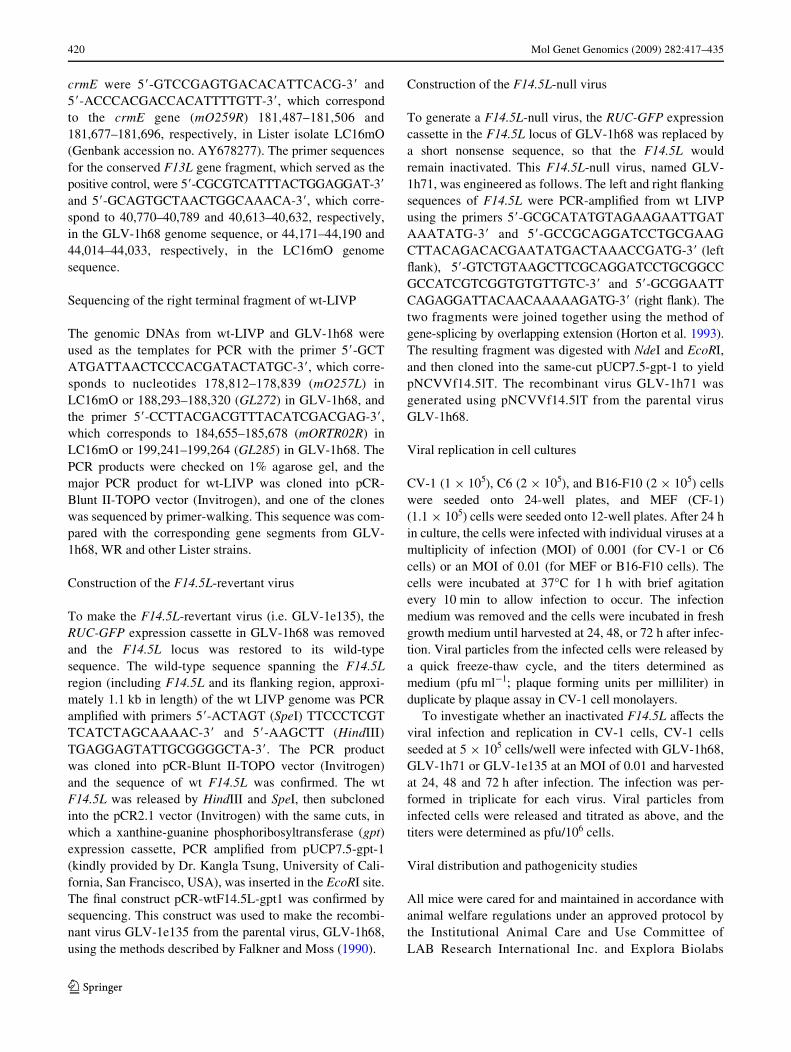
GLV-1h68 as a new virus with therapeutic potential wassequenced and then compared to other VACVs. We consid-ered (see “Discussion”) in particular the well known WRstrain and the COP strain. Further phylogenetic analysis
indicated that two Lister strains (Lister and LC16mO) werehighly similar to GLV-1h68, and that all three strains had arecent common ancestor (Fig. 1). GLV-1h68 seemed to becloser to WR than to COP, which was also supported bysequence comparison results that indicated that GLV-1h68shared more orthologous ORFs with WR than with COP(suppl. Fig. S1). Together with other VACV strains, such asACAM, DUKE and MVA, these strains form a vacciniacluster. The closest neighbors to the vaccinia cluster werecowpox virus (CP_GRI) and two monkeypox strains(MP_SL and MP_ZAI), followed by the camelpox strain(CAM_CMS). The three variola strains and the ectromeliavirus strain (ECT_MOS) were relatively distinct in the gen-erated tree.
Genomic features of GLV-1h68
The complete sequence of GLV-1h68 allowed bioinformaticanalysis of all genes and genomic features detected. GLV-1h68 was generated through three steps of construction asdescribed previously. The resulting virus contained threeforeign insertions of 1,958 bp, 8,606 bp, and 2,326 bp in theF14.5L, J2R, and A56R loci, respectively. With these threeinsertions, the size of the engineered GLV-1h68 virus wasfound to be 203,057 bp with 65.78% A + T content. Withoutthe insertions, the viral genome was found to be comprisedof 190,167 bp with 66.57% A + T content, and can be trans-lated into 238 “major” ORFs (not embedded ORFs, seebelow). Most of the ORFs encoded protein products largerthan 50 amino acids and were non-overlapping. Only a fewweakly overlapped with other ORFs. In addition, 13 shortORFs were included as “major” ORFs since they are presentin annotated ORFs of WR or COP. We also found 61“minor” ORFs embedded in larger ORFs within thegenome. In total, 299 potential ORFs, listed consecutivelyfrom left to right as they appeared in the genome, are sum-marized in suppl. Table S1 and S2. The homologs present inthe WR genome are also listed for comparison, and thenomenclature for the COP genome is used as reference. Thelengths, annotations and descriptions of some extremelyshort ORFs are listed in suppl. Table S3. The most strikingdiVerences among the ORFs of the three VACV strains arepresented in suppl. Table S4. The numbers of vaccinia majorand minor ORFs are summarized in suppl. Table S5.
Based on the annotation, we classiWed the major ORFs intosix functional categories. Figure 2 shows their distribution inthe viral genome. Sixty-one ORFs were likely to be involvedin host interactions and immune modulation, and most ofthese were found to be positioned in the left and right terminalregions and the edges of the central conserved region. Forty-nine ORFs encoding proteins involved in viral structure andassembly were located in the central conserved region, includ-ing viral core proteins, plaque forming, and EEV and IEV
123
422 Mol Genet Genomics (2009) 282:417–435
membrane proteins (pink). Genes involved in metabolism (41ORFs, orange) and genes involved in DNA replication, RNAtranscription and modiWcations (28 ORFs, green) were alsolocated in the central conserved region. The functions of 58ORFs remained uncertain or unidentiWed (marked with cleararrows). GL068 (F14.5L) was split into two parts by theRUC-GFP expression cassette (marked in gray, Fig. 2). Otherforeign insertions included lacZ in the TK locus, and gusA inthe HA locus.
The sequence data for the wt-LIVP showed sub-strainsand heterogeneity at the termini. There was a central con-served genomic region (from GL016 to GL276). It showedan unambiguous single nucleotide substitution (a GAAcodon was changed into TAA as compared to the sequencein WR or COP) in the middle of the J2R gene that created apremature stop codon. We further performed an in vitroassay (Mackett et al. 1982) which also demonstrated thatthe LIVP strain indeed had a TK negative phenotype for itswild type population (data not shown).
Comparison of ORFs involved in host-range selection and immune modulation
The majority of the diVerences in ORFs among the GLV-1h68, WR and COP strains can be attributed to the ORFs
encoding ankyrin-like proteins (Table 1). Table 1 presentsadditionally a variety of viral encoded complement, cyto-kine and chemokine inhibitors, and caspase inhibitors.Most of these immunomodulators were very well con-served among three VACV strains, usually with over 99%identity in the amino acid sequences. However, we foundseveral genes that were diVerent in GLV-1h68 than in WR.GLV-1h68 had an intact A39R, encoding a semaphorin-likeprotein, whereas, in WR, this protein was not expressed dueto the presence of a frame-shift mutation. In addition, theTNF-receptor-like protein (vTNFR) encoded by A53R geneseemed to be intact in GLV-1h68, but was fragmented inWR. On the other hand, SPI-2/CrmA, which inhibits Fas-mediated apoptosis, was intact in WR (WR195) but frag-mented in GLV-1h68. It is also interesting to note thatGLV-1h68 had two copies of genes encoding for serineprotease inhibitor (SPI-1) and interleukin-18-binding pro-tein, whereas in WR only one copy of each was present.
GLV-1h68 is a unique isolate from LIVP
SpeciWc genomic features point to GLV-1h68 being aunique isolate from LIVP. Thus VACV strains USSR,Lister, and Evans produce an additional soluble vTNFR,named CrmE. To our surprise, however, we could not
Fig. 1 Phylogenetic tree con-structed from orthopoxvirus genomes. The unrooted tree was generated by neighbour-joining, comparing highly conserved 82 kb nucleotide sequence among 16 OPV genomes. Boot-strapping values (percentage; 1,000 trials) are shown beside the nodes. Please see the list of abbreviations and accession numbers for virus identiWcation
123

Mol Genet Genomics (2009) 282:417–435 423
identify a crmE-like sequence in the genome of GLV-1h68.To conWrm this Wnding, the genomic DNAs from wt-LIVP,GLV-1d27, and GLV-1h68 viruses were used as the tem-plates for PCR ampliWcation of the speciWc crmE gene frag-ment. The PCR results indicated that wt-LIVP was crmEpositive, but that GLV-1d27 and GLV-1h68 were not(Fig. 3a).
This unexpected Wnding regarding the loss of the crmEgene led us to further analyze the diVerences betweenGLV-1h68, wt-LIVP and other Lister strains. The genomicDNAs from GLV-1h68 and wt-LIVP were used as the tem-plates for PCR with primers corresponding to GL272 andGL285 in GLV-1h68 or mO257L and mORTR02R inLC16mO, respectively. As predicted, the PCR product forGLV-1h68 was 10,971 bps (Fig. 3b), which was also thesize of the PCR product from its parental virus GLV-1d27or GLV-1f65 (data not shown). The PCR product forwt-LIVP, however, was much smaller, close to the sizepredicted for LC16mO (5,866 bps) (Fig. 3b).
The sequence of the major PCR product from wt-LIVPwas analyzed and compared with the corresponding regionsfrom GLV-1h68, WR, and other Lister strains (e.g.LC16mO) (Fig. 4). In addition to the missing crmE gene(ORF mO259R), mO260R, mO261R, mO262L andmORTR01R (present in the Lister strain LC16mO and wt-LIVP) were also missing in GLV-1h68. mO261R encodesa newly identiWed anti-apoptotic protein that is locatedin the Golgi, so-called viral Golgi anti-apoptotic protein(v-GAAP).
GLV-1h68 carries genes of WR-like origin
In contrast to the missing Lister genes, GL274 (encodingIFN-�/�-receptor-like secreted glycoprotein) and GL275(encoding ankyrin-like protein) were found in the GLV-1h68 genome, with orthologs present in both WR andCOP. These ORFs were absent from wt-LIVP, and allother Lister strains or derivatives sequenced (AY678275,
Fig. 2 GLV-1h68 genome map. Predicted ORFs and their lengths arerepresented by colored arrows. Sixty-one ORFs involved in host inter-actions and immune modulation are shown in blue. Located mostly inthe central conserved region are genes involved in: viral structure andassembly (49 ORFs, shown in pink), metabolism (41 ORFs, shown in
orange) and DNA replication, RNA transcription and modiWcations(28 ORFs, in green). The functions of 58 ORFs remain uncertain(marked with clear arrows). The three foreign insertions are marked indark grey. The genome size is indicated on the right (nucleotides)
123

424 Mol Genet Genomics (2009) 282:417–435
AY678276, AY678277, DQ121394). Moreover, the neigh-boring ORF upstream of crmE in the Lister strain, mO258R(413aa), was extended into GL273 (574aa) in GLV-1h68,
suggesting an island of sequence insertion within and fol-lowing this ORF. Considering the length diVerences of theinverted terminal repeat (ITR) between GLV-1h68 andother Lister strains, we decided to analyze the ORFs thatwere absent from the right terminus of the Lister strains,i.e., GL277-283. The comparison results indicated thatthese ORFs were copies from the left terminus, includingmO008R which were fragmented into GL013/279 andGL014/278 (Fig. 4). This additional copy of nucleotidesequence largely extended the ITR size in GLV-1h68 incomparison to its parental LIVP or Lister strain.
Viral replication in diVerent cell lines
In our previous studies (Zhang et al. 2007), GLV-1h68 wasevaluated in human breast tumor cell line GI-101A toexamine whether insertions in the F14.5L, J2R or A56Rlocus aVected its activity. In this study, complementary tothe broad comparison of genomic and pathogenomicfeatures just detailed, infection studies were carried out infurther tumor cell lines to compare the infectivity and repli-cation of individual recombinant viruses (Fig. 5b). In CV-1
Table 1 Major ORFs involved in host-range selection and immune modulation
a Denotes the corresponding ORF regions present in both WR and COP strains, with locations listed in the suppl. Table S3b Frame-shift mutation
–, No ortholog present
Fig. 3 The PCR results indicate that the parental strain of GLV-1h68 wasa unique isolate from LIVP. a The genomic DNAs from wt-LIVP, GLV-1d27, and GLV-1h68 viruses were used as the templates for PCR ampliW-cation of the speciWc crmE gene fragment and the conserved F13L genefragment. The PCR results show that, while wt-LIVP population was crmEpositive, GLV-1d27 and GLV-1h68 were not; b The genomic DNAs fromwt-LIVP and GLV-1h68 were used as the templates for PCR with primerscorresponding to mO257L and mORTR02R in LC16mO or GL272 andGL285 in GLV-1h68, respectively. The PCR product for GLV-1h68 was10,971 bps as predicted. The major PCR product for wt-LIVP was muchsmaller, similar to the size predicted for LC16mO (5,866 bps). This furtherconWrms that GLV-1h68 was derived from a unique isolate of LIVP
123

Mol Genet Genomics (2009) 282:417–435 425
cells all recombinant viruses replicated very well, andshowed comparable yield, similar to wt-WR and wt-LIVPviruses. In C6 glioma cells, however, the recombinantviruses, and particularly GLV-1h68, showed a reduced rep-lication of up to 102-fold, compared to wt-LIVP (Fig. 5b).Cells of mouse origins required a higher MOI for detectableinfection, which was true for both primary MEFs and thetumor cell line B16-F10. At an MOI of 0.01, the wt-LIVPreplicated fairly well in MEFs, but the three recombinantLIVP viruses did not replicate. In B16-F10 melanomacells, all viruses showed limited infection and replication(Fig. 5b).
Pathogenicity of individual VACVs
The pathogenicity of individual VACVs was already evalu-ated in female nude mice bearing human GI-101A xeno-grafts (Zhang et al. 2007). The current study examinedwhether the individual VACVs showed diVerential patho-genicity in male mice after a single intravenous injection of1 £ 107 pfu of virus, and whether an intact immune systemcould overcome the infections of VACVs. In C6 glioma-bearing nude male mice, wt-WR was extremely virulent(Fig. 5c). Mice exhibited over 30% body weight loss byday 7 after virus injection, and all mice died by day 11.Pocks on the tail and footpad and swelling of the mouthregion were observed. Male mice injected with the TK¡
WR strain lost 22% of their body weight by the end of the14 days, similar to mice injected with wt-LIVP strain. Incontrast, no signiWcant weight loss was observed in miceinjected with GLV-1h68 or other recombinant viruses. Theimmunocompetent C57BL/6 mice, bearing B16-F10 mela-noma on foot pad, showed a greater tolerance to viral path-ogenicity (Fig. 5c). All mice injected with the wt-WR strainsurvived for the duration of the study; however, on averageover 10% of body weight was lost by day 10. Male miceinjected with the TK¡ WR or the LIVP strains showed littleweight loss, suggesting that a competent T-cell systemplays an important role in Wghting against virus infection.
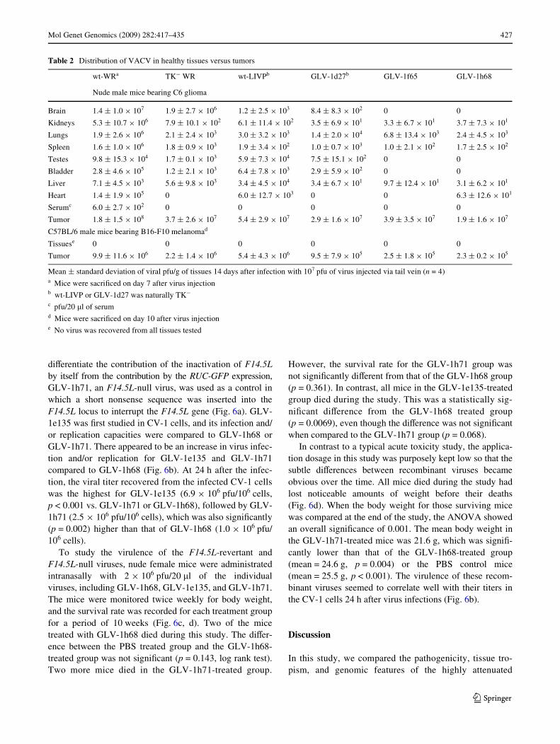
Tissue distribution of individual VACVs
We next looked at virus localization in individual tis-sues. Table 2 summarizes the virus distribution data inC6 tumor-bearing nude male mice after intravenousinjection of individual virus strains. In this experiment,mouse organs were collected 14 days after virus injec-tion. However, mice injected with wt-WR were sacri-Wced and analyzed on day 7 due to extreme toxicity. Asshown in Table 2, the wt-WR injection resulted in abroader distribution of the virus in healthy mouse tis-sues, including an alarmingly high titer in the brain. Theinactivation of the TK gene led to signiWcant reductionsin the viral titers in mice injected with the TK¡ WRstrain; however, viral titers of up to 106 pfu per gramof tissue were still present in the brain tissues. Thewt-LIVP, on the other hand, showed a markedly diVerentdistribution pattern. No viral particles were recoveredfrom the brain tissues in three of the four tumor-bearingmice. Mice injected with GLV-1h68 had no virus recov-ery from brain, testis, or bladder tissues, and very lowlevels of viral particles recovered from kidney, liver, orheart tissues in only one of the four mice. Viral particleswere also recovered from the spleen tissues in two of themice. Lung tissues seemed to be relatively susceptible tothe GLV-1h68 infection, with up to 103 pfu of the virusper gram of tissue found in three mice. The implantedC6 glioma tumors, however, were colonized heavilywith either WR or LIVP strains. The greatly reduced bio-distribution of GLV-1h68 in various mouse organs wasin agreement with the viral replication data in mouse primarycells (MEFs in Fig. 5b).
In melanoma-bearing immunocompetent mice, no virusparticles were recovered from non-tumor tissues 10 daysafter the virus injection (Table 2), even for mice injectedwith wt WR, suggesting the importance of T cells in theWght against viral infection. The viral titers were also lowerin the tumor tissues, in parallel with the in vitro replicationdata (Fig. 5b).
Fig. 4 GLV-1h68 showed a unique genetic makeup that was distinctfrom its parental LIVP strain. Genes outlined by navy blue have corre-sponding paralogs in the left terminus. Genes in light blue are uniquefor WR strain, and those in light orange are unique for GLV-1h68. Theorthologous genes are linked by dashed lines. GL277 (encoding inter-
leukin-18-binding protein) in GLV-1h68 had an ortholog WR013 in theleft terminus of WR genome. The majority of the wt-LIVP populationhad sequences in the CrmE (mO259R) and v-GAAP (mO261R) genesegments that are similar to other Lister strains
123

426 Mol Genet Genomics (2009) 282:417–435
The contribution of F14.5L inactivation
Given the promising data on GLV-1h68 from infectionstudies and genome comparisons, direct experimental testsprobed next the pathogenicity eVect of individual genes.Unlike the well known TK or HA gene in the VACV
genome, the F14.5L is relatively new and less studied. Wetherefore focused our direct experimental tests on whetherthe insertion of an expression cassette in the F14.5L con-tributed to the attenuation of GLV-1h68. We constructed anF14.5L-revertant virus GLV-1e135, in which the F14.5Lwas restored to its wild-type LIVP sequence (Fig. 6a). To
Fig. 5 Replication and pathoge-nicity of diVerent VACV strains. a Genetic constructs for diVerent VACV strains (reproduced from Zhang et al. 2007). A single base substitution created a premature stop codon; therefore, the TK gene in LIVP wt or GLV-1d27 was naturally inactivated. pSEL, VACV synthetic early late pro-moter; p7.5 and p11K, VACV 7.5 early/late and 11k promoters; TFR, human transferrin recep-tor. b The replication of diVerent VACV strains was studied in various cell cultures. c The path-ogenicity was evaluated by change in body weight after a single, intravenous injection of each individual virus at 107 pfu per mouse, in nude mice bearing C6 glioma, and C57BL/6 mice bearing B16-F10 melanoma. Change of body weight (%) was calculated as follows: [(b� ¡ t�) ¡ (b ¡ t)] £ 100/(b ¡ t), where b and t are the body weight and tumor weight (estimated by tumor size; 1 cm3 = 1 g) on the day of virus injection, and b� and t� are the corresponding weights on the day of monitoring. The average weight change for each group is presented
123
Mol Genet Genomics (2009) 282:417–435 427
diVerentiate the contribution of the inactivation of F14.5Lby itself from the contribution by the RUC-GFP expression,GLV-1h71, an F14.5L-null virus, was used as a control inwhich a short nonsense sequence was inserted into theF14.5L locus to interrupt the F14.5L gene (Fig. 6a). GLV-1e135 was Wrst studied in CV-1 cells, and its infection and/or replication capacities were compared to GLV-1h68 orGLV-1h71. There appeared to be an increase in virus infec-tion and/or replication for GLV-1e135 and GLV-1h71compared to GLV-1h68 (Fig. 6b). At 24 h after the infec-tion, the viral titer recovered from the infected CV-1 cellswas the highest for GLV-1e135 (6.9 £ 106 pfu/106 cells,p < 0.001 vs. GLV-1h71 or GLV-1h68), followed by GLV-1h71 (2.5 £ 106 pfu/106 cells), which was also signiWcantly(p = 0.002) higher than that of GLV-1h68 (1.0 £ 106 pfu/106 cells).
To study the virulence of the F14.5L-revertant andF14.5L-null viruses, nude female mice were administratedintranasally with 2 £ 106 pfu/20 �l of the individualviruses, including GLV-1h68, GLV-1e135, and GLV-1h71.The mice were monitored twice weekly for body weight,and the survival rate was recorded for each treatment groupfor a period of 10 weeks (Fig. 6c, d). Two of the micetreated with GLV-1h68 died during this study. The diVer-ence between the PBS treated group and the GLV-1h68-treated group was not signiWcant (p = 0.143, log rank test).Two more mice died in the GLV-1h71-treated group.
However, the survival rate for the GLV-1h71 group wasnot signiWcantly diVerent from that of the GLV-1h68 group(p = 0.361). In contrast, all mice in the GLV-1e135-treatedgroup died during the study. This was a statistically sig-niWcant diVerence from the GLV-1h68 treated group(p = 0.0069), even though the diVerence was not signiWcantwhen compared to the GLV-1h71 group (p = 0.068).
In contrast to a typical acute toxicity study, the applica-tion dosage in this study was purposely kept low so that thesubtle diVerences between recombinant viruses becameobvious over the time. All mice died during the study hadlost noticeable amounts of weight before their deaths(Fig. 6d). When the body weight for those surviving micewas compared at the end of the study, the ANOVA showedan overall signiWcance of 0.001. The mean body weight inthe GLV-1h71-treated mice was 21.6 g, which was signiW-cantly lower than that of the GLV-1h68-treated group(mean = 24.6 g, p = 0.004) or the PBS control mice(mean = 25.5 g, p < 0.001). The virulence of these recom-binant viruses seemed to correlate well with their titers inthe CV-1 cells 24 h after virus infections (Fig. 6b).
Discussion
In this study, we compared the pathogenicity, tissue tro-pism, and genomic features of the highly attenuated
Table 2 Distribution of VACV in healthy tissues versus tumors
Mean § standard deviation of viral pfu/g of tissues 14 days after infection with 107 pfu of virus injected via tail vein (n = 4)a Mice were sacriWced on day 7 after virus injectionb wt-LIVP or GLV-1d27 was naturally TK¡
c pfu/20 �l of serumd Mice were sacriWced on day 10 after virus injectione No virus was recovered from all tissues tested
wt-WRa TK¡ WR wt-LIVPb GLV-1d27b GLV-1f65 GLV-1h68
Nude male mice bearing C6 glioma
Brain 1.4 § 1.0 £ 107 1.9 § 2.7 £ 106 1.2 § 2.5 £ 103 8.4 § 8.3 £ 102 0 0
Kidneys 5.3 § 10.7 £ 106 7.9 § 10.1 £ 102 6.1 § 11.4 £ 102 3.5 § 6.9 £ 101 3.3 § 6.7 £ 101 3.7 § 7.3 £ 101
Lungs 1.9 § 2.6 £ 106 2.1 § 2.4 £ 103 3.0 § 3.2 £ 103 1.4 § 2.0 £ 104 6.8 § 13.4 £ 103 2.4 § 4.5 £ 103
Spleen 1.6 § 1.0 £ 106 1.8 § 0.9 £ 103 1.9 § 3.4 £ 102 1.0 § 0.7 £ 103 1.0 § 2.1 £ 102 1.7 § 2.5 £ 102
Testes 9.8 § 15.3 £ 104 1.7 § 0.1 £ 103 5.9 § 7.3 £ 104 7.5 § 15.1 £ 102 0 0
Bladder 2.8 § 4.6 £ 105 1.2 § 2.1 £ 103 6.4 § 7.8 £ 103 2.9 § 5.9 £ 102 0 0
Liver 7.1 § 4.5 £ 103 5.6 § 9.8 £ 103 3.4 § 4.5 £ 104 3.4 § 6.7 £ 101 9.7 § 12.4 £ 101 3.1 § 6.2 £ 101
Heart 1.4 § 1.9 £ 105 0 6.0 § 12.7 £ 103 0 0 6.3 § 12.6 £ 101
Serumc 6.0 § 2.7 £ 102 0 0 0 0 0
Tumor 1.8 § 1.5 £ 108 3.7 § 2.6 £ 107 5.4 § 2.9 £ 107 2.9 § 1.6 £ 107 3.9 § 3.5 £ 107 1.9 § 1.6 £ 107
C57BL/6 male mice bearing B16-F10 melanomad
Tissuese 0 0 0 0 0 0
Tumor 9.9 § 11.6 £ 106 2.2 § 1.4 £ 106 5.4 § 4.3 £ 106 9.5 § 7.9 £ 105 2.5 § 1.8 £ 105 2.3 § 0.2 £ 105
123

428 Mol Genet Genomics (2009) 282:417–435
recombinant GLV-1h68 with WR, COP and related viridae.The in vivo studies show the reduced pathogenicityphenotype of GLV-1h68, previously conWrmed in femalenude mice bearing GI-101A breast tumor xenografts(Zhang et al. 2007), and now in male nude mice bearingC6 glioma and a melanoma model in immunocompetentmice. The comparative genome sequence analysis oVereddetailed functions of all infection-involved individualgenes, and showed the uniqueness of GLV-1h68 and itsevolutionary relationship. These Wndings provided a foun-dation for further studies on the eVects of individual genes
and gene variants. Detailed experimental examination ofthe eVects of individual ORFs is time-consuming and willrequire a number of separate studies. Our study herefocused on the eVects of individual disruptions andinserted cassettes in the F14.5L, J2R and A56R loci ofGLV-1h68 that resulted in reduced pathogenicity in mice.This is the Wrst study attempting to diVerentiate the contri-bution of an inactivated viral gene (e.g. F14.5L) from thatof an expression cassette (in the F14.5L locus) on theattenuation of a VACV (e.g. GLV-1h68). In the followingdiscussion, we present the combined insights from both
Fig. 6 Contribution of an expression cassette in the F14.5L locus on the attenuation of GLV-1h68. a The schematic presentation of the F14.5L-null and revertant viruses derived from GLV-1h68. b The replica-tion of the F14.5L-null and revertant viruses was studied in CV-1 cells, and compared to that of GLV-1h68. The virulence of the F14.5L-null and revertant viruses was investigated in nude mice by an intranasal application of 2 £ 106 pfu of individual virus per mouse. The survival and body weight of the mice were monitored for 10 weeks and presented in c and d
123

Mol Genet Genomics (2009) 282:417–435 429
the bioinformatics and the experimental approaches appliedin this study.
Comparison choice regarding diVerent vaccinia strains
Some of the vvDD strains and the Wyeth TK-strains havebeen used as oncolytic strains under clinical investiga-tion. They would be ideal candidates for comparison withGLV-1h68, which is now also as GL-ONC-1 undergoinga Phase I/II trial. Unfortunately, the genome sequence forany of these strains is not available or publicly accessi-ble. The vvDD strains were derived from the WR strain,with the double deletion of VGF and TK. However, it isnot safe to assume that a vvDD strain would have a simi-lar genomic sequence as that of wt WR, given the factthat GLV-1h68, derived from an isolate from LIVP,showed a unique genetic make up very diVerent fromLIVP. Therefore, we chose to use the WR wt, which has acomplete genome sequence for analyzing, and a WRTK¡ strain, since we know that LIVP (wt) was a TK¡
strain, as the best available strains for comparison. On topof that, the WR strain has been the most widely studiedstrain in laboratories all over the world. We would gainmore insight from the comparison with these laboratoryWR strains.
Even though we did not directly compare our strainswith the vvDD strains or the Wyeth TK¡ strains, from pub-lished data, we can get a general idea what could be pre-dicted from a direct comparison. During the construction ofa vvDD strain, two copies of the viral VGF genes were Wrstinactivated by the insertion of the lacZ gene under the con-trol of the p11 promoter, and the resulting mutant vSC20(Buller et al. 1988) was used as the parental virus for thedouble deletion mutant vvDD, in which the TK gene wasalso inactivated by the insertion of one or more foreigngene expression cassettes. In one of the vvDD strains,vvDD-GFP (McCart et al. 2001), the EGFP gene under thecontrol of a synthetic early-late promoter, and the gpt geneunder the control of the p7.5 promoter were inserted in theTK locus. In nude mice injected with 107 pfu of vvDD-GFP, the median viral recovery from ovary tissues was ashigh as 8.6 £ 106 pfu/mg total protein 8 days after theinfection, suggesting that neither VGF nor TK was respon-sible for gonadotropism in female mice. However, bothVGF¡ and TK¡ contributed to reduced viral replication inmouse brain. If we would like to Wnd out which gene(s)contribute to gonadotropism in female mice, a genomiccomparison with GLV-1h68 would help to narrow downthe selection of the candidates. Of course, considerableamount of studies are required to pinpoint the exactgene(s). We hope that with this work including the detailedtriple genome comparison, the combined eVorts from ourscientiWc community would speed up the discovery
process, and eventually, an optimized oncolytic virus withmaximum oncolytic activity yet minimum pathogenicitywill become available to beneWt cancer patients.
ORFs involved in host-range selection
Many diVerences in ORFs among the GLV-1h68, WR andCOP strains can be attributed to the ORFs encoding anky-rin-like proteins (Table 1). Because these proteins controlthe interactions between the integral membrane proteinsand the elements of the cell cytoskeleton, the proteinscould be involved in functions related to transcriptionalinitiation, cell cycle regulation, cytoskeleton formation,ion transport and signal transduction (Lux et al. 1990;Mosavi et al. 2004). It has been proposed that diVerent setsof the ankyrin-like proteins encoded by VACVs, as well asthe level of their synthesis, allow for the Wne regulation ofvirus replication and the determination of tissue tropism ininfected animals (Shchelkunov et al. 1993). Recent studieson the M-T5 host-range protein of myxoma virus, whichcontains multiple ankyrin repeats, illustrate that interac-tions of M-T5 with Akt and cullin-1 (both components ofhost cell signaling networks) can have a profound impacton poxvirus tropism (Johnston et al. 2005; Wang et al.2006).
It has been described that the deletion of two vacciniahost-range genes (K1L and C7L) greatly reduces the abilityof VACV to replicate in a variety of human cell lines(Gillard et al. 1986). Restoration of the K1L gene in the MVAgenome, however, failed to release the virus from its hostrestriction on monkey or human cells (Perkus et al. 1990),suggesting that there are other host-range determinationgenes yet to be recognized. K1L and C7L seemed to be wellconserved among GLV-1h68, WR and COP. Yet, we foundthat, while GLV-1h68, along with the other two recombi-nant viruses, infected and replicated in human, monkey andrat cells, it did not replicate in mouse embryonic Wbroblasts(MEFs) (Zhang et al. 2007; Fig. 5b). In contrast, wt-LIVPdid replicate in MEFs, giving a virus yield of just 10-foldless than wt-WR (Fig. 5b). It is reasonable to speculate thatthe restricted replication of these three recombinant virusesin MEFs was caused by the inactivation of the F14.5Llocus. Our unpublished data, however, suggest that the dis-rupted F14.5L was not responsible for this restricted repli-cation. We have observed that a virus isolate derived fromGLV-1h68, which had a restored F14.5L, failed to replicatein the MEFs as well (data not shown). Now we know thatwt-LIVP was a mixed population, and that GLV-1h68 wasderived from a unique isolate from this mixed population,a genome sequencing and comparative analysis with anLIVP isolate capable of replicating in MEFs would help toidentify genes responsible for the restricted replication ofGLV-1h68 in primary mouse cells.
123

430 Mol Genet Genomics (2009) 282:417–435
ORFs involved in host immune modulation
B28R and A53R, encoding two vTNFR proteins, are equiv-alent to Cowpox virus (CPV) genes cytokine responsemodiWer B (crmB) and crmC, respectively. However, bothB28R and A53R are fragmented and found to be inactive inmost VACV strains, including WR and COP (Alcami et al.1999). Nevertheless, GLV-1h68 was found to carry anintact A53R gene (GL241), which encoded a secreted TNF-receptor-like protein of 186aa that was the same lengthas CPV crmC (Fig. 7). CrmC was also found intact inthe Lister isolate VACV107 (GenBank accession no.DQ121394). As a soluble vTNFR that interferes with hostimmune functions, A53R in GLV-1h68 could be a potentialcandidate for disruption, in order to further attenuate thevirus.
Unlike USSR, Evans and other Lister derivatives,GLV-1h68 did not have a crmE-like sequence in thegenome. USSR recombinants lacking crmE were found tobe attenuated, while the expression of an inserted crmEgene in the genome of WR resulted in enhanced virulence(Reading et al. 2002). The crmE deWciency in GLV-1d27or GLV-1h68, therefore, may have contributed to agreater attenuation in comparison to wt-LIVP (the major-ity of wt-LIVP was crmE positive). VACV strains Lister,USSR and Evans also express the v-GAAP protein. Thedeletion of this gene from VACV does not aVect virusreplication in cell cultures, but signiWcantly alters virusvirulence in murine infection models (Gubser et al. 2007).GLV-1h68 does not have a v-GAAP gene. This genewould therefore be a good candidate to be inserted intothe genome of GLV-1h68 to bring additional attenuationto the virus.
It has been shown that deletion of the CPV 38 K geneencoding the well-known orthopoxvirus serpin (also knownas SPI-2 or CrmA) in either the cowpox or rabbitpox virusgenome resulted in attenuation in murine intranasal models(Thompson et al. 1993). The deletion mutants from the WRstrain that lack the SPI-2 or SPI-1 coding sequence displaysimilar in vitro replication proWles and in vivo virulence aswt-WR and the revertant viruses (Kettle et al. 1995). How-ever, the derivative of the WR strain with both SPI-1 andSPI-2 deleted has been shown to be signiWcantly attenuatedand less pathogenic to mice (Guo et al. 2005). The incon-clusive Wndings on SPI-1 and SPI-2 may have resulted fromthe diVerence in the way the “deletion” mutants are con-structed (more details in later discussion). Nevertheless, ifinactivation of the SPI-2 gene indeed reduces virulence ofthe virus, the fragmented SPI-2 gene in GLV-1h68 (GL266and GL267) may have contributed to its attenuation. Fur-thermore, the lack of a functional SPI-2 gene product mayenhance the immunogenicity of the virus (Zhou et al.1990).
It has been observed that A39R induces robust responsesin human monocytes, including cell aggregation, inductionof proinXammatory cytokines, and upregulation of themonocyte cell surface marker ICAM-1. All of these eVectssuggest that A39R inXuences the immune responses of vac-cinia-infected hosts (Comeau et al. 1998). The A39R pro-tein is found to be expressed by VACV strains COP, Evans,Lister, and USSR, but not WR. Expression of an insertedCOP A39R gene in the WR strain was found to be associ-ated with an increase in inWltration and oedema formation,suggesting that the A39R protein has pro-inXammatoryproperties (Gardner et al. 2001). The fact that A39R wasintact in LIVP and in GLV-1h68, but truncated in WR,
Fig. 7 Sequence alignment of crmC gene for selected poxvirusstrains. The ORFs for CrmC in selected poxvirus strains were trans-lated into amino acids and their sequences were aligned using Clu-stalX. GLV-1h68 exhibits an intact crmC gene (GL241), which ishighly identical to List172 in Lister isolate VACV107, and A56 gene
in Cowpox virus, all of which contain a typical signal peptide sequencepredicted by SignalP 3 software. Lister strain LC16mO seems to havea full length protein, but its N-terminal sequence suggests that this pro-tein is not secreted. The crmC genes in VACV strains Copenhagen andWR are truncated
123

Mol Genet Genomics (2009) 282:417–435 431
suggests diVerences in immunogenicity, but requires furtherexperimental conWrmation.
Genome evolution of GLV-1h68
Our data also suggested a unique infection and replicationproWle of GLV-1h68 that was distinctively diVerent fromits parental strain LIVP. This distinction was particularlyevident in mouse primary cells, which warranted a closerlook at its genome evolution. A comparison of the sequenceof the major PCR product on the right terminal region ofwt-LIVP with GLV-1h68 supports our hypothesis thatGLV-1h68 was a unique isolate that diVered from themajority of the wt-LIVP population (Figs. 3, 4). GLV-1h68showed a unique genetic makeup which shared genes simi-lar to those of WR strains.
These changes illustrated that the parental strain ofGLV-1h68 probably underwent two major rearrangementevents. First, there was perhaps a genetic exchange with aWR-like VACV strain regarding the right terminal region.The C-terminal of GL273 was most likely the site of therecombination segment, since it was highly homologous toWR199. GL274, GL275 and GL276 were also acquired dur-ing the same genetic exchange, as they were similar to thedownstream WR200, WR202 and WR201 genes, respec-tively. A second major rearrangement occurred when theparental strain lost part of its newly acquired WR-likesequences at the region, and a copy of its left terminalsequence, GL001 to GL015 genes, was duplicated, and thenappeared in its right terminal region. This exchange proba-bly occurred in a laboratory where WR strains and theparental strains of GLV-1h68 were used at the same time.The sequence of the major PCR product on the right termi-nal region of GLV-1d27 or GLV-1f65 was not determined,but it is very likely to be similar, if not identical, to that ofGLV-1h68, given the fact that the same size PCR productwas obtained, and the history of stepwise virus constructionand puriWcation.
The contribution of F14.5L inactivation
F14.5L, reported as WR053.5 in WR, encodes a protein of49 amino acids with a secretory signal peptide. This ORFwas very well conserved across diVerent VACV strains, aswell as in other poxviruses. There is a NotI restriction sitein the middle of F14.5L sequence, which was used to inserta RUC-GFP expression cassette to create the recombinantvirus GLV-1d27, and subsequently the double-mutant virusGLV-1f65, and the triple-mutant GLV-1h68 (Timiryasovaet al. 2000; Zhang et al. 2007). Our studies with theF14.5L-revertant and the F14.5L-null viruses suggest thatthe F14.5L locus in viral genome can be disrupted as a wayto attenuate VACV. The eVect of inactivation of F14.5L
locus by itself was not very substantial, but it would likelybecome statistically signiWcant with a larger sample size.The expression of an exogenous gene in the F14.5L locusputs extra transcriptional and translational burden on thevirus and, together with the inactivation of F14.5L, resultedin a signiWcantly attenuated virus: GLV-1h68. It is possiblethat the expressed GFP could be toxic to some host cellsthat would inhibit viral replication. However, our unpub-lished data suggest that other foreign genes expressed in thesame locus also attenuated the virus to similar extent; there-fore the reduced virulence was not likely due to the toxicitybrought by GFP. In a recent publication, WR53.5 is foundto be an envelope protein of an intracellular mature virusthat mediates cell adhesion (Izmailyan and Chang 2008). Inthat study, the WR53.5-deletion mutant virus, which con-tains a gpt expression cassette in the WR53.5 locus, exhibitsreduced virulence in mice. It is conceivable that the expres-sion of gpt, in addition to the inactivation of WR53.5L, mayhave contributed to the reduced virulence.
The contribution of J2R inactivation
The TK negative phenotype was conWrmed for the wildtype LIVP population. A TK¡ virus requires TTP for DNAsynthesis, leading to a preferential viral replication in divid-ing cells and ultimately enhanced tumor speciWcity andminimal infection of neighbor tissues. The TK¡ phenotypemay explain, at least in part, why the wt-LIVP strainappeared to be less virulent than wt-WR in animal studies(Fig. 5c; Table 2); however, it cannot explain the observeddiVerences in neurovirulence and gonadotropism betweenLIVP and WR strains. We saw earlier in female nudetumor-bearing mice that, even as disruption of the TK genein the WR genome contributed signiWcantly to reducedviral entry or replication in brain tissues (a 3-log reduction),merely a 2.5-fold reduction was observed in ovary tissues(Zhang et al. 2007). In this study, the disruption of the TKgene in the WR genome resulted in on average a 7-fold anda 58-fold reduction in viral titers in the brain and testis tis-sues, respectively (Table 2). We therefore conclude that,while a TK¡ phenotype in the WR strain signiWcantlyreduced its neurovirulence (more evident in female mice)and reduced gonadotropism in male mice, it had little eVecton gonadotropism in female mice. In contrast, despite thefact that the brain and ovary tissue were found to be free ofviral particles in mice treated with either wt-LIVP or GLV-1h68 (both were TK¡) (Zhang et al. 2007), the wt-LIVP didshow a similar viral titer in testes as the WR strains in thisnew study (Table 2). We suspect that gene(s) other than theTK¡ phenotype contributed to the absence of gonadotro-pism in mice observed for the three recombinant virusesderived from LIVP, including GLV-1h68. However, it wasnot clear how much a TK¡ phenotype in the LIVP strains
123

432 Mol Genet Genomics (2009) 282:417–435
might have contributed to the absence or reduced neurovi-rulence and gonadotropism in mice.
It was also shown that the distribution of GLV-1f65 wasfurther reduced in the normal tissues when compared tothose injected with GLV-1d27 (Table 2). Both viruses wereTK¡, and the only diVerence was that GLV-1f65 containeda foreign gene expression cassette in its J2R locus. Similarto the foreign gene (RUC-GFP) expression in the F14.5Llocus that contributed to the attenuation of GLV-1h68, theexpression of lacZ in the J2R locus may have further con-tributed to the attenuation of GLV-1f65, and the attenuationof GLV-1h68. In the survival study reported previously(Zhang et al. 2007) the mice injected with GLV-1f65 didoutlive the ones injected with GLV-1d27.
The contribution of A56R inactivation
The A56R gene, which encodes hemagglutinin, is anothernonessential gene that can be inactivated to obtain a moreattenuated virus. It has been observed that inactivation ofthe HA gene leads to greater attenuation of WR than ofLC16mO (a temperature-sensitive and lower neuroviru-lence strain isolated from the Lister LO strain) and to noapparent attenuation of LO-1 (an isolate of Lister Elstreestrain) (Shida et al. 1988). In our studies, disruption of theHA gene by the insertion of an expression cassette furtherattenuated the virus (Zhang et al. 2007). While miceinjected with GLV-1f65 (F14.5L¡ and TK¡) lived up to116 days after virus injection, all of the mice injected withGLV-1h68 (F14.5L¡, TK¡ and HA¡) survived for the dura-tion of study (130 days post virus infection). Further studiesare needed to investigate how much of the attenuation inGLV-1h68 could be attributed to the inactivation of HAitself, and how much was contributed by the expression of aforeign gene (gusA) in the A56R locus.
The contribution of foreign gene expression
GFP expression could be toxic to the host cells and mayhave reduced the replication of GLV-1h68, thus the GFPwould be the cause of the reduced pathogenicity. Data pub-lished before (Zhang et al. 2007) and here suggest that theexpression of �-galactosidase in the TK locus attenuatedthe virus as well. We are currently conducting a whole setof studies in which the strength of diVerent viral promoters,the size of the transgenes, the 2nd structure of the tran-scripts, and the locus of the insertions are the factors underinvestigation. Our preliminary data suggest that the obser-vation that transgene expression appears to reduce viralreplication is generally true. All these data, when theybecome available, will be presented in a new study.
A review of published data so far by other investigatorsalso suggests that the expression of inserted foreign gene(s)
may play a role in reduced virulence observed with thoseengineered viruses. For example, when the SPI-1 or SPI-2gene was simply deleted, no reduced virulence wasobserved (Kettle et al. 1995). But when both SPI-1 andSPI-2 genes were inactivated by the insertion of a func-tional gpt expression cassette and an Escherichia coli lacZgene expression cassette, respectively, the resulting mutantwas markedly attenuated (Guo et al. 2005). It is possiblethat the eVect of individual gene deletion was not signiW-cant, but the inactivation of both genes made the attenua-tion more evident. The way the double-mutant virus wasconstructed, however, has made us wonder how much ofthe attenuation was actually contributed by the expressionof transgenes.
General considerations and conclusions
We investigated the genetic diVerences between GLV-1h68and related VACVs. A Wrst comprehensive analysis includ-ing function predictions for all found genome diVerences iseasily gained by bioinformatics and comparative genomeanalysis, even though this method cannot detect and ruleout further diVerences on smaller polymorphisms (e.g.SNPs). Such more subtle eVects and diVerences requireextensive genetic and molecular studies to test individualgenes in a number of strains and their direct eVects in diVer-ent infection scenarios. Table 3 summarizes a number ofgenome diVerences, as examined and discussed here. It isimportant to realize that the functions of viral genes couldbe strain-speciWc, tissue-speciWc, or host-speciWc. The viru-lence genes, which are important for one VACV strain,may play a minimal role in the virulence for another VACVstrain. The inactivation of a certain viral gene may inXu-ence the replication of VACV in one tissue type but notother tissues in the same host, or may reduce the pathoge-nicity of the virus to one host species but not to others.There might also be synergistic eVects involving multipleviral genes. In this report, we focused our analysis on thediVerences between the viral genes that were absent or frag-mented in certain strains and the corresponding viral genesin the Lister, WR and COP strains. Using sequences avail-able from additional VACV genomes, in combination withfunctional transcription and proteome analysis, it is possi-ble to rationally design more optimized oncolytic virusstrains to beneWt cancer therapy in human patients. Theanalysis of GLV-1h68 is an important step towards thisgoal, as demonstrated here by direct sequence data, func-tional tests and tissue tropism.
This study also suggests that insertion of a foreigngene expression cassette in a nonessential locus in theviral genome is a practical way to attenuate VACVs,especially if the nonessential locus itself contains avirulence gene. The challenge will then be to reduce the
123

Mol Genet Genomics (2009) 282:417–435 433
virulence of the virus without compromising too muchon the replication competency of the virus, the key to itsoncolytic activity.
Acknowledgments This work was supported by research and devel-opment grants from Genelux Corporation and in part by DeutscheForschungsgemeinschaft (DFG) TR34/A5 and Bundesministeriumfür Bildung und Forschung (BMBF) Pathogenomik (CL) and LandBavaria (TD). The authors wish to thank Dr. Tatyana Timiryasova forher scientiWc and technical contribution, Mr. Terry Trevino andMs. Melody Jing for their technical support, and Ms. Andrea Feathersfor editorial support.
Open Access This article is distributed under the terms of the Crea-tive Commons Attribution Noncommercial License which permits anynoncommercial use, distribution, and reproduction in any medium,provided the original author(s) and source are credited.
References
Al’tshtein AD, Zakharova LG, Loparev VN, Pashvykina GV,Gorodetskii SI (1985) Isolation of a recombinant vaccinia virusbased on the LIVP strain inducing the surface antigen of thehepatitis B virus. Dokl Akad Nauk SSSR 285:696–699
Table 3 SigniWcant speciWc diVerences
123

434 Mol Genet Genomics (2009) 282:417–435
Alcami A, Khanna A, Paul NL, Smith GL (1999) Vaccinia virus strainsLister, USSR and Evans express soluble and cell-surface tumournecrosis factor receptors. J Gen Virol 80:949–959
Antoine G, ScheiXinger F, Dorner F, Falkner FG (1998) The completegenomic sequence of the modiWed vaccinia Ankara strain: com-parison with other orthopoxviruses. Virology 244:365–396
Bateman A, Coin L, Durbin R, Finn RD, Hollich V, GriYths-Jones S,Khanna A, Marshall M, Moxon S et al (2004) The Pfam proteinfamilies database. Nucl Acid Res 32:D138–D141
Besemer J, Borodovsky M (2005) GeneMark: web software for geneWnding in prokaryotes, eukaryotes and viruses. Nucl Acid Res33:W451–W454
Buller RM, Smith GL, Cremer K, Notkins AL, Moss B (1985)Decreased virulence of recombinant vaccinia virus expressionvectors is associated with a thymidine kinase-negative phenotype.Nature 317:813–815
Buller RM, Chakrabarti S, Cooper JA, Twardzik DR, Moss B (1988)Deletion of the vaccinia virus growth factor gene reduces virusvirulence. J Virol 62:866–874
Comeau MR, Johnson R, DuBose RF, Petersen M, Gearing P, Vanden-Bos T, Park L, Farrah T, Buller RM et al (1998) A poxvirus-en-coded semaphorin induces cytokine production from monocytesand binds to a novel cellular semaphorin receptor, VESPR.Immunity 8:473–482
Delcher AL, Bratke KA, Powers EC, Salzberg SL (2007) Identifyingbacterial genes and endosymbiont DNA with Glimmer. Bioinfor-matics 23:673–679
Earl PL, Moss B, Wyatt LS, Carroll MW (1998) Generation of recom-binant vaccinia viruses. In: Ausubel F, Brent R, Kingston R,Moore D, Seidman J, Smith J, Struhl K (eds) Current protocols inmolecular biology, vol 3. Wiley, New York, pp 16.17.1–16.19.7
Falkner FG, Moss B (1990) Transient dominant selection of recombi-nant vaccinia viruses. J Virol 64:3108–3111
Gardner JD, Tscharke DC, Reading PC, Smith GL (2001) Vacciniavirus semaphorin A39R is a 50–55 kDa secreted glycoproteinthat aVects the outcome of infection in a murine intradermal model.J Gen Virol 82:2083–2093
Gillard S, Spehner D, Drillien R, Kirn A (1986) Localization andsequence of a vaccinia virus gene required for multiplication inhuman cells. Proc Natl Acad Sci USA 83:5573–5577
Goebel SJ, Johnson GP, Perkus ME, Davis SW, Winslow JP, PaolettiE (1990) The complete DNA sequence of vaccinia virus. Virol-ogy 179:247–266, 517–563
Gubser C, Bergmaschi D, Hollinshead M, Lu X, van Kuppeveld FJM,Smith GL (2007) A new inhibitor of apoptosis from vaccinia virusand eukaryotes. PLoS Pathog 3:e17
Guo ZS, Naik A, O’Malley ME, Popovic P, Demarco R, Hu Y, Yin X,Yang S, Zeh HJ et al (2005) The enhanced tumor selectivity of anoncolytic vaccinia lacking the host range and antiapoptosis genesSPI-1 and SPI-2. Cancer Res 65:9991–9998
Henderson DA, Moss B (1999) Smallpox and vaccinia. In: Plotkin S,Orenstein W (eds) Vaccines. W.B. Saunders, Philadelphia,pp 74–97
Horton RM, Ho SN, Pullen JK, Hunt HD, Cai Z, Pease LR (1993)Gene splicing by overlap extension. Methods Enzymol 217:270–279
Huson DH, Bryant D (2006) Application of phylogenetic networks inevolutionary studies. Mol Biol Evol 23:254–267
Izmailyan R, Chang W (2008) Vaccinia virus WR53.5/F14.5L proteinis a new component of intracellular mature virus and is importantfor calcium-independent cell adhesion and vaccinia virus viru-lence in mice. J Virol 82:10079–10087
Johnston JB, Wang G, Barrett JW, Nazarian SH, Colwill K, Moran M,McFadden G (2005) Myxoma virus M-T5 protects infected cellsfrom the stress of cell cycle arrest through its interaction with hostcell cullin-1. J Virol 79:10750–10763
Joklik WK (1962) The puriWcation of four strains of poxvirus. Virol-ogy 18:9–18
Katoh K, Kuma K, Toh H, Miyata T (2005) MAFFT version 5:improvement in accuracy of multiple sequence alignment. NuclAcids Res 33:511–518
Kettle S, Blake NW, Law KM, Smith GL (1995) Vaccinia virus serpinsB13R (SPI-2) and B22R (SPI-1) encode M(r) 38.5 and 40 K,intracellular polypeptides that do not aVect virus virulence in amurine intranasal model. Virology 206:136–147
Li G, Chen N, Feng Z, Buller RML, Osborne J, Harms T, Damon I,Upton C, Esteban DJ (2006) Genomic sequence and analysisof a vaccinia virus isolate from a patient with a smallpox vaccinia-related complication. Virol J 3:88
Liang C, Dandekar T (2006) InGeno—an integrated genome andortholog viewer for improved genome to genome comparisons.BMC Bioinform 7:461
Lux SE, John KM, Bennett V (1990) Analysis of cDNA for humanerythrocyte ankyrin indicates a repeated structure with homologyto tissue-diVerentiation and cell-cycle control proteins. Nature344:36–42
Mackett M, Smith GL, Moss B (1982) Vaccinia virus: a selectableeukaryotic cloning and expression vector. Proc Natl Acad SciUSA 79:7415–7419
McCart JA, Ward JM, Lee J, Hu Y, Alexander HR, Libutti SK, MossB, Bartlett DL (2001) Systemic cancer therapy with a tumor-selective vaccinia virus mutant lacking thymidine kinase and vac-cinia growth factor genes. Cancer Res 61:8751–8757
Mosavi LK, Cammett TJ, Desrosiers DC, Peng ZY (2004) The ankyrinrepeat as molecular architecture for protein recognition. ProteinSci 13:1435–1448
Perkus ME, Goebel SJ, Davis SW, Johnson GP, Limbach K, NortonEK, Paoletti E (1990) Vaccinia virus host range genes. Virology179:276–286
Perrière G, Gouy M (1996) WWW-query: an on-line retrieval systemfor biological sequence banks. Biochimie 78:364–369
Reading PC, Khanna A, Smith GL (2002) Vaccinia virus CrmE en-codes a soluble and cell surface tumor necrosis factor receptorthat contributes to virus virulence. Virology 292:285–298
Shchelkunov SN, Blinov VM, Sandakhchiev LS (1993) Ankyrin-likeproteins of variola and vaccinia viruses. FEBS Lett 319:163–165
Shen Y, Nemunaitis J (2005) Fighting cancer with vaccinia virus:teaching new tricks to an old dog. Mol Ther 11:180–195
Shida H, Hinuma Y, Hatanaka M, Morita M, Kidokoro M, Suzuki K,Maruyama T, Takahashi-Nishimaki F, Sugimoto M et al (1988)EVects and virulences of recombinant vaccinia viruses derivedfrom attenuated strains that express the human T-cell leukemiavirus type I envelope gene. J Virol 62:4474–4480
Sigrist CJ, Cerutti L, Hulo N, Gattiker A, Falquet L, Pagni M, BairochA, Bucher P (2002) PROSITE: a documented database using pat-terns and proWles as motif descriptors. Brief Bioinform 3:265–274
Smith TF, Waterman MS (1981) IdentiWcation of common molecularsubsequences. J Mol Biol 147:195–197
Sonnhammer EL, Durbin R (1995) A dot-matrix program with dynamicthreshold control suited for genomic DNA and protein sequenceanalysis. Gene 167:GC1–GC10
Stajich JE, Block D, Boulez K, Brenner SE, Chervitz SA, DagdigianC, Fuellen G, Gilbert JGR, Korf I et al (2002) The Bioperl toolkit:Perl modules for the life sciences. Genome Res 12:1611–1618
Thompson JP, Turner PC, Ali AN, Crenshaw BC, Moyer RW (1993)The eVects of serpin gene mutations on the distinctive pathobiol-ogy of cowpox and rabbitpox virus following intranasal inocula-tion of Balb/c mice. Virology 197:328–338
Thompson JD, Higgins DG, Gibson TJ (1994) CLUSTAL W: improv-ing the sensitivity of progressive multiple sequence alignmentthrough sequence weighting, position-speciWc gap penalties andweight matrix choice. Nucl Acids Res 22:4673–4680
123
Mol Genet Genomics (2009) 282:417–435 435
Thorne SH, Bartlett DL, Kirn DH (2005) The use of oncolytic vacciniaviruses in the treatment of cancer: a new role for an old ally? CurrGene Ther 5:429–443
Timiryasova TM, Yu YA, Shabahang S, Fodor I, Szalay AA (2000)Visualization of vaccinia virus infection using the renilla-lucifer-ase-GFP fusion protein. In: Case J, Herring P, Robison B,Haddock S, Kricka L, Stanley P (eds) Proceeding of the 11thinternational symposium on bioluminescence & chemilumines-cence. World ScientiWc Publishing Co. Pt. Ltd. River Edge (NJ),pp 457–460
Upton C, Slack S, Hunter AL, Ehlers A, Roper RL (2003) Poxvirusorthologous clusters: toward deWning the minimum essential pox-virus genome. J Virol 77:7590–7600
Verardi PH, Jones LA, Aziz FH, Ahmad S, Yilma TD (2001) Vacciniavirus vectors with an inactivated gamma interferon receptorhomolog gene (B8R) are attenuated in vivo without a concomitantreduction in immunogenicity. J Virol 75:11–18
Wang G, Barrett JW, Stanford M, Werden SJ, Johnston JB, Gao X, SunM, Cheng JQ, McFadden G (2006) Infection of human cancercells with myxoma virus requires Akt activation via interactionwith a viral ankyrin-repeat host range factor. Proc Natl Acad SciUSA 103:4640–4645
Yu YA, Shabahang S, Timiryasova TM, Zhang Q, Beltz R, GentschevI, Goebel W, Szalay AA (2004) Visualization of tumors andmetastases in live animals with bacteria and vaccinia virus encod-ing light-emitting proteins. Nat Biotechnol 22:313–320
Zhang Q, Yu YA, Wang E, Chen N, Danner RL, Munson PJ, MarincolaFM, Szalay AA (2007) Eradication of solid human breast tumorsin nude mice with an intravenously injected light-emitting oncolyticvaccinia virus. Cancer Res 67:10038–10046
Zhou J, Crawford L, McLean L, Sun XY, Stanley M, Almond N, SmithGL (1990) Increased antibody responses to human papillomavirustype 16 L1 protein expressed by recombinant vaccinia virus lackingserine protease inhibitor genes. J Gen Virol 71:2185–2190
123
Related Documents