The hemopexin domain of MMP3 is responsible for mammary epithelial invasion and morphogenesis through extracellular interaction with HSP90b Ana Luı ´sa Correia, 1,2 Hidetoshi Mori, 1 Emily I. Chen, 3 Fernando C. Schmitt, 4 and Mina J. Bissell 1,5 1 Life Sciences Division, Lawrence Berkeley National Laboratory, Berkeley, California 94720, USA; 2 Graduate Program in Areas of Basic and Applied Biology, Abel Salazar Biomedical Sciences Institute, University of Porto, 4050-313 Porto, Portugal; 3 Department of Pharmacological Sciences and Proteomics Center, Stony Brook University, Stony Brook, New York 11794, USA; 4 Medical Faculty of Porto, Institute of Molecular Pathology and Immunology, University of Porto, 4200-465 Porto, Portugal Matrix metalloproteinases (MMPs) are crucial mediators in sculpting tissue architecture and are required for many physiological and pathological processes. MMP3 has been shown to regulate branching morphogenesis in the mammary gland. Ectopic expression of proteolytically active MMP3 in mouse mammary epithelia triggers supernumerary lateral branching and, eventually, tumors. Using a three-dimensional collagen-I (Col-1) gel assay that simulates epithelial invasion and branching, we show that it is the hemopexin domain that directs these processes. Using three different engineered constructs containing a variation on MMP3 structural domains, we confirmed the importance of the hemopexin domain also in primary organoids of the mammary gland. A proteomic screen of MMP3-binding partners surprisingly revealed that the intracellular chaperone heat-shock protein 90 b (HSP90b) is present extracellularly, and its interaction with the hemopexin domain of MMP3 is critical for invasion. Blocking of HSP90b with inhibitory antibodies added to the medium abolished invasion and branching. These findings shift the focus from the proteolytic activity of MMP3 as the central player to its hemopexin domain and add a new dimension to HSP90b’s functions by revealing a hitherto undescribed mechanism of MMP3 regulation. Our data also may shed light on the failure of strategies to use MMP inhibitors in cancer treatment and other related disorders. [Keywords: mammary morphogenesis; epithelial invasion and branching; MMP3; hemopexin domain; HSP90b] Supplemental material is available for this article. Received December 3, 2012; revised version accepted March 18, 2013. Prior to the defining functions of the mammary gland (i.e., pregnancy and lactation), the female mammal de- velops an epithelial tree through branching morphogen- esis. During this process, epithelial cells have to mobilize the necessary machinery for invasion of the growing ducts into the fat pad and the formation of secondary and tertiary branches to complete the eventual adult mammary architecture. It has been shown that the success of this process relies on the activities of a number of matrix metalloproteinases (MMPs) (Fata et al. 2004; Khokha and Werb 2011). Paradoxically, the loss of mammary structure also is dependent on MMPs. Indeed, we showed two decades ago that during the process of involution, up- regulation of MMP3 is responsible for the collapse and remodeling of the alveoli of lactating mice, indicating the intimate connection between functional differentiation and tissue structure (Talhouk et al. 1991, 1992). Condi- tional activation of MMP3 in functionally normal mouse mammary epithelial cells led to cleavage of E-cadherin and epithelial-to-mesenchymal transitions (EMT) (Lochter et al. 1997a). We also showed that ectopic expression of constitutively active MMP3 in mammary epithelia en- hanced lateral branching and induced precocious alveolar development in virgin mice (Sympson et al. 1994). As these animals aged, the stroma was profoundly altered in both structure and function (Thomasset et al. 1998), and mice eventually developed mammary tumors that exhibited chromosomal aberrations (Sternlicht et al. 1999). The mechanism involved a change in the cytoskeleton and cell shape through induction of RAC1B, a spliced isoform of RAC1 found in human breast tumors (Schnelzer et al. 2000). Addition of MMP3 or the expression of RAC1B also led to formation of reactive oxygen species (ROS) and genomic instability (Radisky et al. 2005). 5 Corresponding author Email [email protected] Article is online at http://www.genesdev.org/cgi/doi/10.1101/gad.211383.112. GENES & DEVELOPMENT 27:805–817 Ó 2013 by Cold Spring Harbor Laboratory Press ISSN 0890-9369/13; www.genesdev.org 805

Welcome message from author
This document is posted to help you gain knowledge. Please leave a comment to let me know what you think about it! Share it to your friends and learn new things together.
Transcript

The hemopexin domain of MMP3is responsible for mammary epithelialinvasion and morphogenesis throughextracellular interaction with HSP90b
Ana Luı́sa Correia,1,2 Hidetoshi Mori,1 Emily I. Chen,3 Fernando C. Schmitt,4 and Mina J. Bissell1,5
1Life Sciences Division, Lawrence Berkeley National Laboratory, Berkeley, California 94720, USA; 2Graduate Program in Areasof Basic and Applied Biology, Abel Salazar Biomedical Sciences Institute, University of Porto, 4050-313 Porto, Portugal;3Department of Pharmacological Sciences and Proteomics Center, Stony Brook University, Stony Brook, New York 11794, USA;4Medical Faculty of Porto, Institute of Molecular Pathology and Immunology, University of Porto, 4200-465 Porto, Portugal
Matrix metalloproteinases (MMPs) are crucial mediators in sculpting tissue architecture and are required for manyphysiological and pathological processes. MMP3 has been shown to regulate branching morphogenesis in themammary gland. Ectopic expression of proteolytically active MMP3 in mouse mammary epithelia triggerssupernumerary lateral branching and, eventually, tumors. Using a three-dimensional collagen-I (Col-1) gel assaythat simulates epithelial invasion and branching, we show that it is the hemopexin domain that directs theseprocesses. Using three different engineered constructs containing a variation on MMP3 structural domains, weconfirmed the importance of the hemopexin domain also in primary organoids of the mammary gland. Aproteomic screen of MMP3-binding partners surprisingly revealed that the intracellular chaperone heat-shockprotein 90 b (HSP90b) is present extracellularly, and its interaction with the hemopexin domain of MMP3 iscritical for invasion. Blocking of HSP90b with inhibitory antibodies added to the medium abolished invasion andbranching. These findings shift the focus from the proteolytic activity of MMP3 as the central player to itshemopexin domain and add a new dimension to HSP90b’s functions by revealing a hitherto undescribedmechanism of MMP3 regulation. Our data also may shed light on the failure of strategies to use MMP inhibitors incancer treatment and other related disorders.
[Keywords: mammary morphogenesis; epithelial invasion and branching; MMP3; hemopexin domain; HSP90b]
Supplemental material is available for this article.
Received December 3, 2012; revised version accepted March 18, 2013.
Prior to the defining functions of the mammary gland(i.e., pregnancy and lactation), the female mammal de-velops an epithelial tree through branching morphogen-esis. During this process, epithelial cells have to mobilizethe necessary machinery for invasion of the growingducts into the fat pad and the formation of secondary andtertiary branches to complete the eventual adult mammaryarchitecture. It has been shown that the success of thisprocess relies on the activities of a number of matrixmetalloproteinases (MMPs) (Fata et al. 2004; Khokha andWerb 2011). Paradoxically, the loss of mammary structurealso is dependent on MMPs. Indeed, we showed twodecades ago that during the process of involution, up-regulation of MMP3 is responsible for the collapse andremodeling of the alveoli of lactating mice, indicating the
intimate connection between functional differentiationand tissue structure (Talhouk et al. 1991, 1992). Condi-tional activation of MMP3 in functionally normal mousemammary epithelial cells led to cleavage of E-cadherin andepithelial-to-mesenchymal transitions (EMT) (Lochteret al. 1997a). We also showed that ectopic expression ofconstitutively active MMP3 in mammary epithelia en-hanced lateral branching and induced precocious alveolardevelopment in virgin mice (Sympson et al. 1994). As theseanimals aged, the stroma was profoundly altered in bothstructure and function (Thomasset et al. 1998), and miceeventually developed mammary tumors that exhibitedchromosomal aberrations (Sternlicht et al. 1999). Themechanism involved a change in the cytoskeleton andcell shape through induction of RAC1B, a spliced isoformof RAC1 found in human breast tumors (Schnelzer et al.2000). Addition of MMP3 or the expression of RAC1Balso led to formation of reactive oxygen species (ROS) andgenomic instability (Radisky et al. 2005).
5Corresponding authorEmail [email protected] is online at http://www.genesdev.org/cgi/doi/10.1101/gad.211383.112.
GENES & DEVELOPMENT 27:805–817 � 2013 by Cold Spring Harbor Laboratory Press ISSN 0890-9369/13; www.genesdev.org 805
Because the proteolytic activity of MMPs resides withinthe catalytic domain, it has been generally assumed thatthis domain is responsible for all of the functions of MMPs.More recently, some biochemical literature has indicatedthat the noncatalytic domains of certain MMPs, such asMMP9, MMP12, and MMP14, may also have activities inmammalian cell lines (Mori et al. 2002; Wang et al. 2004;Dufour et al. 2008; Sakamoto and Seiki 2009). The failureof clinical trials based on inhibitors of MMP catalyticdomains (Overall and Kleifeld 2006) suggested to us thatthe other domains of MMP3 may have functions in in-vasion and possibly cancer.
Here we show that overexpression of MMP3 constructswithout catalytic activity is sufficient to direct mammaryepithelial invasion in collagen-I (Col-1) gels. Additionally,the functional activity requires the surprising interactionof heat-shock protein 90 b (HSP90b) with MMP3 in theextracellular milieu. This interaction is necessary forinvasion and branching in not only cultured cells, but alsoprimary organoids where the mammary architecture re-mains intact. We believe that these findings introduce analternative to the classic paradigm of MMP3 activityand point to an HSP90b-mediated regulation of MMP3function essential for epithelial invasion and mammarymorphogenesis.
Results
The hemopexin domain of MMP3 is requiredfor a change in cell shape in two-dimensional (2D)substrata and invasion in Boyden chambers
To investigate the function of different domains of MMP3,we engineered three Flag-tagged constructs containingdifferent domains of the MMP3 molecule: the full-length (FL)MMP3, a mutant lacking the hemopexin-like domain (dPEX),and a construct containing a point mutation, E219A (EA),at the catalytic core (Fig. 1A). We overexpressed the dis-tinct MMP3 constructs in SCp2 (Fig. 1B), a mammary cellline shown to undergo EMT upon expression of MMP3(Lochter et al. 1997a; Radisky et al. 2005). SCp2 cells havea low level of endogenous MMP3 activity that resemblesthat found in vivo in mammary epithelia; we chose tomaintain this activity advisedly to have a positive controlfor the overexpression of the human homologs in murinecells. This was additionally useful because we observedthat the concurrent knockdown of endogenous MMP3 andthe introduction of the exogenous levels of the humanconstructs would lead to aberrant cell behavior. To com-pare the cultures transduced with different constructswith each other and with the control, we ensured thatthe endogenous as well as the exogenous levels of MMP3were comparable in all engineered cell lines (SupplementalFig. S1). Overexpression of the exogenous constructs inSCp2 showed that the proteolytic activity (measured bycasein-quenched degradation) in dPEX was similar to FLand that they both were higher than EA-SCp2 or controlcells (Fig. 1C).
Cell scattering is a functional consequence of EMT(Vincent-Salomon and Thiery 2003); overexpression of
FL-MMP3 induced scattering in 2D cultures (Fig. 1D, firstand second rows). The EA mutant also stimulated a spin-dle-shaped morphology and scattered phenotype, albeitto a lower extent (Fig. 1D, third row). In contrast, dPEX-SCp2 did not scatter and resembled the control cultures(Fig. 1D, fourth row). We and others showed that E-cadherinis a substrate for MMP3, and its loss is associated withscattering (Lochter et al. 1997a; Noe et al. 2001). Consis-tent with these observations, we found that FL and dPEX-MMP3 both reduced the expression of E-cadherin (Fig. 1E,second and fourth rows) by shedding its extracellulardomain (Supplemental Fig. S2A). Surprisingly, however,EA-SCp2 cells (which lack the proteolytic activity) stillexhibited a stretched phenotype even in the presence ofE-cadherin levels similar to control cultures (Fig. 1E, thirdrow), suggesting that the ability of MMP3 to disruptepithelial morphology was due to activities residing inits other domains.
Using changes in cell morphology and reorganization offilamentous actin (F-actin) as additional endpoints, weobserved that in dPEX-SCp2 and control cultures, F-actinwas predominantly organized in cortical bundles, andcells had a classical epithelial morphology in 2D (Fig. 1F,first and last rows). In sharp contrast, actin filaments wereextended in FL and EA-SCp2 cultures, and cells wereelongated (Fig. 1F, second and third rows). We quantifiedthese morphological changes by calculating the ratio ofthe longest (length) to the shortest (width) axis of the cell,which we refer to as the cellular elliptical factor (Fig. 1G).Whereas FL and EA-SCp2 displayed an elliptical factor >2,cells expressing control vector or dPEX had ellipticalfactors close to 1. These observations show a critical rolefor the MMP3 hemopexin domain in altering epithelialcell shape.
Despite the small amount of proteolytic activity ofSCp2 cells, these exhibit little invasive behavior (Lochteret al. 1997b); the same is true in SCp2 cells transducedwith control vector (Fig. 1H, control). SCp2 transducedwith FL-MMP3 had the highest invasive rate, followed byEA and dPEX-SCp2, respectively (Fig. 1H). These dataindicate that despite the background proteolytic activity,MMP3 requires the hemopexin domain to induce in-vasion in SCp2 cells. A similar trend was obtained withEpH4, another mouse mammary epithelial cell line (Sup-plemental Fig. S2B–E).
Proteomic screen identifies HSP90b as interactingwith the hemopexin domain of MMP3
Because MMP3 is a secreted protein, we asked whetherthe secreted form of this enzyme and its mutants wererequired to induce the morphological and functionalchanges observed (Supplemental Fig. S3). Conditionedmedium (CM) from FL-SCp2 was sufficient to inducescattering, elongated shape, and a substantial increase ininvasion in parental SCp2 cells. Whereas dPEX-SCp2 CMdid not trigger scattering or enhance the elliptical factor,there was a small but significant increase in invasion.However, when the proteolytic activity of the MMP3construct was ablated (CM from EA-SCp2), there was still
Correia et al.
806 GENES & DEVELOPMENT

a considerable increase in invasion, and cells were elon-gated. This finding additionally supports the fact that thehemopexin domain is required for invasion in SCp2 cellsand raises the question of whether MMP3 functions aloneor depends on other factors being present in CM. Thehemopexin domain of MMPs is known to interact with
other proteins. The MMP14 hemopexin domain was re-ported to be required for invasion through Col-1 (Tam et al.2002; Wang et al. 2004) and for binding to the adhesionreceptor CD44 and integrin-b1 (Mori et al. 2002, 2013).
To explore what other factors may be required for thefunctional activities of MMP3, we isolated Flag-tagged FL
Figure 1. The MMP3 hemopexin domain induces altered morphology and invasion in mammary epithelial cells. (A) Schematicrepresentation of engineered constructs: the full-length MMP3 (FL) and two mutants (EA and dPEX). (B) Overexpression of MMP3and its mutants in SCp2 cells assessed by Western blotting (WB). CM was isolated from cells transduced with each of the MMP3constructs and the control vector. Flag epitope tag was detected with anti-Flag antibody. Both latent (lat) and activated (act) forms ofMMP3 were recognized. (C) MMP3 proteolytic activity of SCp2 cells overexpressing each construct assayed by casein degradation. CMwas incubated with a dye-quenching casein substrate (BODIPY TR-X casein). MMP3-mediated degradation of casein generatedfluorescent dye-labeled peptides that were monitored over time. Fluorescence intensity is indicated as arbitrary units (AU).(D) Overexpression of MMP3 containing the hemopexin domain induces scattering in SCp2 cells. Scattering ability was evaluatedin cells transduced with each construct upon stimulation with epidermal growth factor (EGF). Bars, 20 mm. (E) The presence of thehemopexin domain of MMP3 is required to disrupt adherens junctions. Immunofluorescence images show E-cadherin distribution(green) in cells expressing each construct. Arrows depict areas of cell–cell contact. Nuclei were stained with DAPI (blue). Bars, 10 mm.(F) The MMP3 hemopexin domain induces reorganization of F-actin. Images show F-actin (magenta) and nuclei (DAPI; blue) in eachculture. Bars, 10 mm. (G) Quantification of morphological changes in each culture by calculation of the cellular elliptical factor. This isdefined as the ratio of the longest (length) to the shortest (width) axis of the cell. The box plot shows the median and the interquartilerange, and the whiskers show the extreme values. n = 100 cells for each stable cell line. (***) P < 0.0001 by Student’s t-test. (H) TheMMP3 hemopexin domain is required for invasion. Invasiveness in each condition was assayed in Boyden chambers. Results areindicated as mean 6 SD from three independent experiments (10 bright-field images in 203 magnification were counted). (***) P <
0.0001; (*) P < 0.05 by Student’s t-test.
HSP90b is critical for MMP3 function
GENES & DEVELOPMENT 807

or dPEX protein complexes from CM and performeda proteomic analysis to identify proteins that interactwith the MMP3 hemopexin domain (Fig. 2A). Based onspectra counts, we selected proteins with abundances>1.5-fold change in FL compared with dPEX (Fig. 2B, left;
Supplemental Fig. S4). Amongst the 75 proteins thatpassed the selection criteria, we selected myristoylatedalanine-rich C-kinase substrate (MARCKS) and annexinA2 (ANXA2), which were previously implicated in regu-lation of cell shape, motility, and invasion in Xenopus
Figure 2. Proteomic screen of MMP3-binding partners reveals an extracellular role for HSP90b, ANXA2, and MARCKS in MMP3-driven invasion via the hemopexin domain. (A) Strategy for screening MMP3-binding partners through the hemopexin domain.(B) Selection of MARCKS, ANXA2, and HSP90b from proteomic analysis. (Left) Venn diagram showing the spectrum of proteinsdetected in FL and/or dPEX Flag-immunoprecipitated samples. (Right) Heat map illustrating the relative difference in abundance ofproteins detected in both FL and dPEX but much higher in FL. Proteins were sorted by the highest ratio between FL and dPEX. (C) Co-IPof each mutant shows the association between MMP3 and the selected targets via the hemopexin domain. Flag-tagged MMP3 FL, EA,and dPEX were immunoprecipitated from CM with an anti-Flag antibody and blotted with antibodies for its binding partners. (D–F)Blots showing shRNA-mediated silencing of HSP90b (D), ANXA2 (E), and MARCKS (F) in SCp2 cells overexpressing each of the MMP3constructs and the control vector. Knockdowns were reproduced using two other shRNAs for each one of the targets (Supplemental Fig.S5A–C,G–I). Nontargeting shRNA was used as negative control. Knockdowns were verified by Western blotting of whole-cell lysateswith antibodies specific for each target protein. a-Tubulin was used as loading control. (G–I) Silencing of HSP90b, ANXA2, andMARCKS reduces MMP3-driven invasion in SCp2 cells when the hemopexin domain of MMP3 is present. SCp2 cells werecotransduced with each of the MMP3 constructs and either nontargeting shRNA or shRNAs selectively targeting HSP90b (G),ANXA2 (H), or MARCKS (I). SCp2 parental cells were treated with CM from each engineered cell line and assayed for invasiveness inBoyden chambers. Parental cells treated with CM from SCp2 cells expressing each of the MMP3 constructs and the control vector(untreated CM) were used as control. Results are expressed as mean 6 SD from three independent experiments (10 bright-field images in203 magnification were counted in each experiment). (**) P < 0.001; (*) P < 0.05 by Student’s t-test. The biological effects of shRNA-mediated knockdowns were reproduced with two other shRNAs for each of the three interacting proteins (Supplemental Fig. S5D–F,J–L).
Correia et al.
808 GENES & DEVELOPMENT

embryos and canine kidney cells (Iioka et al. 2004; deGraauw et al. 2008). Additionally, we selected HSP90b,detected in both FL and dPEX but much higher in FL (Fig.2B, right). We validated the interaction of the hemopexindomain of MMP3 with these three proteins by coimmu-noprecipitation (co-IP) (Fig. 2C).
We then asked whether this interaction is functionallysignificant and required for MMP3-induced invasion. Wegenerated SCp2 cell lines coexpressing each of the MMP3constructs and either nontargeting shRNA (negative con-trol) or shRNA selectively targeting each of the threeproteins (Fig. 2D–F). We treated parental SCp2 with CMfrom each engineered cell line and screened for invasionusing Boyden chambers (Fig. 2G–I). Whereas the knock-down did not affect invasion of cells treated with CM fromcontrol or dPEX-SCp2, it significantly reduced the inva-siveness of cells treated with FL or EA-SCp2 CM. Theseresults indicate that binding of each one of these threeproteins to the hemopexin domain of MMP3 has functionalsignificance, but the inhibition was much more dramaticwhen HSP90b was inhibited (Fig. 2G). The nature of thecomplexes containing MMP3 and HSP90b was clarifiedfurther by reverse co-IP of HSP90b protein complexes fromCM of control SCp2 cells (Supplemental Fig. S6). Whereasthe association of MMP3 and HSP90b was confirmed inreverse, ANXA2 and MARCKS could not be recovered inthe immunoprecipitated fraction under these conditions.This suggests that either these proteins do not exist in asingle complex at a given time—but may instead repre-sent a network of proteins interacting with one another atdifferent times for different purposes—or the interactionof the other two proteins is weak and thus cannot bedetected easily by the reverse co-IP. These data alsojustify the importance of HSP90b as the major player inregulation of MMP3 function.
The levels of extracellular HSP90b determineMMP3-induced invasion
Given the significance of HSP90b in cellular and tissuefunction, we concentrated on understanding the role ofthis molecule in regulating MMP3. The levels of HSP90b
in each engineered cell line were tuned by adding either arecombinant protein or a specific inhibitor (CCT018159)(Sharp et al. 2007). Increasing HSP90b levels enhancedinvasion significantly in FL and EA-SCp2 (SupplementalFig. S7A, second and third panels) but did not raise theinvasive potential of dPEX-SCp2 or control cells signifi-cantly (Supplemental Figure S7A, first and last panels).Conversely, inhibition of HSP90b reduced invasion in FLand EA-SCp2 (Supplemental Fig. S7B, second and thirdpanels) and had no significant effect on dPEX-SCp2 orcontrol cells (Supplemental Figure S7B, first and lastpanels). The above pattern was reproduced when we useda function-blocking antibody against HSP90b and dem-onstrated that inhibition of extracellular HSP90b wassufficient to reduce invasiveness of FL and EA-SCp2(Supplemental Fig. S7C). These data show that MMP3 isunable to perform much of its invasive functions withoutinteracting with HSP90b in the extracellular milieu.
The hemopexin domain is required for the invasivefunction of MMP3 during branching morphogenesis
The finding of the critical role of the hemopexin domainin the invasion function of MMP3 in cell lines needed tobe confirmed in a more physiological context. We usedtwo culture models that simulate the normal processes ofmammary invasion and branching: primary mammaryorganoids (Fig. 3A; Simian et al. 2001) and cell clustersof a functionally normal mouse mammary epithelial cellline (EpH4) (Supplemental Fig. S8A; Hirai et al. 1998;Mori et al. 2013) embedded in Col-1 gels. The physiolog-ical relevance of this model is illustrated by the presenceof copious amounts of Col-1 in the stroma surroundingepithelial ducts in the murine mammary gland (Williamsand Daniel 1983).
There are a number of advantages of using the versatileassay using organoids from the mammary gland. Cell–celland cell–matrix interactions remain intact, and the archi-tecture of the tissue is not disrupted. Additionally, we canprepare enough mammary organoids from a single mouse(;1200) and infect with the four distinct constructs. Eveninbred mice are known to change biochemical and mor-phological characteristics at different stages of the estro-gen cycle as well as in response to handling and context. Inthis way, we could control for all variations and avoid theexcessive use of animals but also achieve statistical sig-nificance. Last, we could mark them: The presence of theGFP in the constructs indicated that >80% of the cellswere infected. These cultures allow us to not only createa physiological condition where the organoids regener-ate an epithelial tree-like structure, but also observe andcontrol extracellular events much more robustly.
The functional significance of the hemopexin domainwas reproduced in our three-dimensional (3D) assays withclustered EpH4 cells (Supplemental Fig. S8B) and, mostimportantly, with primary organoids (Fig. 3B). We usedtwo different criteria to quantify invasion and branchingof organoids: the number of extended sprouts and processesdeveloped from each structure (Fig. 3C) and the spatialnetwork per organoid (Fig. 3D). As expected, organoidsoverexpressing FL-MMP3 had the highest number ofextending processes and the longest spatial network,indicating that the proteolytic activity would still benecessary if the path is obstructed.
For the purpose of the current experiments, we did notdistinguish between branches that were more than onecell layer thick and demonstrated basal and apical polar-ity and strands that grew as a single file. However, therewere very few of the latter in dPEX-overexpressing andcontrol cultures. As mentioned above, we advisedly de-cided against inhibiting the endogenous MMP3 activityusing multiple genetic manipulations because both cellsand organoids were sensitive to more than one set of viralinfections. We therefore used a peptide that has beenshown to inhibit MMP3 proteolytic activity effectivelyand specifically (Fotouhi et al. 1994; Farina et al. 2002).Inhibition of both endogenous and exogenous MMP3proteolytic activity decreased branches in a dose-dependentmanner in all organoids (Supplemental Fig. S9). Nevertheless,
HSP90b is critical for MMP3 function
GENES & DEVELOPMENT 809

there still was invasion of cells individually or in a singlefile, with less branching than untreated cultures (Supple-mental Fig. S9A). These data indicate that the hemopexindomain of MMP3 allows epithelial invasion, but in thepresence of proteolytic activity, there are more multilayeredbranches. Additionally, when we knocked down MMP3 incontrol organoids, there was a significant decrease in in-vasion and branching (Supplemental Fig. S10). This reaf-firms the requirement for MMP3 for mammary branchingmorphogenesis and provides an additional reason for ourchoice of preserving the endogenous MMP3 intact.
The interaction of HSP90b with MMP3 is requiredfor invasion
Having shown the relevance of the distinct domains forinvasion and branching also in organoids, we examinedthe requirement of HSP90b in organoids transduced withdifferent constructs receiving either recombinant protein(Fig. 4A) or a function-blocking antibody against HSP90b
(Fig. 4B). The recombinant HSP90b added extracellularlyenabled the secreted MMP3 to induce the most exuberantbranched structures (Fig. 4A, bottom right) and the longest
spatial network observed so far (Fig. 4C, right). Impor-tantly, blocking the extracellular HSP90b with inhibi-tory antibodies added to the medium abolished branch-ing ability in all organoids, including controls (Fig. 4B[bottom], D). Organoids receiving the construct with adeleted hemopexin domain were essentially identical tothe controls. Additionally, there was very little co-IP ofMMP3 with HSP90b in the absence of exogenous HSP90b
(data not shown). These findings identify the crucial roleof extracellular HSP90b in mammary epithelial invasionand branching, with binding occurring in the presence ofthe hemopexin domain of MMP3.
Discussion
The importance of MMPs for sculpting the architectureof branched organs is well accepted. This statement isdemonstrated in particular in the mammary gland. Weand others showed that overexpression of MMP3 in mam-mary epithelia enhanced lateral branching and precociousalveolar development in virgin mice (Sympson et al. 1994;Witty et al. 1995). These mice eventually developed tumorsthat exhibited chromosomal aberrations (Sternlicht et al.
Figure 3. The hemopexin domain of MMP3 is necessary to direct the epithelial invasion of mammary organoids in 3D Col-1 gels evenwithout the proteolytic activity. (A) Schematic representation of primary mammary organoid preparation and culture in 3D Col-1 gels.(B) Overexpression of MMP3 containing the hemopexin domain enhances the invasion of mammary organoids in Col-1. Images ofmaximum intensity projection of mammary organoids transduced with each of the MMP3 constructs as well as the control vector andcultured in 3 mg/mL Col-1 gels for 3 d. Organoids invaded and branched only in the presence of the growth factor (TGFa). Structureswere stained for F-actin (red) and nuclei (DAPI; blue). Image background was pseudo-colored in gray. Bars, 100 mm. (C) The presence ofthe hemopexin domain of MMP3 increases the number of extending processes developed from each organoid invading through Col-1(150 organoids were counted per culture). (***) P < 0.0001; (**) P < 0.001 by Student’s t-test. (D) The size of the spatial network perorganoid is increased by overexpression of MMP3 containing the hemopexin domain. The spatial network per organoid is defined as thesum of the length of all the extending processes of an organoid (50 organoids were counted per culture). (***) P < 0.0001 by Student’st-test.
Correia et al.
810 GENES & DEVELOPMENT

1999) through a mechanism dependent on ROS and RAC1B,a spliced variant of RAC1 (Radisky et al. 2005). Conversely,we showed that MMP3 controls lateral branching in vivo(Wiseman et al. 2003) and in Col-1 gels (Simian et al. 2001).
In many of these experiments, we and others had as-sumed that the catalytic domain of MMP3 was responsiblefor these functions. More recently, there has been somebiochemical evidence that the hemopexin domain of someMMPs has a role in the nonproteolytic function. Mori et al.(2002) and Dufour et al. (2008) examined the role of thehemopexin domain of MMP14 and MMP9 in cancer cellsand fibroblasts, respectively, and showed that it is necessaryfor cell migration. Likewise, the hemopexin domain, butnot the catalytic activity, of MMP12 was shown to berequired for the antimicrobial function of this enzyme(Houghton et al. 2009). The only clear evidence for thephysiological relevance of the hemopexin domain in vivocame from a report by Glasheen et al. (2009) in Drosophila;these investigations showed that whereas the catalyticdomain was still required for all MMP functions, thehemopexin domain was specifically implicated in invasionduring metamorphosis.
Neither the requirement for the hemopexin domain ofMMP3 nor the surprising interaction with extracellularHSP90b were known or reported previously. Here weshow that cells and tissues that overexpress MMP3 butlack catalytic activity can invade and branch easily in 3DCol-1 gels. Additionally and importantly, we show that thefunctional activity of the hemopexin domain of MMP3requires extracellular interaction with HSP90b (Fig. 5).
The previous literature on functions of HSP90 place itsactivity essentially within the cell, where it works asa ‘‘hub of protein homeostasis’’ by facilitating the matu-ration of a wide range of proteins (Taipale et al. 2010). It isonly with regard to HSP90a that the extracellular func-tion has been mentioned. A number of investigators haveshown that the a isoform of HSP90 is present in CM ofeither cancer cells or ‘‘wounded cultures’’ (Eustace et al.2004; Li et al. 2007; Cheng et al. 2008). Our discovery thatthe extracellular HSP90b is essential for MMP3-driveninvasion and branching adds a new dimension to thischaperone’s functions. Despite the fact that HSP90a andHSP70, which was shown previously to increase theassociation between MMP2 and HSP90a in vitro (Sims
Figure 4. Extracellular HSP90b modulates MMP3 function in invasion and branching of mammary epithelial organoids. (A)Recombinant HSP90b added to the medium increases the invasiveness of mammary organoids expressing MMP3. Images of maximumintensity projection from confocal z-stacks of mammary organoids overexpressing FL-MMP3 or control vector embedded in 3 mg/mLCol-1 gels. Organoids were cultured for 3 d in the presence or absence of a recombinant HSP90b. Structures were stained for F-actin(red) and nuclei (DAPI; blue). Image background was pseudo-colored in gray. Bars, 100 mm. (B) Inhibition of extracellular HSP90b
abolishes the branching ability of mammary organoids. Images of maximum intensity projection from confocal z-stacks of mammaryorganoids overexpressing FL MMP3 or control vector embedded in 3 mg/mL Col-1 gels. Organoids were cultured for 3 d witha function-blocking antibody against HSP90b or a control IgG. Structures were stained for F-actin (red) and nuclei (DAPI; blue). Imagebackground was pseudo-colored in gray. Bars, 100 mm. (C,D) Quantification of invasion and branching by measuring the spatial networkper organoid (50 organoids were counted per culture). (***) P < 0.0001; (*) P < 0.05 by Student’s t-test.
HSP90b is critical for MMP3 function
GENES & DEVELOPMENT 811
et al. 2011), are present intracellularly in our model, theyare not found in the extracellular milieu (data not shown).That HSP90b has a crucial extracellular function wasshown by addition of specific inhibitory antibodies to themedium, resulting in complete inhibition of branching(Fig. 4B,D). These data indicate that the presence ofHSP90b in the medium is a selective process and is notdue to cell lysis or apoptosis.
Mice deficient for HSP90b fail to develop a placentallabyrinth and die around mid-gestation (Voss et al. 2000).This fact prevented us from characterizing their mam-mary gland development in vivo. Additionally, despitethe fact that Mmp3-null mice are viable and fertile, theycompensate the reduced secondary and tertiary branch-ing phenotype by day 70 (Wiseman et al. 2003). The use ofECM gels, however, has allowed us to elucidate the role ofdifferent domains of MMP3 as well as prove that extra-cellular HSP90b regulates MMP3 function in invasion
and branching through interaction with the hemopexindomain. The primary organoids develop into hundredsof minimammary epithelial trees, thus offering a modelof mammary epithelial development in a robust and ma-nipulable format.
The signaling pathways and regulatory mechanismsthat drive branching in mammalian organs have beendescribed by a number of laboratories, including ours, andinvolve multiple members of the receptor tyrosine kinase(RTK) family (for review, see Lu and Werb 2008). Sus-tained activation of MAPKERK-1,2 in response to hepato-cyte growth factor was shown to be required for kidneyepithelial morphogenesis in Col-1 gels (Maroun et al.2000). We showed that the MAPKERK-1,2 pathway alsointegrates distinct and antagonistic signals from TGFa
and FGF7 to determine the final morphogenetic responseof mammary organoids cultured in lrECM; sustainedMAPK activation downstream from TGFa and EGFR in-
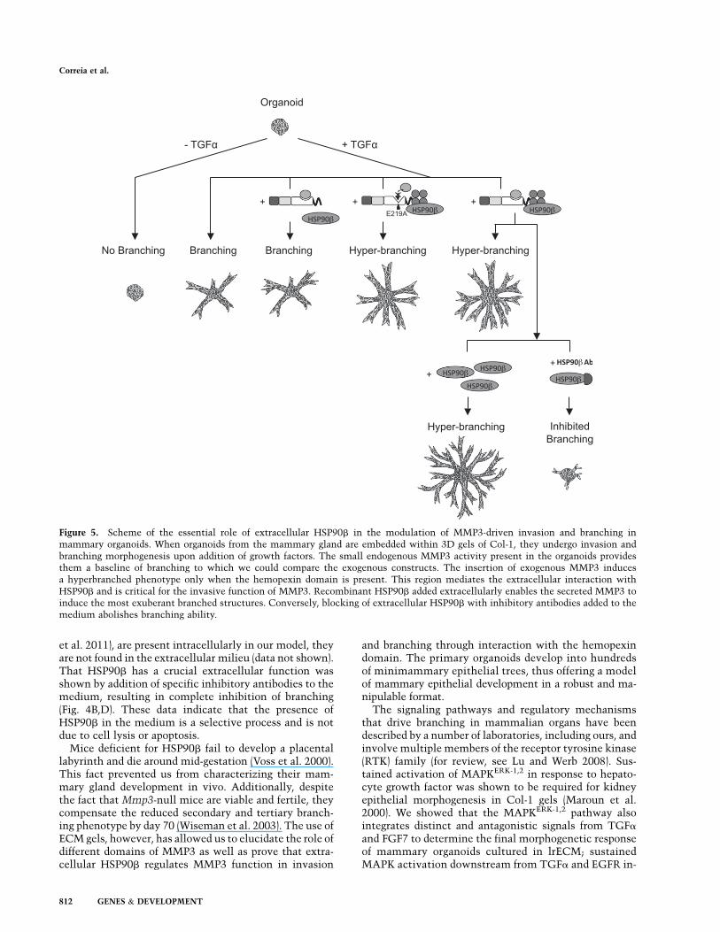
Figure 5. Scheme of the essential role of extracellular HSP90b in the modulation of MMP3-driven invasion and branching inmammary organoids. When organoids from the mammary gland are embedded within 3D gels of Col-1, they undergo invasion andbranching morphogenesis upon addition of growth factors. The small endogenous MMP3 activity present in the organoids providesthem a baseline of branching to which we could compare the exogenous constructs. The insertion of exogenous MMP3 inducesa hyperbranched phenotype only when the hemopexin domain is present. This region mediates the extracellular interaction withHSP90b and is critical for the invasive function of MMP3. Recombinant HSP90b added extracellularly enables the secreted MMP3 toinduce the most exuberant branched structures. Conversely, blocking of extracellular HSP90b with inhibitory antibodies added to themedium abolishes branching ability.
Correia et al.
812 GENES & DEVELOPMENT

duces branching, whereas its transient activation down-stream from FGF7 and FGFR2 stimulates proliferationbut not branching (Fata et al. 2007). FGF7 acts in part bysuppressing the expression of MMP3, and inhibition ofthe latter reduces branching significantly both in cultureand in vivo (Simian et al. 2001; Wiseman et al. 2003). Ourdiscovery that extracellular HSP90b is critical for MMP3function in invasion and branching places HSP90b as animportant player in the signaling pathways that deter-mine the final mammary morphogenetic fate.
The presence of HSP90 in murine mammary glandswas reported in 1989 (Catelli et al. 1989); therefore, it issurprising that its role in functional and morphogeneticaspects of the mammary gland is still poorly understood.The fact that HSPs have been postulated as molecularchaperones that mitigate the life-threatening effects ofheat and other stresses on the proteome (Taipale et al.2010) poses the question of whether HSP90b may alsoplay a role in stabilization and maturation of MMP3. Weare now beginning to understand that HSP90’s functionsextend well beyond stress tolerance, and associated changesin its clients can then exert marked effects on the relation-ship between genotype and phenotype, influencing humanhealth, disease, and evolutionary processes (Rutherford andLindquist 1998; Queitsch et al. 2002; Cowen and Lindquist2005). The presence of HSP90b in the medium and thefunctional significance of its interaction with MMP3 arefurther proof that HSP90-mediated events are above andbeyond the heat-shock response. Our preliminary data in-dicate that the extracellular source of HSP90b for luminalepithelial branching most probably is the myoepithelialcells in vivo (data not shown). These data, combined withsome evidence that MMP3 is mainly produced by stromalfibroblasts (Witty et al. 1995; Kouros-Mehr and Werb2006), raise the exciting possibility that extracellular in-teraction of HSP90b with MMP3 may be a way for differentcell types to communicate in the coordination of the normalprocesses of invasion and branching.
In the initial mass spectrometry data, we found manyadditional molecules that appear to be interacting withMMP3. In particular, we show that ANXA2 and MARCKSwere coimmunoprecipitated with MMP3, with binding oc-curring in the presence of the hemopexin domain. Ourpreliminary data also showed that depletion of each of theseproteins reduced invasiveness in SCp2 cells. Unlike theinteraction between HSP90b and MMP3 that happened inboth directions, the reverse co-IP of ANXA2 and MARCKSwith HSP90b could not be confirmed under these condi-tions. In addition, our proteomic screen identified otherproteases such as ADAM10, ADAMTS15, and CathepsinsA and L as possible proteins that may interact extracel-lularly with MMP3 (Supplemental Fig. S4). The func-tional significance of these latter proteins remains to bedetermined. Our data from the mass spectrometry, how-ever, tentatively suggest that a cascade of proteases mightfunction collectively to orchestrate epithelial invasion.
Finally, we showed most recently that the signalingmodule for MMP14, a membrane-bound MMP, in branch-ing of the end bud of the mammary gland of virgin mice isits transmembrane/cytoplasmic domain in conjunction
with integrin-b1 (Mori et al. 2013). Thus, the findingspresented here, along with the above work, may providea compelling explanation for why inhibitors of MMPsfailed so dramatically in the clinic (Overall and Kleifeld2006). Targeting noncatalytic sites of MMPs as well as theinteracting partners with agents such as small inhibi-tors or antibodies for the binding sites of integrin-b1 andHSP90b may yield more effective and tissue-specificinhibitors.
Materials and methods
Restriction enzymes, antibodies, proteins, and chemical
reagents
All restriction enzymes were acquired from New EnglandBiolabs. Bovine dermis acid-solubilized Col-1 solution (IAC-50)was purchased from Koken. Antibodies against the followingproteins were obtained as indicated: Flag (F1804, M2, Sigma;1:500 for Western blotting), E-cadherin (13-1900, clone ECCD-2,Invitrogen; 1:1000 for Western blotting; 1:200 for immunofluo-rescence), HSP90b (5087, Cell Signaling; 1:1000 for Westernblotting), HSP90b (NBP1-61773, Novus Biologicals; 40 mg/mLfor function-blocking experiments; 10 mg for co-IP experiments),MARCKS (P0370, Sigma; 1:1000 for Western blotting), ANXA2(AF3928, R&D Systems; 1:1000 for Western blotting), MMP3(ab18898, Abcam; 1:1000 for Western blotting), a-tubulin (T6074,clone B-5-1-2, Sigma; 1:5000 for Western blotting), and rabbitIgG (2729, Cell Signaling; 40 mg/mL for function-blocking exper-iments). Alexa Fluor 594 Phalloidin (A12381, Molecular Probes;1:400) was used to stain F-actin. DAPI (Sigma) was used to stainnuclei. HSP90b inhibitor CCT018159 (385920), MMP3-specificpeptide-based inhibitor (444218), and recombinant HSP90b
(385903) were purchased from Calbiochem/EMD Millipore.
Construction of expression plasmids
All MMP3 mutants were constructed using a PCR-based method(details in the Supplemental Material). The cDNA sequence usedas a template was cloned from a human breast cell line andsequence-confirmed by comparison with gene accession numberNM_002422.3. FL contains the full-length MMP3 cDNA. EA isa catalytically inactive mutant, holding a point mutation E219Aat the catalytic core. dPEX is a hemopexin-like domain-deletedmutant (DN289-C477). A mammalian expression vector, pCDH-EF1-MCS-T2A-copGFP (System Biosciences), was used to ex-press the gene products. To detect MMP3 protein, the Flagepitope (Asp–Tyr–Lys–Asp–Asp–Asp–Asp–Lys) was inserted atthe C terminus of every construct generated. All cDNA con-structs were confirmed by DNA sequencing.
shRNA-mediated knockdowns
shRNA constructs selectively targeting HSP90b, ANXA2,MARCKS, or MMP3 were purchased from MISSION shRNAlibrary (Sigma) (sequences are detailed in Supplemental TableS1). Control cells were infected with nontargeting shRNA(SHC002, Sigma). Knockdown efficiency was verified by Westernblotting with the appropriate antibodies.
Cell culture and transduction
SCp2 cells were cultured in Dulbecco’s modified Eagle’s me-dium/Ham’s F-12 nutrient mixture (DMEM/F-12) supplementedwith 5% fetal bovine serum (FBS), 5 mg/mL insulin, and 50 mg/mL
HSP90b is critical for MMP3 function
GENES & DEVELOPMENT 813

gentamicin and maintained as previously described (Desprez et al.1993). EpH4 cells were cultured in DMEM/F-12 medium supple-mented with 2% FBS, 5 mg/mL insulin, and 50 mg/mL gentamicinand maintained as previously described (Reichmann et al. 1989).For transduction, cells were seeded in 24-well plates (1 3 105 cellsper well) and infected with lentiviral particles carrying differentexpression plasmids using MISSION ExpressMag Beads (Sigma)according to the manufacturer’s instructions. Cells transducedwith lentivirus carrying shRNA constructs were additionally se-lected with 2 mg/mL puromycin.
Preparation of primary mammary organoids and transduction
Primary epithelial organoids were isolated from 8-wk-old virginFVB mice as previously described (Fata et al. 2007). Briefly,inguinal glands were removed, minced with two parallel razorblades, and gently shaken for 30 min at 37°C in a 50-mLcollagenase/trypsin mixture (0.2% trypsin, 0.2% type-IV colla-genase, 5% FBS, 5 mg/mL insulin in DMEM/F-12). After centri-fugation at 80g for 10 min, supernatant was discarded, and thecell pellet was resuspended in DMEM/F-12. The suspension waspelleted again, resuspended in 4 mL of DMEM/F-12 containing80 U of DNase I (Sigma), and incubated for 5 min at roomtemperature with occasional shaking. After the suspension wasspun at 80g for 10 min, a series of differential centrifugations inDMEM/F-12 was implemented to separate the epithelial organoidsfrom single cells, fibroblasts, and fibrillar extracellular matrices.The final pellet was resuspended in the desired amount of medium.For transduction, organoids were seeded in 24-well polyhema-coated plates (1000 organoids per well) and infected with lentivirusin the presence of 8 mg/mL polybrene for 24 h.
Preparation of cell clusters and transduction
EpH4 cells suspended in growth medium were plated in six-wellpolyhema-coated plates (1 3 105 cells per well) and incubatedovernight at 37°C, yielding rounded clusters. Single cells wereremoved by differential centrifugation, and the final pellet wasresuspended in the desired amount of medium.
Branching morphogenesis assay
Primary organoids or clustered EpH4 cells were embedded in 3DCol-1 gels as previously published (Simian et al. 2001; Mori et al.2013). In brief, acid-solubilized Col-1 solution was mixed gentlyon ice with 1 vol of 103 DMEM/F-12 (pH adjusted to 7.4 with 0.1M NaOH), and the concentration was adjusted to 3 mg/mL withDMEM/F-12. A basal layer of 80 mL of Col-1 was poured into eachwell of an eight-well chambered coverglass (155409, ThermoScientific) and allowed to gel for 5 min at 37°C. A second layerof 200 mL of Col-1 containing 150 organoids or EpH4 clusters wasadded to each well and placed immediately at 37°C. Aftergelation, 400 mL of chemically defined medium (DMEM/F-12containing 1% insulin/transferrin/selenium, 1% penicilin/strep-tomycin) with 9 nM TGFa (Sigma) or 9 nM bFGF (Sigma) wasadded to each well (unless stated otherwise) and replaced everyother day.
After 3 d of culture, gels were fixed with 4% formalin for 30min and stained with phalloidin and DAPI for 1 h. Structureswere imaged with an upright Zeiss LSM710 using a 0.8 NA 203
air objective. An organoid or cell cluster was defined as invadingand branching when it had at least three independent extendingprocesses that were at least half the diameter of the center of theorganoid or cell cluster. The number of extending processes andtheir average length were determined using the Imaris program(Bitplane). We defined a new metric of invasion and branching,
which we refer to as the spatial network per organoid. This isdefined as the sum of the length of all of the extending processesdeveloped from each organoid. Fifty structures were countedper condition, and the experiments were executed at least threetimes.
Caseinase activity assay
CM was incubated with a casein derivative-quenching red-fluorescent dye (BODIPY TR-X Casein, E6639, Invitrogen).Protease-catalyzed hydrolysis released highly fluorescent BODIPYTR-X dye-labeled peptides. The accompanying increase in fluo-rescence is proportional to MMP3 proteolytic activity and wasmonitored with a microplate reader. A control without BODIPYcasein was used to subtract residual fluorescence background.
Cell scatter assay
SCp2 cells were seeded in six-well plates at low density (1 3 105
cells per well), allowed to form colonies (;48 h), and serum-starved for 24 h. Epithelial cell islets were then stimulated with9 nM epidermal growth factor (EGF) (Sigma) and imaged at 48 hwith a Zeiss Imager Z1 microscope using a 103 objective.
Immunofluorescence
SCp2 cells were cultured for 72 h on glass coverslips, fixed with4% paraformaldehyde/PBS for 10 min, washed with PBS, andpermeabilized in 0.25% Triton X-100/PBS for 10 min. Sampleswere blocked with 1% BSA and 5% goat serum/PBS for 1 h,followed by incubation with the primary antibody in blockingbuffer overnight at 4°C and the secondary antibody for 1 h atroom temperature. Images were acquired with an upright ZeissLSM710 using a 1.4 NA 633 oil immersion.
Morphometry analysis
Cell edges were outlined in F-actin-stained cells using an ‘‘ObjectIdentification Module’’ from CellProfiler software (Carpenteret al. 2006). Cellular elliptical factors, defined as the ratio ofthe longest (length) to the shortest (width) axis of the cell, werecalculated for 100 random cells per culture.
Invasion assay
Cell culture inserts (8 mm, 24-well format; BD Biosciences) wereevenly coated with 20 mL of diluted (1:5 in DMEM/F-12 medium)Matrigel (BD Biosciences). Cells (1 3 105) in 200 mL of DMEM/F-12 medium or different CM (as indicated in each experiment)were added to the upper compartment of the chamber. The lowercompartment of the chamber was filled with 300 mL of mediumcontaining 10% FBS as a chemoattractant. After 48 h of in-cubation at 37°C, the top side of the insert was cleared fromnoninvasive cells with a cotton swab and washed with serum-freeDMEM/F-12. The remaining (invasive) cells at the lower surfaceof the filter were fixed and stained with a solution of CoomassieBlue 0.125% in methanol:acetic acid:H2O (45%:10%:45% [v/v/v])for 15 min. Invasive cells were scored by counting 10 3 20magnification fields per filter with a Zeiss Imager Z1 microscopeusing a 203 objective. Mouse embryonic fibroblast NIH/3T3cells were routinely included as a positive control. Results areexpressed as mean 6 SD from three independent experiments.
Western blotting
Cells were lysed with a buffer containing 1% Triton X-100,1% NP-40, and protease and phosphatase inhibitor cocktails
Correia et al.
814 GENES & DEVELOPMENT

(Calbiochem/EMD Millipore) in PBS, and the lysates were clari-fied by centrifugation at 16,000g for 15 min. Protein concentrationwas determined using the BCA Protein Assay kit (ThermoScientific) according to the manufacturer’s instructions. Proteinsamples were mixed with electrophoresis sample buffer contain-ing 5% (v/v) 2-b-mercaptoethanol and 5% (v/v) bromophenol blueand boiled for 5 min at 95°C. Samples were loaded in equalamounts into precast 4%–20% gradient polyacrylamide gels(Invitrogen) and separated by SDS-PAGE. Resolved proteins weretransferred to a nitrocellulose membrane (Whatman) at 130 V for90 min, followed by blocking of nonspecific binding with 5% BSAin 0.05% Tween-20/PBS for 1 h at room temperature. The mem-branes were probed with primary antibodies specific to each pro-tein overnight at 4°C and then with HRP-conjugated secondaryantibodies (Thermo Scientific and Santa Cruz Biotechnology).Blots were visualized with an ECL detection system (Thermo Sci-entific) according to the manufacturer’s instructions, and chemi-luminescent signal was captured with a FluorChem IS-8900(Alpha Innotech). Each Western blot was done at least three times,and here we show representative experiments.
Co-IP
For co-IP of Flag-tagged MMP3 protein complexes, CM wasincubated with anti-Flag M2 antibody-conjugated agarose beads(F2426, Sigma) for 16 h at 4°C. The beads were then washed threetimes with 0.05% Tween/PBS, and the immune complexes weredirectly eluted with electrophoresis sample buffer and analyzedby Western blotting. For liquid chromatography-tandem massspectrometry (LC-MS/MS) analysis, beads were washed with0.05% Tween/PBS, and protein complexes were eluted with aFlag peptide (F3290, Sigma) in 0.05% Tween/PBS. Samples werethen precipitated with trichloroacetic acid and reconstitutedwith a buffer (Invitrosol, MS10007, Invitrogen) suitable for massspectrometry analysis.
For co-IP of HSP90b protein complexes, CM was incubatedwith 10 mg of control rabbit IgG or anti-HSP90b antibody for 16 hat 4°C. Precipitation was performed with protein G sepharosebeads (17-0618-01, GE Healthcare) for 4 h at 4°C. The beads werethen washed three times with 0.05% Tween/PBS, and theimmune complexes were directly eluted with electrophoresissample buffer and analyzed by Western blotting.
Mass spectrometry analysis
Mass spectrometry analysis is described in the SupplementalMaterial. Scaled signal intensities were log2 transformed andanalyzed by R software.
Statistical analysis
Statistical analyses were performed using GraphPad Prism 5.0software. Student’s t-test (unpaired with Welch’s correction, two-tailed, 95% confidence interval) was used to determine statisticalsignificance. Statistical analyses were always performed in re-lation to vector control cells (unless stated otherwise).
Acknowledgments
We thank Alexandre Bruni-Cardoso and Cyrus Ghajar for criticalreading of the manuscript, Joao Guimaraes for helpful sugges-tions and assistance with R software, Derek Radisky for initialadvice, and Richard Schwarz, Joni Mott, Douglas Brownfield,Alvin Lo, and Joana Paredes for their constructive discussion andhelp. This work was supported by a predoctoral Fellowship(SFRH/BD/33249/2007) from the Portuguese Foundation for
Science and Technology awarded to A.L.C. The work fromM.J.B.’s laboratory is supported by grants from the National CancerInstitute (awards R37CA064786, U54CA126552, R01CA057621,U54CA112970, U01CA143233, and U54CA143836, Bay Area Phys-ical Sciences-Oncology Center, University of California at Berkeley,Berkeley, California); from the U.S. Department of Energy, Office ofBiological and Environmental Research and Low-Dose RadiationProgram (contract no. DE-AC02-05CH1123); and from the U.S.Department of Defense (W81XWH0810736) and in part by a grantfrom The Breast Cancer Research Foundation. E.I.C. is supported bygrants from Manhasset Women’s Coalition Against Breast Cancerand Carol M. Baldwin Foundation For Breast Cancer Research.
References
Carpenter AE, Jones TR, Lamprecht MR, Clarke C, Kang IH,Friman O, Guertin DA, Chang JH, Lindquist RA, Moffat J,et al. 2006. CellProfiler: Image analysis software for identi-fying and quantifying cell phenotypes. Genome Biol 7:R100.
Catelli MG, Ramachandran C, Gauthier Y, Legagneux V, QuelardC, Baulieu EE, Shyamala G. 1989. Developmental regulationof murine mammary-gland 90 kDa heat-shock proteins. Bio-
chem J 258: 895–901.Cheng CF, Fan J, Fedesco M, Guan S, Li Y, Bandyopadhyay B,
Bright AM, Yerushalmi D, Liang M, Chen M, et al. 2008.Transforming growth factor a (TGFa)-stimulated secretion ofHSP90a: Using the receptor LRP-1/CD91 to promote humanskin cell migration against a TGFb-rich environment duringwound healing. Mol Cell Biol 28: 3344–3358.
Cowen LE, Lindquist S. 2005. Hsp90 potentiates the rapidevolution of new traits: Drug resistance in diverse fungi.Science 309: 2185–2189.
de Graauw M, Tijdens I, Smeets MB, Hensbergen PJ, DeelderAM, van de Water B. 2008. Annexin A2 phosphorylationmediates cell scattering and branching morphogenesis viacofilin Activation. Mol Cell Biol 28: 1029–1040.
Desprez P, Roskelley C, Campisi J, Bissell MJ. 1993. Isolation offunctional cell lines from a mouse mammary epithelial cellstrain: The importance of basement membrane and cell-cellinteraction. Mol Cell Differ 1: 99–110.
Dufour A, Sampson NS, Zucker S, Cao J. 2008. Role of thehemopexin domain of matrix metalloproteinases in cellmigration. J Cell Physiol 217: 643–651.
Eustace BK, Sakurai T, Stewart JK, Yimlamai D, Unger C,Zehetmeier C, Lain B, Torella C, Henning SW, Beste G,et al. 2004. Functional proteomic screens reveal an essentialextracellular role for hsp90a in cancer cell invasiveness. Nat
Cell Biol 6: 507–514.Farina AR, Tacconelli A, Cappabianca L, Gulino A, Mackay AR.
2002. Inhibition of human MDA-MB-231 breast cancer cellinvasion by matrix metalloproteinase 3 involves degradationof plasminogen. Eur J Biochem 269: 4476–4483.
Fata JE, Werb Z, Bissell MJ. 2004. Regulation of mammary glandbranching morphogenesis by the extracellular matrix and itsremodeling enzymes. Breast Cancer Res 6: 1–11.
Fata JE, Mori H, Ewald AJ, Zhang H, Yao E, Werb Z, Bissell MJ.2007. The MAPK(ERK-1,2) pathway integrates distinct andantagonistic signals from TGFa and FGF7 in morphogenesisof mouse mammary epithelium. Dev Biol 306: 193–207.
Fotouhi N, Lugo A, Visnick M, Lusch L, Walsky R, Coffey JW,Hanglow AC. 1994. Potent peptide inhibitors of stromelysinbased on the prodomain region of matrix metalloproteinases.J Biol Chem 269: 30227–30231.
Glasheen BM, Kabra AT, Page-McCaw A. 2009. Distinct func-tions for the catalytic and hemopexin domains of a Drosoph-
HSP90b is critical for MMP3 function
GENES & DEVELOPMENT 815

ila matrix metalloproteinase. Proc Natl Acad Sci 106: 2659–2664.
Hirai Y, Lochter A, Galosy S, Koshida S, Niwa S, Bissell MJ.1998. Epimorphin functions as a key morphoregulator formammary epithelial cells. J Cell Biol 140: 159–169.
Houghton AM, Hartzell WO, Robbins CS, Gomis-Ruth FX,Shapiro SD. 2009. Macrophage elastase kills bacteria withinmurine macrophages. Nature 460: 637–641.
Iioka H, Ueno N, Kinoshita N. 2004. Essential role of MARCKSin cortical actin dynamics during gastrulation movements.J Cell Biol 164: 169–174.
Khokha R, Werb Z. 2011. Mammary gland reprogramming:Metalloproteinases couple form with function. Cold SpringHarb Perspect Biol 3: a0044333.
Kouros-Mehr H, Werb Z. 2006. Candidate regulators of mam-mary branching morphogenesis identified by genome-widetranscript analysis. Dev Dyn 235: 3404–3412.
Li W, Li Y, Guan S, Fan J, Cheng CF, Bright AM, Chinn C, ChenM, Woodley DT. 2007. Extracellular heat shock protein-90a:Linking hypoxia to skin cell motility and wound healing.EMBO J 26: 1221–1233.
Lochter A, Galosy S, Muschler J, Freedman N, Werb Z, BissellMJ. 1997a. Matrix metalloproteinase stromelysin-1 triggers acascade of molecular alterations that leads to stable epithe-lial-to-mesenchymal conversion and a premalignant pheno-type in mammary epithelial cells. J Cell Biol 139: 1861–1872.
Lochter A, Srebrow A, Sympson CJ, Terracio N, Werb Z, BissellMJ. 1997b. Misregulation of stromelysin-1 expression inmouse mammary tumor cells accompanies acquisition ofstromelysin-1-dependent invasive properties. J Biol Chem
272: 5007–5015.Lu P, Werb Z. 2008. Patterning mechanisms of branched organs.
Science 322: 1506–1509.Maroun CR, Naujokas MA, Holgado-Madruga M, Wong AJ, Park
M. 2000. The tyrosine phosphatase SHP-2 is required forsustained activation of extracellular signal-regulated kinaseand epithelial morphogenesis downstream from the metreceptor tyrosine kinase. Mol Cell Biol 20: 8513–8525.
Mori H, Tomari T, Koshikawa N, Kajita M, Itoh Y, Sato H,Tojo H, Yana I, Seiki M. 2002. CD44 directs membrane-type 1matrix metalloproteinase to lamellipodia by associating withits hemopexin-like domain. EMBO J 21: 3949–3959.
Mori H, Lo AT, Inman JL, Alcaraz J, Ghajar CM, Mott JD,Nelson CM, Chen CS, Zhang H, Bascom JL, et al. 2013.Transmembrane/cytoplasmic, rather than catalytic, domainsof Mmp14 signal to MAPK activation and mammary branch-ing morphogenesis via binding to integrin b1. Development
140: 343–352.Noe V, Fingleton B, Jacobs K, Crawford HC, Vermeulen S,
Steelant W, Bruyneel E, Matrisian LM, Mareel M. 2001.Release of an invasion promoter E-cadherin fragment bymatrilysin and stromelysin-1. J Cell Sci 114: 111–118.
Overall CM, Kleifeld O. 2006. Tumour microenvironment—opinion: Validating matrix metalloproteinases as drug targetsand anti-targets for cancer therapy. Nat Rev Cancer 6: 227–239.
Queitsch C, Sangster TA, Lindquist S. 2002. Hsp90 as a capacitorof phenotypic variation. Nature 417: 618–624.
Radisky DC, Levy DD, Littlepage LE, Liu H, Nelson CM, FataJE, Leake D, Godden EL, Albertson DG, Nieto MA, et al.2005. Rac1b and reactive oxygen species mediate MMP-3-induced EMT and genomic instability. Nature 436: 123–127.
Reichmann E, Ball R, Groner B, Friis RR. 1989. New mammaryepithelial and fibroblastic cell clones in coculture formstructures competent to differentiate functionally. J Cell Biol
108: 1127–1138.
Rutherford SL, Lindquist S. 1998. Hsp90 as a capacitor formorphological evolution. Nature 396: 336–342.
Sakamoto T, Seiki M. 2009. Cytoplasmic tail of MT1-MMPregulates macrophage motility independently from its pro-tease activity. Genes Cells 14: 617–626.
Schnelzer A, Prechtel D, Knaus U, Dehne K, Gerhard M, GraeffH, Harbeck N, Schmitt M, Lengyel E. 2000. Rac1 in humanbreast cancer: Overexpression, mutation analysis, and char-acterization of a new isoform, Rac1b. Oncogene 19: 3013–3020.
Sharp SY, Boxall K, Rowlands M, Prodromou C, Roe SM,Maloney A, Powers M, Clarke PA, Box G, Sanderson S,et al. 2007. In vitro biological characterization of a novel,synthetic diaryl pyrazole resorcinol class of heat shockprotein 90 inhibitors. Cancer Res 67: 2206–2216.
Simian M, Hirai Y, Navre M, Werb Z, Lochter A, Bissell MJ.2001. The interplay of matrix metalloproteinases, morpho-gens and growth factors is necessary for branching ofmammary epithelial cells. Development 128: 3117–3131.
Sims JD, McCready J, Jay DG. 2011. Extracellular heat shock pro-tein (Hsp)70 and Hsp90a assist in matrix metalloproteinase-2activation and breast cancer cell migration and invasion.PLoS ONE 6: e18848.
Sternlicht MD, Lochter A, Sympson CJ, Huey B, Rougier JP,Gray JW, Pinkel D, Bissell MJ, Werb Z. 1999. The stromalproteinase MMP3/stromelysin-1 promotes mammary carci-nogenesis. Cell 98: 137–146.
Sympson CJ, Talhouk RS, Alexander CM, Chin JR, Clift SM,Bissell MJ, Werb Z. 1994. Targeted expression of stromelysin-1in mammary gland provides evidence for a role of proteinasesin branching morphogenesis and the requirement for an intactbasement membrane for tissue-specific gene expression. J Cell
Biol 125: 681–693.Taipale M, Jarosz DF, Lindquist S. 2010. HSP90 at the hub of
protein homeostasis: Emerging mechanistic insights. Nat
Rev Mol Cell Biol 11: 515–528.Talhouk RS, Chin JR, Unemori EN, Werb Z, Bissell MJ. 1991.
Proteinases of the mammary gland: Developmental regula-tion in vivo and vectorial secretion in culture. Development
112: 439–449.Talhouk RS, Bissell MJ, Werb Z. 1992. Coordinated expression
of extracellular matrix-degrading proteinases and their in-hibitors regulates mammary epithelial function during in-volution. J Cell Biol 118: 1271–1282.
Tam EM, Wu YI, Butler GS, Stack MS, Overall CM. 2002.Collagen binding properties of the membrane type-1 matrixmetalloproteinase (MT1-MMP) hemopexin C domain. Theectodomain of the 44-kDa autocatalytic product of MT1-MMP inhibits cell invasion by disrupting native type Icollagen cleavage. J Biol Chem 277: 39005–39014.
Thomasset N, Lochter A, Sympson CJ, Lund LR, Williams DR,Behrendtsen O, Werb Z, Bissell MJ. 1998. Expression ofautoactivated stromelysin-1 in mammary glands of trans-genic mice leads to a reactive stroma during early develop-ment. Am J Pathol 153: 457–467.
Vincent-Salomon A, Thiery JP. 2003. Host microenvironment inbreast cancer development: Epithelial–mesenchymal tran-sition in breast cancer development. Breast Cancer Res 5:101–106.
Voss AK, Thomas T, Gruss P. 2000. Mice lacking HSP90b fail todevelop a placental labyrinth. Development 127: 1–11.
Wang P, Nie J, Pei D. 2004. The hemopexin domain of mem-brane-type matrix metalloproteinase-1 (MT1-MMP) is notrequired for its activation of proMMP2 on cell surface but isessential for MT1-MMP-mediated invasion in three-dimen-sional type I collagen. J Biol Chem 279: 51148–51155.
Correia et al.
816 GENES & DEVELOPMENT

Williams JM, Daniel CW. 1983. Mammary ductal elongation:Differentiation of myoepithelium and basal lamina duringbranching morphogenesis. Dev Biol 97: 274–290.
Wiseman BS, Sternlicht MD, Lund LR, Alexander CM, Mott J,Bissell MJ, Soloway P, Itohara S, Werb Z. 2003. Site-specificinductive and inhibitory activities of MMP-2 and MMP-3orchestrate mammary gland branching morphogenesis. J Cell
Biol 162: 1123–1133.Witty JP, Wright JH, Matrisian LM. 1995. Matrix metallopro-
teinases are expressed during ductal and alveolar mammarymorphogenesis, and misregulation of stromelysin-1 in trans-genic mice induces unscheduled alveolar development. Mol
Biol Cell 6: 1287–1303.
HSP90b is critical for MMP3 function
GENES & DEVELOPMENT 817
Related Documents