The Heart • A muscular double pump • Pulmonary circuit – takes blood to and from the lungs • Systemic circuit – vessels transport blood to and from body tissues • Atria – receive blood from the pulmonary and systemic circuits • Ventricles – the pumping chambers of the heart

The Heart A muscular double pump Pulmonary circuit – takes blood to and from the lungs Systemic circuit – vessels transport blood to and from body tissues.
Dec 20, 2015
Welcome message from author
This document is posted to help you gain knowledge. Please leave a comment to let me know what you think about it! Share it to your friends and learn new things together.
Transcript

The Heart
• A muscular double pump• Pulmonary circuit – takes blood to and from the lungs
• Systemic circuit – vessels transport blood to and from body tissues• Atria – receive blood from the pulmonary and systemic
circuits
• Ventricles – the pumping chambers of the heart

The Pulmonary and Systemic Circuits
Figure 18.1

Location and Orientation within the Thorax
Figure 18.2
Structure of the Heart – Coverings
Figure 18.3

Structure of the Heart – Layers of the Heart Wall
• Epicardium – visceral layer of the serous pericardium
• Myocardium – consists of cardiac muscle• Muscle arranged in circular and spiral patterns
• Endocardium – endothelium resting on a layer of connective tissue• Lines the internal walls of the heart
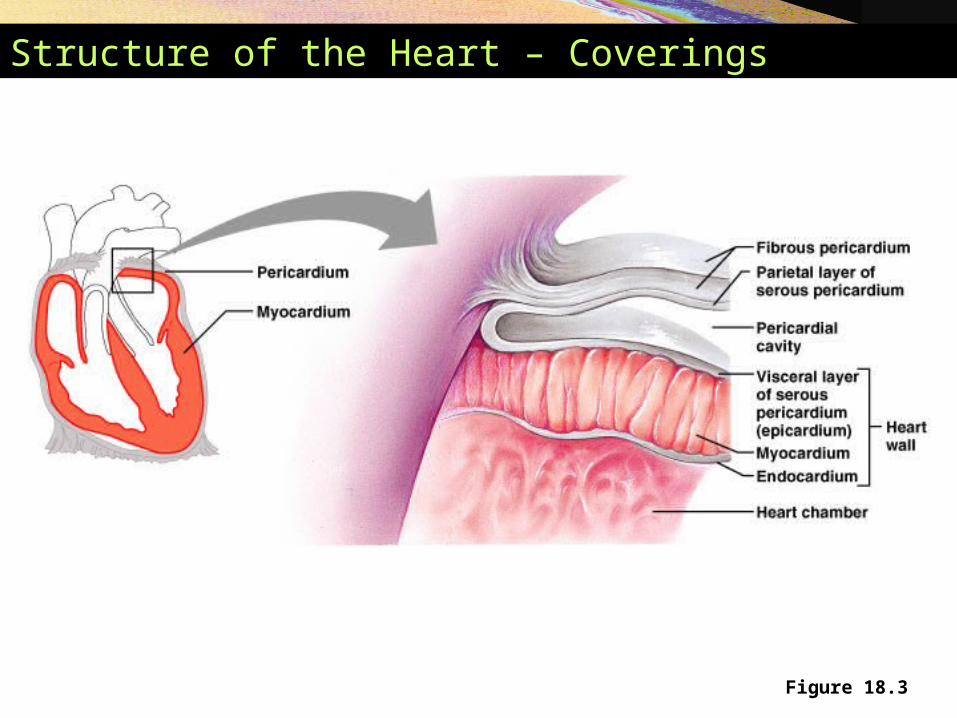
Heart Chambers
• Internal divisions• Atria and ventricles
• Interventricular and interatrial septa
• External markings • Coronary sulcus
• Anterior and posterior interventricular sulcus

Heart Chambers
Figure 18.5b

Heart Chambers
Figure 18.5e
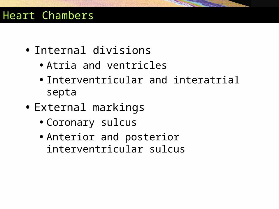
Inferior View of the Heart
Figure 18.5d
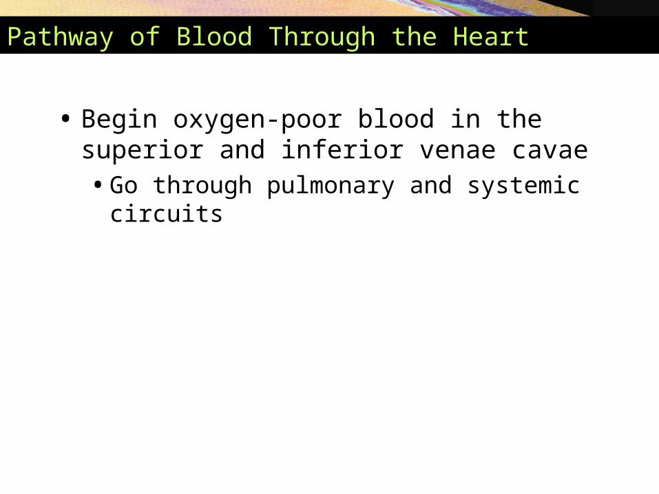
Pathway of Blood Through the Heart
• Begin oxygen-poor blood in the superior and inferior venae cavae• Go through pulmonary and systemic circuits

Blood Flow Through the Heart
Figure 18.6
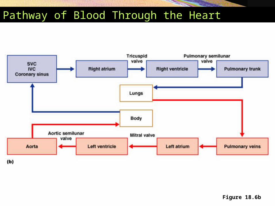
Pathway of Blood Through the Heart
Figure 18.6b
Heartbeat
• 70–80 beats/minute at rest
• Systole – contraction
• Diastole – expansion
• Systole and diastole also refer to:• Stage of heartbeat when ventricles contract and
expand

Structure of Heart Wall
• Walls differ in thickness• Atria – thin walls
• Ventricles – thick walls

Structure of Heart Wall
• Left ventricle – three times thicker than right• Exerts more
pumping force
• Flattens right ventricle into a crescent shape
Figure 18.7
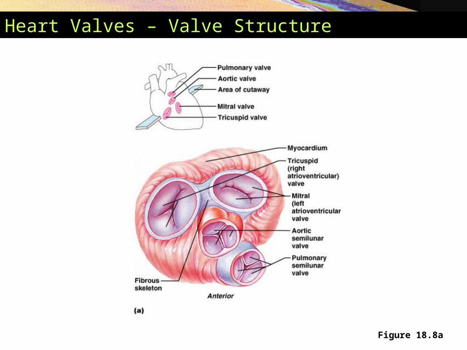
Heart Valves – Valve Structure
Figure 18.8a
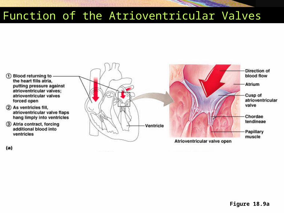
Function of the Atrioventricular Valves
Figure 18.9a

Function of the Atrioventricular Valves
Figure 18.9b
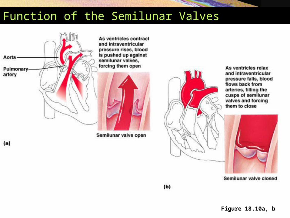
Function of the Semilunar Valves
Figure 18.10a, b
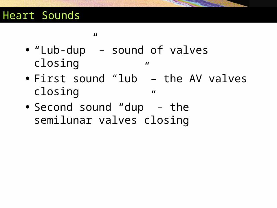
Heart Sounds
• “Lub-dup” – sound of valves closing
• First sound “lub” – the AV valves closing
• Second sound “dup” – the semilunar valves closing

Heart Sounds
Figure 18.8a
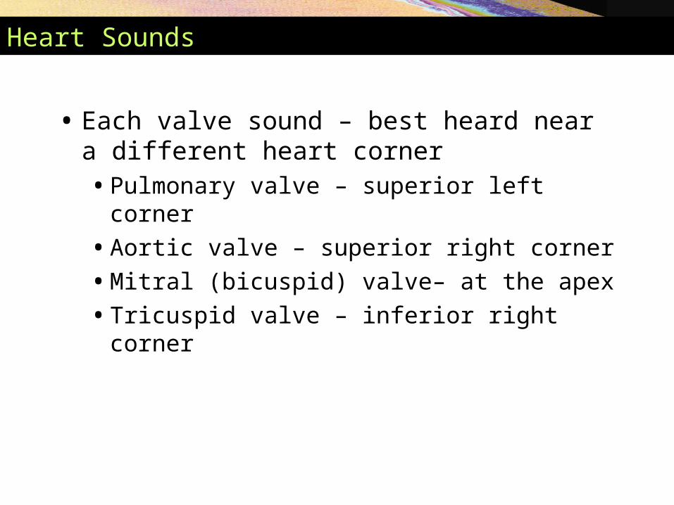
Heart Sounds
• Each valve sound – best heard near a different heart corner• Pulmonary valve – superior left corner
• Aortic valve – superior right corner
• Mitral (bicuspid) valve– at the apex
• Tricuspid valve – inferior right corner
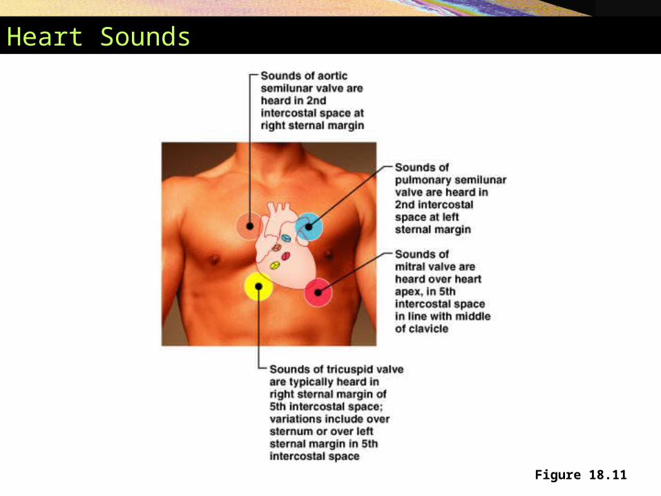
Heart Sounds
Figure 18.11
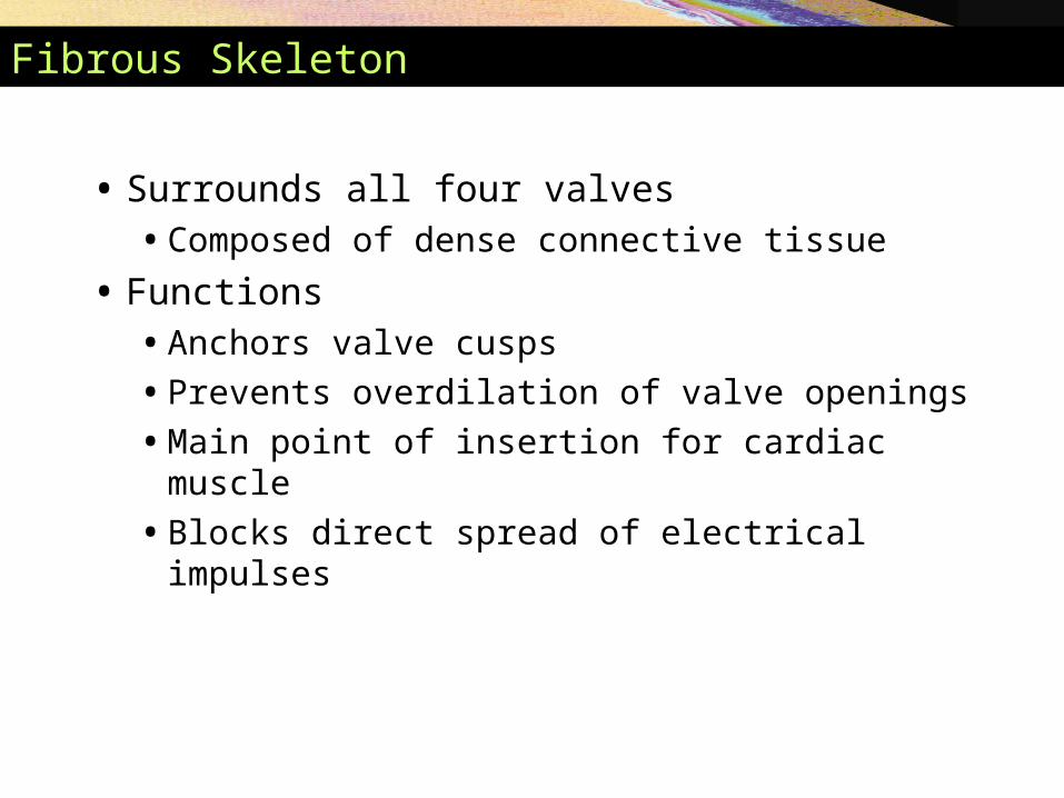
Fibrous Skeleton
• Surrounds all four valves• Composed of dense connective tissue
• Functions• Anchors valve cusps
• Prevents overdilation of valve openings
• Main point of insertion for cardiac muscle
• Blocks direct spread of electrical impulses

Conducting System
• Cardiac muscle tissue has intrinsic ability to:• Generate and conduct impulses
• Signal these cells to contract rhythmically
• Conducting system • A series of specialized cardiac muscle cells
• Sinoatrial (SA) node sets the inherent rate of contraction

Microscopic Anatomy of Heart Muscle

Cardiac Muscle Contraction
Heart muscle:
Is stimulated by nerves and is self-excitable (automaticity)
Contracts as a unit
Has a long (250 ms) absolute refractory period
Cardiac muscle contraction is similar to skeletal muscle contraction

Heart Physiology: Intrinsic Conduction System
Autorhythmic cells:
Initiate action potentials
Have unstable resting potentials called pacemaker potentials
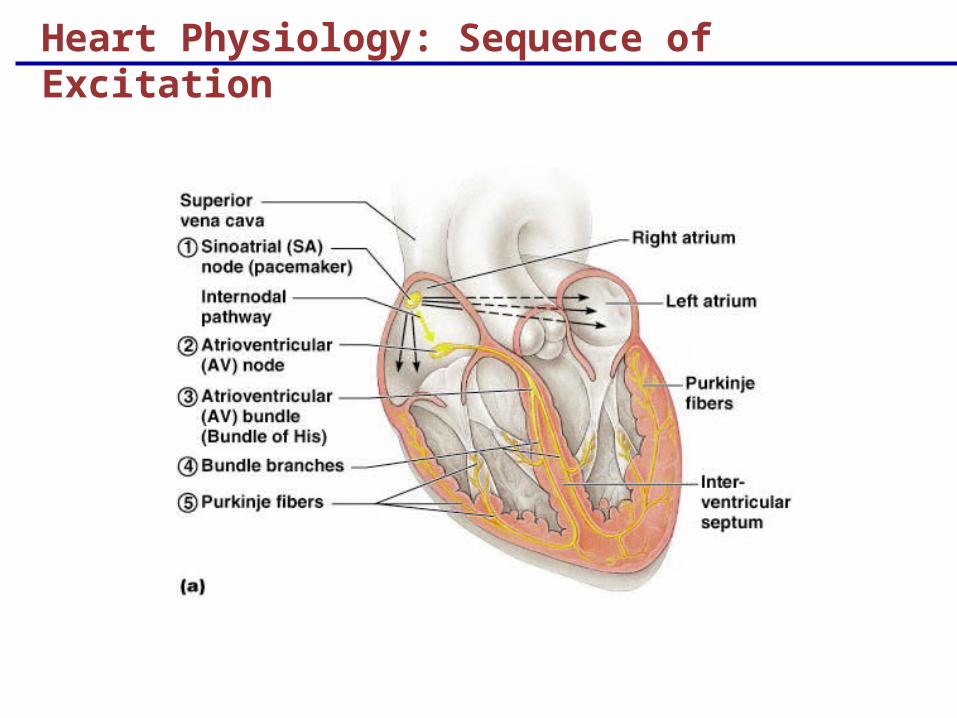
Heart Physiology: Sequence of Excitation

Heart Physiology: Sequence of Excitation
Sinoatrial (SA) node generates impulses about 75 times/minute
Atrioventricular (AV) node delays the impulse approximately 0.1 second
Impulse passes from atria to ventricles via the atrioventricular bundle (bundle of His)

Heart Physiology: Sequence of Excitation
AV bundle splits into two pathways in the interventricular septum (bundle branches)
Bundle branches carry the impulse toward the apex of the heart
Purkinje fibers carry the impulse to the heart apex and ventricular walls
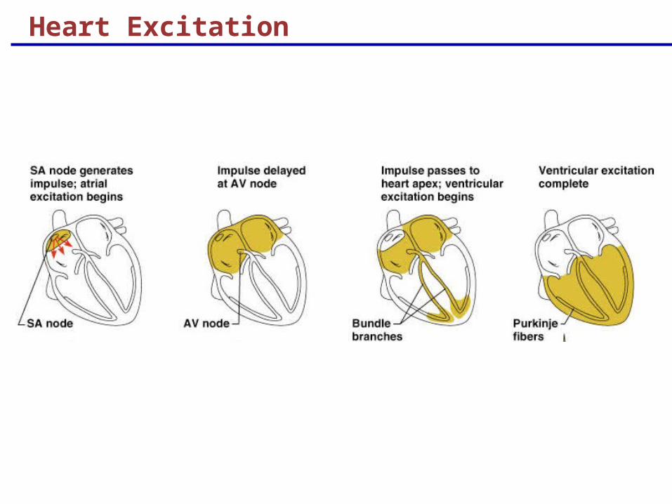
Heart Excitation

Innervation
• Heart rate is altered by external controls
• Nerves to the heart include:• Visceral sensory fibers
• Parasympathetic branches of the vagus nerve
• Sympathetic fibers – from cervical and upper thoracic chain ganglia
Figure 18.13

Blood Supply to the Heart
• Functional blood supply• Coronary arteries
• Arise from the aorta• Located in the coronary sulcus
• Main branches • Left and right coronary arteries

Blood Supply to the Heart
Figure 18.14

Disorders of the Heart
• Coronary artery disease• Atherosclerosis – fatty deposits
• Angina pectoris – chest pain
• Myocardial infarction – blocked coronary artery
• Silent ischemia – no pain or warning
Disorders of the Heart
• Heart failure• Progressive weakening of the heart• Cannot meet the body’s demands for oxygenated
blood
• Congestive heart failure – heart enlarges• Pumping efficiency declines
• Cor pulmonale • Enlargement and potential failure of the right
ventricle
Disorders of Conduction
• Ventricular fibrillation • Rapid, random firing of electrical impulses in the
ventricles
• Atrial fibrillation • Multiple waves of impulses randomly signal the
AV node
• Signals ventricles to contract quickly and irregularly

The Heart Throughout Life
• Blood vessels• Begin as condensations of mesodermal
mesenchyme
• Embryonic heart• Pair of tubes fuse at day 21
• Heart starts pumping at day 22
• Bulges develop along heart tube

Congenital Heart Defects
Figure 18.17a, b

Congenital Heart Defects
Figure 18.17c, d

Congenital Heart Defects
Figure 18.17e, f
The Heart in Adulthood and Old Age
• Age-related changes• Hardening and thickening of valve cusps
• Decline in cardiac reserve• Sympathetic control over heart is less efficient
• Less severe in the physically active
• Fibrosis of cardiac muscle tissue • Lowers the amount of blood the heart can pump
Related Documents