The HBx Oncoprotein of Hepatitis B Virus Deregulates the Cell Cycle by Promoting the Intracellular Accumulation and Re-Compartmentalization of the Cellular Deubiquitinase USP37 Nehul Saxena, Vijay Kumar* Virology Group, International Center for Genetic Engineering and Biotechnology, Aruna Asaf Ali Marg, New Delhi, India Abstract The HBx oncoprotein of hepatitis B Virus has been accredited as one of the protagonists in driving hepatocarcinogenesis. HBx exerts its influence over the cell cycle progression by potentiating the activity of cyclin A/E-CDK2 complex, the Cyclin A partner of which is a well-known target of cellular deubiquitinase USP37. In the present study, we observed the intracellular accumulation of cyclin A and USP37 proteins under the HBx microenvironment. Flow cytometry analysis of the HBx- expressing cells showed deregulation of cell cycle apparently due to the enhanced gene expression and stabilization of USP37 protein and deubiquitination of Cyclin A by USP37. Our co-immunoprecipitation and confocal microscopic studies suggested a direct interaction between USP37 and HBx. This interaction promoted the translocation of USP37 outside the nucleus and prevented its association and ubiquitination by E3 ubiquitin ligases - APC/CDH1 and SCF/b-TrCP. Thus, HBx seems to control the cell cycle progression via the cyclin A-CDK2 complex by regulating the intracellular distribution and stability of deubiquitinase USP37. Citation: Saxena N, Kumar V (2014) The HBx Oncoprotein of Hepatitis B Virus Deregulates the Cell Cycle by Promoting the Intracellular Accumulation and Re- Compartmentalization of the Cellular Deubiquitinase USP37. PLoS ONE 9(10): e111256. doi:10.1371/journal.pone.0111256 Editor: Michael J. Bouchard, Drexel University College of Medicine, United States of America Received May 31, 2014; Accepted September 16, 2014; Published October 27, 2014 Copyright: ß 2014 Saxena, Kumar. This is an open-access article distributed under the terms of the Creative Commons Attribution License, which permits unrestricted use, distribution, and reproduction in any medium, provided the original author and source are credited. Data Availability: The authors confirm that all data underlying the findings are fully available without restriction. All relevant data are within the paper and its Supporting Information files. Funding: This work was supported by grant no. BT/PR12658/BRB/10/712/2009 of the Department of Biotechnology (DBT), Ministry of Science and Technology, Government of India (to VK). The funders had no role in study design, data collection and analysis, decision to publish, or preparation of the manuscript. Competing Interests: The authors have declared that no competing interests exist. * Email: [email protected] Introduction The momentum of cell cycle is governed by the temporal synthesis, maintenance and degradation of cell cycle regulators. A plethora of E3 ubiquitin ligases and deubiquitinases (DUBs) capable of reversing ubiquitination, are now considered integral to the regulation of cell cycle [1–4]. So far fifteen different DUBs including USP2, USP3, USP7, USP13, USP17L2, USP19, USP28, USP37, USP39, USP44, USP50, COP9 sinnalosome subunit 5 (CSN5), BRCA1 associated protein-1 (BAP1), Cylin- dromatosis protein (CYLD) and Ovarian tumor domain contain- ing subunit 6B (OTUD-6B) have been implicated in cell cycle regulation [5]. Particularly, USP37 which belongs to the ubiquitin- specific protease family of DUBs, regulates cell cycle by antagonizing the activity of APC/CDH1 complex during the G1/S boundary, S and G2 phases to stabilize its substrate Cyclin A [6]. The USP37 gene is transcriptionally activated by transcription factor E2F followed by its translation during the G1/S boundary of cell cycle. The USP37 protein becomes fully functional upon its Cyclin A/CDK2-mediated phosphorylation at Ser-628 residue [6] and remains active throughout the S phase upto G2/M boundary. Apparently, the degradation of USP37 occurs in a bi-phasic manner. At the G2/M boundary, polo like kinase 1 (Plk1)-dependent phosphorylation of serine residues in DSGXXS consensus motif makes USP37 vulnerable to Skp1- Cullin1-F-box ubiquitin ligase/beta-transducin repeat containing protein complex (SCF/b-TRCP)-mediated ubiquitination and proteasomal degradation [7]. Also, during the M phase, upon depletion of Cyclin A and subsequent disappearance of CDK2 activity, the residual un-phosphorylated USP37 undergoes pro- teasomal degradation following its APC/CDH1-mediated KEN- box dependent ubiquitination [6]. Apart from its physiological relevance, USP37 is also reported to play an important role in cancer. For instance, increased USP37 expression is correlated with poor prognosis in non-small cell lung cancer [8]. It also confers resistance to Acute promyelocytic leukemia cells against arsenic trioxide and all-trans retinoic acid treatment by preserving the PLZF-RARA (promyelocytic leukemia zinc finger and retinoic acid receptor alpha) fusion protein [9]. Ambiguously, the transcription of USP37 is suppressed in medulloblastoma cells through the activity of RE1 silencing transcription factor to prevent the USP37-mediated stabilization of the cyclin-dependent kinase inhibitor p27, which is known to act as a negative regulator of cell cycle [10]. The HBx oncoprotein of hepatitis B virus (HBV) is a multifaceted transactivator protein that can induce growth promoting signaling pathways, inhibit DNA damage response, stabilize cell cycle regulators and destabilize inhibitors of cell cycle PLOS ONE | www.plosone.org 1 October 2014 | Volume 9 | Issue 10 | e111256

Welcome message from author
This document is posted to help you gain knowledge. Please leave a comment to let me know what you think about it! Share it to your friends and learn new things together.
Transcript

The HBx Oncoprotein of Hepatitis B Virus Deregulatesthe Cell Cycle by Promoting the IntracellularAccumulation and Re-Compartmentalization of theCellular Deubiquitinase USP37Nehul Saxena, Vijay Kumar*
Virology Group, International Center for Genetic Engineering and Biotechnology, Aruna Asaf Ali Marg, New Delhi, India
Abstract
The HBx oncoprotein of hepatitis B Virus has been accredited as one of the protagonists in driving hepatocarcinogenesis.HBx exerts its influence over the cell cycle progression by potentiating the activity of cyclin A/E-CDK2 complex, the Cyclin Apartner of which is a well-known target of cellular deubiquitinase USP37. In the present study, we observed the intracellularaccumulation of cyclin A and USP37 proteins under the HBx microenvironment. Flow cytometry analysis of the HBx-expressing cells showed deregulation of cell cycle apparently due to the enhanced gene expression and stabilization ofUSP37 protein and deubiquitination of Cyclin A by USP37. Our co-immunoprecipitation and confocal microscopic studiessuggested a direct interaction between USP37 and HBx. This interaction promoted the translocation of USP37 outside thenucleus and prevented its association and ubiquitination by E3 ubiquitin ligases - APC/CDH1 and SCF/b-TrCP. Thus, HBxseems to control the cell cycle progression via the cyclin A-CDK2 complex by regulating the intracellular distribution andstability of deubiquitinase USP37.
Citation: Saxena N, Kumar V (2014) The HBx Oncoprotein of Hepatitis B Virus Deregulates the Cell Cycle by Promoting the Intracellular Accumulation and Re-Compartmentalization of the Cellular Deubiquitinase USP37. PLoS ONE 9(10): e111256. doi:10.1371/journal.pone.0111256
Editor: Michael J. Bouchard, Drexel University College of Medicine, United States of America
Received May 31, 2014; Accepted September 16, 2014; Published October 27, 2014
Copyright: � 2014 Saxena, Kumar. This is an open-access article distributed under the terms of the Creative Commons Attribution License, which permitsunrestricted use, distribution, and reproduction in any medium, provided the original author and source are credited.
Data Availability: The authors confirm that all data underlying the findings are fully available without restriction. All relevant data are within the paper and itsSupporting Information files.
Funding: This work was supported by grant no. BT/PR12658/BRB/10/712/2009 of the Department of Biotechnology (DBT), Ministry of Science and Technology,Government of India (to VK). The funders had no role in study design, data collection and analysis, decision to publish, or preparation of the manuscript.
Competing Interests: The authors have declared that no competing interests exist.
* Email: [email protected]
Introduction
The momentum of cell cycle is governed by the temporal
synthesis, maintenance and degradation of cell cycle regulators. A
plethora of E3 ubiquitin ligases and deubiquitinases (DUBs)
capable of reversing ubiquitination, are now considered integral to
the regulation of cell cycle [1–4]. So far fifteen different DUBs
including USP2, USP3, USP7, USP13, USP17L2, USP19,
USP28, USP37, USP39, USP44, USP50, COP9 sinnalosome
subunit 5 (CSN5), BRCA1 associated protein-1 (BAP1), Cylin-
dromatosis protein (CYLD) and Ovarian tumor domain contain-
ing subunit 6B (OTUD-6B) have been implicated in cell cycle
regulation [5]. Particularly, USP37 which belongs to the ubiquitin-
specific protease family of DUBs, regulates cell cycle by
antagonizing the activity of APC/CDH1 complex during the
G1/S boundary, S and G2 phases to stabilize its substrate Cyclin
A [6]. The USP37 gene is transcriptionally activated by
transcription factor E2F followed by its translation during the
G1/S boundary of cell cycle. The USP37 protein becomes fully
functional upon its Cyclin A/CDK2-mediated phosphorylation at
Ser-628 residue [6] and remains active throughout the S phase
upto G2/M boundary. Apparently, the degradation of USP37
occurs in a bi-phasic manner. At the G2/M boundary, polo like
kinase 1 (Plk1)-dependent phosphorylation of serine residues in
DSGXXS consensus motif makes USP37 vulnerable to Skp1-
Cullin1-F-box ubiquitin ligase/beta-transducin repeat containing
protein complex (SCF/b-TRCP)-mediated ubiquitination and
proteasomal degradation [7]. Also, during the M phase, upon
depletion of Cyclin A and subsequent disappearance of CDK2
activity, the residual un-phosphorylated USP37 undergoes pro-
teasomal degradation following its APC/CDH1-mediated KEN-
box dependent ubiquitination [6]. Apart from its physiological
relevance, USP37 is also reported to play an important role in
cancer. For instance, increased USP37 expression is correlated
with poor prognosis in non-small cell lung cancer [8]. It also
confers resistance to Acute promyelocytic leukemia cells against
arsenic trioxide and all-trans retinoic acid treatment by preserving
the PLZF-RARA (promyelocytic leukemia zinc finger and retinoic
acid receptor alpha) fusion protein [9]. Ambiguously, the
transcription of USP37 is suppressed in medulloblastoma cells
through the activity of RE1 silencing transcription factor to
prevent the USP37-mediated stabilization of the cyclin-dependent
kinase inhibitor p27, which is known to act as a negative regulator
of cell cycle [10].
The HBx oncoprotein of hepatitis B virus (HBV) is a
multifaceted transactivator protein that can induce growth
promoting signaling pathways, inhibit DNA damage response,
stabilize cell cycle regulators and destabilize inhibitors of cell cycle
PLOS ONE | www.plosone.org 1 October 2014 | Volume 9 | Issue 10 | e111256

to favor unchecked cellular proliferation and create an ambience
conducive for the development of hepatocellular carcinoma
(HCC) in the host [11]. Under the HBx microenvironment, the
Cyclin E/A-CDK2 complex is constitutively activated to hyper-
phosphorylate and inactivate pRb to accelerate the G1/S phase
transition by activating E2F transcription factor [12]. Deviating
from normalcy, HBx also stabilizes and maintains Cyclin A
protein levels throughout the cell cycle [13] in contrast to its usual
degradation during mitosis by anaphase promoting complex and
its adaptor CDC20 homologue 1 (APC/CDH1) [14]. Thus, a
premature surge in Cyclin A/CDK2 activity [13] and downreg-
ulation of CDH1 protein levels [15] under the HBx microenvi-
ronment, may create an ambience conducive for enhanced USP37
activity. Akin to this, earlier studies illustrating the close association
of USP37 with cell cycle regulation [6,10] and tumorigenesis [8–
10] makes USP37 a likely target that could be manoeuvred by
HBx to orchestrate HCC development.
The present study revealed the intracellular accumulation of
USP37 under the HBx microenvironment resulting in the
stabilization of its target and key cell cycle regulator cyclin A.
The stabilization of USP37 and Cyclin A and consequent increase
in cyclin-CDK2 activity apparently led to deregulation of the cell
cycle. Further, we observed that HBx interacted with USP37 and
chaperoned it out of nucleus to prevent its ubiquitination and
degradation by E3 ubiquitin ligases.
Materials and Methods
DNA recombinantsThe HA-tagged HBx expression construct was developed by
cloning HBx gene in pSG5 vector (Stratagene) [16]; X0-MBP was
obtained by cloning HBx gene into a modified pMal-Xa vector
(NEB) [17] and the shRNA against HBx (X-E) was obtained by
cloning 59-phosphorylated and annealed oligonucleotides corre-
sponding the siRNA sequence targeting transactivation domain of
HBx into pSilencer 1.0-U6 (Ambion, USA) [18]. X-E shRNA
construct was validated by monitoring the expression of X0-GFP
construct upon its co-transfection with Scrambled (Sc) shRNA or
X-E shRNA construct, using bright field and fluorescent
microscopy (Figure S2D in file S1). X0-GFP recombinant
construct was received from Addgene. The recombinants HA-
CDH1, Flag-USP37, and Flag-USP37-DUB-Dead were kindly
provided by Dr. Vishwa Mohan Dixit (Genentech) [6]; X0-
NESM-GFP construct was a kind gift from Dr. Xin Wei Wang
(National Institutes of Health, Bethesda, Maryland, US) [19];
FLAG-Emi1 construct was kindly provided by Dr. Anindya Dutta
(University of Virginia, Charlottesville, VA, US) [20]; pCDNA3-b-
TRCP and pCDNA3-DF-b-TRCP were kindly provided by Dr.
Kei-ichi Nakayama (Department of Molecular and cellular
biology, Kyushu university, Japan) [21]; wild-type E2F1 (pCMV-
E2F1) and its transactivation defective mutant pCMV-E2F1-1-374
(DC) from obtained as kind gifts from Dr Xin Lu (Ludwig Institute
for Cancer Research, Cambridge, UK) [22] and Myc-ubiquitin
construct was kindly provided by Dr. Michael MC Lai (Institute of
Molecular Biology, Academia Sinica, Taipei, Taiwan) [23].
AntibodiesUSP37 antibody was acquired from Proteintech; Flag (1:4000)
antibody was acquired from Sigma; b-TRCP antibody was
acquired from Cell Signaling; Emi1 (1:500), GAPDH, CDC6,
phospho-CDC6, Geminin, b-catenin, Histone H1, Myc-tag, b-
TRCP/HOS, HBx, Myc, Ubiquitin, HA, Anti-rabbit and Anti-
mouse–HRP conjugates were acquired from Santa Cruz Biotech-
nology; CDH1 from Abcam and phospho-Serine (4A4) antibody
was acquired from Millipore. Anti-mouse-Alexa-Fluor-488 and
Anti-rabbit Alex-Fluor-594 were obtained from Life technologies.
All the primary antibodies were used at a dilution of 1:1000 for
western blotting and 1:250 dilution for confocal microscopy unless
mentioned otherwise. The secondary antibodies were used at
dilution of 1:5000 for western blotting and 1:1500 for confocal
microscopy.
Cell Culture and transfectionHuman hepatoma Huh7 cells and Immortalized human
hepatocytes IHH cells were received as kind gifts from Dr. Aleem
Siddiqui (University of Colorado, Denver) [16] and Dr. F. Danniel
(Institut National de la Sante et de la Recherche Medicale Unite
481, Universite Paris 7, Paris, France) [17], respectively. Human
embryonic kidney Cells (HEK293T) and Human bone osteosar-
coma epithelial (U2OS) cells were obtained from ATCC. All the
cell lines were maintained in DMEM supplemented with 10% FBS
at 37uC in 5% CO2. Transfection was carried out in culture dish
with indicated plasmids by Lipofectamine (Invitrogen) according
to the manufacturer’s instructions. In general, a total of 1 ug DNA
was transfected per well of 12 well dish; 2 ug DNA was transfected
in 60 mm dish and 5 ug DNA was transfected in 100 mm dish,
unless mentioned otherwise. During co-transfection equal ratio of
each plasmid DNA was added to maintain this stoichiometry.
shRNA (Scrambled or X-E) and HBx constructs (HA-HBx or
HBx-GFP) were transfected at a ratio of 2:1. IHH cells were
synchronized by serum starvation for 72 h followed by release in
10% DMEM for designated time. Where indicated cells were
treated with 20 mM MG132 for 6 h (Calbiochem); 300 mg
Cycloheximide for indicated time intervals (Amresco); 100 nM
Leptomycin B (Sigma) for 4 h; 10 mM CDK2 inhibitor II
compound 3 (Calbiochem) for 6 h, 100 mM PLK1 inhibitor SBE
13 hydrochloride (Sigma-Aldrich) for 8 h and 0.1% (v/v) Methyl
methane sulphonate (97% w/v) for 30 min.
Brd-U incorporation assay5-Bromo-29-deoxy-uridine detection and labeling kit I (Roche)
was used to perform Brd-U incorporation assay as per the
manufacturer’s protocol for cells grown on coverslips.
Co-immunoprecipitation assay and Western BlottingCo-imunoprecipitation was performed using Thermo-Fischer
Pierce Co-immunoprecipitation Kit as per manufacturer’ proto-
col. The beads were boiled in 2x lysis buffer to release the
immuno-complex. The samples were resolved on SDS-PAGE gel
were transferred onto the nitro-cellulose membrane (MDI). The
blots were blocked with 5% blocking at 37uC for 1 h followed by
incubation with primary antibody overnight at 4uC. Subsequently,
the blots were washed thrice for 5 minutes each with 1x PBST
(Phosphate-buffered saline with 1% Triton-100) and were then
incubated with secondary antibody for 2 h at 37uC. The blots were
then washed thrice for 5 min each with 1x PBST. The blots were
developed on X-ray films (Amersham or Kodak) after incubation
with ECL reagent (Immunocruz, Santa Cruz biotechnology).
Cytoplasmic-Nuclear fractionationHuh7 cells transfected with desired constructs (5 ug DNA of
Vector, HBx-GFP and HBx-NESM-GFP, respectively in 100 mm
dishes or 3 ug vector with 2 ug Scrambled shRNA, 1 ug HBx with
2 ug Scrambled shRNA equalized with 2 ug of vector or 1 ug HBx
with 2 ug X-E shRNA equalized with 2 ug of vector, respectively
in 100 mm dishes) were harvested 48 hours post-transfection. Cells
were incubated in buffer A [10 mM Hepes (pH-7.9), 10 mM KCl,
HBx-USP37 Interaction
PLOS ONE | www.plosone.org 2 October 2014 | Volume 9 | Issue 10 | e111256
0.1 mM EGTA, 0.1 mM EDTA, 1 mM DTT, 1 mM PMSF and
1x PIC) for 15 min at 4uC. 10% NP40 was added to cells
suspended in Buffer A and vortexed vigorously for 15 sec. The
nuclear (pellet) and cytoplasmic fractions (supernatant) by centri-
fuging at 13,000 r.p.m. at 4uC for 30 seconds. Nuclear fraction
was resuspended in buffer B [20 mM Hepes (pH, 7.9), 1 mM
EDTA, 1 mM EGTA, 1 mM DTT, 400 mM NaCl, 20%
Glycerol (V/V), 1 mM PMSF and 1x PIC] and incubated at
4uC for 45 minutes on a Nutator. After centrifugation at 5000
r.p.m. for 5 min at 4uC, the nuclear fraction (supernatant) was
collected in separate tube. Protein was quantified by using
Bradford’s Dye (BioRad), electrophoretically separated on SDS-
PAGE gel and western blotted with desired antibodies.
Flow CytometryIHH cells 24 h post-transfection were starvedfor 72 h and then
werestimulated with serum for the indicated timeperiods. After
washing with PBS, cells were fixed at 4uC with 70% ethanol,
washed again with PBS and incubated in PBS containing 100 mg/
ml RNaseA at 37uC for 30 min. After staining with propidium
iodide overnight at 4uC the DNA content of cells was analyzed
with a FACS Calibur Flow Cytometer (BD Biosciences) using the
Cell Quest software. The cell synchronization was ascertained by
monitoring the Cyclin E, Cyclin A and p27 levels in cell
population harvested at various time points post-serum stimulation
in parallel with FACS analysis (Figure S1A in file S1).
Immunoflorescence microscopyCells grown on cover slips were fixed with 2% paraformalde-
hyde for 20 min, permeabilized with 0.4% Triton X-100 for
20 min and then blocked with PBS containing 0.5% bovine serum
antigen for 1 h at room temperature. Immunostaining was
performed with appropriate primary antibody followed by
incubation with corresponding fluorescent-labelled secondary
antibody. Nuclei were stained with DAPI and mounted using
prolong-Antifade (Invitrogen). Photomicrographs were captured at
60x magnification in Nikon A1R confocal microscope. Images
were processed and co-localization co-efficient were determined
using NIS Elements AR 3.0 software (Nikon).
In-vitro ubiquitination assayCells transfected with regulatory plasmid or control plasmid;
bait DNA and Myc-Ubiquitin construct (A total of 6 ug DNA was
transfected in a 100 mm dish) were treated with 20 mM MG132
6 h before harvesting and lysed in 2x cell lysis buffer. After
incubation with desired antibody, The samples immunoprecipi-
tated with the indicated antibody using the Thermo-Fischer Pierce
Co-immunoprecipitation Kit as per manufacturer’ protocol,
electrophoretically separated on SDS-PAGE gel, transferred onto
the nitro-cellulose membrane (MDI) and immunoblotted with
anti-ubiquitin antibody.
MTT AssayHuh7 cells overexpressing indicated plasmids or given indicated
treatments were incubated with 200 ul of MTT solution (1 mg/ml
3-(4,5-dimethylthiazol-2-yl)-2,5-diphenyltetrazolium bromide
(MTT) in 1 ml DMEM without Phenol Red) at 37uC for
30 min in dark. The media was discarded and cells were extracted
in 1 ml DMSO and the absorbance was measured at 560 nm.
Quantitative real-time RT-PCRTotal RNA was isolated from cells using TRIzol reagent as per
manufacturer’s instructions (Invitrogen). Reverse transcriptase–
PCR (RTPCR) was performed with M-MuLV reverse transcrip-
tase (Fermentas) according to the manufacturer’s guidelines.
Prepared cDNA samples were amplified using specific primers
(Table S1 in File S1) and analyzed by quantitativereal-time PCR
with 2x Brilliant III SYBR Green qPCR Master Mix (BioRad)
using Step One plus Real Time PCR System Thermal cycling
Block (Applied Biosystems). Each sample was assessedin sets of
triplicate. Relative mRNA levels were normalized to GAPDH
mRNA and calculated using thecomparative threshold cycle
method (22DDCt) [26].
Statistical analysisData are expressed as mean 6 S.E. Statistical significance was
calculated using Student’s t test. P values,0.05 were considered
significant.
Results
Intracellular accumulation of Cyclin A in the presence ofHBx is dependent on USP37
HBx has been attributed to deregulate cell cycle by multiple
mechanisms. Among these, HBx helps maintaining the Cyclin A
levels and potentiates cyclin A-CDK2 complex activity to
accelerate S phase entry without cellular catastrophy [12,13]. As
USP37 is well known to deubiquitinate and stabilize cyclin A, we
wondered if the sustained levels of Cyclin A and consequent
increase in the CDK2 activity under the HBx micro-environment
was mediated by USP37. We used two expression constructs of
USP37-full length (USP37) and DUB dead mutant (USP37-DD)
to evaluate the stability of Cyclin A in Huh7 cells. As reported
earlier [6], we observed a decline in the levels of Cyclin A in the
presence of USP37-DD but not USP37 (Figure 1A). Next, we
examined the ubiquitination status of Cyclin A in the presence of
HBx and both the USP37 recombinants. As shown in Figure 1B,
Cyclin A ubiquitination was relatively lower in the presence of
HBx alone or HBx along with USP37 (lanes 3,4). However,
USP37-DD relieved the restraint on Cyclin A ubiquitination
(Figure 1B, lane 2). Analysis of Cyclin A expression in the
synchronized population of IHH cells revealed that while its levels
were maintained in HBx transfected cells upto 24 h post serum
stimulation, the Cyclin A levels started declining 16 h post serum
stimulation in control cells (Figure 1C). We then monitored the
levels of Cyclin A in IHH cells co-transfected with HBx-USP37
and HBx-USP37-DD. We observed that while HBx and USP37
co-transfection helped in maintaining the levels of Cyclin A, HBx
and USP37-DD co-transfection caused a decline in Cyclin A levels
at 16 h post serum stimulation (Figure 1C). As expected,
transfection with USP37 alone conferred stability to CyclinA
when compared to USP37-DD mutant (Figure S1B in file S1).
These results indicated that USP37 plays a niche role in HBx-
mediated CyclinA homeostasis. Further, FACS analysis of these
cells revealed that majority of cells transfected with HBx alone or
HBx and USP37 circulated in S phase irrespective of time of
serum stimulation (0–20 h) while the vector and HBx-USP37-DD
transfected cells showed some variation in the distribution of cells
(Figure 1D). This result indicated that USP37 along with HBx
hastened the S phase entry of cells. Further, MTT assay of these
cells ruled out the possibility of cell cycle arrest as the HBx-USP37
co-transfected cells showed higher viability just as c-Myc
transfected cells compared to HBx-USP37-DD mutant co-
transfected, MMS treated cells or control cells (Figure S1C infile S1). As Cyclin A expression is critical for DNA synthesis and S
phase progression, we reasoned that cells with stable Cyclin A
expression might show better 5-bromo-29-deoxy-uridine (Brd-U)
HBx-USP37 Interaction
PLOS ONE | www.plosone.org 3 October 2014 | Volume 9 | Issue 10 | e111256
incorporation. Not surprisingly, the number of Brd-U positive cells
was higher (at least ,2 fold) in HBx alone and HBx-USP37 co-
transfected cells compared to those transfected with HBx-USP37-
DD construct or control cells (Figure 1E). Interestingly, the
USP37 over-expressing cells also exhibited higher Brd-U incor-
poration as compared to the cells over-expressing USP37-DD
construct (Figure S1D in file S1). These observations implicated
USP37 as a crucial player in the HBx-mediated deregulation of
cell cycle.
HBx augments the expression of USP37Since USP37 appeared to be a mediator of HBx activity, we
next investigated the influence of HBx on USP37. We found that
ectopic expression of HBx resulted in an increase in the levels of
USP37 protein both in Huh7 and IHH cells (Figures 2A andS2A). The HBx-dependent up-regulation of USP37 could be seen
even in non-hepatic cells such as HEK293T and U2OS (FigureS2B and S2C in file S1). As USP37 is already known to be
transcribed in an E2F1-dependent fashion [6] and E2F1 is
transcriptionally up-regulated by HBx [27], we next analyzed the
expression of USP37 mRNA in the presence of HBx. As shown in
Figure 2B, there was a marked increase in USP37 transcription
(p,0.001) in the presence of HBx or E2F1 alone or HBx along
with E2F1. Further, the HBx-dependent expression of USP37
mRNA was inhibited in the presence of a transactivation domain
mutant of E2F1-1-374 (DC) [24] (Figure 2B) thereby, reflecting
on the dependence of HBx on E2F1 in trans-activating the USP37
gene.
The rise in protein expression can often be correlated to an
upsurge in transcript levels as previously observed in case of the
replication licensing factor CDC6 [27]. Surprisingly, a minimal or
no change was observed in the USP37 protein levels upon
transfection of cells with either E2F1 alone or E2F1-1-374 (DC)
along with HBx. Note that CDC6 protein level that remained
static in HBx or HBx and E2F1 co-expressing cells but was down-
regulated after co-transfecting HBx and E2F1-1-374 (DC) (FigureS2E in file S1). These observations thereby indicated the role of
other mechanisms in USP37 protein up-regulation. As the
observed increase in USP37 levels in the presence of HBx could
be due to enhanced protein stability, we measured the half-life of
USP37 by blocking the de-novo protein synthesis with cyclohex-
imide. As shown in Figure 2C, there was a marked improvement
in USP37 stability under these conditions. Since USP37 is
degraded by the proteasomal machinery after ubiquitination by
SCF/b-TrCP and APC/CDH1 complex, we wondered if USP37
could escape proteasomal degradation machinery in the presence
of HBx. The levels of USP37 protein were monitored after treating
the cells with proteasomal inhibitor MG132. Not surprisingly, the
MG132-treated cells showed a marked increase in USP37 levels
equivalent to HBx transfected cells, when compared to untreated
cells (Figure 2D). Thus, HBx seemed to facilitate the accumu-
lation of USP37 by preventing its proteasomal degradation. In
support, ubiquitination assay confirmed that HBx interfered with
USP37 ubiquitination and the effect could be reversed by using sh-
RNA against HBx (Figure 2E).
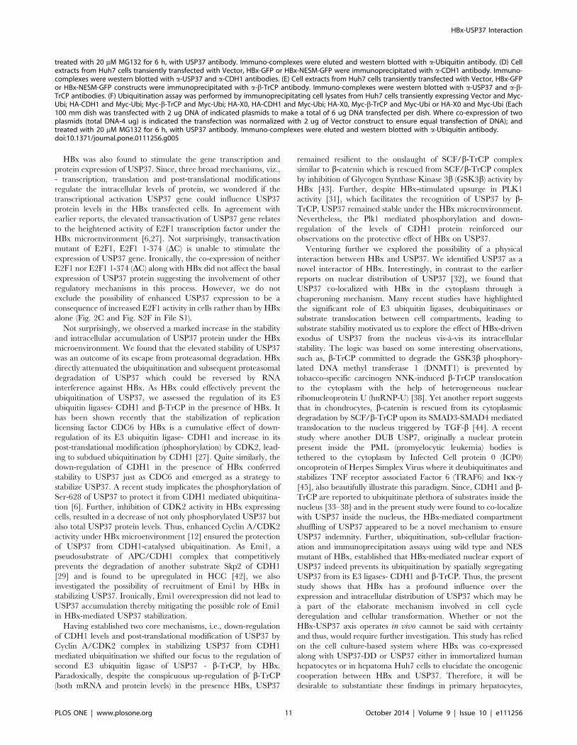
HBx differentially regulates E3 ubiquitin ligases tostabilize USP37
USP37 is degraded in a biphasic fashion by two well established
E3 ubiquitin ligase complex -SCF/b-TrCP during the G2/M
phase and APC/CDH1 from mitosis to early G1 phase. Having
established the role of HBx in USP37 gene expression and protein
stability, we next monitored the regulation of CDH1 and b-TrCP
in HBx microenvironment. Since, HBx is known to interfere with
the ubiquitination of CDC6 by negatively regulating protein levels
of CDH1 [27], we wondered if HBx similarly conferred protection
to USP37 from CDH1-mediated degradation. Not surprisingly,
CDH1 levels were down-regulated in the presence of HBx
(Figure 3A). Incidentally, HBx had only a marginal effect on
the basal CDH1 transcripts (Figure S3A in file S1). Further, we
found that ectopic expression of CDH1 led to decreased levels of
USP37 protein (Figure 3B). Conversely, HBx rescued USP37
from CDH1-mediated down-regulation similar to CDC6 but not
the other CDH1 substrate, Geminin [28] (Figure 3C). Consis-
tently, the HBx-mediated protection of USP37 from CDH1 was
attenuated by RNA interference against HBx (Figure S3B in fileS1).
Early mitotic inhibitor 1 (Emi1) has been reported to act as
psuedosubstrate of CDH1 to provide stability to CDH1 substrate
by interfering with CDH1-substrate association [29]. Addressing
the possibility of Emi1 subjugating the activity of CDH1, USP37
protein levels were monitored after co-expressing Emi1 and HBx.
Ironically, no change in the USP37 protein levels was observed
either by Emi1 alone or along with HBx (Figure S3C in file S1).
As HBx is known to potentiate Cyclin-associated CDK2 activity
which endows stability to CDC6 by protecting it against APC/
CDH1 catalyzed ubiquitination [27], we next investigated the role
of CDK2-mediated phosphorylation in the stabilization and
accumulation of USP37 protein in the presence of HBx. We
observed that just as CDC6, the levels of USP37 protein and
phosphorylated USP37 decreased in the presence of CDK2
inhibitor (Figure 3D). Interestingly, the USP37 protein and
phosphorylated USP37 protein levels remained static in the vector
transfected cells irrespective of CDK2 inhibitor treatment thereby
highlighting the significance of CDK2 mediated phosphorylation
in HBx-mediated accumulation of USP37(Figure 3D). Thus, the
down-regulation of CDH1 and CDK2-dependent phosphoryla-
tion seem responsible for conferring intracellular stability to
USP37 in HBx microenvironment.
We next sought to understand the involvement of other E3
ubiquitin ligase, b-TrCPin the regulation of USP37 levels by HBx.
We found that, in stark contrast to CDH1, b-TrCP was
upregulated in the presence of HBx, both at the protein and
transcript levels (Figure 3E and 3F). As reported earlier [7], b-
TrCP overexpression led to the downregulation of USP37 protein
levels in the cell (Figure S3D in file S1). While the ectopic
expression of DF-box deletion mutant of b-TrCP [23] could rescue
the levels of its substrates like USP37, b-catenin [23] and Ikba[30], HBx over-expression also conferred stability to b-catenin
similar to USP37 (Figure 3E). Further, RNA interference against
HBx counteracted its ability to vanquish b-TrCP catalyzed down-
regulation of USP37 (Figure S3E in file S1).
Plk1-mediated phosphorylation of USP37 plays an important
role in its recognition by SCF-b-TrCP [7]. Besides, several line of
evidence also indicate that PLK1 can be activated in the presence
of HBx [31]. Hence, we next investigated whether USP37 was
insulated from PLK1 activity in the HBx microenvironment.
Incidentally, the inhibition of PLK1 had no influence on USP37
protein levels in the HBx over-expressing cells unlike that of
CDH1 which was rescued in the presence of PLK1 inhibitor
(Figure S3F in file S1). Thus, USP37 remains protected from
SCF-b-TrCP-mediated ubiquitination despite PLK1 activity. The
dichotomy in the action of two E3 ligases targeting USP37 further
inspired us to investigate the possibility of direct or indirect
interaction between HBx and USP37 which could reveal a bigger
role of HBx in protection of USP37.
HBx-USP37 Interaction
PLOS ONE | www.plosone.org 4 October 2014 | Volume 9 | Issue 10 | e111256
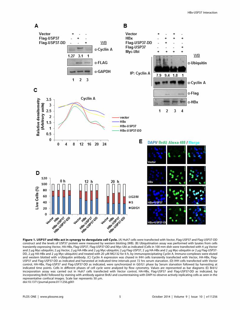
Figure 1. USP37 and HBx act in synergy to deregulate cell Cycle. (A) Huh7 cells were transfected with Vector, Flag-USP37 and Flag-USP37-DDconstruct and the levels of USP37 protein were measured by western blotting (WB). (B) Ubiquitination assay was performed with lysates from cellstransiently expressing Vector, HA-HBx, Flag-USP37, Flag-USP37-DD and Myc-Ubi as indicated (Cells in 100 mm dish were transfected with 4 mg Vectorand 2 mg Myc-ubiquitin; 2 mg Vector, 2 mg HA-HBx and 2 mg Myc-ubiquitin; 2 mg Flag-USP37, 2 mg HA-HBx and 2 mg Myc-ubiquitin or 2 mg Flag-USP37-DD, 2 mg HA-HBx and 2 mg Myc-ubiquitin) and treated with 20 mM MG132 for 6 h, by immunoprecipitating Cyclin A. Immuno-complexes were elutedand western blotted with a-Ubiquitin antibody. (C) Cyclin A expression was chased in IHH cells transiently transfected with Vector, HA-HBx, Flag-USP37 and Flag-USP37-DD as indicated and harvested at indicated time intervals post 72 hrs serum starvation. (D) IHH cells transfected with Vectorcontrol, HA-HBx, Flag-USP37 and Flag-USP37-DD as indicated, were synchronized in G0/G1 phase by Serum starvation followed by harvesting atindicated time points. Cells in different phases of cell cycle were analyzed by flow cytometry. Values are represented as bar diagrams (E) Brd-Uincorporation assay was carried out in Huh7 cells transfected with Vector control, HA-HBx, Flag-USP37 and Flag-USP37-DD as indicated, byincorporating BrdU followed by staining with antibody against BrdU and counterstaining with DAPI to observe actively replicating cells as seen in therepresentative confocal images. Scale bar represents 50 mm.doi:10.1371/journal.pone.0111256.g001
HBx-USP37 Interaction
PLOS ONE | www.plosone.org 5 October 2014 | Volume 9 | Issue 10 | e111256

Figure 2. HBx upregulates USP37 mRNA and protein levels. (A) Cell lysates from Huh7 cells transfected with Vector control or HA-X0construct were run on SDS-PAGE gel and were blotted for USP37 and normalized against GAPDH (B) Total RNA was isolated and USP37 mRNA levelswere measured by RT-qPCR using specific primers (Table S1 in File S1) in Huh7 cells overexpressing Vector, HA-HBx, E2F1 and E2F1-1-374-DC asindicated. Data (bar diagrams) are shown as mean 6 SD of three independent observations # represents statistically significant difference of p,0.001. (C)Stability of USP37 protein was monitored in Huh7 cells transfected with vector or HA-HBx then treated with 20 mg/ml cycloheximide for theindicated durations. Change in Endogenous USP37 protein levels were detected by western blotting using antiUSP37 antibody as indicated in linegraph. GAPDH was used as a control. Data (line graph) are shown as mean 6 SD of three independent observations. (D) Cell extracts from Huh7 cellstransfected with Vector or HA-X0 as indicated, were treated with 20 mM of MG132 for 6 hrs and western blotted for USP37 protein and normalizedwith GAPDH. (E) Ubiquitination assay was performed by immunoprecipitating USP37 from cell lysates from Huh7 cells transiently transfected withVector, HA-HBx, X-E and Myc-Ubiquitin as indicated (1 ug vector, 2 ug scrambled shRNA and 2 ug Myc-ubiquitin; 1 ug HA-HBx, 2 ug scrambled shRNAand 2 ug Myc-ubiquitin or 1 ug HA-HBx, 2 ug X-E shRNA and 2 ug Myc-ubiquitin were transfected in 100 mm dishes) and treated with MG132, asmentioned above and western blotting the immino-complexes with anti-Ubiquitin Anitibody.doi:10.1371/journal.pone.0111256.g002
HBx-USP37 Interaction
PLOS ONE | www.plosone.org 6 October 2014 | Volume 9 | Issue 10 | e111256

Figure 3. HBx differentially regulate CDH1 and b-TrCP to circumvent USP37 downregulation. (A) Cell extracts of Huh7 cells ectopicallyexpressing Vector or HA-HBx were western blotted with anti-CDH1 antibody and normalized with GAPDH. (B) Relative m-RNA levels of CDH1 inVector or HA-HBx transfected cells, were measured by performing qRT-PCR with primers mentioned in Table S1 in File S1. GAPDH was used ascontrol. Data (bar diagrams) are shown as mean 6 SD of three independent observations. (C) Cell lysates from cells expressing vector alone; co-expressing vector and HA-CDH1 and HA-CDH1 and HA-X0 were western blotted for USP37, CDC6, Geminin and GAPDH with indicated antibodies. (D)
HBx-USP37 Interaction
PLOS ONE | www.plosone.org 7 October 2014 | Volume 9 | Issue 10 | e111256
HBx interacts with USP37 and translocates it tocytoplasm
USP37 has been reported previously to majorly localize in the
nucleoplasm [32]. However, we observed that under HBx
environment, USP37 translocated in the peri-nuclear-cytoplasmic
region and co-localized with HBx (Pearson’s co-efficient of
correlation = 0.994292; Mander’s co-efficient of correla-
tion = 0.658119; n = 25). In contrast, USP37 was primarily nuclear
in the control cells (Figure 4A1). These findings were further
substantiated by nuclear and cytoplasmic fractionation of cells
transfected with HBx and its hairpin construct (Figure S4A infile S1). Interestingly, the cytoplasmic compartmentalization of
USP37 could be reversed in the presence of HBx sh-RNA (X-E). It
has been reported earlier nuclear export signal of HBx is
responsible for its nuclear export in a cellular milieu [21].
Moreover, mutation of its two Leucine residues to Alanine (L98A,
L100A) in ‘NES element’ renders it nucleus bound [21]. The
nuclear export of HBx was also sensitive to Leptomycin B
treatment (CRM1/XPO dependent nuclear transport inhibitor)
[21]. Intriguingly, USP37 migration to cytoplasm in the presence
of HBx, was also found to be sensitive to Leptomycin B treatment.
This observation prompted us to investigate if HBx acted as
chaperone for USP37. As expected, over-expression of the nuclear
export signal mutant of HBx (HBx-NESM-GFP) abolished the
nuclear export of USP37 (Figure 4A2). Consistent with this, our
subcellular fractionation studies revealed that while USP37 was
present both in the nucleus as well as in cytoplasm in the presence
of HBx, it was majorly nuclear in the HBx-NESM transfected cells
(Figure 4B). Since, HBx and USP37 co-localized in cells, we next
examined the possibility of a physical interaction between HBx
and USP37. Our co-immunoprecipitation studies revealed that the
purified recombinant HBx-MBP fusion protein interacted with
endogenous USP37 present in HEK293T cell lysates but not with
MBP (Figure S4B in file S1). The HBx-USP37 interaction was
further confirmed by co-immunoprecipitation of HBx by Flag-
tagged USP37 from cells co-transfected with USP37 and HBx
(Figure S4C in file S1). In addition, HBx was found to interact
with endogenously expressed USP37 in cells overexpressing HBx
(Figure 4C).
Nuclear export of USP37 rescues it from ubiquitinationand proteasomal degradation
Earlier studies have identified nucleus as the site for the E3
ubiquitin ligase activity of CDH1 and b-TrCP [33–38]. Since,
USP37 is targeted by both these ubiquitin ligases and HBx
chaperoned USP37 out of the nucleus, we wondered if it was
safeguard mechanism to prevent the ubiquitination and degrada-
tion of USP37. Our, Confocal experiments confirmed that bulk of
USP37 interacted with its E3 ligases- CDH1 (Pearson’s co-efficient
of correlation = 0.984381; Mander’s co-efficient of correla-
tion = 0.581855; n = 25) and b-TrCP (Pearson’s co-efficient of
Correlation = 0.979543; Mander’s co-efficient of correla-
tion = 0.557357; n = 25) inside the nucleus (Figure 5A). Not
surprisingly, over-expression of HBx-NESM mutant did not confer
stability to USP37 protein (Figure 5B) owing to its inability to
interfere with its ubiquitination (Figure 5C). As expected,
overexpression of HBx unlike NESM mutant abrogated the
interaction of USP37 with its cognate E3 ligases- CDH1 and b-
TrCP (Figure 5D and 5E). Moreover, our ubiquitination assay
confirmed that HBx did not interfere with the ubiquitination of
USP37 by CDH1 and b-TrCP (Figure 5F). These observations
suggested that HBx could stabilize USP37 by protecting it from
ubiquitination and subsequent proteasomal degradation.
Discussion
HBx is a bonafide oncoprotein of HBV that extends its
influence over a range of host cell functions like cell cycle
progression, signaling pathways, DNA damage response, gene
expression and regulation of ubiquitin-proteasomal system to
facilitate virus-mediated carcinogenesis [11]. Not surprisingly,
Ubiquitin ligases are increasingly being recognized as being
instrumental in oncogenesis. HBx is now also known to
manipulate few E3 ligases and their adaptors like SCF/S-phase
kinase associated protein 2 (SCF/Skp2), SCF/F-box/WD repeat-
containing protein 7 a (SCF/Fbw7a), SCF/Suppressor of cytokine
signaling 3 (SCF/SOCS3), Adenomatosis Polyposis Coli, APC/
CDC20 or APC/CDH1 [39]. Ironically, the interaction of HBx
with cellular deubiquitinases has not studied. Recently, deubiqui-
tinase USP37 has been recognized as a cell cycle regulator which
reverses APC/CDH1 mediated ubiquitination of cyclin A to
promote S phase entry of cells [6]. Besides, its normal functions,
USP37 is gaining relevance in context of cancer development. For
instance, increased USP37 expression is correlated with poor
prognosis in non-small cell lung cancer [8]. USP37 also plays a
role in stabilizing tumor suppressor p27 in medulloblastoma cells
and promyelocytic leukemia zinc finger and retinoic acid receptor
alpha (PLZF-RARA) oncogenic fusion protein in Acute promy-
elocytic leukemia [9,10]. As HBx has been shown to stabilze cyclin
A in order to potentiate CDK2 activity [13], we wondered if HBx
engaged USP37 in conferring intracellular stability to cyclin A.
The present study showed that HBx involved USP37 in the
stabilization of Cyclin A and the effect could be reversed in the
presence of USP37-DD. Furthermore, FACS analysis revealed
that USP37 played a crucial role in HBx-mediated deregulation of
cell cycle by accelerating the S phase entry and the effect could be
reversed in the presence of USP37-DD mutant. Earlier reports
have suggested that stable expression of Cyclin A inside the cells
can cause cell cycle arrest or apoptosis due to checkpoint
activation [40]. In contrast, our Brd-U and MTT assays revealed
that cells co-transfected with HBx and USP37 showed improved
cellular viability and active DNA replication. As HBx has been
documented to overcome cell cycle checkpoints in order to support
unfettered cell cycle progression [31,41], the higher viability of
cells co-transfected with HBx and USP37 could be attributed to
HBx-mediated checkpoint inactivation.
Untreated and CDK2 Inhibitor II compound 3 (10 mM for 6 h) treated Vector or HBx transfected cell extract were western blotted for USP37, CDC6 andGAPDH. Immunoprecipitation assay was performed using USP37 antibody with cell lysates from untreated or CDK2 inhibitor II compound 3 (10 mMfor 6 h) treated Vector or HBx transfected cells. The immune-complexes were blotted with phospho-serine and USP37 antibody. Total cell lysateswere also western blotted with phospho-CDC6 (Ser-54) and GAPDH antibody. (E) Cell lysates from cells expressing Vector or HA-HBx were westernblotted with anti-b-TrCP antibody and normalized with GAPDH. (F) Relative m-RNA levels of b-TrCP in Vector or HA-HBx transfected Huh7 cells, weremeasured by performing qRT-PCR with primers mentioned in Table S1 in File S1. GAPDH was used as control. Data (bar diagrams) are shown as mean6 SD of three independent observations # represents statistically significant difference of p,0.001. (G) Myc-b-TrCP, Myc-DF-b-TrCP and HA-HBxtransfected huh7 cells (as indicated) were lysed and lysates was separated on SDS-PAGE gel western blotted for USP37, b-catenin and CDH1. GAPDHwas used as a loading control.doi:10.1371/journal.pone.0111256.g003
HBx-USP37 Interaction
PLOS ONE | www.plosone.org 8 October 2014 | Volume 9 | Issue 10 | e111256

Figure 4. HBx interacts with USP37 and promotes USP37 translocation from Nucleus to cytoplasm. Representative confocal image ofmock-treated Huh7 cells transiently expressing Vector, X0-GFP or XO-NESM-GFP constructs and cells expressing X0-GFP construct treated withLeptomycin B (100 nM, 4 h), fixed and stained with anti-HBx and anti-USP37 primary antibody and corresponding fluorescent-labelled secondaryantibody. Nuclei were counterstained with DAPI. Images were captured at 60x magnification in Nikon A1R confocal microscope. Scale bar represents10 mm unless mentioned otherwise. (B) Cytoplasmic-nuclear fractionation was performed with cells transfected with vector, HBx-GFP or HBx-NESM-GFP constructs as per the protocol mentioned earlier. The Nuclear (N) and the cytoplasmic (C) fractions of cells were western blotted with USP37,HBx, Histone H1 and GAPDH antibodies. (C) USP37 was immunoprecipitated from cell lysate of Huh7 cells transfected with Vector or HA-HBx.Immuno-complexes were separated on SDS-PAGE gel and were immunoblotted using USP37 and HBx anitibody. Input was probed with HA antibody.doi:10.1371/journal.pone.0111256.g004
HBx-USP37 Interaction
PLOS ONE | www.plosone.org 9 October 2014 | Volume 9 | Issue 10 | e111256
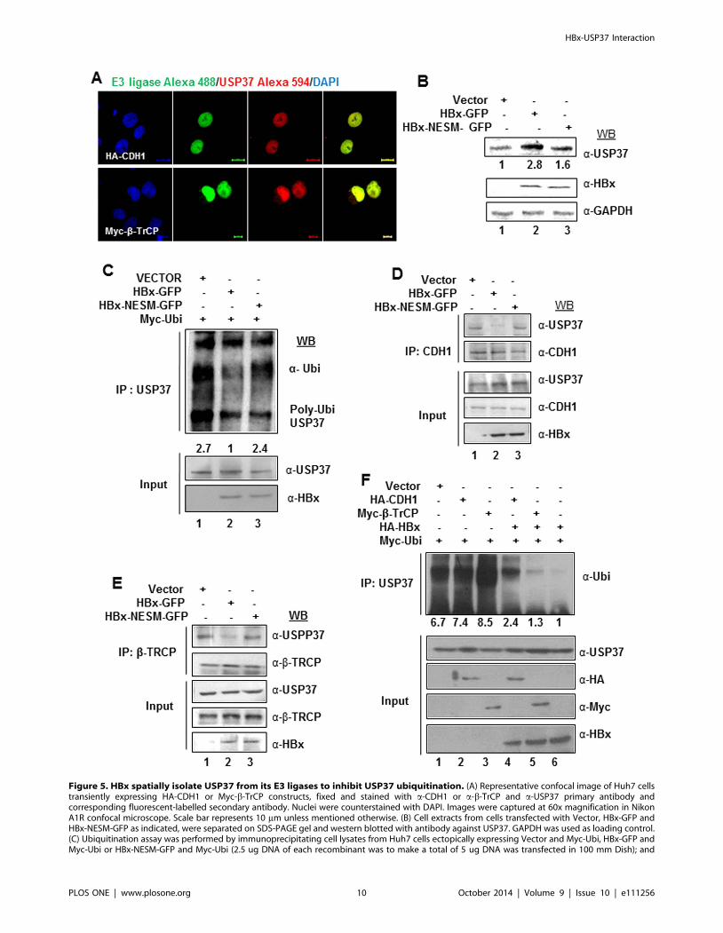
Figure 5. HBx spatially isolate USP37 from its E3 ligases to inhibit USP37 ubiquitination. (A) Representative confocal image of Huh7 cellstransiently expressing HA-CDH1 or Myc-b-TrCP constructs, fixed and stained with a-CDH1 or a-b-TrCP and a-USP37 primary antibody andcorresponding fluorescent-labelled secondary antibody. Nuclei were counterstained with DAPI. Images were captured at 60x magnification in NikonA1R confocal microscope. Scale bar represents 10 mm unless mentioned otherwise. (B) Cell extracts from cells transfected with Vector, HBx-GFP andHBx-NESM-GFP as indicated, were separated on SDS-PAGE gel and western blotted with antibody against USP37. GAPDH was used as loading control.(C) Ubiquitination assay was performed by immunoprecipitating cell lysates from Huh7 cells ectopically expressing Vector and Myc-Ubi, HBx-GFP andMyc-Ubi or HBx-NESM-GFP and Myc-Ubi (2.5 ug DNA of each recombinant was to make a total of 5 ug DNA was transfected in 100 mm Dish); and
HBx-USP37 Interaction
PLOS ONE | www.plosone.org 10 October 2014 | Volume 9 | Issue 10 | e111256
HBx was also found to stimulate the gene transcription and
protein expression of USP37. Since, three broad mechanisms, viz.,
- transcription, translation and post-translational modifications
regulate the intracellular levels of protein, we wondered if the
transcriptional activation USP37 gene could influence USP37
protein levels in the HBx transfected cells. In agreement with
earlier reports, the elevated transactivation of USP37 gene relates
to the heightened activity of E2F1 transcription factor under the
HBx microenvironment [6,27]. Not surprisingly, transactivation
mutant of E2F1, E2F1 1-374 (DC) is unable to stimulate the
expression of USP37 gene. Ironically, the co-expression of neither
E2F1 nor E2F1 1-374 (DC) along with HBx did not affect the basal
expression of USP37 protein suggesting the involvement of other
regulatory mechanisms in this process. However, we do not
exclude the possibility of enhanced USP37 expression to be a
consequence of increased E2F1 activity in cells rather than by HBx
alone (Fig. 2C and Fig. S2F in File S1).
Not surprisingly, we observed a marked increase in the stability
and intracellular accumulation of USP37 protein under the HBx
microenvironment. We found that the elevated stability of USP37
was an outcome of its escape from proteasomal degradation. HBx
directly attenuated the ubiquitination and subsequent proteasomal
degradation of USP37 which could be reversed by RNA
interference against HBx. As HBx could effectively prevent the
ubiquitination of USP37, we assessed the regulation of its E3
ubiquitin ligases- CDH1 and b-TrCP in the presence of HBx. It
has been shown recently that the stabilization of replication
licensing factor CDC6 by HBx is a cumulative effect of down-
regulation of its E3 ubiquitin ligase- CDH1 and increase in its
post-translational modification (phosphorylation) by CDK2, lead-
ing to subdued ubiquitination by CDH1 [27]. Quite similarly, the
down-regulation of CDH1 in the presence of HBx conferred
stability to USP37 just as CDC6 and emerged as a strategy to
stabilize USP37. A recent study implicates the phosphorylation of
Ser-628 of USP37 to protect it from CDH1 mediated ubiquitina-
tion [6]. Further, inhibition of CDK2 activity in HBx expressing
cells, resulted in a decrease of not only phosphorylated USP37 but
also total USP37 protein levels. Thus, enhanced Cyclin A/CDK2
activity under HBx microenvironment [12] ensured the protection
of USP37 from CDH1-catalysed ubiquitination. As Emi1, a
pseudosubstrate of APC/CDH1 complex that competitively
prevents the degradation of another substrate Skp2 of CDH1
[29] and is found to be upregulated in HCC [42], we also
investigated the possibility of recruitment of Emi1 by HBx in
stabilizing USP37. Ironically, Emi1 overexpression did not lead to
USP37 accumulation thereby mitigating the possible role of Emi1
in HBx-mediated USP37 stabilization.
Having established two core mechanisms, i.e., down-regulation
of CDH1 levels and post-translational modification of USP37 by
Cyclin A/CDK2 complex in stabilizing USP37 from CDH1
mediated ubiquitination we shifted our focus to the regulation of
second E3 ubiquitin ligase of USP37 - b-TrCP, by HBx.
Paradoxically, despite the conspicuous up-regulation of b-TrCP
(both mRNA and protein levels) in the presence HBx, USP37
remained resilient to the onslaught of SCF/b-TrCP complex
similar to b-catenin which is rescued from SCF/b-TrCP complex
by inhibition of Glycogen Synthase Kinase 3b (GSK3b) activity by
HBx [43]. Further, despite HBx-stimulated upsurge in PLK1
activity [31], which facilitates the recognition of USP37 by b-
TrCP, USP37 remained stable under the HBx microenvironment.
Nevertheless, the Plk1 mediated phosphorylation and down-
regulation of the levels of CDH1 protein reinforced our
observations on the protective effect of HBx on USP37.
Venturing further we explored the possibility of a physical
interaction between HBx and USP37. We identified USP37 as a
novel interactor of HBx. Interestingly, in contrast to the earlier
reports on nuclear distribution of USP37 [32], we found that
USP37 co-localized with HBx in the cytoplasm through a
chaperoning mechanism. Many recent studies have highlighted
the significant role of E3 ubiquitin ligases, deubiquitinases or
substrate translocation between cell compartments, leading to
substrate stability motivated us to explore the effect of HBx-driven
exodus of USP37 from the nucleus vis-a-vis its intracellular
stability. The logic was based on some interesting observations,
such as, b-TrCP committed to degrade the GSK3b phosphory-
lated DNA methyl transferase 1 (DNMT1) is prevented by
tobacco-specific carcinogen NNK-induced b-TrCP translocation
to the cytoplasm with the help of heterogeneous nuclear
ribonucleoprotein U (hnRNP-U) [38]. Yet another report suggests
that in chondrocytes, b-catenin is rescued from its cytoplasmic
degradation by SCF/b-TrCP upon its SMAD3-SMAD4 mediated
translocation to the nucleus triggered by TGF-b [44]. A recent
study where another DUB USP7, originally a nuclear protein
present inside the PML (promyelocytic leukemia) bodies is
tethered to the cytoplasm by Infected Cell protein 0 (ICP0)
oncoprotein of Herpes Simplex Virus where it deubiquitinates and
stabilizes TNF receptor associated Factor 6 (TRAF6) and Ikk-c[45], also beautifully illustrate this paradigm. Since, CDH1 and b-
TrCP are reported to ubiquitinate plethora of substrates inside the
nucleus [33–38] and in the present study were found to co-localize
with USP37 inside the nucleus, the HBx-mediated compartment
shuffling of USP37 appeared to be a novel mechanism to ensure
USP37 indemnity. Further, ubiquitination, sub-cellular fraction-
ation and immunoprecipitation assays using wild type and NES
mutant of HBx, established that HBx-mediated nuclear export of
USP37 indeed prevents its ubiquitination by spatially segregating
USP37 from its E3 ligases- CDH1 and b-TrCP. Thus, the present
study shows that HBx has a profound influence over the
expression and intracellular distribution of USP37 which may be
a part of the elaborate mechanism involved in cell cycle
deregulation and cellular transformation. Whether or not the
HBx-USP37 axis operates in vivo cannot be said with certainty
and thus, would require further investigation. This study has relied
on the cell culture-based system where HBx was co-expressed
along with USP37-DD or USP37 either in immortalized human
hepatocytes or in hepatoma Huh7 cells to elucidate the oncogenic
cooperation between HBx and USP37. Therefore, it will be
desirable to substantiate these findings in primary hepatocytes,
treated with 20 mM MG132 for 6 h, with USP37 antibody. Immuno-complexes were eluted and western blotted with a-Ubiquitin antibody. (D) Cellextracts from Huh7 cells transiently transfected with Vector, HBx-GFP or HBx-NESM-GFP were immunoprecipitated with a-CDH1 antibody. Immuno-complexes were western blotted with a-USP37 and a-CDH1 antibodies. (E) Cell extracts from Huh7 cells transiently transfected with Vector, HBx-GFPor HBx-NESM-GFP constructs were immunoprecipitated with a-b-TrCP antibody. Immuno-complexes were western blotted with a-USP37 and a-b-TrCP antibodies. (F) Ubiquitination assay was performed by immunoprecipitating cell lysates from Huh7 cells transiently expressing Vector and Myc-Ubi; HA-CDH1 and Myc-Ubi; Myc-b-TrCP and Myc-Ubi; HA-X0, HA-CDH1 and Myc-Ubi; HA-X0, Myc-b-TrCP and Myc-Ubi or HA-X0 and Myc-Ubi (Each100 mm dish was transfected with 2 ug DNA of indicated plasmids to make a total of 6 ug DNA transfected per dish. Where co-expression of twoplasmids (total DNA-4 ug) is indicated the transfection was normalized with 2 ug of Vector construct to ensure equal transfection of DNA); andtreated with 20 mM MG132 for 6 h, with USP37 antibody. Immuno-complexes were eluted and western blotted with a-Ubiquitin antibody.doi:10.1371/journal.pone.0111256.g005
HBx-USP37 Interaction
PLOS ONE | www.plosone.org 11 October 2014 | Volume 9 | Issue 10 | e111256

USP37 null cells as well as in experimental animal models of viral
HBx. Nevertheless, it appears that HBx could employ cellular
USP37 as a novel strategy to deregulate cell cycle and induce cell
transformation.
Supporting Information
File S1 Supporting files. This file contains Table S1, Figure
S1, Figure S2, Figure S3, and Figure S4. Figure S1, Status of cell
cycle regulators and cell viability under different experimental
conditions. Figure S2, Validation of HBx expression and
stimulation of USP37 by HBx. Figure S3, Regulation of
USP37 under HBx microenvironment. Figure S4, Intracellular
distribution of USP37 and its interaction with HBx. Table S1,
tabulates primer names and sequences used for RT PCR.
(PDF)
Acknowledgments
We are grateful to the following scientists for kindly providing us the
expression vectors for genes and cell lines: Dr. Vishwa Mohan Dixit
(Genentech, USA) for HA-CDH1, USP37-Flag, and USP37-DUB-Dead-
Flag constructs; Dr. Xin Wei Wang (National Institutes of Health,
Bethesda, Maryland, US) for X0-NESM-GFP construct; Dr. Anindya
Dutta (University of Virginia, USA) for FLAG-Emi1 construct; Dr. Kei-
ichi Nakayama (Department of Molecular and cellular biology, Kyushu
university, Japan) for pCDNA3-b-TRCP and pCDNA3-DF-b-TRCP; Dr
Xin Lu (Ludwig Institute for Cancer Research, Cambridge, UK) for wild-
type E2F1 (pCMV-E2F1) and its transactivation defective mutant pCMV-
E2F1-1-347 (DC), Dr. Michael MC Lai (Institute of Molecular Biology,
Academia Sinica, Taipei, Taiwan) for Myc-ubiquitin construct and Dr.
Aleem Siddiqui (University of Colorado, Denver) for Human hepatoma
Huh7 cells and Dr. F. Danniel (Institut National de la Sante et de la
Recherche Medicale Unite 481, Universite Paris 7, Paris, France) for
Immortalized human hepatocytes IHH cells.
Author Contributions
Conceived and designed the experiments: VK. Performed the experiments:
NS. Analyzed the data: NS VK. Contributed reagents/materials/analysis
tools: VK. Contributed to the writing of the manuscript: NS VK.
References
1. Glickman MH, Ciechanover A (2002) The ubiquitin-proteasome proteolytic
pathway: destruction for the sake of construction. Physiol Rev 82: 373–428.
2. Schnell JD, Hicke L (2003) Non-traditional functions of ubiquitin and ubiquitin-binding proteins. J BiolChem 278: 35857–35860.
3. Weissman AM, Shabek N, Ciechanover A (2011) The predator becomes theprey: regulating the ubiquitin system by ubiquitylation and degradation. Nat
Rev Mol Cell Biol 12: 605–620.
4. Komander D, Clague MJ, Urbe S (2009) Breaking the chains: structure andfunction of the deubiquitinases. Nat Rev Mol Cell Bio l10: 550–563.
5. Saxena N, Kumar V (2013) Oncogenic viruses: DUBbing their way to cancer.
VirolDiscov 1: 5.
6. Huang X, Summers MK, Pham V, Lill JR, Liu J, et al. (2011) Deubiquitinase
USP37 is activated by CDK2 to antagonize APC(CDH1) and promote S phaseentry. Mol Cell 42: 511–523.
7. Burrows AC, Prokop J, Summers MK (2012) Skp1-Cul1-F-box ubiquitin ligase
(SCF(bTrCP))-mediated destruction of the ubiquitin-specific protease USP37during G2-phase promotes mitotic entry. J BiolChem 287: 39021–39029.
8. Bianchi F, Nuciforo P, Vecchi M, Bernard L, Tizzoni L, et al. (2007) Survival
prediction of stage I lung adenocarcinomas by expression of 10 genes. J ClinInvest 17: 3436–3444.
9. Yang WC, Shih HM (2012) Thedeubiquitinating enzyme USP37 regulates theoncogenic fusion protein PLZF/RARA stability. Oncogene 32: 5167–5175.
10. Das CM, Taylor P, Gireud M, Singh A, Lee D, et al. (2012) Thedeubiquitylase
USP37 links REST to the control of p27 stability and cell proliferation.Oncogene 32: 1691–1701.
11. Kapoor NR, Kumar V (2012) Hepatitis B virus: A molecular perspective.
ProcNatlAcadSciSectBBiolSci 82: 31–41.
12. Benn J, Schneider RJ (1995) Hepatitis B virus HBx protein deregulates cell cycle
checkpoint controls. ProcNatlAcadSci U S A 92: 11215–11219.
13. Mukherji A, Janbandhu VC, Kumar V (2007) HBx-dependent cell cyclederegulation involves interaction with cyclinE/A-cdk2 complex and destabili-
zation of p27Kip1. Biochem J 401: 247–256.
14. Visintin R, Prinz S, Amon A (1997) CDC20 and CDH1: a family of substrate-
specific activators of APC-dependent proteolysis. Science 278: 460–463.
15. Fukushima H, Ogura K, Wan L, Lu Y, Li V, et al. (2013) SCF-mediated Cdh1degradation defines a negative feedback system that coordinates cell-cycle
progression. Cell Rep 4: 803–816.
16. Nakabayashi H, Taketa K, Miyano K, Yamane T, Sato J (1982) Growth ofhuman hepatoma cells lines with differentiated functions in chemically defined
medium. Cancer Res 42(9): 3858–63.
17. Schippers JI, Moshage H, Roelofsen H, Muller M, Heymans HAS, et al. (1997)
Immortalized human hepatocytes as a tool for the study of hepatocytic (de-)
differentiation. Cell Biol Toxicol 13(4–5): 375–86.
18. Kumar V, Jayasuryan N, Kumar R (1996) A truncated mutant (residues 58–140)
of the hepatitis B virus X protein retains transactivation function. ProcNatlA-
cadSci USA 93: 5647–5652.
19. Sidhu K, Kumar V (2014) Mass spectrometric determination of disulfide bonds
in the biologically active recombinant HBx protein of hepatitis B virus. (In Press).
20. Hung L, Kumar V (2004) Specific inhibition of gene expression and
transactivation functions of hepatitis B virus X protein and c-myc by small
interfering RNAs. FEBS Lett 560: 210–214.
21. Forgues M, Marrogi AJ, Spillare EA, Wu CG, Yang Q, et al. (2001) Interaction
of the hepatitis B virus X protein with the Crm1-dependent nuclear export
pathway. J BiolChem 276: 22797–22803.
22. Machida YJ, Dutta A (2007) The APC/C inhibitor, Emi1, is essential for
prevention of rereplication. Genes Dev 21: 184–194.
23. Kitagawa M, Hatakeyama S, Shirane M, Matsumoto M, Ishida N, et al. (1999)
An F-box protein, FWD1, mediates ubiquitin-dependent proteolysis of beta-
catenin. EMBO J 18: 2401–2410.
24. Hsieh JK, Fredersdorf S, Kouzarides T, Martin K, Lu X (1997) E2F1-induced
apoptosis requires DNA binding but not transactivation and is inhibited by the
retinoblastoma protein through direct interaction. Genes Dev 11: 1840–1852.
25. Liao TL, Wu CY, Su WC, Jeng KS, Lai MM (2010) Ubiquitination and
deubiquitination of NP protein regulates influenza A virus RNA replication.
EMBO J 22: 3879–90.
26. Schmittgen TD, Livak KJ (2008) Analyzing real-time PCR data by the
comparative C(T) method. Nat Protoc 3: 1101–1108.
27. Pandey V, Kumar V (2012) HBx protein of hepatitis B virus promotes
reinitiation of DNA replication by regulating expression and intracellular
stability of replication licensing factor CDC6. J BiolChem 287: 20545–10554.
28. McGarry TJ, Kirschner MW (1998) Geminin, an inhibitor of DNA replication,
is degraded during mitosis. Cell 93: 1043–1053.
29. Margottin-Goguet F, Hsu JY, Loktev A, Hsieh HM, Reimann JD, et al. (2003)
Prophase destruction of Emi1 by the SCF(betaTrCP/Slimb) ubiquitin ligase
activates the anaphase promoting complex to allow progression beyond
prometaphase. Dev Cell 4: 813–826.
30. Da Silva-Ferrada E, Torres-Ramos M, Aillet F, Campagna M, Matute C, et al.
(2011) Role of monoubiquitylation on the control of IkBa degradation and NF-
kB activity. PLoS One 6: e25397.
31. Studach L, Wang WH, Weber G, Tang J, Hullinger RL, et al. (2010) Polo-like
kinase 1 activated by the hepatitis B virus X protein attenuates both the DNA
damage checkpoint and DNA repair resulting in partial polyploidy. J BiolChem
285: 30282–30293.
32. Tanno H, Shigematsu T, Nishikawa S, Hayakawa A, Denda K, et al. (2014)
Ubiquitin-interacting motifs confer full catalytic activity, but not ubiquitin chain
substrate specificity, to deubiquitinatingenzyme USP37. J BiolChem 289: 2415–
2423.
33. Zhou Y, Ching YP, Ng RW, Jin DY (2003) Differential expression, localization
and activity of two alternatively spliced isoforms of human APC regulator
CDH1. Biochem J 374: 349–358.
34. Lassot I, Segeral E, Berlioz-Torrent C, Durand H, Groussin L, et al. (2001)
ATF4 degradation relies on a phosphorylation-dependent interaction with the
SCF(betaTrCP) ubiquitin ligase. Mol Cell Biol 21: 2192–2202.
35. Davis M, Hatzubai A, Andersen JS, Ben-Shushan E, Fisher GZ, et al. (2002)
Pseudosubstrate regulation of the SCF(beta-TrCP) ubiquitin ligase by hnRNP-
U. Genes Dev 16: 439–451.
36. Nakayama KI, Nakayama K (2005) Regulation of the cell cycle by SCF-type
ubiquitin ligases. Semin Cell DevBiol 16: 323–333.
37. von Mikecz A (2006) The nuclear ubiquitin-proteasome system. J Cell Sci 119:
1977–1984.
38. Lin RK, Hsieh YS, Lin P, Hsu HS, Chen CY, et al. (2010) The tobacco-specific
carcinogen NNK induces DNA methyltransferase 1 accumulation and tumor
suppressor gene hypermethylation in mice and lung cancer patients. J Clin
Invest 120: 521–532.
39. Ahuja R, Jamal A, Nosrati N, Pandey V, Rajput P, et al. (2014) Human
Oncogenic Viruses and Cancer. Curr Sci (In press).
HBx-USP37 Interaction
PLOS ONE | www.plosone.org 12 October 2014 | Volume 9 | Issue 10 | e111256
40. Jacobs HW, Keidel E, Lehner CF (2001) A complex degradation signal in Cyclin
A required for G1 arrest, and a C-terminal region for mitosis. EMBO J 20:2376–2386.
41. Chae S, Ji JH, Kwon SH, Lee HS, Lim JM, et al. (2013) HBxAPa/Rsf-1-
mediated HBx-hBubR1 interactions regulate the mitotic spindle checkpoint andchromosome instability. Carcinogenesis 34: 1680–1688.
42. Zhao Y, Tang Q, Ni R, Huang X, Wang Y, et al. (2013) Early mitotic inhibitor-1, an anaphase-promoting complex/cyclosome inhibitor, can control tumor cell
proliferation in hepatocellular carcinoma: correlation with Skp2 stability and
degradation of p27(Kip1). Hum Pathol 44: 365–373.
43. Jung JK, Kwun HJ, Lee JO, Arora P, Jang KL (2007) Hepatitis B virus X
protein differentially affects the ubiquitin-mediated proteasomal degradation ofbeta-catenin depending on the status of cellular p53. J Gen Virol88(Pt 8): 2144–
54. PubMed: 17622616.
44. Zhang M, Wang M, Tan X, Li TF, Zhang YE, et al. (2010) Smad3 preventsbeta-catenin degradation and facilitates beta-catenin nuclear translocation in
chondrocytes. J BiolChem 285: 8703–8710.45. Daubeuf S, Singh D, Tan Y, Liu H, Federoff HJ, et al. (2009) HSV ICP0
recruits USP7 to modulate TLR-mediated innate response. Blood 113: 3264–
3275.
HBx-USP37 Interaction
PLOS ONE | www.plosone.org 13 October 2014 | Volume 9 | Issue 10 | e111256


Supplementary Information
Primers for RT-PCR Sequence 5'-->3'
Cdh1 Fwd primer AGATCTCCAAGATCCCCTTCA
Cdh1 Rev primer CCTCCAACATGGACAGCTTCT
USP37 Fwd primer CCAGTGGAGCGAAACAAAGC
USP37 Rev primer CCTCTGCATCCTTACTTGGTACT
B-trcp Fwd primer ATGCAAGCGAATTCTCACAGG
B-trcp Rev primer GGAACGATCTTTGGAGCAGGT
Table S1
Fwd, forward; Rev, reverse

Figure S1
C
A
B
OD
at
560 n
m
0
0.5
1
1.5
2
2.5
3
0 4 8 12 16 20 24
USP37
USP37-DD
Cyclin A
*
α-Cyclin E
α-Cyclin A
a-p27
a-GAPDH
WB Serum Stv. 0 4 8 12 16 20 24 28
Time (h)
USP37
USP37 DD
D
0
0.5
1
1.5
2
2.5
3
Asynchronous Synchronized
0 h 12 h 20 h

Figure S1. Status of cell cycle regulators and cell viability under
different experimental conditions. (A) IHH cells were serum starved for
72 h followed by serum stimulation and harvesting at indicated time
points. Cell cycle analysis was performed by flow cytometry after staining
with propidium iodide . The expression of Cyclin E, Cyclin A, p27 and
GAPDH was analyzed by western blotting. (B) IHH cells were transfected
with Flag-USP37 and Flag-USP37-DD and the levels of Cyclin A were
measured at indicated time periods. (C) Huh7 cells were transfected with
control vector; HBx; Flag-USP37; Flag-USP37-DD; HBx and Flag-USP37
and HBx and Flag-USP37-DD and cell viability was measured by MTT
assay. Cells transfected with HA-Myc and vector and treated with Methyl
Methane Sulphonate (97% w/v) at 0.1% v/v for 30 min were used as
positive and negative control, respectively. Data (bar diagrams) are shown
as mean ± SD of three independent observations . * represents
statistically significant difference of p<0.005. (D) Huh7 cells transfected
with Flag-USP37 and Flag-USP37-DD were analyzed for Brd-U
incorporation and confocal imaging. Scale bar represents 50µm.

Figure S2
Bright Field / Flourescence
HBx-GFP +
Sc-shRNA
HBx-GFP +
X-E shRNA
I
II
III
D
E E2F1 - + -
E2F1-1-374(DC) - - +
HA-HBx + + +
α-USP37
α-CDC6
α-GAPDH
1 1.3 1
1 2 3
α-HBx
IHH cells A C
1 2
Vector + -
HA-HBx - +
α-USP37
α-HBx
α-GAPDH
1 1.8
Vector + -
HA-HBx - + Vector + -
HA-HBx - +
α-USP37
α-HBx
α-GAPDH
B
α-USP37
α-HBx
α-GAPDH
1 2
1 2.3
WB WB WB
1 2
1 1.7
HEK293T U2OS
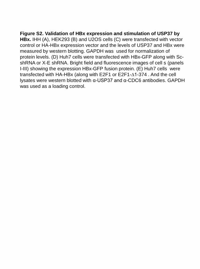
Figure S2. Validation of HBx expression and stimulation of USP37 by
HBx. IHH (A), HEK293 (B) and U2OS cells (C) were transfected with vector
control or HA-HBx expression vector and the levels of USP37 and HBx were
measured by western blotting. GAPDH was used for normalization of
protein levels. (D) Huh7 cells were transfected with HBx-GFP along with Sc-
shRNA or X-E shRNA. Bright field and fluorescence images of cell s (panels
I-III) showing the expression HBx-GFP fusion protein. (E) Huh7 cells were
transfected with HA-HBx (along with E2F1 or E2F1-∆1-374 . And the cell
lysates were western blotted with α-USP37 and α-CDC6 antibodies. GAPDH
was used as a loading control.

Figure S3
D
α-USP37
α-Myc
α-GAPDH
Vector + -
Myc -β-TrCP - + WB
1 2
1 0.2
Myc -β-TrCP + + +
Sc-shRNA + + -
HA-HBx - + +
X-E shRNA - - +
α-USP37
α-β- Catenin
α-Myc
α-HBx
α-GAPDH
WB
1 2 3
E
1 2.1 1.3
HA-HBx + +
Plk1i - +
α-USP37
α-CDH1
α-HBx
α-GAPDH
WB
1 2
F
1 1.2
A
α-USP37
α-HA
α-GAPDH
WB
1 2
Vector + -
HA-CDH1 - +
1 0.25
B C
Vector + - - -
HA-HBx - - + +
Flag-Emi1 - + - +
α-USP37
α-Flag
α-HBx
α-GAPDH
1 2 3 4
WB
1 1 1.8 2
α-USP37
α-Cdc6
α-HA
α-HBx
α-GAPDH
WB
1 2 3
HA-CDH1 + + +
Sc-shRNA + + -
HA-HBx - + +
X-E shRNA - - +
1 1.9 1.2

Figure S3. Regulation of USP37 under HBx microenvironment. Huh7
cells were transfected with indicated recombinants and western blotted
for the expression of specific antigens: (A) transfection with vector or
HA-CDH1 constructs and immunoblotting for USP37; (B) transfection
with HA-CDH1, HA-HBx, Sc-shRNA and/or Sc-shRNA and probing with
α-USP37, α-Cdc6 , α-HBx and α-HA antibodies; (C) transfection with
vector, Flag-Emi1 and HA-HBx and western blotting with α-USP37, α-
Flag and α-HBx antibodies; (D) transfection with vector and Myc-β-TrcP
constructs and western blotting for USP37; (E) transfection with
combinations of Myc-β-TrcP, HA-HBx, Sc-shRNA and X-E-shRNA as
indicated and western blotted with α-USP37 and α-β-catenin antibodies;
(F) transfection with HBx, treated with PLK1 inhibitor SBE13
hydrochloride (100µM) for 8h, and western blotted for USP37 and CDH1.
GAPDH levels were used for normalization in the above panels.
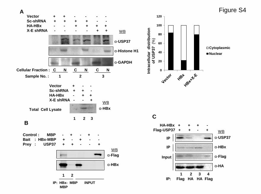
Figure S4
B
α-Flag
α-HBx
Control : MBP - + - + -
Bait : HBx-MBP + - + - -
Prey : USP37 + + - - +
WB
1 2
IP: HBx- MBP INPUT
MBP
A
Intr
acellu
lar
dis
trib
uti
on
of
US
P37
(%)
0
20
40
60
80
100
120
Cytoplasmic
Nuclear
Total Cell Lysate
Vector + - -
Sc-shRNA + + -
HA-HBx - + +
X-E shRNA - - +
α-HBx
WB
1 2 3
Vector + + - - - -
Sc-shRNA + + + + - -
HA-HBx - - + + + +
X-E shRNA - - - - + +
Cellular Fraction : C N C N C N
Sample No. : 1 2 3
α-USP37
α-Histone H1
α-GAPDH
WB
C
HA-HBx + + + -
Flag-USP37 + + - +
IP: Flag HA HA Flag
α-USP37
α-HBx
α-Flag
α-HA
IP
Input
WB
1 2 3 4
IP

Figure S4. Intracellular distribution of USP37 and its interaction
with HBx. (A) HEK293T cells were transfected with vector, HA-HBx,
Scrambled (Sc)-shRNA and X-E shRNA constructs, and the nuclear
(N) and cytoplasmic (C) fractions of cells were western blotted with
USP37, Histone H1, HBx and GAPDH antibodies. (B) Amylose
beads were bound with recombinant MBP (Control) or HBx-MBP
fusion proteins (Prey) and incubated with cell lysates from HEK293T
cells transfected with vector or Flag-USP37 (Bait). Eluted immuno-
complexes were western blotted with α-Flag and α-HBx antibodies.
(C) Cell lysates of Huh7 cells overexpressing HA-HBx and Flag-
USP37 were immunoprecipitated using α-Flag and α-HA antibodies
as indicated. Eluted immuno-complexes were western blotted with
α-USP37, α-HBx, α-HA and α-Flag antibodies.
Related Documents


![Deletion of the Transcriptional Regulator cyAbrB2 ...Deletion of the Transcriptional Regulator cyAbrB2 Deregulates Primary Carbon Metabolism in Synechocystis sp. PCC 68031[W] Yuki](https://static.cupdf.com/doc/110x72/610439f45249fe5f98300be5/deletion-of-the-transcriptional-regulator-cyabrb2-deletion-of-the-transcriptional.jpg)


