The genus Paracyphocrania Redtenbacher, 1908 (Phasmatodea: Phasmatinae: Phasmatini) F.H. Hennemann & O.V. Conle Hennemann, F.H. & Conle, O.V. The genus Paracyphocrania Redtenbacher, 1908 (Phasmatodea: Phasma- tinae: Phasmatini). Zool. Med. Leiden 80-4 (9), 10.xi.2006: 91-101, figs 1-6.— ISSN 0024-0672. Frank H. Hennemann, Triftstrasse 104, 67663 Kaiserslautern, Germany (e-mail: hennemann@phasmato- dea.com). Oskar V. Conle, Goldbachweg 24, 87538 Bolsterlang, Germany (e-mail: [email protected]). Com- mon Website: www.Phasmatodea.com Key words: Phasmatodea; Phasmatinae; Phasmatini; Paracyphocrania; Paracyphocrania lativentris; Vasil- issa tecticollis; Sulawesi; Philippines; description; egg. The little-known monotypic genus Paracyphocrania Redtenbacher, 1908 (Phasmatinae: Phasmatini) is re- viewed and now comprises two species: P. lativentris Redtenbacher, 1908, and P. tecticollis (Redtenbacher, 1908) comb. nov., which are redescribed and illustrated. A neotype is designated for P. lativentris which is newly recorded from Sulawesi. Introduction While examining the phasmid material deposited in the Nationaal Natuurhistor- isch Museum, Leiden (RMNH), the authors came across an unidentified female Phas- matini from Central Sulawesi, subsequently identified as belonging to the monotypic genus Paracyphocrania Redtenbacher, 1908. Paracyphocrania lativentris Redtenbacher, 1908 was described in the third part of the well known monograph “Die Insektenfamilie der Phasmiden” (1906-1908) by C. Brun- ner v. Wattenwyl (1907) and J. Redtenbacher (1906 & 1908). The original description was based on a unique female but unfortunately the exact depository and locality of the specimen were unknown to Redtenbacher. The holotype has not subsequently been traced in any European museum, and hence presumed lost. Neither the characteriza- tion of the genus nor the original description of P. lativentris are sufficiently detailed. However, the specimen in RMNH matches so well with the characters given by Redten- bacher that there can be no doubt it belongs to Paracyphocrania Redtenbacher and repre- sents the only known species, P. lativentris Redtenbacher. These circumstances justify a redescription of Redtenbacher’s genus and designation of a neotype for P. lativentris Redtenbacher. Further research showed the so far unknown male of Paracyphocrania to have been described from the Philippines by Redtenbacher (1908) himself in the genus Vasilissa Kirby, 1896. The diverse but not apparently rich phasmid fauna of Sulawesi is still poorly known. The most recent publication on the island’s fauna, which included the descrip- tion of new taxa and a catalogue of recorded species, is that of Hennemann (1997). Paracyphocrania lativentris Redtenbacher, 1908 is a new record for Sulawesi and un- doubtedly amongst the most striking representatives of the island’s Phasmatodea. In addtion to Phasma gigas (Linné, 1758) and Phasma marosensis Hennemann, 1997, it is the third representative of the tribe Phasmatini to be known from the island. According to

Welcome message from author
This document is posted to help you gain knowledge. Please leave a comment to let me know what you think about it! Share it to your friends and learn new things together.
Transcript

The genus Paracyphocrania Redtenbacher, 1908 (Phasmatodea: Phasmatinae: Phasmatini)
F.H. Hennemann & O.V. Conle
Hennemann, F.H. & Conle, O.V. The genus Paracyphocrania Redtenbacher, 1908 (Phasmatodea: Phasma-
tinae: Phasmatini).
Zool. Med. Leiden 80-4 (9), 10.xi.2006: 91-101, fi gs 1-6.— ISSN 0024-0672.
Frank H. Hennemann, Triftstrasse 104, 67663 Kaiserslautern, Germany (e-mail: hennemann@phasmato-
dea.com).
Oskar V. Conle, Goldbachweg 24, 87538 Bolsterlang, Germany (e-mail: [email protected]). Com-
mon Website: www.Phasmatodea.com
Key words: Phasmatodea; Phasmatinae; Phasmatini; Paracyphocrania; Paracyphocrania lativentris; Vasil-issa tecticollis; Sulawesi; Philippines; description; egg.
The little-known monotypic genus Paracyphocrania Redtenbacher, 1908 (Phasmatinae: Phasmatini) is re-
viewed and now comprises two species: P. lativentris Redtenbacher, 1908, and P. tecticollis (Redtenbacher,
1908) comb. nov., which are redescribed and illustrated. A neotype is designated for P. lativentris which
is newly recorded from Sulawesi.
Introduction
While examining the phasmid material deposited in the Nationaal Natuurhistor-
isch Museum, Leiden (RMNH), the authors came across an unidentifi ed female Phas-matini from Central Sulawesi, subsequently identifi ed as belonging to the monotypic
genus Paracyphocrania Redtenbacher, 1908.
Paracyphocrania lativentris Redtenbacher, 1908 was described in the third part of the
well known monograph “Die Insektenfamilie der Phasmiden” (1906-1908) by C. Brun-
ner v. Wattenwyl (1907) and J. Redtenbacher (1906 & 1908). The original description was
based on a unique female but unfortunately the exact depository and locality of the
specimen were unknown to Redtenbacher. The holotype has not subsequently been
traced in any European museum, and hence presumed lost. Neither the characteriza-
tion of the genus nor the original description of P. lativentris are suffi ciently detailed.
However, the specimen in RMNH matches so well with the characters given by Redten-
bacher that there can be no doubt it belongs to Paracyphocrania Redtenbacher and repre-
sents the only known species, P. lativentris Redtenbacher. These circumstances justify a
redescription of Redtenbacher’s genus and designation of a neotype for P. lativentris
Redtenbacher. Further research showed the so far unknown male of Paracyphocrania to
have been described from the Philippines by Redtenbacher (1908) himself in the genus
Vasilissa Kirby, 1896.
The diverse but not apparently rich phasmid fauna of Sulawesi is still poorly
known. The most recent publication on the island’s fauna, which included the descrip-
tion of new taxa and a catalogue of recorded species, is that of Hennemann (1997).
Paracyphocrania lativentris Redtenbacher, 1908 is a new record for Sulawesi and un-
doubtedly amongst the most striking representatives of the island’s Phasmatodea. In
addtion to Phasma gigas (Linné, 1758) and Phasma marosensis Hennemann, 1997, it is the
third representative of the tribe Phasmatini to be known from the island. According to
92 Hennemann & Conle. The genus Paracyphocrania Redtenbacher, 1908. Zool. Med. Leiden 80 (2006)
the catalogue of Phasmatodea of Sulawesi provided by Hennemann (1997) the number
of represented genera increases to 36 and the number of recorded species to 72.
Abbreviations
MNHN: Museum National d’Histoire Naturelle, Paris / France.
RMNH: Nationaal Natuurhistorisch Museum, Leiden / Netherlands.
Paracyphocrania Redtenbacher, 1908
Type-species: Paracyphocrania lativentris Redtenbacher, 1908: 466, by monotypy.
Paracyphocrania Redtenbacher, 1908: 466; Brock & Hasenpusch, 2001: 5; Otte & Brock, 2005: 246.
Vasilissa, Redtenbacher, 1908: 383, pl. 22: 6 (in part); Otte & Brock, 2003: 370 (in part)
Diagnosis.— �, �: Small to medium (body length < 15.0 cm), rather slender (��)
to very massive (��), green Phasmatini, with a short mesothorax, long tegmina and
fully developed alae. Head indistinctly longer than wide, unarmed, vertex strongly
rounded and convex (��) or fl at (��). No ocelli. Antennae longer than head and pro-
notum combined, antennomeres strongly shortened. Mesothorax gradually widening
towards posterior margin, less than 1.5× longer than head and pronotum combined.
Mesonotum tectiform, sparsely tuberculate. Meso- and metasternum simple. Tegmina
large, oval, projecting over posterior margin of median segment (��) or tergite II (��).
Alae well developed, at least reaching tergite VI; anal region yellowish or orange with
bold blackish brown markings. Abdomen cylindrical; segments II-VI parallel-sided, al-
most quadrangular (��) or distinctly longer than wide ��). Tergites VI-X of �� with
a median carina; V and VI of �� without lateral lobes or expansions. Anal segment of
�� with an impressed median line, posterior margin rounded; of �� strongly tecti-
form and with a rounded posteromedial excavation. Supraanal plate triangular and
tectiform in ��, very indistinct in ��. Cerci laterally compressed, apically truncate
and at best half as long as anal segment. Subgenital plate of �� lanceolate, projecting
over anal segment by combined length of tergites IX-X; tapered, apex pointed. Poculum
of �� indistinct, scoop-like, reaching about half way along tergite IX. Legs short and
stout, meso- and metafemora trapezoidal, tibiae triangular in cross-section, dorsal cari-
nae of latter strongly converging. Meso-, metafemora and mesotibiae with a distinct
sub-apical tooth on posterodorsal carina. All ventral carinae of mid- and hind legs
minutely serrate. Ventral surfaces of meso- and metafemora with a row of minute
spines. Mesobasitarsus shorter than following three tarsomeres combined.
Diagnosis of the eggs.— Large (capsule length 5.2 mm), capsule longer than high,
oval in cross-section with a blunt bulge on dorsal and ventral surfaces, and across polar
end. Capsule surface slightly rugulose. Micropylar plate long, almost reaching from
polar-area to operculum, slightly broadened posteriorly; open internally. Operculum
fl at, oval and with a small capitulum on a short stalk.
Differentiation.— Closely related to Phasma Lichtenstein, 1796 (Type -species: Phas-ma empusa Lichtenstein, 1796, = Gryllus (Mantis) gigas Linné, 1758) but easily distin-
guished by: the short, not conspicuously elongated and foliaceous cerci; lack of ocelli
Hennemann & Conle. The genus Paracyphocrania Redtenbacher, 1908. Zool. Med. Leiden 80 (2006) 93
and tectiform mesonotum of both sexes. Furthermore, �� differ by the lack of lateral
lobes on tergites V-VI and the distinctly elongated, lanceolate subgenital plate; �� by
the longitudinal median keel of abdominal tergites III-X and much smaller, less bulgy
poculum. The egg shows resemblance to that of Phasma marosensis Hennemann, 1997
but is distinct by having the capsule more laterally compressed and surrounded by a
blunt keel on the dorsal and ventral surfaces, and across polar area.
Redtenbacher (1908: 466) distinguished Paracyphocrania from Phasma Lichtenstein,
1796 by the simple apical spines of the tibiae, the short and broad mesonotum, long lan-
ceolate subgenital plate and short ovate cerci. Two further genera of Phasmatini are typi-
fi ed as having shortened cerci but both are restricted to Australia, this is Cigarrophasma
Brock & Hasenpusch, 2001 (Type-species: Cigarrophasma tessellata Brock & Hasenpusch,
2001) and Onchestus Stål, 1875 (Type-species: Lopaphus gorgus Westwood, 1859). From
Cigarrophasma it differs by: the unarmed head; smooth body surface (except for tubercles
of the mesonotum); tectiform mesonotum of both sexes; the lack of lateral lobes on
tergites VII-VIII and pointed, lanceolate subgenital plate of ��. Eggs are distinct by the
laterally compressed and keeled capsule. From Onchestus it is readily distinguished by:
the unarmed head; smooth body surface (except for tubercles of the mesonotum); tecti-
form mesonotum and different pattern of the alae of both sexes; elongate and lanceolate
subgenital plate of �� and lack of bulgy, longitudinal keels of the egg capsule.
In general appearance �� show striking similarity to those of Platycrana Gray,
1835 (subfamily Platycraninae) perhaps the reason why the RMNH specimen was
found amongst a series of Platycrana viridana (Olivier, 1792). Platycrana Gray is how-
ever not closely related and clearly separated from Paracyphocrania by: the broader
body, not tectiform mesonotum and plain hyalinous anal region of the alae of both
sexes; smooth body surface, laterally expanded mesonotum and abdominal tergites as
well as the elongated and fi liform gonapophyses of ��; and small, cylindrical cerci
and produced vomer of ��. Furthermore, the eggs of Platycrana Gray lack a dorsov-
entral bulge and have the micropylar plate with conspicuous L-shaped lateral exapan-
sions and closed internally.
Distribution.— Sulawesi & Philippines.
Species included.— 1. Paracyphocrania lativentris Redtenbacher, 1908: 466. [Central
Sulawesi]. 2. Vasilissa tecticollis Redtenbacher, 1908: 383, pl. 22: 6 (�). [Philippines]
Paracyphocrania lativentris Redtenbacher, 1908
(fi gs 1-5)
Paracyphocrania lativentris Redtenbacher, 1908: 466. Holotype, �: locality unknown (lost). Neotype, �
[here designated]: Indonesia: C. Sulawesi nr. Luwuk, Salodik c. 400 m, 1.-14. XI. 1989, Mal. Trap 14b,
RMNH, C. van Achterberg (RMNH); Brock & Hasenpusch, 2001: 5; Otte & Brock, 2005: 246.
Differentiation.— The second Paracyphocrania species, P. tecticollis (Redtenbacher,
1908) comb. nov., from the Philippines, is only known from the �, therefore a satisfac-
tory complementary description is hard to provide. However, the comparatively small
size, relatively longer mesothorax, less tectiform mesonotum and only known record in
Central Sulawesi indicate P. lativentris Redtenbacher to be a distinct species, rather than
representing the � of P. tecticollis (Redtenbacher).
94 Hennemann & Conle. The genus Paracyphocrania Redtenbacher, 1908. Zool. Med. Leiden 80 (2006)
1
54
3
2
Hennemann & Conle. The genus Paracyphocrania Redtenbacher, 1908. Zool. Med. Leiden 80 (2006) 95
Description.— Redtenbacher’s (1908: 466) original description of the type-species P. lativentris is very brief: “Viridifl afescens, unicolor. Alarum area postica lutescens, nigro-
tessellata. Meso- et metsternum tuberculata, margine laterali denticulato.”.
Below is a detailed description of the neotype � in RMNH. Although quite re-
cently collected the insect is rather incomplete and has suffered badly from damage
by parasites. Apart from three legs being glued, it lacks the following extremities:
both fore legs, complete left and most parts of the right antenna, apical parts of the
metatibiae and metatarsi. Due to former conservation in ethanol the original coloura-
tion has strongly faded and changed to pale yellow. Certainly the insect was bright
green when alive.
� Neotype (fi gs 1-3).— Rather small (body length 120.0 mm, including subgenital
plate 130.0 mm) and broad Phasmatini (maximum body width at abdominal tergite IV
12.5 mm) with the fi rst fi ve abdominal tergites strongly swollen, long tegmina (28.5
mm), fully developed alae (59.0 mm) and a long, lanceolate subgenital plate (29.5 mm).
Body surface smooth except for a number of granules and tubercles on meso- and me-
tathorax. General colouration of body and legs more or less uniformly yellowish brown
(presumably bright green when alive), the pronotum, anterior and posterior sections of
the mesonotum as well as the metanotum and median segment mid brown (due to
preservation). Tegmina and costal region of alae pale yellow (believed to be green when
alive). Bases of alae reddish brown, anal region orange with irregular, bold blackish
brown markings. The outer ones larger and roughly arranged in radial rows.
Head.— Large, almost 1.5× longer than wide, entirely smooth; vertex strongly swol-
len, convex. Eyes prominent, greyish brown, circular, convex and projecting hemi-
spherically from head capsule. Antennae mid brown, at least reaching posterior margin
of pronotum (broken in the unique specimen). Scapus less than 2× longer than wide,
with ledge-like lateral dilations and distinctly constricted towards base. Pedicellus cy-
lindrical, as long as wide. Following antennomeres strongly shortened, about 2× longer
than wide and slightly constricted towards their bases.
Thorax.— Pronotum distinctly shorter and narrower than head, slightly longer
than wide, anterior margin strongly concave and with a raised transverse carina; pos-
terior margin slightly convex and transverse. Median transverse depression distinct
and curved, not reaching lateral margins of segment, median line impressed. Mesotho-
rax strongly constricted at anterior margin and gradually widening towards posterior
margin. Mesonotum slightly narrowing towards anterior margin, 2.5× longer than
wide, strongly convex and covered with several rounded tubercles in anterior half of
segment; median line distinctly raised. Mesopleurae with a distinct longitudinal me-
dian keel which bears 3-4 slightly pointed tubercles in the anterior half. Mesosternum
with several rounded granules, ± placed in two parallel, longitudinal rows. Metaster-
num like mesosternum, metapleurae with a few minor granules near ventrolateral
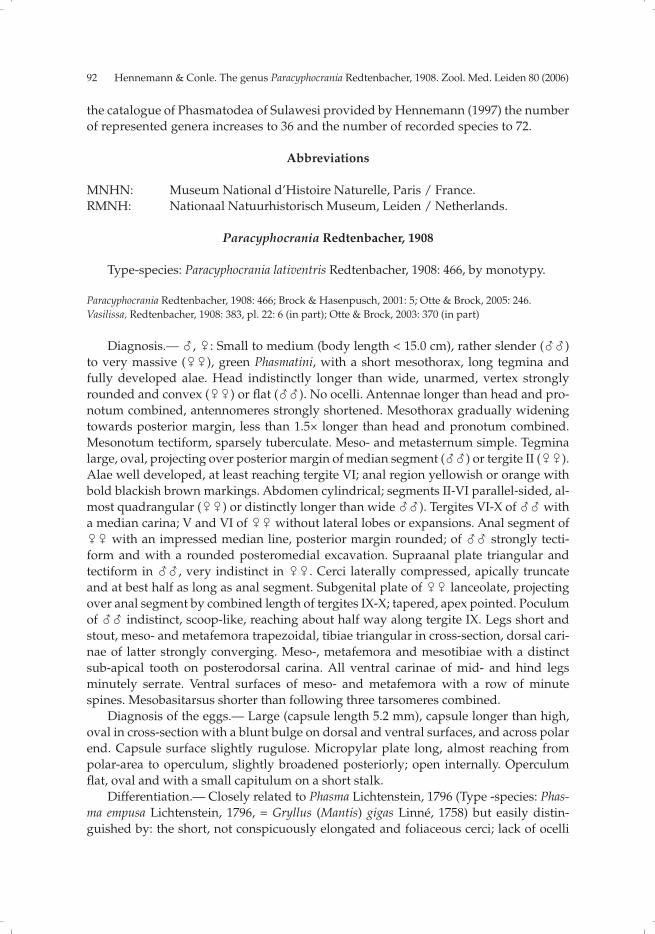
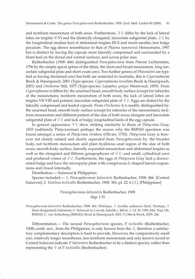
Figs 1-5. Paracyphocrania lativentris Redtenbacher, 1908, Neotype, �
1. Neotype, � (RMNH), dorsal view (partly reconstructed).
2. Head (lateral view).
3. Terminal abdominal segments (lateral view, subgenital plate partly reconstructed).
4. Egg (dorsal view).
5. Egg (lateral view).
�
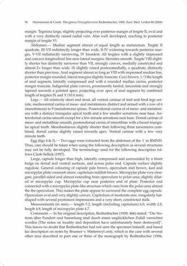
96 Hennemann & Conle. The genus Paracyphocrania Redtenbacher, 1908. Zool. Med. Leiden 80 (2006)
margin. Tegmina large, slightly projecting over posterior margin of tergite II, oval and
with a very distinctly raised radial vein. Alae well developed, reaching to posterior
margin of tergite VI.
Abdomen.— Median segment almost of equal length as metanotum. Tergite II
quadrate, III-VII indistinctly longer than wide, II-IV widening towards posterior mar-
gin, V-VII indistinctly narrowing, IV broadest. All tergites with a slightly impressed
and concave longitudinal line near lateral margins. Sternites smooth. Tergite VIII slight-
ly shorter but distinctly narrower than VII, strongly convex, medially constricted and
almost 2× longer than wide. IX slightly raised posteromedially, ± quadrate, distinctly
shorter than previous. Anal segment almost as long as VIII with impressed median line,
posterior margin rounded, lateral margins slightly truncate. Cerci brown, 1/3 the length
of anal segment, laterally compressed and with a rounded median carina, posterior
margin truncate. Subgenital plate convex, prominently keeled, lanceolate and strongly
tapered towards a pointed apex; projecting over apex of anal segment by combined
length of tergites IX and X (10.0 mm).
Legs.— All relatively short and stout, all ventral carinae of mid and hind legs ser-
rate, medioventral carina of meso- and metafemora distinct and armed with a row of 6
(mesofemora) to 9 (metafemora) spines. Posterodorsal carina of of meso- and metafem-
ora with a distinct triangular apical tooth and a few smaller serrations near base. An-
terodorsal carina smooth except for a few minute serrations near base. Dorsal carinae of
meso- and metatibiae smooth, posterodorsal carina of mesotibiae with a broad triangu-
lar apical tooth. Mesobasitarsus slightly shorter than following three tarsomeres com-
bined, dorsal carina slightly raised towards apex. Ventral carinae with a few very
minute teeth.
Egg (fi gs 4 & 5).— Two eggs were extracted from the abdomen of the � in RMNH.
Thus, care should be taken when using the following description as several structures
may not be fully developed. The terminology used for the following description fol-
lows Clark-Sellick (1997).
Large, capsule longer than high, laterally compressed and surrounded by a blunt
bulge on dorsal and ventral surfaces, and across polar end. Capsule surface slightly
rugulose. General colouring of capsule pale brown, operculum mid brown, keel and
micropylar plate creamish straw, capitulum reddish brown. Micropylar plate very elon-
gate, parallel-sided and almost extending from operculum to polar-area; slightly dilat-
ed at micropylar cup. Micropylar cup near posterior end of plate. Posterior end
connected with a micropylar plate-like structure which runs from the polar-area almost
the the operculum. This makes the plate appear to surround the complete egg capsule.
Operculum oval and very slightly convex. Capitulum of moderate size, irregularly hat-
shaped with several prominent impressions and a very short, constricted stalk.
Measurements (in mm).— length 5.2, length (including capitulum) 6.0, width 2.8,
heigth 4.9, length of micropylar plate 4.7.
Comments.— In his original description, Redtenbacher (1908: 466) stated: “Die No-
tizen über Fundort und Sammlung sind durch einen unglücklichen Zufall vernichtet
worden [The notes on locality and deposition have unfortunately been destroyed]“.
This leaves no doubt that Redtenbacher had not seen the specimen himself, and based
his description on notes by Brunner v. Wattenwyl only, which is the case with several
other taxa described in part one or three of the monograph by Redtenbacher (1906,
Hennemann & Conle. The genus Paracyphocrania Redtenbacher, 1908. Zool. Med. Leiden 80 (2006) 97
1908). Although Redtenbacher did not state the holotype was lost, the specimen has so
far not been traced. Brock & Hasenpusch (2001: 5) as well as Otte & Brock (2005: 246)
stated the holotype to be lost. Due to it being a rather large and striking insect it is very
unlikely to have been overlooked during all of the extensive searches in European mu-
seum collections conducted by several recent authors (e.g. Brock, Conle, Hennemann &
Zompro). Unfortunately, Redtenbacher’s original description of the genus and charac-
terization of the single included species, Paracyphocrania lativentris, are rather brief, but
the � in RMNH from Central Sulawesi matches very well with Redtenbacher’s original
description except for being slightly smaller (130.0 mm instead of 140.0 mm given by
Redtenbacher for the holotype, ➝ see table 1) and having the tubercles of the mesoster-
num slightly less distinct. Furthermore, the metafemora of the holotype are seen to be
about 25% longer than in the neotype (35.0 mm compared to 28.0 mm), but other cases
have already shown Redtenbacher’s measurements to be not always accurate. There-
fore, the RMNH � is here designated as the neotype of P. lastiventris and serves to pro-
vide a new diagnosis of the genus Paracyphocrania as well as a detailed redescription
and illustration of its type-species.
Paracyphocrania tecticollis (Redtenbacher, 1908) comb. nov.
(fi gs 6-9)
Vasilissa tecticollis Redtenbacher, 1908: 383, pl. 22: 6 (�). Holotype, �: Museum Paris, Philippines, Type,
136. Vasilissa tecticollis Redt. n. sp. (Type !) (MNHN); Otte & Brock, 2005: 339.
Differentiation.— P. lativentris Redtenbacher is only known from a rather damaged
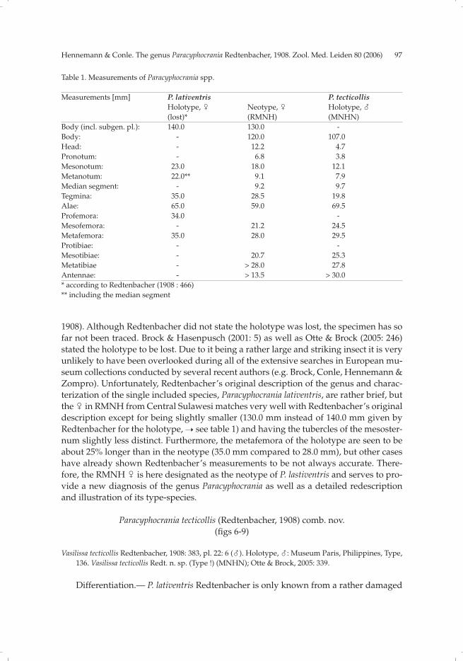
Table 1. Measurements of Paracyphocrania spp.
Measurements [mm] P. lativentris P. tecticollis Holotype, � Neotype, � Holotype, �
(lost)* (RMNH) (MNHN)
Body (incl. subgen. pl.): 140.0 130.0 -
Body: - 120.0 107.0
Head: - 12.2 4.7
Pronotum: - 6.8 3.8
Mesonotum: 23.0 18.0 12.1
Metanotum: 22.0** 9.1 7.9
Median segment: - 9.2 9.7
Tegmina: 35.0 28.5 19.8
Alae: 65.0 59.0 69.5
Profemora: 34.0 -
Mesofemora: - 21.2 24.5
Metafemora: 35.0 28.0 29.5
Protibiae: - -
Mesotibiae: - 20.7 25.3
Metatibiae - > 28.0 27.8
Antennae: - > 13.5 > 30.0
* according to Redtenbacher (1908 : 466)
** including the median segment
98 Hennemann & Conle. The genus Paracyphocrania Redtenbacher, 1908. Zool. Med. Leiden 80 (2006)
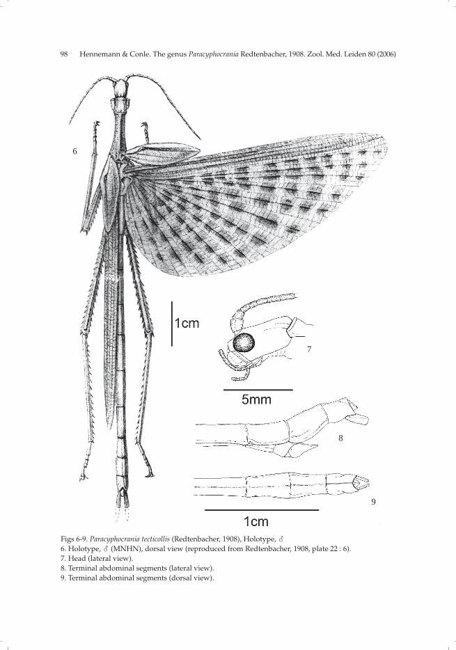
Figs 6-9. Paracyphocrania tecticollis (Redtenbacher, 1908), Holotype, �
6. Holotype, � (MNHN), dorsal view (reproduced from Redtenbacher, 1908, plate 22 : 6).
7. Head (lateral view).
8. Terminal abdominal segments (lateral view).
9. Terminal abdominal segments (dorsal view).
6
9
8
7

Hennemann & Conle. The genus Paracyphocrania Redtenbacher, 1908. Zool. Med. Leiden 80 (2006) 99
� from Central Sulawesi and P. tecticollis only from a unique �. Thus it is diffi cult to
provide a satisfying complementary description, but the large size (if compared to the
� of P. lativentris), relatively shorter mesothorax, more prominently tectiform mesono-
tum and distribution indicate V. tecticollis Redtenbacher to be a distinct species, rather
than representing the � of P. lativentris Redtenbacher.
Description.— The � holotype lacks both forelegs, the left mid leg and great parts
of the antennae. Due to former conservation in ethanol the original colouration has
strongly faded and changed to yellow. Certainly the insect was bright green when
alive. Although the cerci are complete, Redtenbacher (1908: 383) erroneously stated
they were broken and partly missing (indicated as broken in Redtenbacher’s illustra-
tion ➝ see Fig. 6).
� Holotype (fi gs 6-9).— Medium sized (body length 107.0 mm), moderately robust
Phasmatini with long, lanceolate tegmina (19.8 mm) and fully developed alae (69.5 mm).
Mesothorax densely and roughly granulose, remaining parts of thorax less distinctly
and abdominal sterna very minutely granulose. General colouration of body and legs
plain yellow (due to preservation); antennae pale brown. Tegmina and costal region of
alae yellow (believed to be green in life); tegmina with anterior margin dark brown and
followed by a broad longitudinal white band; costal region of alae with a longitudinal
white line running some 2/3 the way along alae and pink towards the base. Anal region
of alae slightly yellowish, sub-transparent, with darker yellow veins and numerous
more or less rectangular brown markings which become less distinct and fi nally disap-
pear towards the base of the wing.
Head.— Sub-cylindrical, 1.5× longer than wide, broadest at eyes, vertex fl at and
with two longitudinal impressed dorsolateral lines. Eyes very large, hemispherical and
prominently projecting from head capsule; pale orange brown. Antennae projecting
over posterior margin of mesonotum (broken in the holotype). Scapus 1.5× times longer
than wide, compressed dorsoventrally and constricted basally. Pedicellus cylindrical
and about 2/3 the length of scapus. Following antennomeres increasing in length.
Thorax.— Pronotum about as long as head, slightly medially constricted, transverse
median depression indistinct. Mesothorax 2× longer than pronotum, slightly medially
constricted and widening towards posterior margin. Mesonotum distinctly tectiform
and with a prominent, blunt median keel; complete surface densely granulose, with
larger granules roughly placed in longitudinal rows. Meso- and metapleurae and sterna
densely but minutely granulose. Tegmina elongate, lanceolate, apically tapered and al-
most reaching to posterior margin of median segment; with a small but pointed hump
in basal third. Alae reaching half way along tergite VII.
Abdomen.— Segments II-VI cylindrical, VII very slightly widening towards the
posterior; II-V smooth, VI-IX with a fi ne median carina. II-V increasing in length, II
slightly less than 3×, V 3.5× longer than wide; V-VII of equal length. VIII about 2/3 the
length of VII and gradually widening towards the posterior. IX strongly convex, paral-
lel-sided, longer than VIII. Sternites II-VII very minutely granulose. Anal segment
strongly tectiform with a slight triangular posteromedian excavation. Supraanal plate
distinct and projecting over posterior margin of anal segment; triangular and distinctly
tectiform. Poculum small, slightly convex, spoon-like and reaching about half way
along tergite IX. Cerci about 2/3 the length of anal segment, strongly laterally fl attened
and more or less rectangular if seen in lateral aspect.

100 Hennemann & Conle. The genus Paracyphocrania Redtenbacher, 1908. Zool. Med. Leiden 80 (2006)
Legs.— Mesofemora reaching to posterior margin of median segment, hind legs
almost reaching apex of abdomen. Ventral carinae of meso- and metafemora set with
numerous minute teeth (less in number on anteroventral carinae); only a very few small
teeth on dorsal carinae. Medioventral carina with a longitudinal row of minute spines.
Dorsal carinae of meso- and metatibiae smooth. Ventral carinae of mesotibiae very in-
distinctly spinose. Ventral carinae of metatibiae densely set with minute, slightly nee-
dle-like teeth which gradually increase in size towards the apex of tibia and are lacking
in the basal quarter of tibia. Basitarsi as long as remaining segments combined except
claw, simple.
Comments.— When describing Vasilissa tecticollis from the Philippines, Redten-
bacher (1908: 383) was uncertain about its generic position “Ob jedoch die Spezies tat-
sächlich hierher gehört, vermag ich nicht mit Bestimmtheit zu behaupten. [I am not able
to confi rm that this species really belongs here (in the genus Vasilissa Kirby, 1896)]”.
Comparison of the � holotype of Vasilissa tecticollis Redtenbacher in MNHN with the
type-species of Vasilissa Kirby, V. walkeri Kirby, 1896 from NW-Australia, has clearly
shown V. tecticollis Redtenbacher not to be a member of Vasilissa Kirby, but to represent
the second species and previously unknown � of Paracyphocrania Redtenbacher, 1908.
Acknowledgements
The authors would like to thank Dr J. van Tol (RMNH) as well as Dr C. Amedegnato
and S. Poulain (MNHN) for access to the collections of the corresponding institutions,
loan of specimens and providing information. Paul D. Brock (Slough, England) and Dr
Phil E. Bragg (Nottinghamshire, England) shall be thanked for helpful comments on the
manuscript.
References
Brock, P.D. & J. Hasenpusch, 2001. Cigarrophasma, a new genus of stick-insect (Phasmatidae) from Aus-
tralia.— Phasmid Studies, 9 (1 & 2): 4-10.
Brunner v. Wattenwyl, C., 1907. Die Insektenfamilie der Phasmiden. II. Phasmidae Anareolatae (Clit-umnini, Lonchodini, Bacunculini).— W. Engelmann, Leipzig, pp. 181-340, pls. 7-15.
Clark-Sellick, J.T., 1997. Descriptive terminology of the phasmid egg capsule, with an extended key to
the phasmid genera based on egg structure.— Systematic Entomology, 22: 97-122.
Gray, G.R., 1835. Synopsis of Phasmidae.— Longmans, London.
Hennemann, F.H., 1997. Ein Beitrag zur Kenntnis der Phasmidenfauna von Sulawesi. Mit einem Katalog
der bisher bekannt gewordenen Arten.— Mitteilungen aus dem Museum für Naturkunde in Berlin,
Zoologische Reihe, 74(1): 95-128.
Kirby, W.F., 1896. On some new or rare Phasmidae in the Collection of the British Museum.— Transac-
tions of the Linnean Society of London, Series 2, 6(6): 447-473.
Lichtenstein, A.A.H., 1796. Catalogus Musei zoologi ditissimi Hamburgi, d III February 1796.— Ac-
tionis lege distrahendi. Section 3, Hamburg.
Linné, C. von, 1758. Systema Naturae, 10th ed. 1, [iv +] 824 pp.— Holmiae.
Olivier, A.G., 1792. Encyclopédie Méthodique, ou par Ordre de Matières: par un Société de Gens de
Lettres, de Savans et d’Artistes.— Histoire Naturelle. Volume 7, Paris.
Otte, D. & P.D. Brock, 2005. Phasmida Species File. Catalog of stick and leaf insects of the world. Second
Edition.— The Insect Diversity Association and the Academy of Natural Sciences, Philadelphia,
CafePress.com., 414 pages.
Redtenbacher, J., 1906. Die Insektenfamilie der Phasmiden. I. Phasmidae Areolatae.— W. Engelmann,
Hennemann & Conle. The genus Paracyphocrania Redtenbacher, 1908. Zool. Med. Leiden 80 (2006) 101
Leipzig, pp. 1-180, pls. 1-6.
Redtenbacher, J., 1908. Die Insektenfamilie der Phasmiden. III. Phasmidae Anareolatae (Phibalosomini, Acrophyllini, Necrosciini). W. Engelmann, Leipzig, pp. 341-589, pls. 16-27.
Stål, C., 1875. Recensio Orthopterorum.Revue critique des Orthoptères déscrits par Linné, de Geer et
Thunberg, 3 : 4-105.— P. A. Norstedt & Söner, Stockholm.
Westwood, J.O., 1859. Catalogue of Orthopterous insects in the collection of the British Museum. Part 1:
Phasmidae.— British Museum, London.
Received: 2.vi.2005
Accepted: 15.ii.2006
Edited: C. van Achterberg

Related Documents
![THE REGISTRATION ACT, 1908 - Tripurarevenue.tripura.gov.in/sites/default/files/Registration_Act.pdf · THE REGISTRATION ACT, 1908 (16 OF 1908) [18th December, 1908] An Act to consolidate](https://static.cupdf.com/doc/110x72/5aa329097f8b9a1f6d8e32f7/the-registration-act-1908-registration-act-1908-16-of-1908-18th-december.jpg)


![ACT NO. V OF 1908 [21st March 1908] PRELIMINARYma-law.org.pk/pdflaw/CODE OF CIVIL PROCEDURE 1908.pdf · Code of Civil Procedure, 1908. ACT NO. V OF 1908 [21st March 1908] An Act to](https://static.cupdf.com/doc/110x72/5a703e007f8b9abb538bc596/act-no-v-of-1908-21st-march-1908-preliminaryma-laworgpkpdflawcode-of-civil.jpg)


![ACT NO. V OF 1908 [21st March 1908] PRELIMINARY OF CIVIL PROCEDURE 1908.pdf · V OF 1908 [21st March 1908] An Act to consolidate and amend the laws relating to the Procedure of the](https://static.cupdf.com/doc/110x72/5a8a4cf57f8b9afe568bcac2/act-no-v-of-1908-21st-march-1908-preliminary-of-civil-procedure-1908pdfv-of.jpg)


