The Freshwater Sponge Ephydatia fluviatilis Harbours Diverse Pseudomonas Species (Gammaproteobacteria, Pseudomonadales) with Broad-Spectrum Antimicrobial Activity Tina Keller-Costa 1,4 , Alexandre Jousset 2 , Leo van Overbeek 3 , Jan Dirk van Elsas 4 , Rodrigo Costa 4,5 * 1 Centre of Marine Sciences (CCMAR), University of Algarve, Faro, Algarve, Portugal, 2 Department of Ecology and Biodiversity, Utrecht University, Utrecht, The Netherlands, 3 Plant Research International, Wageningen University and Research Centre, Wageningen, The Netherlands, 4 Department of Microbial Ecology, Centre for Ecological and Evolutionary Studies, University of Groningen, Groningen, The Netherlands, 5 Microbial Ecology and Evolution Research Group, Centre of Marine Sciences (CCMAR), University of Algarve, Faro, Algarve, Portugal Abstract Bacteria are believed to play an important role in the fitness and biochemistry of sponges (Porifera). Pseudomonas species (Gammaproteobacteria, Pseudomonadales) are capable of colonizing a broad range of eukaryotic hosts, but knowledge of their diversity and function in freshwater invertebrates is rudimentary. We assessed the diversity, structure and antimicrobial activities of Pseudomonas spp. in the freshwater sponge Ephydatia fluviatilis. Polymerase Chain Reaction – Denaturing Gradient Gel Electrophoresis (PCR-DGGE) fingerprints of the global regulator gene gacA revealed distinct structures between sponge-associated and free-living Pseudomonas communities, unveiling previously unsuspected diversity of these assemblages in freshwater. Community structures varied across E. fluviatilis specimens, yet specific gacA phylotypes could be detected by PCR-DGGE in almost all sponge individuals sampled over two consecutive years. By means of whole-genome fingerprinting, 39 distinct genotypes were found within 90 fluorescent Pseudomonas isolates retrieved from E. fluviatilis. High frequency of in vitro antibacterial (49%), antiprotozoan (35%) and anti-oomycetal (32%) activities was found among these isolates, contrasting less-pronounced basidiomycetal (17%) and ascomycetal (8%) antagonism. Culture extracts of highly predation-resistant isolates rapidly caused complete immobility or lysis of cells of the protozoan Colpoda steinii. Isolates tentatively identified as P. jessenii, P. protegens and P. oryzihabitans showed conspicuous inhibitory traits and correspondence with dominant sponge-associated phylotypes registered by cultivation-independent analysis. Our findings suggest that E. fluviatilis hosts both transient and persistent Pseudomonas symbionts displaying antimicrobial activities of potential ecological and biotechnological value. Citation: Keller-Costa T, Jousset A, van Overbeek L, van Elsas JD, Costa R (2014) The Freshwater Sponge Ephydatia fluviatilis Harbours Diverse Pseudomonas Species (Gammaproteobacteria, Pseudomonadales) with Broad-Spectrum Antimicrobial Activity. PLoS ONE 9(2): e88429. doi:10.1371/journal.pone.0088429 Editor: Melanie R. Mormile, Missouri University of Science and Technology, United States of America Received May 22, 2013; Accepted January 7, 2014; Published February 12, 2014 Copyright: ß 2014 Keller-Costa et al. This is an open-access article distributed under the terms of the Creative Commons Attribution License, which permits unrestricted use, distribution, and reproduction in any medium, provided the original author and source are credited. Funding: This research was partially supported by the European Regional Development Fund (ERDF) through the COMPETE (Operational Competitiveness Programme) and national funds through FCT (Foundation for Science and Technology), under the project ‘‘PEst-C/MAR/LA0015/2011’’. Further financial support was obtained from the FCT-funded project PTDC/BIA-MIC/3865/2012. Tina Keller-Costa received a mobility grant from the Federation of European Microbiological Societies (FEMS) to perform field and laboratory work in Groningen, The Netherlands. The funders had no role in study design, data collection and analysis, decision to publish, or preparation of the manuscript. Competing Interests: The authors have declared that no competing interests exist. * E-mail: [email protected] Introduction Sponges (Porifera) are sessile filter-feeding organisms that primarily lack evasive or behavioural defence strategies [1]. Besides mechanical deterrence enabled by their spicules [2], sponges seem to mainly rely on chemical defence to prevent predation (e.g. by fishes and molluscs), avoid microbial biofilm formation and impede fouling [2–5]. There is increasing evidence that bacterial symbionts are the actual producers of many sponge- derived antagonistic metabolites [6–10], and this aspect has triggered much research interest in the diversity and bioactive potential of bacteria from marine sponges [9,11,12]. Conversely, knowledge of microbial communities in freshwater sponges remains limited. Their ubiquity in continental water bodies [13], coupled to recent molecular findings on highly selected commu- nities and specific lineages of bacteria that inhabit them [14] make freshwater sponges valuable models in symbiosis research. Although inland water sponges likely synthesize less secondary metabolites than marine species [15], they are prolific producers of fatty acids, lipids and sterols. Indeed, more than 100 distinct such compounds have been recorded for freshwater sponges and some might be of bacterial origin [16]. Commensal bacterial commu- nities may therefore fulfil important services required for the survival of their freshwater sponge host. Pseudomonas species (Gammaproteobacteria, Pseudomonadales) are one ubiquitous group of metabolically versatile, eukaryote-associated bacteria with important effects on host health and survival, where they display a multitude of behaviours [17]. Often commensalistic, pseudomonads may act as opportunistic pathogens e.g. in plants [18,19], fish [20,21] and humans [22,23]. In contrast, they are PLOS ONE | www.plosone.org 1 February 2014 | Volume 9 | Issue 2 | e88429

Welcome message from author
This document is posted to help you gain knowledge. Please leave a comment to let me know what you think about it! Share it to your friends and learn new things together.
Transcript
The Freshwater Sponge Ephydatia fluviatilis HarboursDiverse Pseudomonas Species (Gammaproteobacteria,Pseudomonadales) with Broad-Spectrum AntimicrobialActivityTina Keller-Costa1,4, Alexandre Jousset2, Leo van Overbeek3, Jan Dirk van Elsas4, Rodrigo Costa4,5*
1 Centre of Marine Sciences (CCMAR), University of Algarve, Faro, Algarve, Portugal, 2 Department of Ecology and Biodiversity, Utrecht University, Utrecht, The
Netherlands, 3 Plant Research International, Wageningen University and Research Centre, Wageningen, The Netherlands, 4 Department of Microbial Ecology, Centre for
Ecological and Evolutionary Studies, University of Groningen, Groningen, The Netherlands, 5 Microbial Ecology and Evolution Research Group, Centre of Marine Sciences
(CCMAR), University of Algarve, Faro, Algarve, Portugal
Abstract
Bacteria are believed to play an important role in the fitness and biochemistry of sponges (Porifera). Pseudomonas species(Gammaproteobacteria, Pseudomonadales) are capable of colonizing a broad range of eukaryotic hosts, but knowledge oftheir diversity and function in freshwater invertebrates is rudimentary. We assessed the diversity, structure and antimicrobialactivities of Pseudomonas spp. in the freshwater sponge Ephydatia fluviatilis. Polymerase Chain Reaction – DenaturingGradient Gel Electrophoresis (PCR-DGGE) fingerprints of the global regulator gene gacA revealed distinct structuresbetween sponge-associated and free-living Pseudomonas communities, unveiling previously unsuspected diversity of theseassemblages in freshwater. Community structures varied across E. fluviatilis specimens, yet specific gacA phylotypes couldbe detected by PCR-DGGE in almost all sponge individuals sampled over two consecutive years. By means of whole-genomefingerprinting, 39 distinct genotypes were found within 90 fluorescent Pseudomonas isolates retrieved from E. fluviatilis.High frequency of in vitro antibacterial (49%), antiprotozoan (35%) and anti-oomycetal (32%) activities was found amongthese isolates, contrasting less-pronounced basidiomycetal (17%) and ascomycetal (8%) antagonism. Culture extracts ofhighly predation-resistant isolates rapidly caused complete immobility or lysis of cells of the protozoan Colpoda steinii.Isolates tentatively identified as P. jessenii, P. protegens and P. oryzihabitans showed conspicuous inhibitory traits andcorrespondence with dominant sponge-associated phylotypes registered by cultivation-independent analysis. Our findingssuggest that E. fluviatilis hosts both transient and persistent Pseudomonas symbionts displaying antimicrobial activities ofpotential ecological and biotechnological value.
Citation: Keller-Costa T, Jousset A, van Overbeek L, van Elsas JD, Costa R (2014) The Freshwater Sponge Ephydatia fluviatilis Harbours Diverse PseudomonasSpecies (Gammaproteobacteria, Pseudomonadales) with Broad-Spectrum Antimicrobial Activity. PLoS ONE 9(2): e88429. doi:10.1371/journal.pone.0088429
Editor: Melanie R. Mormile, Missouri University of Science and Technology, United States of America
Received May 22, 2013; Accepted January 7, 2014; Published February 12, 2014
Copyright: � 2014 Keller-Costa et al. This is an open-access article distributed under the terms of the Creative Commons Attribution License, which permitsunrestricted use, distribution, and reproduction in any medium, provided the original author and source are credited.
Funding: This research was partially supported by the European Regional Development Fund (ERDF) through the COMPETE (Operational CompetitivenessProgramme) and national funds through FCT (Foundation for Science and Technology), under the project ‘‘PEst-C/MAR/LA0015/2011’’. Further financial supportwas obtained from the FCT-funded project PTDC/BIA-MIC/3865/2012. Tina Keller-Costa received a mobility grant from the Federation of European MicrobiologicalSocieties (FEMS) to perform field and laboratory work in Groningen, The Netherlands. The funders had no role in study design, data collection and analysis,decision to publish, or preparation of the manuscript.
Competing Interests: The authors have declared that no competing interests exist.
* E-mail: [email protected]
Introduction
Sponges (Porifera) are sessile filter-feeding organisms that
primarily lack evasive or behavioural defence strategies [1].
Besides mechanical deterrence enabled by their spicules [2],
sponges seem to mainly rely on chemical defence to prevent
predation (e.g. by fishes and molluscs), avoid microbial biofilm
formation and impede fouling [2–5]. There is increasing evidence
that bacterial symbionts are the actual producers of many sponge-
derived antagonistic metabolites [6–10], and this aspect has
triggered much research interest in the diversity and bioactive
potential of bacteria from marine sponges [9,11,12]. Conversely,
knowledge of microbial communities in freshwater sponges
remains limited. Their ubiquity in continental water bodies [13],
coupled to recent molecular findings on highly selected commu-
nities and specific lineages of bacteria that inhabit them [14] make
freshwater sponges valuable models in symbiosis research.
Although inland water sponges likely synthesize less secondary
metabolites than marine species [15], they are prolific producers of
fatty acids, lipids and sterols. Indeed, more than 100 distinct such
compounds have been recorded for freshwater sponges and some
might be of bacterial origin [16]. Commensal bacterial commu-
nities may therefore fulfil important services required for the
survival of their freshwater sponge host.
Pseudomonas species (Gammaproteobacteria, Pseudomonadales) are one
ubiquitous group of metabolically versatile, eukaryote-associated
bacteria with important effects on host health and survival, where
they display a multitude of behaviours [17]. Often commensalistic,
pseudomonads may act as opportunistic pathogens e.g. in plants
[18,19], fish [20,21] and humans [22,23]. In contrast, they are
PLOS ONE | www.plosone.org 1 February 2014 | Volume 9 | Issue 2 | e88429

found in synergistic association with arbuscular mycorrhizae [24]
and plant roots where they play beneficial roles in plant growth
promotion and disease control [25,26]. The two-component
regulatory system GacS/GacA mediates the interaction between
Pseudomonas spp. and their hosts. It controls the biosynthesis of
several secondary metabolites and exoenzymes at the post-
transcriptional level [25,27]. Mutations in gacA and gacS genes
induce phenotypic variation in Pseudomonas spp. [28,29], affecting
host colonization and persistence traits such as motility, biofilm
formation, biosurfactant synthesis and protein secretion [28–31].
Previous studies demonstrated that the gacA gene is a high-
resolution phylogenetic marker to the study of Pseudomonas spp.
[32,33].
Pseudomonads co-dominate the culturable fraction of the
freshwater sponge microbiome [34] and have been previously
detected in E. fluviatilis by cultivation-independent means [14].
Recently, Lipko et al. [35] reported on polyketide synthase (PKS)-
encoding genes from a freshwater sponge pseudomonad. It is well
known that Pseudomonas genomes are equipped with a wide range
of secondary metabolite biosynthetic gene clusters, including PKS
clusters [36,37]. However, complete genome sequences from - and
dedicated studies of - freshwater Pseudomonas spp. are scarce and
our understanding of their diversity, secondary metabolite
production capacity and adaptive strategies limited. Here, we
combine culture-dependent and -independent methods to unveil
the structure, diversity, and antimicrobial properties of Pseudomonas
spp. in the freshwater sponge Ephydatia fluviatilis. To this end, PCR-
DGGE fingerprinting of Pseudomonas-specific 16S rRNA and gacA
genes was used to test the hypotheses of selectivity and temporal
stability of Pseudomonas assemblages in the animal host. We further
identify Pseudomonas species cultured from the sponge and
determine their genome-wide diversity, antagonism towards
several microorganisms and distribution/dominance across E.
fluviatilis individuals. We finally address the potential biotechno-
logical value of E. fluviatilis as a promising source of novel
pseudomonads presenting antimicrobial activities.
Materials and Methods
SamplingEphydatia fluviatilis specimens were collected in the Vinkeveense
Plassen lake (VP, (52u149N, 4u579E), an artificial lake located in
the northwest of the province of Utrecht in the Netherlands on
June 6, 2007 and June 4, 2008. The specimens were found at a
depth of 9 m along a ,60 m transect on woody material of a
shipwreck scuttled at the diving point (‘zandeiland 4’) of VP and/
or on zebra mussels (Dreissenia polymorpha) attached to wrecks and
hard substrate. Each year, four sponge individuals (2007: SA – SD;
2008: SE – SH, about 10 g wet weight each) were sampled by
scuba diving and placed in 50 mL falcon tubes filled with lake
water. Four bulk water samples (W1–W4) were collected in sterile
500 mL flasks in year 2007 at about 3 m depth. This sampling
scheme enabled us to address the hypothesis of selective shaping of
Pseudomonas assemblages in E. fluviatilis (sponge vs. water compar-
ison, 2007 samples) and to detect Pseudomonas phylotypes
consistently associated with E. fluviatilis through time, if any
(2007 vs. 2008 comparison). Further results on sponge vs. water
comparisons for samples collected in 2008 have been described
elsewhere [14]. Samples were transported to the laboratory
(,2.5 h) in a cooling box and immediately processed for DNA
extraction and culturing. Because sampling involved invertebrate
animals not representing endangered or protected species and did
not occur within privately owned or protected areas, no specific
permits were required for the described field studies. Sampling
procedures were minimally intrusive and preserved sponge
individuals at the field site.
Total community DNA extractionTotal community DNA extraction from sponge and bulk water
samples took place as described before [14]. Briefly, homogenates
were obtained by grinding sponge samples with mortar and pestle.
These were subjected to differential centrifugation to yield sponge-
derived microbial cell pellets, which were used for DNA extraction
with the Fast DNAH Spin Kit for Soil (Bio101, Q-Biogene,
Heidelberg, Germany). The same kit was applied to extract DNA
from water samples concentrated on a sterile 0.2 mm nitrocellulose
membrane filter (Carl Roth GmbH, Karlsruhe, Germany).
Pseudomonas-specific PCR-DGGE fingerprintingCultivation-independent analysis of Pseudomonas diversity in E.
fluviatilis and bulk water DNA samples was carried out using PCR-
DGGE primer systems and cycling conditions as previously
described for the amplification of Pseudomonas-specific 16S rRNA
[38] and gacA [33] gene fragments. All PCRs were performed on a
Verititm thermal cycler (Applied Biosystems, Foster City, CA).
Denaturing gradient gel electrophoresis (DGGE) was performed
with a PhorU-2 gradient system (Ingeny International, Goes, The
Netherlands). Pseudomonas-specific 16S rRNA and gacA gene
amplicons were applied onto polyacrylamide gels containing a
46.5–65% gradient of denaturants (100% denaturants defined as
7M urea and 40% formamide) and a 6–9% gradient of acrylamide
[14]. Mixtures of 16S rRNA PCR products from five bacterial
species (Arthrobacter sp., Burkholderia sp., Enterobacter sp., Listeria sp.
and Rhizobium sp.) were applied at the edges of the 16S rRNA gel
and used as standard to control run quality. The PCR-DGGE
electrophoretic mobility of gacA gene fragments amplified from 36
E. fluviatilis derived Pseudomonas isolates shown to differ in whole
genome content (see ‘‘BOX-PCR fingerprinting’’ below) was
determined. These PCR products were pooled and used as a
reference on the gacA gel to look for band matches in the sponge
metagenomic DNA samples that could be assigned to these
isolates. Electrophoresis was performed in 1x TAE buffer (pH 8.0)
at 58uC and 140 V for 16 h. Gels were silver-stained [39] and air-
dried at room temperature (RT). Processing of the gel images and
multivariate statistical analysis of PCR-DGGE data were accom-
plished following the procedures of Costa et al. [40]. Briefly, the
software package GelCompar 4.5 was used to calculate a ‘‘samples
vs. band abundance’’ contingency table for each gel, which was
then used as input data for multivariate statistics with Canoco for
windows 4.5. Firstly, detrending correspondence analysis was
applied to both 16S rRNA and gacA gene datasets to check the
length of gradient in PCR-DGGE fingerprint variation. The linear
model of distribution fitted both data best and, therefore,
redundancy analysis (RDA) was chosen to estimate the extent to
which the variation in the relative abundance of Pseudomonas
DGGE bands are explained by the independent variables, namely
sample origin and year of sampling.
Isolation of fluorescent Pseudomonas spp. from E.fluviatilis
One gram of each sponge specimen sampled in year 2008 (SE –
SH) was cut and homogenized in 4 mL sterile 0.85% saline
solution using mortar and pestle. Serial dilutions were prepared
and spread onto Gould’s S1 (GS1) agar [41] plates in triplicate.
Volumes of 1, 10 and 50 mL freshwater were filtered in triplicate
per sample through sterile 0.2 mm nitrocellulose filters (47 mm;
Carl Roth GmbH, Germany). Filters were then placed onto GS1
Pseudomonas spp. in Ephydatia fluviatilis
PLOS ONE | www.plosone.org 2 February 2014 | Volume 9 | Issue 2 | e88429
agar plates. All plates were incubated at 28uC. Fluorescent colony
forming units (CFU) were counted under UV light after 2 and 5
days of incubation. Ninety colonies from sponge samples (18 to 26
per specimen) were randomly picked and purified with successive
streaks on modified King’s B (KB) agar plates (1.5% agar, 1.0%
peptone, 0.75% glycerol, 0.075% K2HPO4, 0.075% MgSO4).
Purified isolates were stored in KB broth with 20% glycerol at
280uC.
Whole-genome diversity of Pseudomonas isolatesTwo millilitre aliquots of cultures shaken overnight were
centrifuged at 16,000 g for 20 min. The supernatant was discarded
and genomic DNA extracted from the resulting cell pellets with the
WizardH Genomic DNA Purification Kit (Promega, Madison, WI,
USA). To determine genotypic diversity within Pseudomonas
isolates, whole-genome BOX-PCR genotyping was performed
with the BOXA1R primer (CTA CGG CAA GGC GAC GCT
GAC G) as previously described [42]. This primer is complemen-
tary to the boxA subunit of the conserved BOX element, a
repetitive DNA sequence present in most gram-negative bacteria,
including pseudomonads. Its selective amplification leads to a
genotypic fingerprint that permits differentiation to the strain level.
BOX-PCR amplicons were resolved by electrophoresis at 80 V on
2% agarose gels. Gels were visualized under UV light (256 nm)
after 30 min staining in ethidium bromide (1 mg mL21). Cluster
analysis of the resulting BOX-PCR profiles was performed using
the GelCompar II software (Applied Maths, Ghent, Belgium) as
described elsewhere [42]. The Shannon measure of diversity
(H’) was applied to estimate genotypic diversity within fluorescent
pseudomonads isolated from each E. fluviatilis specimen.
Identification and phylogeny of Pseudomonas isolatesIsolates displaying singular BOX-PCR profiles (singletons) and
representatives of each BOX-PCR genotypic group determined by
cluster analysis were selected for 16S rRNA and gacA gene
sequencing. 16S rRNA gene fragments (,1500 bp) were amplified
with primers F8 (59-AGA GTT TGA TCM TGG CTC AG-59)
and R1492 (59-GGT TAC CTT GTT ACG ACT T-39) [43] with
56uC annealing temperature in 30 PCR cycles. GacA gene
fragments (575 bp) were amplified using the forward primer
gacA-1F [33] and the reverse primer gacA-2 [32] as explained by
Costa et al. [33]. 16S rRNA and gacA gene PCR products were
sequenced at MACROGEN Inc. (Seoul, South Korea) using
standard Sanger sequencing procedures. Sequences were edited
with the Sequence Scanner Software version 1.0 (Applied
Biosystems, Foster, CA, USA). Closest matches to sequence
queries were identified using the blast algorithm of NCBI (http://
blast.ncbi.nlm.nih.gov/Blast.cgi). Closest type-strain 16S rRNA
gene relatives to each sequenced isolate were determined using the
RDP II sequence match tool (http://rdp.cme.msu.edu/seqmatch)
and served as reference for the tentative identification of the
isolates at the species level. For single gene-based diversity
assessments and phylogeny inference, 16S rRNA gene partial
sequences were aligned using the SINA web aligner tool and
imported into the ARB software. Analysis of gacA gene sequences
succeeded through the construction of a gacA gene database in
ARB with subsequent slow and accurate alignment using
ClustalX. 16S rRNA and gacA gene alignments were manually
corrected using the ARB sequence editor window. Phylip format
alignments were used as input files for the generation of 16S rRNA
and gacA gene sequence similarity matrices with the dnadist
software, applying the Kimura-2 parameter as distance algorithm.
Sequences were assigned operational taxonomic units (OTU)
using DOTUR [44]. The frequency data assigned to a ‘unique’
OTU at 99%, 97% and 95% similarity levels were used to yield
rarefaction curves for each gene. Maximum likelihood phyloge-
netic inference was carried out for the gacA gene dataset with the
general-time reversible (GTR) evolutionary model with a discrete
gamma-distribution of among-site rate variation (C4) and a
proportion of invariant sites (I). This was regarded the best-fit
model of nucleotide substitution for the dataset as determined by
analysis run on MrModelTest version 2.3 [45]. Phylogenetic
analysis and tree construction were conducted with the software
package MEGA version five [46]. Sequences were deposited in the
European Molecular Biology Laboratory database under the
accession numbers HE794891–HE794933 (16S rRNA gene) and
HE794934–HE794974 (gacA gene).
Biofilm formation in static microcosmsAn air-liquid (A-L)-interface assay for biofilm formation was
performed using the static microcosm approach developed by Ude
et al. [47]. Briefly, static microcosms consisted of glass tubes
containing 5 mL of liquid KB. Fifty microliter inocula from
overnight shaking cultures were used to start the biofilm formation
assay. Tubes were incubated for 6 days, vibration-free, at 23uCwith lids loosely attached to allow air exchange. Biofilm material, if
present, was taken from the A-L interface with a wire loop and
used to inoculate a second, fresh microcosm. If no biofilm was
observed by naked eye or as material present on the wire loop,
then the microcosm was vortex-shaken for 30 sec and a 50 mL
aliquot was taken for inoculation of a new microcosm. All 90
isolates were tested in triplicate through three successive passages
over a maximum of 18 days. A microcosm was scored as biofilm-
positive if - at least in one replicate - material at the surface could
be observed by eye (score +), or detected using a wire loop (score
+/2). Pseudomonas protegens strain CHA0, which yields a mucilag-
inous biofilm under the tested circumstances, was employed as a
positive control in the assays.
In vitro antimicrobial activityIn vitro antagonistic activity of Pseudomonas spp. isolated from E.
fluviatilis was tested towards the phytopathogenic fungi Rhizoctonia
solani strain AG3 (basidiomycete) and Fusarium moniliforme strain
CBS 218.76 (ascomycete), the phytopathogenic oomycete Pythium
ultimum strain 67-1 and the commensalistic bacterium Bacillus
subtilis F6 Rpr. For both fungi and P. ultimum, tests were performed
in dual-culture assays as described before [48] on modified Potato
Dextrose Agar (1.95% PDA, 0.5% Peptone, 1.5% Agar).
Antagonistic potential against B. subtilis F6 Rpr was tested
following the agar plug assay procedure of Hentschel et al. [49].
All experiments were set up in triplicate and the degree of
antagonism was estimated by measuring the size of the haloes of
inhibition of B. subtilis after 2 days, of P. ultimum and R. solani after
4 days and of F. moniliforme after 7 days of incubation at 28uC. The
growth of the target organisms in the absence of potential
antagonists was monitored and used as blank. The soil-derived,
root-colonizing bio-control strain Pseudomonas protegens CHA0,
which inhibits all tested strains in vitro, was used as a positive
control for antagonism in all trials and replicates.
Resistance against protozoan predationThe resistance of Pseudomonas strains to predation by Colpoda
steinii strain Sp1 was evaluated in a microtiter plate assay adapted
from Jousset et al. [50]. C. steinii culture stocks were maintained in
LB broth diluted 1:10 in sterile amoeba saline (AS) solution [51]
and purified in two centrifugation steps (1000 g, 1 min) prior to the
start of the experiments. Fresh Pseudomonas cultures were incubated
overnight with gentle agitation (50 rpm) at RT in 96-well plates
Pseudomonas spp. in Ephydatia fluviatilis
PLOS ONE | www.plosone.org 3 February 2014 | Volume 9 | Issue 2 | e88429
containing 150 mL LB broth 1:20 diluted in AS. Twenty
microliters of overnight cultures were transferred to a new 96-
well microtiter plate, each well containing 80 mL AS mixed with
20 mL of washed and active C. steinii suspension (set at final density
of 1,000 cells mL21). Plates were incubated with gentle agitation
(50 rpm) at RT, and bacterial densities (OD600) in the presence
and absence of C. steinii were measured at regular intervals over a
50 h period with an M200 plate photometer (Tecan, Mannedorf,
Switzerland). Bacterial sensitivity to predation was defined as the
relative reduction of the OD600 value after its stabilization, usually
at 22 h, in the presence of the protozoan compared to the initial
OD600 value at 0 h readily after protozoan inoculation. Wells
containing only bacterial isolates or protozoan cells were
monitored in parallel to control for vitality and growth of the
tested organisms. Bacteria with less than 40% reduction of the
initial optical density after 22 h of exposure to C. steinii were
counted as ‘‘predation-resistant’’. The objective of this assay was to
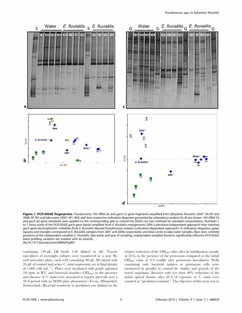
Figure 1. PCR-DGGE fingerprints. Pseudomonas 16S rRNA (a) and gacA (c) gene fragments amplified from Ephydatia fluviatilis (2007: SA-SD and2008: SE-SH) and lake water (2007: W1–W4) and their respective ordination diagrams generated by redundancy analysis (b, d) are shown. 16S rRNA (S)and gacA (G) gene standards were applied on the corresponding gels to control the DGGE run (see methods for standard compositions). Numbers 1to 7 show some of the PCR-DGGE gacA gene bands amplified from E. fluviatilis metagenomic DNA (cultivation-independent approach) that matchedgacA gene electrophoretic mobilities from E. fluviatilis-derived Pseudomonas isolates (cultivation-dependent approach). In ordination diagrams, greensquares and triangles correspond to E. fluviatilis samples from 2007 and 2008, respectively; and blue circles to lake water samples. Blue stars: centroidpositions of the independent variables E. Fluviatilis, lake water and year of sampling. Independent variables found to significantly influence PCR-DGGEband profiling variation are marked with an asterisk.doi:10.1371/journal.pone.0088429.g001
Pseudomonas spp. in Ephydatia fluviatilis
PLOS ONE | www.plosone.org 4 February 2014 | Volume 9 | Issue 2 | e88429
Pseudomonas spp. in Ephydatia fluviatilis
PLOS ONE | www.plosone.org 5 February 2014 | Volume 9 | Issue 2 | e88429
perform one single screening for resistant isolates to be used in the
short-term toxicity assay against C. steinii (see below).
Short-term toxicity of Pseudomonas spp. extracts towardColpoda steinii
Predation-resistant isolates, determined as described above,
were grown for 2.5 days in 20 mL LB broth (10x diluted in AS) at
27uC under shaking at 200 rpm. The cell suspensions were then
transferred to 50 mL Falcon tubes containing 2.2 mL of 1 M HCl
and vortexed, after which 20 mL ethyl acetate was added. Tubes
were then shaken for 90 min at 37uC and centrifuged (1800 g,
10 min). The solvent-phase supernatants (,10 mL) were carefully
transferred to new tubes and dried to air. The dried bacterial
extracts were resuspended in 100 mL methanol and kept at
220uC. One microliter volumes of the respective cell extracts were
added to 100 mL freshly grown C. steinii suspensions in 96-well
microtiter plates, and suspensions were gently homogenized by
shaking. Controls were monitored in parallel, replacing the cell
extracts by 1 mL methanol. The extract of P. protegens CHA0 grown
under the same conditions was included as positive control. The
effect of extracts (and methanol) on C. steinii was evaluated in
duplicate after 5, 15, 30, 60 and 120 min, respectively, using an
inversion microscope (Axiovert 25, Carl Zeiss, Jena, Germany;
magnification 6200 and 6400).
Multivariate analysis of antagonistic propertiesMultivariate analysis was performed with the objective of
grouping the Pseudomonas isolates based on their profiles of
antagonistic features. These were determined, per isolate, as the
collective scores obtained in antibacterial, antifungal, anti-oomyce-
tal and predation resistance in in vitro assays (semi-quantitative data),
along with biofilm formation tests as described above, in accordance
with the approaches of Costa et al. [33] and Adesina et al. [52].
Thus, the 90 Pseudomonas isolates were introduced in the statistics as
samples, whereas their respective antimicrobial properties served as
descriptors of each sample (dependent variables). The affiliation of
Pseudomonas isolates to a certain E. fluviatilis specimen (i.e. specimens
E, F, G, H) was used as a nominal (i.e. binary), independent
variable. This way, correlations between antimicrobial properties
and their association with each E. fluviatilis specimen could be
explored. Analyses were carried out using the software package
Canoco for Windows 4.5. After preliminary inspection of overall
dataset variation by detrended correspondence analysis (DCA),
principal components analysis (PCA) was selected as the most
appropriate ordination method (linear, unconstrained) to be used in
the analysis [53]. PCA was run with focus on inter-sample distances
and results were illustrated in an ordination diagram.
Results
Cultivation-independent analysis of Pseudomonascommunities
PCR-DGGE analysis of Pseudomonas assemblages using specific
16S rRNA gene primers revealed low levels of band richness in all
samples (Fig. 1a). Two up to three dominant bands in bulk water
fingerprints and a single dominant band in E. fluviatilis profiles
were visible, with fingerprints in general displaying less than 10
detectable bands. All profiles were characterized by low and
statistically similar (p = 0.827; One-Way-ANOVA) Shannon
diversity indices, with values of 1.67360.35, 1.54360.26 and
1.56260.35 (means 6 SD, N = 4) obtained for bulk water (2007)
and E. fluviatilis (2007, 2008) samples, respectively. Regarding
community structure as revealed by 16S rRNA gene fingerprint-
ing, Monte Carlo permutation tests confirmed sample origin as a
significant factor differentiating the profiles (p = 0.048 and
p = 0.044 for the independent variables ‘‘water’’ and ‘‘E. fluviatilis’’,
respectively); however, yearly variation in sponge profiles was not
significant (p = 0.132 and p = 0.142 for the independent variables
‘‘2007’’ and ‘‘2008’’, respectively; Fig. 1b). In contrast with 16S
rRNA gene analyses, the relative abundance of some Pseudomonas
gacA bands from bulk water was enhanced in E. fluviatilis while
several other bulk water gacA bands could not be readily detected
in the sponge samples, resulting in reduced gacA band diversity in
the latter PCR-DGGE fingerprints in comparison with the former
(Fig. 1c). Significantly higher (p,0.001; F2,9 = 19.404; OW-
ANOVA followed by Tukey-test) gacA diversity indices were found
in bulk water (3.21060.13) than in E. fluviatilis profiles from years
2007 (2.54060.146) and 2008 (2.40260.28). Diversity measures
obtained for sponge gacA fingerprints from 2007 and 2008 were
statistically similar. Monte Carlo permutation tests confirmed the
independent variables ‘‘water’’ and ‘‘E. fluviatilis’’ to significantly
differentiate the PCR-DGGE profiles (p = 0.002 and 0.006,
respectively), with samples belonging to these microhabitats clearly
separated along the horizontal axis of the ordination diagram
(Fig. 1d). As opposed to 16S rRNA gene fingerprinting, year of
sampling did contribute significantly to distinguish the gacA
fingerprints of E. fluviatilis specimens (p = 0.03 for both indepen-
dent variables 2007 and 2008, respectively). Overall, PCR-DGGE
fingerprinting data showed statistically higher Pseudomonas gacA
gene diversity than predicted by 16S rRNA gene analysis in both
water (p = 0.003; F1,3 = 77.844; OW-Repeated Measures-ANOVA
followed by Tukey-test) and E. fluviatilis (p = 0.002; Two-Way-RM-
ANOVA followed by Tukey-test) samples.
Isolation of fluorescent Pseudomonas spp. from E.fluviatilis
Numbers of fluorescent Pseudomonas were low in the VP bulk
water samples, ranging from 3.1–4.16100 CFU mL21. In con-
trast, numbers in E. fluviatilis specimens were in the range of 1.5–
3.66103 CFU g (fresh wt)21, surpassing counts obtained for bulk
water by c. 3 orders of magnitude. In total, 90 isolates were
obtained from E. fluviatilis specimens sampled in 2008 and
thoroughly inspected for diversity, biofilm formation capacity
and antimicrobial activities.
Whole-genome diversity and identity of Pseudomonasisolates
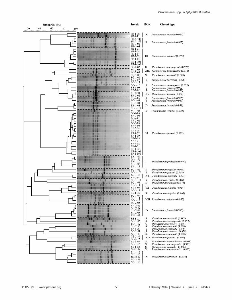
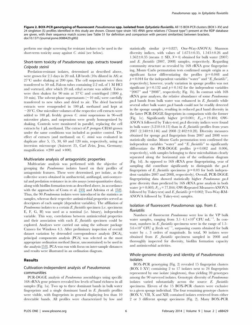
BOX-PCR genotyping (Fig. 2) revealed 15 fingerprint clusters
(BOX I–XV) containing 2 to 17 isolates next to 24 fingerprints
represented by one isolate (singletons), thus yielding 39 genotypes
among the 90 surveyed isolates. Genotypic diversity of Pseudomonas
isolates varied substantially across the source E. fluviatilis
specimens. Eleven of the 15 BOX-PCR clusters were exclusive
to a given sponge individual. The four remaining genotype clusters
(BOX V, VII, X and XII) contained isolates retrieved from either
2 or 3 different sponge specimens (Fig. 2). Many BOX-PCR
Figure 2. BOX-PCR genotyping of fluorescent Pseudomonas spp. isolated from Ephydatia fluviatilis. All 15 BOX-PCR clusters (BOX I–XV) and24 singleton (S) profiles identified in this study are shown. Closest type strain 16S rRNA gene relatives (‘‘Closest type’’) present at the RDP databaseare given, with their sequence match scores (see Table S1 for definition and comparison with percent similarities) between brackets.doi:10.1371/journal.pone.0088429.g002
Pseudomonas spp. in Ephydatia fluviatilis
PLOS ONE | www.plosone.org 6 February 2014 | Volume 9 | Issue 2 | e88429
singletons (10 of 24) were detected in E. fluviatilis specimen SG.
This sponge sample yielded the highest BOX-PCR-based
Shannon diversity index (Table 1). Forty-three representative
isolates covering the 39 observed BOX-PCR profiles were all
identified as members of the genus Pseudomonas by 16S rRNA gene
sequencing (Table S1). Twelve of the 39 BOX-PCR genotypes
shared 100% 16S rRNA gene sequence similarity with type strains
of species such as P. mandelii, P. migulae, P. monteilii, P. reinekei, P.
brenneri and P. cedrina (Table S1). Overall, 40 and 13 isolates
resembled P. jessenii and P. umsongensis based on .99% 16S rRNA
gene similarities between genotype representatives and type strains
of these species (Tables S1 and S2). These isolates encompassed
several BOX-PCR genotypes and antagonistic profiles, represent-
ing the most prominent species tentatively identified in this study
(Table S2). The four BOX-PCR genotypes retrieved from more
than one sponge specimen had highest resemblance to P. koreensis
(BOX V), P. migulae (BOX VII), P. umsongensis (BOX X) and P.
monteilii (BOX XII) type strains (Fig. 2, Table S1). BOX cluster I
comprised five isolates from specimen SH (Fig. 2). The biological
control strains Pf-5 and CHA0 (T), belonging to the recently
described species P. protegens (Ramette et al., 2011), exhibited the
highest genetic similarity to this group (Table S1). Seventeen of the
18 isolates from sponge specimen F belonged to the same BOX-
PCR cluster (VI). P. jessenii CIP 105274 was the closest type strain
to this group (Table S1).
Diversity and phylogeny of Pseudomonas isolatesNucleotide heterogeneities among partial, high quality 16S
rRNA and gacA gene sequences obtained for 36 Pseudomonas
isolates were compared. These isolates represented all 15 BOX-
PCR clusters and 21 of the 24 single BOX-PCR genotypes found
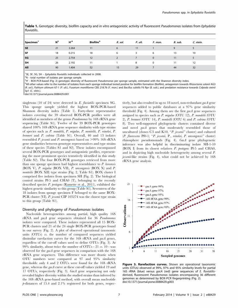
in our survey (Fig. 2). A plot of observed operational taxonomic
units (OTUs) vs. the number of compared sequences yielded
dissimilar rarefaction curves for the 16S rRNA and gacA genes,
regardless of the cut-off values used to define OTUs (Fig. 3). At
99% similarity, about twice the number of OTUs - 21 vs. 10 - was
observed for the gacA gene sequences in comparison with the 16S
rRNA gene sequences. This difference was more drastic when
OTU numbers were compared at 97 and 95% similarity
thresholds: only 4 and 2 OTUs were found for the 16S rRNA
gene, whereas the gacA curve at these cut-off values yielded 19 and
17 OTUs, respectively (Fig. 3). GacA gene sequencing not only
revealed higher diversity within the studied strains than inferred by
the 16S rRNA gene-based method, with overall mean nucleotide
p-distances of 15.4 and 2.1% registered for both genes, respec-
tively, but also resulted in up to 18 novel, non-redundant gacA gene
sequences added to public databases at a 97% gene similarity
threshold (Fig. 4). Among them are the first gacA gene sequences
assigned to species such as P. migulae (OTU 12), P. mandelii (OTU
2), P. brenneri (OTU 14), P. monteilli (OTU 6) and P. cedrina (OTU
8). Two well-supported phylogenetic clusters contained diverse
and novel gacA genes that moderately resembled those of
uncultured (clones C3 and K10, ‘‘P. jessenii’’ cluster) and cultured
(P. fluorescens Pf0-1; ‘‘P. jessenii, P., reinekei, P. umsongensis’’ cluster)
rhizosphere pseudomonads (Fig. 4). GacA gene phylogenetic
inference was also helpful in discriminating isolate SH-1-10
(BOX I) from its closest relatives P. protegens Pf-5 and CHA0,
and in depicting high nucleotide heterogeneity among several P.
jessenii-like strains (Fig. 4), what could not be achieved by 16S
rRNA gene analysis.
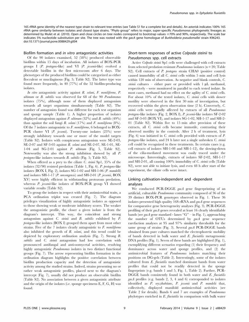
Table 1. Genotypic diversity, biofilm capacity and in vitro antagonistic activity of fluorescent Pseudomonas isolates from Ephydatiafluviatilis.
Specimen1 N2 H’3 Biofilm4 R. sol. P. ult. F. mon. B. sub. C. stein.
SE 21 2.264 11 6 11 1 9 5
SF 18 0.215 18 6 3 6 13 10
SG 25 2.754 12 2 7 0 11 5
SH 26 2.192 11 1 8 0 11 12
All 90 1.404 52 15 29 7 44 32
1SE, SF, SG, SH – Ephydatia fluviatilis individuals collected in 2008.2N - total number of isolates per sponge sample.3H’ - BOX-PCR-based (Fig. 2) genotypic diversity of fluorescent Pseudomonas per sponge sample, estimated with the Shannon diversity index.4All other values refer to the number of isolates from each sponge individual tested positive for biofilm formation (Biofilm), antagonism towards Rhizoctonia solanii AG3(R. sol.), Pythium ultimum 67-1 (P. ult.), Fusarium moniliforme CBS 218.76 (F. mon.) and Bacillus subtilis F6 Rpr (B. sub.), and predation resistance towards Colpoda steiniiSp1 (C. stein.).doi:10.1371/journal.pone.0088429.t001
Figure 3. Rarefaction curves. Shown are operational taxonomicunits (OTUs) observed at 99%, 97% and 95% similarity levels for partial16S rRNA (blue) versus gacA (red) gene sequences of E. fluviatilis-derived, fluorescent Pseudomonas isolates encompassing 36 differentgenomes as determined by BOX-PCR fingerprinting (Fig. 2).doi:10.1371/journal.pone.0088429.g003
Pseudomonas spp. in Ephydatia fluviatilis
PLOS ONE | www.plosone.org 7 February 2014 | Volume 9 | Issue 2 | e88429
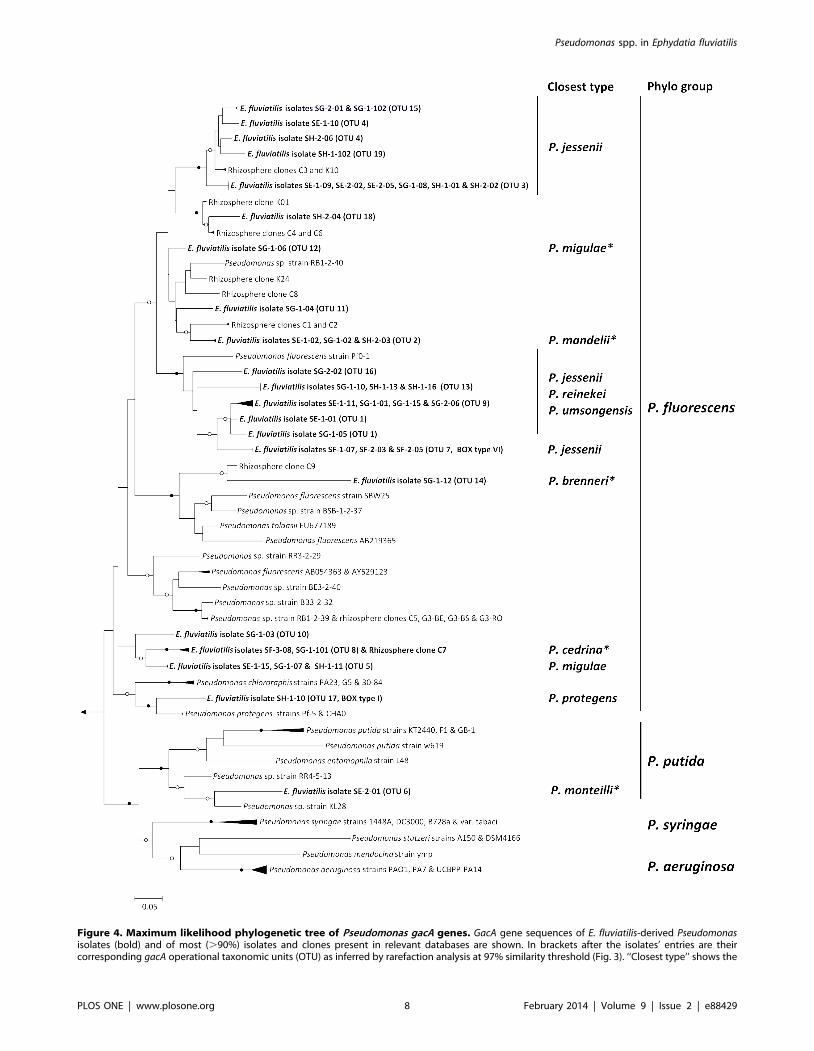
Figure 4. Maximum likelihood phylogenetic tree of Pseudomonas gacA genes. GacA gene sequences of E. fluviatilis-derived Pseudomonasisolates (bold) and of most (.90%) isolates and clones present in relevant databases are shown. In brackets after the isolates’ entries are theircorresponding gacA operational taxonomic units (OTU) as inferred by rarefaction analysis at 97% similarity threshold (Fig. 3). ‘‘Closest type’’ shows the
Pseudomonas spp. in Ephydatia fluviatilis
PLOS ONE | www.plosone.org 8 February 2014 | Volume 9 | Issue 2 | e88429
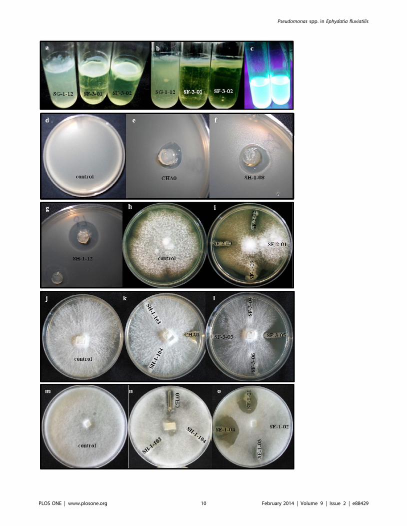
Biofilm formation and in vitro antagonistic activitiesOf the 90 isolates examined, 52 (58%) produced observable
biofilms within 15 days of incubation. All isolates of BOX-PCR
groups I (P. protegens-like) and VI (P. jessenii-like) evolved a
detectable biofilm in the first microcosm after 6 days. The
phenotypes of the produced biofilms could be categorized as either
flocculent or mucilaginous (Fig. 5; Table S2). The latter type was
found more frequently, in 40 (77%) of the 52 biofilm-producing
isolates.
In vitro antagonistic activity against R. solani, F. moniliforme, P.
ultimum or B. subtilis was observed for 68 of the 90 Pseudomonas
isolates (75%), although none of them displayed antagonism
towards all target organisms simultaneously (Table S2). The
number of antagonists found was different for each test organism
and sponge sample (Table 1). A higher proportion of isolates
displayed antagonism against P. ultimum (32%) and B. subtilis (49%)
than against the soil fungi R. solani (17%) and F. moniliforme (8%).
Of the 7 isolates antagonistic to F. moniliforme, 6 belonged to BOX-
PCR cluster VI (P. jessenii). Twenty-one isolates (23%) were
strongly inhibitory towards one or more of the model targets
(Table S2). Isolates with conspicuous antagonism were SE-2-03,
SE-2-07 and SF-3-05 against R. solani and SE-2-07, SE-1-01, SE-
1-04 and SG-2-01 against P. ultimum (Fig. 5, Table S2).
Noteworthy was also the strong inhibition showed by all P.
protegens-like isolates towards B. subtilis (Fig. 5; Table S2).
When offered as a prey to the ciliate C. steinii Sp1, 35% of the
isolates (32/90) resisted predation (Table 1, Fig. 6). P. protegens-like
isolates (BOX I, Fig. 2), isolates SG-1-02 and SH-1-06 (P. mandelli)
and isolates SH-1-17 (P. umsongensis) and SH-2-01 (P. jessenii, BOX
XV) were highly efficient in withstanding protozoan predation,
whereas P. jessenii-like isolates of BOX-PCR group VI showed
variable results (Table S2).
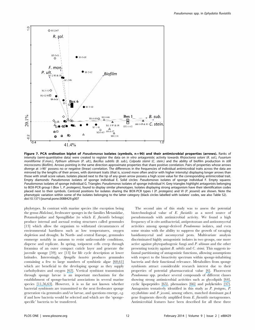
To group the isolates in respect with their antimicrobial traits, a
PCA ordination triplot was created (Fig. 7). The diagram
privileges visualization of highly antagonistic isolates as opposed
to those showing weak or moderate inhibitory scores. The weaker
the antagonistic profile, the closer a given isolate is from the
diagram’s intercept. This way, the coincident and strong
antagonism against C. steinii and B. subtilis exhibited by P.
protegens-like isolates (BOX I) grouped them apart from most other
strains. Five of the 7 isolates clearly antagonistic to F. moniliforme
also inhibited the growth of R. solani, and this trend could be
depicted by exploratory ordination analysis (Fig. 7). Strong B.
subtilis and C. steinii antagonism had low correlation with
pronounced antifungal and anti-oomycetal activities, resolving
highly antagonistic Pseudomonas isolates in two distinct functional
groups (Fig. 7). The arrow representing biofilm formation in the
ordination diagram highlights the positive correlation between
biofilm production capacity and the detection of antagonistic
activity among the studied strains. Indeed, strains presenting no or
rather weak antagonistic profiles, placed next to the diagram’s
intercept (Fig. 7), usually did not produce an observable biofilm
(Table S2). No association between a given antagonistic attribute
and the origin of the isolates (i.e. sponge specimens E, F, G, H) was
found.
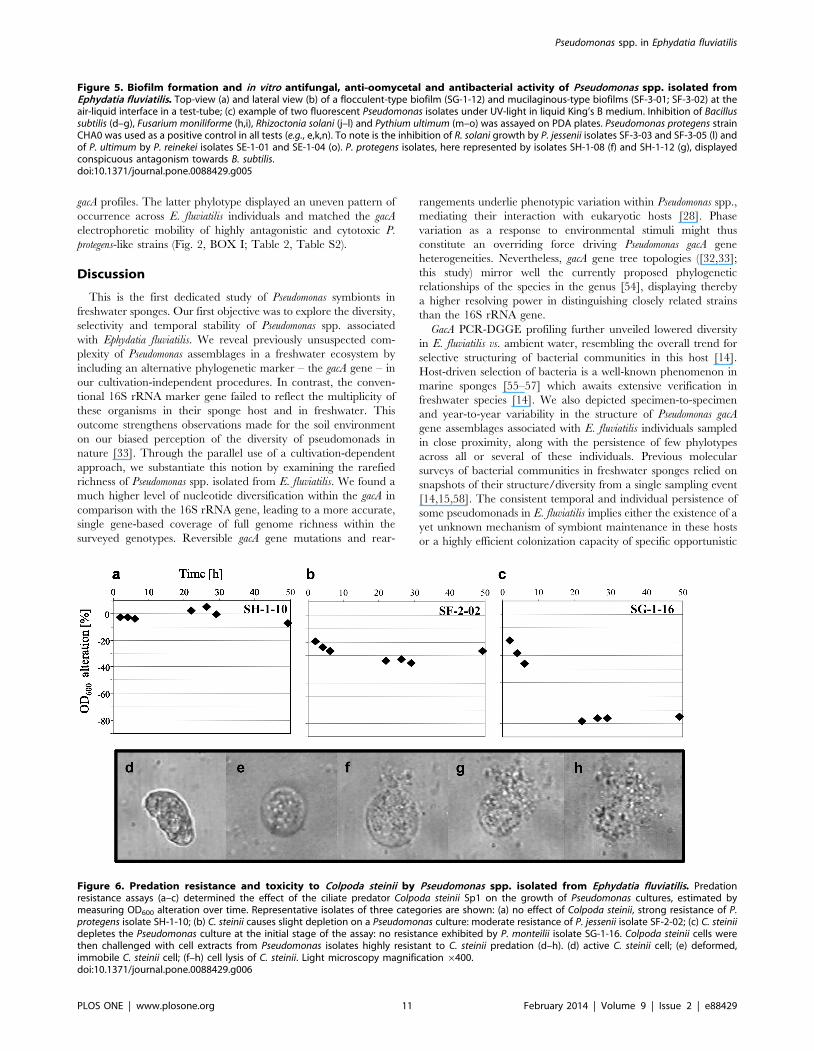
Short-term responses of active Colpoda steinii toPseudomonas spp. cell extracts
Active Colpoda steinii Sp1 cells were challenged with cell extracts
from selected predation resistant Pseudomonas isolates (n = 20; Table
S2). Cell extracts of P. protegens strain CHA0 (positive control)
caused immobility of all C. steinii cells within 5 min and cell lysis
within 120 min of observation. As negative and blank controls, C.
steinii cultures – either pure or provided with 1 mL methanol,
respectively - were monitored in parallel to each tested isolate. In
most cases, methanol had no effect on the agility of C. steinii cells.
For about 10% of the tested isolates, C. steinii cells with slower
motility were observed in the first 30 min of investigation, but
recovered within the given observation time (2 h). Conversely, C.
steinii cells were rapidly affected by extracts of all Pseudomonas
protegens-like isolates (Fig. 2, BOX I), P. jessenii-like isolates SF-2-02
and SF-3-03 (BOX VI), and isolates SG-1-02, SH-1-17 and SH-2-
01 (Table S2). Within five to 15 min after provision of these
extracts, all C. steinii cells became immotile, contrasting their
observed motility in the controls. After 2 h of treatment, lysis
(Fig. 6) was initiated in C. steinii cells provided with extracts of P.
protegens-like isolates, and 24 h later not a single unbroken C. steinii
cell could be recognized in these treatments. In certain cases (e.g.
cell extracts of isolates SH-1-08 and SH-1-12), the slowing-down
of the cilia-mediated movement was easily traced under the
microscope. Interestingly, extracts of isolates SF-2-02, SH-1-17
and SH-2-01, all causing 100% immobility of C. steinii cells (Table
S2), were not able to initiate lysis, and even 48 h after start of the
experiment, the ciliate cells were intact.
Linking cultivation-independent and -dependentanalyses
We conducted PCR-DGGE gacA gene fingerprinting of an
artificial, culturable Pseudomonas community composed of 36 of the
39 distinct BOX-PCR genotypes observed in Fig. 1. These 36
isolates presented high quality 16S rRNA and gacA gene sequences
for comparative gene heterogeneity analyses (Fig. 3). PCR-DGGE
profiling of their gacA genes revealed at least 16 clearly identifiable
bands (see gacA gene standard - lanes ‘‘G’’ – in Fig. 1), approaching
the number of OTUs determined by gacA gene sequence
rarefaction analyses at 95 and 97% similarity thresholds for the
same group of strains (Fig. 3). Several gacA PCR-DGGE bands
obtained from pure cultures matched the electrophoretic mobility
of bands detected in bulk water and E. fluviatilis metagenomic
DNA profiles (Fig. 1). Seven of these bands are highlighted (Fig. 1),
exemplifying different scenarios regarding (1) their frequency and
dominance across water and sponge profiles, and (2) the
antimicrobial features of Pseudomonas isolates matching their
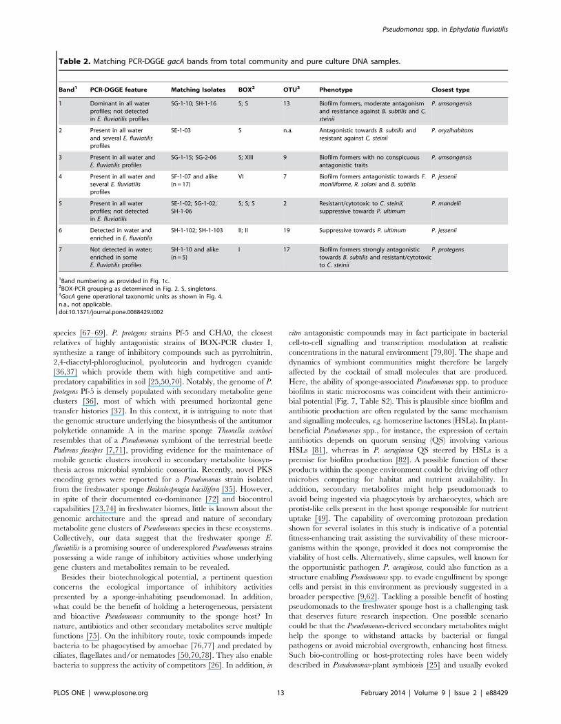
positions on DG-gels (Table 2). Interestingly, some of the isolates
cultured from E. fluviatilis matched dominant bands from water
profiles that could not be readily detected in the sponge
fingerprints (e.g. bands 1 and 5, Fig. 1, Table 2). Further, PCR-
DGGE bands consistently found in both water and E. fluviatilis
gacA profiles (e.g. bands 2, 3, 4 and 6) corresponded to isolates
identified as P. oryzihabitans, P. jessenii and P. mandelii that,
collectively, displayed manifold antimicrobial activities (see
Table 2 for details). Bands 6 and 7 are examples of Pseudomonas
phylotypes enriched in E. fluviatilis in comparison with bulk water
16S rRNA gene identity of the nearest type strain to relevant tree entries (see Table S1 for a complete list and details). An asterisk indicates 100% 16SrRNA gene similarity between isolates and closest type strains. ‘‘Phylo group’’ refers to major, super-specific Pseudomonas phylogenetic lineages asdetermined by Mulet et al. (2010). Open and close circles on tree nodes correspond to bootstrap values $70% and 90%, respectively. The scale barindicates 5% nucleotide substitution per site. The tree was rooted with the gacA gene homologue of Burkholderia pseudomallei strain K96243.doi:10.1371/journal.pone.0088429.g004
Pseudomonas spp. in Ephydatia fluviatilis
PLOS ONE | www.plosone.org 9 February 2014 | Volume 9 | Issue 2 | e88429
Pseudomonas spp. in Ephydatia fluviatilis
PLOS ONE | www.plosone.org 10 February 2014 | Volume 9 | Issue 2 | e88429
gacA profiles. The latter phylotype displayed an uneven pattern of
occurrence across E. fluviatilis individuals and matched the gacA
electrophoretic mobility of highly antagonistic and cytotoxic P.
protegens-like strains (Fig. 2, BOX I; Table 2, Table S2).
Discussion
This is the first dedicated study of Pseudomonas symbionts in
freshwater sponges. Our first objective was to explore the diversity,
selectivity and temporal stability of Pseudomonas spp. associated
with Ephydatia fluviatilis. We reveal previously unsuspected com-
plexity of Pseudomonas assemblages in a freshwater ecosystem by
including an alternative phylogenetic marker – the gacA gene – in
our cultivation-independent procedures. In contrast, the conven-
tional 16S rRNA marker gene failed to reflect the multiplicity of
these organisms in their sponge host and in freshwater. This
outcome strengthens observations made for the soil environment
on our biased perception of the diversity of pseudomonads in
nature [33]. Through the parallel use of a cultivation-dependent
approach, we substantiate this notion by examining the rarefied
richness of Pseudomonas spp. isolated from E. fluviatilis. We found a
much higher level of nucleotide diversification within the gacA in
comparison with the 16S rRNA gene, leading to a more accurate,
single gene-based coverage of full genome richness within the
surveyed genotypes. Reversible gacA gene mutations and rear-
rangements underlie phenotypic variation within Pseudomonas spp.,
mediating their interaction with eukaryotic hosts [28]. Phase
variation as a response to environmental stimuli might thus
constitute an overriding force driving Pseudomonas gacA gene
heterogeneities. Nevertheless, gacA gene tree topologies ([32,33];
this study) mirror well the currently proposed phylogenetic
relationships of the species in the genus [54], displaying thereby
a higher resolving power in distinguishing closely related strains
than the 16S rRNA gene.
GacA PCR-DGGE profiling further unveiled lowered diversity
in E. fluviatilis vs. ambient water, resembling the overall trend for
selective structuring of bacterial communities in this host [14].
Host-driven selection of bacteria is a well-known phenomenon in
marine sponges [55–57] which awaits extensive verification in
freshwater species [14]. We also depicted specimen-to-specimen
and year-to-year variability in the structure of Pseudomonas gacA
gene assemblages associated with E. fluviatilis individuals sampled
in close proximity, along with the persistence of few phylotypes
across all or several of these individuals. Previous molecular
surveys of bacterial communities in freshwater sponges relied on
snapshots of their structure/diversity from a single sampling event
[14,15,58]. The consistent temporal and individual persistence of
some pseudomonads in E. fluviatilis implies either the existence of a
yet unknown mechanism of symbiont maintenance in these hosts
or a highly efficient colonization capacity of specific opportunistic
Figure 5. Biofilm formation and in vitro antifungal, anti-oomycetal and antibacterial activity of Pseudomonas spp. isolated fromEphydatia fluviatilis. Top-view (a) and lateral view (b) of a flocculent-type biofilm (SG-1-12) and mucilaginous-type biofilms (SF-3-01; SF-3-02) at theair-liquid interface in a test-tube; (c) example of two fluorescent Pseudomonas isolates under UV-light in liquid King’s B medium. Inhibition of Bacillussubtilis (d–g), Fusarium moniliforme (h,i), Rhizoctonia solani (j–l) and Pythium ultimum (m–o) was assayed on PDA plates. Pseudomonas protegens strainCHA0 was used as a positive control in all tests (e.g., e,k,n). To note is the inhibition of R. solani growth by P. jessenii isolates SF-3-03 and SF-3-05 (l) andof P. ultimum by P. reinekei isolates SE-1-01 and SE-1-04 (o). P. protegens isolates, here represented by isolates SH-1-08 (f) and SH-1-12 (g), displayedconspicuous antagonism towards B. subtilis.doi:10.1371/journal.pone.0088429.g005
Figure 6. Predation resistance and toxicity to Colpoda steinii by Pseudomonas spp. isolated from Ephydatia fluviatilis. Predationresistance assays (a–c) determined the effect of the ciliate predator Colpoda steinii Sp1 on the growth of Pseudomonas cultures, estimated bymeasuring OD600 alteration over time. Representative isolates of three categories are shown: (a) no effect of Colpoda steinii, strong resistance of P.protegens isolate SH-1-10; (b) C. steinii causes slight depletion on a Pseudomonas culture: moderate resistance of P. jessenii isolate SF-2-02; (c) C. steiniidepletes the Pseudomonas culture at the initial stage of the assay: no resistance exhibited by P. monteilii isolate SG-1-16. Colpoda steinii cells werethen challenged with cell extracts from Pseudomonas isolates highly resistant to C. steinii predation (d–h). (d) active C. steinii cell; (e) deformed,immobile C. steinii cell; (f–h) cell lysis of C. steinii. Light microscopy magnification 6400.doi:10.1371/journal.pone.0088429.g006
Pseudomonas spp. in Ephydatia fluviatilis
PLOS ONE | www.plosone.org 11 February 2014 | Volume 9 | Issue 2 | e88429
phylotypes. In contrast with marine species (the exception being
the genus Haliclona), freshwater sponges in the families Metaniidae,
Potamolepidae and Spongillidae (to which E. fluviatilis belongs)
produce internal and asexual resting structures called gemmules
[13] which allow the organism to withstand circumstances of
environmental hardiness such as low temperatures, oxygen
depletion and drought. In North- and central Europe, gemmules
emmerge notably in autumn to resist unfavourable conditions,
disperse and replicate. In spring, totipotent cells creep through
foramina of an outer compact cuticle layer and generate the
juvenile sponge [59]; see [13] for life cycle description at lower
latitudes. Interestingly, Spongilla lacustris produces gemmules
containing a few to large numbers of symbiotic algae [60,61]
which are beneficial to the developing sponge as they supply
carbohydrates and oxygen [62]. Vertical symbiont transmission
through sponge larvae is an important mechanism for the
establishment of sponge-bacterial associations in several marine
species [11,56,63]. However, it is so far not known whether
bacterial symbionts are transmitted to the next freshwater sponge
generation via gemmules and/or larvae, and questions emerge, e.g.
if and how bacteria would be selected and which are the ‘sponge-
specific’ bacteria to be transferred.
The second aim of this study was to assess the potential
biotechnological value of E. fluviatilis as a novel source of
pseudomonads with antimicrobial activity. We found a high
frequency of in vitro antibacterial, antiprotozoan and antioomycetal
activities among sponge-derived Pseudomonas isolates, and even
some strains with the ability to suppress the growth of ravaging
basidiomycetal and ascomycetal pests. Multivariate analysis
discriminated highly antagonistic isolates in two groups, one more
active against phytopathogenic fungi and P. ultimum and the other
presenting toxicity against B. subtilis and C. steinii. This suggests in-
faunal partitioning of antagonistic functions, allowing assumptions
with respect to the bioactivity spectrum within sponge-inhabiting
bacteria and their functional relevance. Metabolites from sponge
symbionts attract considerable research interest due to their
properties of potential pharmaceutical value [6]. Fluorescent
Pseudomonas spp. produce several compounds of different classes
showing strong antimicrobial activities such as glycolipids [64],
cyclic lipopeptides [65], phenazines [66] and polyketides [37].
Antagonists tentatively identified in this study as P. protegens, P.
oryzihabitans and P. jessenii, among others, matched dominant gacA
gene fragments directly amplified from E. fluviatilis metagenomes.
Antimicrobial features have been described for all these three
Figure 7. PCA ordination biplot of Pseudomonas isolates (symbols, n = 90) and their antimicrobial properties (arrows). Ranks ofintensity (semi-quantitative data) were created to register the data on in vitro antagonistic activity towards Rhizoctonia solani (R. sol.), Fusariummoniliforme (F.mon.), Pythium ultimum (P. ult.), Bacillus subtilis (B. sub.), Colpoda steinii (C. stein.) and the ability of biofilm production in stillmicrocosms (Biofilm). Arrows pointing in the same direction approximate properties that share positive correlation. Pairs of properties whose arrowsdiverge at $90u possess no or negative (linear) correlation. The differences in the frequencies of individual antimicrobial traits across the data aremirrored by the lengths of their arrows, with dominant traits (that is, scored more often and/or with higher intensity) displaying longer arrows thanthose with small score values. Isolates placed next to the tip of any given arrow possess a high score value for the corresponding antimicrobial trait.Empty diamonds: Pseudomonas isolates of sponge individual E. Solid circles: Pseudomonas isolates of sponge individual F. Empty squares:Pseudomonas isolates of sponge individual G. Triangles: Pseudomonas isolates of sponge individual H. Grey triangles highlight antagonists belongingto BOX-PCR group I (Box 1, P. protegens), found to display similar phenotypes. Isolates displaying strong antagonism have their identification codesplaced next to their symbols. Centroid positions for isolates sharing the BOX-PCR types I (P. protegens) and VI (P. jessenii) are shown. Note thephenotypic variation within some of the isolates belonging to the latter category (black circles labelled with isolates’ codes, see also Table S2).doi:10.1371/journal.pone.0088429.g007
Pseudomonas spp. in Ephydatia fluviatilis
PLOS ONE | www.plosone.org 12 February 2014 | Volume 9 | Issue 2 | e88429
species [67–69]. P. protegens strains Pf-5 and CHA0, the closest
relatives of highly antagonistic strains of BOX-PCR cluster I,
synthesize a range of inhibitory compounds such as pyrrolnitrin,
2,4-diacetyl-phloroglucinol, pyoluteorin and hydrogen cyanide
[36,37] which provide them with high competitive and anti-
predatory capabilities in soil [25,50,70]. Notably, the genome of P.
protegens Pf-5 is densely populated with secondary metabolite gene
clusters [36], most of which with presumed horizontal gene
transfer histories [37]. In this context, it is intriguing to note that
the genomic structure underlying the biosynthesis of the antitumor
polyketide onnamide A in the marine sponge Theonella swinhoei
resembles that of a Pseudomonas symbiont of the terrestrial beetle
Padereus fuscipes [7,71], providing evidence for the maintenace of
mobile genetic clusters involved in secondary metabolite biosyn-
thesis across microbial symbiotic consortia. Recently, novel PKS
encoding genes were reported for a Pseudomonas strain isolated
from the freshwater sponge Baikalospongia bacillifera [35]. However,
in spite of their documented co-dominance [72] and biocontrol
capabilities [73,74] in freshwater biomes, little is known about the
genomic architecture and the spread and nature of secondary
metabolite gene clusters of Pseudomonas species in these ecosystems.
Collectively, our data suggest that the freshwater sponge E.
fluviatilis is a promising source of underexplored Pseudomonas strains
possessing a wide range of inhibitory activities whose underlying
gene clusters and metabolites remain to be revealed.
Besides their biotechnological potential, a pertinent question
concerns the ecological importance of inhibitory activities
presented by a sponge-inhabiting pseudomonad. In addition,
what could be the benefit of holding a heterogeneous, persistent
and bioactive Pseudomonas community to the sponge host? In
nature, antibiotics and other secondary metabolites serve multiple
functions [75]. On the inhibitory route, toxic compounds impede
bacteria to be phagocytised by amoebae [76,77] and predated by
ciliates, flagellates and/or nematodes [50,70,78]. They also enable
bacteria to suppress the activity of competitors [26]. In addition, in
vitro antagonistic compounds may in fact participate in bacterial
cell-to-cell signalling and transcription modulation at realistic
concentrations in the natural environment [79,80]. The shape and
dynamics of symbiont communities might therefore be largely
affected by the cocktail of small molecules that are produced.
Here, the ability of sponge-associated Pseudomonas spp. to produce
biofilms in static microcosms was coincident with their antimicro-
bial potential (Fig. 7, Table S2). This is plausible since biofilm and
antibiotic production are often regulated by the same mechanism
and signalling molecules, e.g. homoserine lactones (HSLs). In plant-
beneficial Pseudomonas spp., for instance, the expression of certain
antibiotics depends on quorum sensing (QS) involving various
HSLs [81], whereas in P. aeruginosa QS steered by HSLs is a
premise for biofilm production [82]. A possible function of these
products within the sponge environment could be driving off other
microbes competing for habitat and nutrient availability. In
addition, secondary metabolites might help pseudomonads to
avoid being ingested via phagocytosis by archaeocytes, which are
protist-like cells present in the host sponge responsible for nutrient
uptake [49]. The capability of overcoming protozoan predation
shown for several isolates in this study is indicative of a potential
fitness-enhancing trait assisting the survivability of these microor-
ganisms within the sponge, provided it does not compromise the
viability of host cells. Alternatively, slime capsules, well known for
the opportunistic pathogen P. aeruginosa, could also function as a
structure enabling Pseudomonas spp. to evade engulfment by sponge
cells and persist in this environment as previously suggested in a
broader perspective [9,62]. Tackling a possible benefit of hosting
pseudomonads to the freshwater sponge host is a challenging task
that deserves future research inspection. One possible scenario
could be that the Pseudomonas-derived secondary metabolites might
help the sponge to withstand attacks by bacterial or fungal
pathogens or avoid microbial overgrowth, enhancing host fitness.
Such bio-controlling or host-protecting roles have been widely
described in Pseudomonas-plant symbiosis [25] and usually evoked
Table 2. Matching PCR-DGGE gacA bands from total community and pure culture DNA samples.
Band1 PCR-DGGE feature Matching Isolates BOX2 OTU3 Phenotype Closest type
1 Dominant in all waterprofiles; not detectedin E. fluviatilis profiles
SG-1-10; SH-1-16 S; S 13 Biofilm formers, moderate antagonismand resistance against B. subtilis and C.steinii
P. umsongensis
2 Present in all waterand several E. fluviatilisprofiles
SE-1-03 S n.a. Antagonistic towards B. subtilis andresistant against C. steinii
P. oryzihabitans
3 Present in all water andE. fluviatilis profiles
SG-1-15; SG-2-06 S; XIII 9 Biofilm formers with no conspicuousantagonistic traits
P. umsongensis
4 Present in all water andseveral E. fluviatilisprofiles
SF-1-07 and alike(n = 17)
VI 7 Biofilm formers antagonistic towards F.moniliforme, R. solani and B. subtilis
P. jessenii
5 Present in all waterprofiles; not detectedin E. fluviatilis
SE-1-02; SG-1-02;SH-1-06
S; S; S 2 Resistant/cytotoxic to C. steinii;suppressive towards P. ultimum
P. mandelii
6 Detected in water andenriched in E. fluviatilis
SH-1-102; SH-1-103 II; II 19 Suppressive towards P. ultimum P. jessenii
7 Not detected in water;enriched in someE. fluviatilis profiles
SH-1-10 and alike(n = 5)
I 17 Biofilm formers strongly antagonistictowards B. subtilis and resistant/cytotoxicto C. steinii
P. protegens
1Band numbering as provided in Fig. 1c.2BOX-PCR grouping as determined in Fig. 2. S, singletons.3GacA gene operational taxonomic units as shown in Fig. 4.n.a., not applicable.doi:10.1371/journal.pone.0088429.t002
Pseudomonas spp. in Ephydatia fluviatilis
PLOS ONE | www.plosone.org 13 February 2014 | Volume 9 | Issue 2 | e88429
as a likely function of secondary metabolites produced by marine
sponge associated bacteria [11]. Although Pseudomonas spp. did not
rank among the most common bacteria in E. fluviatilis as suggested
by previous molecular analyses [14], their sharply enriched
numbers in this organism, as revealed in this study, and status as
a prevalent member of the culturable freshwater sponge micro-
biome [34] are indicative of a most likely active bacterium
consortium inhabiting these hosts.
In summary, with a complementary cultivation-dependent and -
independent approach, this study gives first insights into the
ecology of pseudomonads in freshwater sponges. It suggests a
distinct and mixed assemblage of both persistent and transient
Pseudomonas spp. inhabiting the model organism E. fluviatilis. The
increased abundance of these symbionts in the sponge host as
compared with bulk water along with their wide genotypic and
phenotypic heterogeneities highlight freshwater sponges as reser-
voirs of diverse Pseudomonas spp. with broad in vitro antimicrobial
activity (Table S2) of potential biotechnological value, e.g. in the
search for novel chemical structures with microbial inhibitory
capacities. Future focus on the temporal stability of a wider range
of microbial symbionts (e.g. the domains Bacteria and Archaea), and
on their occurrence in/on resting structures such as gemmules, will
shed further light on our understanding of the dynamics and
evolution of the freshwater sponge holobiont. Further, ecoge-
nomics of Pseudomonas species arises as a much needed approach to
unveil their roles and adaptive strategies as host-associated and
free-living bacteria in freshwater biomes. As genome sequencing of
Pseudomonas spp. persists heavily biased towards animal and plant
pathogens and soil-borne specimens, the sponge-associated strains
reported here emerge as a promising source for novel bioactive
secondary metabolites and future genome mining endeavours.
Supporting Information
Table S1 Closest 16S rRNA and gacA gene relatives offluorescent Pseudomonas spp. isolated from Ephydatiafluviatilis.
(XLSX)
Table S2 In vitro antagonistic activity profiles ofPseudomonas spp. isolated from Ephydatia fluviatilis.
(XLSX)
Acknowledgments
We thank Prof. Dr. Kirsten Kusel (Jena University, Germany) for
providing the lab facilities to test the response of Colpoda steinii to
Pseudomonas spp. cell extracts. We are grateful to Dr. Joana R. Xavier for
the identification of the sponge specimens analysed in this study.
Author Contributions
Conceived and designed the experiments: TKC AJ JDvE RC. Performed
the experiments: TKC AJ LvO RC. Analyzed the data: TKC RC.
Contributed reagents/materials/analysis tools: LvO JDvE RC. Wrote the
paper: TKC AJ LvO JDvE RC.
References
1. Braekman JC, Daloze D (1986) Chemical defense in sponges. Pure & Appl
Chem 58: 357–364.
2. Reiswig HM, Frost TM, Ricciardi A (2010) Porifera. In: Thorp JH, Alan PC,editors. Ecology and classification of North American freshwater invertebrates.
Third ed: Academic Press, London. pp. 91–124.
3. Davis AR, Targett NM, McConnel OJ, Young CM (1989) Epibiosis of marine
algae and benthic invertebrates: natural products chemistry and other
mechanisms inhibiting settlement and overgrowth. Bioorg & Mar Chem 3:85–114.
4. Wahl M (1989) Marine Epibiosis .1. Fouling and antifouling – some basicaspects. Mar Ecol Prog Ser 58: 175–189.
5. Kelly SR, Jensen PR, Henkel TP, Fenical W, Pawlik JR (2003) Effects of
Caribbean sponge extracts on bacterial attachment. Aquat Microb Ecol 31:175–182.
6. Piel J (2004) Metabolites from symbiotic bacteria. Nat Prod Rep 21: 519–538.
7. Piel J, Hui D, Wen G, Butzke D, Platzer M, et al. (2004) Antitumor polyketide
biosynthesis by an uncultivated bacterial symbiont of the marine sponge Theonella
swinhoei. Proc Natl Acad Sci USA 101: 16222–16227.
8. Thomas TRA, Kavlekar DP, LokaBharathi PA (2010) Marine drugs from
sponge-microbe association - a review. Mar Drugs 8: 1417–1468.
9. Hentschel U, Piel J, Degnan SM, Taylor MW (2012) Genomic insights into the
marine sponge microbiome. Nat Rev Microbiol 10: 641–U675.
10. Fisch KM, Gurgui C, Heycke N, van der Sar SA, Anderson SA, et al. (2009)Polyketide assembly lines of uncultivated sponge symbionts from structure-based
gene targeting. Nat Chem Biol 5: 494–501.
11. Taylor MW, Radax R, Steger D, Wagner M (2007) Sponge-associated
microorganisms: evolution, ecology, and biotechnological potential. MicrobiolMol Biol Rev 71: 295–347.
12. Webster NS, Taylor MW (2012) Marine sponges and their microbial symbionts:
love and other relationships. Environ Microbiol 14: 335–346.
13. Manconi R, Pronzato R (2008) Global diversity of sponges (Porifera:
Spongillina) in freshwater. Hydrobiologia 595: 27–33.
14. Costa R, Keller-Costa T, Gomes NCM, da Rocha UN, van Overbeek L, et al.
(2013) Evidence for selective bacterial community structuring in the freshwater
sponge Ephydatia fluviatilis. Microb Ecol 65: 232–244.
15. Gernert C, Glockner FO, Krohne G, Hentschel U (2005) Microbial diversity of
the freshwater sponge Spongilla lacustris. Microb Ecol 50: 206–212.
16. Dembitsky VM, Rezanka T, Srebnik M (2003) Lipid compounds of freshwater
sponges: family Spongillidae class Demospongiae. Chem & Phys Lip 123: 117–
155.
17. Silby MW, Winstanley C, Godfrey SAC, Levy SB, Jackson RW (2011)
Pseudomonas genomes: diverse and adaptable. FEMS Microbiol Rev 35: 652–680.
18. Buell CR, Joardar V, Lindeberg M, Selengut J, Paulsen IT, et al. (2003) The
complete genome sequence of the Arabidopsis and tomato pathogen Pseudomonas
syringae pv. tomato DC3000. Proc Natl Acad Sci USA 100: 10181–10186.
19. Rico A, McCraw SL, Preston GM (2011) The metabolic interface between
Pseudomonas syringae and plant cells. Curr Opin Microbiol 14: 31–38.
20. Nishimori E, Kita-Tsukamoto K, Wakabayashi H (2000) Pseudomonas plecoglossi-
cida sp nov., the causative agent of bacterial haemorrhagic ascites of ayu,
Plecoglossus altivelis. Int J Syst Evol Microbiol 50: 83–89.
21. Ferguson HW, Collins RO, Moore M, Coles M, MacPhee DD (2004)
Pseudomonas anguilliseptica infection in farmed cod, Gadus morhua L. J Fish Dis
27: 249–253.
22. Bleves S, Viarre V, Salacha R, Michel GPF, Filloux A, et al. (2010) Protein
secretion systems in Pseudomonas aeruginosa: A wealth of pathogenic weapons.
Int J Med Microbiol 300: 534–543.
23. Saeidi N, Wong CK, Lo T-M, Nguyen HX, Ling H, et al. (2011) Engineering
microbes to sense and eradicate Pseudomonas aeruginosa, a human pathogen. Mol
Systs Biol 7: 521
24. Artursson V, Finlay RD, Jansson JK (2006) Interactions between arbuscular
mycorrhizal fungi and bacteria and their potential for stimulating plant growth.
Environ Microbiol 8: 1–10.
25. Haas D, Defago G (2005) Biological control of soil-borne pathogens by
fluorescent pseudomonads. Nat Rev Microbiol 3: 307–319.
26. Mendes R, Kruijt M, de Bruijn I, Dekkers E, van der Voort M, et al. (2011)
Deciphering the rhizosphere microbiome for disease-suppressive bacteria.
Science 332: 1097–1100.
27. Brencic A, McFarland KA, McManus HR, Castang S, Mogno I, et al. (2009)
The GacS/GacA signal transduction system of Pseudomonas aeruginosa acts
exclusively through its control over the transcription of the RsmY and RsmZ
regulatory small RNAs. Mol Microbiol 73: 434–445.
28. van den Broek D, Chin-A-Woeng TFC, Eijkemans K, Mulders IHM,
Bloemberg GV, et al. (2003) Biocontrol traits of Pseudomonas spp. are regulated
by phase variation. Mol Plant-Microbe Interact 16: 1003–1012.
29. Lalaouna D, Fochesato S, Sanchez L, Schmitt-Kopplin P, Haas D, et al. (2012)
Phenotypic switching in Pseudomonas brassicacearum involves GacS- and GacA-
dependent Rsm small RNAs. Appl Environ Microbiol 78: 1658–1665.
30. Drenkard E, Ausubel FM (2002) Pseudomonas biofilm formation and antibiotic
resistance are linked to phenotypic variation. Nature 416: 740–743.
31. Achouak W, Conrod S, Cohen V, Heulin T (2004) Phenotypic variation of
Pseudomonas brassicacearum as a plant root-colonization strategy. Mol Plant-
Microbe Interact 17: 872–879.
32. de Souza JT, Mazzola M, Raaijmakers JM (2003) Conservation of the response
regulator gene gacA in Pseudomonas species. Environ Microbiol 5: 1328–1340.
33. Costa R, Gomes NCM, Kroegerrecklenfort E, Opelt K, Berg G, et al. (2007)
Pseudomonas community structure and antagonistic potential in the rhizosphere:
insights gained by combining phylogenetic and functional gene-based analyses.
Environ Microbiol 9: 2260–2273.
Pseudomonas spp. in Ephydatia fluviatilis
PLOS ONE | www.plosone.org 14 February 2014 | Volume 9 | Issue 2 | e88429

34. Parfenova V, Terkina I, Kostornova TY, Nikulina I, Chernykh V, et al. (2008)
Microbial community of freshwater sponges in Lake Baikal. Bio Bull 35: 374–379.
35. Lipko IA, Kalyuzhnaya OV, Kravchenko OS, Parfenova VV (2012)
Identification of polyketide synthase genes in genome of Pseudomonas fluorescens
strain 28Bb-06 from freshwater sponge Baikalospongia bacillifera. Mol Biol 46:
609–611.36. Loper JE, Gross H (2007) Genomic analysis of antifungal metabolite production
by Pseudomonas fluorescens Pf-5. Eur J Plant Pathol 119: 265–278.
37. Gross H, Loper JE (2009) Genomics of secondary metabolite production byPseudomonas spp. Nat Prod Rep 26: 1408–1446.
38. Milling A, Smalla K, Maidl FX, Schloter M, Munch JC (2005) Effects oftransgenic potatoes with an altered starch composition on the diversity of soil
and rhizosphere bacteria and fungi. Plant Soil 266: 23–39.39. Heuer H, Wieland J, Schonfeld J, Schonwalder A, Gomes N, et al. (2001)
Bacterial community profiling using DGGE or TGGE analysis. In: Rochelle PA,
editor. Environmental molecular microbiology: protocols and applications.Wymondham, United Kingdom: Horizon Scientific Press. pp. 177–190.
40. Costa R, Salles JF, Berg G, Smalla K (2006) Cultivation-independent analysis ofPseudomonas species in soil and in the rhizosphere of field-grown Verticillium dahliae
host plants. Environ Microbiol 8: 2136–2149.
41. Gould W, Hagedorn C, Bardinelli T, Zablotowicz R (1985) New selective mediafor enumeration and recovery of fluorescent pseudomonads from various
habitats. Appl Environ Microbiol 49: 28–32.42. Costa R, Gomes NCM, Peixoto RS, Rumjanek N, Berg G, et al. (2006) Diversity
and antagonistic potential of Pseudomonas spp. associated to the rhizosphere ofmaize grown in a subtropical organic farm. Soil Biol & Biochem 38: 2434–2447.
43. Weisburg WG, Barns SM, Pelletier DA, Lane DJ (1991) 16S ribosomal DNA
amplification for phylogenetic study. J Bacteriol 173: 697–703.44. Schloss PD, Handelsman J (2005) Introducing DOTUR, a computer program
for defining operational taxonomic units and estimating species richness. ApplEnviron Microbiol 71: 1501–1506.
45. Nylander JAA (2008) MrModeltest 2.3. Available: http://www.abc.se/
,nylander/mrmodeltest2/mrmodeltest2.html.46. Tamura K, Peterson D, Peterson N, Stecher G, Nei M, et al. (2011) MEGA5:
molecular evolutionary genetics analysis using maximum likelihood, evolution-ary distance, and maximum parsimony methods. Mol Biol Evol 28: 2731–2739.
47. Ude S, Arnold DL, Moon CD, Timms-Wilson T, Spiers AJ (2006) Biofilmformation and cellulose expression among diverse environmental Pseudomonas
isolates. Environ Microbiol 8: 1997–2011.
48. Berg G, Kurze S, Buchner A, Wellington EM, Smalla K (2000) Successfulstrategy for the selection of new strawberry-associated rhizobacteria antagonistic
to Verticillium wilt. Can J Microbiol 46: 1128–1137.49. Hentschel U, Schmid M, Wagner M, Fieseler L, Gernert C, et al. (2001)
Isolation and phylogenetic analysis of bacteria with antimicrobial activities from
the Mediterranean sponges Aplysina aerophoba and Aplysina cavernicola. FEMSMicrobiol Ecol 35: 305–312.
50. Jousset A, Lara E, Wall LG, Valverde C (2006) Secondary metabolites helpbiocontrol strain Pseudomonas fluorescens CHA0 to escape protozoan grazing. Appl
Environ Microbiol 72: 7083–7090.51. Page FC (1988) A new key to freshwater and soil Gymnaboeae. Freshwater
Biological Association, Ambleside, United Kigdom.
52. Adesina MF, Lembke A, Costa R, Speksnijder A, Smalla K (2007) Screening ofbacterial isolates from various European soils for in vitro antagonistic activity
towards Rhizoctonia solani and Fusarium oxysporum: Site-dependent composition anddiversity revealed. Soil Biol & Biochem 39: 2818–2828.
53. Leps J, Smilauer P (2003) Multivariate analysis of ecological data using
CANOCO: Cambridge University Press. 269 p.54. Mulet M, Lalucat J, Garcia-Valdes E (2010) DNA sequence-based analysis of the
Pseudomonas species. Environ Microbiol 12: 1513–1530.55. Taylor MW, Schupp PJ, De Nys R, Kjelleberg S, Steinberg PD (2005)
Biogeography of bacteria associated with the marine sponge Cymbastela concentrica.
Environ Microbiol 7: 419–433.56. Webster NS, Taylor MW, Behnam F, Lucker S, Rattei T, et al. (2010) Deep
sequencing reveals exceptional diversity and modes of transmission for bacterialsponge symbionts. Environ Microbiol 12: 2070–2082.
57. Hardoim CCP, Esteves AIS, Pires FR, Goncalves JMS, Cox CJ, et al. (2012)Phylogenetically and spatially close marine sponges harbour divergent bacterial
communities. PLoS ONE 7: e53029.
58. Kaluzhnaya OV, Itskovich VB, McCormack GP (2011) Phylogenetic diversity ofbacteria associated with the endemic freshwater sponge Lubomirskia baicalensis.
World J Microbiol Biotechnol 27: 1955–1959.
59. Weltner W (1909) Spongillidae, Sußwasserschwamme. In: Brauer A, editor. Die
Sußwasserfauna Deutschlands Mollusca, Nemertini, Bryozoen, Turbellaria,
Tridadida, Spongillidae, Hydrozoa Verlag von Gustav Fischer, Jena. pp. 177–
191.
60. Jørgensen CB (1946) On the gemmule of Spongilla lacustris auct. together with
some remarks on the taxonomy of the species. Vid Medd Dansk Nat Foren 109:
69–79.
61. Gilbert JJ, Simpson TL (1976) Gemmule polymorphism in the freshwater sponge
Spongilla lacustris. Arch Hydrobiol 78: 268–277.
62. Wilkinson CR (1980) Nutrient translocation from green algal symbionts to the
freshwater sponge Ephydatia fluviatilis. Hydrobiologia 75: 241–250.
63. Schmitt S, Weisz J, Lindquist N, Hentschel U (2007) Vertical transmission of a
phylogenetically complex microbial consortium in the viviparous sponge Ircinia
felix. Appl Environ Microbiol 73: 2067–2078.
64. Kim BS, Lee JY, Hwang BK (2000) In vivo control and in vitro antifungal activity
of rhamnolipid B, a glycolipid antibiotic, against Phytophthora capsici and
Colletotrichum orbiculare. Pest Man Sci 56: 1029–1035.
65. de Bruijn I, de Kock MJD, Yang M, de Waard P, van Beek TA, et al. (2007)
Genome-based discovery, structure prediction and functional analysis of cyclic
lipopeptide antibiotics in Pseudomonas species. Mol Microbiol 63: 417–428.
66. Turner JM, Messenger AJ (1986) Occurrence, biochemistry and physiology of
phenazine pigment production. Adv Microb Physiol 27: 211–275.
67. Vagelas IK, Pembroke B, Gowen SR, Davies KG (2007) The control of root-
knot nematodes (Meloidogyne spp.) by Pseudomonas oryzihabitans and its immuno-
logical detection on tomato roots. Nematology 9: 363–370.
68. Deora A, Hashidoko Y, Tahara S (2008) Antagonistic effects of Pseudomonas
jessenii against Pythium aphanidermatum: morphological, ultrastructural and
cytochemical aspects. J Basic Microbiol 48: 71–81.
69. Ramette A, Frapolli M, Fischer-Le Saux M, Gruffaz C, Meyer J-M, et al. (2011)
Pseudomonas protegens sp nov., widespread plant-protecting bacteria producing the
biocontrol compounds 2,4-diacetylphloroglucinol and pyoluteorin. Syst Appl
Microbiol 34: 180–188.
70. Jousset A, Rochat L, Pechy-Tarr M, Keel C, Scheu S, et al. (2009) Predators
promote defence of rhizosphere bacterial populations by selective feeding on
non-toxic cheaters. ISME J 3: 666–674.
71. Piel J (2002) A polyketide synthase-peptide synthetase gene cluster from an
uncultured bacterial symbiont of Paederus beetles. Proc Natl Acad Sci USA 99:
14002–14007.
72. Michaud L, Caruso C, Mangano S, Interdonato F, Bruni V, et al. (2012)
Predominance of Flavobacterium, Pseudomonas, and Polaromonas within the
prokaryotic community of freshwater shallow lakes in the northern Victoria
Land, East Antarctica. FEMS Microbiol Ecol 82: 391–404.
73. Jung SW, Kim BH, Katano T, Kong DS, Han MS (2008) Pseudomonas fluorescens
HYK0210-SK09 offers species-specific biological control of winter algal blooms
caused by freshwater diatom Stephanodiscus hantzschii. J Appl Microbiol 105: 186–
195.
74. Kang Y-H, Jung SW, Joo J-H, Han M-S (2012) Use of immobilized algicidal
bacteria to control natural freshwater diatom blooms. Hydrobiologia 683: 151–
162.
75. Vining LC (1990) Functions of secondary metabolites. Annu Rev Microbiol 44:
395–427.
76. Singh BN (1945) The selection of bacterial food by soil amoebae, and the toxic
effects of bacterial pigments and other products on soil protozoa. Brit J Exp
Pathol 26: 316–325.
77. Jousset A (2012) Ecological and evolutive implications of bacterial defences
against predators. Environ Microbiol 14: 1830–1843.
78. Neidig N, Paul RJ, Scheu S, Jousset A (2011) Secondary metabolites of
Pseudomonas fluorescens CHA0 drive complex non-trophic interactions with
bacterivorous nematodes. Microb Ecol 61: 853–859.
79. Yim G, Wang HH, Davies J (2007) Antibiotics as signalling molecules. Phil
Trans R Soc B 362: 1195–1200.
80. Clardy J, Fischbach MA, Currie CR (2009) The natural history of antibiotics.
Curr Biol 19: R437–R441.
81. Chin-A-Woeng TFC, van den Broek D, de Voer G, van der Drift K, Tuinman
S, et al. (2001) Phenazine-1-carboxamide production in the biocontrol strain
Pseudomonas chlororaphis PCL1391 is regulated by multiple factors secreted into the
growth medium. Mol Plant-Microbe Interact 14: 969–979.
82. Parsek MR, Greenberg EP (2005) Sociomicrobiology: the connections between
quorum sensing and biofilms. TRENDS Microbiol 13: 27–33.
Pseudomonas spp. in Ephydatia fluviatilis
PLOS ONE | www.plosone.org 15 February 2014 | Volume 9 | Issue 2 | e88429
Related Documents