210 DSJUOG The Fetal Thorax 1 Aleksandar Ljubic, 2 Tatjana Bozanovic ABSTRACT Although relatively uncommon, congenital abnormalities in the thorax are important because of the potential effect on lung growth as well as the effect of the intrinsic abnormality. Keywords: Congenital abnormalities, Fetal therapy, Fetus, Ultrasound, Thorax. How to cite this article: Ljubic A, Bozanovic T. The Fetal Thorax. Donald School J Ultrasound Obstet Gynecol 2017;11(3):210-224. Source of support: Nil Conflict of interest: None DEVELOPMENTAL ANATOMY AND ULTRASONOGRAPHIC CORRELATIONS The ossification of the thoracic cage/ribs begins at the end of the first trimester. 1 The lower aperture of the thorax with the lower ribs is one of the landmarks for measure- ment of the abdomen. The thoracic transverse diameter, the mean abdominal diameter, and the abdominal circum- ference are measurements taken at virtually the same level of the fetal body. Measurement of the thoracic length in a long axis of the midline section is done from the superior end of the sternum to the level of the diaphragm. The rib length is measured by tracing the lateral edge of a rib at the level of the four-chamber view. Its size correlates well with fetal age; thus, this parameter can be useful in the management of a pregnancy with fetal skeletal dysplasia. 2 The sternum shows extremely individual variation in its development: The number of sonographically visible ossification centers varies (up to six), and the first 2 to 3 ossification centers appear at 19 weeks of gestation, a fourth center during weeks 22 to 23, and five ossification centers are usually visible from 29 weeks onward. 1 The main structures in the fetal thorax that are subject to examination are the heart, the lungs, and eventually the esophagus. The anatomy of the heart is of interest because it can be influenced by extracardiac anomalies within the thorax, e.g., unilateral pulmonal hypoplasia or REVIEW ARTICLE 1,2 Professor 1 Deparment of Perinatal Medicine, Medigroup Hospital Belgrade, Republic of Serbia 2 Clinical Center of Serbia, School of Medicine, University of Belgrade, Belgrade, Republic of Serbia Corresponding Author: Aleksandar Ljubic, Professor Deparment of Perinatal Medicine, Medigroup Hospital, Belgrade Republic of Serbia, e-mail: [email protected] 10.5005/jp-journals-10009_1525 cystic adenomatoid lung malformation. While the heart occupies approximately half of the size of the thorax during the embryonic period, it occupies one-third of the thoracic area, and is positioned in the middle of the thorax with the apex pointing toward the left during the second and third trimesters. 2 The lungs are homoge- neously echogenic and largely isoechoic compared with other mediastinal structures. The left lung is limited in its extent by the heart. Due to its complex anatomy and importance, the detailed anatomy and anomalies of the heart are discussed in a different chapter. Diaphragm The borderline between the thorax (lungs, heart) and the abdominal content is the diaphragm. Sonographically, the diaphragm appears as a thin, dark, arched line; it usually shows a dome on each side, where the right side seems higher than the left side. However, recent anatomical studies 3 did not find any difference between the height of the left and the right diaphragmatic domes. The costodia- phragmatic recess is most commonly located at the level of the 9th rib. 3 The function of the diaphragm, namely breathing, has been the subject of extensive ultrasound research. As early as the 1970s, ultrasound studies of fetal breathing movements (FBMs) in human fetuses showed that their presence was indicative of well-being, while the absence of FBM, though less reliably, was a sign of pregnancy disorder. Movements of the diaphragm start as early as 9 to 10 weeks. 4 Hiccups can be registered first, followed by breathing movements during week 10. At 8 weeks’ gestation, clear identification of the differ- ence between thoracic and abdominal content is already possible, especially in those cases with a mild fluid accu- mulation in the thorax or pericardiac cavity; however, at that stage, the pleuroperitoneal canals are still open. The diaphragm, or rather the dividing line between the thoracic and abdominal contents, becomes detectable at approximately 10 to 11 weeks. Lungs The development of the vertebrate lung has been sub- divided into five distinct periods based on the anatomi- cal changes that occur in lung architecture: Embryonic (3–7 weeks), pseudoglandular (7–17 weeks), canalicular (17–29 weeks), saccular (24–36 weeks), and alveolar (36 weeks to maturity). Initially, the tracheobronchial tubules are formed from the pulmonary diverticulum that

Welcome message from author
This document is posted to help you gain knowledge. Please leave a comment to let me know what you think about it! Share it to your friends and learn new things together.
Transcript
Aleksandar Ljubic, Tatjana Bozanovic
210
DSJUOG
The Fetal Thorax1Aleksandar Ljubic, 2Tatjana Bozanovic
ABSTRACTAlthough relatively uncommon, congenital abnormalities in the thorax are important because of the potential effect on lung growth as well as the effect of the intrinsic abnormality.
Keywords: Congenital abnormalities, Fetal therapy, Fetus, Ultrasound, Thorax.
How to cite this article: Ljubic A, Bozanovic T. The Fetal Thorax. Donald School J Ultrasound Obstet Gynecol 2017;11(3):210-224.
Source of support: Nil
Conflict of interest: None
DEVELOPMENTAL ANATOMY AND ULTRASONOGRAPHIC CORRELATIONS
The ossification of the thoracic cage/ribs begins at the end of the first trimester.1 The lower aperture of the thorax with the lower ribs is one of the landmarks for measure-ment of the abdomen. The thoracic transverse diameter, the mean abdominal diameter, and the abdominal circum-ference are measurements taken at virtually the same level of the fetal body. Measurement of the thoracic length in a long axis of the midline section is done from the superior end of the sternum to the level of the diaphragm.
The rib length is measured by tracing the lateral edge of a rib at the level of the four-chamber view. Its size correlates well with fetal age; thus, this parameter can be useful in the management of a pregnancy with fetal skeletal dysplasia.2
The sternum shows extremely individual variation in its development: The number of sonographically visible ossification centers varies (up to six), and the first 2 to 3 ossification centers appear at 19 weeks of gestation, a fourth center during weeks 22 to 23, and five ossification centers are usually visible from 29 weeks onward.1
The main structures in the fetal thorax that are subject to examination are the heart, the lungs, and eventually the esophagus. The anatomy of the heart is of interest because it can be influenced by extracardiac anomalies within the thorax, e.g., unilateral pulmonal hypoplasia or
REVIEW ARTICLE
1,2Professor1Deparment of Perinatal Medicine, Medigroup Hospital Belgrade, Republic of Serbia2Clinical Center of Serbia, School of Medicine, University of Belgrade, Belgrade, Republic of Serbia
Corresponding Author: Aleksandar Ljubic, Professor Deparment of Perinatal Medicine, Medigroup Hospital, Belgrade Republic of Serbia, e-mail: [email protected]
10.5005/jp-journals-10009_1525
cystic adenomatoid lung malformation. While the heart occupies approximately half of the size of the thorax during the embryonic period, it occupies one-third of the thoracic area, and is positioned in the middle of the thorax with the apex pointing toward the left during the second and third trimesters.2 The lungs are homoge-neously echogenic and largely isoechoic compared with other mediastinal structures. The left lung is limited in its extent by the heart. Due to its complex anatomy and importance, the detailed anatomy and anomalies of the heart are discussed in a different chapter.
Diaphragm
The borderline between the thorax (lungs, heart) and the abdominal content is the diaphragm. Sonographically, the diaphragm appears as a thin, dark, arched line; it usually shows a dome on each side, where the right side seems higher than the left side. However, recent anatomical studies3 did not find any difference between the height of the left and the right diaphragmatic domes. The costodia-phragmatic recess is most commonly located at the level of the 9th rib.3 The function of the diaphragm, namely breathing, has been the subject of extensive ultrasound research. As early as the 1970s, ultrasound studies of fetal breathing movements (FBMs) in human fetuses showed that their presence was indicative of well-being, while the absence of FBM, though less reliably, was a sign of pregnancy disorder. Movements of the diaphragm start as early as 9 to 10 weeks.4 Hiccups can be registered first, followed by breathing movements during week 10.
At 8 weeks’ gestation, clear identification of the differ-ence between thoracic and abdominal content is already possible, especially in those cases with a mild fluid accu-mulation in the thorax or pericardiac cavity; however, at that stage, the pleuroperitoneal canals are still open. The diaphragm, or rather the dividing line between the thoracic and abdominal contents, becomes detectable at approximately 10 to 11 weeks.
Lungs
The development of the vertebrate lung has been sub-divided into five distinct periods based on the anatomi-cal changes that occur in lung architecture: Embryonic (3–7 weeks), pseudoglandular (7–17 weeks), canalicular (17–29 weeks), saccular (24–36 weeks), and alveolar (36 weeks to maturity). Initially, the tracheobronchial tubules are formed from the pulmonary diverticulum that
The Fetal Thorax
Donald School Journal of Ultrasound in Obstetrics and Gynecology, July-September 2017;11(3):210-224 211
DSJUOG
forms at the medial tracheolaryngeal sulcus in the ventral wall of the foregut. Branching of the trachea produces two lobar bronchi on the left and three on the right side, defin-ing the lobar anatomy of the human lung. The esophagus and trachea separate, bronchial tubules subdivide to form the bronchial pulmonary segments, and the splanchnic mesenchyme undergoes differentiation and organization to form blood vessels, lymphatics, and other supporting structures. In the pseudoglandular period (7–17 weeks), there is rapid expansion of the conducting airways and peripheral lung tubules, which continue to branch and bud to form acinar tubules. The expansion of these small tubules in the periphery of the lung produces a glandular appearance. The peripheral lung mesenchyme thins out and becomes increasingly vascularized. Neuroendocrine bodies, nerves, and organized smooth muscle are observed in the developing areas. Cartilage rings form around the segmental bronchi. The pleura and peritoneal cavity close, and the diaphragm thickens and becomes increasingly muscularized.5
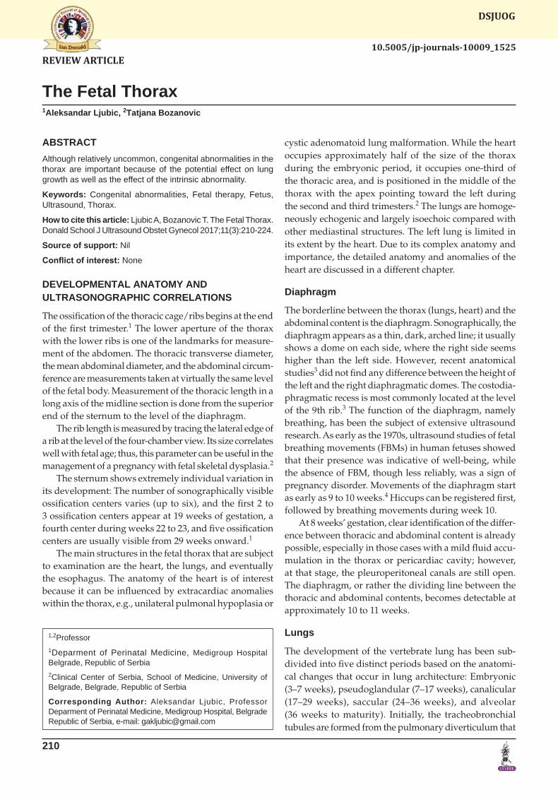
The echogenicity of the lungs usually appears some-what brighter than the echogenicity of the liver; this becomes clearer by applying harmonic imaging6 (Fig. 1). Tekesin et al tried to quantify this subjective assessment of the tissue appearance by quantitative ultrasonic tissue characterization.6 They concluded that the echogenicity of the fetal lung showed a particular changing pattern during pregnancy: The mean gray value of the lungs was almost the same as that of the liver at 22 to 23 weeks gesta-tion, decreased between 23 and 31 weeks, and increased again later in pregnancy. They thought that these changes of echogenicity coincided with the saccular and alveolar phases of fetal lung development. Normal data for the pulmonal size are important to assess lung development in a fetus at risk for pulmonary hypoplasia, such as con-genital diaphragmatic hernia (CDH), cystic adenomatoid
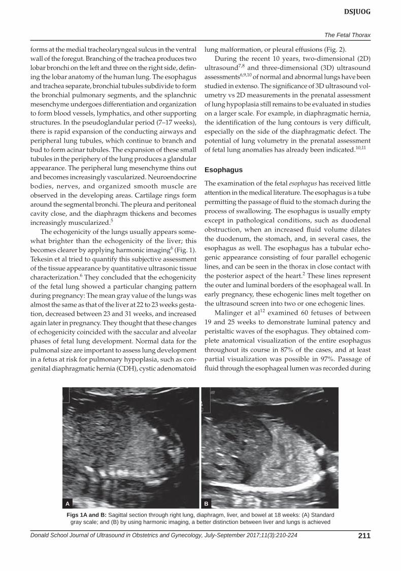
lung malformation, or pleural effusions (Fig. 2).During the recent 10 years, two-dimensional (2D)
ultrasound7,8 and three-dimensional (3D) ultrasound assessments6,9,10 of normal and abnormal lungs have been studied in extenso. The significance of 3D ultrasound vol-umetry vs 2D measurements in the prenatal assessment of lung hypoplasia still remains to be evaluated in studies on a larger scale. For example, in diaphragmatic hernia, the identification of the lung contours is very difficult, especially on the side of the diaphragmatic defect. The potential of lung volumetry in the prenatal assessment of fetal lung anomalies has already been indicated.10,11
Esophagus
The examination of the fetal esophagus has received little attention in the medical literature. The esophagus is a tube permitting the passage of fluid to the stomach during the process of swallowing. The esophagus is usually empty except in pathological conditions, such as duodenal obstruction, when an increased fluid volume dilates the duodenum, the stomach, and, in several cases, the esophagus as well. The esophagus has a tubular echo-genic appearance consisting of four parallel echogenic lines, and can be seen in the thorax in close contact with the posterior aspect of the heart.2 These lines represent the outer and luminal borders of the esophageal wall. In early pregnancy, these echogenic lines melt together on the ultrasound screen into two or one echogenic lines.
Malinger et al12 examined 60 fetuses of between 19 and 25 weeks to demonstrate luminal patency and peristaltic waves of the esophagus. They obtained com-plete anatomical visualization of the entire esophagus throughout its course in 87% of the cases, and at least partial visualization was possible in 97%. Passage of fluid through the esophageal lumen was recorded during
Figs 1A and B: Sagittal section through right lung, diaphragm, liver, and bowel at 18 weeks: (A) Standard gray scale; and (B) by using harmonic imaging, a better distinction between liver and lungs is achieved
A B
Aleksandar Ljubic, Tatjana Bozanovic
212
5 minutes of examination in 90% of the fetuses. Thus, it is possible to evaluate the esophagus in fetuses with suspected obstruction.13
Thymus
The three-vessel view is a parallel plan to the four-cham-ber view, but a little bit more cranial, which is a plan also for evaluation of the thymus. The thymus is just in front of the great arteries in the three-vessel plan. It is easier to recognize from the late second trimester onward, when it starts to undergo significant hypertrophy. It appears as a well-defined, roundish, solid structure interposed between the great vessels, just behind the sternum, and is weakly hypoechoic, in which this different echogenicity is not easy to pick up, especially if a high-frequency (>5 MHz) transducer is not used. The thymus is located on top of the heart and unlike the lungs shows movements synchronous with the cardiac cycle.
Scanning Techniques
The fetus is usually scanned with a 3.5 to 5.0 MHz probe. The thorax is scanned in a transaxial plane, supplemented by sagittal and coronal planes as needed. The four-chamber heart scan is useful to assess chest size and lung status. Color flow Doppler is a valuable adjunct to assess vascular structures and their connections.
Before doing the detailed examination of the thoracic contents, an overall view of the whole trunk should be obtained to make sure that the relative proportions are correct.
The transverse section of the thoracic circumference at the level of the heart should be roughly similar to the abdominal circumference at the level of umbilical vein.
The ribs should enclose about two-thirds of the thorax, which should be almost circular in the transverse section.
The major thoracic contents are the heart and great
vessels and the lungs. The heart occupies about one-third of the chest, and is situated with the apex toward the left side, with the left ventricle lying posterolaterally and the right anteromedially.
The left lung lies behind the heart and is smaller than the right. The continuity of the diaphragm should be checked in sagittal and coronal views, thus confirming that the liver and bowels are separated from the heart and lungs.
The echogenicity of the lungs is usually brighter than the liver, which becomes more distinctive by harmonic imaging. The echogenicity of the fetal lung showed a particular characteristic pattern during the course of pregnancy: It is almost the same as that of the liver at 22 to 23 weeks gestation, but later on decreased between 23 and 31 weeks and increased again later in pregnancy. The esti-mation of the lungs’ volume is important to assess lung development in a fetus at risk for pulmonary hypoplasia, such as CDH, cystic adenomatoid lung malformation, or hydrothorax.
Lung echogenicity does not correlate with lung maturity. Although well studied, lung lengths are not easily reproducible. Thoracic perimeter measurements are made at the level of the four-chamber view and do not include the soft tissue areas beyond the ribs. The thoracic circumference/abdominal circumference ratio is constant through the late second trimester and in the third trimester: >0.80. The 3D reconstruction techniques are reliable and reproducible for fetal lung volumes. During recent years, 2D and 3D ultrasound assessments of normal and abnormal structures of the lungs have been studied extensively. The significance of 3D ultrasound volumetry in the antenatal assessment of lung hypoplasia still remains in discussion.
PATHOLOGY: DIAPHRAGMATIC HERNIA
The diaphragm is a borderline between the thorax and the abdominal cavity. Sonographically, the diaphragm appears as a thin, dark, hypoechoic, arched line. Continuity of this thin and dark line is actually a kind of demarcation between abdominal and thoracic contents. If this line is interrupted or cannot be seen clearly, then diaphragmatic hernia should be questioned. The costo-diaphragmatic recess is most commonly located at the level of the ninth rib. The function of the diaphragm has been studied extensively by ultrasound. Movements of the diaphragm start as early as 9 to 10 weeks. Hiccups can be seen first, followed by breathing movements at the 10th week of gestation (Fig. 3).
Diaphragmatic hernia represents herniation of the abdominal contents into the chest, through a defect in the diaphragm. The defect in the diaphragm exists from the 10th week of gestation, but the herniation of the gut
Fig. 2: Harmonic imaging anterior parasagittal section of the lungs. Diaphragm represent clear demarcation from the abdominal content. In the posterior part—spine is clearly seen
The Fetal Thorax
Donald School Journal of Ultrasound in Obstetrics and Gynecology, July-September 2017;11(3):210-224 213
DSJUOG
into the chest in about 50% of the cases may not occur before the 24th week; this is probably the cause of the relatively low detection rate at the 18 to 20 weeks scan, though the evidence suggests that the marker for those cases where the prognosis is likely to be poor is increased nuchal translucency at the 11 to 14 weeks scan.14
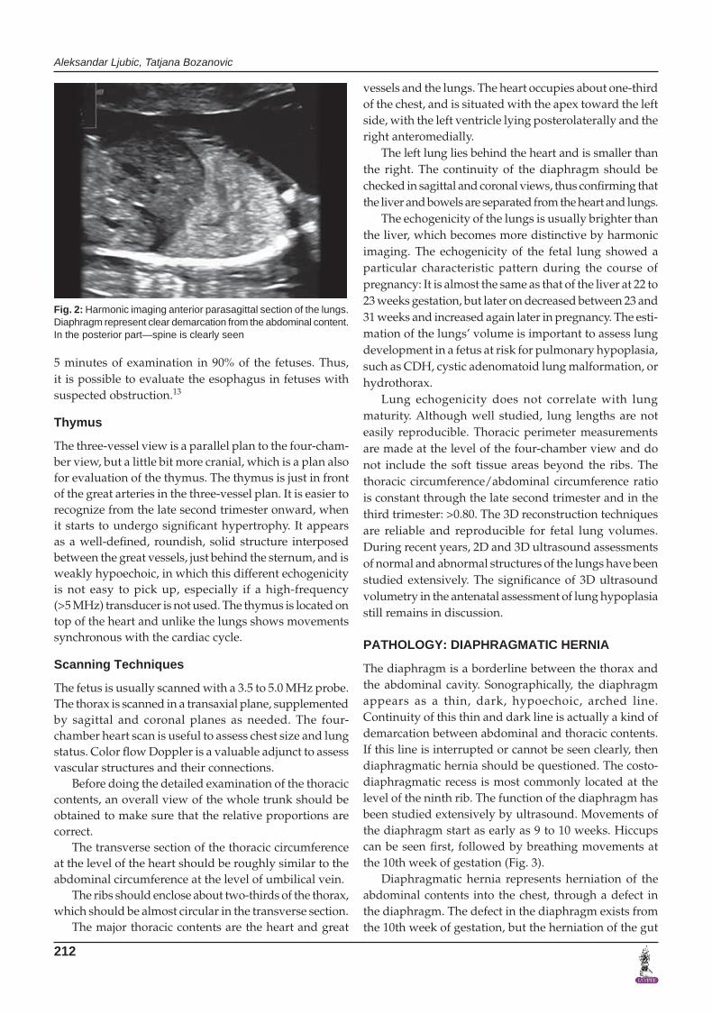
The incidence of diaphragmatic hernia on birth is 1/2,500 to 4,000. There are three types of hernias (Fig. 4):• Posterolateral defect, or Bochdalek’s hernia, which
accounts for about 90% of cases found in the neonatal period. It occurs on the left side in 80% of cases, on the right in 15%, and may be bilateral in approximately 5%. The commonest contents of a left-sided hernia are stomach, bowel, and spleen. If the defect is right-sided, the usual intrathoracic organs are liver and gallbladder.
• Parasternaldefect,orMorgagni’shernia—itaccountsfor 1 to 2% of cases and is more often right sided or bilateral. It usually contains the liver.
• Eventrationofthediaphragmoccursin5%ofcases,and it is more commonly reported on the right.Prenatal diagnosis by ultrasound is based on:
• Demonstrationofabdominalorganswithinthetho-racic cavity. On longitudinal scanning, a defect in the posterior aspect of the diaphragm may be seen, at least for the most common Bochdalek type of hernia.
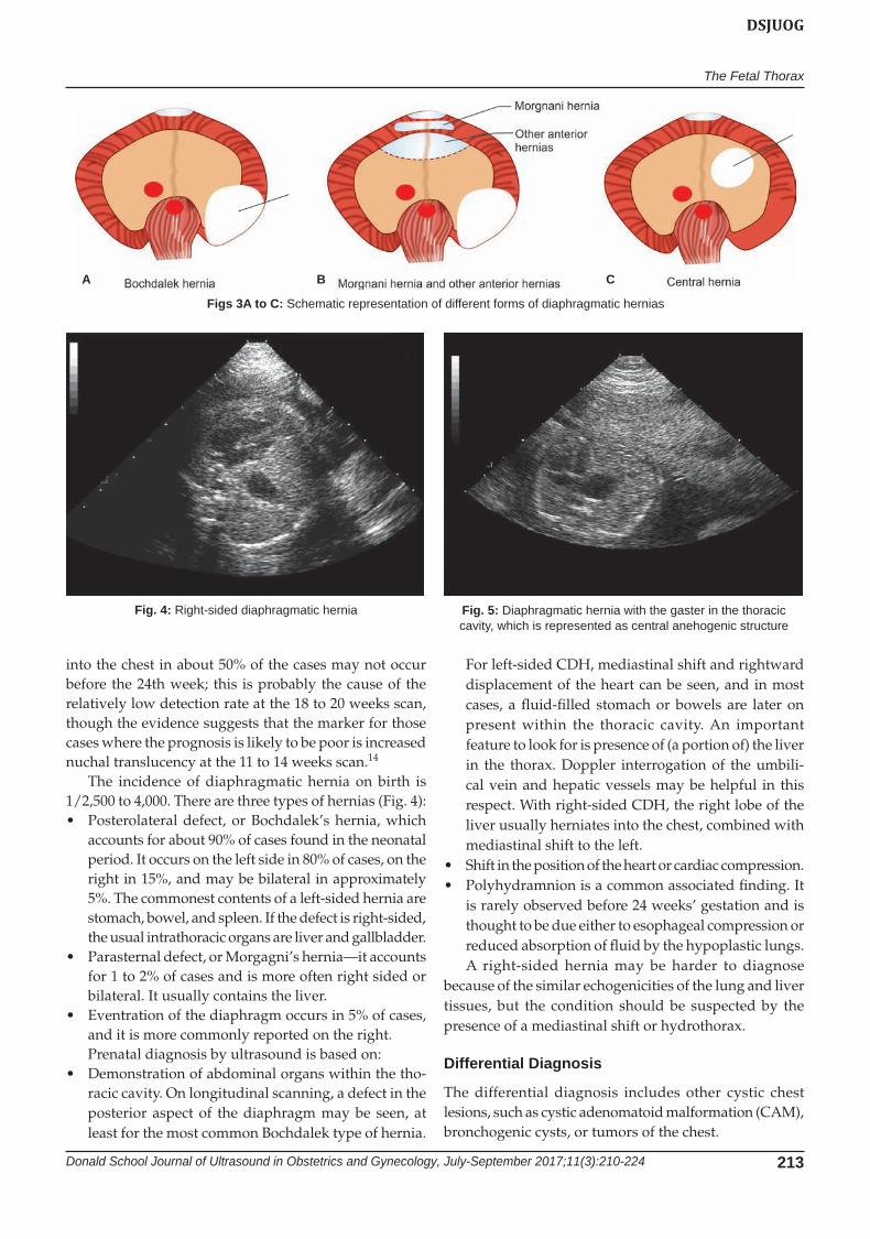
For left-sided CDH, mediastinal shift and rightward displacement of the heart can be seen, and in most cases, a fluid-filled stomach or bowels are later on present within the thoracic cavity. An important feature to look for is presence of (a portion of) the liver in the thorax. Doppler interrogation of the umbili-cal vein and hepatic vessels may be helpful in this respect. With right-sided CDH, the right lobe of the liver usually herniates into the chest, combined with mediastinal shift to the left.
• Shiftinthepositionoftheheartorcardiaccompression.• Polyhydramnionisacommonassociatedfinding.It
is rarely observed before 24 weeks’ gestation and is thought to be due either to esophageal compression or reduced absorption of fluid by the hypoplastic lungs.A right-sided hernia may be harder to diagnose
because of the similar echogenicities of the lung and liver tissues, but the condition should be suspected by the presence of a mediastinal shift or hydrothorax.
Differential Diagnosis
The differential diagnosis includes other cystic chest lesions, such as cystic adenomatoid malformation (CAM), bronchogenic cysts, or tumors of the chest.
Figs 3A to C: Schematic representation of different forms of diaphragmatic hernias
Fig. 4: Right-sided diaphragmatic hernia Fig. 5: Diaphragmatic hernia with the gaster in the thoracic cavity, which is represented as central anehogenic structure
A B C
Aleksandar Ljubic, Tatjana Bozanovic
214
Associated Anomalies
In prenatal series, about 50% of fetuses have an isolated diaphragmatic defect and, in the rest, a chromosomal abnormality may be found, usually trisomy 18, as well as a major defect including congenital heart disease, exomphalos, and renal, brain, and spinal abnormalities.
The incidence of aneuploidy was reported in overall 14% of pregnancies, particularly trisomies 13 and 18. Comprehensive anatomy scanning including fetal echocardiography is essential to detect other structural anomalies because at least 33% of cases will have other anomalies including cardiac defects.
Syndromes associated with diaphragmatic hernia:Pallister–Killian syndrome (Tetrasomy 12p): It pres-
ents with polyhydramnios, rhizomelic limb shortening, abnormal facial profile with prominent philtrum, and fetal somatic overgrowth in addition to diaphragmatic hernia. These are variable and even absent. Pallister–Killian syn-drome is a mosaicism for an isochromosome of 12p, which is an extra two short arms of chromosome-12. Therefore, the fetus has four copies of 12p in some cell lines. In
cases of highly suspicious Pallister–Killian syndrome, the amniocentesis should be done for correct diagnosis because the cytogenetic abnormality may not be detected in either chorionic villi or fetal blood.
Fryns syndrome is a rare autosomal recessive dis-order of multiple congenital abnormalities, but can be found in about 4% of fetuses of CDH cases with normal chromosomes. The main criteria for diagnosis include CDH, hypoplasia of distal limb and nail, and abnormal facies. Cataracts may be detected later on in gestation. Intrauterine growth restriction (IUGR) and an increased nuchal fold together with CDH are the cases to be sus-pected for Fryns syndrome. Prognosis is very poor and 86% present with an early lethal outcome. This is a dif-ficult diagnosis in a fetus in the absence of a history of a previously affected child or parental consanguinity; therefore, it is important to have genetic consultation when a diaphragmatic hernia is found together with other anomalies and normal karyotype.
The Simpson–Golabi–Behmel syndrome: It is an X-linked recessive disorder due to mutations in glypican
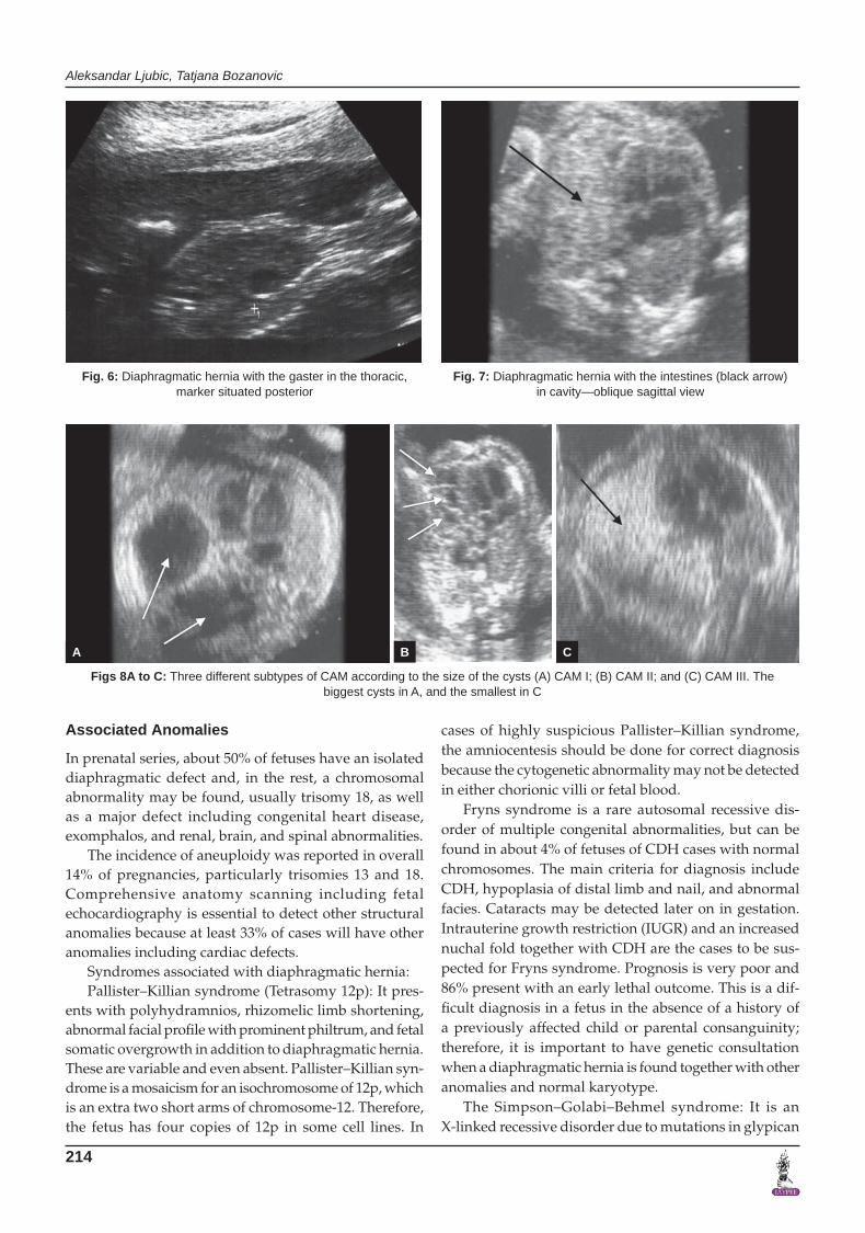
Fig. 6: Diaphragmatic hernia with the gaster in the thoracic, marker situated posterior
Fig. 7: Diaphragmatic hernia with the intestines (black arrow) in cavity—oblique sagittal view
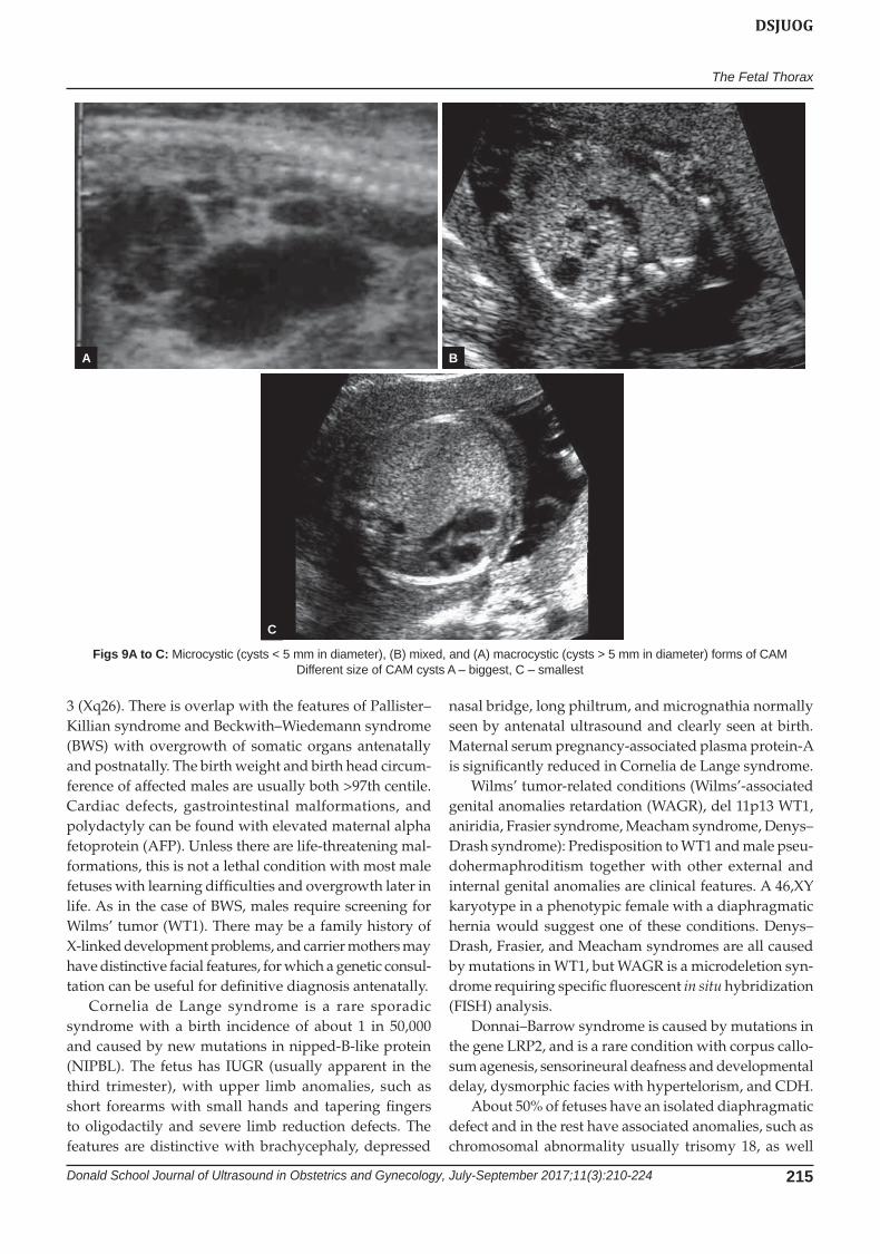
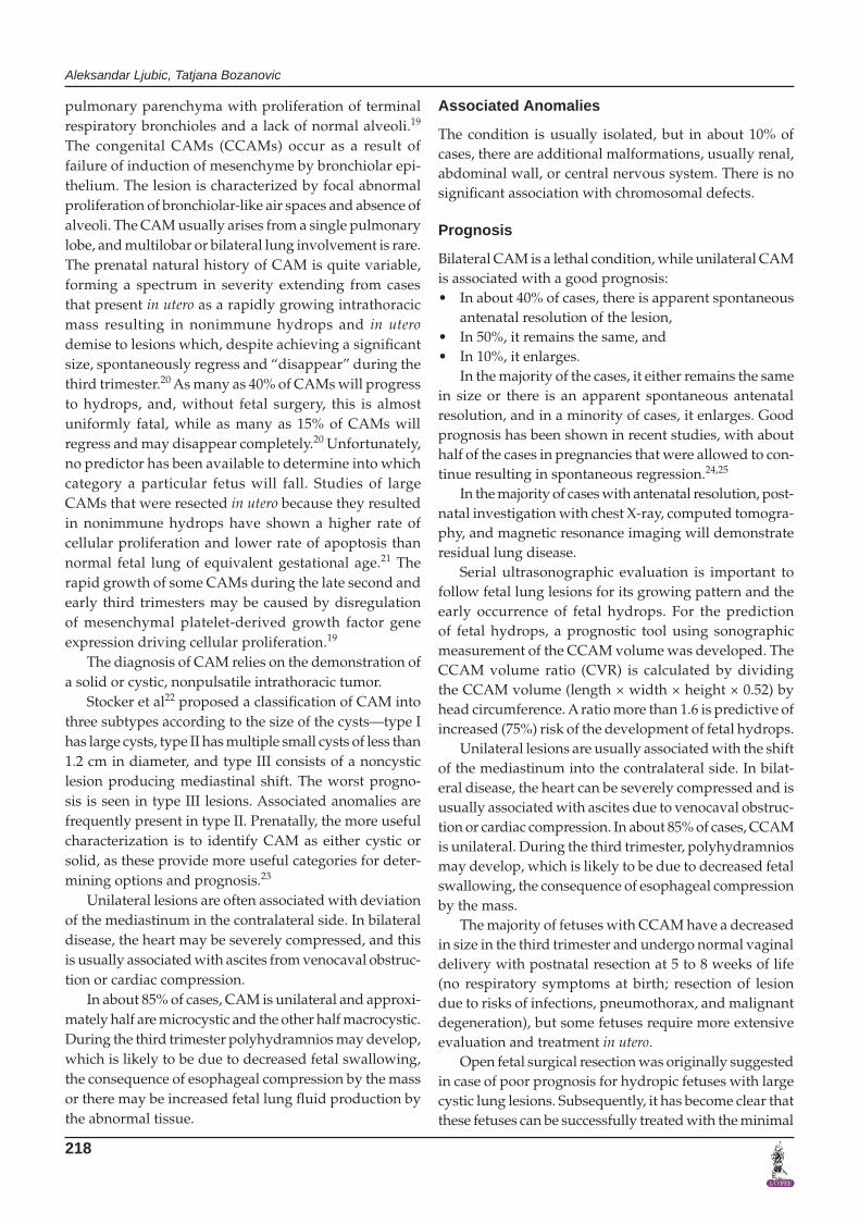
Figs 8A to C: Three different subtypes of CAM according to the size of the cysts (A) CAM I; (B) CAM II; and (C) CAM III. The biggest cysts in A, and the smallest in C
A B C
The Fetal Thorax
Donald School Journal of Ultrasound in Obstetrics and Gynecology, July-September 2017;11(3):210-224 215
DSJUOG
3 (Xq26). There is overlap with the features of Pallister–Killian syndrome and Beckwith–Wiedemann syndrome (BWS) with overgrowth of somatic organs antenatally and postnatally. The birth weight and birth head circum-ference of affected males are usually both >97th centile. Cardiac defects, gastrointestinal malformations, and polydactyly can be found with elevated maternal alpha fetoprotein (AFP). Unless there are life-threatening mal-formations, this is not a lethal condition with most male fetuses with learning difficulties and overgrowth later in life. As in the case of BWS, males require screening for Wilms’ tumor (WT1). There may be a family history of X-linked development problems, and carrier mothers may have distinctive facial features, for which a genetic consul-tation can be useful for definitive diagnosis antenatally.
Cornelia de Lange syndrome is a rare sporadic syndrome with a birth incidence of about 1 in 50,000 and caused by new mutations in nipped-B-like protein (NIPBL). The fetus has IUGR (usually apparent in the third trimester), with upper limb anomalies, such as short forearms with small hands and tapering fingers to oligodactily and severe limb reduction defects. The features are distinctive with brachycephaly, depressed
nasal bridge, long philtrum, and micrognathia normally seen by antenatal ultrasound and clearly seen at birth. Maternal serum pregnancy-associated plasma protein-A is significantly reduced in Cornelia de Lange syndrome.
Wilms’ tumor-related conditions (Wilms’-associated genital anomalies retardation (WAGR), del 11p13 WT1, aniridia, Frasier syndrome, Meacham syndrome, Denys–Drash syndrome): Predisposition to WT1 and male pseu-dohermaphroditism together with other external and internal genital anomalies are clinical features. A 46,XY karyotype in a phenotypic female with a diaphragmatic hernia would suggest one of these conditions. Denys–Drash, Frasier, and Meacham syndromes are all caused by mutations in WT1, but WAGR is a microdeletion syn-drome requiring specific fluorescent in situ hybridization (FISH) analysis.
Donnai–Barrow syndrome is caused by mutations in the gene LRP2, and is a rare condition with corpus callo-sum agenesis, sensorineural deafness and developmental delay, dysmorphic facies with hypertelorism, and CDH.
About 50% of fetuses have an isolated diaphragmatic defect and in the rest have associated anomalies, such as chromosomal abnormality usually trisomy 18, as well
Figs 9A to C: Microcystic (cysts < 5 mm in diameter), (B) mixed, and (A) macrocystic (cysts > 5 mm in diameter) forms of CAM Different size of CAM cysts A – biggest, C – smallest
A B
C
Aleksandar Ljubic, Tatjana Bozanovic
216
as a major defect, including congenital heart disease, exomphalos, renal anomalies, brain anomalies and spinal abnormalities, and Fryns, Goldenhar, BWS, Cornelia De Lange, Apert syndromes, and lethal pterygium.
Management of Pregnancy
When diaphragmatic hernia is diagnosed antenatally, fetal karyotyping and detailed ultrasound examination of the fetus (in particular the heart) should be undertaken. Fetal echocardiography should be arranged as well as consultation with a pediatric surgeon.
Prediction of Outcome
In neonates with isolated diaphragmatic hernia, the primary determinant of survival is the presence of pul-monary hypoplasia and hypertension. The mortality has remained high despite optimal postnatal manage-ment and the introduction of extracorporal membrane oxygenation.
Antenatal prediction of pulmonary hypoplasia is difficult and it is the most important part in counseling parents and selecting those cases that may benefit from prenatal surgery. In isolated cases of CDH, prognosis is poor when the liver is intrathoracic15 and the lung-to-head ratio (LHR) is less than 1.16
When making a measurement of the LHR, a transverse section of the chest is taken at the level of the four-chamber view.Thecontralaterallungismeasured—thelongestaxisis measured and multiplied by the longest measurement perpendicular to it. This is placed over the head circumfer-ence, which is measured at the standard biparietal view, typically showing two equal hemispheres, the septum cavum pellucidum, one-third of the way from the front to the back, and the posterior horns of the lateral ventricles.17
Recent research shows that in isolated CDH, fetal lung volume measurement by 3D ultrasound may be a potential predictor for pulmonary hypoplasia and postnatal outcome.11,18
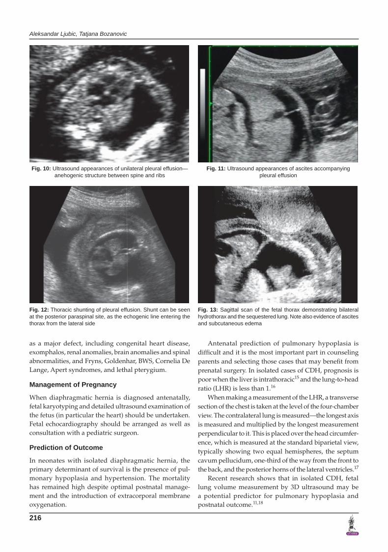
Fig. 10: Ultrasound appearances of unilateral pleural effusion—anehogenic structure between spine and ribs
Fig. 11: Ultrasound appearances of ascites accompanying pleural effusion
Fig. 12: Thoracic shunting of pleural effusion. Shunt can be seen at the posterior paraspinal site, as the echogenic line entering the thorax from the lateral side
Fig. 13: Sagittal scan of the fetal thorax demonstrating bilateral hydrothorax and the sequestered lung. Note also evidence of ascites and subcutaneous edema
The Fetal Thorax
Donald School Journal of Ultrasound in Obstetrics and Gynecology, July-September 2017;11(3):210-224 217
DSJUOG
Fetal surgery can be a reasonable approach in the treatment of CDH. The aim of surgery is to prevent lung hypoplasia. Although fetal surgery initially involved reduction of the hernia by open repair in utero, current practice involves ballooning of the trachea. Tracheal occlusion results in increased lung volume and acceler-ated lung maturity.
The initial fetal surgery was a patch closure of the diaphragmatic defect with abdominal silo with an open fetal surgery. Also, fetuses with herniation of the left lobe of the liver could not be treated in this way; because of this, reduction of the herniated liver led to kinking of the umbilical vein, which compromises the blood flow.
More recently, fetal tracheal occlusion (FETO) has been used as a treatment modality for CDH. It has been shown that the dynamics of fetal lung fluid affect lung growth. Although tracheal occlusion might offer a relatively simple approach to accelerate lung growth in human fetuses with CDH, further experimental studies in the sheep model also demonstrated that tracheal occlusion had a detrimental effect on lung maturation because of disappearance of type II pneumocytes. Other studies demonstrated that released tracheal occlusion at an interval prior to delivery
induced type II pneumocytes and significant lung growth. Evidence suggested that lung function was not restored to normal, because abnormally thick wall causing in a limited gas exchange.
A minimally invasive approach to tracheal occlusion in the sheep model utilizing a deployable balloon technol-ogy placed via a single small trocar has been proposed. Overall outcomes in 210 FETO interventions increased survival from 24 to 49% in severe cases with left-sided CDH and from 0 to 35% in right-sided CDH when com-pared with expectantly managed cases.
Despite the use of small-diameter fetoscopes,a high incidence of prematurity (50.0%), extreme prematurity (15.0%), and preterm premature rupture of the mem-branes (35.0%) was present. Infant survival to 6 months was 10% in the FETO group and 4.8% in controls, and severe pulmonary arterial hypertension is 50% in the FETO group and 85.7% in controls.
CYSTIC ADENOMATOID MALFORMATION
The CAM of the lung is a developmental abnormality of the lung characterized by a cystic mass of disordered
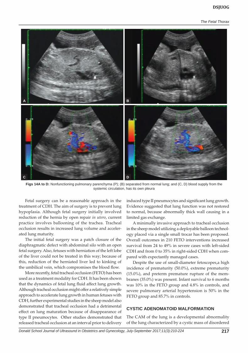
Figs 14A to D: Nonfunctioning pulmonary parenchyma (P); (B) separated from normal lung; and (C, D) blood supply from the systemic circulation, has its own pleura
A
C
B
D
Aleksandar Ljubic, Tatjana Bozanovic
218
pulmonary parenchyma with proliferation of terminal respiratory bronchioles and a lack of normal alveoli.19 The congenital CAMs (CCAMs) occur as a result of failure of induction of mesenchyme by bronchiolar epi-thelium. The lesion is characterized by focal abnormal proliferation of bronchiolar-like air spaces and absence of alveoli. The CAM usually arises from a single pulmonary lobe, and multilobar or bilateral lung involvement is rare. The prenatal natural history of CAM is quite variable, forming a spectrum in severity extending from cases that present in utero as a rapidly growing intrathoracic mass resulting in nonimmune hydrops and in utero demise to lesions which, despite achieving a significant size, spontaneously regress and “disappear” during the third trimester.20 As many as 40% of CAMs will progress to hydrops, and, without fetal surgery, this is almost uniformly fatal, while as many as 15% of CAMs will regress and may disappear completely.20 Unfortunately, no predictor has been available to determine into which category a particular fetus will fall. Studies of large CAMs that were resected in utero because they resulted in nonimmune hydrops have shown a higher rate of cellular proliferation and lower rate of apoptosis than normal fetal lung of equivalent gestational age.21 The rapid growth of some CAMs during the late second and early third trimesters may be caused by disregulation of mesenchymal platelet-derived growth factor gene expression driving cellular proliferation.19
The diagnosis of CAM relies on the demonstration of a solid or cystic, nonpulsatile intrathoracic tumor.
Stocker et al22 proposed a classification of CAM into threesubtypesaccordingtothesizeofthecysts—typeIhas large cysts, type II has multiple small cysts of less than 1.2 cm in diameter, and type III consists of a noncystic lesion producing mediastinal shift. The worst progno-sis is seen in type III lesions. Associated anomalies are frequently present in type II. Prenatally, the more useful characterization is to identify CAM as either cystic or solid, as these provide more useful categories for deter-mining options and prognosis.23
Unilateral lesions are often associated with deviation of the mediastinum in the contralateral side. In bilateral disease, the heart may be severely compressed, and this is usually associated with ascites from venocaval obstruc-tion or cardiac compression.
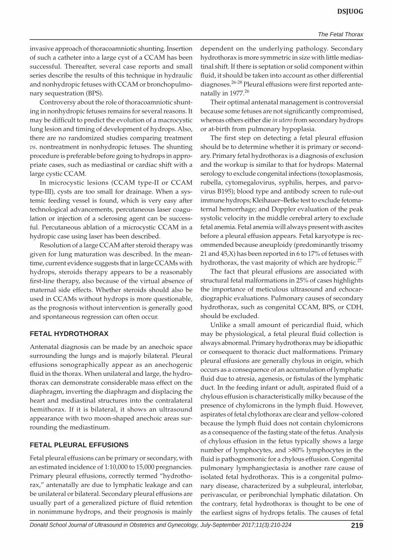
In about 85% of cases, CAM is unilateral and approxi-mately half are microcystic and the other half macrocystic. During the third trimester polyhydramnios may develop, which is likely to be due to decreased fetal swallowing, the consequence of esophageal compression by the mass or there may be increased fetal lung fluid production by the abnormal tissue.
Associated Anomalies
The condition is usually isolated, but in about 10% of cases, there are additional malformations, usually renal, abdominal wall, or central nervous system. There is no significant association with chromosomal defects.
Prognosis
Bilateral CAM is a lethal condition, while unilateral CAM is associated with a good prognosis:• Inabout40%ofcases,thereisapparentspontaneous
antenatal resolution of the lesion,• In50%,itremainsthesame,and• In10%,itenlarges.
In the majority of the cases, it either remains the same in size or there is an apparent spontaneous antenatal resolution, and in a minority of cases, it enlarges. Good prognosis has been shown in recent studies, with about half of the cases in pregnancies that were allowed to con-tinue resulting in spontaneous regression.24,25
In the majority of cases with antenatal resolution, post-natal investigation with chest X-ray, computed tomogra-phy, and magnetic resonance imaging will demonstrate residual lung disease.
Serial ultrasonographic evaluation is important to follow fetal lung lesions for its growing pattern and the early occurrence of fetal hydrops. For the prediction of fetal hydrops, a prognostic tool using sonographic measurement of the CCAM volume was developed. The CCAM volume ratio (CVR) is calculated by dividing the CCAM volume (length × width × height × 0.52) by head circumference. A ratio more than 1.6 is predictive of increased (75%) risk of the development of fetal hydrops.
Unilateral lesions are usually associated with the shift of the mediastinum into the contralateral side. In bilat-eral disease, the heart can be severely compressed and is usually associated with ascites due to venocaval obstruc-tion or cardiac compression. In about 85% of cases, CCAM is unilateral. During the third trimester, polyhydramnios may develop, which is likely to be due to decreased fetal swallowing, the consequence of esophageal compression by the mass.
The majority of fetuses with CCAM have a decreased in size in the third trimester and undergo normal vaginal delivery with postnatal resection at 5 to 8 weeks of life (no respiratory symptoms at birth; resection of lesion due to risks of infections, pneumothorax, and malignant degeneration), but some fetuses require more extensive evaluation and treatment in utero.
Open fetal surgical resection was originally suggested in case of poor prognosis for hydropic fetuses with large cystic lung lesions. Subsequently, it has become clear that these fetuses can be successfully treated with the minimal
The Fetal Thorax
Donald School Journal of Ultrasound in Obstetrics and Gynecology, July-September 2017;11(3):210-224 219
DSJUOG
invasive approach of thoracoamniotic shunting. Insertion of such a catheter into a large cyst of a CCAM has been successful. Thereafter, several case reports and small series describe the results of this technique in hydraulic and nonhydropic fetuses with CCAM or bronchopulmo-nary sequestration (BPS).
Controversy about the role of thoracoamniotic shunt-ing in nonhydropic fetuses remains for several reasons. It may be difficult to predict the evolution of a macrocystic lung lesion and timing of development of hydrops. Also, there are no randomized studies comparing treatment vs. nontreatment in nonhydropic fetuses. The shunting procedure is preferable before going to hydrops in appro-priate cases, such as mediastinal or cardiac shift with a large cystic CCAM.
In microcystic lesions (CCAM type-II or CCAM type-III), cysts are too small for drainage. When a sys-temic feeding vessel is found, which is very easy after technological advancements, percutaneous laser coagu-lation or injection of a sclerosing agent can be success-ful. Percutaneous ablation of a microcystic CCAM in a hydropic case using laser has been described.
Resolution of a large CCAM after steroid therapy was given for lung maturation was described. In the mean-time, current evidence suggests that in large CCAMs with hydrops, steroids therapy appears to be a reasonably first-line therapy, also because of the virtual absence of maternal side effects. Whether steroids should also be used in CCAMs without hydrops is more questionable, as the prognosis without intervention is generally good and spontaneous regression can often occur.
FETAL HYDROTHORAX
Antenatal diagnosis can be made by an anechoic space surrounding the lungs and is majorly bilateral. Pleural effusions sonographically appear as an anechogenic fluid in the thorax. When unilateral and large, the hydro-thorax can demonstrate considerable mass effect on the diaphragm, inverting the diaphragm and displacing the heart and mediastinal structures into the contralateral hemithorax. If it is bilateral, it shows an ultrasound appearance with two moon-shaped anechoic areas sur-rounding the mediastinum.
FETAL PLEURAL EFFUSIONS
Fetal pleural effusions can be primary or secondary, with an estimated incidence of 1:10,000 to 15,000 pregnancies. Primary pleural effusions, correctly termed “hydrotho-rax,” antenatally are due to lymphatic leakage and can be unilateral or bilateral. Secondary pleural effusions are usually part of a generalized picture of fluid retention in nonimmune hydrops, and their prognosis is mainly
dependent on the underlying pathology. Secondary hydrothorax is more symmetric in size with little medias-tinal shift. If there is septation or solid component within fluid, it should be taken into account as other differential diagnoses.26-28 Pleural effusions were first reported ante-natally in 1977.26
Their optimal antenatal management is controversial because some fetuses are not significantly compromised, whereas others either die in utero from secondary hydrops or at-birth from pulmonary hypoplasia.
The first step on detecting a fetal pleural effusion should be to determine whether it is primary or second-ary. Primary fetal hydrothorax is a diagnosis of exclusion and the workup is similar to that for hydrops: Maternal serology to exclude congenital infections (toxoplasmosis, rubella, cytomegalovirus, syphilis, herpes, and parvo-virus B195); blood type and antibody screen to rule-out immune hydrops; Kleihauer–Betke test to exclude fetoma-ternal hemorrhage; and Doppler evaluation of the peak systolic velocity in the middle cerebral artery to exclude fetal anemia. Fetal anemia will always present with ascites before a pleural effusion appears. Fetal karyotype is rec-ommended because aneuploidy (predominantly trisomy 21 and 45,X) has been reported in 6 to 17% of fetuses with hydrothorax, the vast majority of which are hydropic.27
The fact that pleural effusions are associated with structural fetal malformations in 25% of cases highlights the importance of meticulous ultrasound and echocar-diographic evaluations. Pulmonary causes of secondary hydrothorax, such as congenital CCAM, BPS, or CDH, should be excluded.
Unlike a small amount of pericardial fluid, which may be physiological, a fetal pleural fluid collection is always abnormal. Primary hydrothorax may be idiopathic or consequent to thoracic duct malformations. Primary pleural effusions are generally chylous in origin, which occurs as a consequence of an accumulation of lymphatic fluid due to atresia, agenesis, or fistulas of the lymphatic duct. In the feeding infant or adult, aspirated fluid of a chylous effusion is characteristically milky because of the presence of chylomicrons in the lymph fluid. However, aspirates of fetal chylothorax are clear and yellow-colored because the lymph fluid does not contain chylomicrons as a consequence of the fasting state of the fetus. Analysis of chylous effusion in the fetus typically shows a large number of lymphocytes, and >80% lymphocytes in the fluid is pathognomonic for a chylous effusion. Congenital pulmonary lymphangiectasia is another rare cause of isolated fetal hydrothorax. This is a congenital pulmo-nary disease, characterized by a subpleural, interlobar, perivascular, or peribronchial lymphatic dilatation. On the contrary, fetal hydrothorax is thought to be one of the earliest signs of hydrops fetalis. The causes of fetal
Aleksandar Ljubic, Tatjana Bozanovic
220
hydrothorax with hydrops include cardiac and vascu-lar diseases (50%), chromosomal abnormality (more frequently trisomy 21 and Turner syndrome with the rate of 7–10%), anemia and hematological diseases, pulmonary abnormalities, skeletal dysplasia, hepatic or metabolic diseases, and infections including TORCH and parvovirus B19. Other major congenital abnormalities, such as CDH, extralobar sequestration, CCAM, thyroid teratoma, or fetal goiter are found in 25 to 40% of fetuses with nonimmune hydrops fetalis. Perinatal mortality is about >90% in hydropic fetuses with hydrothorax, if another structural abnormality is identified. Therefore, it is important to make a complete anatomic survey in any hydropic fetus with hydrothorax.
Prognosis
Irrespective of the underlying cause, infants affected by pleural effusions usually present, in the neonatal period, with severe and often fatal respiratory insufficiency. This is either a direct result of pulmonary compression caused by the effusions, or due to pulmonary hypoplasia second-ary to chronic intrathoracic compression. The overall mortality of neonates with pleural effusions increases from low in infants with isolated pleural effusions to very high in those with gross hydrops.26 Chromosomal abnormalities, mainly trisomy 21, are found in about 5% of fetuses with apparently isolated pleural effusions.
In the human, isolated fetal pleural effusions may resolve spontaneously antenatally, or they may persist. In some cases, postnatal thoracocentesis may be suffi-cient, but, in others, the chronic compression of the fetal lungs can result in pulmonary hypoplasia and neonatal death. Additionally, mediastinal compression may lead to the development of fetal hydrops and polyhydram-nios, which are associated with a high risk of premature delivery and subsequent perinatal death.26
Unilateral fetal hydrothorax can occur more sporadi-cally and is caused by a congenital malformation of the thoracic duct or the pulmonary lymphatic system. It is usually diagnosed in the second or early third trimester. The prognosis of fetal hydrothorax is difficult to predict with expected perinatal mortality rates between 22 and 53%. Therefore, counseling on perinatal outcome is very hard for the selection of cases for fetal intervention or early delivery.
Irrespective of the underlying cause, fetal hydro-thorax is potentially responsible for fetal and neonatal death due to pulmonary hypoplasia caused by chronic intrathoracic compression and due to hydrops caused by mediastinal shift, cardiac compression, and vena caval obstruction with low cardiac output;it is also due to the prematurity as a consequence of polyhydramnios caused by esophagus compression (mediasitnal shift) and/or low
amnitoic fluid uptake of the lungs. Infants affected by fetal hydrothorax present usually with severe respiratory problems and insufficiency. Sometimes, it can be seen as an associated maternal morbidity, as in the Mirror syn-drome characterized by a generalized edema, due to the hydropic placenta that produces vasoactive substances.
Parameters associated with a better prognostic include later gestational age at diagnosis, spontaneous resolution of the effusion prior to delivery, lack of hydrops, isolated effusion, and unilateral effusion.
For prognostic features, several investigators reported outcomes of fetuses with antenatally diagnosed hydrotho-rax. Adverse prognostic indicators included bilaterality, presence of hydrops, absence of spontaneous resolu-tion, and premature delivery. Polyhydramnios has also prognostic significance, with the uterine overdistention predisposing to preterm labor and delivery. Even without hydrops, large pleural effusions can cause pulmonary hypoplasia due to compression. The time of onset, size, and duration of the pleural effusion influence the devel-opment of pulmonary hypoplasia. The most common cause of neonatal mortality with fetal hydrothorax is respiratory insufficiency due to pulmonary hypoplasia.
Therapy
There are several options in the management of fetuses with isolated hydrothorax; the options depend on gesta-tional age, severity of effusion, evidence of progression, hydrops, polyhydramnios, or mediastinal shift. The outcome of fetal hydrothorax is significantly worsened by prematurity (less than 32 weeks of gestation), the presence of hydrops, and lack of fetal therapy. When the hydrothorax is small, isolated, and well tolerated, expectant management with frequent follow-up may be most beneficial because of the possibility of spontane-ous resolution. If the fetal pleural effusion is very large or increases by gestational age, then a fetal intervention is needed. Fetal intervention for pleural effusion can include a single and serial thoracocentesis (less effective) or thoracoamniotic shunting to allow the drainage of fetal fluid into the amniotic fluid.
Thoracocentesis is a diagnostic procedure to obtain pleural fluid for cell count, culture, and establish whether the effusion is chylous. Additionally and importantly, ultrasound evaluation should be done again after decom-pression because undiagnosed cardiac abnormality or other intrathoracic lesion may become apparent which cannot be seen easily due to compressed organs and anatomy.
When hydrothorax re-accumulates after the initial thoracocentesis, and with the development of mediastinal shift or fetal hydrops, a thoracoamniotic shunt should be done.
The Fetal Thorax
Donald School Journal of Ultrasound in Obstetrics and Gynecology, July-September 2017;11(3):210-224 221
DSJUOG
LUNG SEQUESTRATION/PULMONARY SEQUESTRATION
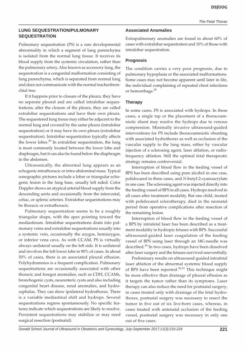
Pulmonary sequestration (PS) is a rare developmental abnormality in which a segment of lung parenchyma is isolated from the normal lung tissue. It receives its blood supply from the systemic circulation, rather than the pulmonary artery. Also known as accessory lung, the sequestration is a congenital malformation consisting of lung parenchyma, which is separated from normal lung and does not communicate with the normal tracheobron-chial tree.
If it happens prior to closure of the pleura, they have no separate pleural and are called intralobar seques-trations; after the closure of the pleura, they are called extralobar sequestrations and have their own pleura. The sequestered lung tissue may either be adjacent to the normal lung and covered by the same pleura (intralobar sequestration) or it may have its own pleura (extralobar sequestration). Intralobar sequestration typically affects the lower lobes.29 In extralobar sequestration, the lung is most commonly located between the lower lobe and diaphragm, but it can also be found below the diaphragm in the abdomen.
Ultrasonically, the abnormal lung appears as an echogenic intrathoracic or intra-abdominal mass. Typical sonographic pictures include a lobar or triangular echo-genic lesion in the lung base, usually left sided. Color Doppler shows an atypical arterial blood supply from the descending aorta and occasionally from the intercostal, celiac, or splenic arteries. Extralobar sequestrations may be thoracic or extrathoracic.
Pulmonary sequestration seems to be a roughly triangular shape, with the apex pointing toward the mediastinum. Intralobar sequestrations drain into pul-monary veins and extralobar sequestrations usually into a systemic vein, occasionally the azygos, hemiazygos, or inferior vena cava. As with CCAM, PS is virtually always unilateral usually on the left side. It is unilateral and involves the left lower lobe in 90% of cases. In about 50% of cases, there is an associated pleural effusion. Polyhydramnios is a frequent complication. Pulmonary sequestrations are occasionally associated with other thoracic and foregut anomalies, such as CDH, CCAMs, bronchogenic cysts, neurenteric cysts and also including congenital heart disease, renal anomalies, and hydro-cephalus. They can show ipsilateral hydrothorax. There is a variable mediastinal shift and hydrops. Several sequestrations regress spontaneously. No specific fea-tures indicate which sequestrations are likely to resolve. Persistent sequestrations may stabilize or may need surgical resection (postnatal).
Associated Anomalies
Extrapulmonary anomalies are found in about 60% of cases with extralobar sequestration and 10% of those with intralobar sequestration.
Prognosis
The condition carries a very poor prognosis, due to pulmonary hypoplasia or the associated malformations. Some cases may not become apparent until later in life, the individual complaining of repeated chest infections or hemorrhage.29
Therapy
In some cases, PS is associated with hydrops. In these cases, a single tap or the placement of a thoracoam-niotic shunt may resolve the hydrops due to venous compression. Minimally invasive ultrasound-guided interventions for PS include thoracoamniotic shunting with associated hydrothorax as well as occlusion of the vascular supply to the lung mass, either by vascular injection of a sclerosing agent, laser ablation, or radio-frequency ablation. Still the optimal fetal therapeutic strategy remains controversial.
Interruption of blood flow in the feeding vessel of BPS has been described using pure alcohol in one case, polidocanol in three cases, and N-butyl-2-cyanoacrylate in one case. The sclerosing agent was injected directly into the feeding vessel of BPS in all cases. Hydrops resolved in all cases after treatment modality. But one child, treated with polidocanol sclerotherapy, died in the neonatal period from operative complications after resection of the remaining lesion.
Interruption of blood flow in the feeding vessel of a BPS by intrafetal laser has been described as a treat-ment modality in hydropic fetuses with BPS. Successful ultrasound-guided laser coagulation of the feeding vessel of BPS using laser through an 18G-needle was described.30 In two cases, hydrops have been dissolved after laser surgery and the fetuses survived uneventfully.
Preliminary results on ultrasound-guided intrafetal laser ablation of the abnormal systemic blood supply of BPS have been reported.30,31 This technique might be more effective than drainage of pleural effusion as it targets the tumor rather than its symptoms. Laser therapy can also reduce the need for postnatal surgery; in cases treated only with drainage of the fetal hydro-thorax, postnatal surgery was necessary to resect the tumor in five out of six live-born cases, whereas, in cases treated with antenatal occlusion of the feeding vessel, postnatal surgery was necessary in only one out of five cases.
Aleksandar Ljubic, Tatjana Bozanovic
222
Syndromic Associations with Congenital Anomalies of the Fetal Thorax
Conditions to Consider in the Presence of a Diaphragmatic Hernia
Pallister–Killian syndrome (Tetrasomy 12p): In addition to diaphragmatic hernia, polyhydramnios, rhizomelic limb shortening, and an abnormal facial profile with promi-nent philtrum are found in association with fetal somatic overgrowth. These features are variable and may even be absent. Pallister–Killian syndrome is a chromosomal abnormality with mosaicism for an isochromosome of 12p, which is an extra chromosome comprising the two short arms of 12. Thus, the fetus has four copies of 12p in some cell lines. In the neonate, this chromosome abnormality is usually present in skin fibroblasts, but not usually in blood. For this reason, the cytogenetic abnormality may not be detected in either chorionic villi or fetal blood, the latter being the least indicated method because of the low frequency of the isochromosome in lymphocytes. In cases thought to have a normal karyotype following chorionic villus sampling (CVS) , amniocentesis should be consid-ered if this diagnosis is suspected, as this is most likely to reveal the mosaicism, particularly if FISH and interphase FISH on noncultured cells are performed.29
Fryns syndrome: Fryns syndrome is a rare autosomal recessive disorder.32
Prenatally, suspicion of this condition is raised in fetuses with a CDH, IUGR, and an increased nuchal fold. Cataracts may be detected later in gestation.
Simpson–Golabi–Behmel syndrome: It is an X-linked recessive disorder.
Maternal AFP is elevated. Unless there are life-threat-ening malformations, this is not a lethal condition with most boys presenting at later age with other manifesta-tions of the syndrome, such as learning difficulties and overgrowth.33
Cornelia de Lange syndrome: Cornelia de Lange (de Lange) syndrome is a rare sporadic syndrome with a birth incidence of about 1 in 50,000.
There are distinctive craniofacial features (micro-brachycephaly, depressed nasal bridge with anteverted nares, long smooth overhanging philtrum and microgna-thia) which may be recognized with prenatal ultrasound and are clearly seen at birth.34 Maternal serum pregnancy associated plasma protein-A is reported to be significantly reduced in de Lange syndrome.
CONGENITAL CYSTIC LUNG LESIONS
If a cystic lung lesion is isolated, unilateral karyotyp-ing is not indicated. “Bilateral” lesions may represent laryngeal/tracheal atresia, thus consideration should be given to the autosomal recessive Fraser–Crytophthalmos
syndrome where the likely mechanism for pulmonary hyperplasia is retention of fetal lung fluid by laryngeal or tracheal obstruction.
Fraser Syndrome (Fraser–Cryptophthalmos Syndrome)
Fraser syndrome is inherited in an autosomal recessive fashion and can be caused by mutation in the FRAS1 gene or in the FREM2 gene and there may be yet further unidentified gene(s). The classic features are a combina-tion of cryptophthalmos, laryngeal stenosis, syndactyly, renal agenesis, and genital abnormalities (fused labia and enlarged clitoris).34 It is now recognized that not all infants with Fraser syndrome have cryptophalmos. Some authors observed pulmonary hyperplasia and laryngeal stenosis in two siblings with Fraser syndrome. Markedly enlarged echogenic lungs may be demonstrable on ultra-sound in this condition.
Esophageal/Tracheoesophageal Atresia
Tracheoesophageal atresia (TOF) results from incomplete separation of the foregut into the trachea and esophagus, and 85% of TOF is associated with esophageal atresia.
Laryngeal/tracheal atresia are rare congenital anoma-lies, which are associated with demise soon after birth, unless treated antenatally. These arise consequent to either subglottic laryngeal atresia, tracheal stenosis or atresia, or, tracheal webs or cysts and are also known as congenital high airways obstruction. Pathologically, failure of efflux of fluid from the fetal lung results in exag-gerated lung development. Ultrasound features include symmetric enlargement of both lungs with squeezed and anterior displacement of the heart and reduced cardiac angle (sometimes to zero) by high intrathoracic pressure. The lungs are homogeneously echogenic, often similar to autosomal recessive infantile polycystic kidneys, since the underlying lesion consists of numerous fluid-filled spaces. The diaphragm is flat or inverted and cutaneous edema is common as is hydrops. Polyhydramnios is seen consequent to esophageal compression. The distal trachea and bronchii may be identified as tubular bulging fluid-filled structures in the mediastinum.
The risk of chromosomal abnormality is extremely low, but extremely high risks for Fraser syndrome exist with extremely unfavorable outcomes (laryngeal atresia, cleft lip/palate, congenital heart disease, microphthal-mia, syndactyly, external ear anomalies, and bilateral renal agenesis, genital abnormalities with fused labia and enlarged clitoris) (184). Fraser syndrome shows autosomal recessive inheritance caused by mutation in the FRAS1 gene or in the FREM2 gene. Postnatal therapy is the only available option to manage the fetus with
The Fetal Thorax
Donald School Journal of Ultrasound in Obstetrics and Gynecology, July-September 2017;11(3):210-224 223
DSJUOG
laryngeal atresia is the EXIT procedure (ex utero intra-partum treatment).
About half the number of infants with a TOF will have other malformations, in particular, anomalies of the heart, limbs, and vertebrae. Chromosomal anomalies, including Trisomy 21, Trisomy 18, and deletion of 22q11, and devel-opmental and genetic syndromes are relatively frequent and need to be carefully considered and investigated.35 If the pregnancy is terminated, a detailed postmortem examination should be performed to establish if the mal-formation is isolated or part of a syndrome or association. This should include cytogenetic testing and consent for tissue/deoxyribonucleic acid storage.
We can conclude that in the majority of pregnancies where the fetus is diagnosed with an isolated lung lesion, the parents can be reassured that the outcome is likely favorable. In the absence of hydrops, even large lesions can be treated expectantly with weekly or biweekly moni-toring. Also, selection of an appropriate site for delivery is another important point which is crucial for better outcome. If there is a growing pattern of the pathology by gestational age, especially with a mediastinal shift and displacement of the heart, there is indication for fetal surgery. In microcystic CCAMs with hydrops, a course of steroids may be beneficial for fetuses less than 32 weeks of gestational age. Although promising, more evidence is needed to establish its role.30,31 Minimally invasive fetal interventions, such as thoracoamniotic shunting of fetal hydorthorax and large cysts, or occlusion of the feeding artery of microcystic CCAM or PSs, should be taken into account in appropriate cases. Therefore, those cases should be taken care of in a fetal or perinatal center where this surgery is performed because it is not so easy and possible sometimes to predict the prognosis for expectant or interventional management.
REFERENCES
1. Zalel Y, Lipitz S, Soriano D. The development of the fetal sternum: a cross-sectional sonography. Ultrasound Obstet Gynecol 1999 Mar;13(3):187-190.
2. Blaas HG, Eik-Nes SH. Sonographic development of the normal foetal thorax and abdomen across gestation. Prenat Diagn 2008 Jul;28(7):568-580.
3. Malas MA, Evcil EH, Desdicioglu K. Size and location of the fetal diaphragma during the fetal period in human fetuses. Surg Radiol Anat 2007 Mar;29(2):155-164.
4. de Vries JI, Fong BF. Normal fetal motility: an overview. Ultrasound Obstet Gynecol 2006 Jun;27(6):701-711.
5. Whitsett JA, Wert SE, Trapnell BC. Genetic disorders influenc-ing lung formation and function at birth. Human Mol Genet 2004 Oct 1;13 Spec No. 2:R207-R215.
6. Kalache KD, Espinoza J, Chgaiworapongsa T. Three-dimensional ultrasound fetal lung volume measurement: a systematic study comparing the multiplanar method with
the rotational technique. Ultrasound Obstet Gynecol 2003 Feb;21(2):111-118.
7. Peralta CF, Cavoretto P, Csapo B, Vandecruys H, Nicolaides KH. Assessment of lung area in normal fetuses at 12–32 weeks. Ultrasound Obstet Gynecol 2005 Dec;26(7):718-724.
8. Jani J, Peralta CF, Benachi A, Deprest J, Nicolaides KH. Assessment of lung area in fetuses with congenital diaphrag-matic hernia. Ultrasound Obstet Gynecol 2007 Jul;30(1): 72-76.
9. Chang CH, Yu CH, Chang FM, Ko HC, Chen HY. Volumetric assessment of normal fetal lungs using three-dimensional ultrasound. Ultrasound Med Biol 2003 Jul;29(7):935-942.
10. Peralta CF, Cavoretto P, Csapo B, Falcon O, Nicolaides KH. Lung and heart volumes by three-dimensional ultrasound in normal fetuses at 12–32 weeks’ gestation. Ultrasound Obstet Gynecol 2006 Feb;27(2):128-133.
11. Osada H, Iitsuka Y, Masuda K, Sakamoto R, Kaku K, Seki K, Sekiya S. Application of lung volume measurement by three-dimensional ultrasonography for clinical assessment of fetal lung development. J Ultrasound Med 2002 Aug;21(8):841-847.
12. Malinger G, Levine A, Rotmensch S. The fetal esophagus: ana-tomical and physiological ultrasonographic characterization using a high-resolution linear transducer. Ultrasound Obstet Gynecol 2004 Oct;24(5):500-505.
13. Brantberg A, Blaas HG, Haugen SE, Eik-Nes SH. Esophageal obstruction—prenataldetectionrateandoutcome.UltrasoundObstet Gynecol 2007 Aug;30(2):180-187.
14. Variet F, Bousquet F, Clemenson A, Chauleur C, Kopp-Dutour N, Tronchet M, Tyssier G, Prieur F, Varlet MN. Congenital diaphragmatic hernia. Two cases with early prenatal diagnosis and increased nuchal translucency. Fetal Diagn Ther 2003 Jan-Feb;18(1):33-35.
15. Kitano Y, Nakagawa S, Kuroda T, Honna T, Itoh Y, Nakamura T, Morikawa N, Shimizu N, Kashima K, Hayashi S, et al. Liver position in fetal congenital diaphragmatic hernia retains a prognostic value in the era of lung-protective strategy. J Pediatr Surg 2005 Dec;40(12):1827-1832.
16. Jani J, Benachi A, Keller R, Favre R, Moreno O, Vandecruys H, Harrison M, Matis J, Gratacos E, Nicolaides K, et al. Lung-to-head ratio and liver position to predict outcome in early diagnosed isolated left sided diaphragmatic hernia fetuses: a multicenter study. Ultrasound Obstet Gynecol 2005 Sep;26(4):331.
17. Deprest J, Jani J, Van Schoubroeck D, Cannie M, Gallot D, Dymarkowski S, Fryns JP, Naulaers G, Gratacos G, Nicolaides K. Current consequences of prenatal diagnosis of congenital diaphragmatic hernia. J Pediatr Surg 2006 Feb;41(2):423-430.
18. Ruano R, Benachi A, Joubin L, Aubry MC, Thalabard JC, Dumez Y, Dommergues M. Three-dimensional ultrasono-graphic assessment of fetal lung volume as prognostic factor in isolated congenital diaphragmatic hernia. BJOG 2004 May;111(5):423-429.
19. Crombleholme T, Coleman B, Hedrick H, Liechty K, Howell L, Flake AW, Johnson M, Adzick NS. Cystic adenomatoid malformation volume ratio predicts outcome in prenatally diagnosed cystic adenomatoid malformation of the lung. J Pediatric Surg 2002 Mar;37(3):331-338.
20. Winters WD, Effmann EL, Nghiam HV, Nyberg DA. Disappearing fetal lung masses: importance of postnatal imaging studies. Pediatr Radiol 1997 Jun;27(6):535-539.
Aleksandar Ljubic, Tatjana Bozanovic
224
21. Cass DL, Quinn TM, Yang EY, Liechty KW, Crombleholme TM, Flake AW, Adzick NS. Increased cell proliferation and decreased apoptosis characterizes congenital cystic adenomatoid mal-formation of the lung. J Pediatr Surg 1998 Jul;33(7):1043-1047.
22. Stocker JT, Madewell JE, Drake RM. Congenital cystic adenomatoid malformation of the lung. Classification and morphologic spectrum. Hum Pathol 1977 Mar;8(2):155-171.
23. Gilbert-Barness E, Debich-Spicer D. Respiratory system. In: Gilbert-Barness E, Debich-Spicer D, editors. Embryo and fetal pathology. Cambridge (UK): Cambridge University Press; 2004. pp. 470-489.
24. Duncombe G, Dickinson J, Kikiros C. Prenatal diagnosis and management of congenital cystic adenomatoid malformation of the lung. Am J Obstet Gynecol 2002 Oct;187(4):950-954.
25. Laberge JM, Flageole H, Pugash D, Khalife S, Blair G, Filiatrault D, Russo P, Lees G, Wilson RD. Congenital cystic adenomatoid malformation of the lung: prognosis when diagnosed in utero. Saudi Med J 2004;24(Suppl 5):S33.
26. Yinon Y, Kelly E, Rzan G. Fetal pleural effusions. Best Pract Res Clin Obstet Gynaecol 2008;22(1):77-96.
27. Waller K, Chaithongwongwatthana S, Yamasmit W, Donnenfeld AE. Chromosomal abnormalities among 246 fetuses with pleural effusions detected on prenatal ultrasound examination: factors associated with an increased risk of aneuploidy. Genet Med 2005 Jul-Aug;7(6):417-421.
28. Frazier AA, Dosado DE, Cristenson ML, Stocker JT, Templeton PA. Intralobar seqestration: radiologic-pathologic correlation. Radiographics 1997 May-Jun;17(3):725-745.
29. Doray B, Girard-Lemaire F, Gasser B, Baldauf JJ, De Geeter B, Spizzo M, Zeidan C, Flori E. Pallister-Killian syndrome: dif-ficulties of prenatal diagnosis. Prenat Diagn 2002 Jun;22(6): 470-477.
30. Mallmann MR, Geipel A, Bludau M, Matil K, Gottschalk I, Hoopmann M, Müller A, Bachour H, Heydweiller A, Gembruch U, et al. Bronchopulmonary sequestration with massive pleural effusion: pleuroamniotic shunting vs intrafe-tal vascular laser ablation. Ultrasound Obstet Gynecol 2014 Oct;44(4):441-446.
31. Ruano R, da Silva MM, Salustiano EM, Kilby MD, Tannuri U, Zugaib M. Percutaneous laser ablation under ultrasound guidance for fetal hyperechogenic microcystic lung lesions with hydrops: a single center cohort and a literature review. Prenat Diagn 2012 Dec;32(12):1127-1132.
32. Slavotinek A. Fryns syndrome: a review of the phenotype and diagnostic guidelines. Am J Med Genet 2004 Feb 1; 124A(4):427-433.
33. Slavotinek AM. Single gene disorders associated with con-genital diaphragmatic hernia. Am J Med Genet C Semin Med Genet 2007 May 15;145C(2):172-183.
34. Hurst J, Firth H, Chitty L. Syndromic associations with con-genital anomalies of the fetal thorax and abdomen Prenat Diagn 2008 Jul;28(7):676-684.
35. Shaw-Smith C. Oesophageal atresia, tracheo-esophageal fistula, and the VACTERL association: review of genetics and epidemiology. J Med Genet 2006 Jul;43(7):545-554.
Related Documents