The fate of ultrafast degrading polymeric implants in the brain Dan Y. Lewitus a , Karen L. Smith b , William Shain b,1 , Durgadas Bolikal a , and Joachim Kohn a,* Dan Y. Lewitus: [email protected]; Karen L. Smith: [email protected]; Durgadas Bolikal: [email protected] a New Jersey Center for Biomaterials Rutgers – The State University of New Jersey, 145 Bevier Rd., Piscataway, NJ, USA, 08854 b Wadsworth Center, New York State Department of Health, Empire State Plaza, Albany, NY, USA, 12201 Abstract We have recently reported on an ultra fast degrading tyrosine-derived terpolymer that degrades and resorbs within hours, and is a suitable for use in cortical neural prosthetic applications. Here we further characterize this polymer, and describe a new tyrosine-derived fast degrading terpolymer in which the poly(ethylene glycol) (PEG) is replaced by poly(trimethylene carbonate) (PTMC). This PTMC containing terpolymer showed similar degradation characteristics but its resorption was negligible in the same period. Thus, changes in the polymer chemistry allowed for the development of two ultrafast degrading polymers with distinct difference in resorption properties. The in vivo tissue response to both polymers used as intraparenchymal cortical devices was compared to poly(lactic-co-glycolic acid) (PLGA). Slow resorbing, indwelling implant resulted in continuous glial activation and loss of neural tissue. In contrast, the fast degrading tyrosine-derived terpolymer that is also fast resorbing, significantly reduced both the glial response in the implantation site and the neuronal exclusion zone. Such polymers allow for brain tissue recovery, thus render them suitable for neural interfacing applications. Keywords brain tissue response; biodegradation; bioerosion; ultrafast-degrading polymers; tyrosine-derived terpolymer 1. Introduction Biodegradable, erodible, and non-degradable polymeric implants have been explored for diverse central nervous system (CNS) bioengineering applications[1–10]. Examples of CNS efforts vary from carriers for electronic and micro-fluidic devices[5–11], scaffolds for tissue regeneration[1, 2, 4, 12–14], and localized drug delivery vehicles via nanoparticles, solid © 2011 Elsevier Ltd. All rights reserved. * Corresponding author. Address: New Jersey Center for Biomaterials, 145 Bevier Rd. Piscataway, NJ, 08854, USA., [email protected], Tel: +1 732 445 0488, Fax: +1 732 445 5006. 1 Present address: Center for Integrated Brain Research, Seattle Children’s Research Institute, and University of Washington School of Medicine, Department of Neurological Surgery and Seattle Children’s Research Institute, 1900 Ninth Avenue Seattle, WA, USA, 98101. [email protected] Publisher's Disclaimer: This is a PDF file of an unedited manuscript that has been accepted for publication. As a service to our customers we are providing this early version of the manuscript. The manuscript will undergo copyediting, typesetting, and review of the resulting proof before it is published in its final citable form. Please note that during the production process errors may be discovered which could affect the content, and all legal disclaimers that apply to the journal pertain. NIH Public Access Author Manuscript Biomaterials. Author manuscript; available in PMC 2012 August 1. Published in final edited form as: Biomaterials. 2011 August ; 32(24): 5543–5550. doi:10.1016/j.biomaterials.2011.04.052. NIH-PA Author Manuscript NIH-PA Author Manuscript NIH-PA Author Manuscript

Welcome message from author
This document is posted to help you gain knowledge. Please leave a comment to let me know what you think about it! Share it to your friends and learn new things together.
Transcript
The fate of ultrafast degrading polymeric implants in the brain
Dan Y. Lewitusa, Karen L. Smithb, William Shainb,1, Durgadas Bolikala, and JoachimKohna,*
Dan Y. Lewitus: [email protected]; Karen L. Smith: [email protected]; Durgadas Bolikal:[email protected] New Jersey Center for Biomaterials Rutgers – The State University of New Jersey, 145 BevierRd., Piscataway, NJ, USA, 08854b Wadsworth Center, New York State Department of Health, Empire State Plaza, Albany, NY,USA, 12201
AbstractWe have recently reported on an ultra fast degrading tyrosine-derived terpolymer that degradesand resorbs within hours, and is a suitable for use in cortical neural prosthetic applications. Herewe further characterize this polymer, and describe a new tyrosine-derived fast degradingterpolymer in which the poly(ethylene glycol) (PEG) is replaced by poly(trimethylene carbonate)(PTMC). This PTMC containing terpolymer showed similar degradation characteristics but itsresorption was negligible in the same period. Thus, changes in the polymer chemistry allowed forthe development of two ultrafast degrading polymers with distinct difference in resorptionproperties. The in vivo tissue response to both polymers used as intraparenchymal cortical deviceswas compared to poly(lactic-co-glycolic acid) (PLGA). Slow resorbing, indwelling implantresulted in continuous glial activation and loss of neural tissue. In contrast, the fast degradingtyrosine-derived terpolymer that is also fast resorbing, significantly reduced both the glialresponse in the implantation site and the neuronal exclusion zone. Such polymers allow for braintissue recovery, thus render them suitable for neural interfacing applications.
Keywordsbrain tissue response; biodegradation; bioerosion; ultrafast-degrading polymers; tyrosine-derivedterpolymer
1. IntroductionBiodegradable, erodible, and non-degradable polymeric implants have been explored fordiverse central nervous system (CNS) bioengineering applications[1–10]. Examples of CNSefforts vary from carriers for electronic and micro-fluidic devices[5–11], scaffolds for tissueregeneration[1, 2, 4, 12–14], and localized drug delivery vehicles via nanoparticles, solid
© 2011 Elsevier Ltd. All rights reserved.*Corresponding author. Address: New Jersey Center for Biomaterials, 145 Bevier Rd. Piscataway, NJ, 08854, USA.,[email protected], Tel: +1 732 445 0488, Fax: +1 732 445 5006.1Present address: Center for Integrated Brain Research, Seattle Children’s Research Institute, and University of Washington School ofMedicine, Department of Neurological Surgery and Seattle Children’s Research Institute, 1900 Ninth Avenue Seattle, WA, USA,98101. [email protected]'s Disclaimer: This is a PDF file of an unedited manuscript that has been accepted for publication. As a service to ourcustomers we are providing this early version of the manuscript. The manuscript will undergo copyediting, typesetting, and review ofthe resulting proof before it is published in its final citable form. Please note that during the production process errors may bediscovered which could affect the content, and all legal disclaimers that apply to the journal pertain.
NIH Public AccessAuthor ManuscriptBiomaterials. Author manuscript; available in PMC 2012 August 1.
Published in final edited form as:Biomaterials. 2011 August ; 32(24): 5543–5550. doi:10.1016/j.biomaterials.2011.04.052.
NIH
-PA Author Manuscript
NIH
-PA Author Manuscript
NIH
-PA Author Manuscript
constructs, or their combination[1, 3, 15–18]. Both natural and synthetic polymers have beensuggested for use in such applications, with varying chemistries, degradation rates, andresorption rates (if any). Recently reported naturally occurring polymers for use in the CNSare silk protein, gelatin and agarose. These were studied specifically for brain computerinterface applications, as carriers for delivery of neural prosthetics[6, 7], or substrates for theformation of the electrode itself[19]. Synthetic biodegradable polymers allow the controland adjustment of material properties such as degradation rates, mechanical properties, andlack of immunogenicity. The most commonly studied polymers are the syntheticbiodegradable poly(α-hydroxy acids) such as poly(lactic acid) (PLA), poly(glycolic acid)(PGA) and copolymers thereof (PLGA)[8, 9, 20, 21]. The latter are used to allow for bettercontrol of biodegradation properties, where variants such as copolymerization ratio andinitial molecular weight can result in faster degradation rates than the homopolymers[22,23]. Poly(ethylene glycol) (PEG) is an additional synthetic polymer intensively investigatedin CNS applications, either in its homopolymer form (as device insertion support construct)[10], or in copolymerized form (for localized drug delivery or hydrogel regenerationscaffolds)[1, 2, 4, 13, 24].
However, in brain tissue applications, the effect of the aforementioned materials and theirpossible degradation products on adjacent neuronal population in the parenchyma is rarelyreported[6–9, 21, 25]. These data are essential for evaluation of the temporal performance ofintracortical devices. In particular, it is widely accepted that the persistent immune responseto indwelling brain implant (in the form of gliosis) will result in localized continuousneurodegeneration[5, 26, 27].
Here we describe the brain tissue response to degrading polymers of new chemistries. Theirsynthesis, fabrication into implantable monofilaments, and the evaluation to their effect onsurrounding tissue in vivo are portrayed. We chose to use the latest sub-group of tyrosine-derived biodegradable polymers as they have been shown to be biologically benign andpossess the ability to alter their degradation rates through terpolymer structure which allowsfor the investigation of the effect of resorption and degradation kinetics on their surroundingtissue in vivo[11, 28, 29]. We synthesized and evaluated new types of terpolymers, wheredesaminotyrosyl-tyrosine ethyl esters (DTE), desaminotyrosyl-tyrosine (DT) and PEG wereused as the degradation rate governing elements[11, 28, 29], while PTMC was used as amore hydrophobic alternative component to PEG that allowed for a significant modificationof the resorption properties of the final polymer. Furthermore, the potential of thesepolymers for use in neural tissue applications was studied by way of evaluation of the fate ofimplanted polymeric monofilaments in rat brains. Assessment of the brain tissue response tothe implants were carried out through the observation and quantification of the surroundingcellular response. The performance of the new polymers was compared to the implants madeof a 50:50 PLGA polymer, a commercially available and commonly used fast degradingpolymer[22, 23], previously studied in brain tissue applications[8, 15]. A significantdifference in glial and neuronal response to the changes in polymer chemistries wasmeasured. These changes reported here were materialized through continuousneurodegeneration as a result of indwelling implants, which also is inclusive of the brainsability to recover from the insertion and presence of fast resorbing polymers.
2. Materials and Methods2.1 Polymer synthesis
All chemicals used were reagent grade. Trimethylene carbonate was obtained fromBoehringer Ingelheim USA (Ridgefield, CT). Tin (II) 2-ethylhexanoate and 1,3-propanediolwere obtained from Sigma-Aldrich Chemical Co. (St. Louis, MO). Dichloromethane (DCM)and methanol were obtained from Fisher Scientific Co. (Pittsburg, PA). Molecular weights
Lewitus et al. Page 2
Biomaterials. Author manuscript; available in PMC 2012 August 1.
NIH
-PA Author Manuscript
NIH
-PA Author Manuscript
NIH
-PA Author Manuscript

and polydispersity (PD) were determined using GPC relative to polystyrene standards. TheGPC system consisted of a 515 HPLC pump, 717plus autosampler, a 2414 RI detector andEmpower 2 software (Waters Corporation, Milford, MA). Two PL gel columns (PolymerLaboratories, Amherst, MA) 1000 and 100,000 Å were used in series. Tetrahydrofuran(THF, flow rate 1 mL/min) or dimethylformamide (DMF, flow rate of 0.8 mL/min) bothfrom Omnisolv, EMD Chemicals, (Gibbstown, NJ) were used as the mobile phase (DMFalso contained 0.1% TFA). 1H NMR measurements were made using a Varian 300 MHzInstrument (Lexington, MA). Preparation of poly(trimethylene carbonate) of Mn 1250(PTMC1250): Modified literature procedure was used to synthesize PTMC1250 [30]. Into a100 mL round-bottomed flask were sequentially added 3.32 g (44 mmol) of 1,3-propanediol,0.259 g (0.64 mmol) of Tin(II) 2-ethylhexanoate, and 51.2 g (500 mmol) of trimethylenecarbonate along with a 1″ egg-shaped magnetic stir bar. The flask was evacuated andmaintained under static vacuum. The flask was immersed into an oil bath maintained at 130°C using a stirrer/hot plate. The reaction was continued for 5 h under constant stirring. Aftercooling to room temperature the reaction mixture was dissolved in 200 mL of DCM and theproduct was precipitated with 1 L of methanol. The supernatant was decanted off and theoily precipitate was stirred with 200 mL of hexane. After removal of hexane layer, the oilyprecipitate was dried under vacuum at 40 °C for 24 h. GPC of the product (THF) gave apolystyrene equivalent Mw of 2841 Da with polydispersity of 1.36. 1H NMR (DMSO-d6,ppm) 4.51 (1, OH), 4.12 (t, (O)CO-CH2CH2CH2-OC(O)), 3.42 (m, HO-CH2CH2CH2-OC(O)), 1.93 (m, (O)CO-CH2CH2CH2-OC(O)), 1.72 (m, HO-CH2CH2CH2-OC(O)).Number average molecular weight (Mn) of 1380 Da was obtained by the comparison of theintegration area of the peaks at 4.12 ppm and 3.42 ppm, which is in agreement with theexpected theoretical value based on the ratio of the reagents used (1236 Da).
Preparation of poly(45%DTE-co-50%DT-co-5% PEG2000 carbonate): This terpolymer wasprepared using procedures of described in Magno et al[28]. Preparation of poly(40%DTE-co-50%DT-co-10% PTMC1250 carbonate): Similar synthetic procedure was used to the oneemployed for the preparation poly(45%DTE-co-50%DT-co-5% PEG2000 carbonate)[28]with the following modifications: DTE (10.0 g, 28.0 mmol), DTtBu (11.7 g, 30.4 mmol) andPTMC1250 (3.04 g, 2.4 mmol) were dissolved in 150 mL of DCM and 19 mL of pyridine. Asolution of triphosgene in 20 mL of DCM (6.5 g, 22 mmol equivalent to 66 mmol ofphosgene) was added to the reaction mixture and stirred for a two-hour period at roomtemperature. After the desired molecular weight was reached, TFA (90 mL) was added tothe reaction mixture and stirred overnight. The polymer was precipitated using isopropylalcohol (IPA, 400 mL) in 4 L industrial blender and then subsequently washed with IPA,IPA: H2O (1:1) and H2O. The polymer was dried under the N2 gas overnight and followedby vacuum drying at 40 °C for 48 hours. The polymer purity and composition wasdetermined by 1H-NMR (in DMSO-d6 or CDCl3). The ratio integration of DTE, DT andPTMC 1H-NMR peaks was used to confirm polymer composition.
To simplify the naming of the tyrosine-derived terpolymers, the notation E5005(2k) is usedto name poly(45%DTE-co-50%DT-co-5% PEG2000 carbonate). Similarly, whenpolytrimethylene carbonate (PTMC) was used, the notation E5010(PTMC) was employed toname poly(40%DTE-co-50%DT-co-10% PTMC1250 carbonate). The PLGA notation wasused for the poly(DL-lactide-co-glycolide), with 50:50 ratio of monomers purchased fromSigma (P2191) and was used as received.
2.2 Monofilament fabricationTo allow for a consistent implantable sample preparation, a mold system was created. First,an aluminum positive mold was machined into a 25×100 mm basin with full width ridgesmeasuring 25 mm wide × 0.18 mm tall × 0.18 mm thick, and 10mm spacing between them.Polydimethyl-siloxane (Fisher Scientific, Pittsburg, PA) was molded and set over the
Lewitus et al. Page 3
Biomaterials. Author manuscript; available in PMC 2012 August 1.
NIH
-PA Author Manuscript
NIH
-PA Author Manuscript
NIH
-PA Author Manuscript
aluminum mold to create a flexible negative mold. Polymer powder was dissolved at 20%w/v in THF with the addition of 0.5% (w/w) of hydrophobic rhodamine B (Sigma, St. Louis,MO) to allow for visualization of the translucent polymers [31]. Using a 27G needle (FisherScientific), the solvent was filled into the groves in the PDS mold and left to evaporateovernight under nitrogen in a fume hood, followed by 7 days of vacuum drying at roomtemperature. Formed filaments were then trimmed under a dissecting microscope (FisherScientific) using a surgical scalpel (Fisher Scientific).
2.3 Polymer degradation and erosionFor in vitro evaluations of polymer degradation (loss of molecular weight), monofilamentsamples were placed in separate scintillation vials containing pre-warmed (37 °C) phosphatebuffer saline (PBS, pH 7.4, Sigma St. Louis, MO). Final polymer concentration was of 0.5mg filament/1.0 mL buffer. Vials were incubated at 37 °C for the duration of the study. Atpredetermined time-intervals, three samples of each polymer were removed from theincubator and visually inspected for structural integrity. Vials were frozen at −20 °C to stopdegradation process and lyophilized to remove all water. Dried samples were dissolved in1.5 mL of DMF containing 0.1% TFA and filtered (0.45μm filters, Whatman, Piscataway,NJ) in preparation for molecular weight measurements, which were determined as describedabove. Molecular weights of degraded samples were compared to that of pristine polymerfilaments stored at −20 °C in airtight bags. For the evaluation of polymer erosion,Monofilaments were manually inserted into agarose gel capsules placed inside wells withina 6 well plate. Agarose gel was prepared at 0.6% w/w agarose (Sigma, St. Louis, MO) inPBS and cut into 10 mL cubic blocks used as phantom brain models[11] as previouslydescribed. This allowed for time dependant observation of the filament integrity within thephantom model. Images of filaments in capsules were taken using an inverted microscope(Axio Observer-D1, Carl Zeiss MicroImaging GmbH, Göttingen, Germany) with a 10xobjective in phase contrast mode. To avoid dehydration between time points of imageacquisition PBS was added to each well, prior to being covered and placed in an incubator at37°C.
2.4 Sterilization, implantation, and in vivo characterization (monofilaments)To allow accurate placement and smooth insertion of the monofilaments, guiding insertioncatheters were used (see supplementary data). Prior to use, catheters with monofilamentswere placed in self-sealing sterilizable pouches and sterilized with ethylene oxide gas(Anderson Products, Chapel Hill, NC) followed by 10 days aeration.
The Institutional Animal Care and Use Committee (IACUC) at Wadsworth Center approvedall surgical procedures involving animals. Surgeries were performed according previouslydescribed procedures with slight modifications[32]. Briefly, 160 g male Sprague–Dawleyrats were anesthetized using isoflurane maintained at 2% (in oxygen) for the duration of theprocedure (approximately 60 min) and placed in a stereotaxic holder. Four craniotomy holeswere drilled using electric drill (Dremel, Racine, WI), (two on each side of midline, oneanterior to bregma and one posterior to lambda). The dura was transected from the area ofinterest. Using a stereotaxic holder, catheters were accurately placed above the insertion areaallowing for smooth insertion of the monofilaments. In each brain, three types of fibers wereimplanted (E5005(2k), E5010(PTMC), and PLGA) while one craniotomy site was leftempty as control. With each animal, the fiber implantation locations were altered toeliminate placement dependant response variability. Cellulose dialysis film (FisherScientific) was cut to 5 × 5 mm squares and applied over the exposed tissue, adhered to theskull (Instant krazy glue, Elmer’s products Columbus OH) and the skin was closed usingsurgical staples.
Lewitus et al. Page 4
Biomaterials. Author manuscript; available in PMC 2012 August 1.
NIH
-PA Author Manuscript
NIH
-PA Author Manuscript
NIH
-PA Author Manuscript
2.5 Tissue processing and immunohistochemistry (IHC)Animals were sacrificed by first being anesthetized with a ketamine/xylazine mixture,followed by transcardial perfusion and storage of tissue in 4% paraformaldehide for 24h[33]. Brain tissue was obtained using previously established procedures[32–34]. Horizontal100 μm thick tissue slices were cut using a vibratory microtome (Vibratom®, model 1000,Bannockburn, IL) and stored individually in HEPES-buffered Hanks saline solution (HBSS)containing sodium azide until further use. Sections used for analysis were collected 900–1100 μm below the dorsal surface of the brain. To minimize variability between samples,IHC was performed on all the tissue slices, using specific labeling for three cell types.Primary antibodies: (1) Astrocytes, rat anti-GFAP (13-0300, dilution 1:200, Invitrogen,Carlsbad, CA) (2) Microglia, rabbit anti-Iba1 (019-19741, dilution 1:800, Wako, Richmond,VA) (3) Neurons, mouse anti-neuron-specific protein (NeuN clone A60 MAB377, dilution1:250, Millipore, Billerica, MA). Secondary antibodies: (1) Goat anti-rabbit (Alexa Flour488 A11008, dilution 1:200, Invitrogen), (2) Goat anti-rat (Alexa Flour 546 A110081,dilution 1:200, Invitrogen), (3) Goat anti-mouse (Alexa Fluor 594 A21125, dilution 1:250,Invitrogen). Sections were mounted on glass slides with anti-fade reagent (P36930,Invitrogen) for confocal imaging.
2.6 Data collectionImages of histological samples were collected in the form of 3D data sets using a TCS SP2confocal laser-scanning inverted microscope (Leica, Bannockburn, IL). To minimizevariability, imaging of all samples was performed consecutively. The exposure time of eachmarker was consistent throughout the imaging process using the same laser power anddetector and gain setting for each specific antibody stained tissue. Images were stacked intoX, Y maximal projections of the entire Z dimension of the sample to allow for evaluation ofcellular populations surrounding insertion sites. Images of the insertion site and at least twoadjacent lateral fields were collected for each sample. Composite images of extendedregions of tissue were formed using the image-processing program ImageJ (NationalInstitute of Health version 1.43) by applying a 3D stitching application. For the analysis ofastrocyte and microglia activation, the pixel intensity was calculated as a function ofdistance from center of the implant site using ImageJ. Individual channels were convertedfrom 32 bit to 8-bit, consisting of 256 shades of grey from black to white followed by abackground correction performed by normalizing the average pixel intensity of the wholeimage to that of the most distant uninjured section. To limit aliasing, automated contrastenhancement was performed to each image. The grayscale intensity of a linear array of lineprofiles originated from the center of the implant site and expanded laterally with 2.5 μmspacing generated a spatial intensity profile. A simplified illustration demonstrating thistechnique can be found in the supplementary data file. The profiles were averaged for eachtime point and experimental condition (polymer) and the standard deviations werecalculated. This allowed for generation of an average intensity profile plot for all collectedsamples (in arbitrary Fluorescence Intensity units (FI)), and represented the spatialdistribution and extent of tissue reaction and infiltration into the implant site. The peakintensities formed at the implant-tissue interface were used as the initial point for plottingGFAP and Iba1 reactivity. Statistical differentiation analysis was performed in accordanceto the peak’s spatial location and its intensity. We defined a “neuronal exclusion zone -NEZ”[35] using the quantitative radial profile intensity analysis of the NeuN markersurrounding the implant with 3 μm increments of from the center. A simplified illustrationdemonstrating this technique can be found in the supplementary data Fig. S2. The spatiallocations of the initial readings of NeuN signal were used for determination of the NEZexpansion. To ease the comparison between the various groups, a fluorescent intensitythreshold of 40 FI units was assigned as a marker of initial return to normal levels for neuralcell population.
Lewitus et al. Page 5
Biomaterials. Author manuscript; available in PMC 2012 August 1.
NIH
-PA Author Manuscript
NIH
-PA Author Manuscript
NIH
-PA Author Manuscript
2.7 Statistical analysisStatistical analyses were performed using Addinsoft statistical software. For comparisonsinvolving multiple conditions, one-way standard analysis of variance (ANOVA) was used.When a significant difference was found between groups Tukey’s Honesty SignificantDifference, (HSD) post-hoc test was utilized to identify pairwise differences. A p-value ofless than 0.01 was considered significant.
3. Results and discussion3.1 Polymers synthesis
The chemical structure of the tyrosine-derived polymers and PLGA used in this work areshown in Fig. 1. The weight average molecular weights of each polymer measured via GPCis as follows: E5010 (PTMC) =138 kDa, E5005(2k) =148 kDa, PLGA =111 kDa.
3.2 Polymer degradation and erosion in vitroPolymer degradation (in form of molecular weight retention) for the E5005(2k),E5010(PTMC) and PLGA is depicted in Fig. 2. The PTMC based tyrosine-derived polymerdemonstrated a similar rapid reduction in molecular weight to the PEG-based polymer(E5005(2k) with loss of 80% of polymer chain length within 75 hours. During the sameperiod, the PLGA showed less than 20% loss of its molecular weight. In contrast to thedegradation data, the PTMC-based terpolymer did not rapidly erode compared to PEG-basedterpolymers (images shown in supplementary data file Fig. S3), and behaved comparably tothe slower degrading and slow eroding PLGA. Even though PLGA has been reported to be afast degrading polyester[23] it is possible that either due to the high initial molecular weightof the polymer used in this study or relatively short period, its degradation and resorptionproperties were less profound[23]. We used a high molecular weight PLGA for two reasons:(a) so that all three polymer would be similar in their initial molecular weight (over 100kD),and (b) to allow improved mechanical strength of filaments.
PTMC is a hydrophobic molecule compared to the hydrophilic PEG[36] and hence reducedwater uptake was expected with the PTMC based terpolymer compared to the PEG basedone. The fast rate of E5010(PTMC) degradation can be possibly attributed of the presence ofhigh levels of the DT constituent in its backbone (50 mole percent), the monomeric elementwe have previously shown to accelerate and govern chain scission[11, 28, 29]. However, thehydrophobic nature of the PTMC possibly led to the E5010(PTMC) lack of erosioncompared to the PEG based E5005(2k), since PEG has been shown to accelerate wateruptake, dissolution and degradation[11, 28, 29].
3.3 Qualitative brain tissue response to implantable polymer monofilamentsImages of two explanted brains, one and four weeks post insertion, are shown in Fig. 3. Theresulting images conform to the in vitro data: complete resorption of E5005(2K) and little tono resorption of E5010(PTMC) and PLGA. After one week, the E5005(2K) insertion siteshowed some swelling at the implant site, while at the 4-week time point it wasindistinguishable from control craniotomy (with dura removal) site.
Fig. 4 illustrates representative immunofluorescent images obtained from harvested andprocessed brain sections at one and four weeks after the craniotomy and implantation ofPLGA, E5010(PTMC), and E5005(2K) monofilaments. These sections were processedusing immunohistochemistry techniques staining of microglia (blue), astrocytes (orange),neurons (green). Fluorescence intensities were measured for each channel and used forqualitative assessment of the effect of the monofilament on the brain tissue. Based on thevisualization at one week post implantation, PLGA and E5010(PTMC) monofilaments were
Lewitus et al. Page 6
Biomaterials. Author manuscript; available in PMC 2012 August 1.
NIH
-PA Author Manuscript
NIH
-PA Author Manuscript
NIH
-PA Author Manuscript
still present in the tissue. However, tissue processing caused dislodging of the slicedmonofilament, completely in the case of the PLGA and partially in the case of theE5010(PTMC) (seen as a light blue colored reflectance). This is evident by an empty spacethat was previously occupied by the monofilaments. In contrast, the E5005(2K)monofilament was completely resorbed from the area of insertion by one week. Moreover,the space that was previously occupied by the monofilament was significantly smallercompared to the other polymers, and showed signs of cellular infiltration and tissuecontraction. The control craniotomy sites showed no tissue loss but still demonstrated a milddegree of gliosis and neuronal exclusion[37, 38]. The trauma caused by the craniotomy andinsertion processes is enough to cause tissue damage visible within the one-week time point.Such phenomenon is reported when neural prosthetics are used[35], but less so when tissuescaffolds are inserted to brain parenchyma[12, 14]. An immediate (acute) reactive glialresponse should be expected regardless of the levels of trauma caused to the brain. Oncecraniotomy is performed, activated microglia and astrocytes appear near the injury site[37,38]. Penetration trauma further accentuates this phenomenon, specifically with indwellingimplants resulting in a chronic glial response[35, 39]. Brain implant’s ability to allowrecovery from the trauma (to the extent possible) and minimize the transition from the acutestage to the chronic stage is an advantageous feature.
Visual inspection of tissue four weeks post implantation, showed little, if any, signs ofPLGA resorption. However, unlike the one-week sample, the four-week PLGA polymerfilament remained intact within the histological slice after processing as indicated by a lightblue reflectance of the polymer seen in the implantation hole (Fig. 4, PLGA 4wk). Weobserved during the tissue slicing and processing that the polymer became softer and morepliable. Thus, the observed denser layer of astrocytes formed at the filament interface (Fig.5b) could explain why the PLGA monofilament became more stable within the tissue. Kouet al.[15], reported the nearly complete in vitro and in vivo resorption (via bulk erosion) ofPLGA (with molecular weights of 4.5 and 22 KD) within four weeks in brain tissue. Thediscrepancy in our findings may be attributed to the significant difference in the molecularweight of the polymer used in our study (> 100 KD), which led to the reduction in the rate oferosion[15, 40].
E5010(PTMC) monofilament showed further signs of surface fractioning at four-weeks postimplantation. The core of the monofilament was fractured and removed during processing,however the monofilament surface disintegrated into fragments that remained within thetissue. It is possible that the rapid loss of molecular weight (Fig. 2) caused the disintegrationof the monofilament into smaller fragments without actually being fully resorbed[41]. It isalso possible that the formation of a dense glial response surrounding both theE5010(PTMC)) and the PLGA contributed to its full or partial presence within the tissue.This observation is further clarified through the examination of each individual cellularchannel, shown in the supplementary data file (Fig. S4).
In contrast, four week insertion sites of the fast resorbing E5005(2k) displayed asignificantly smaller damaged area. Cellular infiltration consisting of mostly microglia andastrocytes (blue and yellow stains) can be seen in the area previously occupied by theresorbed monofilament. The size of the astrocytic sheath surrounding the insertion siteappears to be smaller after four weeks when compared to the one-week post implantationand other tested monofilaments.
3.4 Quantitative brain tissue response to implantable polymer monofilamentsQuantification of the above-described observations was performed through the measurementof the cellular response to the implanted monofilaments as grey scale intensity versus.distance, from the peak intensity to the area of background intensity. This allowed for
Lewitus et al. Page 7
Biomaterials. Author manuscript; available in PMC 2012 August 1.
NIH
-PA Author Manuscript
NIH
-PA Author Manuscript
NIH
-PA Author Manuscript
evaluation of the size of each implant, contribution of each cell type to the reactive response,and evaluation of cellular infiltration into the implant site (in the E5005(2K) case).
Within one week, the microglia intensity shown in Fig. 5a indicated a slightly higher(though statistically insignificant) cellular response to the slow resorbing PLGA andE5010(PTMC) monofilaments (80 FI vs. 70 FI). The microglia response gradually reducedto the normal tissue levels further away from the implanted monofilament, spanningapproximately 250 μm from the peak. This phenomenon is a typical response to a foreignmaterial insertion into the brain parenchyma[5, 26, 32, 39, 42]. A temporal reduction in thestate of activation of microglia is expected after implantation with silicon and microwiredevices[39, 43]. However, in the case of PLGA and E5010(PTMC) monofilaments, thetemporal extent of microglia response did not change with time and was similar in both oneand four-week tissue samples. This retention of microglia response can possibly beattributed to the fact that the slow resorbing polymers are constantly secreting theirdegradation products to the surrounding tissue, resulting in constant activation of microgliafor the removal of the degraded material[26].
The E5005(2K) instigated a different kind of microglia response (Fig. 5a). One-week postimplantation, large number of activated microglia were present at the center of the implantsite, at the space previously occupied by the fully resorbed polymer. After four weeks, themicroglia signal was subdued not only in its intensity (from 80 FI to 55 FI), but in its spatialextent as well, indicated by FI intensity return to normal background levels 100 μm awayfrom the center (compared to 250 μm after one week). This reduction in the extent ofmicroglia response possibly indicates tissue recovery from both the injury and presence ofthe degraded monofilament. In addition, this microglia response was comparable to thecontrol craniotomy sites indicative to the benignity of the E5005(2k) and its degradationproducts.
Fig. 5b illustrates both spatial and temporal difference of the extent of astrocyte reaction tothe fast and slow resorbing monofilaments. In all cases, the intensity of astrocyte signal(GFAP) as a function of distance from the implant was somewhat reduced from one to fourweek. In addition, the data suggest that the slow resorbing PLGA and E5010(PTMC)exhibited the formation of a more compact sheath of astrocytes surrounding themonofilaments. This was evident by the increased peak in GFAP intensity at themonofilament interface (from approximately 30 FI unites to 45 FI units) and a sharperreduction further away (return to normal levels at approximately 280 μm compared to 350μm at the one week time point). This phenomenon is expected for indwelling brainimplants[44]. The control sites exhibited similar levels of astrocyte intensity when comparedto the E5005(2k) after the first week of implantation. However, the control had a smallerspatial distribution (return to normal levels at 80 μm compared to 120 μm). After fourweeks, the control’s peak intensity and spatial extent were reduced, though not entirely. TheE5005(2k) did not show a reduction in peak intensity after four weeks, but did demonstrate areduction in spatial expansion of astrocyte response (return to normal levels at 150 μmcompared to 220 μm).
Unlike glial cells, mature neurons do not proliferate, and are possibly the most importantcellular indicators to the extent of damage caused by the insertion of the foreign body intothe brain. Moreover, the neural response to implanted devices is considered as an imperativeparameter in design and evaluation of neuroprosthetics[5, 27]. Calculated NEZ for all testedconditions are presented in Fig. 5c. To ease the qualitative comparison between conditions,we depicted a 40 FI units threshold, as an indicator of the neural tissue return to approximatenormal levels. One week post implantation, possibly due to the sheer physical presence ofthe E5010(PTMC) and PLGA filaments, a reduction in the measured neuron intensity was
Lewitus et al. Page 8
Biomaterials. Author manuscript; available in PMC 2012 August 1.
NIH
-PA Author Manuscript
NIH
-PA Author Manuscript
NIH
-PA Author Manuscript
seen. The region of neural exclusion roughly conforms to the area of maximal activation ofastrocytes and microglia, measured as complete neuronal loss at 80 μm and 120 μm awayfrom implant center for the E5010(PTMC) and the PLGA, respectively. The thresholdcrossing for these materials was observed at 150 μm and 200 μm. Four-week postimplantation, unlike the glial response that remained the same or even subdued, the NEZincreased compared to one week. The crossing of threshold levels was at approximately 280μm away from the implant site for both slow resorbing polymers. This data indicates acontinuous neural regression and degeneration, possibly triggered by the constant orprolonged presence of activated microglia[27].
In the case of fast resorbing E5005(2K), its effect on NEZ was more comparable to that ofthe control craniotomy sites rather than that of the other monofilaments. After one week,complete neuron absence was observed 30 μm away from implant center with thresholdcrossing 200 μm away. At four weeks, an upward shift of neural levels occurred, indicatedby presence of neurons at the center of implantation site (x-axis origin) and thresholdcrossing 100 μm away. This result can be explained by the rapid elimination of theE5005(2k) monofilament from the implantation site, along with tissue shrinkage andinfiltration. The craniotomy sites demonstrated a similar trend to the E5005(2k), though alesser extent of neuronal loss was seen, which is in agreement with measured findings bothfor neuronal loss and glial response instigated by both craniotomy and dura removal[35, 37,38]. The E5005(2k) permitted tissue infiltration and presence of neurons at the implant site,in a form that resembles the control sites rather than the slow eroding polymers. Eventhough the polymer degrades rapidly it is possible that the removal of the degradationproducts is supported both by their metabolite and non-toxic nature[45, 46], and by the fluidinflux in brain tissue[47]. It is possible that the rate of polymer degradation and erosion,surpasses the rate of transition of the acute glial response to the chronic one[5, 26], thusallow tissue recovery. As we have previously suggested[11], such a material property maybe a useful feature in the field of neuroprosthetics. The E5005(2k) could allows for neuronalproximity when used as a degradable coating or carrier for neural electrodes, a highlydesired feature for neural prosthetics (i.e. where action potentials cannot be recorded fromneurons that are distanced over 150 μm from the electrode[48]). Moreover, such polymer,while causing minimal collateral damage, could potentially be used as implantable devicefor localized drug delivery devices within various tissues.
4. ConclusionsWe have demonstrated the versatility in the synthesis and resulting degradation and erosionproperties of tyrosine-derived polycarbonates through a new terpolymer compositionscontaining poly(trimethylene carbonate). The effect of polymer implant resorption on thefate of surrounding neural tissue was found to depend on the rate of implant resorption. Thefavorable fast resorption and degradation of E5005(2k) resulted in the most rapid andefficient tissue recovery at the implant site. The ultrafast degrading but slow resorbingnature of the E5010(PTMC) did not result in measured differences in tissue responsecompared to the slow degrading slow eroding PLGA. In addition, a continuous insult to glialcells was observed deeming both slow resorbing polymers, regardless of degradation rate,unsuitable for applications where immediate tissue recovery is necessary. These findingslead us to conclude that the E5005(2k) is a strong candidate for localized therapies in thecentral nervous system. Specifically, E5005(2k) –based devices offer the potential for neuralprosthetic applications, where the neuronal survival at the implant site, along withminimization of distance from implant to neuronal soma are crucial requirements.
Lewitus et al. Page 9
Biomaterials. Author manuscript; available in PMC 2012 August 1.
NIH
-PA Author Manuscript
NIH
-PA Author Manuscript
NIH
-PA Author Manuscript
Supplementary MaterialRefer to Web version on PubMed Central for supplementary material.
AcknowledgmentsThe authors thank Dr. Larisa Sheihet for reviewing this manuscript. This work was supported by RESBIO(Integrated Technology Resource for Polymeric Biomaterials), funded by National Institutes of Health (NIBIB andNCMHD) under Grant P41 EB001046, and the Center for Neural Communication Technology (CNCT funded bythe National Institutes of Health under grant P41 EB000203). The content is solely the responsibility of the authorsand does not necessarily represent the official views of the NIH, NIBIB or NCMHD
References1. Zhong YH, Bellamkonda RV. Biomaterials for the central nervous system. J R Soc Med Interface.
2008; 5:957–975.2. Orive G, Anitua E, Pedraz JL, Emerich DF. Biomaterials for promoting brain protection, repair and
regeneration. Nat Rev Neurosci. 2009; 10:682–U47. [PubMed: 19654582]3. Alam MI, Beg S, Samad A, Baboota S, Kohli K, Ali J, et al. Strategy for effective brain drug
delivery. Eur J of Pharm Sci. 2010; 40:385–403. [PubMed: 20497904]4. Pettikiriarachchi JTS, Parish CL, Shoichet MS, Forsythe JS, Nisbet DR. Biomaterials for brain
tissue engineering. Aust J Chem. 2010; 63:1143–1154.5. Leach J, Achyuta AKH, Murthy SK. Bridging the divide between neuroprosthetic design, tissue
engineering and neurobiology. Front Neuroengineering. 2010; 3:12.6. Kim D-H, Viventi J, Amsden JJ, Xiao J, Vigeland L, Kim Y-S, et al. Dissolvable films of silk
fibroin for ultrathin conformal bio-integrated electronics. Nat Mater. 2010; 9:511–517. [PubMed:20400953]
7. Lind G, et al. Gelatine-embedded electrodes—a novel biocompatible vehicle allowing implantationof highly flexible microelectrodes. J Neural Eng. 2010; 7:046005. [PubMed: 20551508]
8. Foley CP, Nishimura N, Neeves KB, Schaffer CB, Olbricht WL. Flexible microfluidic devicessupported by biodegradable insertion scaffolds for convection-enhanced neural drug delivery.Biomedical Microdevices. 2009; 11:915–924. [PubMed: 19353271]
9. Stice P, Gilletti A, Panitch A, Muthuswamy J. Thin microelectrodes reduce gfap expression in theimplant site in rodent somatosensory cortex. J Neural Eng. 2007; 4:42–53. [PubMed: 17409479]
10. Takeuchi S, Suzuki T, Mabuchi K, Fujita H. 3d flexible multichannel neural probe array. JMicromech Microeng. 2004; 14:104–107.
11. Lewitus D, Smith KL, Shain W, Kohn J. Ultrafast resorbing polymers for use as carriers forcortical neural probes. Acta Biomater. In Press, Corrected Proof.
12. Nisbet DR, Rodda AE, Horne MK, Forsythe JS, Finkelstein DI. Neurite infiltration and cellularresponse to electrospun polycaprolactone scaffolds implanted into the brain. Biomaterials. 2009;30:4573–4580. [PubMed: 19500836]
13. Bjugstad KB, Lampe K, Kern DS, Mahoney M. Biocompatibility of poly(ethylene glycol)-basedhydrogels in the brain: An analysis of the glial response across space and time. J Biomed MaterRes A. 2010; 95A:79–91. [PubMed: 20740603]
14. Jurga M, Dainiak MB, Sarnowska A, Jablonska A, Tripathi A, Plieva FM, et al. The performanceof laminin-containing cryogel scaffolds in neural tissue regeneration. Biomaterials. 2011;32:3423–3434. [PubMed: 21324403]
15. Kou JH, Emmett C, Shen P, Aswani S, Iwamoto T, Vaghefi F, et al. Bioerosion andbiocompatibility of poly(d,l-lactic-co-glycolic acid) implants in brain. J Controlled Release. 1997;43:123–130.
16. Chvatal SA, Kim Y-T, Bratt-Leal AM, Lee H, Bellamkonda RV. Spatial distribution and acuteanti-inflammatory effects of methylprednisolone after sustained local delivery to the contusedspinal cord. Biomaterials. 2008; 29:1967–1975. [PubMed: 18255138]
Lewitus et al. Page 10
Biomaterials. Author manuscript; available in PMC 2012 August 1.
NIH
-PA Author Manuscript
NIH
-PA Author Manuscript
NIH
-PA Author Manuscript
17. Fournier E, Passirani C, Montero-Menei CN, Benoit JP. Biocompatibility of implantable syntheticpolymeric drug carriers: Focus on brain biocompatibility. Biomaterials. 2003; 24:3311–3331.[PubMed: 12763459]
18. Huynh GH, Deen DF, Szoka FC. Barriers to carrier mediated drug and gene delivery to braintumors. J Controlled Release. 2006; 110:236–259.
19. Lewitus D, Landers J, Branch J, Callegari GC, Kohn J, Neimark AV. Biohybrid carbon nanotube/agarose fibers for neural tissue engineering. Advanced Functional Materials. In Press, CorrectedProof.
20. Stice PJ, Panitch A, Muthuswamy J. Improved viability of chronic neural implants using thinmicroelectrodes. Proc Eng Med Biol Soc. 2003; 2:1987–1989.
21. Abidian MR, Martin DC. Multifunctional nanobiomaterials for neural interfaces. Adv Fun Mat.2009; 19:573–585.
22. Middleton JC, Tipton AJ. Synthetic biodegradable polymers as orthopedic devices. Biomaterials.2000; 21:2335–2346. [PubMed: 11055281]
23. Grayson ACR, Voskerician G, Lynn A, Anderson JM, Cima MJ, Langer R. Differentialdegradation rates in vivo and in vitro of biocompatible poly(lactic acid) and poly(glycolic acid)homo- and co-polymers for a polymeric drug-delivery microchip. J Biomater Sci Polym Ed. 2004;15:1281–1304. [PubMed: 15559850]
24. Zhong J, Chan A, Morad L, Kornblum HI, Fan Guoping, Carmichael ST. Hydrogel matrix tosupport stem cell survival after brain transplantation in stroke. Neurorehabil Neural Repair. 2010;24:636–644. [PubMed: 20424193]
25. Wong DY, Hollister SJ, Krebsbach PH, Nosrat C. Poly(epsilon-caprolactone) and poly (l-lactic-co-glycolic acid) degradable polymer sponges attenuate astrocyte response and lesion growth in acutetraumatic brain injury. Tissue Eng. 2007; 13:2515–2523. [PubMed: 17655492]
26. Polikov VS, Tresco PA, Reichert WM. Response of brain tissue to chronically implanted neuralelectrodes. J Neurosci Methods. 2005; 148:1–18. [PubMed: 16198003]
27. McConnell GC, Rees HD, Levey AI, Gutekunst CA, Gross RE, Bellamkonda RV. Implantedneural electrodes cause chronic, local inflammation that is correlated with localneurodegeneration. J Neural Eng. 2009; 6:056003. [PubMed: 19700815]
28. Magno MHR, Kim J, Srinivasan A, McBride S, Bolikal D, Darr A, et al. Synthesis, degradationand biocompatibility of tyrosine-derived polycarbonate scaffolds. J Mater Chem. 2010; 20:8885–8893.
29. Lewitus D, Vogelstein RJ, Zhen G, Choi YS, Kohn J, Harshbarger S, et al. Designing tyrosine-derived polycarbonate polymers for biodegradable regenerative type neural interface capable ofneural recording. IEEE Trans Neural Syst Rehabil Eng. 2011; 19:204–212. [PubMed: 21147598]
30. Cha Y, Pitt CG. The biodegradability of polyester blends. Biomaterials. 1990; 11:108–112.[PubMed: 2317532]
31. Yu B, Dong CY, So PT, Blankschtein D, Langer R. In vitro visualization and quantification ofoleic acid induced changes in transdermal transport using two-photon fluorescence microscopy. JInvest Dermatol. 2001; 117:16–25. [PubMed: 11442745]
32. Szarowski DH, Andersen MD, Retterer S, Spence AJ, Isaacson M, Craighead HG, et al. Brainresponses to micro-machined silicon devices. Brain Res. 2003; 983:23–35. [PubMed: 12914963]
33. Bjornsson CS, Lin G, Al-Kofahi Y, Narayanaswamy A, Smith KL, Shain W, et al. Associativeimage analysis: A method for automated quantification of 3d multi-parameter images of braintissue. J Neurosci Methods. 2008; 170:165–178. [PubMed: 18294697]
34. Spataro L, Dilgen J, Retterer S, Spence AJ, Isaacson M, Turner JN, et al. Dexamethasone treatmentreduces astroglia responses to inserted neuroprosthetic devices in rat neocortex. Exp Neurol. 2005;194:289–300. [PubMed: 16022859]
35. Biran R, Martin DC, Tresco PA. Neuronal cell loss accompanies the brain tissue response tochronically implanted silicon microelectrode arrays. Exp Neurol. 2005; 195:115–126. [PubMed:16045910]
36. Zhang Y, Zhuo R-x. Synthesis and drug release behavior of poly (trimethylene carbonate)-poly(ethylene glycol)-poly (trimethylene carbonate) nanoparticles. Biomaterials. 2005; 26:2089–2094.[PubMed: 15576183]
Lewitus et al. Page 11
Biomaterials. Author manuscript; available in PMC 2012 August 1.
NIH
-PA Author Manuscript
NIH
-PA Author Manuscript
NIH
-PA Author Manuscript
37. Shreiber DI, Bain AC, Ross DT, Smith DH, Gennarelli TA, McIntosh TK, et al. Experimentalinvestigation of cerebral contusion: Histopathological and immunohistochemical evaluation ofdynamic cortical deformation. J Neuropathol Exp Neurol. 1999; 58:153–164. [PubMed:10029098]
38. Xu H-T, Pan F, Yang G, Gan W-B. Choice of cranial window type for in vivo imaging affectsdendritic spine turnover in the cortex. Nat Neurosci. 2007; 10:549–551. [PubMed: 17417634]
39. Winslow BD, Tresco PA. Quantitative analysis of the tissue response to chronically implantedmicrowire electrodes in rat cortex. Biomaterials. 2010; 31:1558–1567. [PubMed: 19963267]
40. Braunecker J, Baba M, Milroy GE, Cameron RE. The effects of molecular weight and porosity onthe degradation and drug release from polyglycolide. Int J Pharm. 2004; 282:19–34. [PubMed:15336379]
41. Burkersroda, Fv; Schedl, L.; Göpferich, A. Why degradable polymers undergo surface erosion orbulk erosion. Biomaterials. 2002; 23:4221–4231. [PubMed: 12194525]
42. Zhong YH, Bellamkonda RV. Dexamethasone-coated neural probes elicit attenuated inflammatoryresponse and neuronal loss compared to uncoated neural probes. Brain Res. 2007; 1148:15–27.[PubMed: 17376408]
43. Winslow BD, Christensen MB, Yang W-K, Solzbacher F, Tresco PA. A comparison of the tissueresponse to chronically implanted parylene-c-coated and uncoated planar silicon microelectrodearrays in rat cortex. Biomaterials. 2010; 31:9163–9172. [PubMed: 20561678]
44. Turner JN, Shain W, Szarowski DH, Andersen M, Martins S, Isaacson M, et al. Cerebral astrocyteresponse to micromachined silicon implants. Exp Neurol. 1999; 156:33–49. [PubMed: 10192775]
45. Tangpasuthadol V, Pendharkar SM, Kohn J. Hydrolytic degradation of tyrosine-derivedpolycarbonates, a class of new biomaterials. Part i: Study of model compounds. Biomaterials.2000; 21:2371–2378. [PubMed: 11055284]
46. Luo J, Shi R. Polyethylene glycol inhibits apoptotic cell death following traumatic spinal cordinjury. Brain Res. 2007; 1155:10–16. [PubMed: 17512912]
47. Hammarlund-Udenaes M, Fridén M, Syvänen S, Gupta A. On the rate and extent of drug deliveryto the brain. Pharm Res. 2008; 25:1737–1750. [PubMed: 18058202]
48. Henze DA, Borhegyi Z, Csicsvari J, Mamiya A, Harris KD, Buzsaki G. Intracellular featurespredicted by extracellular recordings in the hippocampus in vivo. J Neurophysiol. 2000; 84:390–400. [PubMed: 10899213]
Lewitus et al. Page 12
Biomaterials. Author manuscript; available in PMC 2012 August 1.
NIH
-PA Author Manuscript
NIH
-PA Author Manuscript
NIH
-PA Author Manuscript
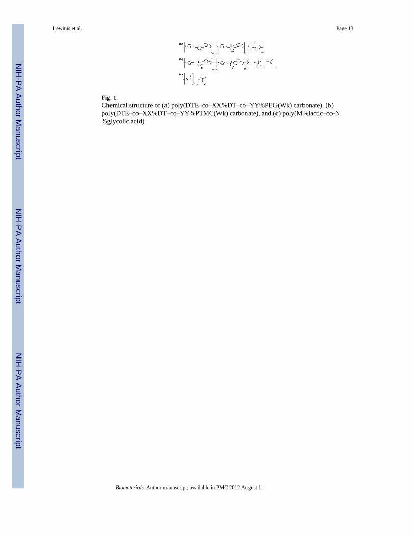
Fig. 1.Chemical structure of (a) poly(DTE–co–XX%DT–co–YY%PEG(Wk) carbonate), (b)poly(DTE–co–XX%DT–co–YY%PTMC(Wk) carbonate), and (c) poly(M%lactic–co-N%glycolic acid)
Lewitus et al. Page 13
Biomaterials. Author manuscript; available in PMC 2012 August 1.
NIH
-PA Author Manuscript
NIH
-PA Author Manuscript
NIH
-PA Author Manuscript
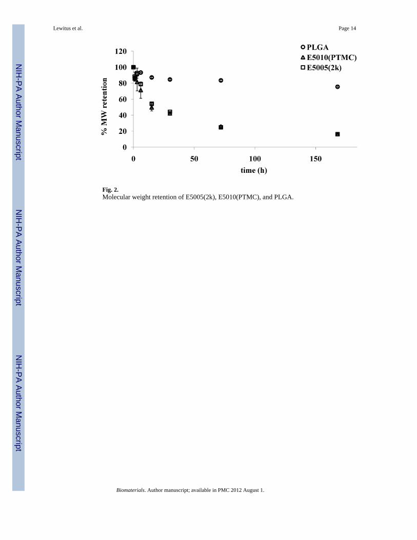
Fig. 2.Molecular weight retention of E5005(2k), E5010(PTMC), and PLGA.
Lewitus et al. Page 14
Biomaterials. Author manuscript; available in PMC 2012 August 1.
NIH
-PA Author Manuscript
NIH
-PA Author Manuscript
NIH
-PA Author Manuscript
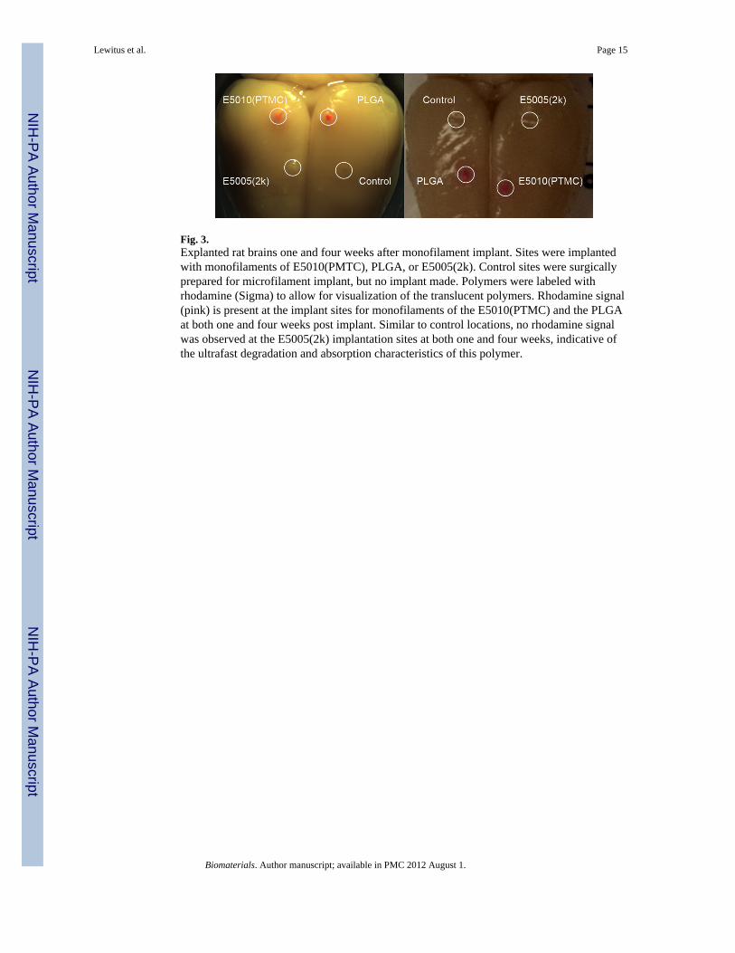
Fig. 3.Explanted rat brains one and four weeks after monofilament implant. Sites were implantedwith monofilaments of E5010(PMTC), PLGA, or E5005(2k). Control sites were surgicallyprepared for microfilament implant, but no implant made. Polymers were labeled withrhodamine (Sigma) to allow for visualization of the translucent polymers. Rhodamine signal(pink) is present at the implant sites for monofilaments of the E5010(PTMC) and the PLGAat both one and four weeks post implant. Similar to control locations, no rhodamine signalwas observed at the E5005(2k) implantation sites at both one and four weeks, indicative ofthe ultrafast degradation and absorption characteristics of this polymer.
Lewitus et al. Page 15
Biomaterials. Author manuscript; available in PMC 2012 August 1.
NIH
-PA Author Manuscript
NIH
-PA Author Manuscript
NIH
-PA Author Manuscript
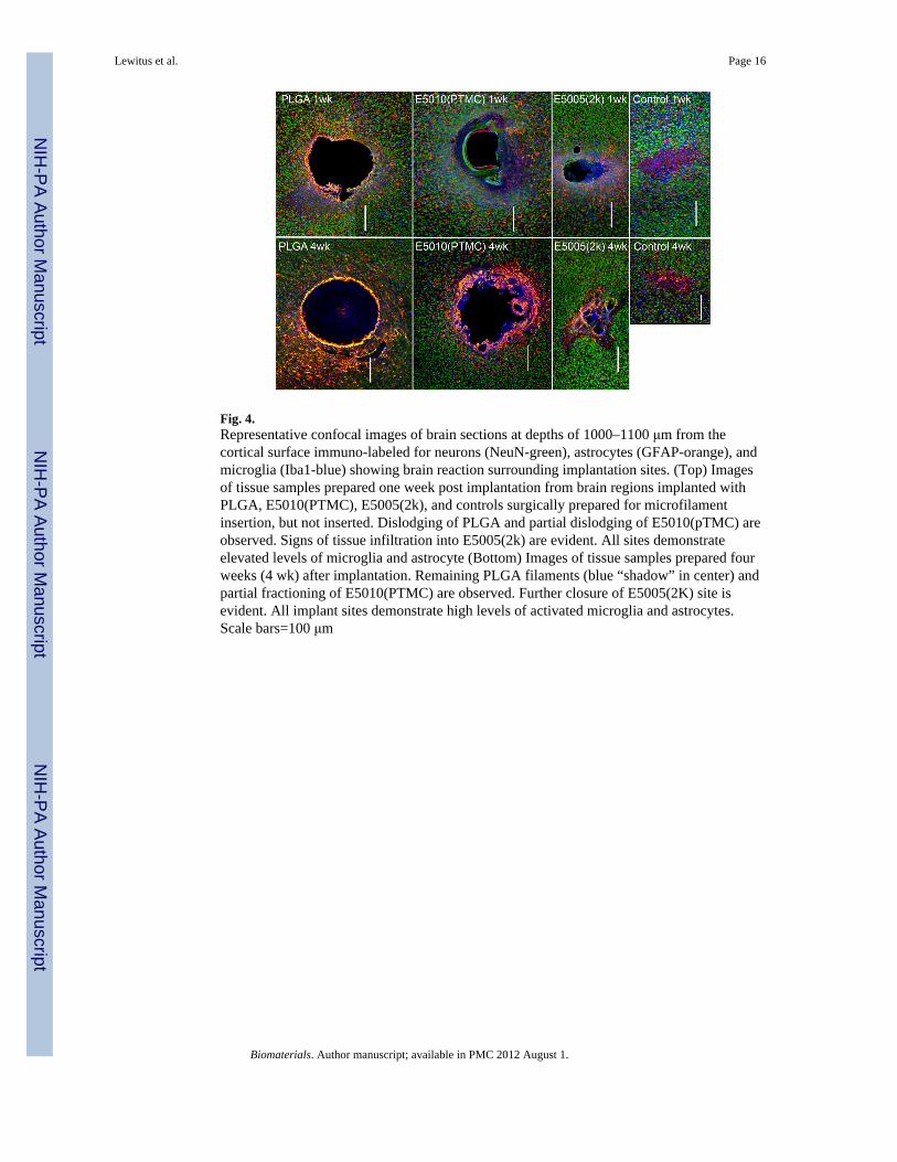
Fig. 4.Representative confocal images of brain sections at depths of 1000–1100 μm from thecortical surface immuno-labeled for neurons (NeuN-green), astrocytes (GFAP-orange), andmicroglia (Iba1-blue) showing brain reaction surrounding implantation sites. (Top) Imagesof tissue samples prepared one week post implantation from brain regions implanted withPLGA, E5010(PTMC), E5005(2k), and controls surgically prepared for microfilamentinsertion, but not inserted. Dislodging of PLGA and partial dislodging of E5010(pTMC) areobserved. Signs of tissue infiltration into E5005(2k) are evident. All sites demonstrateelevated levels of microglia and astrocyte (Bottom) Images of tissue samples prepared fourweeks (4 wk) after implantation. Remaining PLGA filaments (blue “shadow” in center) andpartial fractioning of E5010(PTMC) are observed. Further closure of E5005(2K) site isevident. All implant sites demonstrate high levels of activated microglia and astrocytes.Scale bars=100 μm
Lewitus et al. Page 16
Biomaterials. Author manuscript; available in PMC 2012 August 1.
NIH
-PA Author Manuscript
NIH
-PA Author Manuscript
NIH
-PA Author Manuscript
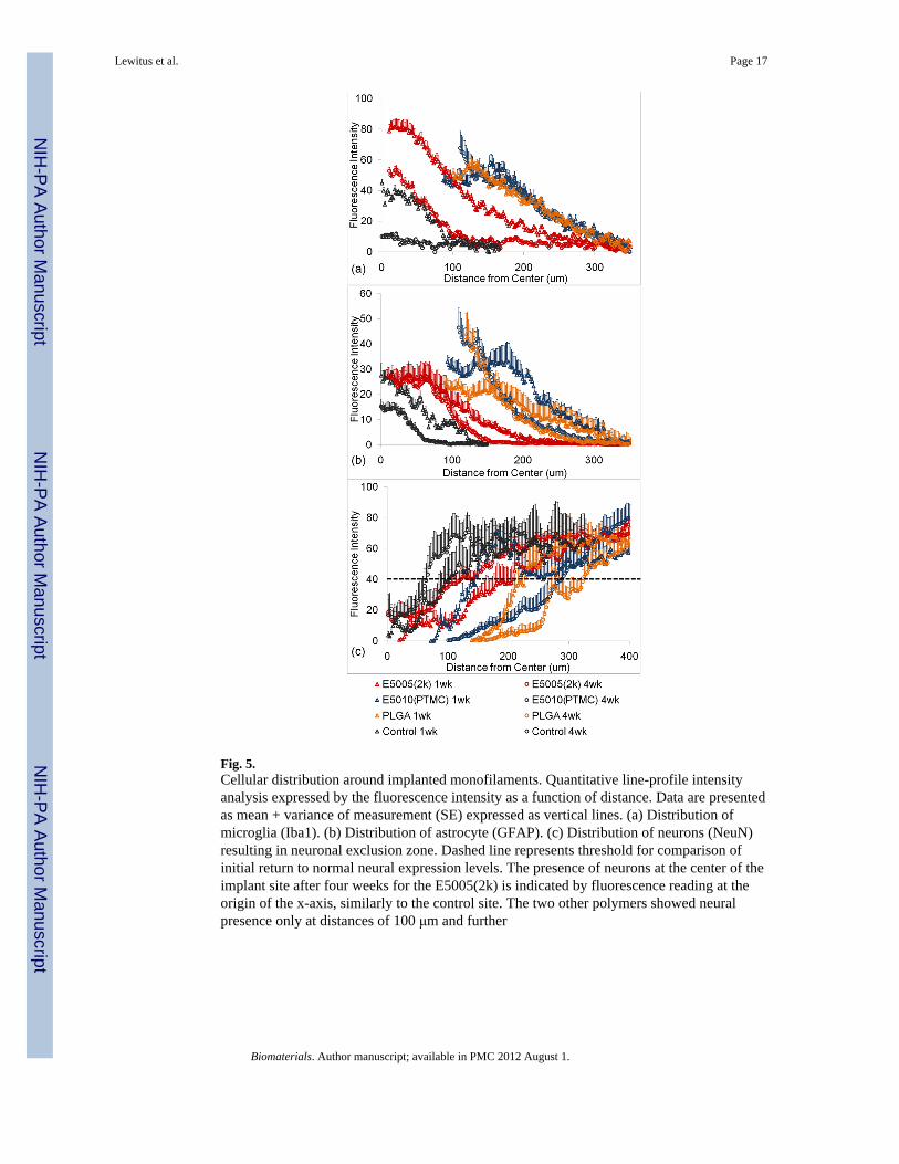
Fig. 5.Cellular distribution around implanted monofilaments. Quantitative line-profile intensityanalysis expressed by the fluorescence intensity as a function of distance. Data are presentedas mean + variance of measurement (SE) expressed as vertical lines. (a) Distribution ofmicroglia (Iba1). (b) Distribution of astrocyte (GFAP). (c) Distribution of neurons (NeuN)resulting in neuronal exclusion zone. Dashed line represents threshold for comparison ofinitial return to normal neural expression levels. The presence of neurons at the center of theimplant site after four weeks for the E5005(2k) is indicated by fluorescence reading at theorigin of the x-axis, similarly to the control site. The two other polymers showed neuralpresence only at distances of 100 μm and further
Lewitus et al. Page 17
Biomaterials. Author manuscript; available in PMC 2012 August 1.
NIH
-PA Author Manuscript
NIH
-PA Author Manuscript
NIH
-PA Author Manuscript
Related Documents