The Eye

The Eye. Iris: the muscle that adjusts the pupil to regulate the amount of light that enters the eye. Pupil: the aperture in the middle of the iris.
Dec 26, 2015
Welcome message from author
This document is posted to help you gain knowledge. Please leave a comment to let me know what you think about it! Share it to your friends and learn new things together.
Transcript

The Eye

Iris: the muscle that adjusts the pupil to regulate the amount of light that enters the eye.
Pupil: the aperture in the middle of the iris of the eye. The size of the aperture can be adjusted to control the amount of light
Lens: a transparent, bi-convex body situated behind the iris of the eye to focus an image on the retina
Retina: the innermost layer of the eye; contains
rods and cones, bipolar cells and ganglion cells

Sclera: the thick, white outer layer thatgives the eye its shape
Cornea: the clear part of the sclera at the front of the eye
Choroid layer: the middle layer of the eye, which absorbs light and prevents internal reflection. This layer forms the iris at the front of the eye
Rods: photoreceptors in the eye; more sensitive to light than cones, but unable to distinguish colour
Cones: colour receptors in the eye (red, green, blue) Fovea centralis: concentration of cones on the retina located directly behind the centre of the lens. Vision is the most acute here.
optic nerve: conducts information received from rods and cones to the brain for interpretation.
Blind spot: an area on the retina where there are no rods or cones present; locate where blood vessels enter the eye

As light enters the eye, the pupil will dilate if there isn’t enough light or it will constrict if there’s too much.
As well, the shape of the lens changes depending on how far away the object is.
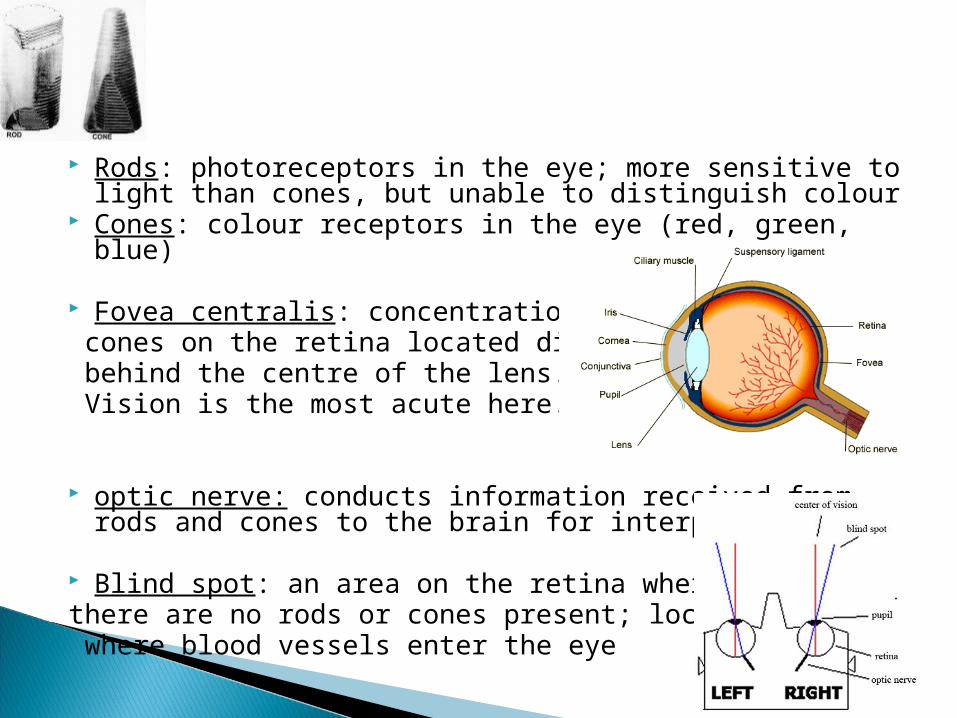
Accommodation: in the eye, adjustment that the ciliary body makes to the shape of the lens to focus on objects at varying distances
When the object is far away, the lens is flattened
When the object is close, the lens is rounded

Light enters the eye through the pupil. As it does, light rays become bent at the cornea and the lens in such a way that an inverted and reversed image of the object focuses on the retina.
Information from this image is captured by rods and cones, which transmit their info to bipolar cells and then ganglion cells (optic nerve). ◦ Cones transmit information to a single bipolar cell, but require more
light to become stimulated. As a result, cones see more detail and are best suited for lighted situations (daytime).
◦ Rods, however, are very sensitive to light and cannot distinguish color. As well, many rods connect to a single bipolar cell (up to 100 rods per bipolar cell). This causes images to be blurry. As a result, rods are best suited to situations where there isn’t much light and details are not important.

Cataracts- cloudy or opaque areas on the lens of the eye that increases in size over time and can lead to blindness if not medically treatment.
Glaucoma – build-up of the aqueous humor in the eye that irreversibly damages the nerve fibres responsible for peripheral Vision.

Myopia – near-sightedness, or difficulty in seeing things that are far away. The condition is caused by too strong ciliary muscles or a too-long eyeball
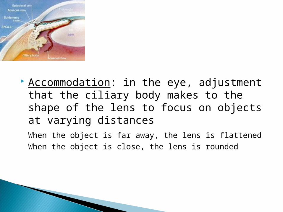
Hyperopia – far-sightedness, or difficulty in seeing near objects. This condition is caused by weak ciliary muscles or a too short eyeball
focus

Astigmatism – abnormality in the shape of the cornea or lens that results in uneven
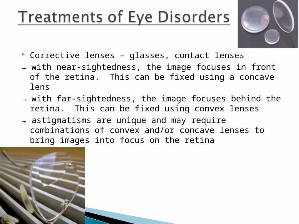
Corrective lenses – glasses, contact lenses→ with near-sightedness, the image focuses in front of the
retina. This can be fixed using a concave lens → with far-sightedness, the image focuses behind the retina.
This can be fixed using convex lenses→ astigmatisms are unique and may require combinations of
convex and/or concave lenses to bring images into focus on the retina
Laser surgery – two types→ Photorefractive keratectomy (PRK): non-
invasive, simple procedure→ LASIK surgery: more complex, some
surgery required (corneal)→ Both surgeries may diminish eyesight
Corneal transplant→ Corneas come from organ donors; no need
to match blood types→ Recovery long; most patients do well
though→ Recurrence of disease unusual

Vitreous humor-the transparent gelatinous substance filling the eyeball behind the lens
Aqueous humor- watery fluid that fills the space between the cornea and the lens in the eye.

The Ear
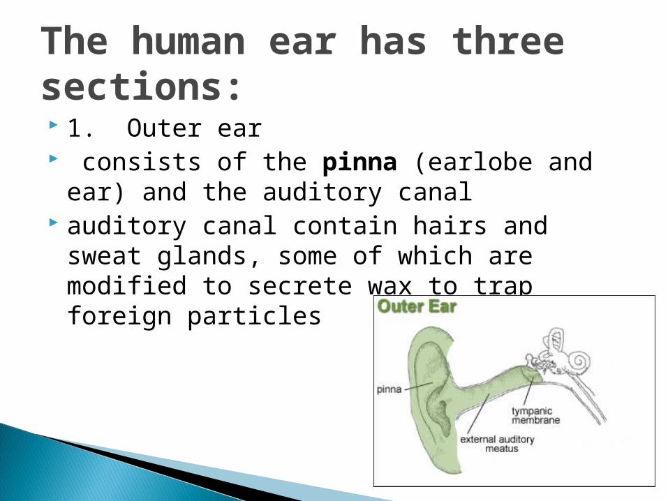
The human ear has three sections: 1. Outer ear consists of the pinna (earlobe and ear) and
the auditory canal auditory canal contain hairs and sweat
glands, some of which are modified to secrete wax to trap foreign particles

The human ear has three sections: Middle ear tympanic membrane: the eardrum; a
membrane of thin skin and fibrous tissue that vibrates in response to sound waves.
ossicles: the group of three small bones between the eardrum and the oval window of the middle ear; transmit sound waves from the eardrum to the inner ear
malleus - hammer incus – anvil stapes – stirrup

Eustachian tube: bony passage extending from the middle ear to the nasopharynx that plays a role in equalizing air pressure on both sides of the eardrum.
Yawning can cause the air to move throughthe tubes and the ear will “pop”

The human ear has three sections:
3. Inner Ear Vestibule: involved in balance and
equilibrium Semicircular canals: three tubes involved in
balance and equilibrium

Cochlea: involved in hearing. A spiral-shaped cavity of the inner ear that resembles a snail shell and contains nerve endings essential for hearing.
Vibrations from the stapes bone hits the oval window, which causes it to vibrate.
The oval window then pushes on the fluid within the vestibular canal of the cochlea.
The movement of the fluid within the cochlea places pressure on the basilar membrane causing it to move. When it moves it causes the hair cells attached to it to move.
The hair cells stimulate the tectorial membrane causing it to generate an impulse. The impulse travels to theauditory nerve where it is sent to the brain.

How the ear works… PINNA-Funnel-shaped flap that directs sound waves into the
auditory canal AUDITORY CANAL-Canal that carries sound waves to the eardrum TYPANIC MEMBRANE- A thin membrane that is vibrated by sound
waves OSSICLES – Three bones () They are the smallest bones in the
human body. The eardrum vibrates, and this vibrates the hammer, than the anvil and the stirrup one after another The stirrup then vibrates the Oval Window COCHLEA- This is a spirally coiled tube containing fluid and the
actual organ of hearing (the Organ of Corti). Each Organ of Corti contains thousands of sensitive hairs that are vibrated by sound waves. The hairs initiate nervous impulses in the
Auditory Nerve which carries messages to the brain

Disorders of the Auditory System Nerve Deafness
◦ caused by damage to hair cells in the spiral organ◦ typically found with aging and cannot be reversed◦ hearing loss uneven, some frequencies more
affected than others
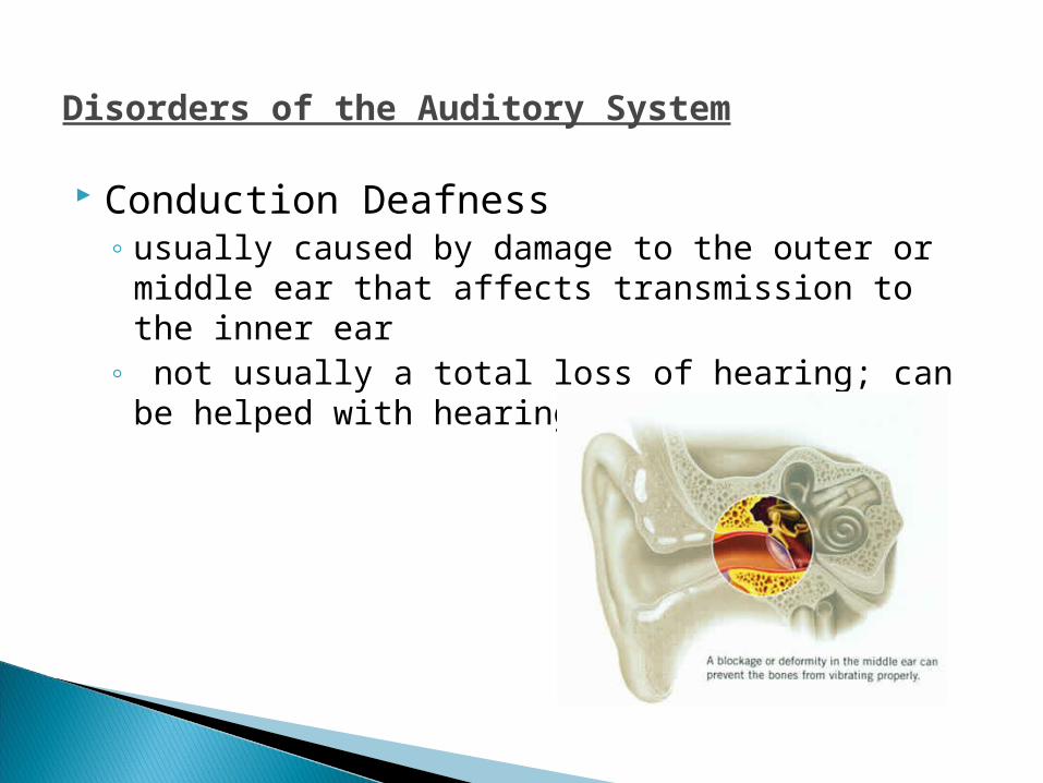
Disorders of the Auditory System Conduction Deafness
◦ usually caused by damage to the outer or middle ear that affects transmission to the inner ear
◦ not usually a total loss of hearing; can be helped with hearing aids
Disorders of the Auditory System Ear Infections
◦ caused by fluid build-up behind the eardrums, common in children
◦ fluid builds up because of the shallow angle of the auditory tube

Treating Auditory Disorders Hearing Aids
Eustachian tube implants◦ also called tympanostomy tube surgery; used to
treat infections◦ tiny plastic tubes are placed in a slit in the
eardrum, relieving the pressure from the built-up fluid and allowing in to drain

Filler Cholinesterase- an enzyme, found in the heart, brain, and blood, that breaks down acetylcholine to acetic acid and choline. If the acetylcholine did not break down then it would remain in the synapse.


Cochlea: involved in hearing. A spiral-shaped cavity of the inner ear that resembles a snail shell and contains nerve endings essential for hearing.
Vibrations from the stapes bone hits the oval window, which causes it to vibrate.
The oval window then pushes on the fluid within the vestibular canal of the cochlea.
The movement of the fluid within the cochlea places pressure on the basilar membrane causing it to move. When it moves it causes the hair cells attached to it to move.
The hair cells stimulate the tectorial membrane causing it to generate an impulse. The impulse travels to theauditory nerve where it is sent to the brain.
How the ear works… PINNA-Funnel-shaped flap that directs sound waves into the
auditory canal AUDITORY CANAL-Canal that carries sound waves to the eardrum TYPANIC MEMBRANE- A thin membrane that is vibrated by sound
waves OSSICLES – Three bones () They are the smallest bones in the
human body. The eardrum vibrates, and this vibrates the hammer, than the anvil and the stirrup one after another The stirrup then vibrates the Oval Window COCHLEA- This is a spirally coiled tube containing fluid and the
actual organ of hearing (the Organ of Corti). Each Organ of Corti contains thousands of sensitive hairs that are vibrated by sound waves. The hairs initiate nervous impulses in the
Auditory Nerve which carries messages to the brain
ATP- adenosine triphosphate: serves as a source of energy used to power the Na/K pump.
Glucose and oxygen and required for cellular respiration to obtain energy.
CNS cannot regenerate. Stroke- lack of oxygen to the brain.
May use blood clotting drugs, which need to be taken within at least 3 hrs. Asprin reduces the thickness of platelets, so it decreases clots form forming.
However if the blood is thinned and an aneurysm (broken blood vessel) occurs then the bleeding can be worst.
Spinal cord injury- Damage is usually permanent and can lead to paralysis(a loss or impairment of voluntary movement in a body part).
Found a gene that inhibits spinal regeneration. A protein that prevents tissue growth is now being researched.
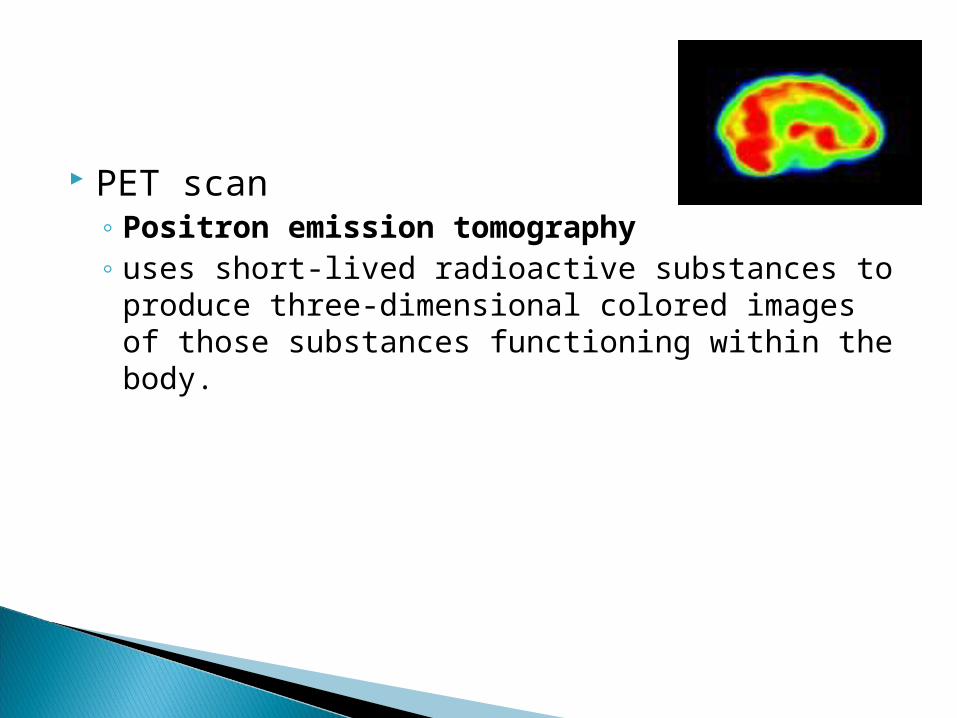
PET scan◦ Positron emission tomography ◦ uses short-lived radioactive substances to
produce three-dimensional colored images of those substances functioning within the body.
Related Documents