THE EMERGENCE OF CRYPTOCOCCUS GATTII IN BRITISH COLUMBIA: VETERINARY ASPECTS A Thesis Submitted to the College of Graduate Studies and Research in Partial Fulfillment of the Requirements for the Degree of Masters of Science in the Department of Large Animal Clinical Studies University of Saskatchewan Saskatoon By Colleen Duncan © Colleen Duncan, June 2005. All rights reserved.

Welcome message from author
This document is posted to help you gain knowledge. Please leave a comment to let me know what you think about it! Share it to your friends and learn new things together.
Transcript
THE EMERGENCE OF CRYPTOCOCCUS GATTII
IN BRITISH COLUMBIA:
VETERINARY ASPECTS
A Thesis Submitted to the College of
Graduate Studies and Research
in Partial Fulfillment of the Requirements
for the Degree of Masters of Science
in the Department of Large Animal Clinical Studies
University of Saskatchewan
Saskatoon
By
Colleen Duncan
© Colleen Duncan, June 2005. All rights reserved.
Permission to use:
In presenting this thesis in partial fulfillment of the requirements for a Postgraduate
degree from the University of Saskatchewan, I agree that the Libraries of this University
may make it freely available for inspection. I further agree that permission for copying
this thesis in any manner, in whole or in part, for scholarly purposes may be granted by
the professor or professors who supervised my thesis work or, in their absence, by the
Head of the Department of the Dean of the College in which my thesis work was done. It
is understood that any copying, publication, or use of this thesis or parts thereof for
financial gain shall not be allowed without my written permission. It is also understood
that due recognition shall be given to me and the University of Saskatchewan in any
scholarly use which may be made of any material in my thesis.
Requests for permission to copy or to make other use of material in this thesis in whole or
in part should be addressed to:
Head of the Department of Large Animal Clinical Sciences
University of Saskatchewan
52 Campus Drive
Saskatoon, Saskatchewan
S7N 5B4
ii
Abstract
A series of presumed or confirmed Cryptococcus gattii cases diagnosed between 1999
and 2003 was compiled through review of records from veterinary laboratories and
human diagnostic services. There was a continual increase in the annual number of
animal, but not human, cases diagnosed; no seasonality was observed. Animal cases
exceeded human cases by almost 75% even though it was hypothesized that animal cases
are more likely to go undiagnosed or unreported when compared to humans. Animal
cryptococcosis cases were identified on Vancouver Island prior to 1999 suggesting the
organism may have emerged in the region prior to its identification as a causative agent
for human disease; therefore animals may serve as a good sentinel for human
cryptococcosis infection.
There were 50% more feline than canine cases and disease appeared more commonly in
middle aged cats and younger dogs. There was no sex predilection for either species.
The primary system involved was most commonly respiratory, followed by central
nervous system (CNS) in both cats and dogs. There was a higher proportion of CNS
disease in dogs relative to cats, and cats were much more likely to have subcutaneous or
dermal masses relative to dogs. Multivariate survival analysis identified only the
presence of neurological symptoms as a statistically significant predictor of mortality;
those animals exhibiting CNS symptoms were over four times more likely to die than
those never showing neural signs. A case-control study identified host and environmental
risk factors for clinical C. gattii infection in dogs and cats suggesting that where an
infectious agent is not uniformly distributed, individual risk increases when the organism
iii
is re-distributed through large scale environmental disturbance, or when the animal has
increased opportunities for exposure through travel or activity level.
Serum samples and material for fungal culture were collected from dogs, cats, horses and
terrestrial mammal species residing within the region where clinical cases had been
diagnosed. Nasal colonization was identified in squirrels (Sciurus carolinensis), horses,
dogs and cats. Most of the animals sampled had no signs of systemic infection however
asymptomatic infection, defined as the presence of cryptococcal antigen in the
bloodstream in the absence of clinical symptoms, was identified in a small number of
dogs and cats. Fourteen months of follow-up testing of asymptomatic animals revealed
that animals can progress to clinical disease, remain sub-clinically infected, or clear the
organism.
iv
Acknowledgements
This project would not have been possible without the support of the veterinary
community of British Columbia. Private practitioners diagnosed and reported cases,
shared medical records, hosted me at their clinics and provided the impetus for this
research. Specialists contributed their skills and shared ideas that directed study and gave
tremendous insight into the project. While there remain many unanswered questions
surrounding cryptococcosis in BC I hope this research provides some baseline
information on the disease and will serve as a starting point upon which to base further
investigations.
Dr. John Campbell, my supervisor, made epidemiology stand out as an obvious vocation,
provided me with the opportunity to pursue graduate training and facilitated what turned
out to be a very cool, and educational, project. His encouragement to do field based
research, infallible support and yet fantastically laid back attitude was the best
environment I could ever have to learn in. Dr. Craig Stephen has been an unofficial co-
supervisor and made the MSc experience far more than a graduate degree. Over coffee
and doughnuts I learned to think and not just regurgitate, challenge ideas and not just
accept the status quo and, most importantly, that you really can make a difference if you
stand up for what you believe in. Thanks also to my other committee members, Dr. Gary
Wobeser and Dr. Terry Carruthers who provided support with the development of the
project and final drafts.
v

Financial support for this study was provided by the University of Saskatchewan inter-
provincial research fellowship, Companion Animal Health Fund, Equine Health Research
Fund, Wildlife Health Fund, Center for Coastal Health and the Central Laboratory for
Veterinarians Ltd.
vi
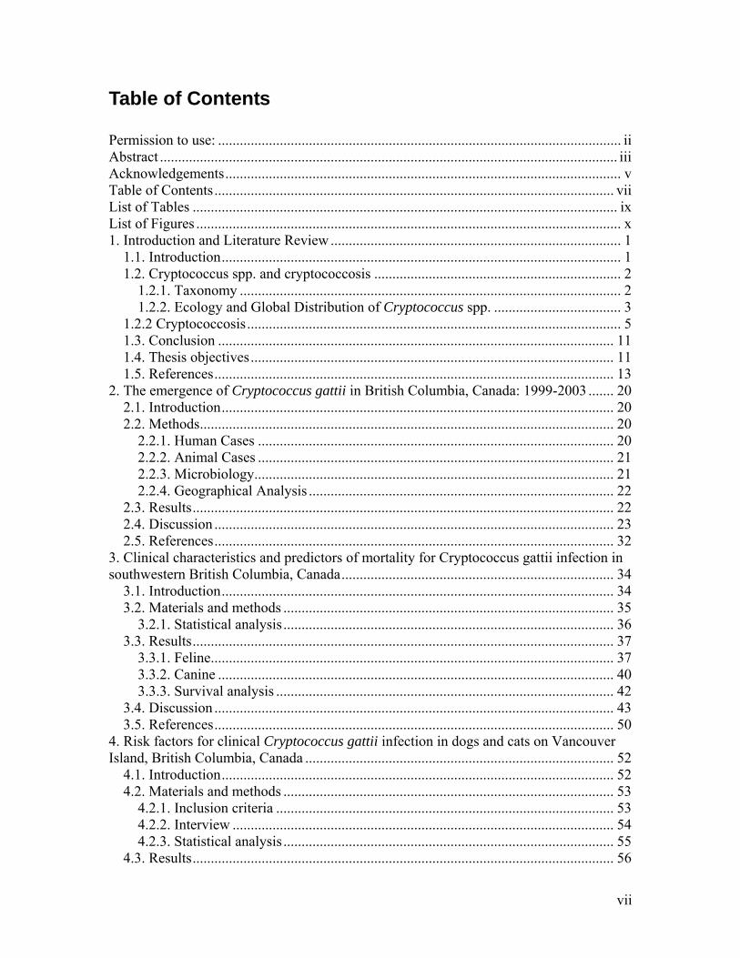
Table of Contents
Permission to use: ............................................................................................................... ii Abstract .............................................................................................................................. iii Acknowledgements............................................................................................................. v Table of Contents.............................................................................................................. vii List of Tables ..................................................................................................................... ix List of Figures ..................................................................................................................... x 1. Introduction and Literature Review ................................................................................ 1
1.1. Introduction.............................................................................................................. 1 1.2. Cryptococcus spp. and cryptococcosis .................................................................... 2
1.2.1. Taxonomy ......................................................................................................... 2 1.2.2. Ecology and Global Distribution of Cryptococcus spp. ................................... 3
1.2.2 Cryptococcosis....................................................................................................... 5 1.3. Conclusion ............................................................................................................. 11 1.4. Thesis objectives.................................................................................................... 11 1.5. References.............................................................................................................. 13
2. The emergence of Cryptococcus gattii in British Columbia, Canada: 1999-2003 ....... 20 2.1. Introduction............................................................................................................ 20 2.2. Methods.................................................................................................................. 20
2.2.1. Human Cases .................................................................................................. 20 2.2.2. Animal Cases .................................................................................................. 21 2.2.3. Microbiology................................................................................................... 21 2.2.4. Geographical Analysis .................................................................................... 22
2.3. Results.................................................................................................................... 22 2.4. Discussion .............................................................................................................. 23 2.5. References.............................................................................................................. 32
3. Clinical characteristics and predictors of mortality for Cryptococcus gattii infection in southwestern British Columbia, Canada........................................................................... 34
3.1. Introduction............................................................................................................ 34 3.2. Materials and methods ........................................................................................... 35
3.2.1. Statistical analysis ........................................................................................... 36 3.3. Results.................................................................................................................... 37
3.3.1. Feline............................................................................................................... 37 3.3.2. Canine ............................................................................................................. 40 3.3.3. Survival analysis ............................................................................................. 42
3.4. Discussion .............................................................................................................. 43 3.5. References.............................................................................................................. 50
4. Risk factors for clinical Cryptococcus gattii infection in dogs and cats on Vancouver Island, British Columbia, Canada ..................................................................................... 52
4.1. Introduction............................................................................................................ 52 4.2. Materials and methods ........................................................................................... 53
4.2.1. Inclusion criteria ............................................................................................. 53 4.2.2. Interview ......................................................................................................... 54 4.2.3. Statistical analysis ........................................................................................... 55
4.3. Results.................................................................................................................... 56
vii
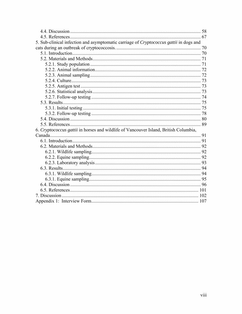
4.4. Discussion .............................................................................................................. 58 4.5. References.............................................................................................................. 67
5. Sub-clinical infection and asymptomatic carriage of Cryptococcus gattii in dogs and cats during an outbreak of cryptococcosis. ....................................................................... 70
5.1. Introduction............................................................................................................ 70 5.2. Materials and Methods........................................................................................... 71
5.2.1. Study population ............................................................................................. 71 5.2.2. Animal information......................................................................................... 72 5.2.3. Animal sampling............................................................................................. 72 5.2.4. Culture............................................................................................................. 73 5.2.5. Antigen test ..................................................................................................... 73 5.2.6. Statistical analysis ........................................................................................... 73 5.2.7. Follow-up testing ............................................................................................ 74
5.3. Results.................................................................................................................... 75 5.3.1. Initial testing ................................................................................................... 75 5.3.2. Follow-up testing ............................................................................................ 78
5.4. Discussion .............................................................................................................. 80 5.5. References.............................................................................................................. 89
6. Cryptococcus gattii in horses and wildlife of Vancouver Island, British Columbia, Canada............................................................................................................................... 91
6.1. Introduction............................................................................................................ 91 6.2. Materials and Methods........................................................................................... 92
6.2.1. Wildlife sampling............................................................................................ 92 6.2.2. Equine sampling.............................................................................................. 92 6.2.3. Laboratory analysis......................................................................................... 93
6.3. Results.................................................................................................................... 94 6.3.1. Wildlife sampling............................................................................................ 94 6.3.1. Equine sampling.............................................................................................. 95
6.4. Discussion .............................................................................................................. 96 6.5. References............................................................................................................ 101
7. Discussion ................................................................................................................... 102 Appendix 1: Interview Form.......................................................................................... 107
viii
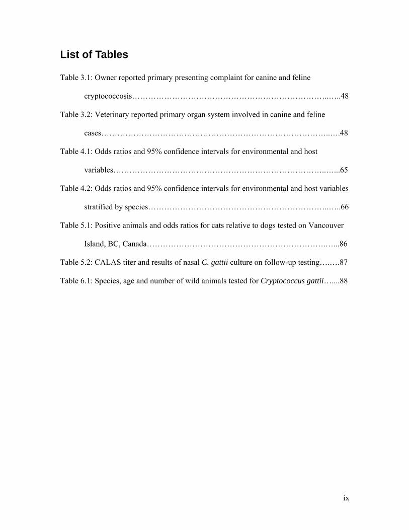
List of Tables
Table 3.1: Owner reported primary presenting complaint for canine and feline
cryptococcosis………………………………………………………………..…..48
Table 3.2: Veterinary reported primary organ system involved in canine and feline
cases…………………………………………………………………………..….48
Table 4.1: Odds ratios and 95% confidence intervals for environmental and host
variables……………………………………………………………………..…...65
Table 4.2: Odds ratios and 95% confidence intervals for environmental and host variables
stratified by species…………………………………………………………..…..66
Table 5.1: Positive animals and odds ratios for cats relative to dogs tested on Vancouver
Island, BC, Canada………………………………………………………….…...86
Table 5.2: CALAS titer and results of nasal C. gattii culture on follow-up testing….….87
Table 6.1: Species, age and number of wild animals tested for Cryptococcus gattii…....88
ix
List of Figures
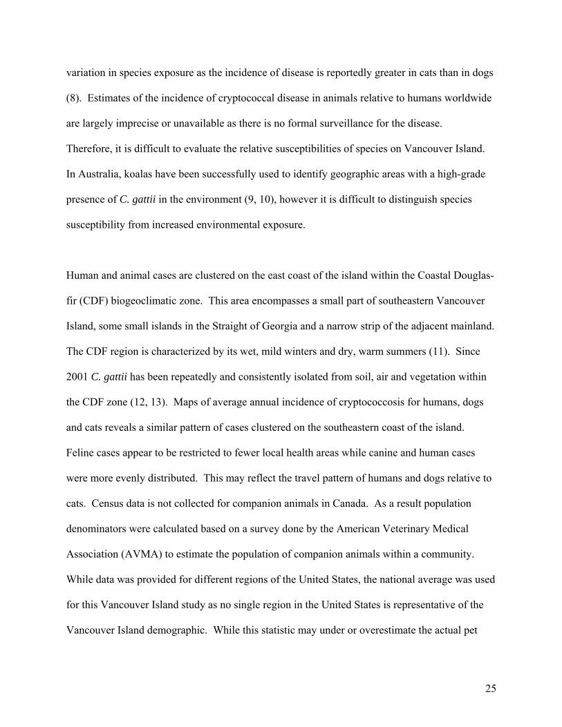
Figure 2.1: Confirmed and probable C. gattii cases by animal species on Vancouver
Island from January 1999 to December 2003……………….………………..….28
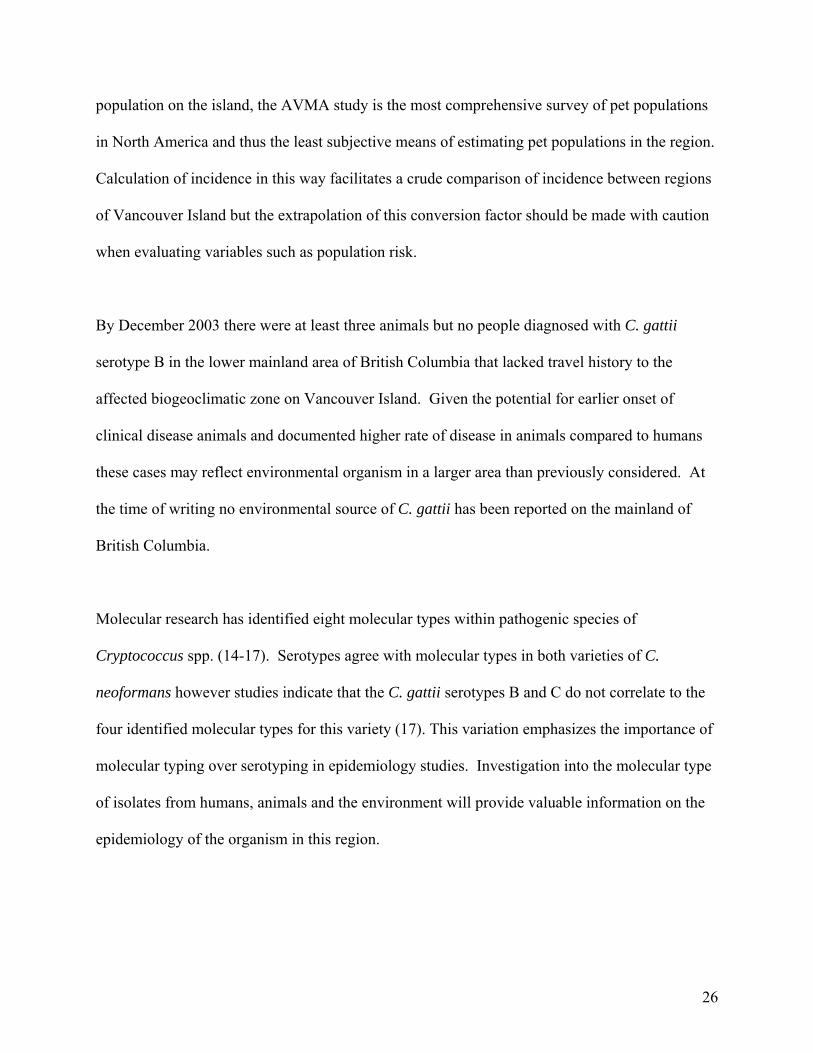
Figure 2.2: Confirmed and probable human and animal C. gattii cases on Vancouver
Island by year from January 1999 to December 2003 .........................….…..…..28
Figure 2.3: Confirmed and probable human and animal C. gattii cases on Vancouver
Island by month from January 1999 to December 2003………..…………..……29
Figure 2.4: Average annual incidence of human cases per 100,000 people by local health
area on Vancouver Island, BC…………………………………………...….…...29
Figure 2.5: Average annual incidence of canine cases per 100,000 by local health area on
Vancouver Island, BC…………………………………………………………....30
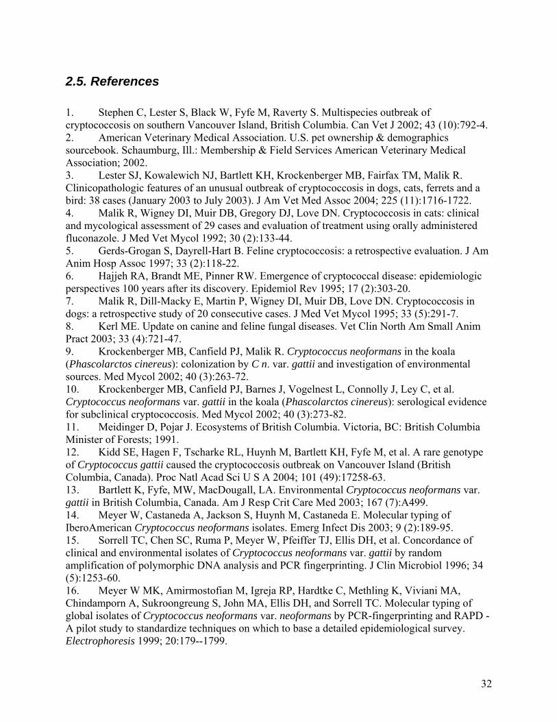
Figure 2.6: Average annual incidence of feline cases per 100,000 by local health area on
Vancouver Island, BC……………………………………………………...….....31
Figure 3.1: Cumulative survival for animals with and without central nervous system
symptoms……………………………………………………………………...…49
Figure 5.1: Location of sampling clinics (clear circles) and distribution of the Coastal
Douglas Fir Biogeoclimatic zone on Vancouver Island, BC, Canada…………...88
x
1. Introduction and Literature Review
1.1. Introduction
The term ‘emerging infectious disease’ (EID) is used to describe the expansion of a known
pathogen to new host species and/or geographic range, or recent identification of a new
infectious agent. Emerging infections have been well documented in human medicine (1) and
are increasingly identified in domestic and wild animals (2, 3) as well as agricultural and wild
plant species. Emerging infectious diseases may impact populations locally, regionally or
globally and effects vary according to relevant host, agent and environment interactions.
Changes in human ecology are a central force influencing EIDs; the emergence of pathogens
within different cohorts of biota is fundamentally driven by different forms of environmental
anthropogenic change (2). Causal factors implicated in emergence include changes in human
behavior patterns, social organization, demographics, movement, industry and land use in
conjunction with microbial adaptation that disrupts the host parasite relationship equilibrium
such that the parasite is favored (2, 4).
In 2001 the public health authorities and veterinary community of southwestern British
Columbia, Canada recognized an increased incidence of human and animal cryptococcosis (5).
The cases were largely restricted to Vancouver Island and most isolates were C. gattii serotype
B; a species classically restricted to tropical and sub-tropical regions of the world. The
appearance of C. gattii in Canada constitutes an EID as the known pathogen has surfaced in a
new geographic region.
1
1.2. Cryptococcus spp. and cryptococcosis
1.2.1. Taxonomy
Cryptococcus spp. are environmental fungi of the phylum Basidiomycota, class
Heterobasidiomycetes, order Filobasidiales, family Filobasidiaceae (6). The genus Cryptococcus
includes over 37 species however only C. neoformans was commonly considered to be
pathogenic. There were previously three recognized varieties of Cryptococcus neoformans: C.
neoformans var. grubii (serotype A), C. neoformans var. neoformans (serotype D) and C.
neoformans var. gattii (serotypes B and C) as well as a hybrid of C. neoformans var. grubii and
C. neoformans var. neoformans (serotype AD) (7-9). Serotypes are based on the antigenicity of
the capsular polysaccharides. Recently proposed changes to the nomenclature suggest that C.
neoformans should be divided into two distinct species including C. neoformans (serotypes A, D
and AD) and C. gattii (serotypes B and C) based on molecular and mating type characteristics
(10). This nomenclature is now widely accepted and will be used here.
Advances in DNA typing methods have led to the identification of eight molecular types based
on polymerase chain reaction (PCR) fingerprinting, random amplified polymorphic DNA
analysis and restriction fragment length polymorphism (RFLP) (11-13). Serotypes are consistent
with the molecular types of C. neoformans var. grubii (serotype A) and C. neoformans var.
neoformans (serotype D) where serotype A is made up of molecular types VNI and VNII and
serotype D equates to VNIV. Serotype AD, the hybrid of C. neoformans var. grubii and C.
neoformans var. neoformans, corresponds to VNIII. In contrast C. gattii is comprised of 4
different molecular types, VGI, VGII, VGIII, and VGIV, which do not correspond well with the
delineations of serotypes B and C (14).
2
1.2.2. Ecology and Global Distribution of Cryptococcus spp.
1.2.2.1. Cryptococcus neoformans
The ecology of C. neoformans var. grubii and C. neoformans var. neoformans are quite similar;
as C. neoformans var. grubii was only proposed as a distinct variety, separate from C.
neoformans var. neoformans, in 1999 it is often difficult to discern differing distributions of the
two varieties in the literature (15). Historically both varieties of C. neoformans were thought to
be associated with avian excreta, particularly that of pigeons (16-22). It was hypothesized that
birds feeding on an unidentified host plant could carry the organism in their gastrointestinal tract
and act as a vector by dispersing yeast cells in their feces. The birds were thought to not be
clinically affected because their body temperature was above that required for replication of the
fungi (23, 24). This hypothesis was challenged by the isolation of both varieties from living and
decaying vegetation and domestic dust worldwide (20, 21, 25, 26). Given that avian excreta is
rich in creatinine and other chemical constituents that promote fungal replication, it is likely that
the environmental niche of the fungus is vegetation but that it is easily isolated from avian
excreta because it provides a good media for growth (21, 24).
C. neoformans var. grubii has a global distribution and is the most common cause of human
fungal meningitis in immunocompromised hosts worldwide (22, 27-32). C. neoformans var.
neoformans (serotypes D and AD) is less commonly recovered from the environment or clinical
cases and appear to be more prevalent in Europe than other parts of the world (32, 33).
3
1.2.2.2 Cryptococcus gattii
Cryptococcus gattii has classically been restricted to the tropics and sub tropics of the world (6,
32). The first environmental isolation of the organism was from eucalyptus trees (Eucalyptus
camaldulensis and E. tereticornis) in Australia (34, 35). Subsequently C. gattii has been
recovered from material associated with eucalypt species in many other parts of the world
including California, India and Brazil (20, 28, 36, 37) and in non-eucalypt tree species from
tropical and subtropical areas worldwide (28, 38-40). Most environmental isolates of C. gattii
have been serotype B, however there are reports of serotype C isolated from almond trees
(Terminallia catappa) in Columbia and vegetation in southern California (37,40). It has been
proposed that dispersal of infectious propagules, the asexual budding yeast or the sexual
basidiospores, is linked to the flowering of the eucalypt trees as airborne organisms had, prior to
the collection of Canadian environmental isolates, only been detected under a tree in flower (23).
Some studies suggest that alternative environmental sources of C. gattii have yet to be
identified, as molecular types isolated from clinical and environmental samples have been
different in western and northern Australia (12, 41, 42) and clinical cryptococcosis caused by C.
gattii has been reported from many regions where an environmental source cannot be discerned,
including parts of Australia, Africa and Papua New Guinea (12, 41, 43-45).
The regional distribution of human disease caused by C. gattii corresponds largely with the
distribution of environmental isolates (6, 29, 34). In an exhaustive study of worldwide human
clinical cryptococcosis, isolates of C. gattii were not found in Austria, Belgium, Denmark,
France, Germany, Holland, Italy, Switzerland, and Japan but were identified at an unusually high
4
prevalence in Australia, Brazil, Cambodia, Hawaii, southern California, Mexico, Paraguay,
Thailand, Vietnam, Nepal and Central Africa (32). Follow up studies have confirmed the high
prevalence of C. gattii in tropical and sub-tropical regions including Brazil (27, 28), Thailand
(31), Papua New Guinea (43), Venezuela (46), South Africa and Mexico (46). Small numbers of
human C. gattii cases have been reported from India, China, Taiwan, Peru , Argentina, Rwanda,
Italy (6, 22, 30, 47, 48). Cases reported from Europe and non-endemic areas of North America
are thought to have been acquired elsewhere (6, 32, 49).
In North America C. gattii is considered to be a rarity (32). The majority of isolates have been
serotype B (50, 51) with the exception of southern California where serotype C is more prevalent
(32) and has been isolated from the environment (37). In Canada, cryptococcosis has been
reported from most provinces, is classically associated with immunosuppression and caused by
C. neoformans (52). Cryptococcus gattii has been isolated once from an AIDS patient in Quebec
suffering with cryptococcosis (53) however this patient had a travel history to a region where C.
gattii is considered endemic (37).
1.2.2 Cryptococcosis
Cryptococcosis affects humans and animals worldwide and can be caused by C. neoformans or
C. gattii. While the exact mode of infection is unknown, it is widely accepted to be through
inhalation of air-borne organism (23, 54). Cryptococcus neoformans has been isolated from the
nasal passages of dogs, cats (55) and koalas (56) in Australia without evidence of infection
suggesting asymptomatic colonization of the nasal mucosa following environmental exposure. It
is not clear what triggers tissue invasion after colonization (6). Direct inoculation has been
reported in humans and experimentally in animals (57, 58). Zoonotic transmission was proposed
5
in one instance in which the same molecular strain was isolated from both human and bird;
however, this may represent shared environmental exposure (59). The nature of the infectious
propagule is hypothesized to be the basidiospore or desiccated yeast cells. Upon entry into tissue
the desiccated cell becomes rehydrated and acquires a thick polysaccharide capsule,
basidiospores convert to encapsulated blastoconidia (23).
Clinical disease is dictated by host characteristics and the variety of infecting organism. C.
neoformans is isolated most commonly from immunosuppressed individuals (29, 60). In contrast
C. gattii is a primary pathogen as it tends to infect immunocompetent hosts. Even in areas where
the organism is endemic, C. gattii is rarely isolated as the cause of cryptococcosis in AIDS
patients (29, 60, 61).
Exposure to environmental sources of the organism is hypothesized to be the primary risk factor
for clinical disease. In Australia, the aboriginal populations living in rural and semi-rural areas
have a high incidence of cryptococcosis caused by C. gattii and further investigation suggests
that this is due to a close association with eucalyptus trees (29). Men in Australia and Papua
New Guinea were at an increased risk of infection with C. gattii, presumably because of
increased contact with the organism in the environment (29, 62). Isolation of C. neoformans var.
grubii from a home was a significant risk factor for AIDS patients developing cryptococcosis
(21).
1.2.2.1 Animal Cryptococcosis
Clinical cryptococcosis has been reported in many domestic and wild animal species worldwide.
The variety of infecting organism is often not identified due to financial constraints or the
6
assumption that isolates are geographically restricted and that the agent can be assumed. There
are, however, differences in the clinical presentation of the different varieties that are important
to recognize. These differences facilitated the identification of C. gattii in British Columbia.
Cryptococcosis is the most common systemic fungal infection in cats (54, 63, 64). Disease is
most frequently reported in middle aged cats, but the age range is broad (65). Males have been
reported to be affected more commonly than females, with the suggestion that males are more
likely to be exposed for behavioral reasons (66-68). Other studies found no sex predisposition
(65). Siamese cats appeared to be over represented in one Australian study (67). Contrary to the
hypothesis that environmental exposure may be a principal risk factor, the disease is frequently
reported in ‘indoor only’ cats (65, 66).
Some seasonality in feline clinical cases has been reported. In Australia there was an observed
tendency for cats to present to veterinarians in the summer (67). In one study in the USA, cats
presenting to the clinic with cryptococcosis were more likely to be outdoor cats in the warm
seasons but strictly indoor cats in the cold seasons (66).
The three clinical syndromes most commonly reported for feline cryptococcosis include upper
respiratory tract disease, dermatomycosis, and meningitis (67-69). Of the three, upper
respiratory tract infection, specifically nasal cavity disease, is most commonly observed.
Clinical signs may include nasal or facial deformity, sneezing, nasal discharge, respiratory noise
or coughing (65, 67, 69). Respiratory signs are often accompanied by mandibular lymph node
involvement (67). Lower respiratory infection is not a common presentation in the cat.
7
Infection may remain localized to the nasal cavity and sinuses or penetrate through the cribriform
plate to the central nervous system where it can cause meningitis. Clinical signs of feline
cryptococcal meningitis include depression, ataxia, paresis, coma, lumbar pain, behavior
changes, vestibular signs and seizures (65, 66, 70). Central nervous symptoms in cats are often
not a primary presenting sign but secondary to respiratory infection (67). Other non-specific
signs including weight loss, anorexia and lethargy are also reported in the cat (65, 66, 69).
Infection from the nasal passages may disseminate hematogenously, often presenting as
cutaneous lumps or ocular lesions. Cutaneous and subcutaneous disease may involve single or
multiple nodules anywhere on the body and has been reported both as dissemination and as a
primary lesion (65-68, 71). Ocular lesions commonly include chorioretinitis from hematogenous
dissemination or optic nerve meningitis causing blindness (63).
The role of immunosuppression in feline cryptococcosis has been debated. In an Australian
case series of 29 cats, the prevalence of FIV in cases was equivalent to that in the hospital
population, however, animals with both infections appeared to have more severe disease (67). In
a study in the USA, 21% of cryptococcosis cases had concurrent FIV or FeLV infection,
compared with 1.4% in the general hospital population. Immunosuppressed cats were more
seriously affected (66). Response to therapy has been less successful in immunosuppressed cats
in both Australia and the USA (67, 68). Another American case series found cryptococcosis
cases with concurrent FeLV or FIV infection to have a less successful treatment outcome (65).
An examination of FIV positive and negative cats in the USA found C. neoformans more
8
commonly in the oropharynx of FIV seropositive cats although no cats had signs of clinical
disease (72).
Canine cryptococcosis is reported to affect relatively young dogs (73, 74). Doberman pincers,
Great Danes and other large breed have been over represented relative to respective hospital
populations suggesting a potential genetic or behavioral factor involved with infection (73, 75).
Unlike cats and humans, there is no apparent sex predilection (73). In one retrospective study of
20 canine cryptococcosis cases in Australia, all dogs infected with C. gattii resided in rural or
suburban environments suggesting that environmental exposure is an important risk factor (73).
The most common presentations of cryptococcosis in dogs are central nervous system, upper
respiratory, ocular and cutaneous (76). Many dogs present with meningitis and clinically have
ataxia, seizures, vestibular disease, cervical pain or tetraparesis (54, 76). Uncommonly
cryptococcosis may present as a spinal cord lesion (77). Nasal cavity infection is less common
in dogs than in cats, but may present as nasal discharge, stridor, and facial deformity (69, 73, 76).
It has been hypothesized that nasal cavity involvement is more prevalent than commonly
reported but that the disease goes undiagnosed in dogs until the central nervous system is
involved or because the infection disseminates from the respiratory tract more rapidly in dogs
than in cats (73). Optic neuritis is the most common cause of blindness in canine cryptococcosis
cases but chorioretinitis is also often reported (76). Atypical presentations of canine
cryptococcosis include pyleonephritis (78) and intra-abdominal masses (79).
9
Cryptococcosis has been reported in many other domestic and wild species. Goats with upper
and lower respiratory tract disease have been reported from Australia and Spain (80-82). Guinea
pigs have been infected naturally and experimentally resulting in skin and respiratory tract
lesions (83-85). Ferrets have been reported to have respiratory, gastrointestinal and dermal
lesions (86-88). Llamas with cryptococcal meningitis and alpacas with pneumonia are not
uncommon (89, 90). Horses have been reported with cryptococcal pneumonia, rhinitis,
meningitis, sinusitis and abdominal cryptococcal granulomas (91-96). Cryptococcus has been
isolated from the reproductive tract and known to cause abortion in mares (97-99). Clinical
disease has been observed in sheep (12). Mastitis has been reported in goats and cattle (100).
Avian cryptococcosis has been reported worldwide in many species (24, 101-105). Clinical
conditions may vary depending on geography and infecting variety; Australian parrots were
commonly infected with C. gattii as a primary pathogen. Cases reported from Europe and North
America had more severe disease and extensive dissemination from the lung to other systems
(24).
Much research has gone into cryptococcosis in the koala (Phascolarctos cinereus) as their
association with eucalyptus trees in Australia makes them a species with a high probability of
exposure (106). Koalas have been identified to have both clinical disease and sub-clinical
infections (56, 106-108). There are numerous reports of cryptococcosis in non-human primates
including a squirrel monkey (Saimiri sciureus), a common marmoset (Callithrix jacchus), tree
shrews (Macroscelides proboscides, Tupaia tana and Tupaia minor), patas monkey
(Erythrocebus patas) and rhesus monkey (Macaca mulatta) (109-112). Isolation of
10
Cryptococcus species have also been made from a wild fox (Vulpes vulpes) (113), a striped
dolphin (Stenella coeruleoalba) (114), a bottlenose dolphin (Tursiops truncates) (115) a cheetah
(Acinonyx jubatus) (116) and an eastern water skink (Eulamprus quoyii) (117).
Cryptococcosis in animals is routinely diagnosed on the basis of histology, cytology and
serology; fungal culture is less common. As a result it is often difficult to determine the exact
geographical distribution of cryptococcal varieties in animal populations. One study has
suggested that culture may be less useful in veterinary medicine as C. gattii is commonly thought
to be restricted to tropical and sub-tropical regions (63). Where the variety of infecting organism
has been determined in animals the pattern appears to follow that of environmental isolates and
clinical human cases (73, 81).
1.3. Conclusion
Cryptococcosis is a sporadic disease of humans and animals with a global distribution. The
variety of infecting organism has historically been restricted by geography and the pattern of
clinical isolates follows that of environmental discovery. The recent isolation of C. gattii
serotype B from humans, animals and the environment of southwestern British Columbia,
Canada (118) challenges the previously accepted ecology of this organism and dictates the need
for investigation into the emergence of the organism in this new environment.
1.4. Thesis objectives
The objectives of this study were to document the pattern of clinical C. gattii infection in humans
and animals of British Columbia from 1999-2003, to describe the clinical presentation, outcomes
and variables influencing survival of canine and feline C. gattii infections, to identify risk factors
11

for clinical C. gattii infection in dogs and cats residing on Vancouver Island, to identify the
prevalence, and outcomes, of sub-clinical cryptococcosis and asymptomatic carriage of C. gattii
in the nasal passages of dogs and cats and to identify terrestrial mammalian wildlife species and
horses that have been exposed to or infected with C. gattii on Vancouver Island.
12
1.5. References
1. Satcher D. Emerging infections: getting ahead of the curve. Emerg Infect Dis 1995; 1 (1):1-6. 2. Daszak P, Cunningham AA, Hyatt AD. Emerging infectious diseases of wildlife--threats to biodiversity and human health. Science 2000; Jan 21, 2000. v. 287 (5452):443-449. 3. Brown C, Bolin C. Emerging diseases of animals. Washington, DC: ASM Press; 2000. 4. Krause RM. The origin of plagues: old and new. Science 1992; 257 (5073):1073-8. 5. Stephen C, Lester S, Black W, Fyfe M, Raverty S. Multispecies outbreak of cryptococcosis on southern Vancouver Island, British Columbia. Can Vet J 2002; 43 (10):792-4. 6. Sorrell TC. Cryptococcus neoformans variety gattii. Med Mycol 2001; 39 (2):155-68. 7. Kwon-Chung KJ, Wickes BL, Stockman L, Roberts GD, Ellis D, Howard DH. Virulence, serotype, and molecular characteristics of environmental strains of Cryptococcus neoformans var. gattii. Infect Immun 1992; 60 (5):1869-74. 8. Kwon-Chung KJ, Polacheck I, Bennett JE. Improved diagnostic medium for separation of Cryptococcus neoformans var. neoformans (serotypes A and D) and Cryptococcus neoformans var. gattii (serotypes B and C). J Clin Microbiol 1982; 15 (3):535-7. 9. Katsu M, Kidd S, Ando A, Moretti-Branchini ML, Mikami Y, Nishimura K, et al. The internal transcribed spacers and 5.8S rRNA gene show extensive diversity among isolates of the Cryptococcus neoformans species complex. FEMS Yeast Res 2004; 4 (4-5):377-88. 10. Kwon-Chung J, Boekhout, T., Fell, J., Diaz, M. Proposal to conserve the name Cryptococcus gattii against C. hondurianus and C. bacillisporus (Basidiomycota, Hymenomycetes, Tremellomycetidae). Taxon 2002; 51:804-806. 11. Meyer W, Castaneda A, Jackson S, Huynh M, Castaneda E. Molecular typing of IberoAmerican Cryptococcus neoformans isolates. Emerg Infect Dis 2003; 9 (2):189-95. 12. Sorrell TC, Chen SC, Ruma P, Meyer W, Pfeiffer TJ, Ellis DH, et al. Concordance of clinical and environmental isolates of Cryptococcus neoformans var. gattii by random amplification of polymorphic DNA analysis and PCR fingerprinting. J Clin Microbiol 1996; 34 (5):1253-60. 13. Meyer W MK, Amirmostofian M, Igreja RP, Hardtke C, Methling K, Viviani MA, Chindamporn A, Sukroongreung S, John MA, Ellis DH, and Sorrell TC. Molecular typing of global isolates of Cryptococcus neoformans var. neoformans by PCR-fingerprinting and RAPD - A pilot study to standardize techniques on which to base a detailed epidemiological survey. Electrophoresis 1999; 20:179--1799. 14. Kidd SE. Molecular Epidemiology and Characterisation of Genetic Structure to Assess Speciation within the Cryptococcus neoformans Complex [Ph.D. Thesis]. Sydney, Australia: University of Sydney; 2003. 15. Franzot SP, Salkin IF, Casadevall A. Cryptococcus neoformans var. grubii: separate varietal status for Cryptococcus neoformans serotype A isolates. J Clin Microbiol 1999; 37 (3):838-40. 16. Hotzel H, Kielstein P, Blaschke-Hellmessen R, Wendisch J, Bar W. Phenotypic and genotypic differentiation of several human and avian isolates of Cryptococcus neoformans. Mycoses 1998; 41 (9-10):389-96. 17. Kielstein P, Hotzel H, Schmalreck A, Khaschabi D, Glawischnig W. Occurrence of Cryptococcus spp. in excreta of pigeons and pet birds. Mycoses 2000; 43 (1-2):7-15.
13
18. Kumlin U, Olsen B, Granlund M, Elmqvist LG, Tarnvik A. Cryptococcosis and starling nests. Lancet 1998; 351 (9110):1181. 19. Irokanulo EO, Makinde AA, Akuesgi CO, Ekwonu M. Cryptococcus neoformans var neoformans isolated from droppings of captive birds in Nigeria. J Wildl Dis 1997; 33 (2):343-5. 20. Montenegro H, Paula CR. Environmental isolation of Cryptococcus neoformans var. gattii and C. neoformans var. neoformans in the city of Sao Paulo, Brazil. Med Mycol 2000; 38 (5):385-90. 21. Passoni LF, Wanke B, Nishikawa MM, Lazera MS. Cryptococcus neoformans isolated from human dwellings in Rio de Janeiro, Brazil: an analysis of the domestic environment of AIDS patients with and without cryptococcosis. Med Mycol 1998; 36 (5):305-11. 22. Li A, Nishimura K, Taguchi H, Tanaka R, Wu S, Miyaji M. The isolation of Cryptococcus neoformans from pigeon droppings and serotyping of naturally and clinically sourced isolates in China. Mycopathologia 1993; 124 (1):1-5. 23. Ellis DH, Pfeiffer TJ. Ecology, life cycle, and infectious propagule of Cryptococcus neoformans. Lancet 1990; 336 (8720):923-5. 24. Malik R, Krockenberger MB, Cross G, Doneley R, Madill DN, Black D, et al. Avian cryptococcosis. Med Mycol 2003; 41 (2):115-24. 25. Lazera MS, Pires FD, Camillo-Coura L, Nishikawa MM, Bezerra CC, Trilles L, et al. Natural habitat of Cryptococcus neoformans var. neoformans in decaying wood forming hollows in living trees. J Med Vet Mycol 1996; 34 (2):127-31. 26. Campisi E, Mancianti F, Pini G, Faggi E, Gargani G. Investigation in Central Italy of the possible association between Cryptococcus neoformans var. Gattii and Eucalyptus camaldulensis. Eur J Epidemiol 2003; 18 (4):357-62. 27. Barreto-de-Oliveira MT, Boekhout T, Theelen B, Hagen F, Baroni FA, Lazera MS, et al. Cryptococcus neoformans shows a remarkable genotypic diversity in Brazil. J Clin Microbiol 2004; 42 (3):1356-9. 28. Nishikawa MM, Lazera MS, Barbosa GG, Trilles L, Balassiano BR, Macedo RC, et al. Serotyping of 467 Cryptococcus neoformans isolates from clinical and environmental sources in Brazil: analysis of host and regional patterns. J Clin Microbiol 2003; 41 (1):73-7. 29. Chen S, Sorrell T, Nimmo G, Speed B, Currie B, Ellis D, et al. Epidemiology and host- and variety-dependent characteristics of infection due to Cryptococcus neoformans in Australia and New Zealand. Australasian Cryptococcal Study Group. Clin Infect Dis 2000; 31 (2):499-508. 30. Padhye AA, Chakrabarti A, Chander J, Kaufman L. Cryptococcus neoformans var. gattii in India. J Med Vet Mycol 1993; 31 (2):165-8. 31. Poonwan N, Mikami Y, Poosuwan S, Boon-Long J, Mekha N, Kusum M, et al. Serotyping of Cryptococcus neoformans strains isolated from clinical specimens in Thailand and their susceptibility to various antifungal agents. Eur J Epidemiol 1997; 13 (3):335-40. 32. Kwon-Chung KJ, Bennett JE. Epidemiologic differences between the two varieties of Cryptococcus neoformans. Am J Epidemiol 1984; 120 (1):123-30. 33. Dromer F, Mathoulin S, Dupont B, Laporte A. Epidemiology of cryptococcosis in France: a 9-year survey (1985-1993). French Cryptococcosis Study Group. Clin Infect Dis 1996; 23 (1):82-90. 34. Ellis DH, Pfeiffer TJ. Natural habitat of Cryptococcus neoformans var. gattii. J Clin Microbiol 1990; 28 (7):1642-4.
14
35. Pfeiffer TJ, Ellis DH. Environmental isolation of Cryptococcus neoformans var. gattii from Eucalyptus tereticornis. J Med Vet Mycol 1992; 30 (5):407-8. 36. Chakrabarti A, Jatana M, Kumar P, Chatha L, Kaushal A, Padhye AA. Isolation of Cryptococcus neoformans var. gattii from Eucalyptus camaldulensis in India. J Clin Microbiol 1997; 35 (12):3340-2. 37. Pfeiffer T, Ellis D. Environmental isolation of Cryptococcus neoformans gattii from California. J Infect Dis 1991; 163 (4):929-30. 38. Lazera MS, Salmito Cavalcanti MA, Londero AT, Trilles L, Nishikawa MM, Wanke B. Possible primary ecological niche of Cryptococcus neoformans. Med Mycol 2000; 38 (5):379-83. 39. Fortes ST, Lazera MS, Nishikawa MM, Macedo RC, Wanke B. First isolation of Cryptococcus neoformans var. gattii from a native jungle tree in the Brazilian Amazon rainforest. Mycoses 2001; 44 (5):137-40. 40. Callejas A, Ordonez N, Rodriguez MC, Castaneda E. First isolation of Cryptococcus neoformans var. gattii, serotype C, from the environment in Colombia. Med Mycol 1998; 36 (5):341-4. 41. Chen SC, Currie BJ, Campbell HM, Fisher DA, Pfeiffer TJ, Ellis DH, et al. Cryptococcus neoformans var. gattii infection in northern Australia: existence of an environmental source other than known host eucalypts. Trans R Soc Trop Med Hyg 1997; 91 (5):547-50. 42. Sorrell TC, Brownlee AG, Ruma P, Malik R, Pfeiffer TJ, Ellis DH. Natural environmental sources of Cryptococcus neoformans var. gattii. J Clin Microbiol 1996; 34 (5):1261-3. 43. Laurenson IF, Trevett AJ, Lalloo DG, Nwokolo N, Naraqi S, Black J, et al. Meningitis caused by Cryptococcus neoformans var. gattii and var. neoformans in Papua New Guinea. Trans R Soc Trop Med Hyg 1996; 90 (1):57-60. 44. Laurenson IF, Lalloo DG, Naraqi S, Seaton RA, Trevett AJ, Matuka A, et al. Cryptococcus neoformans in Papua New Guinea: a common pathogen but an elusive source. J Med Vet Mycol 1997; 35 (6):437-40. 45. Swinne D, Taelman H, Batungwanayo J, Bigirankana A, Bogaerts J. [Ecology of Cryptococcus neoformans in central Africa]. Med Trop (Mars) 1994; 54 (1):53-5. 46. Castanon-Olivares LR, Arreguin-Espinosa R, Ruiz-Palacios-y-Santos G, Lopez-Martinez R. Frequency of Cryptococcus species and varieties in Mexico and their comparison with some Latin American countries. Rev Latinoam Microbiol 2000; 42 (1):35-40. 47. Banerjee U, Datta K, Majumdar T, Gupta K. Cryptococcosis in India: the awakening of a giant? Med Mycol 2001; 39 (1):51-67. 48. Chen YC, Chang SC, Shih CC, Hung CC, Luhbd KT, Pan YS, et al. Clinical features and in vitro susceptibilities of two varieties of Cryptococcus neoformans in Taiwan. Diagn Microbiol Infect Dis 2000; 36 (3):175-83. 49. Grosse P, Tintelnot K, Sollner O, Schmitz B. Encephalomyelitis due to Cryptococcus neoformans var gattii presenting as spinal tumour: case report and review of the literature. J Neurol Neurosurg Psychiatry 2001; 70 (1):113-6. 50. Mirza SA, Phelan M, Rimland D, Graviss E, Hamill R, Brandt ME, et al. The changing epidemiology of cryptococcosis: an update from population-based active surveillance in 2 large metropolitan areas, 1992-2000. Clin Infect Dis 2003; 36 (6):789-94.
15
51. Brandt ME, Hutwagner LC, Klug LA, Baughman WS, Rimland D, Graviss EA, et al. Molecular subtype distribution of Cryptococcus neoformans in four areas of the United States. Cryptococcal Disease Active Surveillance Group. J Clin Microbiol 1996; 34 (4):912-7. 52. Sekhon AS, Bannerjee SN, Mielke BM, Idikio H, Wood G, Dixon JM. Current status of cryptococcosis in Canada. Mycoses 1990; 33 (2):73-80. 53. St-Germain G, Auger P, Lemieux C. Cryptococcus neoformans in Quebec (1985-1986). Mycoses 1988; 31 (3):123-8. 54. Greene CE. Infectious diseases of the dog and cat. 2nd ed. Philadelphia: W.B. Saunders; 1998. 55. Malik R, Wigney DI, Muir DB, Love DN. Asymptomatic carriage of Cryptococcus neoformans in the nasal cavity of dogs and cats. J Med Vet Mycol 1997; 35 (1):27-31. 56. Connolly JH, Krockenberger MB, Malik R, Canfield PJ, Wigney DI, Muir DB. Asymptomatic carriage of Cryptococcus neoformans in the nasal cavity of the koala (Phascolarctos cinereus). Med Mycol 1999; 37 (5):331-8. 57. Nosanchuk JD, Mednick A, Shi L, Casadevall A. Experimental murine cryptococcal infection results in contamination of bedding with Cryptococcus neoformans. Contemp Top Lab Anim Sci 2003; 42 (4):9-12. 58. Hamann ID, Gillespie RJ, Ferguson JK. Primary cryptococcal cellulitis caused by Cryptococcus neoformans var. gattii in an immunocompetent host. Australas J Dermatol 1997; 38 (1):29-32. 59. Nosanchuk JD, Shoham S, Fries BC, Shapiro DS, Levitz SM, Casadevall A. Evidence of zoonotic transmission of Cryptococcus neoformans from a pet cockatoo to an immunocompromised patient. Ann Intern Med 2000; 132 (3):205-8. 60. Speed B, Dunt D. Clinical and host differences between infections with the two varieties of Cryptococcus neoformans. Clin Infect Dis 1995; 21 (1):28-34; discussion 35-6. 61. Mitchell DH, Sorrell TC, Allworth AM, Heath CH, McGregor AR, Papanaoum K, et al. Cryptococcal disease of the CNS in immunocompetent hosts: influence of cryptococcal variety on clinical manifestations and outcome. Clin Infect Dis 1995; 20 (3):611-6. 62. Seaton RA, Hamilton AJ, Hay RJ, Warrell DA. Exposure to Cryptococcus neoformans var. gattii--a seroepidemiological study. Trans R Soc Trop Med Hyg 1996; 90 (5):508-12. 63. Gionfriddo JR. Feline systemic fungal infections. Vet Clin North Am Small Anim Pract 2000; 30 (5):1029-50. 64. Davies C, Troy GC. Deep mycotic infections in cats. J Am Anim Hosp Assoc 1996; 32 (5):380-91. 65. Flatland B, Greene RT, Lappin MR. Clinical and serologic evaluation of cats with cryptococcosis. J Am Vet Med Assoc 1996; 209 (6):1110-3. 66. Gerds-Grogan S, Dayrell-Hart B. Feline cryptococcosis: a retrospective evaluation. J Am Anim Hosp Assoc 1997; 33 (2):118-22. 67. Malik R, Wigney DI, Muir DB, Gregory DJ, Love DN. Cryptococcosis in cats: clinical and mycological assessment of 29 cases and evaluation of treatment using orally administered fluconazole. J Med Vet Mycol 1992; 30 (2):133-44. 68. Jacobs GJ, Medleau L, Calvert C, Brown J. Cryptococcal infection in cats: factors influencing treatment outcome, and results of sequential serum antigen titers in 35 cats. J Vet Intern Med 1997; 11 (1):1-4. 69. Malik R, Martin P, Wigney DI, Church DB, Bradley W, Bellenger CR, et al. Nasopharyngeal cryptococcosis. Aust Vet J 1997; 75 (7):483-8.
16
70. Beatty JA, Barrs VR, Swinney GR, Martin PA, Malik R. Peripheral vestibular disease associated with cryptococcosis in three cats. J Feline Med Surg 2000; 2 (1):29-34. 71. Medleau L, Hall EJ, Goldschmidt MH, Irby N. Cutaneous cryptococcosis in three cats. J Am Vet Med Assoc 1985; 187 (2):169-70. 72. Mancianti F, Giannelli C, Bendinelli M, Poli A. Mycological findings in feline immunodeficiency virus-infected cats. J Med Vet Mycol 1992; 30 (3):257-9. 73. Malik R, Dill-Macky E, Martin P, Wigney DI, Muir DB, Love DN. Cryptococcosis in dogs: a retrospective study of 20 consecutive cases. J Med Vet Mycol 1995; 33 (5):291-7. 74. Nelson RW, Couto CG. Small animal internal medicine. 3rd ed. St. Louis, Mo.: Mosby; 2003. 75. Sutton RH. Cryptococcosis in dogs: a report on 6 cases. Aust Vet J 1981; 57 (12):558-64. 76. Krohne SG. Canine systemic fungal infections. Vet Clin North Am Small Anim Pract 2000; 30 (5):1063-90. 77. Kerwin SC, McCarthy RJ, VanSteenhouse JL, Partington BP, Taboada J. Cervical spinal cord compression caused by cryptococcosis in a dog: successful treatment with surgery and fluconazole. J Am Anim Hosp Assoc 1998; 34 (6):523-6. 78. Newman SJ, Langston CE, Scase TJ. Cryptococcal pyelonephritis in a dog. J Am Vet Med Assoc 2003; 222 (2):180-3, 174. 79. Malik R, Hunt GB, Bellenger CR, Allan GS, Martin P, Canfield PJ, et al. Intra-abdominal cryptococcosis in two dogs. J Small Anim Pract 1999; 40 (8):387-91. 80. Chapman HM, Robinson WF, Bolton JR, Robertson JP. Cryptococcus neoformans infection in goats. Aust Vet J 1990; 67 (7):263-5. 81. Baro T, Torres-Rodriguez JM, De-Mendoza MH, Morera Y, Alia C. First identification of autochthonous Cryptococcus neoformans var. gattii isloated from goats with predominantly severe pulmonary disease in Spain. J Clin Microbiol 1998; 36 (2):458-61. 82. Gutierrez M, Garcia-Marin JF. Cryptococcus neoformans and Mycobacterium bovis causing granulomatous pneumonia in a goat. Vet Pathol 1999; 36 (5):445-8. 83. Riera CM, Masih DT, Nobile R. Experimental cryptococcosis in guinea pigs. Mycopathologia 1983; 82 (3):179-84. 84. van-Herck H, van-den-Ingh TS, van-der-Hage MH, Zwart P. Dermal cryptococcosis in a guinea pig. Lab Anim 1988; 22 (1):88-91. 85. Lima C, Vital JP. Olfactory mucosa response in guinea pigs following intranasal instillation with Cryptococcus neoformans. A histological and immunocytochemical study. Mycopathologia 1994; 126 (2):65-73. 86. Malik R, Alderton B, Finlaison D, Krockenberger MB, Karaoglu H, Meyer W, et al. Cryptococcosis in ferrets: a diverse spectrum of clinical disease. Aust Vet J 2002; 80 (12):749-55. 87. Malik R, Martin P, McGill J, Martin A, Love DN. Successful treatment of invasive nasal cryptococcosis in a ferret. Aust Vet J 2000; 78 (3):158-9. 88. Lewington JH. Isolation of Cryptococcus neoformans from a ferret. Aust Vet J 1982; 58 (3):124. 89. Bildfell RJ, Long P, Sonn R. Cryptococcosis in a llama (Lama glama). J Vet Diagn Invest 2002; 14 (4):337-9. 90. Goodchild LM, Dart AJ, Collins MB, Dart CM, Hodgson JL, Hodgson DR. Cryptococcal meningitis in an alpaca. Aust Vet J 1996; 74 (6):428-30.
17
91. Hilbert BJ, Huxtable CR, Pawley SE. Cryptococcal pneumonia in a horse. Aust Vet J 1980; 56 (8):391-2. 92. Roberts MC, Sutton RH, Lovell DK. A protracted case of cryptococcal nasal granuloma in a stallion. Aust Vet J 1981; 57 (6):287-91. 93. Pearson EG, Watrous BJ, Schmitz JA, Sonn RJ. Cryptococcal pneumonia in a horse. J Am Vet Med Assoc 1983; 183 (5):577-9. 94. Riley CB, Bolton JR, Mills JN, Thomas JB. Cryptococcosis in seven horses. Aust Vet J 1992; 69 (6):135-9. 95. Scott EA, Duncan JR, McCormack JE. Cryptococcosis involving the postorbital area and frontal sinus in a horse. J Am Vet Med Assoc 1974; 165 (7):626-7. 96. Cho DY, Pace LW, Beadle RE. Cerebral cryptococcosis in a horse. Vet Pathol 1986; 23 (2):207-9. 97. Petrites-Murphy MB, Robbins LA, Donahue JM, Smith B. Equine cryptococcal endometritis and placentitis with neonatal cryptococcal pneumonia. J Vet Diagn Invest 1996; 8 (3):383-6. 98. Blanchard PC, Filkins M. Cryptococcal pneumonia and abortion in an equine fetus. J Am Vet Med Assoc 1992; 201 (10):1591-2. 99. Ryan MJ, Wyand DS. Cryptococcus as a cause of neonatal pneumonia and abortion in two horses. Vet Pathol 1981; 18 (2):270-2. 100. Acha PN, Szyfres B, Pan American Sanitary Bureau. Zoonoses and communicable diseases common to man and animals. 3rd ed. Washington, D.C., U.S.A.: Pan American Health Organization Pan American Sanitary Bureau Regional Office of the World Health Organization; 2001. 101. Ensley PK, Davis CE, Anderson MP, Fletcher KC. Cryptococcosis in a male Beccari's crowned pigeon. J Am Vet Med Assoc 1979; 175 (9):992-4. 102. Griner LA, Walch HA. Cryptococcosis in columbiformes at the San Diego Zoo. J Wildl Dis 1978; 14 (3):389-94. 103. Clipsham RC, Britt JO, Jr. Disseminated cryptococcosis in a macaw. J Am Vet Med Assoc 1983; 183 (11):1303-5. 104. Fenwick B, Takeshita K, Wong A. A moluccan cockatoo with disseminated cryptococcosis. J Am Vet Med Assoc 1985; 187 (11):1218-9. 105. Hill FI, Woodgyer AJ, Lintott MA. Cryptococcosis in a North Island brown kiwi (Apteryx australis mantelli) in New Zealand. J Med Vet Mycol 1995; 33 (5):305-9. 106. Krockenberger MB, Canfield PJ, Malik R. Cryptococcus neoformans var. gattii in the koala (Phascolarctos cinereus): a review of 43 cases of cryptococcosis. Med Mycol 2003; 41 (3):225-34. 107. Krockenberger MB, Canfield PJ, Barnes J, Vogelnest L, Connolly J, Ley C, et al. Cryptococcus neoformans var. gattii in the koala (Phascolarctos cinereus): serological evidence for subclinical cryptococcosis. Med Mycol 2002; 40 (3):273-82. 108. Krockenberger MB, Canfield PJ, Malik R. Cryptococcus neoformans in the koala (Phascolarctos cinereus): colonization by C n. var. gattii and investigation of environmental sources. Med Mycol 2002; 40 (3):263-72. 109. Roussilhon C, Postal JM, Ravisse P. Spontaneous cryptococcosis of a squirrel monkey (Saimiri sciureus) in French Guyana. J Med Primatol 1987; 16 (1):39-47. 110. Juan-Salles C, Marco A, Domingo M. Intestinal cryptococcosis in a common marmoset (Callithrix jacchus). J Med Primatol 1998; 27 (6):298-302.
18
111. Sly DL, London WT, Palmer AE, Rice JM. Disseminated cryptococcosis in a patas monkey (Erythrocebus patas). Lab Anim Sci 1977; 27 (5 Pt 1):694-9. 112. Tell LA, Nichols DK, Fleming WP, Bush M. Cryptococcosis in tree shrews (Tupaia tana and Tupaia minor) and elephant shrews (Macroscelides proboscides). J Zoo Wildl Med 1997; 28 (2):175-81. 113. Staib F, Weller W, Brem S, Schindlmayr R, Schmittdiel E. A Cryptococcus neoformans strain from the brain of a wildlife fox (Vulpes vulpes) suspected of rabies: mycological observations and comments. Zentralbl Bakteriol Mikrobiol Hyg [A] 1985; 260 (4):566-71. 114. Gales N, Wallace G, Dickson J. Pulmonary cryptococcosis in a striped dolphin (Stenella coeruleoalba). J Wildl Dis 1985; 21 (4):443-6. 115. Miller WG, Padhye AA, van-Bonn W, Jensen E, Brandt ME, Ridgway SH. Cryptococcosis in a bottlenose dolphin (Tursiops truncatus) caused by Cryptococcus neoformans var. gattii. J Clin Microbiol 2002; 40 (2):721-4. 116. Bolton LA, Lobetti RG, Evezard DN, Picard JA, Nesbit JW, van-Heerden J, et al. Cryptococcosis in captive cheetah (Acinonyx jubatus): two cases. J S Afr Vet Assoc 1999; 70 (1):35-9. 117. Hough I. Cryptococcosis in an eastern water skink. Aust Vet J 1998; 76 (7):471-2. 118. Mak S, Duncan C, Bartlett K, Stephen C, Fyfe M, MacDougall L. Using GIS to track cryptococcosis in BC. In: GisVet; 2004 June; Guelph, ON; 2004.
19
2. The emergence of Cryptococcus gattii in British Columbia,
Canada: 1999-2003
2.1. Introduction
In 2001, an increased incidence of cryptococcosis was identified on southern Vancouver Island,
British Columbia (BC), Canada. All preliminary animal and human isolates available for culture
from BC were C. gattii serotype B; clinical disease was recognized in humans, dogs, cats, ferrets,
porpoises, and llamas resulting in the first multi-species outbreak of cryptococcosis (1). The
following chapter documents the descriptive epidemiology of this outbreak of C. gattii in
humans and animals as it emerged as an important pathogen in the temperate climate of BC
between 1999 and 2003.
2.2. Methods
2.2.1. Human Cases
Human cases diagnosed between January 1999 and December 2003 were identified by the BC
Centre for Disease control both retrospectively, through the Public Health Information System
(PHIS), and prospectively, through reporting of microbiologists and physicians. The human case
definition was specific for C. gattii and required clinical symptoms of cryptococcosis and
isolation of C. gattii from a normally sterile site, or HIV negative status with clinical evidence of
cryptococcosis and one of: isolation of Cryptococcus spp. of unknown variety from a normally
sterile site, cryptococcal organism visualized in cerebral spinal fluid (CSF), cryptococcal antigen
titer >1:8 in the CSF or histological identification of the organism. Probable cases had clinical
20
symptoms and isolation of C. gattii from sputum with no other causal organism present. Case
data collected included location of residence, date of laboratory diagnoses, and microbiological
findings.
2.2.2. Animal Cases
Animal cases diagnosed between January 1999 and December 2003 were identified
prospectively and retrospectively through local veterinarians, record reviews and case reporting
from private and public veterinary diagnostic labs. A confirmed case of animal cryptococcosis
due to C. gattii required clinically compatible illness and culture of C. gattii from a normally
sterile site. A probable animal case included any animal residing on or with a travel history to
Vancouver Island in the previous two years with clinically compatible illness and a laboratory
confirmed diagnosis of cryptococcosis by one of: cytology, histopathology, serum or CSF
cryptococcal antigen titer ≥ 1:2. Upon receipt of owner and veterinary consent, medical records
or case summaries were obtained. Data collected included geographic location of primary
residence, date of diagnosis and microbiological findings.
2.2.3. Microbiology
Culture material from clinical samples were submitted to Dr. Karen Bartlett at the University of
British Columbia School of Occupational and Environmental Hygiene where there were plated
onto Bird Seed Agar and incubated at 30oC. Plates were checked for growth daily for ten days
before being regarded as negative. Colonies conforming to cryptococcal morphology were
serotyped using capsular antibodies (Crypto-check, Iatron Laboratories, Tokyo, Japan).
21
2.2.4. Geographical Analysis
ArcView® 3.2 (Environmental Systems Research Institute Inc, Redlands, CA) was used to map
the average annual incidence of human, canine and feline cases per 100,000 individuals by local
health areas on Vancouver Island. Canine and feline populations were estimated by converting
human census data (Statistics Canada, Census of Canada, 2001. Ottawa, ON) to animal
population using a factor of 0.58 dogs and 0.66 cats per household (2). Geographical analysis
focused on Vancouver Island; mainland cases with and without travel histories were excluded
from maps as no relevant denominator data was available.
2.3. Results
One hundred and fifty six animal (63 confirmed and 93 probable) and 91 human (51 confirmed,
38 probable, 2 of unknown classification) cases were identified and met the inclusion criteria
between January 1999 and December 2003. The majority of animal cases were feline (figure
2.1) or canine but cryptococcosis was reported in Dall’s and harbour porpoises (Phocoenidae
dalli, Phocoena phocoena), llamas (Lama glama), three avian species (eclectus parrot, Eclectus
roratus, lesser suphur-crested cockatoo, Cacatua sulphurea, cockatoo of unknown species),
domestic ferrets (Mustela putorius furo) and a horse. In five animal cases the species of animal
was unknown. These animals are most likely canine or feline cases as material was submitted
from small animal practices.
Date of diagnosis was obtained for 148 animal cases and 89 human cases. Figure 2.2 shows the
number of confirmed and probable animal and human cases diagnosed by year. Confirmed and
probable case counts by month are reported in figure 2.3.
22
Location of primary residence was available for 140 animals (73 feline, 51 canine, 16 other
species) and all 91 human cases. The average annual incidence of cryptococcosis per 100,000
individuals by local health areas on Vancouver Island are shown in figure 2.4 for humans and
figures 2.5 and 2.6 for dogs and cats respectively. Mainland cases were not mapped. Two dogs
with cryptococcosis resided on the mainland but had a travel history to the island in the previous
year. Two cats and one llama from the mainland had no travel history to the island.
2.4. Discussion
The identification of C. gattii in Canada is an important finding that challenges the previously
accepted ecology and epidemiology of the organism. Retrospective analysis of human
cryptococcosis in British Columbia prior to 1999 failed to reveal any cases that met the inclusion
criteria for this study suggesting the emergence of disease in this region in 1999 (McDougall,
unpublished). In contrast, review of animal cryptococcosis cases identified through two
diagnostic labs for the province revealed four cases of cryptococcosis in animals between 1995
and 1999, three of which were from veterinary clinics on Vancouver Island. Fungal culture is
not routine in veterinary medicine so it is unknown if these cases were C. gattii, however given
the relatively low animal population on Vancouver Island relative to the remainder of the
province serviced by this diagnostic laboratory these cases suggest an earlier emergence of the
organism within the region.
The number of human cases increased from 1999 to 2002 but remained relatively stable in 2003.
Human case counts correspond to 8.5 per million people in 1999, 26 per million in 2000, 24 per
million in 2001, 35 per million in 2002 and 2003; the incidence of human cryptococcosis in BC
prior to 1999 was 1-2 per million (McDougall, unpublished). In contrast, animal cases increased
23
consistently with a sharp jump in 2003. This sharp increase may reflect increased testing for the
agent or better reporting by veterinarians. As practitioner awareness grew, more veterinarians
were testing specifically for cryptococcosis which shows few abnormalities on routine
hematology (3). Although record reviews were conducted at the two largest diagnostic labs in
the province, cases that were diagnosed in clinic or by different laboratories may not have been
recorded. The total count of animal cases likely underestimates the true incidence of disease in
the area (3).
Examination of the human and animal cases by month failed to show a seasonal pattern of
disease. Some seasonality in feline clinical cases has been reported in Australia where there was
an observed tendency for cats to present in the summer (4) and in the USA where cats presenting
with cryptococcosis were more likely to be outdoor cats in the warm seasons and in the cold
seasons were strictly indoor cats (5). Seasonal trends in the incidence of human cryptococcosis
(6) or other species (7) have not been reported.
Regardless of the potential underestimation of animal cases there were significantly more clinical
cases in animals compared to humans within this and previously reported time periods (3).
Human cases were identified through computerized health records while animal cases were
sought out by contacting veterinarians and diagnostic facilities individually. While it may be
argued that the animal investigation involved more personal contact with diagnosticians, the
human PHIS system and database compiled by the British Columbia Center for Disease Control
was exhaustive and it is highly unlikely that laboratory diagnosed human clinical cases were
missed. Within animal species there appears to be some degree of species susceptibility or
24
variation in species exposure as the incidence of disease is reportedly greater in cats than in dogs
(8). Estimates of the incidence of cryptococcal disease in animals relative to humans worldwide
are largely imprecise or unavailable as there is no formal surveillance for the disease.
Therefore, it is difficult to evaluate the relative susceptibilities of species on Vancouver Island.
In Australia, koalas have been successfully used to identify geographic areas with a high-grade
presence of C. gattii in the environment (9, 10), however it is difficult to distinguish species
susceptibility from increased environmental exposure.
Human and animal cases are clustered on the east coast of the island within the Coastal Douglas-
fir (CDF) biogeoclimatic zone. This area encompasses a small part of southeastern Vancouver
Island, some small islands in the Straight of Georgia and a narrow strip of the adjacent mainland.
The CDF region is characterized by its wet, mild winters and dry, warm summers (11). Since
2001 C. gattii has been repeatedly and consistently isolated from soil, air and vegetation within
the CDF zone (12, 13). Maps of average annual incidence of cryptococcosis for humans, dogs
and cats reveals a similar pattern of cases clustered on the southeastern coast of the island.
Feline cases appear to be restricted to fewer local health areas while canine and human cases
were more evenly distributed. This may reflect the travel pattern of humans and dogs relative to
cats. Census data is not collected for companion animals in Canada. As a result population
denominators were calculated based on a survey done by the American Veterinary Medical
Association (AVMA) to estimate the population of companion animals within a community.
While data was provided for different regions of the United States, the national average was used
for this Vancouver Island study as no single region in the United States is representative of the
Vancouver Island demographic. While this statistic may under or overestimate the actual pet
25
population on the island, the AVMA study is the most comprehensive survey of pet populations
in North America and thus the least subjective means of estimating pet populations in the region.
Calculation of incidence in this way facilitates a crude comparison of incidence between regions
of Vancouver Island but the extrapolation of this conversion factor should be made with caution
when evaluating variables such as population risk.
By December 2003 there were at least three animals but no people diagnosed with C. gattii
serotype B in the lower mainland area of British Columbia that lacked travel history to the
affected biogeoclimatic zone on Vancouver Island. Given the potential for earlier onset of
clinical disease animals and documented higher rate of disease in animals compared to humans
these cases may reflect environmental organism in a larger area than previously considered. At
the time of writing no environmental source of C. gattii has been reported on the mainland of
British Columbia.
Molecular research has identified eight molecular types within pathogenic species of
Cryptococcus spp. (14-17). Serotypes agree with molecular types in both varieties of C.
neoformans however studies indicate that the C. gattii serotypes B and C do not correlate to the
four identified molecular types for this variety (17). This variation emphasizes the importance of
molecular typing over serotyping in epidemiology studies. Investigation into the molecular type
of isolates from humans, animals and the environment will provide valuable information on the
epidemiology of the organism in this region.
26
Spatial, temporal and microbiological data from clinical cases on Vancouver Island reflect the
linked nature of the emergence of clinical disease caused by C. gattii serotype B within this
temperate region of the world. Further molecular and epidemiological studies are needed to
identify risk factors and other variables related to the emergence of this organism within a
previously unexpected area.
27
Figure 2.1: Confirmed and probable C. gattii cases by animal species on Vancouver Island from
January 1999 to December 2003
78
51
11
5 4 3 3 1
Feline
Canine
Porpoise
Unknown
Ferret
Avian
Llama
Equine
Figure 2.2: Confirmed and probable human and animal C. gattii cases on Vancouver Island by
year from January 1999 to December 2003
0
20
40
60
80
100
120
140
1999 2000 2001 2002 2003
Year
Num
ber o
f Cas
es
AnimalHuman
28
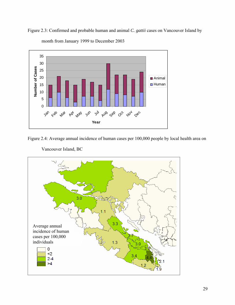
Figure 2.3: Confirmed and probable human and animal C. gattii cases on Vancouver Island by
month from January 1999 to December 2003
0
5
10
15
20
25
30
35
Jan
Feb Mar AprMay Ju
n Jul
Aug Sep Oct NovDec
Year
Num
ber
of C
ases
AnimalHuman
Figure 2.4: Average annual incidence of human cases per 100,000 people by local health area on
Vancouver Island, BC
Average annual incidence of human cases per 100,000 individuals
29
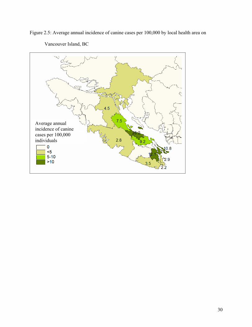
Figure 2.5: Average annual incidence of canine cases per 100,000 by local health area on
Vancouver Island, BC
Average annual incidence of canine cases per 100,000 individuals
30
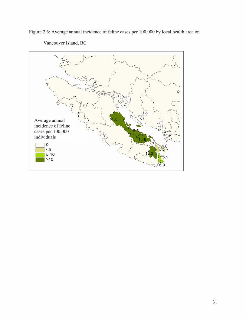
Figure 2.6: Average annual incidence of feline cases per 100,000 by local health area on
Vancouver Island, BC
Average annual incidence of feline cases per 100,000 individuals
31
2.5. References
1. Stephen C, Lester S, Black W, Fyfe M, Raverty S. Multispecies outbreak of cryptococcosis on southern Vancouver Island, British Columbia. Can Vet J 2002; 43 (10):792-4. 2. American Veterinary Medical Association. U.S. pet ownership & demographics sourcebook. Schaumburg, Ill.: Membership & Field Services American Veterinary Medical Association; 2002. 3. Lester SJ, Kowalewich NJ, Bartlett KH, Krockenberger MB, Fairfax TM, Malik R. Clinicopathologic features of an unusual outbreak of cryptococcosis in dogs, cats, ferrets and a bird: 38 cases (January 2003 to July 2003). J Am Vet Med Assoc 2004; 225 (11):1716-1722. 4. Malik R, Wigney DI, Muir DB, Gregory DJ, Love DN. Cryptococcosis in cats: clinical and mycological assessment of 29 cases and evaluation of treatment using orally administered fluconazole. J Med Vet Mycol 1992; 30 (2):133-44. 5. Gerds-Grogan S, Dayrell-Hart B. Feline cryptococcosis: a retrospective evaluation. J Am Anim Hosp Assoc 1997; 33 (2):118-22. 6. Hajjeh RA, Brandt ME, Pinner RW. Emergence of cryptococcal disease: epidemiologic perspectives 100 years after its discovery. Epidemiol Rev 1995; 17 (2):303-20. 7. Malik R, Dill-Macky E, Martin P, Wigney DI, Muir DB, Love DN. Cryptococcosis in dogs: a retrospective study of 20 consecutive cases. J Med Vet Mycol 1995; 33 (5):291-7. 8. Kerl ME. Update on canine and feline fungal diseases. Vet Clin North Am Small Anim Pract 2003; 33 (4):721-47. 9. Krockenberger MB, Canfield PJ, Malik R. Cryptococcus neoformans in the koala (Phascolarctos cinereus): colonization by C n. var. gattii and investigation of environmental sources. Med Mycol 2002; 40 (3):263-72. 10. Krockenberger MB, Canfield PJ, Barnes J, Vogelnest L, Connolly J, Ley C, et al. Cryptococcus neoformans var. gattii in the koala (Phascolarctos cinereus): serological evidence for subclinical cryptococcosis. Med Mycol 2002; 40 (3):273-82. 11. Meidinger D, Pojar J. Ecosystems of British Columbia. Victoria, BC: British Columbia Minister of Forests; 1991. 12. Kidd SE, Hagen F, Tscharke RL, Huynh M, Bartlett KH, Fyfe M, et al. A rare genotype of Cryptococcus gattii caused the cryptococcosis outbreak on Vancouver Island (British Columbia, Canada). Proc Natl Acad Sci U S A 2004; 101 (49):17258-63. 13. Bartlett K, Fyfe, MW, MacDougall, LA. Environmental Cryptococcus neoformans var. gattii in British Columbia, Canada. Am J Resp Crit Care Med 2003; 167 (7):A499. 14. Meyer W, Castaneda A, Jackson S, Huynh M, Castaneda E. Molecular typing of IberoAmerican Cryptococcus neoformans isolates. Emerg Infect Dis 2003; 9 (2):189-95. 15. Sorrell TC, Chen SC, Ruma P, Meyer W, Pfeiffer TJ, Ellis DH, et al. Concordance of clinical and environmental isolates of Cryptococcus neoformans var. gattii by random amplification of polymorphic DNA analysis and PCR fingerprinting. J Clin Microbiol 1996; 34 (5):1253-60. 16. Meyer W MK, Amirmostofian M, Igreja RP, Hardtke C, Methling K, Viviani MA, Chindamporn A, Sukroongreung S, John MA, Ellis DH, and Sorrell TC. Molecular typing of global isolates of Cryptococcus neoformans var. neoformans by PCR-fingerprinting and RAPD - A pilot study to standardize techniques on which to base a detailed epidemiological survey. Electrophoresis 1999; 20:179--1799.
32

17. Kidd SE. Molecular Epidemiology and Characterization of Genetic Structure to Assess Speciation within the Cryptococcus neoformans Complex [Ph.D. Thesis]. Sydney, Australia: University of Sydney; 2003.
33
3. Clinical characteristics and predictors of mortality for Cryptococcus gattii infection in southwestern British Columbia, Canada
3.1. Introduction
Cryptococcosis is a fungal disease found worldwide in human and animal populations. The
causative agent is the organism Cryptococcus spp. which is considered infectious only as a
desiccated yeast cell or basidiospore as found in the environment (1). The genus Cryptococcus
includes over 37 species however only C. neoformans and C. gattii are commonly considered to
be pathogenic. Conventional nomenclature included three recognized varieties of Cryptococcus
neoformans: C. neoformans var. grubii (serotype A), C. neoformans var. neoformans (serotype
D) and C. neoformans var. gattii (serotypes B and C) as well as a hybrid of C. neoformans var.
grubii and C. neoformans var. neoformans (serotype AD) (2-4). Recently proposed changes to
the taxonomy suggest that C. neoformans should be divided into two distinct species including
C. neoformans (serotypes A, D and AD) and C. gattii (serotypes B and C) based on genetic
variability and lack of evidence for genetic recombination between the two varieties (5).
Historically the organism responsible for clinical disease has been thought to be determined by
environmental and ecological factors. Cryptococcus gattii had been restricted to the tropics and
sub tropics while C. neoformans has a global distribution (1, 6). The pattern of clinical disease
corresponds with the distribution of the ecologically limited environmental isolates (1, 7, 8). The
epidemiology of cryptococcosis depends largely on the species of infecting organism as C.
neoformans infects predominantly immunocompromised hosts while C. gattii has not been
associated with a suppressed immune system (1, 9). Historically only C. neoformans has been
34
routinely isolated from animals or humans in Canada without a travel history to a region in
which C. gattii is endemic.
In 2001 the public health authorities and veterinary community of southwestern British
Columbia (BC), Canada recognized an increased incidence of human and animal cryptococcosis
(10, 11). Cryptococcus gattii serotype B was isolated from human and animal cases associated
with the outbreak and affected individuals had a history of travel to, or residence on Vancouver
Island (10, 11). Investigation into the environmental niche of the fungus in Canada revealed the
same organism in the environment within the Coastal Douglas Fir biogeoclimatic zone on the
south east coast of Vancouver Island (12, 13). The following case series describes the clinical
presentation, outcomes and variables influencing survival of canine and feline cryptococcosis
cases caused by C. gattii in British Columbia between January 1999 and December 2003.
3.2. Materials and methods
Feline and canine cryptococcosis cases diagnosed between January 1999 and December 2003
were identified both prospectively and retrospectively from May 2003 through active
surveillance and passive reporting of cases by BC veterinarians, private veterinary laboratories
and the Animal Health Center at the Ministry of Agriculture, Food and Fisheries. A confirmed
case of cryptococcosis due to C. gattii required clinically compatible illness and culture of C.
gattii from a normally sterile site. A probable case included any animal residing on or with a
travel history to Vancouver Island in the previous two years with clinically compatible
symptoms and a laboratory confirmed diagnosis of cryptococcosis by one of: cytology,
histopathology or a latex cryptococcal antigen agglutination test titer > 1:2 from serum or
cerebral spinal fluid.
35
Upon receipt of owner and veterinary consent, medical records or case summaries were obtained
for animals included in the study. Information collected included geographic location, animal
signalment, medical history, date of presentation and diagnosis, presenting complaint and
physical exam findings, diagnostic procedures and treatment. Outcomes were evaluated by
contacting primary veterinarians between six months and two years after diagnosis.
3.2.1. Statistical analysis
Results of the medical record reviews were stratified by species. Based on veterinary records an
animal was classified by presenting complaint and physical exam findings into a category of
principal body system involved. These categories included respiratory, central nervous system
(CNS), subcutaneous mass or dermal lesions, gastrointestinal, generalized illness or other.
Subsequent progression of disease to additional body systems were classified as secondary and
tertiary systems based on chronological order and veterinary evaluation. Descriptive and
comparative statistics were computed using SPSS 12.0 (SPSS Inc., Chicago, Il., USA).
Survival analysis was conducted on all cases presenting with respiratory or CNS symptoms using
SPSS 12.0 (SPSS Inc., Chicago, Il., USA). Endpoints were determined to be death due to or
euthanasia because of cryptococcosis. Cases lost to follow-up or still alive at the time of follow-
up were censored by the date they were last seen by the diagnosing veterinarian or last date
known to be alive. A dichotomous variable, CNS symptoms, was created for animals that
presented with or progressed to CNS disease. To evaluate the potential role of
immunosuppression on survival, three individual variables were created; the presence or absence
of significant illness within two years of diagnosis, history of steroids in the year prior to
36
diagnosis and the combination of steroids and disease history into a dichotomous variable
representing any potential medical or pharmacological induced immunosuppression. These and
other categorical variables including sex, species, antifungal therapy received, primary
presenting system, and location of clinic were evaluated individually using the Kaplan-Meier
survival analysis. Age, a continuous variable, was evaluated independently using Cox
Regression survival analysis. Variables with p < 0.20 on the Log rank test were included in a
multivariate Cox Regression survival analysis. Within the Cox Regression model only variables
with p<0.05 on the Wald test remained in the model.
3.3. Results
3.3.1. Feline
Seventy-eight feline cases suggestive of C. gattii infection were identified between January 1999
and December 2003. Of these, 26 were confirmed C. gattii serotype B on culture and 52 were
probable based on diagnosis, location and travel histories. Case information and primary system
involvement was obtained for 72 and 73 cases respectively. The median age at diagnosis was 7.3
years (minimum 1.2, maximum 14.7 years). There were 41 female (3 intact, 38 spayed) and 31
male (3 intact, 28 neutered) cats.
The initial presenting complaint and veterinary evaluated primary system involved is presented
in tables 3.1 and 3.2 respectively. Twenty (27%) cats were presented to the veterinarian for
generalized illness including weight loss, anorexia and lethargy or behavioral changes. Twenty
(27%) were presented for respiratory problems including nasal discharge, increased respiratory
sounds or effort, coughing and sneezing. Twenty (27%) presented for owner identified skin
37
lumps and 12 (17%) presented for CNS disease including ataxia and seizures. One cat (1%)
presented for dental disease.
Upon veterinary examination the respiratory tract was the primary system involved in the
majority of cases (40 cats, 56%). Many of the skin lumps identified by owners were enlarged
submandibular lymph nodes related to upper respiratory tract infection. Nineteen cats (26%)
were classified as CNS cases and 14 cats (19%) had subcutaneous lumps or dermal lesions.
Eight (20%) cats presenting with clinical respiratory tract disease progressed to central nervous
system disease, one cat with a sub-cutaneous mass on its dorsal thorax progressed to central
nervous system disease.
Diagnosis was based on cytology, serology and histology in 42%, 31% and 28% of the cases
respectively. Serology titers ranged from 1:2 to 1:20,000 and there was no pattern between titer
value and organ system involvement. One cat had a negative titer but chronic nasal discharge
revealed Cryptococcus spp. on cytologic examination.
Fifteen (21%) cats had a reported history of underlying illness that including feline leukemia
virus (2), hyperthyroidism (4), feline lower urinary tract disease (3), irritable bowel syndrome,
renal disease, vaccine reaction within year of diagnosis, allergies, feline asthma and chronic
ectoparasite infestation. Eight cats had received steroids in the year preceding diagnosis.
Twenty-three (32%) cats were diagnosed post-mortem or euthanized upon diagnosis. Treatment
was attempted in 49 (68%) cases. Of those where treatment was initiated 31 (63%) were
38
respiratory cases. Seven of these respiratory cases were alive at the end of the study and were
still being treated with fluconazole (3), itraconazole (3), ketoconazole (1) between 5 and 10
months after diagnosis. Fourteen respiratory cases died or were euthanized within three days to
eight months after diagnosis because disease had progressed to the CNS or the animal failed to
respond to therapy. Seven respiratory cases were classified as clinically recovered by the
diagnosing veterinarian. These animals received fluconazole (4), itraconazole (2) or fluconazole
with amphotericin B. Three respiratory cases were lost to follow-up after initiation of treatment.
Eight (16%) cats receiving therapy had veterinary classified presenting symptoms consistent
with central nervous system disease. Three cats were still undergoing therapy with itraconazole
between five and 11 months after diagnosis. Two cats died within two weeks of diagnosis after
being treated with itraconazole, another three cats were euthanized between two weeks and five
months of treatment with itraconazole and fluconazole.
Ten (20%) cats with subcutaneous masses received antifungal therapy. Two of these cats were
deemed recovered by the diagnosing veterinarian, both had had the masses surgically excised
and one had received itraconazole for one month following excision. One cat was still
undergoing treatment with fluconazole 11 months after diagnosis. Five cats were euthanized,
three within one year of diagnosis, one of which had progressed to central nervous system
disease within two weeks of diagnosis. The remaining two cats were lost to follow-up.
Excluding cases lost to follow-up the overall case fatality of cats included in this case series was
70%. Of those cases where treatment was initiated the case fatality rate was 55%.
39
3.3.2. Canine
There were 51 canine cases suggestive of C. gattii serotype B infection identified between
January 1999 and December 2003. Nineteen were confirmed C. gattii and 32 cases were
classified as probable. Case information was available for 50 cases. There were 23 males (7
intact, 16 neutered) and 27 females (4 intact, 23 spayed). The median age was 2.3 years
(minimum 5 months, maximum 15.5 years).
The primary presenting complaint and veterinary-evaluated primary system involved is presented
in tables 3.1 and 3.2 respectively. Nineteen (38%) dogs were brought to the veterinary clinic for
respiratory problems. Reported complaints included nasal discharge, epistaxis, noisy breathing,
sneezing and coughing. Fifteen (30%) dogs presented for neurological disease including ataxia,
neck pain or seizures. Six (12%) dogs presented for generalized illness including anorexia and
weight loss and six (12%) dogs presented to the veterinarian for ocular problems, four with acute
onset of blindness and two with exopthalmous. Three (6%) dogs were brought in for
subcutaneous lumps on the head or body. One dog presented for an acute onset of vomiting and
diarrhea.
Upon examination by the veterinarian, 26 (52%) of the cases were deemed to be respiratory in
nature, primarily restricted to the upper respiratory tract. Four dogs presenting with or
determined to have exopthalmous on veterinary exam all had concurrent upper respiratory tract
disease. Twenty-one (42%) dogs were classified as central nervous system cases and two (4%)
had subcutaneous masses. The single gastrointestinal case was taken to surgery for an
intusseption caused by an extraluminal cryptococcoma. Six cases (24%) initially classified by
40
the veterinarian as respiratory progressed to central nervous system disease. One dog with
respiratory disease later developed subcutaneous masses.
Diagnosis was made primarily on cytology (44%) or serology (34%) but histology (18%) and
culture (4%) were also used for diagnosis. Titers ranged from 1:2 to 1:25,000 and did not
correlate with presenting complaint or primary system involved.
Six (12%) dogs had a history of underlying disease that could be considered potentially
immunosuppressive. These conditions included immune mediated thrombocytopenia (2), mast
cell tumor, lymphosarcoma, hypothyroid and a ruptured uterus. Ten dogs had received steroids
within the year before diagnosis.
Twenty-four (48%) dogs were treated and 26 (52%) were diagnosed post-mortem or euthanized
upon receipt of diagnosis. Of those undergoing therapy 15 (63%) were respiratory cases. Nine
of the canine respiratory cases were still alive and receiving therapy between five and 13 months
after diagnosis. Three dogs were euthanized after commencing treatment, one for diagnosis of
lymphosarcoma and two for unknown causes. Two dogs were reported as recovered after four
and 12 months of therapy with azole antifungals. One respiratory case was lost to follow-up.
Of the seven (29%) neurological cases where treatment was attempted, two dogs were still alive
and undergoing treatment with azole antifungals alone or in combination with amphotericin B at
the time of writing. Three of the dogs with primary neurological diseases died within one to
41
three weeks of commencing treatment with azole antifungals and or amphotericin B. Two
neurological dogs were lost to follow-up.
One dog with a solitary subcutaneous mass underwent surgical excision of the mass and
recovered with no antifungal therapy. The dog with the abdominal cryptococcoma was treated
with fluconazole for 10 months post surgery and was clinically healthy but maintained a
cryptococcal antigen titer >1:2. Excluding cases lost to follow-up the overall case fatality of
dogs included in this case series was 68%. Of those cases where treatment was initiated the case
fatality rate was 29%.
3.3.3. Survival analysis
Thirty nine feline and 20 canine cases representing animals presenting with respiratory or central
nervous symptoms and receiving treatment were included in the survival analysis. Of these 51%
of canine and 63% feline cases were censored. On initial univariate analysis only the presence of
central nervous system disease (p<0.01), primary system involved (p=0.06) and species (p=0.14)
were significant at the 20% level. Significant medical history (p=0.52), city of diagnosing clinic
(p=0.46), sex (p=0.37), steroids in the previous year (p=0.29), antifungal treatment (p=0.86),
potential immunosuppression (p=0.83) and age at diagnosis (p=0.33) were excluded from further
models.
Using Cox Regression survival analysis, a model created with species, presence of CNS
symptoms and primary presenting system revealed that only the presence of CNS symptoms was
a significant predictor of mortality (p<0.01). A second Cox Regression model (figure 3.1)
including only the presence of CNS symptoms found that those animals that present with or
42
progress to neurological disease are 4.3 times more likely to die than those that never show
neurological symptoms (95% CI for mortality ratio 1.87, 9.89).
3.4. Discussion
During the study period, fifty percent more cats than dogs were identified for inclusion in the
case series. This result likely represents the differing species susceptibilities to the organism.
Cryptococcosis is the most common systemic mycoses of cats and, unlike other fungal diseases,
clinical cryptococcosis has been reported in equal or greater frequency in cats than in dogs (14,
15). A previously reported subset of these BC cases identified more cases in dogs however this
result may be due chance as the time interval was considerably smaller than that of this study
(16).
As the sex ratio of the underlying population is unknown gender cannot be evaluated statistically
however there does not appear to be a sex predisposition. Some studies of cats have identified a
greater proportion of males affected and suggested that males are more likely to be exposed for
behavioral reasons, however other studies found no sex predisposition (17-20). No apparent sex
predilection has been reported in dogs (21).
As has been observed in other case series, feline cryptococcosis is more common in middle aged
cats but cats of all ages may be affected. The age range in this and other case series is wide (17-
19, 22). In contrast to cats, clinical disease was more common in younger dogs as has been
previously documented (21, 23). Breed predisposition could not be evaluated in this case series
as the underlying population of the region is not known. In other studies Doberman Pincers,
Great Danes and other large breed have been over represented relative to respective hospital
43
populations suggesting a potential genetic or behavioral factor involved with infection (21, 24).
Siamese cats appeared overrepresented in one Australian study (19).
Respiratory disease was the most common syndrome in cats (56%) followed by central nervous
system symptoms (26%) and subcutaneous or dermal lesions (19%). While the high proportion
of respiratory cases is similar to previous studies, the proportion of cats with CNS symptoms
exceeded those previously reported (17, 19, 22) and may represent a difference in virulence of C.
gattii compared to C. neoformans which has been more commonly isolated or, based on location,
assumed to be the causative agent in the other case series. Central nervous system signs in cats
have been reported to be secondary to respiratory infection and not a common primary
presenting sign (19), however, this study had a high percentage of cases presenting for CNS
disease without a reported history of respiratory symptoms.
The manifestation of cryptococcosis in dogs of British Columbia is very similar to those reported
from Australia where a retrospective case series identified 55% respiratory cases, 35% central
nervous system, 5% with disseminated disease and 5% with gastrointestinal symptoms (21). It
has been hypothesized that nasal cavity or respiratory involvement is more prevalent than
commonly reported but disease goes undiagnosed in dogs until the central nervous system is
involved or the organism simply disseminates from the respiratory tract faster in dogs than in
cats (21). Similarly, this study found a greater proportion of canine cases were classified as
neurological at first veterinary visit when compared to feline cases. Intra-abdominal masses
have been reported in dogs but may be considered an atypical appearance of cryptococcosis (25).
44
Only respiratory and CNS cases were included in the survival analysis as the relative number of
other cases receiving treatment was too low to evaluate meaningfully. The results of the survival
analysis reveal that the presence of neurological symptoms was a very strong predictor of
mortality. Animals presenting with or progressing to CNS disease were over four times more
likely to die than those never exhibiting neurological symptoms.
The poor survival in animals exhibiting central nervous system symptoms observed in this study
may be explained by a number of factors. Most significant is likely the severity of the disease
once it has entered the central nervous system (15, 17). It is also possible that because endpoints
in this analysis included both death and owner elected euthanasia, treatment may have been
terminated for reasons other than the animal being moribund. Such reasons could include cost of
therapy or owner perceived severity of clinical symptoms and electing humane euthanasia.
Finally survival may be influenced by treatment initiated. While antifungal therapy was not a
significant predictor in the model it is important to note that only the azole antifungals were used
with enough frequency to influence the model. For central nervous system cases effective
therapy requires a drug that can penetrate the blood brain barrier such as amphotericin B,
flucytosine and fluconazole (14, 18, 21, 26). Failure to select one of these agents would result in
a poor response to therapy. Minimum inhibitory concentration (MIC) work done on a subset of
cultures from this case series found all isolates were susceptible to amphotericin B but there was
some intermediate sensitivity and resistance to fluconazole and flucytosine (16). Amphotericin
B, the most affordable and potentially efficacious antifungal drug when dealing with CNS cases,
was largely avoided in these cases out of concern for nephrotoxic side effects. Newly proposed
45
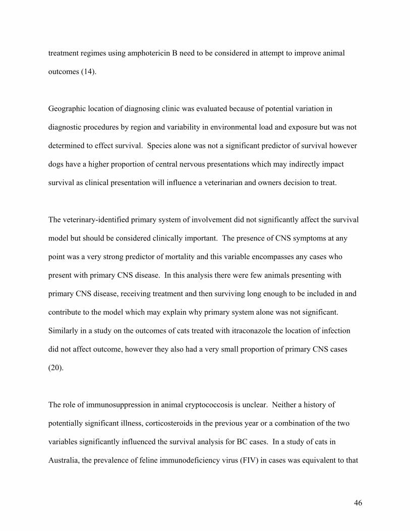
treatment regimes using amphotericin B need to be considered in attempt to improve animal
outcomes (14).
Geographic location of diagnosing clinic was evaluated because of potential variation in
diagnostic procedures by region and variability in environmental load and exposure but was not
determined to effect survival. Species alone was not a significant predictor of survival however
dogs have a higher proportion of central nervous presentations which may indirectly impact
survival as clinical presentation will influence a veterinarian and owners decision to treat.
The veterinary-identified primary system of involvement did not significantly affect the survival
model but should be considered clinically important. The presence of CNS symptoms at any
point was a very strong predictor of mortality and this variable encompasses any cases who
present with primary CNS disease. In this analysis there were few animals presenting with
primary CNS disease, receiving treatment and then surviving long enough to be included in and
contribute to the model which may explain why primary system alone was not significant.
Similarly in a study on the outcomes of cats treated with itraconazole the location of infection
did not affect outcome, however they also had a very small proportion of primary CNS cases
(20).
The role of immunosuppression in animal cryptococcosis is unclear. Neither a history of
potentially significant illness, corticosteroids in the previous year or a combination of the two
variables significantly influenced the survival analysis for BC cases. In a study of cats in
Australia, the prevalence of feline immunodeficiency virus (FIV) in cases was equivalent to that
46
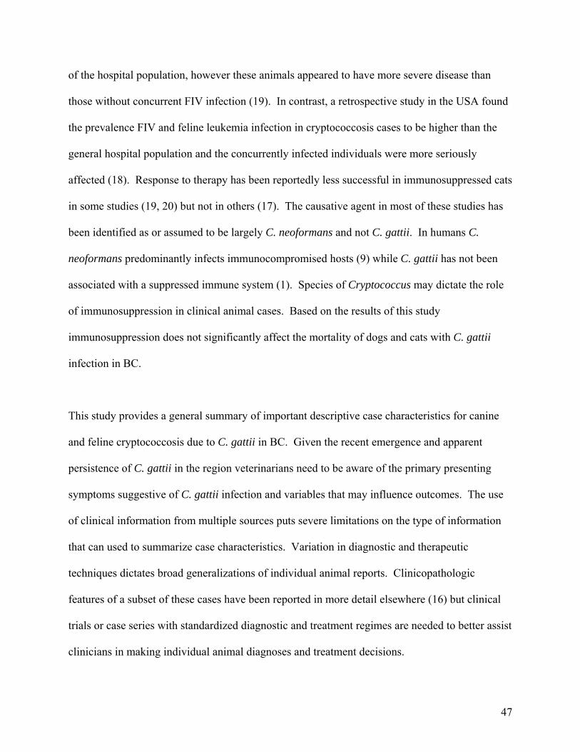
of the hospital population, however these animals appeared to have more severe disease than
those without concurrent FIV infection (19). In contrast, a retrospective study in the USA found
the prevalence FIV and feline leukemia infection in cryptococcosis cases to be higher than the
general hospital population and the concurrently infected individuals were more seriously
affected (18). Response to therapy has been reportedly less successful in immunosuppressed cats
in some studies (19, 20) but not in others (17). The causative agent in most of these studies has
been identified as or assumed to be largely C. neoformans and not C. gattii. In humans C.
neoformans predominantly infects immunocompromised hosts (9) while C. gattii has not been
associated with a suppressed immune system (1). Species of Cryptococcus may dictate the role
of immunosuppression in clinical animal cases. Based on the results of this study
immunosuppression does not significantly affect the mortality of dogs and cats with C. gattii
infection in BC.
This study provides a general summary of important descriptive case characteristics for canine
and feline cryptococcosis due to C. gattii in BC. Given the recent emergence and apparent
persistence of C. gattii in the region veterinarians need to be aware of the primary presenting
symptoms suggestive of C. gattii infection and variables that may influence outcomes. The use
of clinical information from multiple sources puts severe limitations on the type of information
that can used to summarize case characteristics. Variation in diagnostic and therapeutic
techniques dictates broad generalizations of individual animal reports. Clinicopathologic
features of a subset of these cases have been reported in more detail elsewhere (16) but clinical
trials or case series with standardized diagnostic and treatment regimes are needed to better assist
clinicians in making individual animal diagnoses and treatment decisions.
47
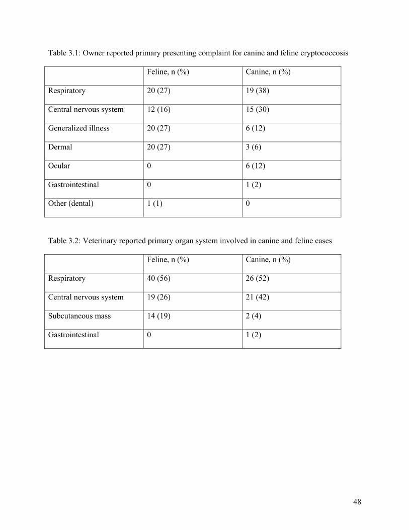
Table 3.1: Owner reported primary presenting complaint for canine and feline cryptococcosis
Feline, n (%) Canine, n (%)
Respiratory 20 (27) 19 (38)
Central nervous system 12 (16) 15 (30)
Generalized illness 20 (27) 6 (12)
Dermal 20 (27) 3 (6)
Ocular 0 6 (12)
Gastrointestinal 0 1 (2)
Other (dental) 1 (1) 0
Table 3.2: Veterinary reported primary organ system involved in canine and feline cases
Feline, n (%) Canine, n (%)
Respiratory 40 (56) 26 (52)
Central nervous system 19 (26) 21 (42)
Subcutaneous mass 14 (19) 2 (4)
Gastrointestinal 0 1 (2)
48
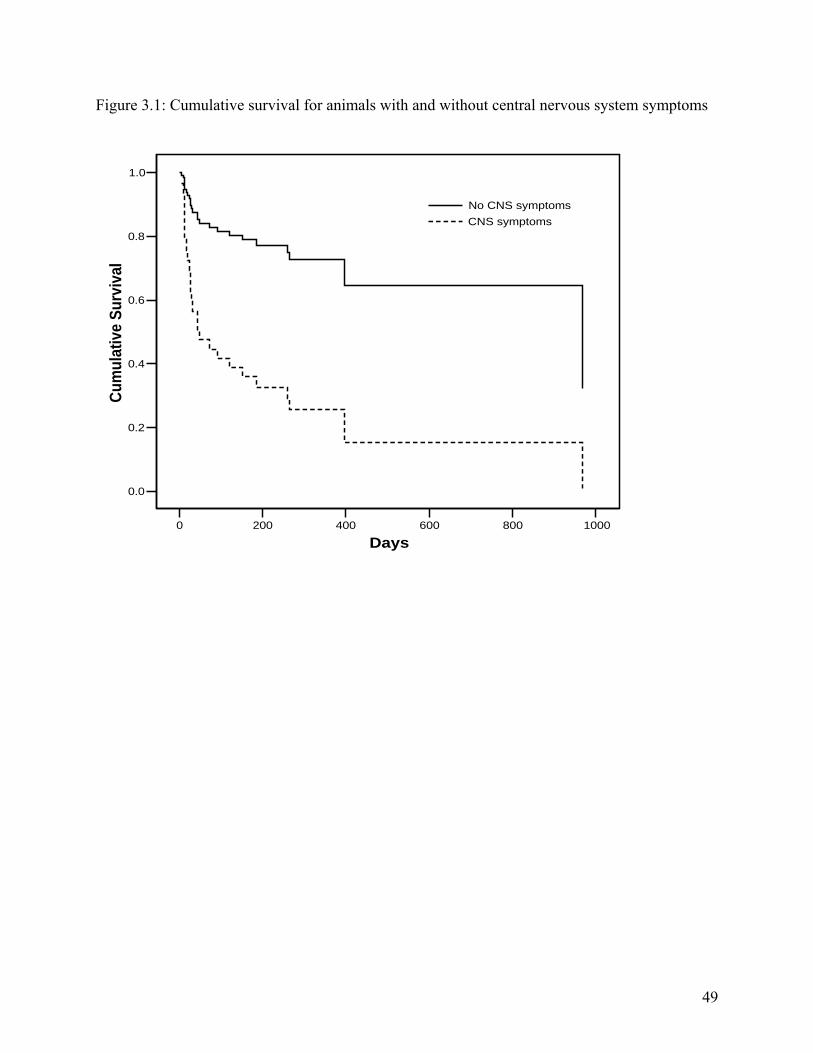
Figure 3.1: Cumulative survival for animals with and without central nervous system symptoms
0 200 400 600 800 1000
Days
0.0
0.2
0.4
0.6
0.8
1.0
Cum
ulat
ive
Surv
ival
No CNS symptomsCNS symptoms
49
3.5. References
1. Sorrell TC. Cryptococcus neoformans variety gattii. Med Mycol 2001; 39 (2):155-68. 2. Kwon-Chung KJ, Wickes BL, Stockman L, Roberts GD, Ellis D, Howard DH. Virulence, serotype, and molecular characteristics of environmental strains of Cryptococcus neoformans var. gattii. Infect Immun 1992; 60 (5):1869-74. 3. Kwon-Chung KJ, Polacheck I, Bennett JE. Improved diagnostic medium for separation of Cryptococcus neoformans var. neoformans (serotypes A and D) and Cryptococcus neoformans var. gattii (serotypes B and C). J Clin Microbiol 1982; 15 (3):535-7. 4. Katsu M, Kidd S, Ando A, Moretti-Branchini ML, Mikami Y, Nishimura K, et al. The internal transcribed spacers and 5.8S rRNA gene show extensive diversity among isolates of the Cryptococcus neoformans species complex. FEMS Yeast Res 2004; 4 (4-5):377-88. 5. Kwon-Chung J, Boekhout, T., Fell, J., Diaz, M. Proposal to conserve the name Cryptococcus gattii against C. hondurianus and C. bacillisporus (Basidiomycota, Hymenomycetes, Tremellomycetidae). Taxon 2002; 51:804-806. 6. Kwon-Chung KJ, Bennett JE. Epidemiologic differences between the two varieties of Cryptococcus neoformans. Am J Epidemiol 1984; 120 (1):123-30. 7. Ellis DH, Pfeiffer TJ. Natural habitat of Cryptococcus neoformans var. gattii. J Clin Microbiol 1990; 28 (7):1642-4. 8. Chen S, Sorrell T, Nimmo G, Speed B, Currie B, Ellis D, et al. Epidemiology and host- and variety-dependent characteristics of infection due to Cryptococcus neoformans in Australia and New Zealand. Australasian Cryptococcal Study Group. Clin Infect Dis 2000; 31 (2):499-508. 9. Speed B, Dunt D. Clinical and host differences between infections with the two varieties of Cryptococcus neoformans. Clin Infect Dis 1995; 21 (1):28-34; discussion 35-6. 10. Stephen C, Lester S, Black W, Fyfe M, Raverty S. Multispecies outbreak of cryptococcosis on southern Vancouver Island, British Columbia. Can Vet J 2002; 43 (10):792-4. 11. Hoang LM, Maguire JA, Doyle P, Fyfe M, Roscoe DL. Cryptococcus neoformans infections at Vancouver Hospital and Health Sciences Centre (1997-2002): epidemiology, microbiology and histopathology. J Med Microbiol 2004; 53 (Pt 9):935-40. 12. Bartlett K, Fyfe, MW, MacDougall, LA. Environmental Cryptococcus neoformans var. gattii in British Columbia, Canada. Am J Resp Crit Care Med 2003; 167 (7):A499. 13. Mak S, Duncan C, Bartlett K, Stephen C, Fyfe M, MacDougall L. Using GIS to track cryptococcosis in BC. In: GisVet; 2004 June; Guelph, ON; 2004. 14. Gionfriddo JR. Feline systemic fungal infections. Vet Clin North Am Small Anim Pract 2000; 30 (5):1029-50. 15. Kerl ME. Update on canine and feline fungal diseases. Vet Clin North Am Small Anim Pract 2003; 33 (4):721-47. 16. Lester SJ, Kowalewich NJ, Bartlett KH, Krockenberger MB, Fairfax TM, Malik R. Clinicopathologic features of an unusual outbreak of cryptococcosis in dogs, cats, ferrets and a bird: 38 cases (January 2003 to July 2003). J Am Vet Med Assoc 2004; 225 (11):1716-1722. 17. Flatland B, Greene RT, Lappin MR. Clinical and serologic evaluation of cats with cryptococcosis. J Am Vet Med Assoc 1996; 209 (6):1110-3. 18. Gerds-Grogan S, Dayrell-Hart B. Feline cryptococcosis: a retrospective evaluation. J Am Anim Hosp Assoc 1997; 33 (2):118-22.
50
19. Malik R, Wigney DI, Muir DB, Gregory DJ, Love DN. Cryptococcosis in cats: clinical and mycological assessment of 29 cases and evaluation of treatment using orally administered fluconazole. J Med Vet Mycol 1992; 30 (2):133-44. 20. Jacobs GJ, Medleau L, Calvert C, Brown J. Cryptococcal infection in cats: factors influencing treatment outcome, and results of sequential serum antigen titers in 35 cats. J Vet Intern Med 1997; 11 (1):1-4. 21. Malik R, Dill-Macky E, Martin P, Wigney DI, Muir DB, Love DN. Cryptococcosis in dogs: a retrospective study of 20 consecutive cases. J Med Vet Mycol 1995; 33 (5):291-7. 22. Medleau L, Jacobs GJ, Marks MA. Itraconazole for the treatment of cryptococcosis in cats. J Vet Intern Med 1995; 9 (1):39-42. 23. Nelson RW, Couto CG. Small animal internal medicine. 3rd ed. St. Louis, Mo.: Mosby; 2003. 24. Sutton RH. Cryptococcosis in dogs: a report on 6 cases. Aust Vet J 1981; 57 (12):558-64. 25. Malik R, Hunt GB, Bellenger CR, Allan GS, Martin P, Canfield PJ, et al. Intra-abdominal cryptococcosis in two dogs. J Small Anim Pract 1999; 40 (8):387-91. 26. Tiches D, Vite CH, Dayrell-Hart B, Steinberg SA, Gross S, Lexa F. A case of canine central nervous system cryptococcosis: management with fluconazole. J Am Anim Hosp Assoc 1998; 34 (2):145-51.
51
4. Risk factors for clinical Cryptococcus gattii infection in dogs and cats on Vancouver Island, British Columbia, Canada
4.1. Introduction
Cryptococcosis is a sporadic disease found worldwide in human and animal populations. The
causative agent is Cryptococcus spp. which is considered infectious only as a desiccated yeast
cell or basidiospore as found in the environment (1). The genus includes many species but only
C. neoformans and C. gattii are commonly regarded as pathogenic. Cryptococcus neoformans
has two recognized varieties: C. neoformans var. grubii (serotype A) and C. neoformans var.
neoformans (serotype D) as well as a hybrid of C. neoformans var. grubii and C. neoformans var.
neoformans (serotype AD) (2, 3). Cryptococcus gattii (serotypes B and C) has recently been
identified as a species distinct from C. neoformans based on genetic variability and lack of
evidence for genetic recombination between C. neoformans and C. gattii (4).
Cryptococcus gattii had historically been geographically confined to the tropics and subtropics
while C. neoformans had a global distribution (1, 5); the pattern of clinical disease corresponded
largely with the ecologically restricted environmental organism (1, 6, 7). Since 1999 C. gattii
(serotype B) has been isolated from sick people and animals in southwestern British Columbia
(BC) as well as from air, soil and vegetation samples collected on the south east coast of the
island (8-11). These findings are in stark contrast to the organisms previously described ecology
and associated epidemiology.
52
Prior to the emergence of C. gattii, cryptococcosis was considered a rare disease of companion
animals in Canada. Vancouver Island veterinarians were challenged by an increasing number of
cases and a lack of information on risk reduction measures. The purpose of this study was to
identify risk factors for clinical C. gattii infection in dogs and cats residing on Vancouver Island,
BC.
4.2. Materials and methods
4.2.1. Inclusion criteria
As two previously reported case series of animal cryptococcosis identified a relationship between
C. gattii infection and southeastern Vancouver Island, BC this area was selected as a focal point
for the study (8, 11). Cryptococcosis in animals is not a reportable disease in Canada so incident
cases of feline and canine cryptococcosis meeting the case definition were identified by
contacting veterinary clinics on Vancouver Island and private diagnostic laboratories servicing
BC. Cases included in this study were identified during two distinct time periods; August 2001
to February 2002 and May to December 2003.
A confirmed case of cryptococcosis due to C. gattii required clinically compatible illness (12)
and culture of C. gattii from a normally sterile site. A probable case included any animal
residing on or with a travel history to Vancouver Island in the previous two years with clinically
compatible symptoms (12) and a laboratory confirmed diagnosis of cryptococcosis by one or
more of: cytology, histopathology or a latex cryptococcal antigen agglutination test titer > 1:2
from serum or cerebral spinal fluid. Many cases were diagnosed without material for fungal
culture. Given that C. gattii was identified in virtually all previously reported cases of animal
53
cryptococcosis on Vancouver Island during the study period where culture material was
available, confirmed and probable cases were included in the analysis (8, 11).
Control animals were obtained from the veterinary clinic that diagnosed the case. Controls were
matched on species, age (within 1 year of case age) and dogs were size matched where large
dogs were defined as > 20 kg and small dogs were < 20 kg. Matching was used to control
confounding by these variables and increase the power of the study as sample size was expected
to be small. Veterinary clinics were sent a letter outlining the control animals required and,
beginning on a random day, asked any client presenting to the veterinary clinic whose animal
met the control description if they would take part in the study. If they refused, the next animal
meeting the description was asked until all controls were obtained. Exclusion criteria for control
animals were those meeting the match requirements but presenting to the veterinary clinic with a
diagnosis of, or clinical symptoms strongly suggestive of, cryptococcosis.
4.2.2. Interview
A standardized questionnaire was administered over the telephone or by personal interview for
cases and over the telephone for all controls. The questionnaire focused on two main areas;
environmental variables and animal characteristics. Controls were asked to answer the questions
with reference to the same time period as the matched case. The number of questions in the
questionnaire was modified slightly between the two sampling periods as the investigation into
C. gattii in the region identified other potential risk factors for infection.
Environmental variables focused on factors that may increase exposure of the animal to the
organism. This included proximity to wooded areas or farmland, contact with eucalyptus trees or
54
products and owner activities six months prior to case diagnosis such as: hiking, gardening,
chopping wood, handling birds/cleaning bird roosts or digging soil. They were also asked about
activities taking place within ten kilometers (km) of the animal’s primary residence in the six
months preceding diagnosis that may disturb the environment such as construction, logging or
commercial soil disturbance.
Host characteristics explored included gender of the animal, travel on and off of Vancouver
Island in the year preceding diagnosis, activity level, presence of potentially stressful changes or
owner perceived stressors in the previous year, history of disease, medication, vaccination and
owner supplementation with commercial products. Animal-environment interactions including
hunting and digging in soil were also included. Owners were asked how much time their animal
spent outside a 10 km radius of their home in a given week, how many hours were spent
outdoors on a given day and how many years the animal had lived within the municipality.
All questions were close ended. Animal activity level was recorded on a four point scale (very
low, low, high, very high) reflecting how active owners felt their pet was in certain
environments. Times spent outdoors, in the municipality and outside a radius of 10 km of the
home were continuous variables; the remaining variables were dichotomous.
4.2.3. Statistical analysis
Odds ratios and 95% confidence intervals were calculated from McNemar chi-squared values for
cases versus controls for each potential risk factor (13). Continuous variables were evaluated
using the paired t-test. For animal activity level the four point response scale was converted to a
dichotomous variable with very low and low activity classified as below average while high and
55
very high were classified as above average. Data from both species were pooled together for the
initial analysis and statistically significant risk factors were stratified by species for further
evaluation.
4.3. Results
Over 80 owners of clinical cases were interviewed but only 17 matched controls in 2001/2002
and 32 in 2003 were provided by the diagnosing veterinary clinic. Less than 10 cases meeting
the case description were not interviewed because the owner or diagnosing veterinarian was
unwilling to participate in the study. Based on available clinical information these cases did not
differ from those included. In total 49 cases and matched controls were included in the analysis.
The results, pooled by species, are presented in table 1; only statistically significant risk factors
and continuous variables are reported in the text. Logging and soil disruption within a 10 km
radius of the primary residence were the most significant risk factors for clinical cryptococcosis
with those exposed being 14 (3.00, 65.37) and 18 (4.21, 76.93) times more likely to develop
disease, respectively. Animals that had traveled on Vancouver Island within the last year were 6
(1.61, 22.33) times more likely to become infected than those without a travel history in the
previous 12 months. Likewise cases spent a mean of 2.2% of time outside of a 10 km radius of
the primary residence while controls spent only 0.33%; a difference that was statistically
significant (p=0.027). Owners who went hiking or visited a botanical garden recently were 4
(1.61, 9.90) and 5 (1.28, 19.60) times more likely to have animals become diseased. Activity
level of the animal was a significant risk factor as animals with owner classified activity level of
above average in the house and outside unrestrained were 3 (1.02, 8.80) and 8 (1.40, 45.89) times
more likely to become diseased, respectively.
56
Owner administration of supplements increased the odds of disease 3.67 (1.11, 12.07) times.
The odds ratio for owners knowing people with cryptococcosis or other people with animals
diagnosed with cryptococcosis was 5.67 (1.91, 16.79). Medication in the previous year and the
presence of other pets were both considered protective in the crude analysis. The amount of time
an animal spent outdoors (p=0.498) or living in their current home municipality (p=0.703) were
not significant.
Results of stratification of the statistically significant risk factors by species are presented in
table 4.2. Logging and soil disturbance remained the greatest risk factors for disease. Odds of
disease in the presence of logging was increased 17 (2.12, 136.40) times in dogs and 6 (0.94,
38.48) times in cats; however, only dogs remained statistically significant. The measure of effect
of soil disturbance was 17 (2.12, 136.40) and 20 (1.90, 52.76) for dogs and cats, respectively,
and remained statistically significant for both species. The odds of disease in dogs, but not in
cats, was statistically increased when the animal had traveled on Vancouver Island in the
previous 12 months. Canine cases spent significantly more time outside a 10 km radius of their
home compared to controls (p=0.024) but the difference was not statistically significant for feline
cases (p=0.33).
The odds of disease by species was similar to the pooled result for owners that hiked or visited a
botanical garden in the previous six months, however only hiking of cat owners remained
statistically significant. When the variables reflecting animal activity level were stratified by
species confidence intervals became large and none of the variables remained significant.
57
Supplements and knowing other cryptococcosis cases remained statistically significant risk
factors for cats but not for dogs in the stratified analysis. Medication in the previous 12 months
remained significantly protective for dogs but not for cats, while the presence of other pets was
not statistically significant for either species.
4.4. Discussion
Cryptococcus is a genus of environmental fungi that can be isolated from vegetative matter
worldwide. While the exact mode of infection is unknown it is widely accepted to be through
inhalation of air-borne organism (14, 15). Cryptococcus spp. have been isolated from the nasal
passages of dogs, cats (16, 17) and koalas (18) in Australia and Canada without evidence of
disease suggesting asymptomatic colonization of the nasal mucosa following environmental
exposure. It is not clear what triggers tissue invasion after colonization (1).
Association with environmental sources of organism has been recognized as a primary risk factor
for clinical cryptococcal disease in humans. In Australia, the aboriginal population living in
rural and semi-rural areas were over 12 times more likely to develop of C. gattii infection
relative to the reference population (7). This increased risk was proposed to be due to the
populations close association with eucalyptus trees, a regional environmental reservoir. Men in
Australia and Papua New Guinea are also at an increased risk of infection with C. gattii,
presumably because of increased contact with environmental organism (7, 19).
Likewise in animals, environmental exposure to Cryptococcus spp. is an important risk factor.
Male cats have been reported infected more often than females, with the suggestion that males
are more likely to be exposed for behavioral reasons; however, other studies found no sex
58
predisposition (20-23). In a retrospective study of 20 canine cryptococcosis cases in Australia,
all dogs infected with C. gattii resided in rural or suburban environments suggesting increased
proximity to the organism was a risk factor (24). Doberman Pincers, Great Danes and other
young, large breed dogs have been over represented relative to respective hospital populations
for cryptococcosis cases suggesting a potential behavioral or genetic factor involved with
infection (24, 25). Unlike cats and humans, there is no evidence of sex predilection for
cryptococcal infection in dogs (24).
In this study, residing within 10 km of a site of commercial soil disruption or vegetation clearing
were the most significant risk factors for clinical cryptococcosis due to C. gattii. Physical
disruption of the environment can result in more airborne particulate matter, including C. gattii
(K. Bartlett, unpublished). In contrast, owner induced vegetation and soil disruption at the
residence, such as chopping wood, bringing wood into the house, digging soil, gardening or the
exposure of animals to garden related products and home construction, were not identified as
significant risk factors. This suggests such activities may not result in sufficient aerosolization of
particulate matter to increase the risk of infection when compared to larger scale environmental
disturbance associated with logging and commercial soil excavation. Such findings imply a
threshold level of air contamination exists that influences the risk of animal infection; or that the
source of the infection is not in the immediate residential environment.
Animals that had traveled on Vancouver Island within the last year were six times more likely to
become ill than those with no travel history. Clinical animals also spent a statistically greater
percentage of time outside a 10 km radius of their primary residence than controls suggesting
59
they traveled more often. Stratification of these variables revealed that this pooled result was
influenced more by dogs than cats as dogs are more likely to travel with their owners. Animals
that travel may be at greater risk of disease because of increased opportunities for exposure.
Environmental C. gattii has not been isolated ubiquitously on Vancouver Island (9) so travel
would increase the probability of exposure to a positive site. Duration of time spent in the
residential municipality and time spent outdoors daily were not significant risk factors. Similarly
residing within two km of a wooded or agricultural area was not a risk factor. This supports the
hypothesis that some animals are not being exposed at their home; rather in places to which they
travel where there is a higher environmental load. Animals traveling off of Vancouver Island
were not at increased risk of disease; however, C. gattii had not been isolated from the
environment off of the island at the time of this study (26).
Higher activity levels in the house and outdoors unrestrained were significant risk factors for
disease; the odds of disease was also very high for animals outside and restrained; however, an
extremely large confidence interval deemed the measure to not be significant statistically.
Behavioral factors, including activity level, may significantly increase an animal’s risk of
infection by bringing the individual in contact with C. gattii more frequently. An active animal
may also disturb the environment more and aerosolize additional organisms that can be inhaled.
Gender, previously suggested to be a risk factor for cryptococcosis because of behavioral reasons
was not determined to be significant in this case-control study.
There was an increased risk of disease in animals whose owners hiked and visited a botanical
garden in the six months preceding diagnosis. As the environmental organism is small enough to
60
be transported on fomites it is possible that humans who participate in outdoor recreational
activities transport the organism back to their residence and unwittingly expose their pet. Dogs
often accompany their owners on hiking trips and thus are at an increased risk of direct exposure
through this recreational activity. Stratification of hiking by species resulted in a wide and non-
significant confidence interval for dogs. This is likely the result of a small sample size.
Questions regarding visitation to botanical gardens were included to explore the role of imported
vegetation, most specifically eucalypt trees, on disease. None of the direct eucalyptus variables
were significant and the risk of an owner visiting a botanical garden in the period prior to
diagnosis may indirectly imply owner travel or affinity for gardening and naturalist activities.
Owners who knew people with cryptococcosis or other people who had animals with
cryptococcosis were over five times more likely to have a diseased pet themselves. This result
may be a result of recall bias as owners with a sick animal may be more likely to recall or seek
out others with the same problem. Alternatively the finding may reflect the clustering of positive
environmental sites resulting in clustering of cases at the level of the neighborhood. Owner
administered supplements were a significant risk factor, but protopathic bias may have
influenced this finding as the incubation period is long and, in the period preceding diagnosis,
owners may have attempted to supplement an animal that appeared slightly unwell. Likewise
individuals who choose to supplement a sick pet may be more apt to bring the animal to the
veterinarian in the case of illness or pay for diagnostic procedures that would facilitate
identification of the case and inclusion in the study.
61
Medication in the year preceding diagnosis was identified to be protective; however this finding
may have been influenced by Berkson’s bias, the selection of the control animals from a hospital
population. Control animals were those presenting to the veterinary clinic on a random day
without a diagnosis of or clinical symptoms strongly suggestive of cryptococcosis. Because the
clinic was used as the control selection point animals were more likely than the general
population to be presenting to the veterinarian because of illness and therefore be receiving
medication. Control selection could also have influenced the finding that owning multiple pets
was considered protective when dogs and cats were pooled together. Owners of multiple pets
tend to visit the veterinary clinic more often and thus have greater odds of being selected as a
control.
Variables indicating potential immunosuppression including any history of disease,
administration of steroids in the previous year, environmental changes or owner perceived
stressors were not significantly associated with cryptococcosis cases. The role of
immunosuppression in feline cryptococcosis has been debated in the literature. The prevalence
of FIV in cats with cryptococcosis has been reportedly equivalent to that of a hospital population
in Australia (22) while in the USA concurrent FIV or FeLV infection in cats with cryptococcosis
was much higher that the general hospital population (21). An examination of FIV positive and
negative cats in the USA found C. neoformans more commonly in the oropharynx of FIV
seropositive cats, although no cats had signs of clinical disease (27). It is important to note that
in many of these studies the causative agent has been identified as, or assumed to be C.
neoformans and not C. gattii. Clinical disease is dictated by host characteristics and the variety
of infecting organism; C. neoformans var. neoformans and C. neoformans var. grubii are isolated
62
most commonly from immunosuppressed individuals (7, 28). In contrast C. gattii should be
considered a primary pathogen as it tends to infect immunocompetent hosts; even in areas where
the organism is endemic C. gattii is rarely the cause of cryptococcosis in AIDS patients (7, 28,
29). This study lends further support that immunosuppression is not required for C. gattii
infection.
The first environmental isolation of C. gattii was from eucalyptus trees (Eucalyptus
camaldulensis and E. tereticornis) in Australia (6, 30). Subsequent to this discovery C. gattii has
been recovered from material associated with eucalypt species in many other parts of the world
including California, India and Brazil (31-34) along with non-eucalypt tree species from tropical
and subtropical areas worldwide (31, 35-37). There are studies that suggest alternative
environmental sources of C. gattii have yet to be identified, as molecular types isolated from
clinical and environmental samples have been different in western and northern Australia (38-40)
and clinical cryptococcosis caused by C. gattii has been reported from many regions where an
environmental source has not been identified including parts of Australia, Africa and Papua New
Guinea (38, 40-43). In this study, animal association with eucalyptus trees or products was not a
significant risk factor for cryptococcosis. Cryptococcus gattii has been isolated from multiple
tree species, soil samples and air samples on Vancouver Island, but never in association with a
eucalypt tree (9). This information suggests the existence of an alternative environmental niche
for the organism on Vancouver Island.
Cryptococcus neoformans has historically thought to be associated with avian excreta,
particularly pigeons (32, 44-49). Pet owners handling birds or cleaning up bird roosts was not
63
identified as a risk factor in this case-control study. Given that avian excreta is rich in creatinine
and other chemical constituents that promote fungal replication, it is likely that the
environmental niche of the fungus is soil or vegetation but that the organism is easily isolated
from avian excreta as it provides a good media for growth (48, 50).
Just over half of the requests for clinic matched controls were obliged. The corresponding low
sample size resulted in wide confidence intervals and results that, while biologically plausible,
were occasionally not statistically significant. Further stratification of potential risk factors to
look for correlation between variables was not possible given the sample size restrictions.
Regardless of this imposed limitation this study identified a dramatic increase in the odds of C.
gattii in dogs and cats residing close to sites of major environmental disturbance, animals that
travel on Vancouver Island and animals that are of above average activity level.
It can be concluded that where an environmental organism is not uniformly and ubiquitously
distributed in the environment, risk is increased if the organism is re-distributed through
disruption of its environmental niche or the likelihood of encountering the environmental cluster
is increased through travel or activity level. Owners and veterinarians residing within an
endemic area of environmental organism, such as C. gattii, should be aware of these risk factors
such that risk can be mitigated or complete patient history data can facilitate prompt diagnosis.
It is critical however that veterinarians discuss these risks in context as cryptococcosis remains a
relatively rare disease of companion animals and the benefits of fresh air far exceed the risk of
disease.
64
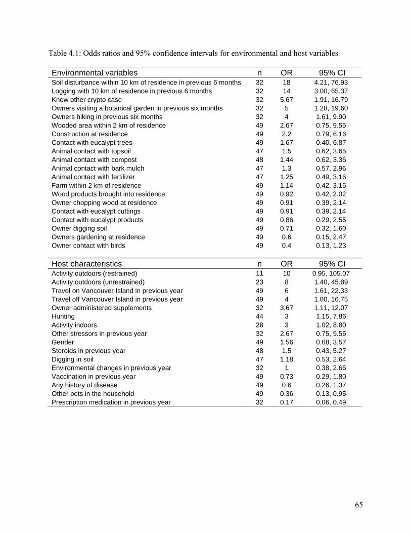
Table 4.1: Odds ratios and 95% confidence intervals for environmental and host variables
Environmental variables n OR 95% CI Soil disturbance within 10 km of residence in previous 6 months 32 18 4.21, 76.93 Logging with 10 km of residence in previous 6 months 32 14 3.00, 65.37 Know other crypto case 32 5.67 1.91, 16.79 Owners visiting a botanical garden in previous six months 32 5 1.28, 19.60 Owners hiking in previous six months 32 4 1.61, 9.90 Wooded area within 2 km of residence 49 2.67 0.75, 9.55 Construction at residence 49 2.2 0.79, 6.16 Contact with eucalypt trees 49 1.67 0.40, 6.87 Animal contact with topsoil 47 1.5 0.62, 3.65 Animal contact with compost 48 1.44 0.62, 3.36 Animal contact with bark mulch 47 1.3 0.57, 2.96 Animal contact with fertilizer 47 1.25 0.49, 3.16 Farm within 2 km of residence 49 1.14 0.42, 3.15 Wood products brought into residence 49 0.92 0.42, 2.02 Owner chopping wood at residence 49 0.91 0.39, 2.14 Contact with eucalypt cuttings 49 0.91 0.39, 2.14 Contact with eucalypt products 49 0.86 0.29, 2.55 Owner digging soil 49 0.71 0.32, 1.60 Owners gardening at residence 49 0.6 0.15, 2.47 Owner contact with birds 49 0.4 0.13, 1.23 Host characteristics n OR 95% CI Activity outdoors (restrained) 11 10 0.95, 105.07 Activity outdoors (unrestrained) 23 8 1.40, 45.89 Travel on Vancouver Island in previous year 49 6 1.61, 22.33 Travel off Vancouver Island in previous year 49 4 1.00, 16.75 Owner administered supplements 32 3.67 1.11, 12.07 Hunting 44 3 1.15, 7.86 Activity indoors 28 3 1.02, 8.80 Other stressors in previous year 32 2.67 0.75, 9.55 Gender 49 1.56 0.68, 3.57 Steroids in previous year 48 1.5 0.43, 5.27 Digging in soil 47 1.18 0.53, 2.64 Environmental changes in previous year 32 1 0.38, 2.66 Vaccination in previous year 49 0.73 0.29, 1.80 Any history of disease 49 0.6 0.26, 1.37 Other pets in the household 49 0.36 0.13, 0.95 Prescription medication in previous year 32 0.17 0.06, 0.49
65
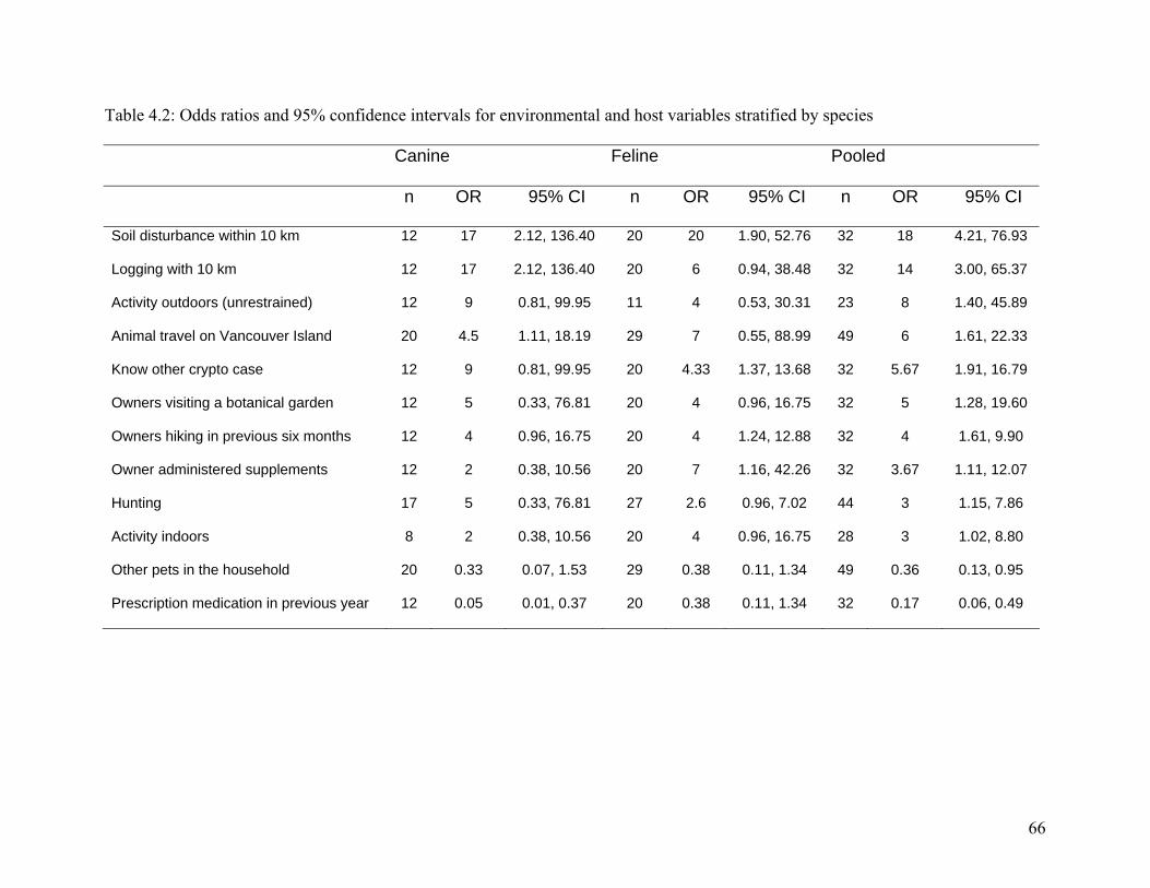
Canine Feline Pooled
n OR 95% CI n OR 95% CI n OR 95% CI
Soil disturbance within 10 km 12 17 2.12, 136.40 20 20 1.90, 52.76 32 18 4.21, 76.93
Logging with 10 km 12 17 2.12, 136.40 20 6 0.94, 38.48 32 14 3.00, 65.37
Activity outdoors (unrestrained) 12 9 0.81, 99.95 11 4 0.53, 30.31 23 8 1.40, 45.89
Animal travel on Vancouver Island 20 4.5 1.11, 18.19 29 7 0.55, 88.99 49 6 1.61, 22.33
Know other crypto case 12 9 0.81, 99.95 20 4.33 1.37, 13.68 32 5.67 1.91, 16.79
Owners visiting a botanical garden 12 5 0.33, 76.81 20 4 0.96, 16.75 32 5 1.28, 19.60
Owners hiking in previous six months 12 4 0.96, 16.75 20 4 1.24, 12.88 32 4 1.61, 9.90
Owner administered supplements 12 2 0.38, 10.56 20 7 1.16, 42.26 32 3.67 1.11, 12.07
Hunting 17 5 0.33, 76.81 27 2.6 0.96, 7.02 44 3 1.15, 7.86
Activity indoors 8 2 0.38, 10.56 20 4 0.96, 16.75 28 3 1.02, 8.80
Other pets in the household 20 0.33 0.07, 1.53 29 0.38 0.11, 1.34 49 0.36 0.13, 0.95
Prescription medication in previous year 12 0.05 0.01, 0.37 20 0.38 0.11, 1.34 32 0.17 0.06, 0.49
66
Table 4.2: Odds ratios and 95% confidence intervals for environmental and host variables stratified by species
4.5. References
1. Sorrell TC. Cryptococcus neoformans variety gattii. Med Mycol 2001; 39 (2):155-68. 2. Kwon-Chung KJ, Polacheck I, Bennett JE. Improved diagnostic medium for separation of Cryptococcus neoformans var. neoformans (serotypes A and D) and Cryptococcus neoformans var. gattii (serotypes B and C). J Clin Microbiol 1982; 15 (3):535-7. 3. Katsu M, Kidd S, Ando A, Moretti-Branchini ML, Mikami Y, Nishimura K, et al. The internal transcribed spacers and 5.8S rRNA gene show extensive diversity among isolates of the Cryptococcus neoformans species complex. FEMS Yeast Res 2004; 4 (4-5):377-88. 4. Kwon-Chung J, Boekhout, T., Fell, J., Diaz, M. Proposal to conserve the name Cryptococcus gattii against C. hondurianus and C. bacillisporus (Basidiomycota, Hymenomycetes, Tremellomycetidae). Taxon 2002; 51:804-806. 5. Kwon-Chung KJ, Bennett JE. Epidemiologic differences between the two varieties of Cryptococcus neoformans. Am J Epidemiol 1984; 120 (1):123-30. 6. Ellis DH, Pfeiffer TJ. Natural habitat of Cryptococcus neoformans var. gattii. J Clin Microbiol 1990; 28 (7):1642-4. 7. Chen S, Sorrell T, Nimmo G, Speed B, Currie B, Ellis D, et al. Epidemiology and host- and variety-dependent characteristics of infection due to Cryptococcus neoformans in Australia and New Zealand. Australasian Cryptococcal Study Group. Clin Infect Dis 2000; 31 (2):499-508. 8. Stephen C, Lester S, Black W, Fyfe M, Raverty S. Multispecies outbreak of cryptococcosis on southern Vancouver Island, British Columbia. Can Vet J 2002; 43 (10):792-4. 9. Kidd SE, Hagen F, Tscharke RL, Huynh M, Bartlett KH, Fyfe M, et al. A rare genotype of Cryptococcus gattii caused the cryptococcosis outbreak on Vancouver Island (British Columbia, Canada). Proc Natl Acad Sci U S A 2004; 101 (49):17258-63. 10. Bartlett K, Fyfe, MW, MacDougall, LA. Environmental Cryptococcus neoformans var. gattii in British Columbia, Canada. Am J Resp Crit Care Med 2003; 167 (7):A499. 11. Lester SJ, Kowalewich NJ, Bartlett KH, Krockenberger MB, Fairfax TM, Malik R. Clinicopathologic features of an unusual outbreak of cryptococcosis in dogs, cats, ferrets and a bird: 38 cases (January 2003 to July 2003). J Am Vet Med Assoc 2004; 225 (11):1716-1722. 12. Kerl ME. Update on canine and feline fungal diseases. Vet Clin North Am Small Anim Pract 2003; 33 (4):721-47. 13. Rothman KJ, Greenland S. Modern epidemiology. 2nd ed. Philadelphia: Lippincott-Raven; 1998. 14. Ellis DH, Pfeiffer TJ. Ecology, life cycle, and infectious propagule of Cryptococcus neoformans. Lancet 1990; 336 (8720):923-5. 15. Greene CE. Infectious diseases of the dog and cat. 2nd ed. Philadelphia: W.B. Saunders; 1998. 16. Malik R, Wigney DI, Muir DB, Love DN. Asymptomatic carriage of Cryptococcus neoformans in the nasal cavity of dogs and cats. J Med Vet Mycol 1997; 35 (1):27-31. 17. Duncan C, Stephen C, Lester S, Bartlett K. Sub-clinical infection and asymptomatic carriage of Cryptococcus gattii in dogs and cats during an outbreak of cryptococcosis. Med Mycol 2005; In Press.
67
18. Connolly JH, Krockenberger MB, Malik R, Canfield PJ, Wigney DI, Muir DB. Asymptomatic carriage of Cryptococcus neoformans in the nasal cavity of the koala (Phascolarctos cinereus). Med Mycol 1999; 37 (5):331-8. 19. Seaton RA, Hamilton AJ, Hay RJ, Warrell DA. Exposure to Cryptococcus neoformans var. gattii--a seroepidemiological study. Trans R Soc Trop Med Hyg 1996; 90 (5):508-12. 20. Flatland B, Greene RT, Lappin MR. Clinical and serologic evaluation of cats with cryptococcosis. J Am Vet Med Assoc 1996; 209 (6):1110-3. 21. Gerds-Grogan S, Dayrell-Hart B. Feline cryptococcosis: a retrospective evaluation. J Am Anim Hosp Assoc 1997; 33 (2):118-22. 22. Malik R, Wigney DI, Muir DB, Gregory DJ, Love DN. Cryptococcosis in cats: clinical and mycological assessment of 29 cases and evaluation of treatment using orally administered fluconazole. J Med Vet Mycol 1992; 30 (2):133-44. 23. Jacobs GJ, Medleau L, Calvert C, Brown J. Cryptococcal infection in cats: factors influencing treatment outcome, and results of sequential serum antigen titers in 35 cats. J Vet Intern Med 1997; 11 (1):1-4. 24. Malik R, Dill-Macky E, Martin P, Wigney DI, Muir DB, Love DN. Cryptococcosis in dogs: a retrospective study of 20 consecutive cases. J Med Vet Mycol 1995; 33 (5):291-7. 25. Sutton RH. Cryptococcosis in dogs: a report on 6 cases. Aust Vet J 1981; 57 (12):558-64. 26. Mak S, Duncan C, Bartlett K, Stephen C, Fyfe M, MacDougall L. Using GIS to track cryptococcosis in BC. In: GisVet; 2004 June; Guelph, ON; 2004. 27. Mancianti F, Giannelli C, Bendinelli M, Poli A. Mycological findings in feline immunodeficiency virus-infected cats. J Med Vet Mycol 1992; 30 (3):257-9. 28. Speed B, Dunt D. Clinical and host differences between infections with the two varieties of Cryptococcus neoformans. Clin Infect Dis 1995; 21 (1):28-34; discussion 35-6. 29. Mitchell DH, Sorrell TC, Allworth AM, Heath CH, McGregor AR, Papanaoum K, et al. Cryptococcal disease of the CNS in immunocompetent hosts: influence of cryptococcal variety on clinical manifestations and outcome. Clin Infect Dis 1995; 20 (3):611-6. 30. Pfeiffer TJ, Ellis DH. Environmental isolation of Cryptococcus neoformans var. gattii from Eucalyptus tereticornis. J Med Vet Mycol 1992; 30 (5):407-8. 31. Nishikawa MM, Lazera MS, Barbosa GG, Trilles L, Balassiano BR, Macedo RC, et al. Serotyping of 467 Cryptococcus neoformans isolates from clinical and environmental sources in Brazil: analysis of host and regional patterns. J Clin Microbiol 2003; 41 (1):73-7. 32. Montenegro H, Paula CR. Environmental isolation of Cryptococcus neoformans var. gattii and C. neoformans var. neoformans in the city of Sao Paulo, Brazil. Med Mycol 2000; 38 (5):385-90. 33. Chakrabarti A, Jatana M, Kumar P, Chatha L, Kaushal A, Padhye AA. Isolation of Cryptococcus neoformans var. gattii from Eucalyptus camaldulensis in India. J Clin Microbiol 1997; 35 (12):3340-2. 34. Pfeiffer T, Ellis D. Environmental isolation of Cryptococcus neoformans gattii from California. J Infect Dis 1991; 163 (4):929-30. 35. Lazera MS, Salmito Cavalcanti MA, Londero AT, Trilles L, Nishikawa MM, Wanke B. Possible primary ecological niche of Cryptococcus neoformans. Med Mycol 2000; 38 (5):379-83. 36. Fortes ST, Lazera MS, Nishikawa MM, Macedo RC, Wanke B. First isolation of Cryptococcus neoformans var. gattii from a native jungle tree in the Brazilian Amazon rainforest. Mycoses 2001; 44 (5):137-40.
68
37. Callejas A, Ordonez N, Rodriguez MC, Castaneda E. First isolation of Cryptococcus neoformans var. gattii, serotype C, from the environment in Colombia. Med Mycol 1998; 36 (5):341-4. 38. Chen SC, Currie BJ, Campbell HM, Fisher DA, Pfeiffer TJ, Ellis DH, et al. Cryptococcus neoformans var. gattii infection in northern Australia: existence of an environmental source other than known host eucalypts. Trans R Soc Trop Med Hyg 1997; 91 (5):547-50. 39. Sorrell TC, Brownlee AG, Ruma P, Malik R, Pfeiffer TJ, Ellis DH. Natural environmental sources of Cryptococcus neoformans var. gattii. J Clin Microbiol 1996; 34 (5):1261-3. 40. Sorrell TC, Chen SC, Ruma P, Meyer W, Pfeiffer TJ, Ellis DH, et al. Concordance of clinical and environmental isolates of Cryptococcus neoformans var. gattii by random amplification of polymorphic DNA analysis and PCR fingerprinting. J Clin Microbiol 1996; 34 (5):1253-60. 41. Laurenson IF, Trevett AJ, Lalloo DG, Nwokolo N, Naraqi S, Black J, et al. Meningitis caused by Cryptococcus neoformans var. gattii and var. neoformans in Papua New Guinea. Trans R Soc Trop Med Hyg 1996; 90 (1):57-60. 42. Laurenson IF, Lalloo DG, Naraqi S, Seaton RA, Trevett AJ, Matuka A, et al. Cryptococcus neoformans in Papua New Guinea: a common pathogen but an elusive source. J Med Vet Mycol 1997; 35 (6):437-40. 43. Swinne D, Taelman H, Batungwanayo J, Bigirankana A, Bogaerts J. [Ecology of Cryptococcus neoformans in central Africa]. Med Trop (Mars) 1994; 54 (1):53-5. 44. Hotzel H, Kielstein P, Blaschke-Hellmessen R, Wendisch J, Bar W. Phenotypic and genotypic differentiation of several human and avian isolates of Cryptococcus neoformans. Mycoses 1998; 41 (9-10):389-96. 45. Kielstein P, Hotzel H, Schmalreck A, Khaschabi D, Glawischnig W. Occurrence of Cryptococcus spp. in excreta of pigeons and pet birds. Mycoses 2000; 43 (1-2):7-15. 46. Kumlin U, Olsen B, Granlund M, Elmqvist LG, Tarnvik A. Cryptococcosis and starling nests. Lancet 1998; 351 (9110):1181. 47. Irokanulo EO, Makinde AA, Akuesgi CO, Ekwonu M. Cryptococcus neoformans var neoformans isolated from droppings of captive birds in Nigeria. J Wildl Dis 1997; 33 (2):343-5. 48. Passoni LF, Wanke B, Nishikawa MM, Lazera MS. Cryptococcus neoformans isolated from human dwellings in Rio de Janeiro, Brazil: an analysis of the domestic environment of AIDS patients with and without cryptococcosis. Med Mycol 1998; 36 (5):305-11. 49. Li A, Nishimura K, Taguchi H, Tanaka R, Wu S, Miyaji M. The isolation of Cryptococcus neoformans from pigeon droppings and serotyping of naturally and clinically sourced isolates in China. Mycopathologia 1993; 124 (1):1-5. 50. Malik R, Krockenberger MB, Cross G, Doneley R, Madill DN, Black D, et al. Avian cryptococcosis. Med Mycol 2003; 41 (2):115-24.
69
5. Sub-clinical infection and asymptomatic carriage of Cryptococcus gattii in dogs and cats during an outbreak of cryptococcosis.
5.1. Introduction
Cryptococcosis is a fungal disease found worldwide in human and animal populations. The
epidemiology of clinical disease depends largely on the species of infecting organism.
Cryptococcus neoformans var. grubii (serotype A) and C. neoformans var. neoformans (serotype
D) are globally distributed and infect predominantly immunocompromised hosts (1).
Cryptococcus gattii (serotypes B and C) has recently been recognized as a species distinct from
C. neoformans based on molecular and mating type characteristics (2). Clinical disease caused
by C. gattii has not been associated with a suppressed immune system (3) and has historically
been restricted to the tropics and sub-tropics, particularly in association with eucalyptus trees (4-
6). The organism is not contagious and considered infectious only as a desiccated yeast cell or
basidiospore as found in the environment (3). Previously only C. neoformans has been routinely
isolated from human or animal cases of cryptococcosis in Canada without a travel history to a
region in which C. gattii is endemic.
In 2001 an increased incidence of cryptococcosis was identified on southern Vancouver Island,
British Columbia (BC), Canada. Clinical disease was recognized in humans, dogs, cats, ferrets,
porpoises, and llamas resulting in the first multi-species outbreak of cryptococcosis (7). All
animal and human isolates available for culture from BC were C. gattii serotype B. Since 1999
over 200 human and animal cases of cryptococcosis have been reported and the species list has
been expanded to include birds and a horse (8). Cases are clustered on the east coast of the
70
island within the Coastal Douglas-fir (CDF) biogeoclimatic zone. Since 2001 C. gattii has been
repeatedly and consistently isolated from soil, air and vegetation within the CDF zone (9).
Infection in animals is thought to be the result of inhalation of the airborne environmental fungi
and subsequent colonization of the nasal cavity and paranasal sinuses (6, 10-12). In Australia it
has been reported that dogs, cats (13) and koalas (14) can carry C. neoformans in their upper
respiratory tract asymptomatically, suggesting that nasal colonization may be much more
common than clinical disease. The following study was conducted to identify the prevalence, and
outcomes, of sub-clinical cryptococcosis and asymptomatic carriage of C. gattii in the nasal
passages of dogs and cats within the CDF zone of Vancouver Island, BC.
5.2. Materials and Methods
5.2.1. Study population
Five veterinary clinics in four cities (figure 5.1) were selected as sampling sites based on
caseload, identification of clinical cryptococcosis in their service area, location within the CDF
region and willingness to participate. At each clinic a fixed weekday was selected where the
daily caseload included both medical and surgical appointments. Owners presenting a dog or cat
over six months of age to the veterinary clinic for reasons other than euthanasia or previously
diagnosed clinical cryptococcosis were offered the opportunity to participate in the study.
Owners completing a consent form and a brief information sheet could elect to have both a nasal
swab and a blood sample collected or one of the two. Sampling was carried out on average once
every three weeks from June to December 2003.
71
5.2.2. Animal information
Information collected from the owner included the animal’s age, sex, breed and the duration of
time it had lived within the CDF zone on Vancouver Island. Owners were asked if, in the last
year, their pet had shown signs suggestive of respiratory tract disease including sneezing,
coughing or nasal discharge, central nervous system disease including behavioral changes,
seizures, poor coordination or balance problems, skin lumps or vision changes. Owners were
also asked to note other health problems observed in the last year that were not included on the
list. Finally owners rated how they perceived the overall health of their pet as one of very poor,
poor, good or very good. Reason for bringing the animal to the veterinarian and body weight of
dogs was also recorded.
5.2.3. Animal sampling
Superficial nasal cultures were collected from unsedated animals. In dogs a single Starplex
StarSwab II (Starplex Scientific, Etobicoke, ON) moistened in transport media was inserted 0.5
to 2 cm into both nasal vestibules and rotated on the mucosa. In cats a similar procedure was
conducted using a Calcium Alginate Fiber Tipped Ultrafine Aluminum Applicator swab (Calgi-
swab, Fisher Scientific, Toronto, ON) moistened with sterile saline (0.9% NaCl). The StarSwabs
were placed in the associated transport media and the Calgi-swabs were placed in a 1.5 ml
Eppendorf microcentrifuge tube (Brinkmann Instruments, Westbury, NY) containing ~0.2 cc
sterile saline. Animals undergoing general anesthesia for any procedure received a second,
deeper nasal swab using a Calgi-swab. The swab was rotated on the nasal mucosa of both nasal
passages at approximately the level of the medial canthus of the eye.
72
Blood was collected using standard venipuncture technique. A minimum of 1ml of blood was
collected from each animal participating in the study, allowed to clot for 15-30 minutes at room
temperature and centrifuged to separate the serum.
5.2.4. Culture
Culture swabs were plated onto Bird Seed Agar and incubated at 30 o C. Plates were examined at
48 hours, then daily for seven days. Cryptococcus neoformans and C. gattii selectively use
caffeic acid in the medium to produce melanin, resulting in brown colonies. Suspect colonies
were transferred to Malt Extract Agar (MEA) and Canavanine-glycine-bromothymol blue (CGB)
agar. Cryptococcus gattii turns the CGB agar cobalt blue while C. neoformans remain negative.
Colonies growing on MEA were serotyped using capsular antibodies (Crypto-check, Iatron
Laboratories, Japan). Biochemical identification was achieved using API 20 AUX strips
(bioMérieux, St. Laurent, Quebec).
5.2.5. Antigen test
Samples were treated with pronase (15) prior to the use of a latex cryptococcal antigen
agglutination test for the measurement of cryptococcal antigen in the sera (Cryptococcal Antigen
Latex Agglutination System (CALAS); Meridian Bioscience, Inc., Cincinnati, Ohio). The
CALAS test cannot identify the organism beyond the level of the genus. Animals with a titer ≥
1:2 were considered positively infected with Cryptococcus spp. (16).
5.2.6. Statistical analysis
When C. gattii was isolated from either the deep or superficial swab the animal was considered
positive on nasal culture. Animals testing positive on either a nasal culture or antigen test were
given an overall rating of positive for Cryptococcus spp. Results were stratified by species. The
73
owner-evaluated overall health rating was converted to a dichotomous variable where scores of
very poor and poor were combined to below average; good and very good were combined to
above average. Dogs were classified into two size categories based on body weight above or
below 15 kilograms.
Descriptive and comparative statistics were computed using SPSS 12.0 (SPSS Inc., Chicago, Il.,
USA). Odds ratios and 95% confidence intervals were used to evaluate the association between
positive test results and animals presenting to the clinic for illness compared to routine
procedures, owner perceived health status, sex, and health problems in the previous year. The
Kolmogorov-Smirnov Z test was used to evaluate the normality of the distribution of continuous
variables including age and duration of residence within the CDF zone. For variables with
normal distribution mean values were reported and the T-test was used to compare means
between positive and negative animals. Where the results of the Kolmogorov-Smirnov Z test
were significant at the 5% level median values were reported and the Mann-Whitney U (MWU)
test was used to compare means between positive and negative animals. The relative proportions
of infected species were compared using odds ratios and 95% confidence intervals. The
prevalence of positive animals in the four sampling cities was compared using a Chi-Square test.
Kappa statistics were calculated to determine the agreement between the two nasal swabs and
between the antigen test and nasal culture.
5.2.7. Follow-up testing
Owners of positive animals identified as sub-clinically infected with Cryptococcus spp. or
carrying C. gattii in their nasal passages were asked to bring their animal to the veterinarian for
follow-up testing as often as possible to a maximum of once a month. Follow-up testing was
74
available until October 2004. At each follow-up visit a serum sample and superficial nasal swab
was collected for CALAS testing and fungal culture. Serial 10 fold dilutions were carried out on
serum samples; when higher dilutions showed agglutination and dilutions were large,
intermediate twofold dilutions were run in an attempt to more accurately determine an endpoint.
5.3. Results
5.3.1. Initial testing
Of serum samples collected from 84 cats, six (7.1%) had an antigen titer ≥ 1:2 (Table 5.1).
Titers ranged from 1:2 to 1:200. Superficial nasal cultures of 94 cats identified three (3.2%)
animals with C. gattii serotype B in the nasal vestibule while deep nasal swabs from 13 cats
under general anesthesia identified two (15.4%) animals with C. gattii serotype B in their nasal
cavity. Overall 7 (7.4%) cats tested positive on one or more tests. One of the seven cats resided
in the Parksville area, five in Nanaimo and one in Duncan but the proportion of cats positive on
one or more tests was not statistically different between cities (p=0.122).
The mean age of cats testing positive on one or more test was 8 years (SD 7.0 years, range 0.7-18
years) and was not significantly different from cats who were not positive on any test performed
(T-test, p=0.54). The median time positive cats had resided in the CDF zone of Vancouver
Island was 5.0 years (range 0.5-18 years) which was not statistically different from the negative
cats (median 4.8, range 0.5-16, MWU p=0.44).
The odds of being positive on one or more test were not statistically different for male cats
relative to females (OR 1.27, 95% CI 0.27, 6.03). The odds of a positive test result was 3.33
75
(95% CI 0.320, 34.71) times greater in cats with an owner perceived health status of poor
relative to good, however, this result was not statistically significant. The odds of a positive test
result was increased in cats with a history of respiratory signs (OR 1.46, 95% CI 0.16, 13.56),
central nervous system symptoms (OR 2.13, 95% CI 0.22, 20.49), skin lumps (OR 2.28, 95% CI
0.24, 22.13) and vision changes (OR 5.50, 95% CI 0.45, 67.49) in the last year relative to those
without these symptoms; however, none of these results were statistically significant. The
presence of other health problems in the previous 12 months was slightly protective (OR 0.68,
95% CI 0.08, 5.858) but this result was not statistically different compared to cats without other
health problems. Cats testing positive on one or both tests for Cryptococcus spp. were less likely
to have presented to the veterinarian for owner perceived illness verses routine veterinary
procedures (OR 0.15, 95% CI 0.02, 1.32); however, this result was not statistically significant.
The breed distribution of the positive cats consisted of five domestic shorthairs, one domestic
longhair and one Himalayan.
Two of 266 (0.8%) dogs had an antigen titer of 1:2 (Table 5.1). Of 280 superficial nasal cultures
C. gattii serotype B was isolated from three (1.1%) dogs. Cryptococcus gattii was not isolated
from any of the 34 dogs from which a deep nasal swab was collected. Overall five (1.7%) dogs
tested positive on one of the tests, no dogs were considered positive on more than one test. Four
of the five dogs positive on any of the tests were from the Duncan area and the remaining
positive dog was in Parksville. This difference in the prevalence of positive dogs by city was
statistically significant (p=0.03).
76
The median age of dogs positive on either test was 6.6 years (range 3.4-11.6 years) which did not
differ statistically from the remainder of the dogs tested (median 5.8, range 0.5-16.5, MWU
p=0.46). Positive dogs had resided in the CDF zone for a median of 5 years (range 3.4-10 years)
which was not statistically different from the negative dogs (median 4.0, range 0.5-16.0, MWU
p=0.51). The odds of a positive test was not statistically different for males relative to females
(OR 0.89, 95% CI 0.15, 5.42). The odds of a positive test result was 3.99 (95% CI 0.44, 36.01)
times greater in dogs with an owner perceived health status of poor relative to good, however,
this result was not statistically significant. The odds of a positive test result was increased in
dogs with a history of respiratory signs (OR 4.09, 95% CI 0.43, 38.79), central nervous system
symptoms (OR 1.78, 95% CI 0.21, 15.38), skin lumps (OR 1.41, 95% CI 0.15, 12.88), vision
problems (OR 3.17, 95% CI 0.36, 28.13) and other health problems (OR 2.37, 95% CI 0.39,
14.52) but these results were not statistically significant. The odds of dogs presenting to the
veterinarian for owner perceived illness being positive for Cryptococcus spp. in was 3.17 (95%
CI 0.35, 28.72) times that of animals brought to the clinic for routine veterinary procedures. Dog
breeds that tested positive included two Labrador Retriever crosses, one German Shepherd, one
Jack Russell Terrier and one Toy Poodle. There was no significant difference in the proportion
of dogs testing positive above and below 15 kilograms (OR 1.46, 95% CI 0.24, 8.92).
Cats were 10.15 times (95% CI OR 2.01, 51.31) more likely to be positive on the antigen test
than dogs. Combination of the superficial and deep nasal culture results into a single
dichotomous variable of positive or negative on culture revealed that cats were 4.10 (95% CI
0.90, 18.68) times more likely than dogs to carry C. gattii in their nasal cavity, however this
result was not statistically significant at the 5% level. When the results of the antigen test and
77
nasal cultures were combined into a single positive or negative variable the difference between
species was significant with cats being 4.42 times (95% CI 1.37, 14.28) more likely than dogs to
be positive on one or both tests.
Three of 12 (25%) positive animals were positive on both the antigen test and nasal culture.
Four of 12 (33%) of animals had a positive nasal culture but negative antigen test and 5 of 12
(42%) were positive on antigen test alone. A computed kappa of 0.39 (95% CI 0.28, 0.49)
suggests only fair agreement between the serum antigen test and nasal culture. Forty-seven
animals had both deep and superficial swabs of which 44 were negative on both cultures, one
was positive on both cultures and one animal was positive on each of the deep and superficial
cultures. The kappa statistic of 0.48 (95% CI 0.19, 0.76) suggests moderate agreement between
the two swab techniques.
5.3.2. Follow-up testing
Twelve animals positive on initial testing had between one and four follow-up samples collected;
the results for each individual animal are shown in table 5.2. Animals 1-7 are cats and 8-12 are
dogs. Of the seven cats, six had a titer on one or more test dates; five of the seven had the
organism isolated from their nasal cavity. One cat (#6) had the organism isolated from its nasal
cavity on two of four occasions but never had a positive titer. Two cats (#’s 5 & 7) had positive
titers on multiple occasions but the organism was never isolated from their nasal cavities. Four
cats (#’s 1, 2, 3 & 4) had both a positive titer and organism in their nasal cavities on one or more
occasions. Titers in cats ranged from 1:2 to 1:2500.
78
Two cats (#’s 1 & 2) progressed to clinical disease. Case 1 was a 19 year old, spayed female
domestic shorthair cat presented initially to the veterinary clinic for illness. Upon initial
examination the owners reported their cat to be in poor overall health with severe arthritis and
regular constipation. In the previous year the owner noted that the cat had a chronic sneeze. The
cat had lived on Vancouver Island all of its life. In February 2004 the cat showed central
nervous symptoms including ataxia and seizures and was humanely euthanized.
Case 2 was a 15 year old, neutered male domestic shorthair cat presented to the veterinarian for
annual vaccination. Upon entry into the study the owners reported the cats overall health to be
very good and noted that in the previous year the cat had no symptoms suggestive of
cryptococcosis. The cat had lived on Vancouver Island all of his life. Upon initial examination
the cat was deemed healthy and received vaccination; although the owners reported regular
sneezing. In late November 2003 the cat showed symptoms of respiratory tract infection
including nasal discharge and sneezing and was started on antifungal therapy in December 2003.
Ten months after initiation of therapy the CALAS titer was negative.
Of the five dogs, two had a positive titer at one or more test dates, four of the five had organism
isolated from their nasal cavity. Three dogs (#’s 10, 11 & 12) had the organism isolated from
their nose on initial testing but never on subsequent tests. One dog (#8) had a single positive
antigen titer and organism isolated from its nasal cavity on a single occasion, but not on the same
date. One dog (#9) had a titer of 1:2 on initial testing but all subsequent CALAS tests were
negative and C. gattii was never isolated from its nasal passages. A CALAS result of 1:2 was
the only titer detected in dogs.
79
5.4. Discussion
Identification of sub-clinical infection and nasal colonization of dogs and cats with Cryptococcus
spp. is an important step in the characterization of the outbreak of clinical cryptococcosis on
Vancouver Island. Of animals sampled within the CDF zone of Vancouver Island 7.4% of cats
and 1.7% of dogs has either a positive nasal culture or antigen titer indicating that they were
colonized by or infected with Cryptococcus spp.
On physical examination none of the animals identified as positive on either test showed signs
consistent with cryptococcal disease. The most common presentations of feline and canine
cryptococcosis include respiratory, central nervous system, dermal or ocular symptoms (17-19).
Owners of positive dogs and cats reported a slightly increased prevalence of symptoms
suggestive of cryptococcosis within the previous 12 months, however these results were not
statistically significant. The odds of an animal testing positive was increased in animals
considered to be in below average health by their owners, however, this result was not
statistically significant and positive animals were equally likely to have presented to the
veterinarian for routine procedures as for owner-perceived illness.
Cryptococcus gattii serotype B was isolated from 4.2% and 1.1% of cats and dogs, respectively.
Although superficial nasal swabs were assumed to confer good agreement with a nasal flush in
koalas (14) numerical results were not reported and extrapolation of this assumption across
species should be made with caution. In this study there was a wide confidence interval with
only moderate agreement between the two swab techniques. The lack of pattern in the disparity
between deep and superficial swabs suggests that both samples may underestimate true nasal
80
colonization. Correspondingly there was only fair agreement between the antigen test and nasal
culture. The kappa statistic is highly dependent on the true prevalence of disease in the
population and where prevalence approaches one or zero kappa is sharply reduced (20). Further
studies on a cohort of animals with a higher prevalence of colonization and infection are required
to evaluate test agreement more meaningfully.
Of the animals positive on one or more tests 33% had a positive nasal culture without a positive
antigen titer. Studies of presumed healthy animals in Australia recovered C. neoformans and C.
gattii from cats, dogs (13) and koalas (14, 21). Based on the lack of cryptococcal antigen or
pathology of the nasal cavity these studies concluded that Cryptococcus spp. can colonize the
nasal passage of animals without an associated local or systemic infection. In contrast 25% of
animals tested in BC that had C. gattii in their nasal cavities also had antigen in their serum
suggesting sub-clinical infection versus nasal colonization. Forty-two percent of animals
positive on any test had an antigen titer without a positive nasal culture. The CALAS test has
been reported to have high specificity in diseased cats and dogs (22, 23) making false positive
reactions unlikely; however, effectiveness of the CALAS test in asymptomatic animals has not
been evaluated. Furthermore, the sensitivity of nasal culture in colonized animals is unknown
and it is possible that the organism was missed during the nasal swab or that the source of
antigen was not in the nasal passage.
Overall cats had significantly greater odds of testing positive on either culture or antigen test or
antigen test alone when compared to dogs. Cats had increased odds of carrying C. gattii in their
nasal cavity but this result was not statistically significant. Given the low number of positive test
81
results this study may lack the power necessary to identify a statistical difference.
Cryptococcosis is the most common systemic mycoses of cats (19) and clinical disease has been
reported with equal or greater frequency in cats than in dogs (18). Likewise, on Vancouver
Island the reported number of clinical cases in cats outweigh that of dogs (8).
Previously published studies on risk factors for animal cryptococcosis focus only on clinical
cases. Cryptococcal disease has been reportedly been more common in young large dogs (12,
24) and male cats (10, 25, 26), potentially for behavioral reasons. This study failed to identify
sex, age or size of dog as statistically significant factors for asymptomatic infection or
colonization by the organism. Elsewhere environmental exposure to infectious organism is
considered a risk factor for cryptococcal disease (12, 27). While C. gattii has only been
identified within the CDF zone on Vancouver Island (9) the duration of time dogs and cats lived
within the region was not a risk factor for asymptomatic infection or colonization. Previously
reported risk factors may be restricted to animals that become clinically ill and not apply to
asymptomatic or otherwise healthy animals. Subsequent investigations into risk factors for
asymptomatic infections should include a larger sample size to increase the study power.
Because the distribution of cat and dog breeds within the study population is unknown the effect
of breed cannot be evaluated.
All of the sampling clinics lie within the CDF zone but the highest proportions of positive
animals were in Duncan and Nanaimo which are located in the center of the testing area. It is
interesting to note that no animals tested positive in Victoria which lies on the southern most
extreme of the CDF zone. In Australia koalas have been used to successfully identify geographic
82
areas with a high-grade presence of C. gattii in the environment (21). Further sampling of dogs
and cats at the edge of the CDF zone in combination with environmental testing may identify
companion animals as a similar sentinel.
Follow-up testing of positive animals revealed that dogs and cats can clear the organism, remain
sub-clinically infected or progress to overt disease. Cryptococcus gattii was initially isolated
from the nasal cavity of three dogs without antigenemia; however the organism was never
recognized on subsequent testing. A single cat had C. gattii isolated from the nasal cavity on
two of four visits while cryptococcal antigen was never found in its serum. These results suggest
the nasal passages of animals residing within a region where environmental C. gattii is present,
may be transiently colonized by the organism without rapid progression to infection. Failure to
re-isolate the organism from any of the dogs suggests that the presence of the organism in the
nose is relatively brief; or that the swab technique is not sufficient to capture the organism at
each sampling interval. Cryptococcus gattii was isolated twice from the cat suggesting better
recovery of the swab, more persistent colonization or re-exposure of the animal to airborne fungi.
As the CALAS test detects only circulating antigen, extension of infection beyond the nasal
mucosa is required before the test can be positive. Factors mediating progression from
colonization to infection eliciting antigenemia are unknown although dose response, concurrent
disease or other forms of immunosuppression have been suggested (3, 21).
One dog had an antigen titer of 1:2 on initial testing but not on any follow-up visits,
Cryptococcus spp. was never isolated from its nose. Two cats had positive titers on more than
one occasion but the organism was never isolated from the nasal cavity. The sensitivity of
83
superficial nasal culture in animals is unknown and it is possible that the organism was missed
during the nasal swab, or that the source of antigen was not in the nasal passage. As the
effectiveness of the CALAS test has not been evaluated in asymptomatic animals false positive
results cannot be ruled out. One dog had a low titer with no culture on initial testing; on follow-
up examination C. gattii was isolated from the nasal passage but the CALAS test was negative,
disparities potentially elicited by the aforementioned limitations of the diagnostic tests. These
four animals with antigenemia on one or more occasion were all CALAS negative at the end of
the study, between five and 11 months after the date of the last positive CALAS test. These
results suggest that the animals may have cleared the infection. Demonstration of cryptococcal
antigen in serum or cerebral spinal fluid implies infection with the organism and a titer of 1:2 has
been reported to be clinically relevant in cats (16). Asymptomatic infection has been proposed to
be a self limiting condition in koalas but individuals with low positive titers may harbor foci of
infection that could reactivate (21).
Four cats had both antigenemia and C. gattii in their nasal cavity; two of these progressed to
clinical disease. Both animals had antigen titers greater than other cats in the study and titer
values increased over the sampling period. The highest titer observed in a cat not showing
clinical signs was 1:32. In a cohort study of koalas in Australia all clinical animals documented
in the study had antigen titers ≥ 1:128 and an increased incidence of nasal colonization; while
animals without clinical symptoms had titers ≤ 1:64 (21). The results of this small study suggest
that asymptomatic animals with a titer equal to or less than 1:32 clear the infection while those
with a higher titer go on to become diseased. Further investigation into the relationship between
84
asymptomatic colonization and clinical disease is warranted as clinical cases in BC have been
diagnosed with titers as low as 1:2 (23).
The findings of this study demonstrate the need for a better understanding of sub-clinical
infection and nasal colonization of C. gattii in companion animals. In areas of Australia where
environmental exposure of koalas to C. gattii is high, sub-clinical infection is relatively common
while progression to clinical disease is rare (21). Given the recent emergence of the organism in
southwestern BC more information on environmental load, variables influencing exposure and
risk factors for progression from colonization to clinical disease is warranted.
85
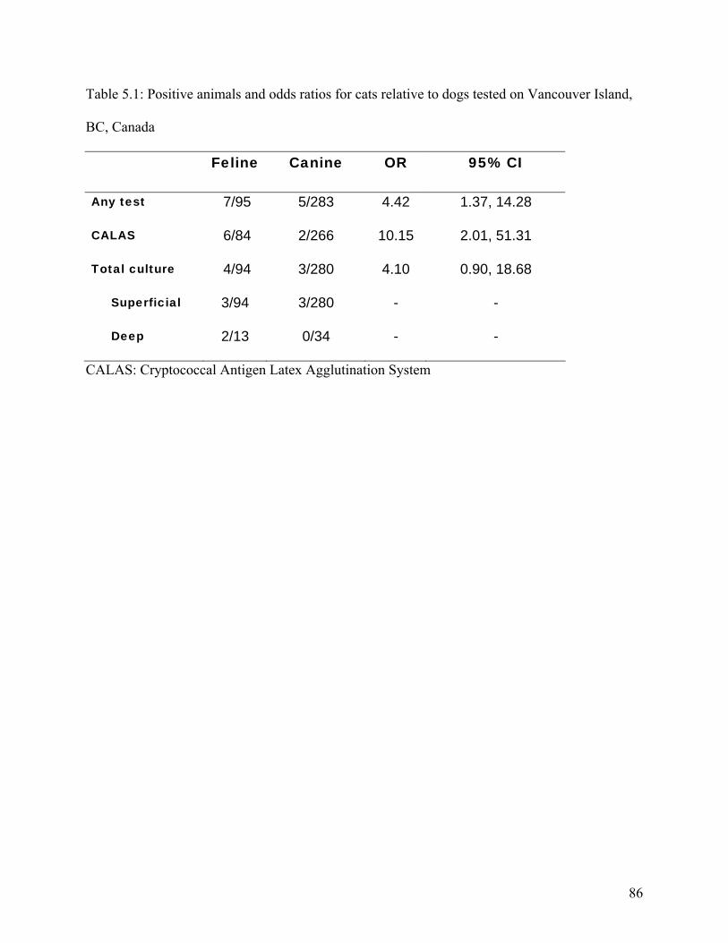
Table 5.1: Positive animals and odds ratios for cats relative to dogs tested on Vancouver Island,
BC, Canada
Feline Canine OR 95% CI
Any test 7/95 5/283 4.42 1.37, 14.28
CALAS 6/84 2/266 10.15 2.01, 51.31
Total culture 4/94 3/280 4.10 0.90, 18.68
Superficial 3/94 3/280 - -
Deep 2/13 0/34 - -
CALAS: Cryptococcal Antigen Latex Agglutination System
86
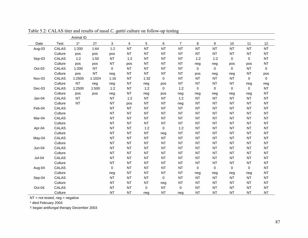
87
Table 5.2: CALAS titer and results of nasal C. gattii culture on follow-up testing Animal ID
Date Test 1* 2† 3 4 5 6 7 8 9 10 11 12 Aug-03 CALAS 1:200 1:64 1:2 NT NT NT NT NT NT NT NT NT
Culture pos pos pos NT NT NT NT NT NT NT NT NT Sep-03 CALAS 1:2 1:50 NT 1:2 NT NT NT 1:2 1:2 0 0 NT
Culture pos pos NT pos NT NT NT neg neg pos pos NT Oct-03 CALAS 1:200 NT 0 NT NT NT NT 0 0 0 NT 0
Culture pos NT neg NT NT NT NT pos neg neg NT pos Nov-03 CALAS 1:2500 1:1024 1:16 NT 1:32 0 NT NT NT NT 0 0
Culture NT neg neg NT neg pos NT NT NT NT neg neg Dec-03 CALAS 1:2500 1:500 1:2 NT 1:2 0 1:2 0 0 0 0 NT
Culture pos pos neg NT neg pos neg neg neg neg neg NT Jan-04 CALAS NT NT 1:2 NT NT 1:2 NT NT NT NT NT
Culture NT NT pos NT NT neg NT NT NT NT NT Feb-04 CALAS NT NT NT NT NT NT NT NT NT NT
Culture NT NT NT NT NT NT NT NT NT NT Mar-04 CALAS NT NT NT NT NT NT NT NT NT NT
Culture NT NT NT NT NT NT NT NT NT NT Apr-04 CALAS NT NT 1:2 0 1:2 NT NT NT NT NT
Culture NT NT NT neg NT NT NT NT NT NT May-04 CALAS NT NT NT NT NT NT NT NT NT NT
Culture NT NT NT NT NT NT NT NT NT NT Jun-04 CALAS NT NT NT NT NT NT NT NT NT NT
Culture NT NT NT NT NT NT NT NT NT NT Jul-04 CALAS NT NT NT NT NT NT NT NT NT NT
Culture NT NT NT NT NT NT NT NT NT NT Aug-04 CALAS 0 NT NT NT NT 0 0 0 0 NT
Culture neg NT NT NT NT neg neg neg neg NT Sep-04 CALAS NT NT NT 0 NT NT NT NT NT NT
Culture NT NT NT neg NT NT NT NT NT NT Oct-04 CALAS NT NT 0 NT 0 NT NT NT NT NT
Culture NT NT neg NT neg NT NT NT NT NT NT = not tested, neg = negative * died February 2004 † began antifungal therapy December 2003
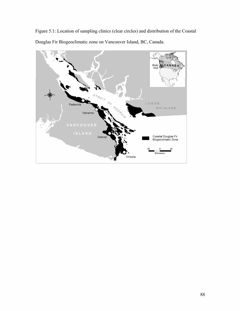
Figure 5.1: Location of sampling clinics (clear circles) and distribution of the Coastal
Douglas Fir Biogeoclimatic zone on Vancouver Island, BC, Canada.
88
5.5. References
1. Speed B, Dunt D. Clinical and host differences between infections with the two varieties of Cryptococcus neoformans. Clin Infect Dis 1995; 21 (1):28-36. 2. Kwon-Chung J, Boekhout, T., Fell, J., Diaz, M. Proposal to conserve the name Cryptococcus gattii against C. hondurianus and C. bacillisporus (Basidiomycota, Hymenomycetes, Tremellomycetidae). Taxon 2002; 51:804-806. 3. Sorrell TC. Cryptococcus neoformans variety gattii. Med Mycol 2001; 39 (2):155-68. 4. Ellis DH. Cryptococcus neoformans var. gattii in Australia. J Clin Microbiol 1987; 25 (2):430-1. 5. Ellis DH, Pfeiffer TJ. Natural habitat of Cryptococcus neoformans var. gattii. J Clin Microbiol 1990; 28 (7):1642-4. 6. Ellis D, Pfeiffer T. The ecology of Cryptococcus neoformans. Eur J Epidemiol 1992; 8 (3):321-5. 7. Stephen C, Lester S, Black W, Fyfe M, Raverty S. Multispecies outbreak of cryptococcosis on southern Vancouver Island, British Columbia. Can Vet J 2002; 43 (10):792-4. 8. Duncan C, Stephen C, Raverty S, Lester S, Fyfe M, Bartlett K. Descriptive epidemiology of a multispecies cluster of Cryptococcus neoformans var. gattii: Veterinary aspects. In: International Veterinary Epidemiology and Economics Conference; 2003; Chile; 2003. 9. Bartlett K, Fyfe, MW, MacDougall, LA. Environmental Cryptococcus neoformans var. gattii in British Columbia, Canada. Am J Resp Crit Care Med 2003; 167 (7):A499. 10. Malik R, Wigney DI, Muir DB, Gregory DJ, Love DN. Cryptococcosis in cats: clinical and mycological assessment of 29 cases and evaluation of treatment using orally administered fluconazole. J Med Vet Mycol 1992; 30 (2):133-44. 11. Levitz SM. The ecology of Cryptococcus neoformans and the epidemiology of cryptococcosis. Rev Infect Dis 1991; 13 (6):1163-9. 12. Malik R, Dill-Macky E, Martin P, Wigney DI, Muir DB, Love DN. Cryptococcosis in dogs: a retrospective study of 20 consecutive cases. J Med Vet Mycol 1995; 33 (5):291-7. 13. Malik R, Martin P, Wigney DI, Church DB, Bradley W, Bellenger CR, et al. Nasopharyngeal cryptococcosis. Aust Vet J 1997; 75 (7):483-8. 14. Connolly JH, Krockenberger MB, Malik R, Canfield PJ, Wigney DI, Muir DB. Asymptomatic carriage of Cryptococcus neoformans in the nasal cavity of the koala (Phascolarctos cinereus). Med Mycol 1999; 37 (5):331-8. 15. Gray LD, Roberts GD. Experience with the use of pronase to eliminate interference factors in the latex agglutination test for cryptococcal antigen. J Clin Microbiol 1988; 26 (11):2450-1. 16. Malik R, McPetrie R, Wigney DI, Craig AJ, Love DN. A latex cryptococcal antigen agglutination test for diagnosis and monitoring of therapy for cryptococcosis. Aust Vet J 1996; 74 (5):358-64.
89
17. Krohne SG. Canine systemic fungal infections. Vet Clin North Am Small Anim Pract 2000; 30 (5):1063-90. 18. Kerl ME. Update on canine and feline fungal diseases. Vet Clin North Am Small Anim Pract 2003; 33 (4):721-47. 19. Gionfriddo JR. Feline systemic fungal infections. Vet Clin North Am Small Anim Pract 2000; 30 (5):1029-50. 20. Thompson WD, Walter SD. A reappraisal of the kappa coefficient. J Clin Epidemiol 1988; 41 (10):949-58. 21. Krockenberger MB, Canfield PJ, Barnes J, Vogelnest L, Connolly J, Ley C, et al. Cryptococcus neoformans var. gattii in the koala (Phascolarctos cinereus): serological evidence for subclinical cryptococcosis. Med Mycol 2002; 40 (3):273-82. 22. Medleau L, Marks MA, Brown J, Borges WL. Clinical evaluation of a cryptococcal antigen latex agglutination test for diagnosis of cryptococcosis in cats. J Am Vet Med Assoc 1990; 196 (9):1470-3. 23. Lester SJ, Kowalewich NJ, Bartlett KH, Krockenberger MB, Fairfax TM, Malik R. Clinicopathologic features of an unusual outbreak of cryptococcosis in dogs, cats, ferrets and a bird: 38 cases (January 2003 to July 2003). J Am Vet Med Assoc 2004; 225 (11):1716-1722. 24. Nelson RW, Couto CG. Small animal internal medicine. 3rd ed. St. Louis, Mo.: Mosby; 2003. 25. Gerds-Grogan S, Dayrell-Hart B. Feline cryptococcosis: a retrospective evaluation. J Am Anim Hosp Assoc 1997; 33 (2):118-22. 26. Jacobs GJ, Medleau L, Calvert C, Brown J. Cryptococcal infection in cats: factors influencing treatment outcome, and results of sequential serum antigen titers in 35 cats. J Vet Intern Med 1997; 11 (1):1-4. 27. Chen S, Sorrell T, Nimmo G, Speed B, Currie B, Ellis D, et al. Epidemiology and host- and variety-dependent characteristics of infection due to Cryptococcus neoformans in Australia and New Zealand. Australasian Cryptococcal Study Group. Clin Infect Dis 2000; 31 (2):499-508.
90
6. Cryptococcus gattii in horses and wildlife of Vancouver Island, British Columbia, Canada
6.1. Introduction
Since 1999, Cryptococcus gattii, a species now distinct from C. neoformans (1), has
emerged as an important pathogen of humans and animals in southwestern British
Columbia (BC) (2-5). Previously only C. neoformans had been isolated from animals or
humans in Canada and C. gattii was thought to be restricted to the tropics and sub tropics
(6, 7). Clinical illness has been identified in humans and numerous animal species in BC
including cats, dogs, ferrets, llamas, porpoises, domestic birds and a horse. Cases to date
have been clustered on the east coast of Vancouver Island; largely within the coastal
Douglas fir (CDF) biogeoclimatic zone (8). Cryptococcus gattii has been routinely
isolated from soil, air and vegetation within the CDF zone since 2001 (4, 9).
Given the airborne nature of this organism, it may be assumed that many species residing
within endemic areas are exposed, but infection has been largely unnoticed.
Asymptomatic carriage of C. gattii has been recognized in companion animal species of
BC; presumably as a result of contact with airborne infectious material (8, 9).
Environmental exposure and asymptomatic colonization of the respiratory tract has been
proposed to be much more common than clinical disease (10, 11), however, variables
influencing the initiation of infection remain unclear. The prevalence of exposure of
humans and animals in BC is unknown.
91
The objective of this study was to identify terrestrial mammalian wildlife species and
horses that have been exposed to or infected with C. gattii on Vancouver Island, BC.
Horses were selected because, on Vancouver Island, they are housed predominantly
outdoors, are dispersed throughout the endemic area, are susceptible to the organism and
public concerns regarding equine cryptococcosis facilitates sampling. Wild mammal
species were targeted because samples were accessible and the majority of domestic
cases on Vancouver Island were mammals.
6.2. Materials and Methods
6.2.1. Wildlife sampling
Between February and August 2004, a deep swab of the nasal mucosa using a Starplex
StarSwab II (Starplex Scientific, Etobicoke, ON) and lung tissue, where possible, were
collected for fungal culture. Sources of live and dead animals included wildlife
rehabilitation facilities, veterinarians, biologists and registered trappers. Any mammalian
species live-trapped or killed and known to reside in the CDF zone on Vancouver Island
between January 1999 and August 2004 were eligible for inclusion in the study. Species
and approximate life stage were recorded along with the location of animal capture when
available.
6.2.2. Equine sampling
All BC veterinary medicine association (BCVMA) registered veterinary clinics servicing
equine clients within the region where clinical cryptococcosis cases had been identified
were contacted for participation. Veterinarians were given maps of their service area that
included sites where C. gattii had been isolated from the environment (Bartlett,
92
unpublished) and an associated 10 km zone around the positive sample locations.
Practitioners were asked to provide names of equine clients residing within the 10 km
zone that would be interested in participating in the study.
Between July 24 and August 9, 2004 samples were collected from horses residing in
identified buffer zones. Material collected included a swab of the nasal mucosa using
StarSwab II (Starplex Scientific, Etobicoke, ON) moistened in transport media, inserted
10-15 cm into both nasal vestibules and rotated on the mucosa. A minimum of 5 ml of
blood was collected from each animal participating in the study using standard
venipuncture technique. Blood was allowed to clot for a minimum of 30 minutes and
centrifuged to separate the serum.
Data collected on each horse tested included age, breed, underlying health problems,
duration lived at sampling location and on Vancouver Island, average time spent outside
per 24 hour period, source of hay and if hay was fed on the ground or in a feeder. The
difference in age, duration of residence on current property and Vancouver Island
between positive and negative horses was evaluated using the Mann-Whitney U test. The
relative number of horses fed on or off of the ground, local or imported hay and housed
outdoors or both in and out were compared using the Fishers exact test.
6.2.3. Laboratory analysis
Culture swabs were plated onto Bird Seed Agar and incubated at 30 o C for 48 hr. Plates
were checked for growth daily for ten days before being regarded as negative. Colonies
conforming to cryptococcal morphology were identified and serotyped using
93
agglutinating antibodies (Crypto-check, Iatron Laboratories, Tokyo, Japan). Lung tissue
was splayed on a sterile surface and dissected to allow access to the interior surface using
a scalpel blade sterilized by alcohol dip and flaming. Internal and external surfaces were
swabbed using a cotton-tipped applicator (Puritan, Fisher Scientific). The applicator was
rolled across a differential agar (Bird Seed Agar) and a rich nutrient agar (Saboraud
Dextrose Agar, BBL). Agar plates were incubated and checked for growth as above.
Serum samples were treated with pronase (12) prior to the use of a latex cryptococcal
antigen agglutination test for the measurement of cryptococcal antigen in the sera
(Cryptococcal Antigen Latex Agglutination System (CALAS); Meridian Bioscience, Inc.,
Cincinnati, Ohio). The CALAS test cannot identify the organism beyond the level of the
genus. Animals with a titer ≥ 1:2 were considered positively infected with Cryptococcus
spp.
6.3. Results
6.3.1. Wildlife sampling
Nasal swabs were collected from 91 individuals representing 14 species; 19 living harbor
seals (Phoca vitulina) and 72 post-mortem samples. A list of all species, the respective
number of isolates and age categories of animals sampled are presented in table 6.1.
Lung tissue was available for culture from 68 animals representing 13 species.
Cryptococcus gattii was isolated from the nasal swab of two Eastern Grey Squirrels
(Sciurus carolinensis) trapped at the same location in the city of Duncan, BC. Lung
tissue was available from one of these squirrels and fungal culture of the tissue yielded no
Cryptococcus spp.
94
6.3.1. Equine sampling
Nasal swabs and serum samples were collected from 260 horses residing within 10 km of
a site where C. gattii had been isolated from the environment. No horses were positive
on the antigen test, but the organism was isolated from the nasal passages of four horses.
All positive horses resided in the area of Duncan, BC with one horse directly adjacent to
the trapping site of the two positive squirrels.
All horses, except for one, had lived on Vancouver Island their entire lives. This horse
had moved to the island approximately six months prior to testing and resided only in the
Duncan area during that time. The median duration of residence on Vancouver Island
was 5 years (minimum 3 months, maximum 9 years) for the positive horses and 8 years
(minimum 3 months, maximum 32 years) for the negative horses; a difference that was
not statistically significant (p=0.198). The median duration of positive horses living on
the property where testing occurred (2.25 years, minimum 3 months, maximum 6 years)
did not differ significantly from that of negative horses (3 years, minimum 3 months,
maximum 23 years). The four positive horses were 4, 6, 9 and 10 years of age; the
median age of positive horses (7.5 years) did not differ significantly from that of horses
without organism in their nose (12 years, minimum 3 months, maximum 35 years,
p=0.202). The proportion of positive horses fed on the ground did not differ statistically
from the negative horses (p=0.264). None of the positive horses had any owner reported
illness or historical medical problems.
95
6.4. Discussion
Recent investigation into subclinical infection in companion animals identified 4.3% of
cats and 1.1% of dogs residing within the CDF zone of Vancouver Island had C. gattii in
their nasal cavity (8). The organism was present on 1.5% of nasal swabs collected from
horses and 2.2% of swabs collected from wild mammals in this study. Identification of
C. gattii in the nasal passages of animals is likely the result of environmental exposure.
Lung tissue was available from one of the positive squirrels but the organism was not
isolated. Cryptococcal antigen was not identified in serum samples collected from any
of the four horses suggesting nasal colonization and not systemic infection. Both grey
squirrels were trapped and humanely euthanized on private property as the species is
considered an invasive alien in this area; both were presumed to be healthy. On gross
post-mortem examination no lesions were observed suggestive of clinical cryptococcosis
or other diseases. Failure to identify pathology or systemic infection in an individual
suggests nasal colonization resulting from environmental exposure and not clinical
infection. Without isolation of the organism from a normally sterile site, histological
examination of tissue from the nasal cavity, or serum upon which to run a cryptococcal
antigen test, it is difficult to make inferences on the status of the organism within the
respiratory tract; however it appears that the squirrels were colonized by and not infected
with the organism as per the horses.
Both positive squirrels and all four horses were from the same geographic location. The
city of Duncan is central in the region in which clinical cryptococcosis cases have been
reported (13) and a cross-sectional study in dogs and cats identified Duncan to have a
96
higher proportion of colonized or sub-clinically infected animals (8). Environmental C.
gattii has not been isolated ubiquitously on Vancouver Island (4) and investigation into
environmental C. gattii identified increased concentration of organism in soil samples
collected from the Duncan area relative to most other parts of Vancouver Island (Bartlett,
unpublished).
As the two squirrels were the only wildlife species submitted from this region it is
difficult to draw conclusions regarding the relative role of location compared to species
of wildlife. The remaining negative squirrels were submitted from Victoria (n=13) and
Salt Spring Island (n=1). Further investigation into nasal colonization of wildlife species
within a region where the environmental organism has been quantified is an important
step in the understanding of the prevalence of the organism in wild populations. In
Australia it has been proposed that heavily colonized or infected koalas may contaminate
previously culture negative vegetation (14). Eastern Grey Squirrels were introduced to
Vancouver Island in the Victoria area; their northward expansion may facilitate
transmission of C. gattii to regions currently free from of the organism.
Over 74% of wild mammals sampled in this study were collected from wildlife
rehabilitation facilities. While younger animals are often over represented, this sampling
technique is an inexpensive way to collect samples from multiple species in a short
period of time. It is important however, to consider the effect of a young sample
population on the results; younger animals will have had a shorter duration of exposure
97
and, given the long incubation period of Cryptococcus spp., younger animals may not
manifest signs of clinical disease at the time of sampling.
Failure to culture the organism from the nasal cavity of any living wildlife may be
influenced by the inability to sample with the same intensity; swabs collected from living
animals were more superficial than those collected post-mortem. Cryptococcus gattii has
been repeatedly isolated from the nasal cavity of living wild and domestic animals
however there are conflicting results concerning the agreement between deep and
superficial nasal swabs (8, 10). Standardization of sampling techniques is important in
cross species studies; given the lack of data for agreement between the sampling
techniques in wild mammals it should be noted that samples collected from living
animals differed from those samples collected post-mortem. The sensitivity of nasal
culture in animals is unknown and it is possible that the organism is missed during a nasal
swab, or that infection is present in a site other than the nasal passage. Antigenemia but
failure to isolate the organism from the nasal cavity has been reported in asymptomatic
animals (8) and clinical cases (Duncan, unpublished). The methodology used in this
study may not be sufficient to identify exposure or infection in all animal samples.
The prevalence of nasal colonization observed in horses in this study is similar to that of
companion animals; however, there has been only one case of clinical C. gattii infection
diagnosed in a horse on Vancouver Island to date (Raverty, unpublished). This
discrepancy may reflect differing species susceptibility to clinical disease or failure to
diagnose clinical cases because they are not being seen by veterinarians or the diagnosis
98
is being missed. In companion animals, feline cases outnumber disease in canines by
over 50% suggesting a variation in species susceptibility (Duncan, unpublished). Horses
may be less susceptible to clinical disease than either dogs or cats.
Age of horse, breed, underlying health problems, duration at sampling location and on
Vancouver Island, average time spent outside per 24 hour period, source of hay and
feeding methods were not identified as statistically significant risk factors for nasal
colonization with C. gattii in this study. It is important to note however that there were
not enough positive horses in this study to make significant conclusions regarding risk
factors. While geographic location relative to environmental organism is likely the most
significant variable influencing exposure, it is important to identify other risk factor, if
present, such that owners and veterinarians can attempt to mitigate risk where possible.
The recent emergence of C. gattii in western Canada dictates the need to identify the
population at risk. Wildlife and horses, by virtue of living outdoors all or most of the
time and therefore being constantly exposed to airborne organism, may be a better
‘environmental indicator’ of human risk than companion animals. The collection of nasal
swabs from wildlife species or horses residing within endemic regions of BC may be an
inexpensive way to survey the environment and quantify environmental load. To date
only one horse has been diagnosed with C. gattii infection on Vancouver Island (Raverty,
unpublished). The impact of environmental Cryptococcus spp. on wildlife of BC remains
largely unknown; further investigation is warranted.
99
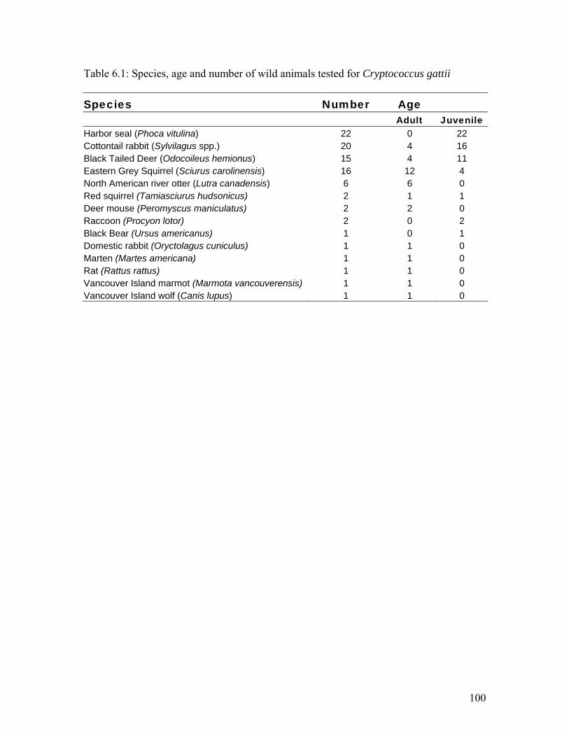
Table 6.1: Species, age and number of wild animals tested for Cryptococcus gattii
Species Number Age Adult Juvenile Harbor seal (Phoca vitulina) 22 0 22 Cottontail rabbit (Sylvilagus spp.) 20 4 16 Black Tailed Deer (Odocoileus hemionus) 15 4 11 Eastern Grey Squirrel (Sciurus carolinensis) 16 12 4 North American river otter (Lutra canadensis) 6 6 0 Red squirrel (Tamiasciurus hudsonicus) 2 1 1 Deer mouse (Peromyscus maniculatus) 2 2 0 Raccoon (Procyon lotor) 2 0 2 Black Bear (Ursus americanus) 1 0 1 Domestic rabbit (Oryctolagus cuniculus) 1 1 0 Marten (Martes americana) 1 1 0 Rat (Rattus rattus) 1 1 0 Vancouver Island marmot (Marmota vancouverensis) 1 1 0 Vancouver Island wolf (Canis lupus) 1 1 0
100
6.5. References
1. Kwon-Chung J, Boekhout, T., Fell, J., Diaz, M. Proposal to conserve the name Cryptococcus gattii against C. hondurianus and C. bacillisporus (Basidiomycota, Hymenomycetes, Tremellomycetidae). Taxon 2002; 51:804-806. 2. Stephen C, Lester S, Black W, Fyfe M, Raverty S. Multispecies outbreak of cryptococcosis on southern Vancouver Island, British Columbia. Can Vet J 2002; 43 (10):792-4. 3. Lester SJ, Kowalewich NJ, Bartlett KH, Krockenberger MB, Fairfax TM, Malik R. Clinicopathologic features of an unusual outbreak of cryptococcosis in dogs, cats, ferrets and a bird: 38 cases (January 2003 to July 2003). J Am Vet Med Assoc 2004; 225 (11):1716-1722. 4. Kidd SE, Hagen F, Tscharke RL, Huynh M, Bartlett KH, Fyfe M, et al. A rare genotype of Cryptococcus gattii caused the cryptococcosis outbreak on Vancouver Island (British Columbia, Canada). Proc Natl Acad Sci U S A 2004; 101 (49):17258-63. 5. Hoang LM, Maguire JA, Doyle P, Fyfe M, Roscoe DL. Cryptococcus neoformans infections at Vancouver Hospital and Health Sciences Centre (1997-2002): epidemiology, microbiology and histopathology. J Med Microbiol 2004; 53 (Pt 9):935-40. 6. Kwon-Chung KJ, Bennett JE. Epidemiologic differences between the two varieties of Cryptococcus neoformans. Am J Epidemiol 1984; 120 (1):123-30. 7. Sorrell TC. Cryptococcus neoformans variety gattii. Med Mycol 2001; 39 (2):155-68. 8. Duncan C, Stephen C, Lester S, Bartlett K. Sub-clinical infection and asymptomatic carriage of Cryptococcus gattii in dogs and cats during an outbreak of cryptococcosis. Med Mycol 2005; In Press. 9. Bartlett K, Fyfe, MW, MacDougall, LA. Environmental Cryptococcus neoformans var. gattii in British Columbia, Canada. Am J Resp Crit Care Med 2003; 167 (7):A499. 10. Connolly JH, Krockenberger MB, Malik R, Canfield PJ, Wigney DI, Muir DB. Asymptomatic carriage of Cryptococcus neoformans in the nasal cavity of the koala (Phascolarctos cinereus). Med Mycol 1999; 37 (5):331-8. 11. Malik R, Wigney DI, Muir DB, Love DN. Asymptomatic carriage of Cryptococcus neoformans in the nasal cavity of dogs and cats. J Med Vet Mycol 1997; 35 (1):27-31. 12. Gray LD, Roberts GD. Experience with the use of pronase to eliminate interference factors in the latex agglutination test for cryptococcal antigen. J Clin Microbiol 1988; 26 (11):2450-1. 13. Duncan C, Stephen C, Raverty S, Lester S, Fyfe M, Bartlett K. Descriptive epidemiology of a multispecies cluster of Cryptococcus neoformans var. gattii: Veterinary aspects. In: International Veterinary Epidemiology and Economics Conference; 2003; Chile; 2003. 14. Krockenberger MB, Canfield PJ, Malik R. Cryptococcus neoformans in the koala (Phascolarctos cinereus): colonization by C n. var. gattii and investigation of environmental sources. Med Mycol 2002; 40 (3):263-72.
101
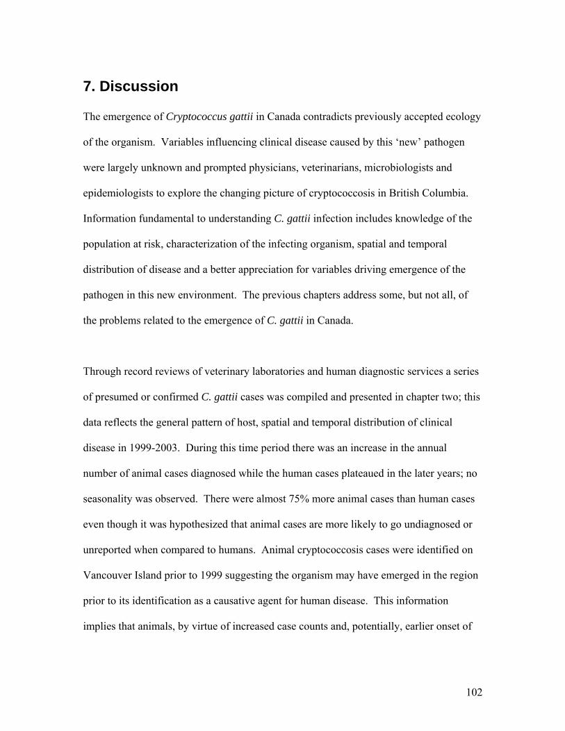
7. Discussion The emergence of Cryptococcus gattii in Canada contradicts previously accepted ecology
of the organism. Variables influencing clinical disease caused by this ‘new’ pathogen
were largely unknown and prompted physicians, veterinarians, microbiologists and
epidemiologists to explore the changing picture of cryptococcosis in British Columbia.
Information fundamental to understanding C. gattii infection includes knowledge of the
population at risk, characterization of the infecting organism, spatial and temporal
distribution of disease and a better appreciation for variables driving emergence of the
pathogen in this new environment. The previous chapters address some, but not all, of
the problems related to the emergence of C. gattii in Canada.
Through record reviews of veterinary laboratories and human diagnostic services a series
of presumed or confirmed C. gattii cases was compiled and presented in chapter two; this
data reflects the general pattern of host, spatial and temporal distribution of clinical
disease in 1999-2003. During this time period there was an increase in the annual
number of animal cases diagnosed while the human cases plateaued in the later years; no
seasonality was observed. There were almost 75% more animal cases than human cases
even though it was hypothesized that animal cases are more likely to go undiagnosed or
unreported when compared to humans. Animal cryptococcosis cases were identified on
Vancouver Island prior to 1999 suggesting the organism may have emerged in the region
prior to its identification as a causative agent for human disease. This information
implies that animals, by virtue of increased case counts and, potentially, earlier onset of
102
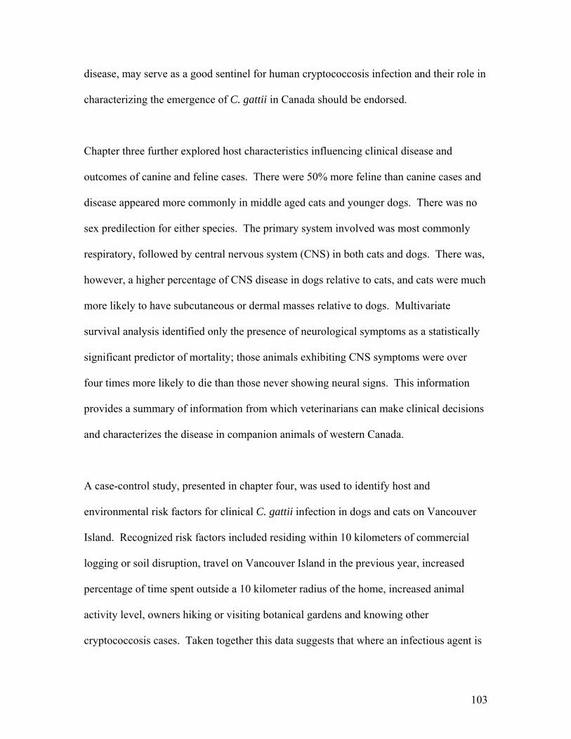
disease, may serve as a good sentinel for human cryptococcosis infection and their role in
characterizing the emergence of C. gattii in Canada should be endorsed.
Chapter three further explored host characteristics influencing clinical disease and
outcomes of canine and feline cases. There were 50% more feline than canine cases and
disease appeared more commonly in middle aged cats and younger dogs. There was no
sex predilection for either species. The primary system involved was most commonly
respiratory, followed by central nervous system (CNS) in both cats and dogs. There was,
however, a higher percentage of CNS disease in dogs relative to cats, and cats were much
more likely to have subcutaneous or dermal masses relative to dogs. Multivariate
survival analysis identified only the presence of neurological symptoms as a statistically
significant predictor of mortality; those animals exhibiting CNS symptoms were over
four times more likely to die than those never showing neural signs. This information
provides a summary of information from which veterinarians can make clinical decisions
and characterizes the disease in companion animals of western Canada.
A case-control study, presented in chapter four, was used to identify host and
environmental risk factors for clinical C. gattii infection in dogs and cats on Vancouver
Island. Recognized risk factors included residing within 10 kilometers of commercial
logging or soil disruption, travel on Vancouver Island in the previous year, increased
percentage of time spent outside a 10 kilometer radius of the home, increased animal
activity level, owners hiking or visiting botanical gardens and knowing other
cryptococcosis cases. Taken together this data suggests that where an infectious agent is
103
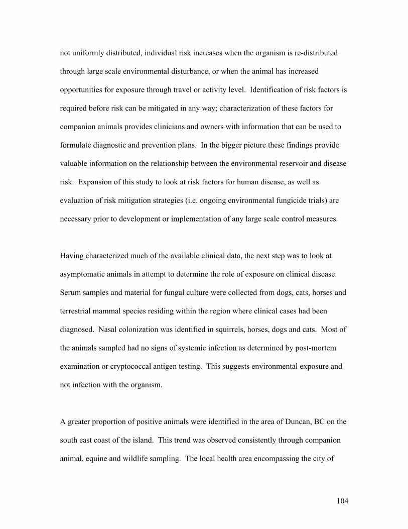
not uniformly distributed, individual risk increases when the organism is re-distributed
through large scale environmental disturbance, or when the animal has increased
opportunities for exposure through travel or activity level. Identification of risk factors is
required before risk can be mitigated in any way; characterization of these factors for
companion animals provides clinicians and owners with information that can be used to
formulate diagnostic and prevention plans. In the bigger picture these findings provide
valuable information on the relationship between the environmental reservoir and disease
risk. Expansion of this study to look at risk factors for human disease, as well as
evaluation of risk mitigation strategies (i.e. ongoing environmental fungicide trials) are
necessary prior to development or implementation of any large scale control measures.
Having characterized much of the available clinical data, the next step was to look at
asymptomatic animals in attempt to determine the role of exposure on clinical disease.
Serum samples and material for fungal culture were collected from dogs, cats, horses and
terrestrial mammal species residing within the region where clinical cases had been
diagnosed. Nasal colonization was identified in squirrels, horses, dogs and cats. Most of
the animals sampled had no signs of systemic infection as determined by post-mortem
examination or cryptococcal antigen testing. This suggests environmental exposure and
not infection with the organism.
A greater proportion of positive animals were identified in the area of Duncan, BC on the
south east coast of the island. This trend was observed consistently through companion
animal, equine and wildlife sampling. The local health area encompassing the city of
104
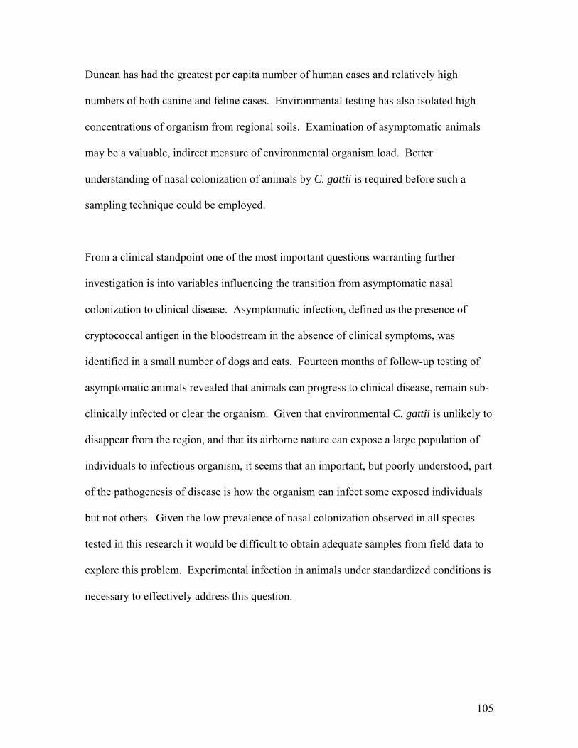
Duncan has had the greatest per capita number of human cases and relatively high
numbers of both canine and feline cases. Environmental testing has also isolated high
concentrations of organism from regional soils. Examination of asymptomatic animals
may be a valuable, indirect measure of environmental organism load. Better
understanding of nasal colonization of animals by C. gattii is required before such a
sampling technique could be employed.
From a clinical standpoint one of the most important questions warranting further
investigation is into variables influencing the transition from asymptomatic nasal
colonization to clinical disease. Asymptomatic infection, defined as the presence of
cryptococcal antigen in the bloodstream in the absence of clinical symptoms, was
identified in a small number of dogs and cats. Fourteen months of follow-up testing of
asymptomatic animals revealed that animals can progress to clinical disease, remain sub-
clinically infected or clear the organism. Given that environmental C. gattii is unlikely to
disappear from the region, and that its airborne nature can expose a large population of
individuals to infectious organism, it seems that an important, but poorly understood, part
of the pathogenesis of disease is how the organism can infect some exposed individuals
but not others. Given the low prevalence of nasal colonization observed in all species
tested in this research it would be difficult to obtain adequate samples from field data to
explore this problem. Experimental infection in animals under standardized conditions is
necessary to effectively address this question.
105
The results of this research emphasize the role of animals in the study of emerging
disease. Identification and reporting of animal cases assisted public health authorities and
microbiologists to define a geographical area in which to focus investigation efforts.
Recent identification of animal C. gattii cases in Washington, USA (M. Leslie, pers.
com., 2005) dictates the need for research into the pattern of disease as it spreads to a new
country. Information gained through the Canadian investigation will be central in the
development of surveillance strategies in the United States and elsewhere.
Given the rate of change in human ecology worldwide it may be assumed that emerging
infectious diseases of man and animals will remain a concern for centuries to come.
Before we can effectively prevent, or even begin to understand new or re-emerging
infection we must take a step back and examine the disease from the angles of host, agent
and, increasingly, the environment. Cryptococcus gattii has afflicted less people and
animals than vehicular traffic on the island’s major roadways; however its emergence in
Canada is unprecedented and may reflect the changing social and ecological environment
in the region. Without a better understanding of variables driving emergence we will be
lack the tools necessary to effectively manage future diseases. In the words of Louis
Pasteur, ‘the microbe is nothing, the terrain is everything’.
106
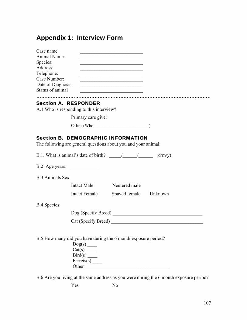
Appendix 1: Interview Form Case name: __________________________ Animal Name: __________________________ Species: __________________________ Address: __________________________ Telephone: __________________________ Case Number: __________________________ Date of Diagnosis __________________________ Status of animal __________________________ ………………………………………………...................................................... Section A. RESPONDER A.1 Who is responding to this interview?
�Primary care giver
�Other (Who_________________________) Section B. DEMOGRAPHIC INFORMATION The following are general questions about you and your animal: B.1. What is animal’s date of birth? _____/______/______ (d/m/y) B.2 Age years: ____________ B.3 Animals Sex:
�Intact Male �Neutered male
�Intact Female �Spayed female �Unknown B.4 Species:
�Dog (Specify Breed) _____________________________________
�Cat (Specify Breed) ______________________________________ B.5 How many did you have during the 6 month exposure period? Dog(s) ____ Cat(s) ____ Bird(s) ____ Ferrets(s) ____
Other ___________________________________ B.6 Are you living at the same address as you were during the 6 month exposure period? �Yes �No
107
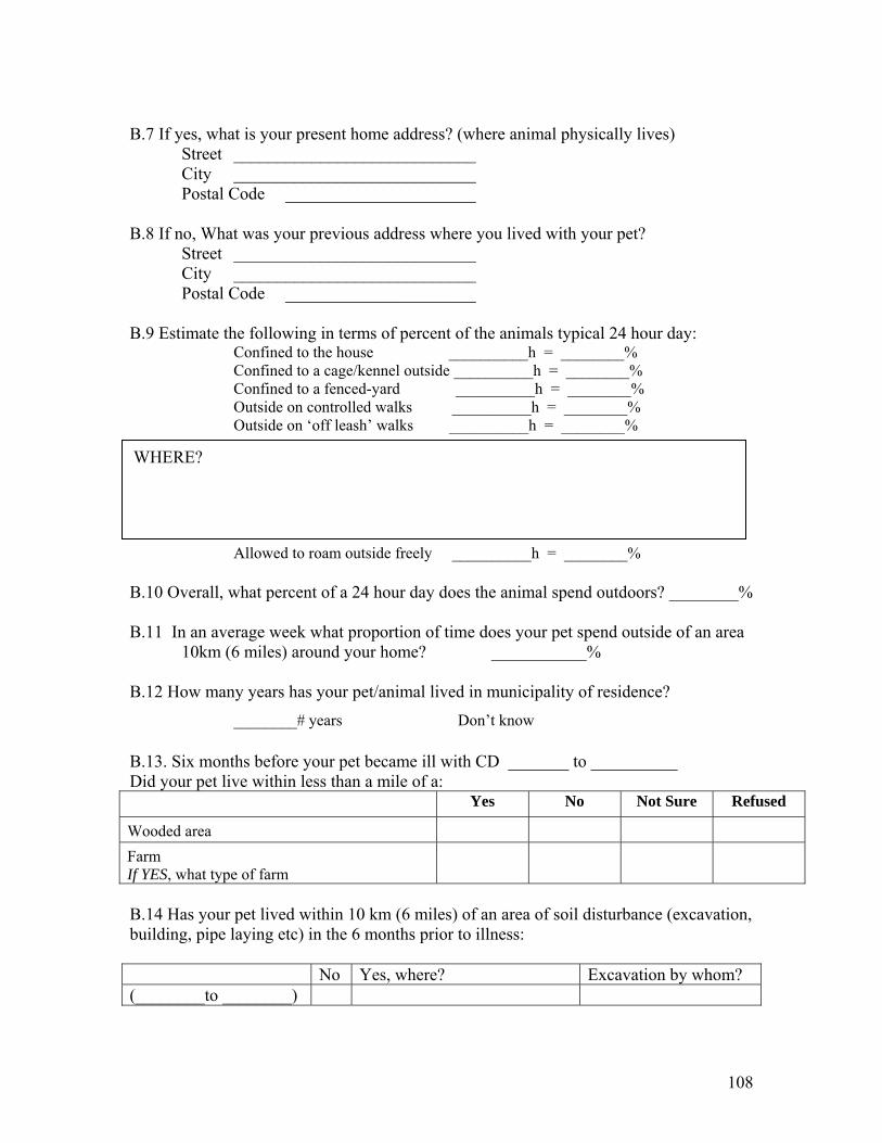
B.7 If yes, what is your present home address? (where animal physically lives) Street ____________________________ City ____________________________ Postal Code ______________________ B.8 If no, What was your previous address where you lived with your pet? Street ____________________________ City ____________________________ Postal Code ______________________ B.9 Estimate the following in terms of percent of the animals typical 24 hour day:
Confined to the house __________h = ________% Confined to a cage/kennel outside __________h = ________% Confined to a fenced-yard __________h = ________% Outside on controlled walks __________h = ________% Outside on ‘off leash’ walks __________h = ________%
WHERE?
Allowed to roam outside freely __________h = ________% B.10 Overall, what percent of a 24 hour day does the animal spend outdoors? ________% B.11 In an average week what proportion of time does your pet spend outside of an area 10km (6 miles) around your home? ___________% B.12 How many years has your pet/animal lived in municipality of residence?
________# years �Don’t know
B.13. Six months before your pet became ill with CD _______ to __________ Did your pet live within less than a mile of a:
Yes No Not Sure Refused
Wooded area Farm If YES, what type of farm
B.14 Has your pet lived within 10 km (6 miles) of an area of soil disturbance (excavation, building, pipe laying etc) in the 6 months prior to illness: No Yes, where? Excavation by whom? (________to ________)
108
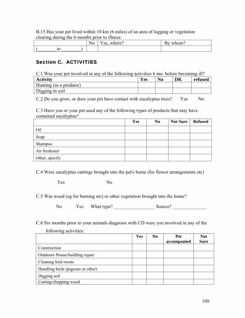
B.15 Has your pet lived within 10 km (6 miles) of an area of logging or vegetation clearing during the 6 months prior to illness: No Yes, where? By whom? (________to ________) Section C. ACTIVITIES C.1 Was your pet involved in any of the following activities 6 mo. before becoming ill? Activity Yes No DK refusedHunting (as a predator) Digging in soil
C.2 Do you grow, or does your pet have contact with eucalyptus trees? �Yes �No C.3 Have you or your pet used any of the following types of products that may have contained eucalyptus? Yes No Not Sure Refused
Oil Soap Shampoo Air freshener Other, specify
C.4 Were eucalyptus cuttings brought into the pet's home (for flower arrangements etc)
�Yes �No
C.5 Was wood (eg for burning etc) or other vegetation brought into the home?
�No �Yes What type? _________________ Source? ______________
C.6 Six months prior to your animals diagnosis with CD were you involved in any of the
following activities:
Yes
No
Pet accompanied
Not Sure
Construction Outdoors House/building repair Cleaning bird roosts Handling birds (pigeons or other) Digging soil Cutting/chopping wood
109

Pruning or branch clean up Hiking If YES, Times/year __________ Where hiked
Visit a botanical garden If YES, where
Gardening IF YES GO TO C.7 ELSE C.8.
C.7 Did you garden: �Year round �Spring �Summer �Fall �Winter C.7.1 How many days a week did you garden 3 months prior to your animal’s illness? �1-3 �4-5 �6-7 �Don’t know C.7.2 Did you do your gardening: �Indoors (greenhouse or in the house) �Outdoor �Both �Don’t know C.7.3 What kind of garden did you have (Mark all that apply)? � Vegetable �Flower �Tree �Other (specify) ____________�Don’t know C.7.4 Did your animal come in contact with: Compost �Y �N Commercial fertilizer �Y �N
Bark Mulch �Y �N Purchased topsoil �Y �N C.8 Travel History: Where have you traveled with your pet… In the last year On Vancouver Island
Off of Vancouver Island
C.9 Do you know of anyone or another animal that has been diagnosed with cryptococcosis? �Yes �No
�Person �Animal Who was it?____________________________
�Don’t know �Refused When did that case occur? ___________
110
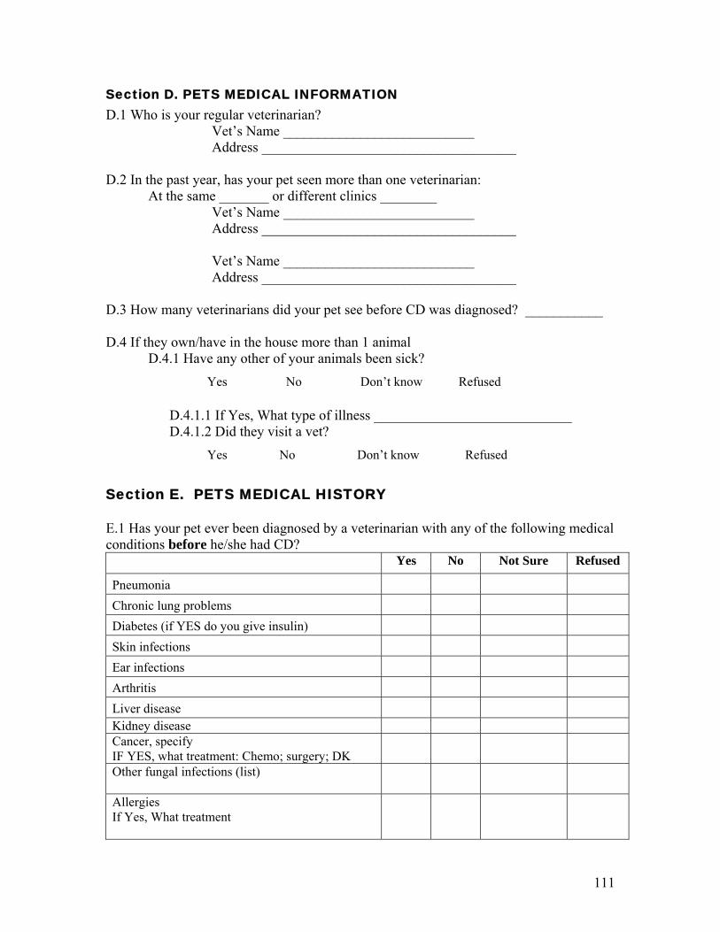
Section D. PETS MEDICAL INFORMATION D.1 Who is your regular veterinarian? Vet’s Name ___________________________ Address ____________________________________ D.2 In the past year, has your pet seen more than one veterinarian: At the same _______ or different clinics ________ Vet’s Name ___________________________ Address ____________________________________ Vet’s Name ___________________________ Address ____________________________________ D.3 How many veterinarians did your pet see before CD was diagnosed? ___________ D.4 If they own/have in the house more than 1 animal D.4.1 Have any other of your animals been sick?
�Yes �No �Don’t know � Refused D.4.1.1 If Yes, What type of illness ____________________________ D.4.1.2 Did they visit a vet?
�Yes �No �Don’t know �Refused Section E. PETS MEDICAL HISTORY E.1 Has your pet ever been diagnosed by a veterinarian with any of the following medical conditions before he/she had CD? Yes No Not Sure Refused
Pneumonia Chronic lung problems Diabetes (if YES do you give insulin) Skin infections Ear infections Arthritis Liver disease Kidney disease Cancer, specify IF YES, what treatment: Chemo; surgery; DK
Other fungal infections (list)
Allergies If Yes, What treatment
111
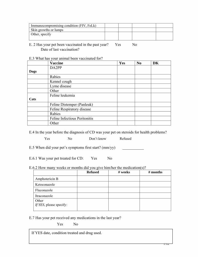
Immunocompromising condition (FIV, FeLk) Skin growths or lumps Other, specify
E. 2 Has your pet been vaccinated in the past year? �Yes �No Date of last vaccination? E.3 What has your animal been vaccinated for? Vaccine Yes No DK
Dogs DA2PP
Rabies Kennel cough Lyme disease Other
Cats Feline leukemia
Feline Distemper (Panleuk) Feline Respiratory disease Rabies Feline Infectious Peritonitis Other E.4 In the year before the diagnosis of CD was your pet on steroids for health problems? �Yes �No �Don’t know �Refused E.5 When did your pet’s symptoms first start? (mm/yy) ___________ E.6.1 Was your pet treated for CD: �Yes �No E.6.2 How many weeks or months did you give him/her the medication(s)?
Refused # weeks # months
Amphotericin B Ketoconazole Fluconazole Itraconazole Other If YES, please specify:
E.7 Has your pet received any medications in the last year? �Yes �No
112
If YES date, condition treated and drug used.
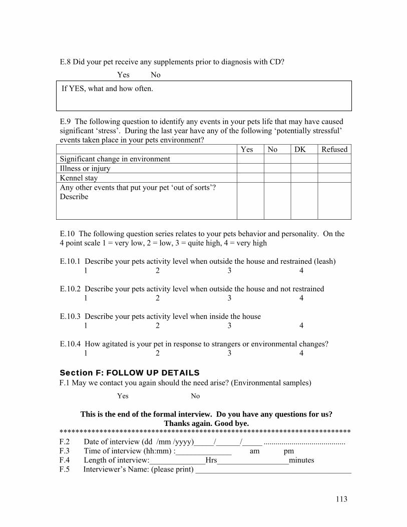
113
E.8 Did your pet receive any supplements prior to diagnosis with CD? �Yes �No
If YES, what and how often.
E.9 The following question to identify any events in your pets life that may have caused significant ‘stress’. During the last year have any of the following ‘potentially stressful’ events taken place in your pets environment? Yes No DK RefusedSignificant change in environment Illness or injury Kennel stay Any other events that put your pet ‘out of sorts’? Describe
E.10 The following question series relates to your pets behavior and personality. On the 4 point scale 1 = very low, 2 = low, 3 = quite high, 4 = very high E.10.1 Describe your pets activity level when outside the house and restrained (leash) 1 2 3 4 E.10.2 Describe your pets activity level when outside the house and not restrained 1 2 3 4 E.10.3 Describe your pets activity level when inside the house 1 2 3 4 E.10.4 How agitated is your pet in response to strangers or environmental changes? 1 2 3 4 Section F: FOLLOW UP DETAILS F.1 May we contact you again should the need arise? (Environmental samples)
�Yes � No
This is the end of the formal interview. Do you have any questions for us? Thanks again. Good bye.
************************************************************************* F.2 Date of interview (dd /mm /yyyy)_____/______/_____ ......................................... F.3 Time of interview (hh:mm) :______________ � am � pm F.4 Length of interview:______________Hrs__________________minutes F.5 Interviewer’s Name: (please print) _______________________________________
Related Documents