Anders Lillevik Thorsen The Emotional Brain in Obsessive-Compulsive Disorder 2019 Thesis for the degree of Philosophiae Doctor (PhD) University of Bergen, Norway

Welcome message from author
This document is posted to help you gain knowledge. Please leave a comment to let me know what you think about it! Share it to your friends and learn new things together.
Transcript

Anders Lillevik Thorsen
The Emotional Brain inObsessive-Compulsive Disorder
2019
Thesis for the degree of Philosophiae Doctor (PhD)University of Bergen, Norway
at the University of Bergen
Avhandling for graden philosophiae doctor (ph.d )
ved Universitetet i Bergen
.
2017
Dato for disputas: 1111
Anders Lillevik Thorsen
The Emotional Brain inObsessive-Compulsive Disorder
Thesis for the degree of Philosophiae Doctor (PhD)
Date of defense: 19.11.2019

The material in this publication is covered by the provisions of the Copyright Act.
Print: Skipnes Kommunikasjon / University of Bergen
© Copyright Anders Lillevik Thorsen
Name: Anders Lillevik Thorsen
Title: The Emotional Brain in Obsessive-Compulsive Disorder
Year: 2019

1
Scientific environment
The work of this thesis has been carried out at the OCD-team at Haukeland
University Hospital, Bergen, Norway; the Departments of Psychiatry and of Anatomy
and Neurosciences at Amsterdam University Medical Centers, Vrije Universiteit
Amsterdam, Amsterdam Neuroscience, The Netherlands, and the Department of
Clinical Psychology at the University of Bergen, Norway. My main supervisor has
been professor Odile A. van den Heuvel, MD PhD (associated with the OCD-team in
Bergen and Amsterdam UMC/Amsterdam Neuroscience in Amsterdam, The
Netherlands), while professor Bjarne Hansen, PhD and professor Gerd Kvale, PhD
(both affiliated with the Department of Clinical Psychology and the OCD-team) have
been my co-supervisors. I have been enrolled at the International Graduate School in
Integrated Neuroscience (IGSIN) at the University of Bergen during my PhD.

2
Acknowledgements
It takes a village to raise a child and even more to raise a scientist, and this thesis
would not be possible without years of work by my supervisors. I wish to thank Gerd
Kvale and Bjarne Hansen for bothering to answer an email from me, a naïve
psychology student, in November 2013. I had just received an offer to revise and
resubmit my first article by a slightly confused editor who probably didn’t understand
what to do with a review paper written by a student with no supervisor. Gerd and
Bjarne decided to give me a chance to work as a research assistant, which became the
first building block of my PhD and our first paper together (Thorsen, van den Heuvel,
Hansen, & Kvale, 2015). I wish to thank Bjarne and Gerd for showing how to
combine being a caring person, clinician, and scientist. In addition, I thank them for
trusting eager new students with important work and for guiding both patients and
students to help them prosper. I wish to thank Odile van den Heuvel for her kind,
intensive, generous, and enlightening guidance on how to combine living a good life
and doing science. I remember travelling to Amsterdam for the first time in June
2014, then an eager and anxious 23-year old. However, my anxiety was soon lifted by
Odile, the members of her research group, and her lovely family. I especially wish to
thank her for her utmost generosity in inviting to me to live with her and her family
for many of my visits to Amsterdam.
Words cannot express my gratitude to my supervisors, and I look forward to working
and spending time together in the future. I wish to thank my family and friends which
make both life and work meaningful. I especially wish to thank Stella J. de Wit for
guidance, discussions, and lots of fun throughout my PhD. I also thank her for
inviting me to get to know her dear family. My visits to Weesp are very dear to me,
and I hope to see you both in the Netherlands and abroad in the future. I also wish to
thank Chris Vriend, Kristen Hagen, Olga Therese Ousdal, and Pernille Hagland for
contributing to the work in this thesis. Lastly, I wish to director Hans Olav Instefjord
and the Division of Psychiatry, as well as the clinicians and patients at the OCD-team
for making science possible.

3
Abstract
Background
Obsessive-compulsive disorder (OCD) is characterized by distressing obsessions and
time-consuming compulsions. The disorder affects 1-3% and can be highly impairing
to daily functioning and detrimental to the quality of life. Cognitive behavioral
therapy is an effective treatment for 50-75% of people with OCD, leaving a
considerable minority who do not benefit from the best available treatments we have
today. Neuroimaging has related the disorder to the function and structure of cortico-
striato-thalamo-cortical and fronto-limbic circuits. A better understanding of these
circuits might contribute to a better understanding of the disorder, how current
treatments change the brain, and how we can help non-responders with better
treatments in the future. This is likely particularly true for fronto-limbic and affective
circuits, given their role in the formation, maintenance, and extinction of fear as well
as motivating behavior. The aim of this dissertation was, first, to investigate how
OCD is related to brain activation during emotional processing of aversive stimuli.
Secondly, we wanted to examine if unaffected siblings of OCD patients showed
similar anxiety, brain activation, and connectivity during emotion provocation and
regulation as their OCD-affected siblings compared to unrelated healthy controls.
Lastly, we wanted to investigate if the resting-state network structure changes in
OCD patients directly after the Bergen 4-Day Treatment (B4DT), a concentrated and
exposure-based psychological therapy.
Methods
Paper I was a meta-analysis of 25 functional neuroimaging studies comparing OCD
patients and healthy controls during emotion processing, when participants were
exposed to aversive or neutral stimuli. In Paper II we used functional magnetic
resonance imaging (fMRI) to investigate distress, brain activation, and fronto-limbic
connectivity during emotion provocation and regulation of neutral, fear-related, and
OCD-related stimuli in 43 unmedicated OCD patients, 19 unaffected siblings, and 38
healthy controls. In Paper III we used resting-state fMRI to study the network
4
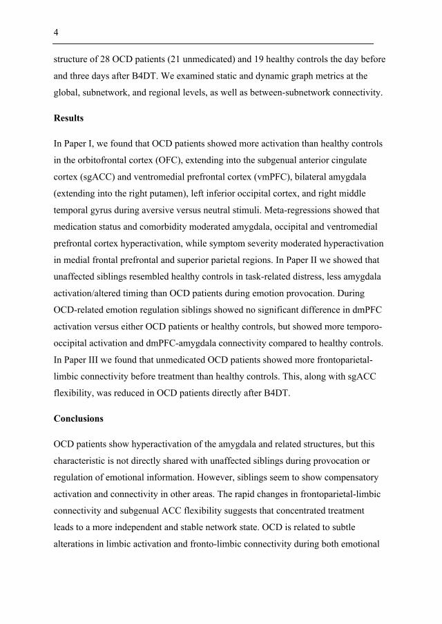
structure of 28 OCD patients (21 unmedicated) and 19 healthy controls the day before
and three days after B4DT. We examined static and dynamic graph metrics at the
global, subnetwork, and regional levels, as well as between-subnetwork connectivity.
Results
In Paper I, we found that OCD patients showed more activation than healthy controls
in the orbitofrontal cortex (OFC), extending into the subgenual anterior cingulate
cortex (sgACC) and ventromedial prefrontal cortex (vmPFC), bilateral amygdala
(extending into the right putamen), left inferior occipital cortex, and right middle
temporal gyrus during aversive versus neutral stimuli. Meta-regressions showed that
medication status and comorbidity moderated amygdala, occipital and ventromedial
prefrontal cortex hyperactivation, while symptom severity moderated hyperactivation
in medial frontal prefrontal and superior parietal regions. In Paper II we showed that
unaffected siblings resembled healthy controls in task-related distress, less amygdala
activation/altered timing than OCD patients during emotion provocation. During
OCD-related emotion regulation siblings showed no significant difference in dmPFC
activation versus either OCD patients or healthy controls, but showed more temporo-
occipital activation and dmPFC-amygdala connectivity compared to healthy controls.
In Paper III we found that unmedicated OCD patients showed more frontoparietal-
limbic connectivity before treatment than healthy controls. This, along with sgACC
flexibility, was reduced in OCD patients directly after B4DT.
Conclusions
OCD patients show hyperactivation of the amygdala and related structures, but this
characteristic is not directly shared with unaffected siblings during provocation or
regulation of emotional information. However, siblings seem to show compensatory
activation and connectivity in other areas. The rapid changes in frontoparietal-limbic
connectivity and subgenual ACC flexibility suggests that concentrated treatment
leads to a more independent and stable network state. OCD is related to subtle
alterations in limbic activation and fronto-limbic connectivity during both emotional

5
tasks and resting-state, which seems to vary with comorbidity and is sensitive to
treatment.

6
Sammendrag
Bakgrunn
Tvangslidelse (obsessive-compulsive disorder, OCD) er definert som angstvekkende
tvangstanker og tidkrevende tvangshandlinger. Lidelsen rammer omtrent 1-3% av
befolkningen og kan være svært hemmende i daglig fungering og livskvalitet.
Kognitiv atferdsterapi er en effektiv behandling for 50-70% av personer med OCD,
mens en betydelig minoritet ikke opplever bedring av de beste behandlingene vi har i
dag. Hjerneavbildning har relatert lidelsen til endret fungering og struktur i kortiko-
striato-thalamo-kortikale og fronto-limbiske hjernebaner. En bedre forståelse av disse
banene kan gi en bedre forståelse av lidelsen, hvordan behandling påvirker hjernen,
og hvordan vi kan hjelpe dem som ikke responderer med mer skreddersydd
behandling i fremtiden. Dette er antakeligvis særlig relevant for fronto-limbiske og
affektive hjernebaner, gitt disses rolle i dannelsen, opprettholdelsen og ekstinksjon av
frykt, så vel som å motivere atferd. Målet med denne avhandlingen var, for det første,
å undersøke hvordan OCD er knyttet til hjerneaktivering under emosjonell
prosessering av aversive stimuli. For det andre ville vi undersøke om friske søsken av
OCD-pasienter viste liknende ubehag, hjerneaktivering og konnektivitet under
emosjonsprovokasjon og -regulering som sine søsken med OCD, sammenlignet med
friske kontrollpersoner uten OCD-pasienter i familien. Til slutt ville vi undersøke om
hjernens funksjonelle nettverksstruktur under hvile endres hos OCD-pasienter
umiddelbart etter Bergen 4-Day Treatment (B4DT), en konsentrert og
eksponeringsbasert behandling.
Metode
Artikkel I var en meta-analyse av 25 funksjonelle hjerneavbildningsstudier som
sammenlignet OCD-pasienter og friske kontrollpersoner under emosjonsprosessering,
når deltakerne ble eksponert for aversive eller nøytrale stimuli. I Artikkel II brukte vi
funksjonell magnetresonnanstomografi (fMRI) for å undersøke ubehag,
hjerneaktivering og fronto-limbisk konnektivitet under emosjonsprovokasjon og
regulering av nøytrale, frykt-relaterte og OCD-relaterte stimuli hos 43 umedisinerte

7
OCD-pasienter, 19 friske søsken og 38 friske kontrollpersoner. Artikkel III brukte vi
fMRI for å undersøke den funksjonelle nettverksstrukturen til 28 OCD-pasienter (21
umedisinerte) og 19 friske kontrollpersoner dagen før og tre dager etter B4DT. Vi
undersøkte statiske og dynamiske grafeteoretiske beregninger på globalt, subnettverk
og regionalt nivå, i tillegg til å undersøke koblingene mellom subnettverk.
Resultater
I Artikkel I fant vi at OCD-pasienter viste mer aktivering enn friske kontrollpersoner
i orbitofrontal korteks (OFC), som strakk seg inn i subgenual anterior cingulate
korteks (sgACC) og ventromedial prefrontal korteks (vmPFC), bilateral amygdala
(som også strakk seg inn i høyre putamen), venstre inferior occipital korteks, og
høyre medial temporal gyrus under aversive versus nøytrale stimuli. Meta-regresjoner
viste at medisinbruk og komorbiditet modererte hyperaktiviteten i amygdala, occipital
og ventromedial prefrontal korteks, mens symptomtrykk modererte hyperaktivering i
mediale frontale og øvre parietale regioner. I Artikkel II viste vi at friske søsken
lignet på friske kontrollpersoner i oppgaverelatert stress, lavere
amygdalaaktivering/endret timing sammenlignet med OCD-pasienter under
emosjonprovokasjon. Under OCD-relatert emosjonsregulering viste søsken ingen
signifikante forskjeller i dmPFC-aktivering fra verken OCD-pasienter eller friske
kontrollpersoner, men viste mer temporo-occipital aktivering og dmPFC-amygdala-
konnektivitet enn friske kontrollpersoner. I Artikkel III fant vi at umedisinerte OCD-
pasienter viste mer frontoparietal-limbisk konnektivitet før behandling enn friske
kontrollpersoner. Dette ble, i tillegg til fleksibilitet i sgACC, redusert hos pasienter
umiddelbart etter B4DT.
Konklusjoner
OCD-pasienter viser hyperaktivering i amygdala og tilknyttede strukturer, men dette
kjennetegnet deles ikke med friske søsken under provokasjon eller regulering av
emosjonelle stimuli. Søsken ser imidlertid ut til å vise kompensatorisk aktivering og
konnektivitet i andre områder. De raske endringene i frontoparietal-limbisk
konnektivitet og fleksibilitet i subgenual ACC foreslår at konsentrert behandling fører

8
til en mer uavhengig og stabil nettverkstilstand. OCD er knyttet til subtile endringer i
limbisk aktivering og fronto-limbisk konnektivitet under både emosjonelle oppgaver
og under hvile, og dette ser ut til både å variere med komorbiditet og være følsomt for
behandling.

9
Abbreviations
ACC Anterior cingulate cortex
B4DT Bergen 4-Day Treatment
CBT Cognitive behavioral therapy
CSTC Cortico-striato-thalamo-cortical circuits
dlPFC Dorsolateral prefrontal cortex
dmPFC Dorsomedial prefrontal cortex
ERP Exposure and response prevention
fMRI Functional magnetic resonance imaging
OCD Obsessive-compulsive disorder
OFC Orbitofrontal cortex
PET Positron emission tomography
SCID Structured Clinical Interview
SSRI Selective serotonin reuptake inhibitors
vmPFC Ventromedial prefrontal cortex
Y-BOCS Yale Brown Obsessive Compulsive Scale

10
List of publications
Thorsen, A. L., Hagland, P., Radua, J., Mataix-Cols, D., Kvale, G., Hansen, B., &
van den Heuvel, O. A. (2018). Emotional processing in obsessive-compulsive
disorder: A systematic review and meta-analysis of 25 functional
neuroimaging studies. Biological Psychiatry: Cognitive Neuroscience and
Neuroimaging, 3(6), 563-571. doi:10.1016/j.bpsc.2018.01.009
Thorsen, A. L., de Wit, S. J., de Vries, F. E., Cath, D. C., Veltman, D. J., van der
Werf, Y. D., Mataix-Cols, D., Hansen, B., Kvale, G., & van den Heuvel, O. A.
(2019). Emotion regulation in obsessive-compulsive disorder, unaffected
siblings, and unrelated healthy control participants. Biological Psychiatry:
Cognitive Neuroscience and Neuroimaging 4(4), 352-360.
doi:10.1016/j.bpsc.2018.03.007
Thorsen, A. L., Vriend, C., de Wit, S. J., Ousdal, O. T., Hagen, K., Hansen,
B., Kvale, G., & van den Heuvel, O. A. Effects of Bergen 4-Day Treatment on
Resting-State Graph Features in Obsessive-Compulsive Disorder. Submitted
for peer review.
Reprints were made with permission from Elsevier. All rights reserved.

11
Related publications which are not included in this thesis
Thorsen, A. L., van den Heuvel, O. A., Hansen, B., & Kvale, G. (2015).
Neuroimaging of psychotherapy for obsessive–compulsive disorder: A
systematic review. Psychiatry Research: Neuroimaging, 233(3), 306-313.
doi:10.1016/j.pscychresns.2015.05.004
Thorsen, A. L., Kvale, G., Hansen, B., & van den Heuvel, O. A. (2018). Symptom
dimensions in obsessive-compulsive disorder as predictors of neurobiology
and treatment response. Current Treatment Options in Psychiatry, 5(1), 182
194. doi:10.1007/s40501-018-0142-4
Kong, X. et al. (in press). Mapping cortical and subcortical asymmetry in obsessive
compulsive disorder: Findings from the ENIGMA consortium. Biological
Psychiatry. doi:10.1016/j.biopsych.2019.04.022

12
Contents
Scientific environment .................................................................................................................................. 1
Acknowledgements ...................................................................................................................................... 2
Abstract ........................................................................................................................................................ 3
List of publications ...................................................................................................................................... 10
Related publications which are not included in this thesis ........................................................................... 11
Contents ..................................................................................................................................................... 12
1. Introduction ...................................................................................................................................... 14
1.1 Obsessive-compulsive disorder ........................................................................................................... 14
1.1.1 Diagnostic criteria, insight and functional impairment ............................................................ 14
1.1.2 Symptom dimensions and subtypes ......................................................................................... 15
1.1.3 Prevalence, onset, course and comorbidity .............................................................................. 16
1.1.4 Risk factors for developing OCD ............................................................................................... 18
1.2 Evidence-based treatments for OCD ................................................................................................... 19
1.2.1 Psychological and pharmacological treatments ....................................................................... 19
1.2.2 Bergen 4-Day Treatment .......................................................................................................... 22
1.3 Neurobiology of OCD ........................................................................................................................... 23
1.3.1 A brief history of functional neuroimaging in OCD ................................................................... 23
1.3.2 Functional connectome during resting-state ............................................................................ 27
1.3.3 Emotions, cognition, and their interaction ............................................................................... 30
1.3.4 Treatment effects on the brain ................................................................................................. 35
1.4 Present thesis ...................................................................................................................................... 39
2. Methods and Results ......................................................................................................................... 41
2.1 Paper I ................................................................................................................................................. 41
2.1.1 Resarch question ...................................................................................................................... 41
2.1.2 Participants ............................................................................................................................... 41
2.1.3 Measures .................................................................................................................................. 41
2.1.4 Preprocessing and statistical analyses ...................................................................................... 41
2.1.5 Ethics ......................................................................................................................................... 42
2.1.6 Results ....................................................................................................................................... 42

13
2.2 Paper II ................................................................................................................................................. 43
2.2.1 Research question ..................................................................................................................... 43
2.2.2 Participants and measures ........................................................................................................ 43
2.2.3 Experimental design of emotion regulation task ....................................................................... 43
2.2.4 Preprocessing and statistical analyses ....................................................................................... 44
2.2.5 Ethics ......................................................................................................................................... 45
2.2.6 Results ....................................................................................................................................... 45
2.3 Paper III ................................................................................................................................................ 46
2.3.1 Research question ..................................................................................................................... 46
2.3.2 Participants ................................................................................................................................ 46
2.3.3 Measures ................................................................................................................................... 46
2.3.4 fMRI preprocessing .................................................................................................................... 47
2.3.5 Graph theoretical measures ...................................................................................................... 47
2.3.6 Statistical analyses ..................................................................................................................... 49
2.3.7 Ethics ......................................................................................................................................... 50
2.3.8 Results ....................................................................................................................................... 50
3. Discussion ......................................................................................................................................... 52
3.1 Findings of Papers I, II and III ............................................................................................................... 52
3.1.1 Limbic involvement in OCD ....................................................................................................... 52
3.1.2 Emotion processing and regulation as a risk or protective factor ............................................. 54
3.1.3 Changes in functional network structure as early marker of treatment response ................... 56
3.2 Methodological considerations ............................................................................................................ 59
3.2.1 Clinical ....................................................................................................................................... 59
3.2.2 Behavioral .................................................................................................................................. 61
3.2.3 Neuroimaging ............................................................................................................................ 62
3.3 Implications for future research ........................................................................................................... 64
3.4 Clinical implications ............................................................................................................................. 67
4. Conclusions ....................................................................................................................................... 69
5. References ........................................................................................................................................ 70

14
1. Introduction
1.1 Obsessive-compulsive disorder
1.1.1 Diagnostic criteria, insight and functional impairment
Obsessive-compulsive disorder (OCD) is defined by the following diagnostic criteria:
to experience either obsessions, compulsions or both. Obsessions are defined as
recurrent and persistent thoughts, urges or impulses that are experienced as intrusive
and anxiety provoking. Examples of obsessions are thoughts of being contaminated
or catching a disease, being afraid of causing harm to others or oneself, an urge for
symmetry to reduce the chance of a catastrophe. Compulsions are defined as
repetitive mental or physical behaviors that are performed to prevent or neutralize
obsessions or reduce anxiety. Compulsions are often not realistically linked to
preventing the feared outcome of obsessions or are clearly excessive (American
Psychiatric Association, 2013; Stein et al., 2016; World Health Organization, 1992).
Symptoms must be time consuming (minimum one hour per day) or cause significant
distress and impairment in personal, work or other aspects of daily life. Furthermore,
these symptoms cannot be better explained by drugs or medication use, or other
physical or mental conditions (American Psychiatric Association, 2013; Stein et al.,
2016; World Health Organization, 1992).
Most patients with OCD realize that their obsessions are unrealistic or exaggerated
and that their compulsions are excessive, at least when they are calm and outside of
situations that trigger their fears (Foa et al., 1995). Approximately 15-30% have poor
or absent insight, and these patients may show higher symptom severity, more
functional impairment, and worse treatment outcomes (Alonso et al., 2008;
Jakubovski et al., 2011; Visser et al., 2017). However, even patients with good
insight often struggle with disregarding obsessions or stopping compulsions once
triggered, and insight can increase during treatment (Alonso et al., 2008; Visser et al.,
2015). This suggests that insight might be a dynamic state rather than a fixed trait,
and is likely influenced by factors such as the present situation, comorbidity, and if

15
the patient has received adequate treatment (Alonso et al., 2008; Jakubovski et al.,
2011; Visser et al., 2017; Visser et al., 2015).
OCD is often highly disabling in family, social, work life and overall quality of life
(Huppert, Simpson, Nissenson, Liebowitz, & Foa, 2009). Results from Swedish
national registries suggest that OCD patients have 17 times higher risk of receiving
disability pension and three times higher risk of up to three months sickness absence
after adjusting for factors such as socioeconomic status and somatic problems (Perez-
Vigil, Mittendorfer-Rutz, Helgesson, Fernandez de la Cruz, & Mataix-Cols, 2018).
There are likely many pathways to disability in OCD, including symptoms interfering
directly with work and personal life, reduced cognitive capacity, worse educational
attainment, and more fatigue (Markarian et al., 2010). The negative impact of OCD
also extends to family members, who also show worse quality of life (Cicek, Cicek,
Kayhan, Uguz, & Kaya, 2013). Importantly, disability and quality of life often
improve after effective treatment (Diefenbach, Abramowitz, Norberg, & Tolin, 2007;
Hollander, Stein, Fineberg, Marteau, & Legault, 2010), which shows how treatment
can be not only immensely important for the individual, but also their relatives and
the society they live in.
1.1.2 Symptom dimensions and subtypes
The content of the obsessions and compulsions can vary widely from one person to
the next (Mataix-Cols, Rosario-Campos, & Leckman, 2005; Thorsen, Kvale, Hansen,
& van den Heuvel, 2018). The heterogeneity of OCD symptoms may complicate
accurate differential diagnosis and make it more difficult to investigate the genetic,
cognitive, and neural correlates of the disorder. A common approach to reduce this
heterogeneity is to categorize symptoms using the Yale Brown Obsessive
Compulsive Scale (Y-BOCS) Symptom Checklist, which is a standardized list of 58
different obsessive and compulsive symptoms (Goodman et al., 1989). Other options
are to use interviews or questionnaires that specifically ask about different symptoms,
such as the dimensional Y-BOCS (DY-BOCS, Rosario-Campos et al., 2006) or the
Obsessive Compulsive Inventory (OCI-R, Foa et al., 2002).

16
Factor analyses have suggested that OCD symptoms can be reduced into
approximately four dimensions: contamination and washing, symmetry and ordering,
sexual, religious and aggressive symptoms, and hoarding and saving (Bloch,
Landeros-Weisenberger, Rosario, Pittenger, & Leckman, 2008; Mataix-Cols et al.,
2005). Hoarding has since been classified as a separate disorder since these
symptoms are more separate than other symptom clusters, often more ego-syntonic,
and they tend to show worse treatment response (American Psychiatric Association,
2013; Mataix-Cols et al., 2010). The symptom dimensions are relatively stable over
time and complete shifts are rare (Fullana et al., 2009; Mataix-Cols et al., 2002). A
debate in the literature has been if different symptoms should be regarded as distinct
subtypes (where patients are placed into the best fitting category) or co-occurring
dimensions (where patients score higher or lower on several axes (McKay et al.,
2004)). A dimensional model has been suggested to more accurately reflect the
disorder since patients often report several kinds of symptoms, but not necessarily
with the same severity (Mataix-Cols et al., 2005). Symptom dimensions have been
related to individual differences in dysfunctional beliefs and cognitive biases
(Brakoulias et al., 2014; Wheaton, Abramowitz, Berman, Riemann, & Hale, 2010),
neuropsychological performance (Hashimoto et al., 2011; Leopold & Backenstrass,
2015), and vulnerability to genetic and environmental risk factors (Iervolino, Rijsdijk,
Cherkas, Fullana, & Mataix-Cols, 2011; van Grootheest, Boomsma, Hettema, &
Kendler, 2008). However, studies into symptom dimensions are often limited by
inconsistent definitions and findings, and little research has investigated the
mechanisms underlying different symptom presentations (Thorsen, Kvale, et al.,
2018) .
1.1.3 Prevalence, onset, course and comorbidity
The prevalence of OCD was estimated to be around 1-3% in the National
Comorbidity Survey Replication study of a representative US sample (Ruscio, Stein,
Chiu, & Kessler, 2010), and Norwegian studies of populations from Oslo and Sogn
og Fjordane have found a somewhat smaller prevalence of around 1% (Kringlen,
Torgersen, & Cramer, 2001, 2006). It should be noted that there are several
challenges with setting an accurate OCD diagnosis in both epidemiological studies

17
and clinical practice. Patients may underreport symptoms due to shame and stigma
related to their symptoms, such as being afraid of being a pedophile or hurting others
(Bruce, Ching, & Williams, 2018; Simonds & Thorpe, 2003) and delay or avoid
seeking help (Torres et al., 2006). Patients with low insight or egosyntonic OCD
often do not perceive their symptoms as exaggerated or excessive, but as external
problems (Belloch, Del Valle, Morillo, Carrio, & Cabedo, 2009). There is also some
overlap in diagnostic criteria with other disorders, such as bodily checking in
hypochondriasis and worrying in GAD, which may require careful differential
diagnosis (Leckman et al., 2010).
The mean age of OCD onset in the United States was approximately 19.5 years, and
males tend to develop the disorder earlier than females, and in patients with a lifetime
OCD diagnosis approximately 80% of males and 60% females had already developed
their first symptoms by the age of 25 (Ruscio et al., 2010). Evidence from a Dutch
study of 377 adult OCD patients suggests that early onset is correlated with higher
symptom severity (Anholt et al., 2014). Naturalistic longitudinal studies show that
OCD is often a chronic disorder, and only a minority appear to recover naturally over
time (Marcks, Weisberg, Dyck, & Keller, 2011; Skoog & Skoog, 1999; Visser, van
Oppen, van Megen, Eikelenboom, & van Balkom, 2014). However, these studies
often do not measure if patients received treatment and whether the treatment was of
high quality or not.
Patients with OCD often have other disorders as well, though OCD is often the
developed first (Ruscio et al., 2010). More comorbid disorders have also been related
to early onset of OCD (Ruscio et al., 2010). The National Comorbidity Survey
Replication study estimated that approximately 75% have a comorbid anxiety
disorder, 63% have a comorbid mood disorder, and 56% have a comorbid
oppositional-defiant or attention-deficit/hyperactivity disorder. Considerable
comorbidity is also reported in international clinical studies (Brakoulias et al., 2017;
Hofmeijer-Sevink et al., 2013), though it is difficult to directly compare rates
between studies due to methodological differences. OCD patients and their family
members also show elevated prevalence of obsessive-compulsive spectrum and other

18
disorders, such as BDD, Tourette and tic disorder, and trichotillomania (Bienvenu et
al., 2012; Phillips et al., 2010).
1.1.4 Risk factors for developing OCD
OCD is more common in some families than others, which may suggest both genetic
and environmental risk factors (Pauls, Abramovitch, Rauch, & Geller, 2014). Twin
and population-based studies suggest that it is a partly heritable disorder, where
genetic factors account for approximately 50% of the risk for developing the disorder
(Mataix-Cols et al., 2013; Pauls, 2010; van Grootheest, Cath, Beekman, & Boomsma,
2005), where genetic factors may account for more risk in early onset cases (Davis et
al., 2013). Family studies have found that the risk of developing OCD increases with
being more closely related, with the highest risk seen in parents, siblings and direct
children of someone with OCD. This risk steadily decreases as the amount of shared
genetic variance decreases, as seen in half siblings, uncles and aunts, or nephews and
nieces (Mataix-Cols et al., 2013). Potential environmental risk factors for OCD
include pre- and perinatal events (birth weight, delivery, smoke exposure during
pregnancy). A recent systematic review suggested that stressful or traumatic life
experiences have also been linked to a higher risk of having OCD (Brander, Rydell,
et al., 2016). There have been largely inconsistent findings for other factors, such as
socioeconomic status, parental rearing style, birth seasons and order, infections, and
household crowding (Brander, Perez-Vigil, Larsson, & Mataix-Cols, 2016). Many
studies of genetic and environmental risk factors share important limitations, such as
few replications, potential recall biases for childhood factors, and inconsistent
measures across studies (Brander, Perez-Vigil, et al., 2016).
Current genetic studies have not found any markers that are significantly related to
having OCD at the whole genome level (Mattheisen et al., 2015; Stewart et al., 2013),
but promising findings have been found in polymorphisms related to glutamate and
serotonin transmission (International Obsessive Compulsive Disorder Foundation
Genetics Collaborative (IOCDF-GC) and OCD Collaborative Genetics Association
Studies (OCGAS), 2018; Taylor, 2013). The lack of clear group-level genetic risk
factors likely reflect that OCD is a multifactorial and heterogenous disorder and that

19
very large sample sizes with more precise phenotyping is needed to uncover genetic
effects (Burton et al., 2018; Katerberg et al., 2010).
The risk for developing OCD is partly heritable, but how it is transmitted within
families is not well understood (Mataix-Cols et al., 2013). One method for finding
familial risk factors is to compare OCD patients, their unaffected family members,
and unrelated people who don’t have the disorder. This could reveal heritable aspects
where OCD patients and their family members are similar to each other but different
from unrelated people, which is called an endophenotype (Gottesman & Gould,
2003). Criteria for a formal endophenotype also requires that it is related to the
disorder in the population, heritable, present even if the person recovers from the
disorder, and stronger in afflicted persons within families (Gottesman & Gould,
2003). Robust endophenotypes could be useful to discover mechanisms for familial
risk of developing a disorder, and more precisely guide genetic and neuroimaging
studies. OCD patients and their relatives have been compared across a variety of
metrics (Taylor, 2012). Some studies have found partial endophenotypes in
dysfunctional beliefs and cognitive biases, such as beliefs about responsibility for
hindering dangers and overestimating situations as threatening (Albert et al., 2015;
Rector, Cassin, Richter, & Burroughs, 2009). OCD patients and their relatives also
show shared worse performance during tasks requiring cognitive flexibility or
response inhibition relative to healthy controls (Chamberlain et al., 2007; Rajender et
al., 2011). These factors may explain some of the familial risk for developing OCD,
but are likely not sufficient to understand why some family members develop OCD
and others do not, which could indicate resiliency to mental disorders. Later sections
will describe how potential endophenotypes have been investigated using
neuroimaging.
1.2 Evidence-based treatments for OCD
1.2.1 Psychological and pharmacological treatments
Treatment guidelines recommend cognitive behavioral therapy (CBT) (including
exposure and response prevention (ERP)) as the first-line treatment for OCD

20
(National Institute for Health and Care Excellence, 2015). Meta-analyses suggesting
that approximately 50% recover after treatment (Öst, Havnen, Hansen, & Kvale,
2015; Skapinakis et al., 2016). Therapist-directed CBT/ERP has been shown to be
effective when provided to individuals, in groups, over telephone or the internet, and
when delivered weekly and intensively (Öst et al., 2015; Patel et al., 2018; Vogel et
al., 2014; Wootton, 2016). Dropout rates are often around 15-20% (Ong, Clyde,
Bluett, Levin, & Twohig, 2016; Öst et al., 2015). Effectiveness studies also show that
ERP is effective when provided in real-life clinical practice (Franklin, Abramowitz,
Kozak, Levitt, & Foa, 2000; Hans & Hiller, 2013; B. Hansen, Kvale, Hagen, Havnen,
& Ost, 2018; Kvale et al., 2018). Lastly, various forms of CBT (including ERP,
cognitive therapy and metacognitive therapy) all seem to be effective and contain
overlapping elements of psychoeducation, exposure, cognitive restructuring, and
stopping compulsions and avoidance behaviors (Papageorgiou et al., 2018).
Selective serotonin reuptake inhibitors (SSRI) are the other recommended first-line
treatment for OCD (National Institute for Health and Care Excellence, 2015). A
recent meta-analysis found that SSRIs lead to a mean improvement of 3.5 points on
the Y-BOCS relative to placebo, with no significant differences between different
types of SSRIs (Skapinakis et al., 2016). High quality studies and meta-analyses
comparing ERP and SSRIs have shown that ERP is more effective, has fewer side
effects, and less dropout than SSRIs treatment alone (Öst et al., 2015; Skapinakis et
al., 2016). ERP has also been shown to be superior to augmenting SSRIs with
risperidone (an antipsychotic medication which is commonly used to augment
pharmacotherapy for patients not responding to SSRIs alone, McLean et al., 2015;
Simpson et al., 2013).
There is an international shortage of therapists with adequate experience and
competency in ERP (McKay, 2018; Shafran et al., 2009). Furthermore, many
therapists often report that they don’t have enough time to implement proper
therapist-directed exposure sessions in clinical practice, that they are afraid to treat
patients with ERP due to concerns of inducing high anxiety levels, or that arousal
reduction strategies are needed to manage anxiety during exposure (Deacon et al.,

21
2013; Pittig, Kotter, & Hoyer, 2019). ERP is therefore often not provided at all or
provided sub-optimally in clinical practice. Considerable effort is needed to provide
therapists with adequate training and supervision, make sure that they provide high
quality treatment, and that results in clinical practice are systematically evaluated
(Kvale & Hansen, 2014; Waller & Turner, 2016).
After effective treatments have been developed, an important goal is to improve
outcomes and reduce drop-out through personalized treatment (Schneider, Arch, &
Wolitzky-Taylor, 2015). Both CBT/ERP and pharmacotherapy in clinical practice
already involves some tailoring to the individual, for example by identifying
individual triggers, compulsions, and exposure tasks, or by adjusting drug dosages
throughout treatment for adequate symptom reduction and tolerable side-effects, but
there are not an evidence-based procedures for systematically tailoring using
individual patient characteristics (Hirschtritt, Bloch, & Mathews, 2017). A
prerequisite for better personalization is uncover factors explaining individual
variation in treatment attrition and outcome. There is a wealth of studies aimed at
identifying such pre-treatment using demographic, clinical or biological factors.
These include age, gender, symptom severity, comorbidity, medication use, cognitive
biases (Steketee, Siev, Yovel, Lit, & Wilhelm, 2018), symptom dimensions (Thorsen,
Kvale, et al., 2018; Williams et al., 2014), functional and structural neuroimaging
(Fullana & Simpson, 2016), and genetic variants (Qin et al., 2016). However, none of
these factors have been adequately replicated as predictors of treatment response
(Knopp, Knowles, Bee, Lovell, & Bower, 2013; Schneider et al., 2015).
The most consistent predictor of outcome after CBT/ERP seem to be patient
compliance, or how much the patient invests in therapy, follows its principles, and
stops engaging in compulsions or anxiety reduction both during and between therapy
sessions (Abramowitz, Franklin, Zoellner, & DiBernardo, 2002; De Araujo, Ito, &
Marks, 1996; Tolin, Maltby, Diefenbach, Hannan, & Worhunsky, 2004; Wheaton,
Galfalvy, et al., 2016). The task dimension of working alliance, which is how much
the patient and therapist agree on what they should do in therapy, may be a possible
mediator of the relationship between compliance and outcome (Hagen et al., 2016;
22
Wheaton, Huppert, Foa, & Simpson, 2016). Lastly, more willingness to experience
anxiety, obsessions and bodily sensations have also been related to more and faster
symptom reduction during ERP (Reid et al., 2017).
1.2.2 Bergen 4-Day Treatment
The Bergen 4-Day Treatment (B4DT) is a concentrated format for ERP which has
been developed by Gerd Kvale and Bjarne Hansen at the OCD-team at Haukeland
University Hospital in Bergen, Norway. It includes separate stages of
psychoeducation and treatment planning, ca. 16 hours of ERP, and relapse
prevention. The difference is that these stages are concentrated into four consecutive
days, where patients vary between individual treatment with at least one certified
therapist per patients and being together with both therapists and other patients in a
group setting. B4DT also includes three weeks of self-exposure, where patients both
perform planned ERP exercises and practice translating the treatment principles into
their daily lives.
B4DT was developed for patients with severe OCD who are entitled to public mental
health, and patients are not excluded based on comorbidity or severity of the
disorders. Patients who are ordinarily not offered B4DT include those with another
disorder that required priority (such as schizophrenia spectrum disorder), or has
severe suicidal ideation, ongoing substance abuse, too low Body Mass Index (BMI)
to start treatment for OCD, ERP treatment is not offered until these issues are dealt
with. Also, patients with mental retardation, are typically not offered the B4DT.
The initial results as well as systematic replications of adult OCD patients found that
approximately 90% of patients responded one week after treatment, where
approximately 75% were classified as recovered using the Y-BOCS (Havnen,
Hansen, Öst, & Kvale, 2017; Havnen, Hansen, Öst, & Kvale, 2014). Similar results
have also been shown and replicated for adolescent patients (Riise, Kvale, Öst,
Skjold, & Hansen, 2018; Riise et al., 2016). These improvements were durable after
three months, six months, one year, and three to four years of follow-up, with no
significant changes between the post-treatment and follow-up time points (B. Hansen,

23
Hagen, Ost, Solem, & Kvale, 2018; B. Hansen, Kvale, et al., 2018; Havnen et al.,
2017; Havnen et al., 2014). Significant improvements were also seen for comorbid
symptoms of depressive and anxiety, quality of life, and ability to work and function
in daily life (B. Hansen, Hagen, et al., 2018; Havnen et al., 2017; Havnen et al.,
2014). The effective transportability of B4DT has also been shown in new clinics in
both Norway and Iceland (Davíðsdóttir et al., 2019; Kvale et al., 2018; Launes et al.,
2019), and clinics in Sweden and the US are currently being trained to deliver the
treatment.
1.3 Neurobiology of OCD
1.3.1 A brief history of functional neuroimaging in OCD
Before the advent of functional neuroimaging, OCD was primarily studied using
neuropsychological, electrophysiological, psychosurgical methods, and lesion case
reports (Khanna, 1988; Turner, Beidel, & Nathan, 1985). Already in the 1980’s, a
hypothesis was that OCD was related to the function of orbitofrontal and limbic
structures (Khanna, 1988; Turner et al., 1985). OCD was among the first mental
disorders to receive focus from functional neuroimaging when Baxter et al. (1987)
used positron emission tomography (PET) to study which parts of the brain used most
glucose (and were thus most active) in OCD patients during resting conditions. They
found that these patients showed higher metabolism of glucose in the right
orbitofrontal cortex (OFC) and bilateral caudate nucleus than healthy controls. The
same group of researchers were also the first to show that treatment could change the
brain, and found reduced glucose metabolism in the right caudate nucleus after
behavioral therapy and fluoxetine for 18 OCD patients (Baxter et al., 1992). The
effect of behavioral therapy was replicated in a later study with nine additional
patients (Schwartz, Stoessel, Baxter, Martin, & Phelps, 1996). These and other early
studies emphasized the role of cortico-striato-thalamo-cortical (CSTC) circuits, which
are involved in many sensorimotor, cognitive and emotional processes (Alexander,
DeLong, & Strick, 1986; Draganski et al., 2008; LeDoux & Pine, 2016). The CSTC
circuits involve excitatory glutaminergic and inhibitory GABAergic pathways that

24
bridge together cortical areas, such as the OFC and ACC, with the basal ganglia
(striatum, putamen, globus pallidus, substantia nigra, subthalamic nucleus) and the
thalamus. These connections form loops and allow for integrated information
processing. An early central hypothesis was that OCD patients show an imbalance
between excitatory direct pathways and inhibitory indirect CSTC pathways, resulting
in a positive feedback loop and a self-reinforcing cycle of obsessions and
compulsions (Graybiel & Rauch, 2000). An explosion of studies using structural and
functional neuroimaging led to the gradual development of newer models with more
complex relationship between different brain circuits. Mataix-Cols and van den
Heuvel (2006) conceptualized OCD as an imbalance between a hyperactive ventral
circuit for emotional processing and motivation and a hypoactive dorsal circuit for
cognitive control. Here, obsessions were thought to be related to less cognitive
control and effective emotion regulation, in combination with more emotional
reactivity to threatening stimuli. This model was later expanded as subsequent
research found that 1) cognitive and emotional functions recruit not only dorsal or
ventral circuits; 2) OCD patients showed widespread abnormal function and
structure, including parietal, visual, cerebellar regions (Menzies, Chamberlain, et al.,
2008); and 3) OCD patients show aberrant communication between brain circuits
(Harrison et al., 2009). This, along with a renewed focus on the role of fear
processing and conditioning, lead Milad and Rauch (2012) to suggest the
involvement of affective, dorsal cognitive and ventral cognitive circuits in OCD.
In recent years, OCD has been extensively investigated using a variety of
neuroimaging methods, including magnetic resonance imaging (MRI) and diffusion
tensor imaging (DTI) for gray and white matter volumes and integrity (Boedhoe et
al., 2018; Boedhoe et al., 2017; de Wit et al., 2014; Norman et al., 2016; Radua et al.,
2014), magnetic resonance spectroscopy (MRS) for neurotransmitter metabolites (S.
Fan et al., 2017; Tadayonnejad et al., 2018; Whiteside, Port, Deacon, & Abramowitz,
2006; Yucel et al., 2007), resting-state fMRI for connectivity between brain regions
(de Vries et al., 2017; Gursel, Avram, Sorg, Brandl, & Koch, 2018; Harrison et al.,
2013), and a range of cognitive and emotional paradigms during functional MRI or

25
PET (Chamberlain et al., 2008; de Vries et al., 2014; de Wit et al., 2012; de Wit et al.,
2015; Milad et al., 2013; Norman et al., 2019; Vaghi et al., 2017; O. A. van den
Heuvel, Veltman, Groenewegen, Witter, et al., 2005). These studies made it clear that
a sole focus on the core CSTC regions was insufficient for describing the
pathophysiology of OCD.
In an effort to integrate both classical and recent findings in OCD, a contemporary
model was recently proposed by O. A. van den Heuvel et al. (2016). This model
suggested that OCD can be related to abnormalities in affective, dorsal and ventral
cognitive, sensorimotor, and fronto-limbic circuits (Table 1). The affective circuit is
thought to be involved in the emotional response to triggering stimuli, reward
processing, and motivating compulsive and avoidance behaviors. This is related to
hyperactivation in the ventromedial prefrontal cortex (vmPFC), subgenual ACC
(sgACC), nucleus accumbens and thalamus, as well the amygdala and hippocampal
complex (O. A. van den Heuvel et al., 2016; O. A. van den Heuvel, Veltman,
Groenewegen, Witter, et al., 2005). This is further supported by a fronto-limbic
circuit which is involved during emotional conditioning and extinction, and
encompasses the vmPFC along with the amygdala and hippocampal complex
(Apergis-Schoute et al., 2017; Milad et al., 2013). The ventral cognitive circuit
governs flexible behavioral preparation and execution, for example by starting and
stopping in response to stimuli. This recruits the inferior frontal gyrus (IFG), anterior
putamen, and pre-supplementary motor area (pre-SMA) (de Wit et al., 2012; Marsh et
al., 2014; van Velzen, Vriend, de Wit, & van den Heuvel, 2014). The dorsal cognitive
circuit is related to top-down control during cognitive tasks, such as planning and
working memory. This recruits areas such as the dorsolateral prefrontal cortex
(dlPFC) and caudate nucleus (de Vries et al., 2014; Heinzel et al., 2018; O. A. van
den Heuvel, Veltman, Groenewegen, Cath, et al., 2005). Lastly, the sensorimotor
circuit is recruited during execution of well learned behaviors, such as habitual
actions. This relies on the premotor cortex and posterior putamen (Gillan et al.,
2015).

26
Table 1 Affected brain circuits in O
CD
Circuit
Function(s) Core areas
Task(s) Clinical relevance
Fronto-limbic
Fear conditioning and
extinction
vmPFC
, amygdala,
hippocampus
Symptom
provocation
and fear conditioning
Conditioning and extinction of feared
stimuli
Affective
Goal-directed
motivational learning
OFC, nucleus
accumbens, am
ygdala
Reward tasks and
symptom
provocation
Exaggerated em
otional and behavioral
response to triggering stimuli,
interference during cognitive tasks
Ventral
cognitive
Motor preparation,
response inhibition
IFG, anterior putam
en,
parietal cortex
Stop signal task, Go-
no go
Cognitive control over com
pulsive
behavior
Dorsal
cognitive
Planning, working
memory, em
otion
regulation
dlPFC, dm
PFC,
caudate nucleus,
parietal cortex
Tower of L
ondon, N-
back, emotion
regulation
Dysfunction in executive function
Sensorimotor
Motor execution,
stimulus-response
learning
Premotor cortex,
posterior putamen
Habit form
ation,
motor sequencing
Habitual use of com
pulsions and
avoidance
.
27
1.3.2 Functional connectome during resting-state The brain is not only a set of distinct regions, but has complex connections that carry
information across regions and circuits. These connections are often referred to as the
connectome of the brain (Bassett & Sporns, 2017; Bullmore & Sporns, 2009). Studies
mapping the connectome has seen an immense growth in the last two decades, and
large-scale projects have shown the intrinsic organization of the brain (Seeley et al.,
2007; Yeo et al., 2011). This research has revealed some subnetworks that are
activated during cognitive or emotional processes and others that are activated during
wakeful rest, where resting-state fMRI can be used to measure the intrinsic
organization of both (Fox et al., 2005; Hugdahl, Raichle, Mitra, & Specht, 2015).
Based on fMRI of 1,000 healthy participants during resting-state, Yeo et al. (2011)
categorized seven visual, somatomotor, dorsal attention, ventral attention, limbic,
frontoparietal and default-mode subnetworks, which were separable into 17
subnetworks at an even finer scale. These subnetworks likely serve specific roles: the
frontoparietal subnetwork is activated during executive tasks (Dosenbach et al., 2007;
Reineberg, Andrews-Hanna, Depue, Friedman, & Banich, 2015). The default-mode
subnetwork supports self-referential and emotional processes (Raichle, 2015). The
dorsal and ventral attention subnetworks are recruited when noticing, interpreting and
allocating cognitive resources to a stimulus, where the ventral attention is especially
active in the early detection of unexpected and arousing stimuli (Vossel, Geng, &
Fink, 2014; Vuilleumier, 2005). The limbic subnetwork is involved in emotional
processing and contributes to emotionally guided decision making, such as approach
and avoidance behavior (LeDoux & Pine, 2016; Pessoa, 2017). The somatomotor
subnetwork is recruited during the execution of motor actions, and relies on the
premotor cortex, posterior insula, and basal ganglia (Choi, Yeo, & Buckner, 2012;
Draganski et al., 2008; Yeo et al., 2011). Lastly, the visual subnetwork is recruited
during perceptual tasks (Wandell, Dumoulin, & Brewer, 2007), and its activation is
also modulated by emotional and cognitive demands (Pessoa & Adolphs, 2010;
Vuilleumier, 2005).
It should be noted that the resting-state subnetworks reported by Yeo et al. (2011)
reflect the organization of the brain in healthy adults, while the model of CSTC and

28
fronto-limbic circuits by O. A. van den Heuvel et al. (2016) describe the altered
subnetworks in OCD and not a general framework of brain organization. For
clarification, the attention and frontoparietal subnetworks in Yeo et al. (2011) are
closely aligned to the respective ventral cognitive and dorsal circuits in O. A. van den
Heuvel et al. (2016), while the limbic subnetwork in Yeo et al. (2011) partly
corresponds with the limbic and affective circuits in O. A. van den Heuvel et al.
(2016).
An important contribution to characterizing the connectome was the application of
graph theory, which uses mathematical models to study relations between
interconnected objects (Bullmore & Sporns, 2009). Graph theory allows for
investigating the topology of a network through defining nodes (e.g. brain regions or
neurons) and connecting edges (e.g. structural or functional connections between
brain regions). Many graph theoretical measures have been developed. For example
for assessing how efficiently a network is organized, defining important hubs, and for
finding local neighborhoods whose nodes are tightly interconnected (Rubinov &
Sporns, 2010). Recently, dynamic graph measures have also been developed, which
allow for a better understanding of how brain networks evolve and change according
to external or internal demands (Avena-Koenigsberger, Misic, & Sporns, 2017).
Dynamic measures have also been used to detect distinctive mental states and the
circuitry involved in switches between them (Allen et al., 2014).
The connectome develops and changes across the lifespan, showing remarkable
plasticity in both structural and functional connections (Collin & van den Heuvel,
2013; Kaiser, 2017). In early childhood this is characterized by massive
developments of connections, followed by a period of pruning and formation of more
efficient connections and hub regions (Collin & van den Heuvel, 2013). During
adolescence and puberty, the connectome becomes more individualized and
distinctive, similar to a fingerprint. Girls are earlier to develop a distinctive
connectome, while boys catch up around the age of 16 (Kaufmann et al., 2017).
Kaufmann et al. (2017) also found that having more symptoms of depression,
attention deficit disorder or schizophrenia was related to a slower development of

29
distinctiveness, which was also evident in the default mode, motor, and frontoparietal
subnetworks. This supports adolescence as an important period of brain development,
where slower maturation is related to mental health problems across diagnostic
categories. In adulthood the brain is typically organized so that information can both
efficiently reach across the brain through key hub regions as well as be processed in
locally segregated clusters (Collin & van den Heuvel, 2013). In late adulthood and
old age the connectome becomes less efficient (Cao et al., 2014), accompanied by
loss of gray matter volume and integrity of white matter tracts (Douaud et al., 2014;
Westlye et al., 2010). This recent body of work has provided a better understanding
of how brain networks develop. It is now important to understand how developing
and recovering from OCD is related to the brain through various developmental
stages. This could also help in disentangling the causes and consequences of OCD,
and guide treatment development in early-onset cases.
Resting-state connectivity and graph theoretical measures may help relate
connectome abnormalities to OCD and other mental disorders (Braun et al., 2018;
Menon, 2011). OCD patients have been reported to show both stronger and weaker
connections within the default-mode subnetwork (Beucke et al., 2014; J. Fan, M.
Zhong, J. Gan, et al., 2017; Hou et al., 2013; E. R. Stern, Fitzgerald, Welsh, Abelson,
& Taylor, 2012). This may reflect the impact of emotional processing and vigilance
on self-referential processing, supported by greater connectivity with the limbic and
ventral attention networks (Beucke et al., 2014; de Vries et al., 2017; J. Fan, M.
Zhong, J. Gan, et al., 2017; Hou et al., 2013; E. R. Stern et al., 2012). Abnormal
connectivity with the limbic and ventral attention subnetwork has also been found for
the executive frontoparietal subnetwork (Gursel et al., 2018). Recent studies have
further found that the global efficiency, or how economically brain regions are
connected, seems to be lower in OCD patients than healthy controls (Jung et al.,
2017; D. J. Shin et al., 2014; Z. Zhang, Telesford, Giusti, Lim, & Bassett, 2016).
OCD patients may also have less differentiated subnetworks (functional modules),
suggesting more cross-talk between them (Gottlich, Kramer, Kordon, Hohagen, &
Zurowski, 2014; D. J. Shin et al., 2014). Both stronger and weaker connections
between neighboring nodes (clustering coefficient) in CSTC circuits has also been

30
reported, which may suggest that the aberrant activation in these structures is also
influenced by their connections with each other (Beucke et al., 2013; Hou et al.,
2014; Jung et al., 2017; Moreira et al., 2017). These findings suggest that the
neurobiology of OCD is not limited to single regions or circuits, but is related to how
circuits communicate with each other.
1.3.3 Emotions, cognition, and their interaction The hallmark of OCD is the loop between experiencing intrusive obsessions, getting
anxious, and trying to manage the anxiety through compulsive rituals, which
maintains a self-reinforcing cycle (American Psychiatric Association, 2013). Much
research has tried to probe what happens in the brain when patients experience
obsessions and become anxious. The most relevant and common paradigm in task-
based fMRI or PET studies is symptom provocation through visual stimuli, for
example by showing aversive (e.g. a dirty toilet) and neutral (e.g. a forest) pictures,
and comparing the levels of distress, brain activation, or psychophysiological
correlates of the two conditions. Early on, such studies often found more activation in
the OFC and ACC, among other areas, during emotional provocation relative to
healthy controls (Adler et al., 2000; Breiter et al., 1996; Nakao et al., 2005). The
amygdala is often a key region looked for in such studies due to its theoretical
importance in the detection of salient stimuli, fear processing, and behavioral
motivation (Etkin & Wager, 2007). However, though some found more activation in
the amygdala in OCD patients compared to controls (Breiter et al., 1996; O. A. van
den Heuvel et al., 2004), others found less amygdala activation in patients
(Cannistraro et al., 2004). This was also reflected in a meta-analysis of emotion
provocation studies, which did not find abnormal amygdala activation, but instead
greater activation in the OFC, ACC, dlPFC, precuneus, and left superior temporal
gyrus in OCD compared to healthy controls (Rotge et al., 2008). This lead some
authors to suggest that “fear/anxiety-related brain regions … do not appear to mediate
the core OCD symptomatology” (L. M. Shin & Liberzon, 2010, p. 180). This was
further considered in the debate on whether OCD should continue to be grouped
among anxiety disorders in the DSM-5 or if it should be classified together with
obsessive-compulsive and related disorders (Stein et al., 2010).

31
Less research has focused on the initiation of compulsive or avoidance behavior
directly. A novel exception was done by Banca et al. (2015) in 15 OCD patients and
15 healthy controls, using live streamed video of therapists disorganizing patients
home or touched the patient with a dirty item during scanning. The patients could
stop the provocation at any time, which allowed for modeling the buildup and release
of activation related to avoidance and presumably compulsive behavior. The results
showed that patients showed a gradual increase right in the seconds before stopping
the provocation, a peak during stopping, and a gradual decrease in the seconds
afterwards. This suggests that the putamen is involved in the regulation of avoidance
and compulsive behavior, shedding some light on the functional role of its altered
activation and structure in OCD patients (Banca et al., 2015).
The search for which regions are activated during emotion provocation in OCD, and
what this meant for how to understand the disorder, is limited by several factors.
Symptom dimensions may be differentially related to brain activation, which could
obscure group differences between heterogenous patients and healthy controls
(Mataix-Cols et al., 2004). SSRIs have substantial effects on amygdala recruitment,
even in low doses in healthy controls (Outhred et al., 2013). Finally, the idiosyncratic
nature of OCD may make it difficult to find personalized and aversive enough stimuli
that can be used in an MRI scanner (Baioui, Pilgramm, Merz, et al., 2013; Simon,
Kaufmann, Musch, Kischkel, & Kathmann, 2010).
Recent research has investigated the role of emotion regulation in OCD (de Wit et al.,
2015), which involves changing emotional responses through processes such as
shifting attention, changing the meaning of an event through cognitive reappraisal, or
suppressing the expression of an emotion (Ochsner, Silvers, & Buhle, 2012). Some
emotion regulation strategies are more automatic (e.g. holding one’s breath or
avoiding looking at distressing stimuli), while others require substantial effortful
control (e.g. deliberately exposing oneself to a stimulus while willfully refraining
from compulsive rituals) (Ochsner et al., 2012). The use of reappraisal strategies are
often found to be linked to better outcomes in terms of well-being, more positive
emotions, and less negative emotions in comparison to suppression or attention

32
shifting strategies (John & Gross, 2004). Emotion regulation recruits a widespread
frontoparietal subnetwork, including the pre-SMA, dACC, dorsomedial prefrontal
cortex (dmPFC), dlPFC, IFG and middle temporal gyrus and parietal
lobule/supramarginal gyrus, and downregulates amygdala activation (Buhle et al.,
2014; Frank et al., 2014). Cognitive reappraisal has been found to most consistently
recruit the entire network, while distancing and suppression strategies are more
limited to the parietal lobule/supramarginal cortex (Morawetz, Bode, Derntl, &
Heekeren, 2017; Ochsner et al., 2012).
Difficulties with emotion regulation, and less successful use of cognitive reappraisal,
has been associated with more mental health problems across diagnostic categories
(Aldao, Nolen-Hoeksema, & Schweizer, 2010). The use of cognitive reappraisal may
also be a transdiagnostic marker of treatment response, as the use of cognitive
reappraisal seems to improve after treatment for anxiety, mood, substance abuse, and
personality disorders (Sloan et al., 2017). In OCD patients and selected student
samples, more use of suppression has been related to both more distress caused by
obsessions and higher symptom severity (Goldberg et al., 2016; Najmi, Riemann, &
Wegner, 2009), whereas more use of cognitive reappraisal strategies has been related
to lower symptom severity (Goldberg et al., 2016). OCD symptom severity has also
been linked to more fear of both negative and positive emotions (Fernandez de la
Cruz et al., 2013; M. R. Stern, Nota, Heimberg, Holaway, & Coles, 2014). This is
line with the cognitive-behavioral model of OCD, which posits that the disorder is
maintained by attempts to take control over or ruminate over thoughts and emotions,
rather than treating them as normal, non-threatening mental events (Foa & McLean,
2016). Some studies have suggested that symptom dimensions have specific
correlates with emotion regulation strategies (Berman, Shaw, & Wilhelm, 2018;
Smith, Wetterneck, Hart, Short, & Björgvinsson, 2012), while others have found
similar relations across symptom presentations (Fergus & Bardeen, 2014).
The first fMRI study of emotion regulation in OCD used an emotion regulation task
where fear-related, OCD-related and neutral stimuli were presented and participants
were asked to either passively view them or actively downregulate their emotions

33
using cognitive reappraisal (de Wit et al., 2015). The study included 43 OCD patients
and 38 healthy controls. During symptom provocation OCD patients showed more
distress during the appraisal of fear and OCD-related stimuli, as well as greater
activation and altered shape of the BOLD response in the amygdala compared to
healthy controls. During emotion regulation, patients showed less activation in the
left dlPFC and parietal cortex for fear-related regulation and more activation in the
dmPFC during OCD-related regulation. OCD patients also showed less dmPFC-
amygdala connectivity during emotion regulation. These findings suggest that OCD
patients show altered recruitment of emotion regulation related regions, as well as
less cognitive control over limbic circuitry (de Wit et al., 2015). Interestingly,
symptom severity was negatively related to recruitment of the dmPFC during OCD-
related, which could suggest that more dmPFC recruitment is a compensatory factor
(de Wit et al., 2015).
Meta-analyses have shown that OCD patients show small to moderate deficits in
general executive function, response inhibition, working memory, planning, and
reversal learning (Abramovitch, Abramowitz, & Mittelman, 2013; Snyder, Kaiser,
Warren, & Heller, 2015). This is also reflected in altered activation of the dorsal
cognitive circuit during planning, response inhibition and working memory, as well
as hyperactivation of premotor cortex during response inhibition (de Vries et al.,
2014; de Wit et al., 2012; Norman et al., 2016; O. A. van den Heuvel, Veltman,
Groenewegen, Cath, et al., 2005). The difference between OCD patients and controls
are also often larger in more difficult task conditions (de Vries et al., 2014; Heinzel et
al., 2018; Vaghi et al., 2017). However, some authors argue that neuropsychological
impairment is not a primary cause or maintaining factor in OCD (Abramovitch,
Mittelman, Tankersley, Abramowitz, & Schweiger, 2015; Snyder et al., 2015). For
one, the difference in neuropsychological performance between OCD patients and
healthy controls are smaller than what is typically characterized as clinically relevant,
and many OCD patients don’t show performance outside the norm (Abramovitch et
al., 2015). Neuropsychological studies in OCD have also been criticized for
methodological limitations in representative recruitment, group matching, and
insufficient focus on the contribution of different patient characteristics (such as

34
medication, symptom dimensions, disease onset and duration, and comorbidity,
Abramovitch et al., 2015). Furthermore, some studies have found increases in
cognitive performance after treatment (Bolton, Raven, Madronal-Luque, & Marks,
2000; Katrin Kuelz et al., 2006), but these findings are inconsistent (Bannon,
Gonsalvez, Croft, & Boyce, 2006; Vandborg et al., 2012).
Abramovitch, Dar, Hermesh, and Schweiger (2012) proposed that worse
neuropsychological performance in OCD is explained by the “executive overload
model”, where worse task performance is an epiphenomenon of obsessions and
anxiety, and not a primary neuropsychological deficit. A recent study also suggested
that OCD patients may perform worse due stereotype threat. This suggests that
internalized negative beliefs about performing worse due to their disorder may
actually lead to worse task performance by itself (Moritz, Spirandelli, Happach, Lion,
& Berna, 2018). Neuroimaging studies provide some support for the “executive
overload model”, as worse task performance has been related to more state distress
and amygdala activation during planning in both OCD, panic disorder, and
hypochondriasis (O. A. van den Heuvel et al., 2011). Further support comes from
fMRI studies of task-related functional connectivity, where OCD patients show
abnormal coupling between the amygdala and dorsal or ventral cognitive circuits,
particularly in patients with the worst task performance (de Vries et al., 2014; Heinzel
et al., 2018; van Velzen et al., 2015). Together, these lines of research suggest that
there are many factors influencing cognitive performance in OCD, and that
longitudinal studies are needed to uncover the relation between state and trait-related
factors.
The partly heritable nature of OCD has motivated researchers to investigate if brain
function and structure could account for the familial risk of developing OCD, and
perhaps even guide future genetic studies (Gottesman & Gould, 2003). This led to
findings that both OCD patients and their family members are similar to each other
and different from unrelated healthy controls in the neural correlates of multiple
cognitive functions. For example, both OCD patients and their siblings show altered
activation relative to unrelated healthy controls in frontoparietal areas during reversal

35
learning (Chamberlain et al., 2008), working memory (de Vries et al., 2014), response
inhibition (de Wit et al., 2012), planning (Vaghi et al., 2017), as well as more error-
related negativity during response inhibition (Riesel, Endrass, Kaufmann, &
Kathmann, 2011). Shared abnormalities have also been found in the volume and
thickness of several brain regions (Menzies, Williams, et al., 2008; Shaw et al.,
2015). Despite this considerable interest there are several outstanding issues before
declaring any findings as reliable endophenotypes. There is limited evidence that
these abnormalities are driven by genetic and not environmental influences, are
present even if the patient recovers from OCD, and that they are causally related to
developing OCD. Finally, it is unknown which abnormalities represent deficits and
which abnormalities represent compensatory mechanisms. Further research is
therefore needed to help understand why unaffected family members show similar
brain structure, activation and connectivity as OCD patients, but without having any
symptoms or reduced cognitive capacity. Indeed, one study suggest that altered
activation during working memory is compensatory as both siblings and the OCD
patients who performed the task most efficiently showed the most abnormal
activation relative to healthy controls (de Vries et al., 2014).
1.3.4 Treatment effects on the brain As treatments can have dramatic treatment effects on symptom severity in OCD, they
could also be used to investigate how the brain changes when patients recover from
the disorder. Treatment studies are therefore important in better understanding how
OCD is related to the brain. In addition, combining treatment and neuroimaging can
potentially reveal more about how effective treatments work, or better understand
why some patients respond quickly while others don’t benefit from treatment.
Finally, it could also disentangle which aspects are stable risk or compensatory
factors, and which are more state-related markers of current OCD symptoms.
As previously mentioned, Baxter et al. (1992) was the first to show that psychological
treatment was associated with reduced and normalized resting-state regional glucose
metabolism in 18 OCD patients. Current studies using CBT/ERP have since used
various imaging modalities, including structural and functional MRI, as well as MRS,

36
PET and electroencephalogram EEG (systematically reviewed by Brooks & Stein,
2015; Thorsen et al., 2015). Investigators have assessed treatment effects during
resting-state (e.g. Feusner et al., 2015; Moody et al., 2017; Saxena et al., 2009),
cognitive (e.g. Freyer et al., 2011; Nakao et al., 2005), and emotional conditions (e.g.
Baioui, Pilgramm, Kagerer, et al., 2013; Morgieve et al., 2014). These studies vary
across many dimensions, such as the efficacy of the treatment, targeted brain
processes or regions, and length of treatment/follow-up period.
Studies using emotional provocation paradigms have most consistently reported
reduced ACC, OFC, and caudate activation after treatment (Baioui, Pilgramm,
Kagerer, et al., 2013; Morgieve et al., 2014; Schiepek et al., 2013). There are also
findings of reduced occipital, hippocampal, thalamic and insula activation during
symptom provocation or Stroop tasks (Nabeyama et al., 2008; Nakao et al., 2005;
Schiepek et al., 2013). In one of the largest and most comprehensive studies,
Morgieve et al. (2014) measured activation to both standard and individualized
symptom provocation paradigms at four time points; before, during, and after
treatment, as well as six months after treatment. They reported a gradual decrease in
symptom severity during treatment and stable improvement between the end of
therapy and follow-up. Using a region-of-interest approach, they found a significant
decrease in dACC and left OFC activation during personalized symptom provocation.
This study also indicated that changes in the brain correlate with symptom
improvement, which supports an earlier finding from a small study that the largest
changes in the brain were found following therapy sessions with the most clinical
change (Schiepek et al., 2013). Morgieve et al. (2014) also saw a large decrease in
activation between the end of therapy and six-month follow-up. Together, these
findings suggest that changes in the brain track the patients progress in therapy
(indicative of direct or short-term treatment effects), but also that some changes in the
brain can happen after a period of normalized behavior (indicative of long-term
recovery).
Studies of executive function have reported increases in dlPFC, parietal cortex and
cerebellar activation during Stroop task after treatment in adult patients (Nabeyama et

37
al., 2008; Nakao et al., 2005). Decreased ACC, OFC, putamen and hippocampal
activation have also been found during Stroop and reversal learning tasks (Freyer et
al., 2011; Nabeyama et al., 2008; Nakao et al., 2005). Increases in dlPFC, ACC and
parietal areas have been reported in studies of pediatric patients using Flanker and
planning tasks after CBT/ERP (Huyser, Veltman, Wolters, de Haan, & Boer, 2010,
2011). Treatment studies have not used functional neuroimaging to investigate other
relevant tasks, such as classical fear conditioning, extinction learning, emotional
Stroop, or working memory tasks. Other tasks, such as planning, has only been used
in pediatric and not adult samples after treatment. There are very few studies on
structural changes after treatment for OCD. Hoexter et al. (2012) investigated
regional brain volumes using T1-weighted voxel-based morphometry in 26 adult
OCD patients, of which half were randomized to CBT/ERP and the other half to
fluoxetine, as well as 36 healthy controls. They found smaller volumes in the left
putamen, OFC and left ACC in patients before treatment, and a small increase in left
putamen volume after treatment in patients treated with fluoxetine. Recently, Zhong
et al. (2019) performed the first CBT/ERP treatment study using DTI in 56 patients.
They found increased fractional anisotropy in orbitofrontal, inferior frontal, temporal
pole, and cerebellar regions, as well as decreased anisotropy in the right putamen
after treatment.
The few treatment studies using SSRI in OCD have used resting-state (D. J. Shin et
al., 2014), symptom provocation (Hendler et al., 2003), and motor tasks (Lazaro et
al., 2008) using PET, fMRI, and single-photon emission computed tomography, as
well as structural neuroimaging (reviewed by Quide, Witteveen, El-Hage, Veltman,
& Olff, 2012). Some studies have reported decreased caudate nucleus metabolism
after SSRI (Baxter et al., 1992; E. S. Hansen, Hasselbalch, Law, & Bolwig, 2002).
Other studies, using MRI, have reported decreased amygdala and temporal volumes
in adolescent patients (Gilbert et al., 2000; Szeszko et al., 2004). A recent crossover
study using intravenous citalopram during symptom provocation in eight OCD
patients and eight healthy controls found that citalopram resulted in less OFC
activation, which correlated with reductions in state anxiety (Bhikram et al., 2016).
There are no large-scale studies comparing if CBT/ERP and SSRIs (the most

38
commonly used treatments for OCD) differ in their effects on the brain, and the few
available studies are underpowered to reliably detect moderate or small differences
between treatments (Apostolova et al., 2010; Baxter et al., 1992; Hoexter et al., 2012;
Nakao et al., 2005).
Pre-treatment neuroimaging characteristics in patients have also been used to predict
treatment efficacy with some success (reviewed by Fullana & Simpson, 2016;
Thorsen, Kvale, et al., 2018). For instance, Olatunji et al. (2014) reported that more
pre-treatment amygdala activation and less dlPFC activation (among other regions)
during symptom provocation was related to a better outcome after exposure therapy
in 12 patients. Structural data from 74 patients further suggested that a thinner left
ACC was related to better outcome (Fullana et al., 2017). Using resting-state fMRI
and machine learning, Reggente et al. (2018) found that functional connectivity
patterns within DMN and visual networks explained 67% of the variance in outcome
in 42 patients after intensive CBT/ERP. However, an important limitation of current
predictor studies is the low rate of replicability, few comparable studies, and no clear
estimate of their predictive validity. These factors, along with the considerable cost of
an (f)MRI scan, likely limit the current clinical utility of existing studies (Fullana &
Simpson, 2016; Thorsen, Kvale, et al., 2018).
The current field of treatment studies is limited considerably by several factors. First,
most have small sample sizes (earlier studies often had around 10 patients), which
markedly increases the risk for both false positive and negative findings (Button et
al., 2013). This problem has been somewhat improved in recent years, with newer
studies having around 30-50 patients (e.g. Moody et al., 2017; Zhong et al., 2019).
Second, there are few studies that are similar enough to directly compare, and very
few systematic replications. This sheds considerable doubt on how replicable the
findings are. Third, some studies show only moderate symptom improvement after
treatment, have considerable attrition or number of patients still showing moderate or
mild OCD after treatment, or report little information on the actual treatment (Baioui,
Pilgramm, Kagerer, et al., 2013; Olatunji et al., 2014; Zhong et al., 2019). Fourth,
many experimental tasks used in case-control or endophenotype studies have not yet
39
been evaluated in treatment studies (e.g. de Vries et al., 2014; de Wit et al., 2012).
This makes it difficult to determine which behavioral aspects of OCD and their
neurobiological correlates are state- or trait-related, or what happens to putative
compensatory factors when patients have recovered. Fifth, there are few studies using
a waitlist controlled or repeated baseline design to separate treatment effects from
natural variation in brain characteristics (Moody et al., 2017). This issue is further
compounded by the moderate and varying test-retest reliability of task- and resting-
state fMRI, which may introduce additional noise in the estimation of any treatment
effects (Braun et al., 2012; Plichta et al., 2012). Lastly, most studies only measure the
brain before and directly after treatment, and few measure changes during treatment
or long-term changes.
In summary, studies combining psychological treatment and neuroimaging have
found that the brain changes after treatment in OCD (Thorsen et al., 2015). These
changes largely occur in affective and cognitive brain circuits that have been
implicated in the pathophysiology of OCD in case-control studies, though there are
some findings of changes outside these classical areas. This suggests that the brain is
plastic and sensitive to symptom improvement in symptoms. However, the field is
limited by small studies, poor replicability, lack of longitudinal studies differentiating
between short- and long-term changes over time, and limited understanding of how
more or less activation in the brain relates to real life behavior, emotions and thoughts
(Thorsen et al., 2015).
1.4 Present thesis
This thesis investigated the role of limbic circuits, emotion processing, and effects of
concentrated psychological treatment in OCD from several perspectives. In Paper I
we performed a meta-analysis of studies using functional neuroimaging to compare
correlates of emotional processing in OCD and healthy controls, and investigated if
variability in study and sample characteristics influence the reported findings.

40
In Paper II we used an emotion regulation task to investigate if provoked distress and
associated brain activation and connectivity is an endophenotype for OCD. This was
done by expanding the analyses of a previous comparison of 43 unmedicated OCD
patients and 38 healthy controls (de Wit et al., 2015) by adding 19 unaffected
siblings. Here, we used the results of Paper I to guide the selection of key regions
during emotion provocation.
In Paper III we investigated if the B4DT leads to short-term changes in brain
topology and network function during resting-state fMRI. This was done by
analyzing key graph theoretical measures acquired the day before treatment and after
one week (i.e. three days after the end of treatment) in 28 OCD patients and 19 age,
gender and education-matched healthy controls. In this study we investigated both the
brain as a whole as well as specific subnetworks and regions, and investigated both
the static network structure and how connections dynamically vary during a scan
session.

41
2. Methods and Results
2.1 Paper I
2.1.1 Research question The goal of Paper I was to investigate if and where OCD patients show abnormal
brain activation during emotional processing compared to healthy controls.
2.1.2 Participants Paper I included summary information from 25 studies and a total of 571 OCD
patients and 564 healthy controls.
2.1.3 Measures The primary measure of Paper I was statistical parametric maps of between-group
differences in activation during the presentation of emotional contrasted with neutral
stimuli (representing group by task interaction effects). We also gathered information
about mean symptom severity (Y-BOCS), percentage of medicated patients,
comorbidity with depressive and anxiety disorders, mean age, mean percentage of
male patients, and mean illness duration.
2.1.4 Preprocessing and statistical analyses Statistical analysis was performed using ES-SDM, a whole-brain meta-analytic
program. We first extracted the coordinates and t-value of the foci from each study,
which were then transformed into MNI space. The contrasts of studies using more
than one emotional condition (e.g. OCD-relevant and general fear stimuli) were
combined. The location and strength of the between-group difference was then
smoothed using an anisotropic Gaussian kernel, which was masked with a gray
matter template. This produced estimated statistical parametric map per study. These
maps were then entered into a random-effect meta-analysis weighted by the number
of OCD patients and healthy controls, between- and within-study heterogeneity. Non-
parametric permutation tests were then used to estimate regions of between-group
differences in activation during emotional processing. The statistical threshold was
set at p < .005, peak voxel Z < 1, and minimum cluster extent of 10 voxels. This is

42
comparable to voxel-level p < .05 corrected for the family-wise error rate. We also
analyzed the moderating role of mean symptom severity (Y-BOCS), percentage of
medicated patients, percentage of patients with comorbid depressive or anxiety
disorders, mean age, mean percentage of male patients, and mean illness duration
using meta-regression analyses which were thresholded at a stricter p < .0005.
2.1.5 Ethics Paper I only included published results of studies with ethical review board approval,
and therefore requires no additional effort or risk from participating researchers or
participants. The paper is therefore not subject to ethical review board approval and
ensures that the original studies are useful for science beyond their original
publication.
2.1.6 Results We found that OCD patients showed more activation in the right OFC (extending into
the sgACC and vmPFC), bilateral amygdala (extending into the right putamen), left
inferior occipital cortex, and right middle temporal gyrus than healthy controls when
viewing emotional versus neutral stimuli. This shows that the exaggerated emotional
processing of aversive stimuli in OCD patients involves a distributed network of
structures.
The meta-regressions showed that studies with more medicated patients reported less
hyperactivation in the right amygdala and left occipital cortex in OCD patients.
Studies with patients with higher Y-BOCS scores showed more hyperactivation in the
sgACC, medial PFC, and precuneus in OCD patients. Finally, studies with more
patients with depressive/anxious comorbidity showed more hyperactivation in the
right insula (extending to the putamen and amygdala) and less hyperactivation in the
left amygdala and right vmPFC in OCD patients. Furthermore, studies with more
patients with longer illness durations showed more hyperactivation of the right
putamen and less hyperactivation of the left temporal pole and OFC in OCD patients.

43
2.2 Paper II
2.2.1 Research question The goal of this paper was to investigate if unaffected siblings of OCD patients show
similar distress, brain activation and functional connectivity during emotion
provocation and regulation as patients, relative to healthy controls without a family
history of OCD.
2.2.2 Participants and measures The study included 43 unmedicated patients with OCD, 19 unaffected siblings of
these patients, and 38 unrelated healthy controls. A diagnosis of OCD and comorbid
disorders was done using the Structural Clinical Interview for DSM-IV (SCID, First,
Spitzer, Gibbon, & Williams, 2002). Symptom severity of OCD was determined
using the Y-BOCS, depressive symptoms were measured using the Montgomery–
Åsberg Depression Rating Scale (MADRS, Montgomery & Asberg, 1979), and use of
reappraisal and suppression as emotion regulation strategies was measured using the
Emotion Regulation Questionnaire (ERQ, Gross & John, 2003).
2.2.3 Experimental design of emotion regulation task All participants performed an emotion regulation task, which has been extensively
used to probe brain regions involved in various forms of emotion regulation in both
healthy participants and participants with mental disorders (de Wit et al., 2015;
Goldin, Manber-Ball, Werner, Heimberg, & Gross, 2009; Ochsner et al., 2004; Rive
et al., 2013). The task involves the appraisal of emotionally neutral pictures, OCD-
related aversive pictures (such as dirty toilets, door handles, water taps, or
asymmetric objects), or general fear-related pictures (such as spiders, bears or guns).
The participants were either instructed to attend the picture naturally or intentionally
try to reduce its emotional relevance using cognitive reappraisal techniques. During
the task the participants were first given the instruction to either “attend” or
“regulate” and where then show a picture for 10 seconds. They were then asked to
rate their distress using a visual analogue scale.
44
2.2.4 Preprocessing and statistical analyses Group differences in demographic characteristics, symptoms of OCD and depression,
ERQ, and distress during emotion provocation and regulation were analyzed using
chi-square or t-tests. Changes in distress during emotion provocation and regulation
were analyzed using repeated-measures ANOVA. Tukey or Games-Howell
corrections were used to adjust for multiple comparisons.
Group comparisons of activation during emotion provocation and regulation were
performed in SPM12. Participant-specific maps of BOLD signal, and its two
derivatives, during provocation (fear attend/regulate > neutral attend, OCD
attend/regulate > neutral attend) were included in separate second-level, random
effects ANOVAs. Three by three ANOVAs for fear and OCD-related provocation
included group (OCD patients, siblings, HC) as a between-subject factor and HRF
(canonical, temporal, dispersion) as a within-subject factor. The interaction between
groups and picture type was modelled in a separate 3 X 2 X 3 ANOVA with group as
a between-subject factor and HRF and picture type (fear, OCD-related) as within-
subject factors. Separate one-way ANOVAS were used for emotion regulation and
regulation-related function connectivity.
In Paper II we primarily focused our analyses to regions of the brain where OCD
patients have previously been shown to differ from healthy controls during emotion
processing or cognitive reappraisal. We therefore formed our hypotheses on emotion
provocation on significant regions from our meta-analysis comparing OCD patients
and healthy controls in Paper I (Thorsen, Hagland, et al., 2018). At that time no meta-
analysis of the neural correlates of emotion regulation in OCD had been published, so
we based our hypotheses on cognitive reappraisal on significant regions from the two
largest meta-analysis of healthy controls (Buhle et al., 2014; Frank et al., 2014). We
then investigated these specific regions using small volume correction (Worsley et
al., 1996), and adjusted the p-values of comparisons between the groups using SISA-
Bonferroni corrections
(http://www.quantitativeskills.com/sisa/calculations/bonhlp.htm; Perneger, 1998).

45
2.2.5 Ethics The study was approved by the ethical approval board of the VU University Medical
Center (Amsterdam, the Netherlands) and all participants provided written informed
consent. The participants were not placed at risk by the procedure and had the right to
withdraw at any time without giving a reason. In accordance with the ethical
approvals, all data were deidentified prior to being available to the Norwegian PhD
student and there was no way of identifying the participants. The PhD student was
also formally associated with VUMC when working with the data.
2.2.6 Results We found that unaffected siblings of OCD patients reported similar levels of distress
as unrelated healthy controls during emotion provocation and regulation, and
significantly less distress than OCD patients. This suggests that they did not appraise
the aversive stimuli as threatening.
During emotion provocation no significant differences was found between the three
groups for fear-related stimuli. For OCD-related stimuli OCD patients showed
significantly altered activation in the right amygdala/hippocampus compared to
healthy controls, which was mainly driven by differences in timing and shape of the
BOLD response. Siblings were intermediate and not significantly different from
either group during OCD-related emotion provocation.
During emotion regulation no significant differences was found between the three
groups for fear-related stimuli, similar to emotion provocation. During OCD-related
regulation siblings showed significantly higher activation in the left temporo-occipital
cortex compared to both OCD patients and healthy controls. OCD patients showed
significantly higher dmPFC activation compared to healthy controls, where siblings
were intermediate and not significantly different from either. Exploratory analyses of
functional connectivity between the dmPFC and amygdala showed that siblings
showed distinctly higher co-activation of these structures during regulation of OCD-
related stimuli, which was significantly greater than OCD patients.

46
2.3 Paper III
2.3.1 Research question The goal of Paper III was to investigate if OCD patients and healthy controls differed
in the topology and organization of resting-state brain networks, and if these
networks change immediately after B4DT. Following a preregistered analysis plan
we investigated these questions both in the entire sample and in a subsample of
unmedicated OCD patients.
2.3.2 Participants This study included 34 patients with OCD who were offered B4DT at the OCD-team
at Haukeland University Hospital (Bergen, Norway). All patients received the
treatment as part of ordinary public mental health care. The patients were interviewed
using the MINI (Sheehan et al., 1997) and Y-BOCS (Goodman et al., 1989) by a
trained local clinical psychologist at the OCD-team as part of standard procedure and
were also interviewed using the SCID (First et al., 2002) and Y-BOCS by a trained
external psychologist who was not part of the treatment. We also recruited a sample
of demographically matched healthy controls with no current or lifetime history of
any mental disorder.
2.3.3 Measures Both OCD patients and healthy controls were assessed using the SCID for lifetime
and current mental disorders before the first fMRI session. Patients were interviewed
by trained independent raters who were not part of the treatment and healthy controls
were interviewed by the first author. Both before the first fMRI session and after one
week, OCD symptom severity was measured using the Y-BOCS, and both OCD
patients and healthy controls provided self-report ratings of OC symptom severity
using the OCI-R (Foa et al., 2002), depressive symptoms using the Patient Health
Questionnaire 9 (PHQ-9, Kroenke, Spitzer, & Williams, 2001) and anxiety symptoms
using the Generalized Anxiety Disorder 7 (GAD-7, Spitzer, Kroenke, Williams, &
Lowe, 2006).

47
2.3.4 fMRI preprocessing FMRIB’s Software Library version 5.0.10 (FSL; Jenkinson, Beckmann, Behrens,
Woolrich, & Smith, 2012) was used to preprocess the fMRI data. The EPI volumes
were motion corrected (with 6 regressors) and spatially smoothed (with a 5 mm
kernel). Participants were excluded if they showed movement exceeding a relative
mean RMS of 0.2 mm or showed more than 20 volumes with RMS above 0.25 mm
(Ciric et al., 2017). Additional movement correction was subsequently performed
using ICA-AROMA (Pruim, Mennes, Buitelaar, & Beckmann, 2015). Nuisance
signals in white matter and cerebrospinal fluid were removed using linear regression
and the data were high-pass filtered (with 100 seconds cut-off). The functional
images were linearly registered to the anatomical T1-weighted images, and the
anatomical image was then parcellated into 226 nodes. Two hundred and ten cortical,
as well as four bilateral dorsolateral and ventromedial putamen nodes, were defined
based on the Brainnetome Atlas (Fan et al., 2016) and warped to the functional
image. The bilateral thalamus, caudate nucleus, pallidum, hippocampus, amygdala,
and nucleus accumbens were individually segmented using FSL FIRST (Patenaude,
Smith, Kennedy, & Jenkinson, 2011). We also applied a mask to the functional
images to account for signal dropout near boundaries between air and tissue during
scanning, which excluded voxels with signal intensities in the lowest quartile. Nodes
with less than four remaining voxels with adequate signal were discarded. Time-
series were then extracted from each node, and Morlet wavelet coherence (Grinsted,
Moore, & Jevrejeva, 2004) in the frequency range of 0.06 to 0.125Hz (Z. Zhang et
al., 2016) was used to calculate the coherence between each pair of nodes to construct
a weighted connectivity matrix per subject and per time point.
2.3.5 Graph theoretical measures Based on these connectivity matrices we calculated the static measures using in-
house scripts and the Brain Connectivity Toolbox (version 2017-15-01; Rubinov &
Sporns, 2010).
For dynamic measures we used a sliding window approach over 136 windows to
assess variation during scanning (window size 25 TRs, each window was shifted 1
48
TR) (Hutchison et al., 2013). Dynamic measures were then calculated using the
toolbox by Sizemore and Bassett (2017). Analyses were performed in MATLAB
R2017a (MathWorks, Inc, Natick, MA, USA). We did not perform any threshold of
the connectivity matrices in order to maximize the information contained therein and
to avoid using arbitrary sparsity levels (Knock et al., 2009). For subnetwork analyses
we assigned all nodes to the visual network, somatomotor network (SMN), dorsal
attention network (DAN), ventral attention network (VAN), limbic network,
frontoparietal network (FPN), or default mode network (DMN) based on a previously
validated parcellation from 1000 healthy controls (Buckner, Krienen, Castellanos,
Diaz, & Yeo, 2011; Choi et al., 2012; Yeo et al., 2011).
We investigated the following graph measures (See Figure 1 for illustration and
Rubinov & Sporns, 2010; Sizemore & Bassett, 2017 for details): Efficiency, which
measures functional integration or how easily information can cross from one side of
the network to the other, is defined as the inverse mean path length. Modularity refers
to the degree to which the network can be divided into functionally different
communities, which maximize the strength of within-module connections and
minimize the strength of between-module connections. The clustering coefficient
measures if a node’s neighbors are also neighbors of each other which, indicates the
level of functional segregation. It is calculated as the fraction of neighbors that are
connected to each other divided by the neighbors that could have been connected.
Betweenness centrality is the ratio of shortest paths through the network that cross
through a given node, and indicates its importance for efficient network
communication. Strength is the total weight of connections to a node, and reflects
how strongly it is connected to rest of the network. We also calculated and compared
the groups’ total functional connectivity strength, as unbalanced connectivity may
bias group comparisons of other graph measures (M. P. van den Heuvel et al., 2017).
For the dynamic graph variables, we measured: Variation in efficiency and clustering
coefficient during a scan. Flexibility refers to how often a node changes which the
module it belongs to. Promiscuity is similar to flexibility but also requires that the
node switches between different modules, not just back and forth between a few.

49
Finally, we assessed the temporal correlation coefficient, which is how stable the
connections between a node’s neighbors are during a scan (similar to the clustering
coefficient). At the global and subnetwork levels we assessed efficiency, clustering
coefficient and dynamic variation in these measures (i.e., temporal correlation
coefficient, flexibility, promiscuity), as well as between-subnetwork connectivity. At
the regional level we assessed node strength, clustering coefficient (and dynamic
variation in these measures), betweenness centrality, temporal correlation coefficient,
flexibility and promiscuity.
2.3.6 Statistical analyses ANOVAs and t-tests were used for clinical measures, and within-group Cohen’s d
were calculated for changes over time (Morris & DeShon, 2002). Group differences
in graph measures were tested using permutated Wilcoxon-Mann–Whitney tests in
the coin package in R (version 3.5.0), while main effects of time, group and group ×
time interactions were performed using the nparLD package (Noguchi, Gel, Brunner,
& Konietschke, 2012). NparLD applies a non-parametric rank-based model, which
yields valid estimates in small sample sizes and data with ties. P-values were
calculated using a modified F-statistic that has been shown to perform well in small
sample sizes (Brunner & Puri, 2001). Within-group changes over time were
calculated using Wilcoxon signed-rank. P-values for Wilcoxon-Mann–Whitney and
Wilcoxon signed-rank tests were calculated based on 10,000 Monte Carlo resamples
using the coin package. The relation between Y-BOCS, graph measures, and changes
in these variables over time was tested using Kendall’s tau correlations. We used the
false discovery rate (FDR; Benjamini & Hochberg, 1995) to correct for multiple
comparisons per graph metric and type of statistical test. We report results of
between-group and longitudinal analyses if they were significant after FDR-
correction (q < .05), except when otherwise specified. P-values of Wilcoxon signed-
ranks were not adjusted for FDR but were only performed when time or group × time
effects were significant. Partial eta squared (η2p) was calculated for time, group and
group × time effects using the modified F-statistic (Lakens, 2013). We calculated the
r effect size for Wilcoxon-Mann–Whitney and Wilcoxon signed-rank tests
(Rosenthal, 1991), which can be regarded as small when ≥ 0.10, medium ≥ 0.30 and
50
large ≥ 0.50 (J. Cohen, 1988). A positive r for Wilcoxon-Mann–Whitney tests
indicates that the value was greater in OCD patients than healthy controls. For
Wilcoxon signed-rank tests a positive r indicates an increase over time.
2.3.7 Ethics The study was approved by the Norwegian Regional Ethics Committee South-East
(2015/936), and all participants provided signed informed consent. The participants
were not placed at risk by participating in the study and had the right to withdraw at
any time without giving a reason.
2.3.8 Results The treatment was highly effective, and 17 (61%) OCD patients were in remission, an
additional 7 (25%) responded, while only 4 (14%) showed no clinically significant
change after one week. There were no significant differences in changes in clinical
measures between medicated and unmedicated patients. Healthy controls showed no
significant changes in OCI-R, PHQ-9, or GAD-7 scores after one week.
We first compared the entire sample of 34 OCD patients (25 unmedicated and 9
medicated) and 28 healthy controls before treatment, and found no significant group
differences after correction for multiple comparisons in static or dynamic measures at
baseline. After excluding medicated patients from the analyses the only difference, at
an uncorrected threshold, was that unmedicated OCD patients showed more
connectivity between the FPN and limbic subnetwork compared to healthy controls (r
= 0.30, p = .03), which was no longer significant after treatment (r = -0.15, p = .36).
We then compared the longitudinal changes in 28 OCD patients (21 unmedicated and
7 medicated) and 19 healthy controls. This showed no significant differences between
the groups (group × time effect), but common changes (main effect of time) at global
and subnetwork but not local levels. Specifically, we observed changes over time in
global efficiency, clustering coefficient, temporal correlation coefficient, total
functional connectivity at the global level. At the subnetwork level we observed
changes in connectivity between the SMN and VAN, efficiency in the SMN, VAN,
and dynamic variation in SMN efficiency. We also found changes in SMN, VAN,

51
DMN and limbic clustering coefficient. Follow-up tests showed that these effects
were mainly driven by increases in healthy controls after one week, while OCD
patients showed no significant changes.
We then compared the longitudinal changes after excluding medicated patients from
the analyses we found significant group differences (group×time effects) in the
change in FPN-limbic connectivity and flexibility in the right sgACC, which were
both driven by significant decreases in OCD patients (both p = .03, r connectivity = -
0.44, r flexibility = -0.52), while healthy controls showed no significant changes.
We found no significant correlations between pre-treatment Y-BOCS, and graph
measures nor between changes in symptom severity and graph measures. Comorbid
anxiety disorders and onset of OCD were not significantly related to change in FPN-
limbic connectivity or sgACC flexibility. When comparing changes in right sgACC
flexibility in depressed and non-depressed OCD patients we found a significant group
× time effect (F(1,19) = 6.11, η2p = 0.26, p = .01), which was driven by a larger
decrease in depressed (r = -0.89, p = .02) than non-depressed patients (r = -0.23, p =
.41).

52
3. Discussion
The present thesis has investigated emotional processing, regulation, and resting-state
connectivity in OCD using meta-analytical, endophenotype, and longitudinal
treatment designs. All three papers have included limbic regions, and the results have
implications for how to understand limbic involvement in the disorder.
The following section will discuss how the findings of the present thesis relate and
contribute to our understanding of OCD. First, it will focus on emotional processing
and regulation. Secondly, it will focus on the plasticity and stability of resting-state
brain network features after treatment. Thirdly, it will consider methodological
concerns of the thesis and general literature. Lastly, it will consider both clinical
implications of our findings as well as the neurobiological implications of clinical
research.
3.1 Findings of Papers I, II and III
3.1.1 Limbic involvement in OCD The role of the fronto-limbic circuit has been the focus of many studies in OCD, but
with inconsistent findings and methodologies (Adler et al., 2000; Breiter et al., 1996;
Cannistraro et al., 2004; Simon et al., 2010; O. A. van den Heuvel et al., 2004).
Inconsistent findings regarding the involvement of limbic structures in OCD,
showing both hyper- and hypoactivation of the amygdala, has even been used as an
argument by for OCD to be classified as an obsessive-compulsive spectrum disorder,
instead of an anxiety disorder (L. M. Shin & Liberzon, 2010; Stein et al., 2010). The
previous meta-analysis on this topic had some important weaknesses that limited its
usefulness, such as not including all relevant studies, including studies without a
healthy control group, and not investigating the role of medication or comorbidity
(Rotge et al., 2008). The purpose of meta-analysis in Paper I was to investigate which
regions are related to hyper- or hypoactivation during emotional processing in OCD,
and identify factors that have contributed to the inconsistent findings in the literature.

53
The results of Paper I challenge previous assertions that OCD is unrelated or
negatively related to limbic activation on the group level, and instead suggest that
group differences are subtle and sensitive to patient characteristics (Outhred et al.,
2013; Thorsen, Hagland, et al., 2018; O. A. van den Heuvel et al., 2016). Classical
models of the detection, evaluation and motivation of action after exposure to
aversive stimuli propose a fast “low” road projecting from the visual cortex to the
amygdala through the thalamus as well as a slower “high” road through cortical
visual areas. Whether the “high” or “low” road “wins” and generates behavior is
thought to be mediated by the affective value, need for conscious evaluation, and
situational factors (LeDoux & Pine, 2016; Pessoa & Adolphs, 2010; Vuilleumier,
2005). Recent models have further suggested many pathways between the visual
cortex and amygdala, which also include parietal, temporal, and orbitofrontal cortices
(Pessoa & Adolphs, 2010; Vuilleumier, 2005). In that context, our findings of
amygdala, OFC, occipital and middle temporal hyperactivation indicates that OCD
patients recruit the visual pathways to larger degree when processing relevant,
aversive stimuli. This is largely encompassed by the affective circuit in the model by
O. A. van den Heuvel et al. (2016). The putamen is thought to be involved in the
preparation and execution of behaviors to neutralize or avoid threats, such as
compulsions (Banca et al., 2015; O. A. van den Heuvel et al., 2016), and the finding
of putamen hyperactivation may reflect the readiness and planning of compulsive or
avoidance behavior.
In summary, Paper I provides an updated view of the neural circuity of emotional
processing in OCD, and highlights important moderating factors. This can provide a
point of reference for future studies and analyses. However, we were not able to
answer further important questions, such as how the implicated regions connect to
each other. Future studies should aim to also investigate task-related functional
connectivity during emotion processing to determine the nature and direction of
regions in the affective and fronto-limbic circuits. It is also important to better
understand the timing and neural correlates of the involved psychological processes,
including noticing, processing emotionally, and deciding on a behavioral response to
disorder-relevant stimuli.
54
3.1.2 Emotion processing and regulation as a risk or protective factor After having investigated the neural correlates of emotion processing in OCD in
Paper I, we intended to investigate if emotion processing and regulation was an
endophenotype that could help explain the familial risk of OCD in Paper II. This
investigation extended the previous work of de Wit et al. (2015), which compared
distress, activation and connectivity during an emotional regulation task in
unmediated OCD patients and healthy controls. In Paper II we added 19 unaffected
siblings of the OCD group. We found that the siblings showed low distress levels
during provocation (lower than patients and similar to healthy controls), suggesting
that only patients were excessively distressed by the fear and OCD-related stimuli
(Thorsen et al., 2019). The siblings showed no significant difference in right
amygdala activation during OCD-related provocation, relative to patients or healthy
controls, while OCD patients showed an altered shape and timing of the BOLD
response in this area. Patients showed greater recruitment of the dmPFC during
regulation of OCD-related stimuli relative to healthy controls, but there was no
significant difference between siblings and patients or controls after correcting for
multiple comparisons. Interestingly, only siblings showed hyperactivation of the left
temporo-occipital cortex during regulation of OCD-related stimuli. Siblings alone
also showed greater dmPFC-amygdala connectivity compared to OCD patients
during regulation of OCD-related stimuli (Thorsen et al., 2019).
Our findings indicate that distress and activation during emotion provocation and
regulation is not a good endophenotype of OCD. However, the greater temporo-
occipital activation and dmPFC-amygdala connectivity during OCD-related
regulation was specific to the sibling group. Previous research has indicated that
neighboring areas on the border of the temporal and parietal cortex is activated more
during distancing than cognitive reappraisal (Morawetz et al., 2017; Ochsner et al.,
2012), which could indicate that siblings rely more on this strategy. The finding that
siblings and OCD patients showed opposite effects in dmPFC-amygdala connectivity
during OCD-related regulation could also indicate a compensatory role, where the
regulatory dmPFC is even more strongly connected in siblings that partly share the
55
environmental and genetic risk of OCD and yet do not develop the disorder.
However, the behavioral role of this finding is unclear since the siblings showed low
distress ratings for all conditions and the specific use of emotion regulation strategy
was not recorded. In summary, more activation in the fronto-limbic circuit during
provocation, less dlPFC activation during fear-related regulation and more dmPFC
activation during OCD-related regulation seem to be characterize patients but not
siblings. This suggests that emotion provocation and regulation does not mediate the
familial risk of OCD (Thorsen et al., 2019).
Despite substantial efforts, there are currently no findings that meet all formal criteria
for an endophenotype in OCD (Gottesman & Gould, 2003; Taylor, 2012).
Endophenotype studies in the same sample found more frontoparietal activation
during working memory and more pre-SMA activation during response inhibition in
both OCD patients and siblings versus healthy controls, with evidence from clinical
and behavioral variables suggesting that both may be compensatory (de Vries et al.,
2014; de Wit et al., 2012). Its noteworthy that this was not found during emotion
processing or regulation, which may indicate that these functions are more state-
related to having the disorder. Unfortunately, the subtle differences found between
unaffected family members of OCD patients relative to healthy controls in the field
has not been systematically replicated, and most current studies have not been
designed to disentangle genetic and environmental effects. The finding with the
strongest evidence for being a possible endophenotype may be greater error-related
negativity as measured with EEG during Flanker tasks. Evidence has suggested that
this is shared by OCD patients and their unaffected first-degree relatives but not
unrelated healthy controls in both adults and adolescents (Carrasco et al., 2013;
Riesel et al., 2011), is largely shared across different symptom dimensions (Riesel,
Kathmann, & Endrass, 2014), and remains unaffected by effective CBT/ERP (Riesel,
Endrass, Auerbach, & Kathmann, 2015). However, more error-related negativity
seem to present in many disorders, and could reflect a general vulnerability to
psychopathology (Olvet & Hajcak, 2008; Riesel, 2019; Riesel et al., 2019).

56
3.1.3 Changes in functional network structure as an early marker of treatment response Paper III measured functional connectivity during resting-state fMRI the day before
the B4DT in 34 OCD patients and 28 healthy controls, and 28 patients and 19 healthy
controls were rescanned after one week (i.e. three days after the end of treatment in
patients). We then used several graph theoretical metrics to describe the functional
connectome at the global, subnetwork and regional level. This included dynamic
metric that capture variation in the network structure during the scanning session
(Bassett & Sporns, 2017; Sizemore & Bassett, 2017), which no studies have
previously investigated in OCD. We found that OCD patients showed more
connectivity between the FPN and limbic subnetworks compared to healthy controls
at an uncorrected threshold. This was only seen when medicated patients were
excluded from the analysis. We also found longitudinal changes after one week,
where OCD patients showed reductions in FPN-limbic connectivity and sgACC
flexibility while healthy controls showed no changes in these measures. This
indicates that symptom improvement directly after concentrated exposure therapy is
related to less crosstalk between subnetworks involved in executive and emotional
processing, extending earlier pretreatment findings of abnormal fronto-limbic
connectivity during resting-state (de Vries et al., 2017; Harrison et al., 2013) and
task-related fMRI (de Vries et al., 2014; de Wit et al., 2015; van Velzen et al., 2015).
However, our results do not answer if the decrease in between-subnetwork
connectivity is driven by changes within one or both subnetworks nor if the
connectivity is bidirectional, top-down or bottom-up. Future analyses using effective
resting-state connectivity might answer this question. Previous studies using such
techniques have shown links from the vmPFC to the amygdala and dorsal striatum
during symptom provocation (Banca et al., 2015) and from the OFC to the nucleus
accumbens at rest (Abe et al., 2015). In comparison, connections from the dlPFC to
the OFC and IFG to amygdala have been found during emotional working memory
and stop signal tasks, respectively (Han et al., 2016; van Velzen et al., 2015). This
suggests that the direction of connectivity is modulated by task demands. It is
possible that activation of task-related regions (i.e. cognitive circuits) are correlated
57
with limbic activation. For instance, when OCD patients experience that their
performance is not good enough they may also try harder at the task (more task-
related activation) which feeds a cycle of increasing anxiety and maladaptive
monitoring (i.e. more limbic activation). This might be particularly observable during
more demanding levels of cognitive tasks (de Vries et al., 2014). In comparison,
OCD patients may show more activation within limbic circuits during resting-state
(Abe et al., 2015; de Vries et al., 2017), reflecting more anxiety and obsessions when
there is no cognitive demand (Paper III also found more state anxiety during resting-
state in OCD patients before treatment relative to healthy controls). I would therefore
expect effective connectivity analyses to show more connectivity from the OFC to
other limbic areas (including the amygdala) in OCD patients before treatment, which
should normalize after treatment.
The sgACC is a central node in the affective circuit, is connected to striatal and
thalamic regions, and is activated during emotional and interoceptive processing
(Pauls et al., 2014; Pessoa, 2017). Flexibility measures how often a node switches
between which functional module it connects the strongest to. A module is a set of
several nodes that have strong connections to each other and weaker connections
outside of the module. Our finding of reduced sgACC flexibility directly after B4DT
in OCD patients suggests a more stable network after treatment. This might be the
result of decreased effort in bridging regions implicated in processes related to
obsessions, emotion regulation, and compulsions. However, since participants were
resting during scanning, we can’t draw firm conclusions about the behavioral or
psychological function of changes in connectivity.
Surprisingly, additional changes over time were seen in global, subnetwork, and
regional measures in healthy controls for both static and dynamic graph measures.
This was particularly seen in the somatomotor subnetwork, including its clustering
coefficient and variation in efficiency. Similar findings in global and regional
measures have been found in healthy controls of previous treatment studies (Li et al.,
2018; D. J. Shin et al., 2014), as well as in a study consisting of many time points in a
single person (Poldrack et al., 2015). These changes are currently poorly understood,

58
and could indicate that OCD patients show less normal increases or variation in
network integration and clustering, or that healthy controls react differently to being
repeatedly scanned. A complicating factor is that healthy controls showed a trend-
significant increase in total functional connectivity (which may influence graph
measures, M. P. van den Heuvel et al., 2017), though the groups were not
significantly different at either before or after treatment.
We were not able to replicate previous findings of less efficiency or modularity in
OCD patients as reported in previous case-control comparisons (Jung et al., 2017; D.
J. Shin et al., 2014; T. Zhang et al., 2011), nor changes in global clustering coefficient
or modularity after treatment (Feusner et al., 2015; D. J. Shin et al., 2014). This went
against our hypotheses, but similar null findings have been reported (T. Zhang et al.,
2011). A major impediment to comparing different studies directly is the variation in
preprocessing pipeline, measures of functional connectivity, scanning duration, and
statistical analyses (Ciric et al., 2017; Murphy & Fox, 2017; Z. Zhang et al., 2016).
Variation in use of medication, time between scans, and clinical effectiveness further
complicates further between-study comparisons (Beucke et al., 2013; Feusner et al.,
2015; Moody et al., 2017). The clinical and methodological variation in the field
highlight the need for more collaboration (including harmonization in data acquisition
and processing) and systematic replications across research groups and scanners.
Network models to better understand the functional connectome is a promising
approach to understand the neural correlates of OCD and plasticity after treatment,
but it is still in its infancy. For it to be a truly useful tool we need to know more about
the functional connectome in general (Avena-Koenigsberger et al., 2017; Bassett &
Sporns, 2017), its relation to behavior (Braun et al., 2015), and reach a consensus of
how to acquire, process and analyze resting-state fMRI (Ciric et al., 2017; Murphy &
Fox, 2017).

59
3.2 Methodological considerations
3.2.1 Clinical OCD is a complicated disorder to study due to its highly heterogenous symptom
presentation, long illness duration, high comorbidity rates, and medication usage that
can influence clinical and biological measures (Brakoulias et al., 2017; Mataix-Cols
et al., 2005; Outhred et al., 2013; Ruscio et al., 2010). Furthermore, it is uncertain
how representative patients participating in studies are for the total population of
people with OCD. For instance, researchers have highlighted the role of symptom
presentation, ethnic and sexual minority status in treatment seeking and inclusion in
research (Bruce et al., 2018; Williams & Farris, 2011; Williams, Powers, Yun, & Foa,
2010; Williams, Turkheimer, Schmidt, & Oltmanns, 2005).
Most studies of OCD rely on a trained clinician to measure the severity of the
disorders (Goodman et al., 1989), which increases the chance that questions are
understood and gives the chance to clarify misunderstandings. However, the overlap
between interview and self-report is not perfect (intraclass correlation of .75), and
less for the obsessions subscale (Federici et al., 2010). We therefore applied both
interviewer and self-report scales in Paper II and III. We also used a trained clinician
who was not part of the groups or local treatment team for both baseline and post-
treatment measures in Paper III. However, the interviewer was not blinded to time
point and had access to additional information about the patient, since blinding the
rater would have been most impractical. The study of Paper III was not designed to
test the effectiveness of the treatment alone or in comparison to others, but we cannot
exclude the possibility of patients or raters under- or overreporting symptoms due to
biases or allegiances (Munder, Brutsch, Leonhart, Gerger, & Barth, 2013).
The studies in Paper II and III recruited patients with varying age, gender and
education status and carefully matched healthy controls on these variables. Varied
patients were also recruited, as reflected in symptom severity, comorbidity rates and
the type of symptoms that they presented with. However, some types of patients were
excluded, such as those with developmental difficulties (such as autism spectrum
60
disorders or intellectual disability) or with ongoing manic or psychotic symptoms.
The sample in Paper III did not include patients who did not want or were unfit for
treatment at the time, for example those with severe self-harm, suicidal intent,
untreated somatic illnesses, or with disorders which needed to be addressed first. In
summary, this suggests that the findings in Paper II and III might generalize to many
types of OCD patients commonly seen in clinical practice, but that caution is
warranted when interpreting the findings in relation to patients with pervasive
developmental difficulties, those not seeking treatment, and patients with low insight.
There is limited knowledge regarding the role of symptom severity, and if and how
higher obsessive-compulsive symptom severity is related to having a more abnormal
brain, or if more symptom improvement after treatment is related to more pre-post
treatment changes in the brain. The results of single studies and meta-analyses are
somewhat inconsistent, and some find no significant relation between symptom
severity and brain characteristics (Boedhoe et al., 2018; Boedhoe et al., 2017; de
Vries et al., 2014; Figee et al., 2011). This was not the case for the meta-analysis in
Paper I, which found that studies including OCD patients with a higher mean Y-
BOCS score showed more prefrontal and precuneus hyperactivation in OCD
(Thorsen, Hagland, et al., 2018). However, in Paper III there was no significant
relation between Y-BOCS scores and graph measures where OCD patients were
significantly different from healthy controls, nor between change in Y-BOCS and
changes in graph measures after treatment. This could be caused by little variation in
the level of symptom improvement, as almost 90% of the patients responded after
treatment. More research is needed to determine the relation between symptom
severity and the brain in OCD.
Future research could also try to understand why some patients improve fast, some
slow, and others not at all. Such studies will require both larger sample sizes and
measures that can help us understand why they did not benefit from treatment. For
instance, one could expect a difference in how emotional brain networks are
organized in patients who are not motivated to perform the most difficult exposure
tasks compared to those who perform all exposures but engage in compulsions

61
afterwards (Aigner et al., 2005; J. Fan, M. Zhong, X. Zhu, et al., 2017). In a
supplemental analysis of Paper III, we excluded the four patients who did not show
significant change after treatment, and found very similar changes in graph measures.
There are few studies reporting the effects of age on changes in the brain after
treatment, while younger age has been linked to more improvement on CBT/ERP,
both in adults (Öst et al., 2015) and children (Öst, Riise, Wergeland, Hansen, &
Kvale, 2016). However, it should also be noted that age has not emerged as a
consistent predictor in systematic reviews (Knopp et al., 2013) or mega-analysis of
adult patients (Steketee et al., 2018). A better understanding of how age influences
brain plasticity and reorganization would be a valuable contribution to the literature,
especially given the large potential for brain development seen in early childhood and
puberty (Collin & van den Heuvel, 2013; Kaufmann et al., 2017).
3.2.2 Behavioral The situations that evoke distress and anxiety in OCD is highly idiosyncratic and can
be difficult to elicit in a highly controlled experimental setting. This is relevant for
both Papers I and II, which study task-induced emotion provocation. Paper II used
generic OCD-related pictures for washing, checking and symmetry dimensions, as the
study did not recruit patients with only one type of symptoms. However, stimuli were
not personalized since this would have complicated interpreting between-group and
between-person analyses. It would also have required considerably more time and
effort to make personalized stimuli. However, this also meant that some patients saw
stimuli that were not particularly relevant for them, which may have resulted in lower
mean distress ratings for OCD than fear-related stimuli (Thorsen et al., 2019). Some
studies have also found that using personalized stimuli is associated with stronger
BOLD responses in relevant regions (Baioui, Pilgramm, Merz, et al., 2013; Morgieve
et al., 2014).
It can be difficult to operationalize even relatively simple psychological functions in
an MRI scanner, and even more difficult with a complex construct such as emotion
regulation. This issue is further complicated in OCD, where patients often try to

62
regulate their emotions using strategies that resemble cognitive reappraisal or
distraction, but often end up trying to reduce distress by relaxing or reasoning
themselves out of obsessive thoughts in a compulsive manner. For instance, the
“regulate” condition in the emotion regulation task of paper II instructed participants
to “imagine a more positive outcome or interpretation of the portrayed events” or
“realize the stimulus is not real-life” (de Wit et al., 2015). This resembles typical
strategies used to regulate emotions in real life in both healthy controls and patients
(John & Gross, 2004; Ochsner et al., 2004). However, it also resembles the
dysfunctional strategies used by patients, as they often attempt to compulsively
rationalize, reimagine or distract themselves from obsessions (even though most
obsessions are normal in terms of content)(Muris, Merckelbach, & Clavan, 1997;
Rachman & de Silva, 1978). This can lead to an increase in distress, obsessional
frequency, and low mood (Najmi et al., 2009; Purdon, Rowa, & Antony, 2005). In
contrast, ERP and other forms of psychological treatment ask patients to
systematically increase their anxiety during exposure, which in turn often leads to a
higher tolerance of emotional distress and less need to neutralize or distract oneself
from obsessions (Grøtte et al., 2015; Reid et al., 2017).
3.2.3 Neuroimaging Functional neuroimaging using fMRI has key strengths such as being non-invasive,
relatively brief, and allowing multimodal imaging. However, it also has critical
limitations that must be taken into account when planning, analyzing and interpreting
data (Poldrack et al., 2008). Scanning requires balancing temporal and spatial detail,
as the number of slices and voxel sizes often increase as repetition time decreases.
This may make it difficult to measure fast processes, such as the communication
between visual cortex and amygdala during detection of aversive stimuli (Boubela,
Kalcher, Nasel, & Moser, 2014; Vuilleumier, 2005). Imaging brain structures of
theoretical importance, such as the amygdala, vmPFC and nucleus accumbens (Figee
et al., 2011; O. A. van den Heuvel et al., 2016), can be difficult due to nearby tissue
boundaries and other confounding physiological variables (Chen, Dickey, Yoo,
Guttmann, & Panych, 2003; Lipp, Murphy, Wise, & Caseras, 2014; Stocker et al.,
2006). Paper I only included summary information from published studies where

63
some likely had better coverage of the brain than others, which we were unable to
assess or control for. In paper II and III we assessed brain coverage of all participants,
and excluded those with poor coverage or artifacts. In Paper III we also ensured that
all segmented brain regions had adequate signal (each region had to have a minimum
of four voxels with signal intensity in the upper three quartiles), which led to the
exclusion of the bilateral nucleus accumbens and the most ventral area of Brodmann
13 in the right hemisphere.
The test-retest reliability (expressed as the intraclass correlation, which measures the
consistency or agreement for a measure taken at two or more time points) of fMRI
during cognitive and emotional tasks has been reported as good (.89-.98) and
acceptable (.66-.97) on whole-brain and regional levels, respectively (Plichta et al.,
2012). However, a recent meta-analysis of 90 task-related studies estimated an
intraclass correlation of .39, and estimates between .07 and .49 in two large
independent datasets (Elliott et al., 2019). Reliability estimates of .50-.60 has also
been reported for common static graph measures, such as global efficiency, during
resting-state fMRI (Braun et al., 2012; Termenon, Jaillard, Delon-Martin, & Achard,
2016), though reliability was somewhat less for static graph measures in emotional
tasks. Both task- and resting-state reliability was influenced by scanning parameters
(such as scan duration) and preprocessing pipeline (Braun et al., 2012; Plichta et al.,
2012). A recent study found that dynamic graph metrics is worse than static
measures, with intraclass correlations under .10 (C. Zhang, Baum, Adduru, Biswal, &
Michael, 2018) These findings illustrate the considerable variability and vulnerability
to confounding variables of fMRI. These issues are highly relevant for both cross-
sectional and longitudinal treatment studies, and should be considered when doing
power analyses or estimating clinically reliable change. We used a preprocessing
pipeline to robustly adjust for confounding motion and physiological noise to the best
of our abilities, but future research should evaluate how test-reliability can be
improved in fMRI.
The choice of processing pipeline is particularly important for Paper III due to the
vulnerability of resting-state fMRI to confounding variables and the heterogenous
64
approaches used in previous studies. Our scan duration of 4 minutes and 48 seconds
was likely long enough to get stable static graph measures and is comparable to
previous studies (Beucke et al., 2013; Fullana et al., 2017). However, a longer
duration would have allowed for more sliding windows for dynamic metrics and
possibly even more robust static estimates (Birn et al., 2013). To adjust for
confounding motion and physiological noise we used linear regressions including six
motion directions followed by ICA-AROMA, which has been shown to detect and
remove motion artifacts better than 24 motion parameters (Pruim et al., 2015). We
used linear regression to remove nuisance signals in white matter and CSF, which
were defined using segmentation of the T1-weighted image (Caballero-Gaudes &
Reynolds, 2017). We also chose to use of wavelet coherence in the 0.06 to 0.125Hz
range as this has been shown to be reliable, robust to outliers and varying
autocorrelation in the BOLD signal, and sensitive to neuropsychiatric disorders
(Bassett et al., 2013; Z. Zhang et al., 2016).
One of the largest problems in neuroimaging is low statistical power, which may
result in both false negative and positive findings, and increased vulnerability to
confounding variables (Button et al., 2013). This issue has received considerable
interest, and at least 80% power to detect a prespecified group difference is often seen
as a minimum (Jacob Cohen, 1992). Methods to calculate power are available for
fMRI, but are often not required by journals and may be difficult to do when the
expected group effect is unknown (Mumford & Nichols, 2008). We did not use a
formal power analysis to plan the studies of Paper II and III, but both were planned to
be among the largest studies at the time that they started including participants. We
also publicly preregistered the hypotheses and methods of Paper III at the Open
Science Foundation to increase transparency and ensure that all our results could be
checked against our initial plan (Munafò et al., 2017).
3.3 Implications for future research
The results of Paper I clearly indicate a role of distributed regions in the affective and
fronto-limbic circuits in OCD, including the amygdala, and shows how the relatively

65
subtle differences between patients and healthy controls can be influenced by
comorbidity, medication status, and other clinical characteristics (Thorsen, Hagland,
et al., 2018). Only a few cross-disorder comparisons have investigated shared and
distinct mechanisms of emotional and cognitive processing in OCD, obsessive-
compulsive spectrum disorders and anxiety disorders (Marin et al., 2017; Milad et al.,
2013; O. A. van den Heuvel et al., 2011). Given the substantial comorbidity between
such disorders, future research should help uncover why some develop these
disorders and how treatment can be improved. One method to answer this question
would be to use population-based studies to separate vulnerability to a disorder from
consequences of having lived with a disorder (including effects of treatment and
chronic medication use). Finally, treatment studies with a lifespan perspective may
show whether children, adolescents, and adults are similar or different in the
neurobiological correlates of recovery.
The results of Paper II further support the role of the fronto-limbic circuit during
emotional provocation, as well as altered dmPFC and temporo-occipital activation
during OCD-related emotion regulation in unaffected siblings of OCD patients.
However, further work is needed to uncover the mechanisms underlining emotion
regulation, and how treatment affects the way OCD patients confront and manage
their symptoms. These questions cannot be adequately answered by commonly used
tasks of today, nor by correlations between brain activation and clinical measures.
Rather, the field needs to develop more ecologically valid paradigms that can, for
example, show what happens when a patient chooses to avoid or confront an aversive
stimulus (Banca et al., 2015). This could involve developing paradigms that are
closer to how psychological therapies are actually done, such as the B4DT. For
instance, performing ERP during fMRI or mobile EEG (Ladouce, Donaldson,
Dudchenko, & Ietswaart, 2016) could allow for imaging the emotional and cognitive
processes during exposure.
The implication of sensitivity to artifacts, uncertain reliability, and few consistent
findings in the field is that large-scale replication efforts with harmonized data
acquisition and analysis is required. The Enhancing NeuroImaging Genetics through

66
Meta-Analysis (ENIGMA) Consortium is a much needed step in the right direction
(Thompson et al., 2014), which pools neuroimaging and genetic data across the world
using harmonized data processing. A next step would be to harmonize data collection
of clinical, neuroimaging and other measures to give better opportunities for cross-
country comparisons. Another step would be to increase the number of replication
studies, which can both test the robustness of earlier findings and provide a good use
of previously collected data (Dinga et al., 2019; Heinzel et al., 2018). The subtle
results found in all three papers of this dissertation highlight the need for more
powerful studies in the future, where sample size is preferably informed by power
analyses rather than tradition (Mumford & Nichols, 2008). This is further supported
by findings from the OCD working group in the ENIGMA consortium, where the
difference between patients and controls are very small when sample sizes are very
large (Boedhoe et al., 2018; Boedhoe et al., 2017).
Paper III was able to highlight short-term changes in resting-state network
communication due the concentrated treatment, which few studies have been able to
investigate so far. However, it cannot speak to the eventual long-term effects of
psychological treatment on the brain. There are very few studies with measurements
at more than two time points, and none that can investigate how the brain changes in
relation to a rapid, non-gradual decrease in symptom severity (Morgieve et al., 2014).
Future studies should investigate treatment-related changes in both the short- and
long-term. The resting-state fMRI data in Paper III has also been collected three
months after treatment, and will be analyzed shortly. These analyses can show if the
short-term changes are stable over time or if other changes emerge after longer
periods of normalized behavior. I would expect that the decrease in frontoparietal-
limbic connectivity will still be present after three months in remitted patients, but not
in those experiencing relapse. Future work at the Bergen Center for Brain Plasticity
will also investigate short- and long-term changes in the brain after B4DT in a larger
sample of OCD and anxiety disorder patients, enabling more specific analyses on
clinical heterogeneity.

67
Finally, many studies in the field only use one source of neurobiological information,
for instance sMRI or fMRI. This could be expanded by including
psychophysiological measures (e.g. skin conductance or heart rate variability) or
multimodal imaging to understand the disorder, and changes after treatment, at
different timescales and biological levels (Robbins, Vaghi, & Banca, 2019). There are
some examples of such studies in the literature and more are underway (Moreira et
al., 2017; Tadayonnejad et al., 2018). One of the aims of the future work at the
Bergen Center for Brain Plasticity is to combine MRI and other biological measures
to a contribute to a more integrated view of the psychobiology of OCD and anxiety
disorders. We also aim to combine these measures with genetics and epigenetics to
better understand how changes in the body and brain are reflected in DNA and its
methylation (Todorov, Mayilvahanan, Ashurov, & Cunha, 2019).
3.4 Clinical implications
Findings of amygdala and affective circuit hyperactivation in Paper I support that the
anxiety and distress seen in OCD patients are largely accompanied by exaggerated
responses in the circuitries known to play an important role in fear conditioning,
extinction learning and emotion regulation (Pessoa, 2017; Pessoa & Adolphs, 2010;
Vuilleumier, 2005). Similar findings were seen during emotion regulation in Paper II,
where patients showed more or less activation than the other groups in regions
previously implicated in meta-analyses of healthy controls (Buhle et al., 2014; Frank
et al., 2014). Reduced and normalized frontoparietal-limbic connectivity after B4DT
suggest that executive resources receive less interference from limbic activation when
patients recover, and provides some indirect support for the executive overload model
of cognitive performance in OCD (Abramovitch et al., 2012). This could suggest that
task-related fronto-limbic connectivity during executive tasks is also sensitive to
improvements in symptom severity (de Vries et al., 2014; van Velzen et al., 2015),
This will be tested in future analyses of the Tower of London and Stop Signal Tasks
before and after B4DT.
68
Even though a substantial amount of time and money has been spent to find
neurobiological and genetic correlates of OCD, few have had any real impact on the
development or innovation of psychological or pharmacological treatments, with the
possible exception of psychosurgery, deep brain stimulation and transcranial
magnetic stimulation (Karas et al., 2018; Zhou, Wang, Wang, Li, & Kuang, 2017). In
comparison, the psychological treatments for OCD have traditionally been rooted in
classical learning theories, and more recently, inhibitory learning, emotion regulation,
and cognitive frameworks (Barlow, Allen, & Choate, 2004; Craske et al., 2008;
Jacoby & Abramowitz, 2016; Wells & Matthews, 1996). I hope that the rapidly
developing field combining neuroimaging with new tasks and approaches can help us
better understand and treat OCD, and lead to a greater degree of integration between
neurobiological and psychological perspectives. Possible advances include using
biological markers to predict how to best tailor currently available treatments to the
individual patient (Fullana & Simpson, 2016; Reggente et al., 2018), using TMS to
augment the effects of CBT/ERP (Carmi et al., 2018), or use recent developments
from cognitive neuroscience to modify current treatments (Kredlow, Eichenbaum, &
Otto, 2018). However, to gain traction in clinical practice such results must be both
robust and accurate, so that patients are not offered ineffective treatments or are
rejected from traditional treatments they could have benefitted from. This would
require more stringent study designs, sample sizes, and advances in neuroimaging to
realize (Button et al., 2013; Elliott et al., 2019; Fullana & Simpson, 2016).

69
4. Conclusions
The present thesis has three main findings that help understand the neurobiological
correlates of having and recovering from OCD: First, OCD patients as a group show
more activation during emotional processing in the amygdala and affective circuit
than healthy controls, but this is dependent on factors such as comorbidity and
medication use. Second, the patterns of activation seen in OCD patients during
emotion provocation and regulation are not found to the same degree in unaffected
first-degree relatives, but the relatives might have specific factors that may protect
them from the familial risk of developing the disorder. Third, rapid reduction in
symptom severity during the B4DT leads to changes in network communication after
only three days, and indicate a more independent and stable network. However, this
finding is also influenced by medication use and comorbidity. Together, the findings
suggest that OCD is related to subtle differences in limbic activation and fronto-
limbic connectivity, and these seem to state-related and sensitive to treatment.
70
5. References
Abe, Y., Sakai, Y., Nishida, S., Nakamae, T., Yamada, K., Fukui, K., & Narumoto, J. (2015). Hyper-influence of the orbitofrontal cortex over the ventral striatum in obsessive-compulsive disorder. European Neuropsychopharmacology, 25(11), 1898-1905. doi:10.1016/j.euroneuro.2015.08.017
Abramovitch, A., Abramowitz, J. S., & Mittelman, A. (2013). The neuropsychology of adult obsessive-compulsive disorder: a meta-analysis. Clinical Psychology Review, 33(8), 1163-1171. doi:10.1016/j.cpr.2013.09.004
Abramovitch, A., Dar, R., Hermesh, H., & Schweiger, A. (2012). Comparative neuropsychology of adult obsessive-compulsive disorder and attention deficit/hyperactivity disorder: implications for a novel executive overload model of OCD. Journal Of Neuropsychology, 6(2), 161-191. doi:10.1111/j.1748-6653.2011.02021.x
Abramovitch, A., Mittelman, A., Tankersley, A. P., Abramowitz, J. S., & Schweiger, A. (2015). Neuropsychological investigations in obsessive–compulsive disorder: A systematic review of methodological challenges. 228(1), 112-120. doi:10.1016/j.psychres.2015.04.025
Abramowitz, J. S., Franklin, M. E., Zoellner, L. A., & DiBernardo, C. L. (2002). Treatment compliance and outcome in obsessive-compulsive disorder. Behavior Modification, 26(4), 447-463. doi:10.1177/0145445502026004001
Adler, C. M., McDonough-Ryan, P., Sax, K. W., Holland, S. K., Arndt, S., & Strakowski, S. M. (2000). fMRI of neuronal activation with symptom provocation in unmedicated patients with obsessive compulsive disorder. Journal Of Psychiatric Research, 34(4-5), 317-324. doi:10.1016/S0022-3956(00)00022-4
Aigner, M., Zitterl, W., Prayer, D., Demal, U., Bach, M., Prayer, L., . . . Lenz, G. (2005). Magnetic resonance imaging in patients with obsessive-compulsive disorder with good versus poor insight. Psychiatry research, 140(2), 173-179. doi:10.1016/j.pscychresns.2005.03.002
Albert, U., Barcaccia, B., Aguglia, A., Barbaro, F., De Cori, D., Brunatto, C., . . . Maina, G. (2015). Obsessive beliefs in first-degree relatives of probands with Obsessive–Compulsive Disorder: Is the cognitive vulnerability in relatives specific to OCD? Personality And Individual Differences, 87, 141-146. doi:10.1016/j.paid.2015.07.047
Aldao, A., Nolen-Hoeksema, S., & Schweizer, S. (2010). Emotion-regulation strategies across psychopathology: A meta-analytic review. Clinical Psychology Review, 30(2), 217-237. doi:10.1016/j.cpr.2009.11.004
Alexander, G. E., DeLong, M. R., & Strick, P. L. (1986). Parallel organization of functionally segregated circuits linking basal ganglia and cortex. Annual Review of Neuroscience, 9, 357-381. doi:10.1146/annurev.ne.09.030186.002041
Allen, E. A., Damaraju, E., Plis, S. M., Erhardt, E. B., Eichele, T., & Calhoun, V. D. (2014). Tracking whole-brain connectivity dynamics in the resting state. Cerebral Cortex, 24(3), 663-676. doi:10.1093/cercor/bhs352
71
Alonso, P., Menchon, J. M., Segalas, C., Jaurrieta, N., Jimenez-Murcia, S., Cardoner, N., . . . Vallejo, J. (2008). Clinical implications of insight assessment in obsessive-compulsive disorder. Comprehensive Psychiatry, 49(3), 305-312. doi:10.1016/j.comppsych.2007.09.005
American Psychiatric Association. (2013). Diagnostic and Statistical Manual of Mental Disorders (5th ed.). Washington, DC: Author.
Anholt, G. E., Aderka, I. M., van Balkom, A. J., Smit, J. H., Schruers, K., van der Wee, N. J., . . . van Oppen, P. (2014). Age of onset in obsessive-compulsive disorder: admixture analysis with a large sample. Psychological Medicine, 44(1), 185-194. doi:10.1017/s0033291713000470
Apergis-Schoute, A. M., Gillan, C. M., Fineberg, N. A., Fernandez-Egea, E., Sahakian, B. J., & Robbins, T. W. (2017). Neural basis of impaired safety signaling in Obsessive Compulsive Disorder. Proceedings Of The National Academy Of Sciences Of The United States Of America, 114(12), 3216-3221. doi:10.1073/pnas.1609194114
Apostolova, I., Block, S., Buchert, R., Osen, B., Conradi, M., Tabrizian, S., . . . Obrocki, J. (2010). Effects of behavioral therapy or pharmacotherapy on brain glucose metabolism in subjects with obsessive-compulsive disorder as assessed by brain FDG PET. Psychiatry Research, 184(2), 105-116. doi:10.1016/j.pscychresns.2010.08.012
Avena-Koenigsberger, A., Misic, B., & Sporns, O. (2017). Communication dynamics in complex brain networks. Nature Reviews. Neuroscience, 19(1), 17-33. doi:10.1038/nrn.2017.149
Baioui, A., Pilgramm, J., Kagerer, S., Walter, B., Vaitl, D., & Stark, R. (2013). Neural correlates of symptom reduction after CBT in obsessive-compulsive washers—An fMRI symptom provocation study. Journal Of Obsessive-Compulsive And Related Disorders, 2(3), 322-330. doi:10.1016/j.jocrd.2013.04.006
Baioui, A., Pilgramm, J., Merz, C. J., Walter, B., Vaitl, D., & Stark, R. (2013). Neural response in obsessive-compulsive washers depends on individual fit of triggers. Frontiers in Human Neuroscience, 7, 143. doi:10.3389/fnhum.2013.00143
Banca, P., Voon, V., Vestergaard, M. D., Philipiak, G., Almeida, I., Pocinho, F., . . . Castelo-Branco, M. (2015). Imbalance in habitual versus goal directed neural systems during symptom provocation in obsessive-compulsive disorder. Brain, 138(3), 798-811. doi:10.1093/brain/awu379
Bannon, S., Gonsalvez, C. J., Croft, R. J., & Boyce, P. M. (2006). Executive functions in obsessive-compulsive disorder: state or trait deficits? The Australian and New Zealand Journal of Psychiatry, 40(11-12), 1031-1038. doi:10.1080/j.1440-1614.2006.01928.x
Barlow, D. H., Allen, L. B., & Choate, M. L. (2004). Toward a unified treatment for emotional disorders. Behavior Therapy, 35(2), 205-230. doi:10.1016/S0005-7894(04)80036-4
Bassett, D. S., & Sporns, O. (2017). Network neuroscience. Nature Neuroscience, 20(3), 353-364. doi:10.1038/nn.4502

72
Bassett, D. S., Wymbs, N. F., Rombach, M. P., Porter, M. A., Mucha, P. J., & Grafton, S. T. (2013). Task-based core-periphery organization of human brain dynamics. Plos Computational Biology, 9(9), e1003171. doi:10.1371/journal.pcbi.1003171
Baxter, L. R., Jr., Phelps, M. E., Mazziotta, J. C., Guze, B. H., Schwartz, J. M., & Selin, C. E. (1987). Local cerebral glucose metabolic rates in obsessive-compulsive disorder. A comparison with rates in unipolar depression and in normal controls. Archives of General Psychiatry, 44(3), 211-218.
Baxter, L. R., Jr., Schwartz, J. M., Bergman, K. S., Szuba, M. P., Guze, B. H., Mazziotta, J. C., . . . et al. (1992). Caudate glucose metabolic rate changes with both drug and behavior therapy for obsessive-compulsive disorder. Archives of General Psychiatry, 49(9), 681-689.
Belloch, A., Del Valle, G., Morillo, C., Carrio, C., & Cabedo, E. (2009). To seek advice or not to seek advice about the problem: the help-seeking dilemma for obsessive-compulsive disorder. Social Psychiatry And Psychiatric Epidemiology, 44(4), 257-264. doi:10.1007/s00127-008-0423-0
Benjamini, Y., & Hochberg, Y. (1995). Controlling the false discovery rate: a practical and powerful approach to multiple testing. Journal Of The Royal Statistical Society. Series B (Methodological), 57(1), 289-300.
Berman, N. C., Shaw, A. M., & Wilhelm, S. (2018). Emotion regulation in patients with obsessive compulsive disorder: unique effects for those with “taboo thoughts”. Cognitive Therapy and Research, 42(5), 674-685. doi:10.1007/s10608-018-9911-z
Beucke, J. C., Sepulcre, J., Eldaief, M. C., Sebold, M., Kathmann, N., & Kaufmann, C. (2014). Default mode network subsystem alterations in obsessive-compulsive disorder. British Journal of Psychiatry, 205(5), 376-382. doi:10.1192/bjp.bp.113.137380
Beucke, J. C., Sepulcre, J., Talukdar, T., Linnman, C., Zschenderlein, K., Endrass, T., . . . Kathmann, N. (2013). Abnormally high degree connectivity of the orbitofrontal cortex in obsessive-compulsive disorder. JAMA Psychiatry, 70(6), 619-629. doi:10.1001/jamapsychiatry.2013.173
Bhikram, T. P., Farb, N. A., Ravindran, L. N., Papadopoulos, Y. G., Conn, D. K., Pollock, B. G., & Ravindran, A. V. (2016). The effect of intravenous citalopram on the neural substrates of obsessive-compulsive disorder. Journal Of Neuropsychiatry And Clinical Neurosciences, 28(3), 243-247. doi:10.1176/appi.neuropsych.15090213
Bienvenu, O. J., Samuels, J. F., Wuyek, L. A., Liang, K. Y., Wang, Y., Grados, M. A., . . . Nestadt, G. (2012). Is obsessive-compulsive disorder an anxiety disorder, and what, if any, are spectrum conditions? A family study perspective. Psychological Medicine, 42(1), 1-13. doi:10.1017/s0033291711000742
Birn, R. M., Molloy, E. K., Patriat, R., Parker, T., Meier, T. B., Kirk, G. R., . . . Prabhakaran, V. (2013). The effect of scan length on the reliability of resting-state fMRI connectivity estimates. Neuroimage, 83, 550-558. doi:10.1016/j.neuroimage.2013.05.099
73
Bloch, M. H., Landeros-Weisenberger, A., Rosario, M. C., Pittenger, C., & Leckman, J. F. (2008). Meta-analysis of the symptom structure of obsessive-compulsive disorder. American Journal of Psychiatry, 165(12), 1532-1542. doi:10.1176/appi.ajp.2008.08020320
Boedhoe, P. S., Schmaal, L., Abe, Y., Alonso, P., Ameis, S. H., Anticevic, A., . . . Beucke, J. C. (2018). Cortical abnormalities associated with pediatric and adult obsessive-compulsive disorder: findings from the ENIGMA obsessive-compulsive disorder working group. American Journal of Psychiatry, 175(5), 453-462. doi:10.1176/appi.ajp.2017.17050485
Boedhoe, P. S., Schmaal, L., Abe, Y., Ameis, S. H., Arnold, P. D., Batistuzzo, M. C., . . . Bose, A. (2017). Distinct subcortical volume alterations in pediatric and adult OCD: a worldwide meta-and mega-analysis. American Journal of Psychiatry, 174(1), 60-69. doi:10.1176/appi.ajp.2016.16020201
Bolton, D., Raven, P., Madronal-Luque, R., & Marks, I. M. (2000). Neurological and neuropsychological signs in obsessive compulsive disorder: interaction with behavioural treatment. Behaviour Research and Therapy, 38(7), 695-708. doi:10.1016/S0005-7967(99)00139-4
Boubela, R. N., Kalcher, K., Nasel, C., & Moser, E. (2014). Scanning fast and slow: current limitations of 3 Tesla functional MRI and future potential. Frontiers in Physics, 2, 00001. doi:10.3389/fphy.2014.00001
Brakoulias, V., Starcevic, V., Belloch, A., Brown, C., Ferrao, Y. A., Fontenelle, L. F., . . . Viswasam, K. (2017). Comorbidity, age of onset and suicidality in obsessive-compulsive disorder (OCD): An international collaboration. Comprehensive Psychiatry, 76, 79-86. doi:10.1016/j.comppsych.2017.04.002
Brakoulias, V., Starcevic, V., Berle, D., Milicevic, D., Hannan, A., & Martin, A. (2014). The relationships between obsessive-compulsive symptom dimensions and cognitions in obsessive-compulsive disorder. The Psychiatric Quarterly, 85(2), 133-142. doi:10.1007/s11126-013-9278-y
Brander, G., Perez-Vigil, A., Larsson, H., & Mataix-Cols, D. (2016). Systematic review of environmental risk factors for Obsessive-Compulsive Disorder: A proposed roadmap from association to causation. Neuroscience And Biobehavioral Reviews, 65, 36-62. doi:10.1016/j.neubiorev.2016.03.011
Brander, G., Rydell, M., Kuja-Halkola, R., Fernandez de la Cruz, L., Lichtenstein, P., Serlachius, E., . . . Mataix-Cols, D. (2016). Association of perinatal risk factors with obsessive-compulsive disorder: a population-based birth cohort, sibling control study. JAMA Psychiatry, 73(11), 1135-1144. doi:10.1001/jamapsychiatry.2016.2095
Braun, U., Plichta, M. M., Esslinger, C., Sauer, C., Haddad, L., Grimm, O., . . . Meyer-Lindenberg, A. (2012). Test-retest reliability of resting-state connectivity network characteristics using fMRI and graph theoretical measures. Neuroimage, 59(2), 1404-1412. doi:10.1016/j.neuroimage.2011.08.044
Braun, U., Schaefer, A., Betzel, R. F., Tost, H., Meyer-Lindenberg, A., & Bassett, D. S. (2018). From maps to multi-dimensional network mechanisms of mental disorders. Neuron, 97(1), 14-31. doi:10.1016/j.neuron.2017.11.007

74
Braun, U., Schafer, A., Walter, H., Erk, S., Romanczuk-Seiferth, N., Haddad, L., . . . Bassett, D. S. (2015). Dynamic reconfiguration of frontal brain networks during executive cognition in humans. Proceedings Of The National Academy Of Sciences Of The United States Of America, 112(37), 11678-11683. doi:10.1073/pnas.1422487112
Breiter, H. C., Rauch, S. L., Kwong, K. K., Baker, J. R., Weisskoff, R. M., Kennedy, D. N., . . . Rosen, B. R. (1996). Functional magnetic resonance imaging of symptom provocation in obsessive-compulsive disorder. Archives of General Psychiatry, 53(7), 595-606.
Brooks, S. J., & Stein, D. J. (2015). A systematic review of the neural bases of psychotherapy for anxiety and related disorders. Dialogues in Clinical Neuroscience, 17(3), 261-279.
Bruce, S. L., Ching, T. H. W., & Williams, M. T. (2018). Pedophilia-themed obsessive-compulsive disorder: assessment, differential diagnosis, and treatment with exposure and response prevention. Archives of Sexual Behavior, 47(2), 389-402. doi:10.1007/s10508-017-1031-4
Brunner, E., & Puri, M. L. (2001). Nonparametric methods in factorial designs. Statistical Papers, 42(1), 1-52.
Buckner, R. L., Krienen, F. M., Castellanos, A., Diaz, J. C., & Yeo, B. T. (2011). The organization of the human cerebellum estimated by intrinsic functional connectivity. Journal Of Neurophysiology, 106(5), 2322-2345. doi:10.1152/jn.00339.2011
Buhle, J. T., Silvers, J. A., Wager, T. D., Lopez, R., Onyemekwu, C., Kober, H., . . . Ochsner, K. N. (2014). Cognitive reappraisal of emotion: a meta-analysis of human neuroimaging studies. Cerebral Cortex, 24(11), 2981-2990. doi:10.1093/cercor/bht154
Bullmore, E., & Sporns, O. (2009). Complex brain networks: graph theoretical analysis of structural and functional systems. Nature Reviews. Neuroscience, 10(3), 186-198. doi:10.1038/nrn2575
Burton, C. L., Park, L. S., Corfield, E. C., Forget-Dubois, N., Dupuis, A., Sinopoli, V. M., . . . Arnold, P. D. (2018). Heritability of obsessive-compulsive trait dimensions in youth from the general population. Translational Psychiatry, 8(1), 191. doi:10.1038/s41398-018-0249-9
Button, K. S., Ioannidis, J. P., Mokrysz, C., Nosek, B. A., Flint, J., Robinson, E. S., & Munafò, M. R. (2013). Power failure: why small sample size undermines the reliability of neuroscience. Nature Reviews Neuroscience, 14(5), 365. doi:10.1038/nrn3475
Caballero-Gaudes, C., & Reynolds, R. C. (2017). Methods for cleaning the BOLD fMRI signal. Neuroimage, 154, 128-149. doi:10.1016/j.neuroimage.2016.12.018
Cannistraro, P. A., Wright, C. I., Wedig, M. M., Martis, B., Shin, L. M., Wilhelm, S., & Rauch, S. L. (2004). Amygdala responses to human faces in obsessive-compulsive disorder. Biological Psychiatry, 56(12), 916-920. doi:10.1016/j.biopsych.2004.09.029
Cao, M., Wang, J. H., Dai, Z. J., Cao, X. Y., Jiang, L. L., Fan, F. M., . . . He, Y. (2014). Topological organization of the human brain functional connectome

75
across the lifespan. Developmental Cognitive Neuroscience, 7, 76-93. doi:10.1016/j.dcn.2013.11.004
Carmi, L., Alyagon, U., Barnea-Ygael, N., Zohar, J., Dar, R., & Zangen, A. (2018). Clinical and electrophysiological outcomes of deep TMS over the medial prefrontal and anterior cingulate cortices in OCD patients. Brain Stimulation, 11(1), 158-165. doi:10.1016/j.brs.2017.09.004
Carrasco, M., Harbin, S. M., Nienhuis, J. K., Fitzgerald, K. D., Gehring, W. J., & Hanna, G. L. (2013). Increased error-related brain activity in youth with obsessive-compulsive disorder and unaffected siblings. Depression and Anxiety, 30(1), 39-46. doi:10.1002/da.22035
Chamberlain, S. R., Fineberg, N. A., Menzies, L. A., Blackwell, A. D., Bullmore, E. T., Robbins, T. W., & Sahakian, B. J. (2007). Impaired cognitive flexibility and motor inhibition in unaffected first-degree relatives of patients with obsessive-compulsive disorder. American Journal of Psychiatry, 164(2), 335-338. doi:10.1176/ajp.2007.164.2.335
Chamberlain, S. R., Menzies, L., Hampshire, A., Suckling, J., Fineberg, N. A., del Campo, N., . . . Sahakian, B. J. (2008). Orbitofrontal dysfunction in patients with obsessive-compulsive disorder and their unaffected relatives. Science, 321(5887), 421-422. doi:10.1126/science.1154433
Chen, N. K., Dickey, C. C., Yoo, S. S., Guttmann, C. R., & Panych, L. P. (2003). Selection of voxel size and slice orientation for fMRI in the presence of susceptibility field gradients: application to imaging of the amygdala. Neuroimage, 19(3), 817-825.
Choi, E. Y., Yeo, B. T., & Buckner, R. L. (2012). The organization of the human striatum estimated by intrinsic functional connectivity. Journal Of Neurophysiology, 108(8), 2242-2263. doi:10.1152/jn.00270.2012
Cicek, E., Cicek, I. E., Kayhan, F., Uguz, F., & Kaya, N. (2013). Quality of life, family burden and associated factors in relatives with obsessive-compulsive disorder. General Hospital Psychiatry, 35(3), 253-258. doi:10.1016/j.genhosppsych.2013.01.004
Ciric, R., Wolf, D. H., Power, J. D., Roalf, D. R., Baum, G. L., Ruparel, K., . . . Satterthwaite, T. D. (2017). Benchmarking of participant-level confound regression strategies for the control of motion artifact in studies of functional connectivity. Neuroimage, 154, 174-187. doi:10.1016/j.neuroimage.2017.03.020
Cohen, J. (1988). Statistical power analysis for the behavioral sciences (2nd ed.). New York, NY: Academic Press.
Cohen, J. (1992). A power primer. Psychological Bulletin, 112(1), 155. Collin, G., & van den Heuvel, M. P. (2013). The ontogeny of the human connectome:
development and dynamic changes of brain connectivity across the life span. Neuroscientist, 19(6), 616-628. doi:10.1177/1073858413503712
Craske, M. G., Kircanski, K., Zelikowsky, M., Mystkowski, J., Chowdhury, N., & Baker, A. (2008). Optimizing inhibitory learning during exposure therapy. Behaviour Research and Therapy, 46(1), 5-27. doi:10.1016/j.brat.2007.10.003
Davíðsdóttir, S. D., Sigurjónsdóttir, Ó., Ludvigsdóttir, S. J., Hansen, B., Laukvik, I. L., Hagen, K., . . . Kvale, G. (2019). Implementation of the Bergen 4-day
76
treatment for obsessive compulsive disorder in Iceland. Clinical Neuropsychiatry, 16(1), 33-38.
Davis, L. K., Yu, D., Keenan, C. L., Gamazon, E. R., Konkashbaev, A. I., Derks, E. M., . . . Scharf, J. M. (2013). Partitioning the heritability of Tourette syndrome and obsessive compulsive disorder reveals differences in genetic architecture. Plos Genetics, 9(10), e1003864. doi:10.1371/journal.pgen.1003864
De Araujo, L. A., Ito, L. M., & Marks, I. M. (1996). Early compliance and other factors predicting outcome of exposure for obsessive-compulsive disorder. British Journal of Psychiatry, 169(6), 747-752. doi:10.1192/bjp.169.6.747
de Vries, F. E., de Wit, S. J., Cath, D. C., van der Werf, Y. D., van der Borden, V., van Rossum, T. B., . . . van den Heuvel, O. A. (2014). Compensatory frontoparietal activity during working memory: an endophenotype of obsessive-compulsive disorder. Biological Psychiatry, 76(11), 878-887. doi:10.1016/j.biopsych.2013.11.021
de Vries, F. E., de Wit, S. J., van den Heuvel, O. A., Veltman, D. J., Cath, D. C., van Balkom, A., & van der Werf, Y. D. (2017). Cognitive control networks in OCD: A resting-state connectivity study in unmedicated patients with obsessive-compulsive disorder and their unaffected relatives. The World Journal of Biological Psychiatry, 1-13. doi:10.1080/15622975.2017.1353132
de Wit, S. J., Alonso, P., Schweren, L., Mataix-Cols, D., Lochner, C., Menchon, J. M., . . . van den Heuvel, O. A. (2014). Multicenter voxel-based morphometry mega-analysis of structural brain scans in obsessive-compulsive disorder. American Journal of Psychiatry, 171(3), 340-349. doi:10.1176/appi.ajp.2013.13040574
de Wit, S. J., de Vries, F. E., van der Werf, Y. D., Cath, D. C., Heslenfeld, D. J., Veltman, E. M., . . . van den Heuvel, O. A. (2012). Presupplementary motor area hyperactivity during response inhibition: a candidate endophenotype of obsessive-compulsive disorder. American Journal of Psychiatry, 169(10), 1100-1108. doi:10.1176/appi.ajp.2012.12010073
de Wit, S. J., van der Werf, Y. D., Mataix-Cols, D., Trujillo, J. P., van Oppen, P., Veltman, D. J., & van den Heuvel, O. A. (2015). Emotion regulation before and after transcranial magnetic stimulation in obsessive compulsive disorder. Psychological Medicine, 45(14), 3059-3073. doi:10.1017/s0033291715001026
Deacon, B. J., Farrell, N. R., Kemp, J. J., Dixon, L. J., Sy, J. T., Zhang, A. R., & McGrath, P. B. (2013). Assessing therapist reservations about exposure therapy for anxiety disorders: the Therapist Beliefs about Exposure Scale. Journal Of Anxiety Disorders, 27(8), 772-780. doi:10.1016/j.janxdis.2013.04.006
Diefenbach, G. J., Abramowitz, J. S., Norberg, M. M., & Tolin, D. F. (2007). Changes in quality of life following cognitive-behavioral therapy for obsessive-compulsive disorder. Behaviour Research and Therapy, 45(12), 3060-3068. doi:10.1016/j.brat.2007.04.014
Dinga, R., Schmaal, L., Penninx, B., van Tol, M. J., Veltman, D. J., van Velzen, L., . . . Marquand, A. F. (2019). Evaluating the evidence for biotypes of depression: Methodological replication and extension of. Neuroimage: Clinical, 22, 101796. doi:10.1016/j.nicl.2019.101796

77
Dosenbach, N. U., Fair, D. A., Miezin, F. M., Cohen, A. L., Wenger, K. K., Dosenbach, R. A., . . . Petersen, S. E. (2007). Distinct brain networks for adaptive and stable task control in humans. Proceedings Of The National Academy Of Sciences Of The United States Of America, 104(26), 11073-11078. doi:10.1073/pnas.0704320104
Douaud, G., Groves, A. R., Tamnes, C. K., Westlye, L. T., Duff, E. P., Engvig, A., . . . Johansen-Berg, H. (2014). A common brain network links development, aging, and vulnerability to disease. Proceedings Of The National Academy Of Sciences Of The United States Of America, 111(49), 17648-17653. doi:10.1073/pnas.1410378111
Draganski, B., Kherif, F., Kloppel, S., Cook, P. A., Alexander, D. C., Parker, G. J., . . . Frackowiak, R. S. (2008). Evidence for segregated and integrative connectivity patterns in the human Basal Ganglia. Journal of Neuroscience 28(28), 7143-7152. doi:10.1523/jneurosci.1486-08.2008
Elliott, M. L., Knodt, A. R., Ireland, D., Morris, M. L., Poulton, R., Ramrakha, S., . . . Hariri, A. R. (2019). Poor test-retest reliability of task-fMRI: New empirical evidence and a meta-analysis. bioRxiv, 681700.
Etkin, A., & Wager, T. D. (2007). Functional neuroimaging of anxiety: a meta-analysis of emotional processing in PTSD, social anxiety disorder, and specific phobia. American Journal of Psychiatry, 164(10), 1476-1488. doi:10.1176/appi.ajp.2007.07030504
Fan, J., Zhong, M., Gan, J., Liu, W., Niu, C., Liao, H., . . . Zhu, X. (2017). Altered connectivity within and between the default mode, central executive, and salience networks in obsessive-compulsive disorder. Journal of Affective Disorders, 223, 106-114. doi:10.1016/j.jad.2017.07.041
Fan, J., Zhong, M., Zhu, X., Gan, J., Liu, W., Niu, C., . . . Tan, C. (2017). Resting-state functional connectivity between right anterior insula and right orbital frontal cortex correlate with insight level in obsessive-compulsive disorder. NeuroImage. Clinical, 15, 1-7. doi:10.1016/j.nicl.2017.04.002
Fan, L., Li, H., Zhuo, J., Zhang, Y., Wang, J., Chen, L., . . . Laird, A. R. (2016). The human brainnetome atlas: a new brain atlas based on connectional architecture. Cerebral Cortex, 26(8), 3508-3526. doi:10.1093/cercor/bhw157
Fan, S., Cath, D. C., van den Heuvel, O. A., van der Werf, Y. D., Schols, C., Veltman, D. J., & Pouwels, P. J. W. (2017). Abnormalities in metabolite concentrations in tourette's disorder and obsessive-compulsive disorder-A proton magnetic resonance spectroscopy study. Psychoneuroendocrinology, 77, 211-217. doi:10.1016/j.psyneuen.2016.12.007
Federici, A., Summerfeldt, L. J., Harrington, J. L., McCabe, R. E., Purdon, C. L., Rowa, K., & Antony, M. M. (2010). Consistency between self-report and clinician-administered versions of the Yale-Brown Obsessive-Compulsive Scale. Journal Of Anxiety Disorders, 24(7), 729-733. doi:10.1016/j.janxdis.2010.05.005
Fergus, T. A., & Bardeen, J. R. (2014). Emotion regulation and obsessive–compulsive symptoms: A further examination of associations. Journal Of Obsessive-Compulsive And Related Disorders, 3(3), 243-248. doi:10.1016/j.jocrd.2014.06.001

78
Fernandez de la Cruz, L., Landau, D., Iervolino, A. C., Santo, S., Pertusa, A., Singh, S., & Mataix-Cols, D. (2013). Experiential avoidance and emotion regulation difficulties in hoarding disorder. Journal Of Anxiety Disorders, 27(2), 204-209. doi:10.1016/j.janxdis.2013.01.004
Feusner, J. D., Moody, T., Lai, T. M., Sheen, C., Khalsa, S., Brown, J., . . . O'Neill, J. (2015). Brain connectivity and prediction of relapse after cognitive-behavioral therapy in obsessive-compulsive disorder. Frontiers in Psychiatry, 6, 74. doi:10.3389/fpsyt.2015.00074
Figee, M., Vink, M., de Geus, F., Vulink, N., Veltman, D. J., Westenberg, H., & Denys, D. (2011). Dysfunctional reward circuitry in obsessive-compulsive disorder. Biological Psychiatry, 69(9), 867-874. doi:10.1016/j.biopsych.2010.12.003
First, M. B., Spitzer, R. L., Gibbon, M., & Williams, J. B. W. (2002). Structured Clinical Interview for DSM-IV-TR axis I Disorders, research version, patient edition (SCID I/P). New York, NY: Biometrics Research, New York State Psychiatric Institute.
Foa, E. B., Huppert, J. D., Leiberg, S., Langner, R., Kichic, R., Hajcak, G., & Salkovskis, P. M. (2002). The Obsessive-Compulsive Inventory: development and validation of a short version. Psychological Assessment, 14(4), 485-496. doi:10.1037/1040-3590.14.4.485
Foa, E. B., Kozak, M. J., Goodman, W. K., Hollander, E., Jenike, M. A., & Rasmussen, S. A. (1995). DSM-IV field trial: obsessive-compulsive disorder. American Journal of Psychiatry, 152(1), 90-96. doi:10.1176/ajp.152.1.90
Foa, E. B., & McLean, C. P. (2016). The efficacy of exposure therapy for anxiety-related disorders and its underlying mechanisms: the case of OCD and PTSD. Annual Review of Clinical Psychology, 12, 1-28. doi:10.1146/annurev-clinpsy-021815-093533
Fox, M. D., Snyder, A. Z., Vincent, J. L., Corbetta, M., Van Essen, D. C., & Raichle, M. E. (2005). The human brain is intrinsically organized into dynamic, anticorrelated functional networks. Proceedings Of The National Academy Of Sciences Of The United States Of America, 102(27), 9673-9678. doi:10.1073/pnas.0504136102
Frank, D. W., Dewitt, M., Hudgens-Haney, M., Schaeffer, D. J., Ball, B. H., Schwarz, N. F., . . . Sabatinelli, D. (2014). Emotion regulation: quantitative meta-analysis of functional activation and deactivation. Neuroscience And Biobehavioral Reviews, 45, 202-211. doi:10.1016/j.neubiorev.2014.06.010
Franklin, M. E., Abramowitz, J. S., Kozak, M. J., Levitt, J. T., & Foa, E. B. (2000). Effectiveness of exposure and ritual prevention for obsessive-compulsive disorder: randomized compared with nonrandomized samples. Journal Of Consulting And Clinical Psychology, 68(4), 594-602.
Freyer, T., Kloppel, S., Tuscher, O., Kordon, A., Zurowski, B., Kuelz, A. K., . . . Voderholzer, U. (2011). Frontostriatal activation in patients with obsessive-compulsive disorder before and after cognitive behavioral therapy. Psychological Medicine, 41(1), 207-216. doi:10.1017/s0033291710000309
Fullana, M. A., Mataix-Cols, D., Caspi, A., Harrington, H., Grisham, J. R., Moffitt, T. E., & Poulton, R. (2009). Obsessions and compulsions in the community:

79
prevalence, interference, help-seeking, developmental stability, and co-occurring psychiatric conditions. American Journal of Psychiatry, 166(3), 329-336. doi:10.1176/appi.ajp.2008.08071006
Fullana, M. A., & Simpson, H. B. (2016). The potential use of neuroimaging biomarkers in the treatment of obsessive-compulsive disorder. Current Treatment Options in Psychiatry, 3(3), 246-252. doi:10.1007/s40501-016-0087-4
Fullana, M. A., Zhu, X., Alonso, P., Cardoner, N., Real, E., Lopez-Sola, C., . . . Soriano-Mas, C. (2017). Basolateral amygdala-ventromedial prefrontal cortex connectivity predicts cognitive behavioural therapy outcome in adults with obsessive-compulsive disorder. Journal Of Psychiatry & Neuroscience, 42(6), 378-385. doi:10.1503/jpn.160215
Gilbert, A. R., Moore, G. J., Keshavan, M. S., Paulson, L. A., Narula, V., Mac Master, F. P., . . . Rosenberg, D. R. (2000). Decrease in thalamic volumes of pediatric patients with obsessive-compulsive disorder who are taking paroxetine. Archives of General Psychiatry, 57(5), 449-456. doi:10.1001/archpsyc.57.5.449
Gillan, C. M., Apergis-Schoute, A. M., Morein-Zamir, S., Urcelay, G. P., Sule, A., Fineberg, N. A., . . . Robbins, T. W. (2015). Functional neuroimaging of avoidance habits in obsessive-compulsive disorder. American Journal of Psychiatry, 172(3), 284-293. doi:10.1176/appi.ajp.2014.14040525
Goldberg, X., Cardoner, N., Alonso, P., López-Solà, C., Real, E., Hernández-Ribas, R., . . . Soriano-Mas, C. (2016). Inter-individual variability in emotion regulation: Pathways to obsessive-compulsive symptoms. Journal Of Obsessive-Compulsive And Related Disorders, 11, 105-112. doi:10.1016/j.jocrd.2016.10.002
Goldin, P. R., Manber-Ball, T., Werner, K., Heimberg, R., & Gross, J. J. (2009). Neural mechanisms of cognitive reappraisal of negative self-beliefs in social anxiety disorder. Biological Psychiatry, 66(12), 1091-1099. doi:10.1016/j.biopsych.2009.07.014
Goodman, W. K., Price, L. H., Rasmussen, S. A., Mazure, C., Fleischmann, R. L., Hill, C. L., . . . Charney, D. S. (1989). The Yale-Brown Obsessive Compulsive Scale. I. Development, use, and reliability. Archives of General Psychiatry, 46(11), 1006-1011. doi:10.1001/archpsyc.1989.01810110048007
Gottesman, II, & Gould, T. D. (2003). The endophenotype concept in psychiatry: etymology and strategic intentions. American Journal of Psychiatry, 160(4), 636-645. doi:10.1176/appi.ajp.160.4.636
Gottlich, M., Kramer, U. M., Kordon, A., Hohagen, F., & Zurowski, B. (2014). Decreased limbic and increased fronto-parietal connectivity in unmedicated patients with obsessive-compulsive disorder. Human Brain Mapping, 35(11), 5617-5632. doi:10.1002/hbm.22574
Graybiel, A. M., & Rauch, S. L. (2000). Toward a neurobiology of obsessive-compulsive disorder. Neuron, 28(2), 343-347.
Grinsted, A., Moore, J. C., & Jevrejeva, S. (2004). Application of the cross wavelet transform and wavelet coherence to geophysical time series. Nonlinear Processes In Geophysics, 11(5/6), 561-566. doi:10.5194/npg-11-561-2004
80
Gross, J. J., & John, O. P. (2003). Individual differences in two emotion regulation processes: implications for affect, relationships, and well-being. Journal Of Personality And Social Psychology, 85(2), 348-362.
Grøtte, T., Solem, S., Vogel, P. A., Güzey, I. C., Hansen, B., & Myers, S. G. (2015). Metacognition, responsibility, and perfectionism in obsessive–compulsive disorder. Cognitive Therapy and Research, 39(1), 41-50. doi:10.1007/s10608-014-9635-7
Gursel, D. A., Avram, M., Sorg, C., Brandl, F., & Koch, K. (2018). Frontoparietal areas link impairments of large-scale intrinsic brain networks with aberrant fronto-striatal interactions in OCD: a meta-analysis of resting-state functional connectivity. Neuroscience And Biobehavioral Reviews, 87, 151-160. doi:10.1016/j.neubiorev.2018.01.016
Hagen, K., Solem, S., Opstad, H. B., Vogel, P. A., Ottesen Kennair, L. E., Kvale, G., & Hansen, B. (2016). Therapist variability in the task/goal dimension of the early working alliance predicts outcome in exposure and response prevention treatment for obsessive-compulsive disorder. Clinical Neuropsychiatry(6).
Han, H. J., Jung, W. H., Yun, J. Y., Park, J. W., Cho, K. K., Hur, J. W., . . . Kwon, J. S. (2016). Disruption of effective connectivity from the dorsolateral prefrontal cortex to the orbitofrontal cortex by negative emotional distraction in obsessive-compulsive disorder. Psychological Medicine, 46(5), 921-932. doi:10.1017/s0033291715002391
Hans, E., & Hiller, W. (2013). A meta-analysis of nonrandomized effectiveness studies on outpatient cognitive behavioral therapy for adult anxiety disorders. Clinical Psychology Review, 33(8), 954-964. doi:10.1016/j.cpr.2013.07.003
Hansen, B., Hagen, K., Ost, L. G., Solem, S., & Kvale, G. (2018). The Bergen 4-day OCD treatment delivered in a group setting: 12-month follow-up. Frontiers in Psychology, 9, 639. doi:10.3389/fpsyg.2018.00639
Hansen, B., Kvale, G., Hagen, K., Havnen, A., & Ost, L. G. (2018). The Bergen 4-day treatment for OCD: four years follow-up of concentrated ERP in a clinical mental health setting. Cognitive Behaviour Therapy, 1-17. doi:10.1080/16506073.2018.1478447
Hansen, E. S., Hasselbalch, S., Law, I., & Bolwig, T. G. (2002). The caudate nucleus in obsessive-compulsive disorder. Reduced metabolism following treatment with paroxetine: a PET study. International Journal Of Neuropsychopharmacology, 5(1), 1-10. doi:10.1017/s1461145701002681
Harrison, B. J., Pujol, J., Cardoner, N., Deus, J., Alonso, P., Lopez-Sola, M., . . . Soriano-Mas, C. (2013). Brain corticostriatal systems and the major clinical symptom dimensions of obsessive-compulsive disorder. Biological Psychiatry, 73(4), 321-328. doi:10.1016/j.biopsych.2012.10.006
Harrison, B. J., Soriano-Mas, C., Pujol, J., Ortiz, H., Lopez-Sola, M., Hernandez-Ribas, R., . . . Cardoner, N. (2009). Altered corticostriatal functional connectivity in obsessive-compulsive disorder. Archives of General Psychiatry, 66(11), 1189-1200. doi:10.1001/archgenpsychiatry.2009.152
Hashimoto, N., Nakaaki, S., Omori, I. M., Fujioi, J., Noguchi, Y., Murata, Y., . . . Furukawa, T. A. (2011). Distinct neuropsychological profiles of three major

81
symptom dimensions in obsessive-compulsive disorder. Psychiatry Research, 187(1-2), 166-173. doi:10.1016/j.psychres.2010.08.001
Havnen, A., Hansen, B., Öst, L., & Kvale, G. (2017). Concentrated ERP delivered in a group setting: A replication study. Behavioural and Cognitive Psychotherapy, 45(5), 530-536. doi:10.1017/S1352465817000091
Havnen, A., Hansen, B., Öst, L.-G., & Kvale, G. (2014). Concentrated ERP delivered in a group setting: An effectiveness study. Journal Of Obsessive-Compulsive And Related Disorders, 3(4), 319-324. doi:10.1016/j.jocrd.2014.08.002
Heinzel, S., Kaufmann, C., Grutzmann, R., Hummel, R., Klawohn, J., Riesel, A., . . . Kathmann, N. (2018). Neural correlates of working memory deficits and associations to response inhibition in obsessive compulsive disorder. Neuroimage: Clinical, 17, 426-434. doi:10.1016/j.nicl.2017.10.039
Hendler, T., Goshen, E., Tzila Zwas, S., Sasson, Y., Gal, G., & Zohar, J. (2003). Brain reactivity to specific symptom provocation indicates prospective therapeutic outcome in OCD. Psychiatry Research, 124(2), 87-103.
Hirschtritt, M. E., Bloch, M. H., & Mathews, C. A. (2017). Obsessive-compulsive disorder: advances in diagnosis and treatment. JAMA, 317(13), 1358-1367. doi:10.1001/jama.2017.2200
Hoexter, M. Q., de Souza Duran, F. L., D'Alcante, C. C., Dougherty, D. D., Shavitt, R. G., Lopes, A. C., . . . Busatto, G. F. (2012). Gray matter volumes in obsessive-compulsive disorder before and after fluoxetine or cognitive-behavior therapy: a randomized clinical trial. Neuropsychopharmacology, 37(3), 734-745. doi:10.1038/npp.2011.250
Hofmeijer-Sevink, M. K., van Oppen, P., van Megen, H. J., Batelaan, N. M., Cath, D. C., van der Wee, N. J., . . . van Balkom, A. J. (2013). Clinical relevance of comorbidity in obsessive compulsive disorder: the Netherlands OCD Association study. Journal of Affective Disorders, 150(3), 847-854. doi:10.1016/j.jad.2013.03.014
Hollander, E., Stein, D. J., Fineberg, N. A., Marteau, F., & Legault, M. (2010). Quality of life outcomes in patients with obsessive-compulsive disorder: relationship to treatment response and symptom relapse. Journal Of Clinical Psychiatry, 71(6), 784-792. doi:10.4088/JCP.09m05911blu
Hou, J. M., Song, L., Zhang, W., Wu, W., Wang, J., Zhou, D., . . . Li, H. (2013). Morphologic and functional connectivity alterations of corticostriatal and default mode network in treatment-naive patients with obsessive-compulsive disorder. Plos One, 8(12), e83931. doi:10.1371/journal.pone.0083931
Hou, J. M., Zhao, M., Zhang, W., Song, L. H., Wu, W. J., Wang, J., . . . Li, H. T. (2014). Resting-state functional connectivity abnormalities in patients with obsessive-compulsive disorder and their healthy first-degree relatives. Journal Of Psychiatry & Neuroscience, 39(5), 304-311. doi:10.1503/jpn.130220
Hugdahl, K., Raichle, M. E., Mitra, A., & Specht, K. (2015). On the existence of a generalized non-specific task-dependent network. Frontiers in Human Neuroscience, 9, 430. doi:10.3389/fnhum.2015.00430
Huppert, J. D., Simpson, H. B., Nissenson, K. J., Liebowitz, M. R., & Foa, E. B. (2009). Quality of life and functional impairment in obsessive–compulsive disorder: a comparison of patients with and without comorbidity, patients in
82
remission, and healthy controls. Depression and Anxiety, 26(1), 39-45. doi:10.1002/da.20506
Hutchison, R. M., Womelsdorf, T., Allen, E. A., Bandettini, P. A., Calhoun, V. D., Corbetta, M., . . . Chang, C. (2013). Dynamic functional connectivity: promise, issues, and interpretations. Neuroimage, 80, 360-378. doi:10.1016/j.neuroimage.2013.05.079
Huyser, C., Veltman, D. J., Wolters, L. H., de Haan, E., & Boer, F. (2010). Functional magnetic resonance imaging during planning before and after cognitive-behavioral therapy in pediatric obsessive-compulsive disorder. Journal Of The American Academy Of Child And Adolescent Psychiatry, 49(12), 1238-1248, 1248.e1231-1235. doi:10.1016/j.jaac.2010.08.007
Huyser, C., Veltman, D. J., Wolters, L. H., de Haan, E., & Boer, F. (2011). Developmental aspects of error and high-conflict-related brain activity in pediatric obsessive-compulsive disorder: a fMRI study with a Flanker task before and after CBT. Journal Of Child Psychology And Psychiatry, And Allied Disciplines, 52(12), 1251-1260. doi:10.1111/j.1469-7610.2011.02439.x
Iervolino, A. C., Rijsdijk, F. V., Cherkas, L., Fullana, M. A., & Mataix-Cols, D. (2011). A multivariate twin study of obsessive-compulsive symptom dimensions. Archives of General Psychiatry, 68(6), 637-644. doi:10.1001/archgenpsychiatry.2011.54
International Obsessive Compulsive Disorder Foundation Genetics Collaborative (IOCDF-GC) and OCD Collaborative Genetics Association Studies (OCGAS). (2018). Revealing the complex genetic architecture of obsessive-compulsive disorder using meta-analysis. Molecular Psychiatry, 23(5), 1181-1188. doi:10.1038/mp.2017.154
Jacoby, R. J., & Abramowitz, J. S. (2016). Inhibitory learning approaches to exposure therapy: A critical review and translation to obsessive-compulsive disorder. Clinical Psychology Review, 49, 28-40. doi:10.1016/j.cpr.2016.07.001
Jakubovski, E., Pittenger, C., Torres, A. R., Fontenelle, L. F., do Rosario, M. C., Ferrao, Y. A., . . . Bloch, M. H. (2011). Dimensional correlates of poor insight in obsessive-compulsive disorder. Progress In Neuro-Psychopharmacology & Biological Psychiatry, 35(7), 1677-1681. doi:10.1016/j.pnpbp.2011.05.012
Jenkinson, M., Beckmann, C. F., Behrens, T. E., Woolrich, M. W., & Smith, S. M. (2012). FSL. Neuroimage, 62(2), 782-790. doi:10.1016/j.neuroimage.2011.09.015
John, O. P., & Gross, J. J. (2004). Healthy and unhealthy emotion regulation: personality processes, individual differences, and life span development. Journal Of Personality, 72(6), 1301-1333. doi:10.1111/j.1467-6494.2004.00298.x
Jung, W. H., Yucel, M., Yun, J. Y., Yoon, Y. B., Cho, K. I., Parkes, L., . . . Kwon, J. S. (2017). Altered functional network architecture in orbitofronto-striato-thalamic circuit of unmedicated patients with obsessive-compulsive disorder. Human Brain Mapping, 38(1), 109-119. doi:10.1002/hbm.23347
Kaiser, M. (2017). Mechanisms of connectome development. Trends In Cognitive Sciences, 21(9), 703-717. doi:10.1016/j.tics.2017.05.010
83
Karas, P. J., Lee, S., Jimenez-Shahed, J., Goodman, W. K., Viswanathan, A., & Sheth, S. A. (2018). Deep Brain Stimulation for Obsessive Compulsive Disorder: Evolution of Surgical Stimulation Target Parallels Changing Model of Dysfunctional Brain Circuits. Frontiers in Neuroscience, 12, 998. doi:10.3389/fnins.2018.00998
Katerberg, H., Delucchi, K. L., Stewart, S. E., Lochner, C., Denys, D. A., Stack, D. E., . . . Cath, D. C. (2010). Symptom dimensions in OCD: item-level factor analysis and heritability estimates. Behavior Genetics, 40(4), 505-517. doi:10.1007/s10519-010-9339-z
Katrin Kuelz, A., Riemann, D., Halsband, U., Vielhaber, K., Unterrainer, J., Kordon, A., & Voderholzer, U. (2006). Neuropsychological impairment in obsessive-compulsive disorder--improvement over the course of cognitive behavioral treatment. Journal Of Clinical And Experimental Neuropsychology, 28(8), 1273-1287. doi:10.1080/13803390500507246
Kaufmann, T., Alnaes, D., Doan, N. T., Brandt, C. L., Andreassen, O. A., & Westlye, L. T. (2017). Delayed stabilization and individualization in connectome development are related to psychiatric disorders. Nature Neuroscience, 20(4), 513-515. doi:10.1038/nn.4511
Khanna, S. (1988). Obsessive-compulsive disorder: is there a frontal lobe dysfunction? Biological Psychiatry, 24(5), 602-613.
Knock, S. A., McIntosh, A. R., Sporns, O., Kotter, R., Hagmann, P., & Jirsa, V. K. (2009). The effects of physiologically plausible connectivity structure on local and global dynamics in large scale brain models. Journal Of Neuroscience Methods, 183(1), 86-94. doi:10.1016/j.jneumeth.2009.07.007
Knopp, J., Knowles, S., Bee, P., Lovell, K., & Bower, P. (2013). A systematic review of predictors and moderators of response to psychological therapies in OCD: do we have enough empirical evidence to target treatment? Clinical Psychology Review, 33(8), 1067-1081. doi:10.1016/j.cpr.2013.08.008
Kredlow, M. A., Eichenbaum, H., & Otto, M. W. (2018). Memory creation and modification: Enhancing the treatment of psychological disorders. American Psychologist, 73(3), 269-285. doi:10.1037/amp0000185
Kringlen, E., Torgersen, S., & Cramer, V. (2001). A Norwegian psychiatric epidemiological study. American Journal of Psychiatry, 158(7), 1091-1098. doi:10.1176/appi.ajp.158.7.1091
Kringlen, E., Torgersen, S., & Cramer, V. (2006). Mental illness in a rural area: a Norwegian psychiatric epidemiological study. Social Psychiatry And Psychiatric Epidemiology, 41(9), 713-719. doi:10.1007/s00127-006-0080-0
Kroenke, K., Spitzer, R. L., & Williams, J. B. (2001). The PHQ-9: validity of a brief depression severity measure. Journal Of General Internal Medicine, 16(9), 606-613. doi:10.1046/j.1525-1497.2001.016009606.x
Kvale, G., & Hansen, B. (2014). Dissemination and intensifying evidence-based treatment for OCD: Norway is in the lead. Nordic Psychiatrist, 3, 14-15.
Kvale, G., Hansen, B., Bjorgvinsson, T., Bortveit, T., Hagen, K., Haseth, S., . . . Ost, L. G. (2018). Successfully treating 90 patients with obsessive compulsive disorder in eight days: the Bergen 4-day treatment. BMC Psychiatry, 18(1), 323. doi:10.1186/s12888-018-1887-4
84
Ladouce, S., Donaldson, D. I., Dudchenko, P. A., & Ietswaart, M. (2016). Understanding minds in real-world environments: toward a mobile cognition approach. Frontiers in Human Neuroscience, 10, 694. doi:10.3389/fnhum.2016.00694
Lakens, D. (2013). Calculating and reporting effect sizes to facilitate cumulative science: a practical primer for t-tests and ANOVAs. Frontiers in Psychology, 4, 863. doi:10.3389/fpsyg.2013.00863
Launes, G., Laukvik, I. L., Sunde, T., Klovning, I., Hagen, K., Solem, S., . . . Kvale, G. (2019). The Bergen 4-Day Treatment for obsessive-compulsive disorder: does it work in a new clinical setting? Frontiers in Psychology, 10, 1069. doi:10.3389/fpsyg.2019.01069
Lazaro, L., Caldu, X., Junque, C., Bargallo, N., Andres, S., Morer, A., & Castro-Fornieles, J. (2008). Cerebral activation in children and adolescents with obsessive-compulsive disorder before and after treatment: a functional MRI study. Journal Of Psychiatric Research, 42(13), 1051-1059. doi:10.1016/j.jpsychires.2007.12.007
Leckman, J. F., Denys, D., Simpson, H. B., Mataix-Cols, D., Hollander, E., Saxena, S., . . . Stein, D. J. (2010). Obsessive-compulsive disorder: a review of the diagnostic criteria and possible subtypes and dimensional specifiers for DSM-V. Depression and Anxiety, 27(6), 507-527. doi:10.1002/da.20669
LeDoux, J. E., & Pine, D. S. (2016). Using neuroscience to help understand fear and anxiety: a two-system framework. American Journal of Psychiatry, 173(11), 1083-1093. doi:10.1176/appi.ajp.2016.16030353
Leopold, R., & Backenstrass, M. (2015). Neuropsychological differences between obsessive-compulsive washers and checkers: a systematic review and meta-analysis. Journal Of Anxiety Disorders, 30, 48-58. doi:10.1016/j.janxdis.2014.12.016
Li, P., Yang, X., Greenshaw, A. J., Li, S., Luo, J., Han, H., . . . Li, Z. (2018). The effects of cognitive behavioral therapy on resting-state functional brain network in drug-naive patients with obsessive-compulsive disorder. Brain and Behavior, 8(5), e00963. doi:10.1002/brb3.963
Lipp, I., Murphy, K., Wise, R. G., & Caseras, X. (2014). Understanding the contribution of neural and physiological signal variation to the low repeatability of emotion-induced BOLD responses. Neuroimage, 86, 335-342. doi:10.1016/j.neuroimage.2013.10.015
Marcks, B. A., Weisberg, R. B., Dyck, I., & Keller, M. B. (2011). Longitudinal course of obsessive-compulsive disorder in patients with anxiety disorders: a 15-year prospective follow-up study. Comprehensive Psychiatry, 52(6), 670-677. doi:10.1016/j.comppsych.2011.01.001
Marin, M. F., Zsido, R. G., Song, H., Lasko, N. B., Killgore, W. D. S., Rauch, S. L., . . . Milad, M. R. (2017). Skin conductance responses and neural activations during fear conditioning and extinction recall across anxiety disorders. JAMA Psychiatry, 74(6), 622-631. doi:10.1001/jamapsychiatry.2017.0329
Markarian, Y., Larson, M. J., Aldea, M. A., Baldwin, S. A., Good, D., Berkeljon, A., . . . McKay, D. (2010). Multiple pathways to functional impairment in

85
obsessive-compulsive disorder. Clinical Psychology Review, 30(1), 78-88. doi:10.1016/j.cpr.2009.09.005
Marsh, R., Horga, G., Parashar, N., Wang, Z., Peterson, B. S., & Simpson, H. B. (2014). Altered activation in fronto-striatal circuits during sequential processing of conflict in unmedicated adults with obsessive-compulsive disorder. Biological Psychiatry, 75(8), 615-622. doi:10.1016/j.biopsych.2013.02.004
Mataix-Cols, D., Boman, M., Monzani, B., Ruck, C., Serlachius, E., Langstrom, N., & Lichtenstein, P. (2013). Population-based, multigenerational family clustering study of obsessive-compulsive disorder. JAMA Psychiatry, 70(7), 709-717. doi:10.1001/jamapsychiatry.2013.3
Mataix-Cols, D., Frost, R. O., Pertusa, A., Clark, L. A., Saxena, S., Leckman, J. F., . . . Wilhelm, S. (2010). Hoarding disorder: a new diagnosis for DSM-V? Depression and Anxiety, 27(6), 556-572. doi:10.1002/da.20693
Mataix-Cols, D., Rauch, S. L., Baer, L., Eisen, J. L., Shera, D. M., Goodman, W. K., . . . Jenike, M. A. (2002). Symptom stability in adult obsessive-compulsive disorder: data from a naturalistic two-year follow-up study. American Journal of Psychiatry, 159(2), 263-268. doi:10.1176/appi.ajp.159.2.263
Mataix-Cols, D., Rosario-Campos, M. C., & Leckman, J. F. (2005). A multidimensional model of obsessive-compulsive disorder. American Journal of Psychiatry, 162(2), 228-238. doi:10.1176/appi.ajp.162.2.228
Mataix-Cols, D., & van den Heuvel, O. A. (2006). Common and distinct neural correlates of obsessive-compulsive and related disorders. The Psychiatric Clinics Of North America, 29(2), 391-410, viii. doi:10.1016/j.psc.2006.02.006
Mataix-Cols, D., Wooderson, S., Lawrence, N., Brammer, M. J., Speckens, A., & Phillips, M. L. (2004). Distinct neural correlates of washing, checking, and hoarding symptom dimensions in obsessive-compulsive disorder. Archives of General Psychiatry, 61(6), 564-576. doi:10.1001/archpsyc.61.6.564
Mattheisen, M., Samuels, J. F., Wang, Y., Greenberg, B. D., Fyer, A. J., McCracken, J. T., . . . Nestadt, G. (2015). Genome-wide association study in obsessive-compulsive disorder: results from the OCGAS. Molecular Psychiatry, 20(3), 337-344. doi:10.1038/mp.2014.43
McKay, D. (2018). Obsessive-compulsive disorder research: Growing in scope, advances unclear. Journal Of Anxiety Disorders, 56, 5-7. doi:10.1016/j.janxdis.2018.03.007
McKay, D., Abramowitz, J. S., Calamari, J. E., Kyrios, M., Radomsky, A., Sookman, D., . . . Wilhelm, S. (2004). A critical evaluation of obsessive-compulsive disorder subtypes: symptoms versus mechanisms. Clinical Psychology Review, 24(3), 283-313. doi:10.1016/j.cpr.2004.04.003
McLean, C. P., Zandberg, L. J., Van Meter, P. E., Carpenter, J. K., Simpson, H. B., & Foa, E. B. (2015). Exposure and response prevention helps adults with obsessive-compulsive disorder who do not respond to pharmacological augmentation strategies. Journal Of Clinical Psychiatry, 76(12), 1653-1657. doi:10.4088/JCP.14m09513
86
Menon, V. (2011). Large-scale brain networks and psychopathology: a unifying triple network model. Trends In Cognitive Sciences, 15(10), 483-506. doi:10.1016/j.tics.2011.08.003
Menzies, L., Chamberlain, S. R., Laird, A. R., Thelen, S. M., Sahakian, B. J., & Bullmore, E. T. (2008). Integrating evidence from neuroimaging and neuropsychological studies of obsessive-compulsive disorder: the orbitofronto-striatal model revisited. Neuroscience And Biobehavioral Reviews, 32(3), 525-549. doi:10.1016/j.neubiorev.2007.09.005
Menzies, L., Williams, G. B., Chamberlain, S. R., Ooi, C., Fineberg, N., Suckling, J., . . . Bullmore, E. T. (2008). White matter abnormalities in patients with obsessive-compulsive disorder and their first-degree relatives. American Journal of Psychiatry, 165(10), 1308-1315. doi:10.1176/appi.ajp.2008.07101677
Milad, M. R., Furtak, S. C., Greenberg, J. L., Keshaviah, A., Im, J. J., Falkenstein, M. J., . . . Wilhelm, S. (2013). Deficits in conditioned fear extinction in obsessive-compulsive disorder and neurobiological changes in the fear circuit. JAMA Psychiatry, 70(6), 608-618; quiz 554. doi:10.1001/jamapsychiatry.2013.914
Milad, M. R., & Rauch, S. L. (2012). Obsessive-compulsive disorder: beyond segregated cortico-striatal pathways. Trends In Cognitive Sciences, 16(1), 43-51. doi:10.1016/j.tics.2011.11.003
Montgomery, S. A., & Asberg, M. (1979). A new depression scale designed to be sensitive to change. British Journal of Psychiatry, 134, 382-389. doi:10.1192/bjp.134.4.382
Moody, T. D., Morfini, F., Cheng, G., Sheen, C., Tadayonnejad, R., Reggente, N., . . . Feusner, J. D. (2017). Mechanisms of cognitive-behavioral therapy for obsessive-compulsive disorder involve robust and extensive increases in brain network connectivity. Translational Psychiatry, 7(9), e1230. doi:10.1038/tp.2017.192
Morawetz, C., Bode, S., Derntl, B., & Heekeren, H. R. (2017). The effect of strategies, goals and stimulus material on the neural mechanisms of emotion regulation: A meta-analysis of fMRI studies. Neuroscience And Biobehavioral Reviews, 72, 111-128. doi:10.1016/j.neubiorev.2016.11.014
Moreira, P. S., Marques, P., Soriano-Mas, C., Magalhaes, R., Sousa, N., Soares, J. M., & Morgado, P. (2017). The neural correlates of obsessive-compulsive disorder: a multimodal perspective. Translational Psychiatry, 7(8), e1224. doi:10.1038/tp.2017.189
Morgieve, M., N'Diaye, K., Haynes, W. I., Granger, B., Clair, A. H., Pelissolo, A., & Mallet, L. (2014). Dynamics of psychotherapy-related cerebral haemodynamic changes in obsessive compulsive disorder using a personalized exposure task in functional magnetic resonance imaging. Psychological Medicine, 44(7), 1461-1473. doi:10.1017/s0033291713002237
Moritz, S., Spirandelli, K., Happach, I., Lion, D., & Berna, F. (2018). Dysfunction by Disclosure? Stereotype Threat as a Source of Secondary Neurocognitive Malperformance in Obsessive-Compulsive Disorder. Journal Of The International Neuropsychological Society, 24(6), 584-592. doi:10.1017/s1355617718000097

87
Morris, S. B., & DeShon, R. P. (2002). Combining effect size estimates in meta-analysis with repeated measures and independent-groups designs. Psychological Methods, 7(1), 105-125.
Mumford, J. A., & Nichols, T. E. (2008). Power calculation for group fMRI studies accounting for arbitrary design and temporal autocorrelation. Neuroimage, 39(1), 261-268. doi:10.1016/j.neuroimage.2007.07.061
Munafò, M. R., Nosek, B. A., Bishop, D. V. M., Button, K. S., Chambers, C. D., Percie du Sert, N., . . . Ioannidis, J. P. A. (2017). A manifesto for reproducible science. Nature Human Behaviour, 1, 0021. doi:10.1038/s41562-016-0021
Munder, T., Brutsch, O., Leonhart, R., Gerger, H., & Barth, J. (2013). Researcher allegiance in psychotherapy outcome research: an overview of reviews. Clinical Psychology Review, 33(4), 501-511. doi:10.1016/j.cpr.2013.02.002
Muris, P., Merckelbach, H., & Clavan, M. (1997). Abnormal and normal compulsions. Behaviour Research and Therapy, 35(3), 249-252.
Murphy, K., & Fox, M. D. (2017). Towards a consensus regarding global signal regression for resting state functional connectivity MRI. Neuroimage, 154, 169-173. doi:10.1016/j.neuroimage.2016.11.052
Nabeyama, M., Nakagawa, A., Yoshiura, T., Nakao, T., Nakatani, E., Togao, O., . . . Kanba, S. (2008). Functional MRI study of brain activation alterations in patients with obsessive-compulsive disorder after symptom improvement. Psychiatry Research, 163(3), 236-247. doi:10.1016/j.pscychresns.2007.11.001
Najmi, S., Riemann, B. C., & Wegner, D. M. (2009). Managing unwanted intrusive thoughts in obsessive-compulsive disorder: relative effectiveness of suppression, focused distraction, and acceptance. Behaviour Research and Therapy, 47(6), 494-503. doi:10.1016/j.brat.2009.02.015
Nakao, T., Nakagawa, A., Yoshiura, T., Nakatani, E., Nabeyama, M., Yoshizato, C., . . . Kanba, S. (2005). Brain activation of patients with obsessive-compulsive disorder during neuropsychological and symptom provocation tasks before and after symptom improvement: a functional magnetic resonance imaging study. Biological Psychiatry, 57(8), 901-910. doi:10.1016/j.biopsych.2004.12.039
National Institute for Health and Care Excellence. (2015). Obsessive-compulsive disorder and body dysmorphic disorder: treatment. Retrieved from https://www.nice.org.uk/guidance/cg31
Noguchi, K., Gel, Y. R., Brunner, E., & Konietschke, F. (2012). nparLD: an R software package for the nonparametric analysis of longitudinal data in factorial experiments. Journal Of Statistical Software, 50(12). doi:10.18637/jss.v050.i12
Norman, L. J., Carlisi, C., Lukito, S., Hart, H., Mataix-Cols, D., Radua, J., & Rubia, K. (2016). Structural and functional brain abnormalities in attention-deficit/hyperactivity disorder and obsessive-compulsive disorder: a comparative meta-analysis. JAMA Psychiatry, 73(8), 815-825. doi:10.1001/jamapsychiatry.2016.0700
Norman, L. J., Taylor, S. F., Liu, Y., Radua, J., Chye, Y., De Wit, S. J., . . . Fitzgerald, K. (2019). Error processing and inhibitory control in obsessive-compulsive disorder: a meta-analysis using statistical parametric maps. Biological Psychiatry, 85(9), 713-725. doi:10.1016/j.biopsych.2018.11.010

88
Ochsner, K. N., Ray, R. D., Cooper, J. C., Robertson, E. R., Chopra, S., Gabrieli, J. D., & Gross, J. J. (2004). For better or for worse: neural systems supporting the cognitive down- and up-regulation of negative emotion. Neuroimage, 23(2), 483-499. doi:10.1016/j.neuroimage.2004.06.030
Ochsner, K. N., Silvers, J. A., & Buhle, J. T. (2012). Functional imaging studies of emotion regulation: a synthetic review and evolving model of the cognitive control of emotion. Annals of the New York Academy of Sciences, 1251, E1-24. doi:10.1111/j.1749-6632.2012.06751.x
Olatunji, B. O., Ferreira-Garcia, R., Caseras, X., Fullana, M. A., Wooderson, S., Speckens, A., . . . Mataix-Cols, D. (2014). Predicting response to cognitive behavioral therapy in contamination-based obsessive-compulsive disorder from functional magnetic resonance imaging. Psychological Medicine, 44(10), 2125-2137. doi:10.1017/s0033291713002766
Olvet, D. M., & Hajcak, G. (2008). The error-related negativity (ERN) and psychopathology: toward an endophenotype. Clinical Psychology Review, 28(8), 1343-1354. doi:10.1016/j.cpr.2008.07.003
Ong, C. W., Clyde, J. W., Bluett, E. J., Levin, M. E., & Twohig, M. P. (2016). Dropout rates in exposure with response prevention for obsessive-compulsive disorder: What do the data really say? Journal Of Anxiety Disorders, 40, 8-17. doi:10.1016/j.janxdis.2016.03.006
Öst, L. G., Havnen, A., Hansen, B., & Kvale, G. (2015). Cognitive behavioral treatments of obsessive-compulsive disorder. A systematic review and meta-analysis of studies published 1993-2014. Clinical Psychology Review, 40, 156-169. doi:10.1016/j.cpr.2015.06.003
Öst, L. G., Riise, E. N., Wergeland, G. J., Hansen, B., & Kvale, G. (2016). Cognitive behavioral and pharmacological treatments of OCD in children: A systematic review and meta-analysis. Journal of Anxiety Disorders, 43, 58-69. doi:10.1016/j.janxdis.2016.08.003
Outhred, T., Hawkshead, B. E., Wager, T. D., Das, P., Malhi, G. S., & Kemp, A. H. (2013). Acute neural effects of selective serotonin reuptake inhibitors versus noradrenaline reuptake inhibitors on emotion processing: Implications for differential treatment efficacy. Neuroscience And Biobehavioral Reviews, 37(8), 1786-1800. doi:10.1016/j.neubiorev.2013.07.010
Papageorgiou, C., Carlile, K., Thorgaard, S., Waring, H., Haslam, J., Horne, L., & Wells, A. (2018). Group cognitive-behavior therapy or group metacognitive therapy for obsessive-compulsive disorder? Benchmarking and comparative effectiveness in a routine clinical service. Frontiers in Psychology, 9, 2551. doi:10.3389/fpsyg.2018.02551
Patel, S. R., Wheaton, M. G., Andersson, E., Ruck, C., Schmidt, A. B., La Lima, C. N., . . . Simpson, H. B. (2018). Acceptability, feasibility, and effectiveness of internet-based cognitive-behavioral therapy for obsessive-compulsive disorder in New York. Behavior Therapy, 49(4), 631-641. doi:10.1016/j.beth.2017.09.003
Patenaude, B., Smith, S. M., Kennedy, D. N., & Jenkinson, M. (2011). A Bayesian model of shape and appearance for subcortical brain segmentation. Neuroimage, 56(3), 907-922. doi:10.1016/j.neuroimage.2011.02.046

89
Pauls, D. L. (2010). The genetics of obsessive-compulsive disorder: a review. Dialogues in Clinical Neuroscience, 12(2), 149-163.
Pauls, D. L., Abramovitch, A., Rauch, S. L., & Geller, D. A. (2014). Obsessive-compulsive disorder: an integrative genetic and neurobiological perspective. Nature Reviews Neuroscience, 15(6), 410-424. doi:10.1038/nrn3746
Perez-Vigil, A., Mittendorfer-Rutz, E., Helgesson, M., Fernandez de la Cruz, L., & Mataix-Cols, D. (2018). Labour market marginalisation in obsessive-compulsive disorder: a nationwide register-based sibling control study. Psychological Medicine, 1-10. doi:10.1017/s0033291718001691
Perneger, T. V. (1998). What's wrong with Bonferroni adjustments. BMJ, 316(7139), 1236-1238.
Pessoa, L. (2017). A network model of the emotional brain. Trends In Cognitive Sciences, 21(5), 357-371. doi:10.1016/j.tics.2017.03.002
Pessoa, L., & Adolphs, R. (2010). Emotion processing and the amygdala: from a 'low road' to 'many roads' of evaluating biological significance. Nature Reviews. Neuroscience, 11(11), 773-783. doi:10.1038/nrn2920
Phillips, K. A., Stein, D. J., Rauch, S. L., Hollander, E., Fallon, B. A., Barsky, A., . . . Leckman, J. (2010). Should an obsessive-compulsive spectrum grouping of disorders be included in DSM-V? Depression and Anxiety, 27(6), 528-555. doi:10.1002/da.20705
Pittig, A., Kotter, R., & Hoyer, J. (2019). The struggle of behavioral therapists with exposure: self-reported practicability, negative beliefs, and therapist distress about exposure-based interventions. Behavior Therapy, 50(2), 353-366. doi:10.1016/j.beth.2018.07.003
Plichta, M. M., Schwarz, A. J., Grimm, O., Morgen, K., Mier, D., Haddad, L., . . . Meyer-Lindenberg, A. (2012). Test-retest reliability of evoked BOLD signals from a cognitive-emotive fMRI test battery. Neuroimage, 60(3), 1746-1758. doi:10.1016/j.neuroimage.2012.01.129
Poldrack, R. A., Fletcher, P. C., Henson, R. N., Worsley, K. J., Brett, M., & Nichols, T. E. (2008). Guidelines for reporting an fMRI study. Neuroimage, 40(2), 409-414. doi:10.1016/j.neuroimage.2007.11.048
Poldrack, R. A., Laumann, T. O., Koyejo, O., Gregory, B., Hover, A., Chen, M. Y., . . . Mumford, J. A. (2015). Long-term neural and physiological phenotyping of a single human. Nature Communications, 6, 8885. doi:10.1038/ncomms9885
Pruim, R. H., Mennes, M., Buitelaar, J. K., & Beckmann, C. F. (2015). Evaluation of ICA-AROMA and alternative strategies for motion artifact removal in resting state fMRI. Neuroimage, 112, 278-287. doi:10.1016/j.neuroimage.2015.02.063
Purdon, C., Rowa, K., & Antony, M. M. (2005). Thought suppression and its effects on thought frequency, appraisal and mood state in individuals with obsessive-compulsive disorder. Behaviour Research and Therapy, 43(1), 93-108. doi:10.1016/j.brat.2003.11.007
Qin, H., Samuels, J. F., Wang, Y., Zhu, Y., Grados, M. A., Riddle, M. A., . . . Shugart, Y. Y. (2016). Whole-genome association analysis of treatment response in obsessive-compulsive disorder. Molecular Psychiatry, 21(2), 270-276. doi:10.1038/mp.2015.32

90
Quide, Y., Witteveen, A. B., El-Hage, W., Veltman, D. J., & Olff, M. (2012). Differences between effects of psychological versus pharmacological treatments on functional and morphological brain alterations in anxiety disorders and major depressive disorder: a systematic review. Neuroscience And Biobehavioral Reviews, 36(1), 626-644. doi:10.1016/j.neubiorev.2011.09.004
Rachman, S., & de Silva, P. (1978). Abnormal and normal obsessions. Behaviour Research and Therapy, 16(4), 233-248.
Radua, J., Grau, M., van den Heuvel, O. A., Thiebaut de Schotten, M., Stein, D. J., Canales-Rodriguez, E. J., . . . Mataix-Cols, D. (2014). Multimodal voxel-based meta-analysis of white matter abnormalities in obsessive-compulsive disorder. Neuropsychopharmacology, 39(7), 1547-1557. doi:10.1038/npp.2014.5
Raichle, M. E. (2015). The brain's default mode network. Annual Review of Neuroscience, 38, 433-447. doi:10.1146/annurev-neuro-071013-014030
Rajender, G., Bhatia, M. S., Kanwal, K., Malhotra, S., Singh, T. B., & Chaudhary, D. (2011). Study of neurocognitive endophenotypes in drug-naive obsessive-compulsive disorder patients, their first-degree relatives and healthy controls. Acta Psychiatrica Scandinavica, 124(2), 152-161. doi:10.1111/j.1600-0447.2011.01733.x
Rector, N. A., Cassin, S. E., Richter, M. A., & Burroughs, E. (2009). Obsessive beliefs in first-degree relatives of patients with OCD: a test of the cognitive vulnerability model. Journal Of Anxiety Disorders, 23(1), 145-149. doi:10.1016/j.janxdis.2008.06.001
Reggente, N., Moody, T. D., Morfini, F., Sheen, C., Rissman, J., O'Neill, J., & Feusner, J. D. (2018). Multivariate resting-state functional connectivity predicts response to cognitive behavioral therapy in obsessive-compulsive disorder. Proceedings Of The National Academy Of Sciences Of The United States Of America, 115(9), 2222-2227. doi:10.1073/pnas.1716686115
Reid, A. M., Garner, L. E., Van Kirk, N., Gironda, C., Krompinger, J. W., Brennan, B. P., . . . Elias, J. A. (2017). How willing are you? Willingness as a predictor of change during treatment of adults with obsessive-compulsive disorder. Depression and Anxiety, 34(11), 1057-1064. doi:10.1002/da.22672
Reineberg, A. E., Andrews-Hanna, J. R., Depue, B. E., Friedman, N. P., & Banich, M. T. (2015). Resting-state networks predict individual differences in common and specific aspects of executive function. Neuroimage, 104, 69-78. doi:10.1016/j.neuroimage.2014.09.045
Riesel, A. (2019). The erring brain: Error-related negativity as an endophenotype for OCD-A review and meta-analysis. Psychophysiology, 56(4), e13348. doi:10.1111/psyp.13348
Riesel, A., Endrass, T., Auerbach, L. A., & Kathmann, N. (2015). Overactive performance monitoring as an endophenotype for obsessive-compulsive disorder: evidence from a treatment study. American Journal of Psychiatry, 172(7), 665-673. doi:10.1176/appi.ajp.2014.14070886
Riesel, A., Endrass, T., Kaufmann, C., & Kathmann, N. (2011). Overactive error-related brain activity as a candidate endophenotype for obsessive-compulsive

91
disorder: evidence from unaffected first-degree relatives. American Journal of Psychiatry, 168(3), 317-324. doi:10.1176/appi.ajp.2010.10030416
Riesel, A., Kathmann, N., & Endrass, T. (2014). Overactive performance monitoring in obsessive-compulsive disorder is independent of symptom expression. European Archives of Psychiatry and Clinical Neuroscience, 264(8), 707-717. doi:10.1007/s00406-014-0499-3
Riesel, A., Klawohn, J., Grutzmann, R., Kaufmann, C., Heinzel, S., Bey, K., . . . Kathmann, N. (2019). Error-related brain activity as a transdiagnostic endophenotype for obsessive-compulsive disorder, anxiety and substance use disorder. Psychological Medicine, 49(7), 1207-1217. doi:10.1017/s0033291719000199
Riise, E. N., Kvale, G., Öst, L.-G., Skjold, S. H., & Hansen, B. (2018). Concentrated exposure and response prevention for adolescents with obsessive-compulsive disorder: A replication study. Journal of Obsessive-Compulsive and Related Disorders, 19, 15-22. doi:10.1016/j.jocrd.2018.07.002
Riise, E. N., Kvale, G., Öst, L.-G., Skjold, S. H., Hansen, H., & Hansen, B. (2016). Concentrated exposure and response prevention for adolescents with obsessive-compulsive disorder: An effectiveness study. Journal of Obsessive-Compulsive and Related Disorders, 11, 13-21. doi:10.1016/j.jocrd.2016.07.004
Rive, M. M., van Rooijen, G., Veltman, D. J., Phillips, M. L., Schene, A. H., & Ruhe, H. G. (2013). Neural correlates of dysfunctional emotion regulation in major depressive disorder. A systematic review of neuroimaging studies. Neuroscience And Biobehavioral Reviews, 37(10 Pt 2), 2529-2553. doi:10.1016/j.neubiorev.2013.07.018
Robbins, T. W., Vaghi, M. M., & Banca, P. (2019). Obsessive-compulsive disorder: puzzles and prospects. Neuron, 102(1), 27-47. doi:10.1016/j.neuron.2019.01.046
Rosario-Campos, M. C., Miguel, E. C., Quatrano, S., Chacon, P., Ferrao, Y., Findley, D., . . . Leckman, J. F. (2006). The Dimensional Yale-Brown Obsessive-Compulsive Scale (DY-BOCS): an instrument for assessing obsessive-compulsive symptom dimensions. Molecular Psychiatry, 11(5), 495-504. doi:10.1038/sj.mp.4001798
Rosenthal, R. (1991). Meta-analytic procedures for social research (2nd ed.). Newbury Park, CA: Sage.
Rotge, J. Y., Guehl, D., Dilharreguy, B., Cuny, E., Tignol, J., Bioulac, B., . . . Aouizerate, B. (2008). Provocation of obsessive-compulsive symptoms: a quantitative voxel-based meta-analysis of functional neuroimaging studies. Journal Of Psychiatry & Neuroscience, 33(5), 405-412.
Rubinov, M., & Sporns, O. (2010). Complex network measures of brain connectivity: uses and interpretations. Neuroimage, 52(3), 1059-1069. doi:10.1016/j.neuroimage.2009.10.003
Ruscio, A. M., Stein, D. J., Chiu, W. T., & Kessler, R. C. (2010). The epidemiology of obsessive-compulsive disorder in the National Comorbidity Survey Replication. Molecular Psychiatry, 15(1), 53-63. doi:10.1038/mp.2008.94

92
Saxena, S., Gorbis, E., O'Neill, J., Baker, S. K., Mandelkern, M. A., Maidment, K. M., . . . London, E. D. (2009). Rapid effects of brief intensive cognitive-behavioral therapy on brain glucose metabolism in obsessive-compulsive disorder. Molecular Psychiatry, 14(2), 197-205. doi:10.1038/sj.mp.4002134
Schiepek, G., Tominschek, I., Heinzel, S., Aigner, M., Dold, M., Unger, A., . . . Karch, S. (2013). Discontinuous patterns of brain activation in the psychotherapy process of obsessive-compulsive disorder: converging results from repeated FMRI and daily self-reports. Plos One, 8(8), e71863. doi:10.1371/journal.pone.0071863
Schneider, R. L., Arch, J. J., & Wolitzky-Taylor, K. B. (2015). The state of personalized treatment for anxiety disorders: A systematic review of treatment moderators. Clinical Psychology Review, 38, 39-54. doi:10.1016/j.cpr.2015.02.004
Schwartz, J. M., Stoessel, P. W., Baxter, L. R., Jr., Martin, K. M., & Phelps, M. E. (1996). Systematic changes in cerebral glucose metabolic rate after successful behavior modification treatment of obsessive-compulsive disorder. Archives of General Psychiatry, 53(2), 109-113.
Seeley, W. W., Menon, V., Schatzberg, A. F., Keller, J., Glover, G. H., Kenna, H., . . . Greicius, M. D. (2007). Dissociable intrinsic connectivity networks for salience processing and executive control. Journal Of Neuroscience, 27(9), 2349-2356. doi:10.1523/jneurosci.5587-06.2007
Shafran, R., Clark, D. M., Fairburn, C. G., Arntz, A., Barlow, D. H., Ehlers, A., . . . Wilson, G. T. (2009). Mind the gap: Improving the dissemination of CBT. Behaviour Research and Therapy, 47(11), 902-909. doi:10.1016/j.brat.2009.07.003
Shaw, P., Sharp, W., Sudre, G., Wharton, A., Greenstein, D., Raznahan, A., . . . Rapoport, J. (2015). Subcortical and cortical morphological anomalies as an endophenotype in obsessive-compulsive disorder. Molecular Psychiatry, 20(2), 224-231. doi:10.1038/mp.2014.3
Sheehan, D., Lecrubier, Y., Sheehan, K. H., Janavs, J., Weiller, E., Keskiner, A., . . . Dunbar, G. (1997). The validity of the Mini International Neuropsychiatric Interview (MINI) according to the SCID-P and its reliability. European Psychiatry, 12(5), 232-241. doi:10.1016/S0924-9338(97)83297-X
Shin, D. J., Jung, W. H., He, Y., Wang, J., Shim, G., Byun, M. S., . . . Kwon, J. S. (2014). The effects of pharmacological treatment on functional brain connectome in obsessive-compulsive disorder. Biological Psychiatry, 75(8), 606-614. doi:10.1016/j.biopsych.2013.09.002
Shin, L. M., & Liberzon, I. (2010). The neurocircuitry of fear, stress, and anxiety disorders. Neuropsychopharmacology, 35(1), 169-191. doi:10.1038/npp.2009.83
Simon, D., Kaufmann, C., Musch, K., Kischkel, E., & Kathmann, N. (2010). Fronto-striato-limbic hyperactivation in obsessive-compulsive disorder during individually tailored symptom provocation. Psychophysiology, 47(4), 728-738. doi:10.1111/j.1469-8986.2010.00980.x

93
Simonds, L. M., & Thorpe, S. J. (2003). Attitudes toward obsessive-compulsive disorders--an experimental investigation. Social Psychiatry And Psychiatric Epidemiology, 38(6), 331-336. doi:10.1007/s00127-003-0637-0
Simpson, H. B., Foa, E. B., Liebowitz, M. R., Huppert, J. D., Cahill, S., Maher, M. J., . . . Campeas, R. (2013). Cognitive-behavioral therapy vs risperidone for augmenting serotonin reuptake inhibitors in obsessive-compulsive disorder: a randomized clinical trial. JAMA Psychiatry, 70(11), 1190-1199. doi:10.1001/jamapsychiatry.2013.1932
Sizemore, A. E., & Bassett, D. S. (2017). Dynamic graph metrics: Tutorial, toolbox, and tale. Neuroimage. doi:10.1016/j.neuroimage.2017.06.081
Skapinakis, P., Caldwell, D. M., Hollingworth, W., Bryden, P., Fineberg, N. A., Salkovskis, P., . . . Lewis, G. (2016). Pharmacological and psychotherapeutic interventions for management of obsessive-compulsive disorder in adults: a systematic review and network meta-analysis. Lancet Psychiatry, 3(8), 730-739. doi:10.1016/s2215-0366(16)30069-4
Skoog, G., & Skoog, I. (1999). A 40-year follow-up of patients with obsessive-compulsive disorder. Archives of General Psychiatry, 56(2), 121-127.
Sloan, E., Hall, K., Moulding, R., Bryce, S., Mildred, H., & Staiger, P. K. (2017). Emotion regulation as a transdiagnostic treatment construct across anxiety, depression, substance, eating and borderline personality disorders: A systematic review. Clinical Psychology Review, 57, 141-163. doi:10.1016/j.cpr.2017.09.002
Smith, A. H., Wetterneck, C. T., Hart, J. M., Short, M. B., & Björgvinsson, T. (2012). Differences in obsessional beliefs and emotion appraisal in obsessive compulsive symptom presentation. Journal Of Obsessive-Compulsive And Related Disorders, 1(1), 54-61. doi:10.1016/j.jocrd.2011.11.003
Snyder, H. R., Kaiser, R. H., Warren, S. L., & Heller, W. (2015). Obsessive-compulsive disorder is associated with broad impairments in executive function: A meta-analysis. Clinical Psychological Science, 3(2), 301-330. doi:10.1177/2167702614534210
Spitzer, R. L., Kroenke, K., Williams, J. B., & Lowe, B. (2006). A brief measure for assessing generalized anxiety disorder: the GAD-7. Archives of Internal Medicine, 166(10), 1092-1097. doi:10.1001/archinte.166.10.1092
Stein, D. J., Fineberg, N. A., Bienvenu, O. J., Denys, D., Lochner, C., Nestadt, G., . . . Phillips, K. A. (2010). Should OCD be classified as an anxiety disorder in DSM-V? Depression and Anxiety, 27(6), 495-506. doi:10.1002/da.20699
Stein, D. J., Kogan, C. S., Atmaca, M., Fineberg, N. A., Fontenelle, L. F., Grant, J. E., . . . Reed, G. M. (2016). The classification of Obsessive-Compulsive and Related Disorders in the ICD-11. Journal of Affective Disorders, 190, 663-674. doi:10.1016/j.jad.2015.10.061
Steketee, G., Siev, J., Yovel, I., Lit, K., & Wilhelm, S. (2018). Predictors and moderators of cognitive and behavioral therapy outcomes for OCD: a patient-level mega-analysis of eight sites. Behavior Therapy, 50(1), 165-176. doi:10.1016/j.beth.2018.04.004
Stern, E. R., Fitzgerald, K. D., Welsh, R. C., Abelson, J. L., & Taylor, S. F. (2012). Resting-state functional connectivity between fronto-parietal and default mode

94
networks in obsessive-compulsive disorder. Plos One, 7(5), e36356. doi:10.1371/journal.pone.0036356
Stern, M. R., Nota, J. A., Heimberg, R. G., Holaway, R. M., & Coles, M. E. (2014). An initial examination of emotion regulation and obsessive compulsive symptoms. Journal Of Obsessive-Compulsive And Related Disorders, 3(2), 109-114. doi:10.1016/j.jocrd.2014.02.005
Stewart, S. E., Yu, D., Scharf, J. M., Neale, B. M., Fagerness, J. A., Mathews, C. A., . . . Pauls, D. L. (2013). Genome-wide association study of obsessive-compulsive disorder. Molecular Psychiatry, 18(7), 788-798. doi:10.1038/mp.2012.85
Stocker, T., Kellermann, T., Schneider, F., Habel, U., Amunts, K., Pieperhoff, P., . . . Shah, N. J. (2006). Dependence of amygdala activation on echo time: results from olfactory fMRI experiments. Neuroimage, 30(1), 151-159. doi:10.1016/j.neuroimage.2005.09.050
Szeszko, P. R., MacMillan, S., McMeniman, M., Lorch, E., Madden, R., Ivey, J., . . . Rosenberg, D. R. (2004). Amygdala volume reductions in pediatric patients with obsessive-compulsive disorder treated with paroxetine: preliminary findings. Neuropsychopharmacology, 29(4), 826-832. doi:10.1038/sj.npp.1300399
Tadayonnejad, R., Deshpande, R., Ajilore, O., Moody, T., Morfini, F., Ly, R., . . . Feusner, J. D. (2018). Pregenual anterior cingulate dysfunction associated with depression in OCD: an integrated multimodal fMRI/(1)h MRS study. Neuropsychopharmacology, 43(5), 1146-1155. doi:10.1038/npp.2017.249
Taylor, S. (2012). Endophenotypes of obsessive–compulsive disorder: Current status and future directions. Journal Of Obsessive-Compulsive And Related Disorders, 1(4), 258-262. doi:10.1016/j.jocrd.2012.06.004
Taylor, S. (2013). Molecular genetics of obsessive-compulsive disorder: a comprehensive meta-analysis of genetic association studies. Molecular Psychiatry, 18(7), 799-805. doi:10.1038/mp.2012.76
Termenon, M., Jaillard, A., Delon-Martin, C., & Achard, S. (2016). Reliability of graph analysis of resting state fMRI using test-retest dataset from the Human Connectome Project. Neuroimage, 142, 172-187. doi:10.1016/j.neuroimage.2016.05.062
Thompson, P. M., Stein, J. L., Medland, S. E., Hibar, D. P., Vasquez, A. A., Renteria, M. E., . . . Drevets, W. (2014). The ENIGMA Consortium: large-scale collaborative analyses of neuroimaging and genetic data. Brain Imaging and Behavior, 8(2), 153-182. doi:10.1007/s11682-013-9269-5
Thorsen, A. L., de Wit, S. J., de Vries, F. E., Cath, D. C., Veltman, D. J., van der Werf, Y. D., . . . van den Heuvel, O. A. (2019). Emotion regulation in obsessive-compulsive disorder, unaffected siblings, and unrelated healthy control participants. Biological Psychiatry: Cognitive Neuroscience and Neuroimaging, 4(4), 352-360. doi:10.1016/j.bpsc.2018.03.007
Thorsen, A. L., Hagland, P., Radua, J., Mataix-Cols, D., Kvale, G., Hansen, B., & van den Heuvel, O. A. (2018). Emotional processing in obsessive-compulsive disorder: A systematic review and meta-analysis of 25 functional

95
neuroimaging studies. Biological Psychiatry: Cognitive Neuroscience and Neuroimaging, 3(6), 563-571. doi:10.1016/j.bpsc.2018.01.009
Thorsen, A. L., Kvale, G., Hansen, B., & van den Heuvel, O. A. (2018). Symptom dimensions in obsessive-compulsive disorder as predictors of neurobiology and treatment response. Current Treatment Options in Psychiatry, 5(1), 182-194. doi:10.1007/s40501-018-0142-4
Thorsen, A. L., van den Heuvel, O. A., Hansen, B., & Kvale, G. (2015). Neuroimaging of psychotherapy for obsessive-compulsive disorder: a systematic review. Psychiatry Research: Neuroimaging, 233, 306-313. doi:10.1016/j.pscychresns.2015.05.004
Todorov, G., Mayilvahanan, K., Ashurov, D., & Cunha, C. (2019). Amelioration of obsessive-compulsive disorder in three mouse models treated with one epigenetic drug: unraveling the underlying mechanism. Scientific Reports, 9(1), 8741. doi:10.1038/s41598-019-45325-6
Tolin, D. F., Maltby, N., Diefenbach, G. J., Hannan, S. E., & Worhunsky, P. (2004). Cognitive-behavioral therapy for medication nonresponders with obsessive-compulsive disorder: a wait-list-controlled open trial. Journal Of Clinical Psychiatry, 65(7), 922-931.
Torres, A. R., Prince, M. J., Bebbington, P. E., Bhugra, D., Brugha, T. S., Farrell, M., . . . Singleton, N. (2006). Obsessive-compulsive disorder: prevalence, comorbidity, impact, and help-seeking in the British National Psychiatric Morbidity Survey of 2000. American Journal of Psychiatry, 163(11), 1978-1985. doi:10.1176/ajp.2006.163.11.1978
Turner, S. M., Beidel, D. C., & Nathan, R. S. (1985). Biological factors in obsessive-compulsive disorders. Psychological Bulletin, 97(3), 430-450.
Vaghi, M. M., Hampshire, A., Fineberg, N. A., Kaser, M., Bruhl, A. B., Sahakian, B. J., . . . Robbins, T. W. (2017). Hypoactivation and dysconnectivity of a frontostriatal circuit during goal-directed planning as an endophenotype for obsessive-compulsive disorder. Biological Psychiatry: Cognitive Neuroscience and Neuroimaging, 2(8), 655-663. doi:10.1016/j.bpsc.2017.05.005
van den Heuvel, M. P., de Lange, S. C., Zalesky, A., Seguin, C., Yeo, B. T. T., & Schmidt, R. (2017). Proportional thresholding in resting-state fMRI functional connectivity networks and consequences for patient-control connectome studies: Issues and recommendations. Neuroimage, 152, 437-449. doi:10.1016/j.neuroimage.2017.02.005
van den Heuvel, O. A., Mataix-Cols, D., Zwitser, G., Cath, D. C., van der Werf, Y. D., Groenewegen, H. J., . . . Veltman, D. J. (2011). Common limbic and frontal-striatal disturbances in patients with obsessive compulsive disorder, panic disorder and hypochondriasis. Psychological Medicine, 41(11), 2399-2410. doi:10.1017/s0033291711000535
van den Heuvel, O. A., van Wingen, G., Soriano-Mas, C., Alonso, P., Chamberlain, S. R., Nakamae, T., . . . Veltman, D. J. (2016). Brain circuitry of compulsivity. European Neuropsychopharmacology, 26(5), 810-827. doi:10.1016/j.euroneuro.2015.12.005
van den Heuvel, O. A., Veltman, D. J., Groenewegen, H. J., Cath, D. C., van Balkom, A. J., van Hartskamp, J., . . . van Dyck, R. (2005). Frontal-striatal dysfunction

96
during planning in obsessive-compulsive disorder. Archives of General Psychiatry, 62(3), 301-309. doi:10.1001/archpsyc.62.3.301
van den Heuvel, O. A., Veltman, D. J., Groenewegen, H. J., Dolan, R. J., Cath, D. C., Boellaard, R., . . . van Dyck, R. (2004). Amygdala activity in obsessive-compulsive disorder with contamination fear: a study with oxygen-15 water positron emission tomography. Psychiatry Research, 132(3), 225-237. doi:10.1016/j.pscychresns.2004.06.007
van den Heuvel, O. A., Veltman, D. J., Groenewegen, H. J., Witter, M. P., Merkelbach, J., Cath, D. C., . . . van Dyck, R. (2005). Disorder-specific neuroanatomical correlates of attentional bias in obsessive-compulsive disorder, panic disorder, and hypochondriasis. Archives of General Psychiatry, 62(8), 922-933. doi:10.1001/archpsyc.62.8.922
van Grootheest, D. S., Boomsma, D. I., Hettema, J. M., & Kendler, K. S. (2008). Heritability of obsessive-compulsive symptom dimensions. American Journal of Medical Genetics. Part B, Neuropsychiatric Genetics, 147b(4), 473-478. doi:10.1002/ajmg.b.30622
van Grootheest, D. S., Cath, D. C., Beekman, A. T., & Boomsma, D. I. (2005). Twin studies on obsessive-compulsive disorder: a review. Twin Research And Human Genetics, 8(5), 450-458. doi:10.1375/183242705774310060
van Velzen, L. S., de Wit, S. J., Ćurĉić-Blake, B., Cath, D. C., de Vries, F. E., Veltman, D. J., . . . van den Heuvel, O. A. (2015). Altered inhibition-related frontolimbic connectivity in obsessive–compulsive disorder. Human Brain Mapping, 36(10), 4064-4075. doi:10.1002/hbm.22898
van Velzen, L. S., Vriend, C., de Wit, S. J., & van den Heuvel, O. A. (2014). Response inhibition and interference control in obsessive-compulsive spectrum disorders. Frontiers in Human Neuroscience, 8, 419. doi:10.3389/fnhum.2014.00419
Vandborg, S. K., Hartmann, T. B., Bennedsen, B. E., Pedersen, A. D., Eskildsen, A., Videbech, P. B., & Thomsen, P. H. (2012). Do cognitive functions in obsessive-compulsive disorder change after treatment? A systematic review and a double case report. Nordic Journal Of Psychiatry, 66(1), 60-67. doi:10.3109/08039488.2011.626869
Visser, H. A., Megen, H. v., Oppen, P. v., Hoogendoorn, A., Glas, G., Neziroglu, F., & Balkom, A. v. (2017). The impact of poor insight on the course of Obsessive-Compulsive Disorder in patients receiving naturalistic treatment. Journal Of Obsessive-Compulsive And Related Disorders, 13, 42-48. doi:10.1016/j.jocrd.2017.03.003
Visser, H. A., van Megen, H., van Oppen, P., Eikelenboom, M., Hoogendorn, A. W., Kaarsemaker, M., & van Balkom, A. J. (2015). Inference-Based Approach versus Cognitive Behavioral Therapy in the Treatment of Obsessive-Compulsive Disorder with Poor Insight: A 24-Session Randomized Controlled Trial. Psychotherapy And Psychosomatics, 84(5), 284-293. doi:10.1159/000382131
Visser, H. A., van Oppen, P., van Megen, H. J., Eikelenboom, M., & van Balkom, A. J. (2014). Obsessive-compulsive disorder; chronic versus non-chronic

97
symptoms. Journal of Affective Disorders, 152-154, 169-174. doi:10.1016/j.jad.2013.09.004
Vogel, P. A., Solem, S., Hagen, K., Moen, E. M., Launes, G., Haland, A. T., . . . Himle, J. A. (2014). A pilot randomized controlled trial of videoconference-assisted treatment for obsessive-compulsive disorder. Behaviour Research and Therapy, 63, 162-168. doi:10.1016/j.brat.2014.10.007
Vossel, S., Geng, J. J., & Fink, G. R. (2014). Dorsal and ventral attention systems: distinct neural circuits but collaborative roles. Neuroscientist, 20(2), 150-159. doi:10.1177/1073858413494269
Vuilleumier, P. (2005). How brains beware: neural mechanisms of emotional attention. Trends In Cognitive Sciences, 9(12), 585-594. doi:10.1016/j.tics.2005.10.011
Waller, G., & Turner, H. (2016). Therapist drift redux: Why well-meaning clinicians fail to deliver evidence-based therapy, and how to get back on track. Behaviour Research and Therapy, 77, 129-137. doi:10.1016/j.brat.2015.12.005
Wandell, B. A., Dumoulin, S. O., & Brewer, A. A. (2007). Visual field maps in human cortex. Neuron, 56(2), 366-383. doi:10.1016/j.neuron.2007.10.012
Wells, A., & Matthews, G. (1996). Modelling cognition in emotional disorder: the S-REF model. Behaviour Research and Therapy, 34(11-12), 881-888.
Westlye, L. T., Walhovd, K. B., Dale, A. M., Bjornerud, A., Due-Tonnessen, P., Engvig, A., . . . Fjell, A. M. (2010). Life-span changes of the human brain white matter: diffusion tensor imaging (DTI) and volumetry. Cerebral Cortex, 20(9), 2055-2068. doi:10.1093/cercor/bhp280
Wheaton, M. G., Abramowitz, J. S., Berman, N. C., Riemann, B. C., & Hale, L. R. (2010). The relationship between obsessive beliefs and symptom dimensions in obsessive-compulsive disorder. Behaviour Research and Therapy, 48(10), 949-954. doi:10.1016/j.brat.2010.05.027
Wheaton, M. G., Galfalvy, H., Steinman, S. A., Wall, M. M., Foa, E. B., & Simpson, H. B. (2016). Patient adherence and treatment outcome with exposure and response prevention for OCD: Which components of adherence matter and who becomes well? Behaviour Research and Therapy, 85, 6-12. doi:10.1016/j.brat.2016.07.010
Wheaton, M. G., Huppert, J. D., Foa, E. B., & Simpson, H. B. (2016). How important is the therapeutic alliance in treating obsessive-compulsive disorder with exposure and response prevention? An empirical report. Clinical Neuropsychiatry(6).
Whiteside, S. P., Port, J. D., Deacon, B. J., & Abramowitz, J. S. (2006). A magnetic resonance spectroscopy investigation of obsessive-compulsive disorder and anxiety. Psychiatry Research, 146(2), 137-147. doi:10.1016/j.pscychresns.2005.12.006
Williams, M. T., & Farris, S. G. (2011). Sexual orientation obsessions in obsessive-compulsive disorder: prevalence and correlates. Psychiatry Research, 187(1-2), 156-159. doi:10.1016/j.psychres.2010.10.019
Williams, M. T., Farris, S. G., Turkheimer, E. N., Franklin, M. E., Simpson, H. B., Liebowitz, M., & Foa, E. B. (2014). The impact of symptom dimensions on

98
outcome for exposure and ritual prevention therapy in obsessive-compulsive disorder. Journal Of Anxiety Disorders, 28(6), 553-558. doi:10.1016/j.janxdis.2014.06.001
Williams, M. T., Powers, M., Yun, Y. G., & Foa, E. (2010). Minority participation in randomized controlled trials for obsessive-compulsive disorder. Journal Of Anxiety Disorders, 24(2), 171-177. doi:10.1016/j.janxdis.2009.11.004
Williams, M. T., Turkheimer, E., Schmidt, K. M., & Oltmanns, T. F. (2005). Ethnic identification biases responses to the Padua Inventory for obsessive-compulsive disorder. Assessment, 12(2), 174-185. doi:10.1177/1073191105275620
Wootton, B. M. (2016). Remote cognitive-behavior therapy for obsessive-compulsive symptoms: A meta-analysis. Clinical Psychology Review, 43, 103-113. doi:10.1016/j.cpr.2015.10.001
World Health Organization. (1992). The ICD-10 classification of mental and behavioural disorders: clinical descriptions and diagnostic guidelines (Vol. 1): World Health Organization.
Worsley, K. J., Marrett, S., Neelin, P., Vandal, A. C., Friston, K. J., & Evans, A. C. (1996). A unified statistical approach for determining significant signals in images of cerebral activation. Human Brain Mapping, 4(1), 58-73. doi:10.1002/(sici)1097-0193(1996)4:1<58::Aid-hbm4>3.0.Co;2-o
Yeo, B. T., Krienen, F. M., Sepulcre, J., Sabuncu, M. R., Lashkari, D., Hollinshead, M., . . . Buckner, R. L. (2011). The organization of the human cerebral cortex estimated by intrinsic functional connectivity. Journal Of Neurophysiology, 106(3), 1125-1165. doi:10.1152/jn.00338.2011
Yucel, M., Harrison, B. J., Wood, S. J., Fornito, A., Wellard, R. M., Pujol, J., . . . Pantelis, C. (2007). Functional and biochemical alterations of the medial frontal cortex in obsessive-compulsive disorder. Archives of General Psychiatry, 64(8), 946-955. doi:10.1001/archpsyc.64.8.946
Zhang, C., Baum, S. A., Adduru, V. R., Biswal, B. B., & Michael, A. M. (2018). Test-retest reliability of dynamic functional connectivity in resting state fMRI. Neuroimage, 183, 907-918. doi:10.1016/j.neuroimage.2018.08.021
Zhang, T., Wang, J., Yang, Y., Wu, Q., Li, B., Chen, L., . . . Gong, Q. (2011). Abnormal small-world architecture of top-down control networks in obsessive-compulsive disorder. Journal Of Psychiatry & Neuroscience : Jpn, 36(1), 23-31. doi:10.1503/jpn.100006
Zhang, Z., Telesford, Q. K., Giusti, C., Lim, K. O., & Bassett, D. S. (2016). Choosing wavelet methods, filters, and lengths for functional brain network construction. Plos One, 11(6), e0157243. doi:10.1371/journal.pone.0157243
Zhong, Z., Yang, X., Cao, R., Li, P., Li, Z., Lv, L., & Zhang, D. (2019). Abnormalities of white matter microstructure in unmedicated patients with obsessive-compulsive disorder: Changes after cognitive behavioral therapy. Brain and Behavior, 9(2), e01201. doi:10.1002/brb3.1201
Zhou, D. D., Wang, W., Wang, G. M., Li, D. Q., & Kuang, L. (2017). An updated meta-analysis: Short-term therapeutic effects of repeated transcranial magnetic stimulation in treating obsessive-compulsive disorder. Journal of Affective Disorders, 215, 187-196. doi:10.1016/j.jad.2017.03.033

Paper I
I


Archival Report
Emotional Processing in Obsessive-CompulsiveDisorder: A Systematic Review and Meta-analysisof 25 Functional Neuroimaging StudiesAnders Lillevik Thorsen, Pernille Hagland, Joaquim Radua, David Mataix-Cols, Gerd Kvale,Bjarne Hansen, and Odile A. van den Heuvel
ABSTRACTBACKGROUND: Patients with obsessive-compulsive disorder (OCD) experience aversive emotions in response toobsessions, motivating avoidance and compulsive behaviors. However, there is considerable ambiguity regardingthe brain circuitry involved in emotional processing in OCD, especially whether activation is altered in the amygdala.METHODS: We conducted a systematic literature review and performed a meta-analysis—seed-based d mapping—of 25 whole-brain neuroimaging studies (including 571 patients and 564 healthy control subjects) using functionalmagnetic resonance imaging or positron emission tomography, comparing brain activation of patients with OCDand healthy control subjects during presentation of emotionally valenced versus neutral stimuli. Meta-regressionswere employed to investigate possible moderators.RESULTS: Patients with OCD, compared with healthy control subjects, showed increased activation in the bilateralamygdala, right putamen, orbitofrontal cortex extending into the anterior cingulate and ventromedial prefrontal cortex,and middle temporal and left inferior occipital cortices during emotional processing. Right amygdala hyperactivationwas most pronounced in unmedicated patients. Symptom severity was related to increased activation in the orbi-tofrontal and anterior cingulate cortices and precuneus. Greater comorbidity with mood and anxiety disorders wasassociated with higher activation in the right amygdala, putamen, and insula as well as with lower activation in the leftamygdala and right ventromedial prefrontal cortex.CONCLUSIONS: Patients with OCD show increased emotional processing-related activation in limbic, frontal, andtemporal regions. Previous mixed evidence regarding the role of the amygdala in OCD has likely been influencedby patient characteristics (such as medication status) and low statistical power.
Keywords: Comorbidity, Emotion, Emotional interference, Medication, Meta-analysis, Symptom provocation
https://doi.org/10.1016/j.bpsc.2018.01.009
Patients with obsessive-compulsive disorder (OCD) oftenexperience aversive emotions such as anxiety, fear, anddisgust in response to obsessive thoughts, urges, or images.These aversive emotions motivate patients to avoid situationsand engage in compulsive behaviors to deal with the provokeddistress and to prevent the catastrophic outcomes that theyanticipate (1).
The neural substrate of emotional processing in OCD hasbeen investigated for nearly 3 decades using a variety ofexperimental tasks comparing patients with OCD with healthycontrol subjects. The central idea in these tasks is to experi-mentally elicit the negative emotions that patients with OCDexperience in daily life, thereby visualizing the brain’s activa-tion in the symptom-provoked state. During symptom provo-cation paradigms, participants view stimuli that resemblesituations in daily life that typically elicit anxiety or an urge toritualize in patients (e.g., potentially contaminated objects orsituations where one could harm someone). The resulting brain
activation patterns are contrasted with a condition with stimulithat are meant to be neutral (e.g., nature scenes, cleanhousehold objects) (2,3). Other studies employ emotional faces(e.g., fearful, disgusted) to induce negative emotions andcontrast the resulting brain activations with those of neutralfacial expressions (4). Another approach is to have participantsperform a cognitive task with emotional interference. In theseparadigms, participants perform the cognitive task under bothneutral and implicitly symptom-provoked states, for example,by naming the color of disorder-related words (5,6).
However, the results from these studies have been some-what inconsistent and hard to reconcile, especially regardingthe role of the amygdala. The largely unclear role of theamygdala in OCD contrasts with theoretical models that pro-pose a central role of this structure in the processing ofemotionally valenced stimuli (7,8). The amygdala is involved inthe unconscious and conscious appraisal of visual stimuli inthe environment (9), the acquisition and extinction of a learned
SEE COMMENTARY ON PAGE 499
ª 2018 Society of Biological Psychiatry. Published by Elsevier Inc. All rights reserved. 563ISSN: 2451-9022 Biological Psychiatry: Cognitive Neuroscience and Neuroimaging June 2018; 3:563–571 www.sobp.org/BPCNNI
BiologicalPsychiatry:CNNI
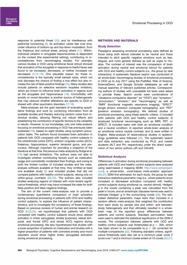
response to potential threat (10), and its interference withprefrontal functioning (9). Its activation varies fast over time,under influence of bottom-up and top-down modulation, fromthe thalamus and cortical areas, among others (11). Within-individual variation in amygdala responsiveness is dependenton the context (the experimental setting), contributing to in-consistencies from neuroimaging studies. For example,various studies in OCD using emotional facial stimuli showedthat activation of the amygdala in response to fearful faces wasfound to be increased, decreased, or neither increased nordecreased (4,12–14). One plausible reason for these in-consistencies is the typically small sample sizes, which notonly decrease the chance of finding a true effect but also in-crease the risk of false-positive findings (15). Many studies alsoinclude patients on selective serotonin reuptake inhibitors,which are known to influence brain activation in regions suchas the amygdala and hippocampus (16). Comorbidity withanxiety or mood disorders is another source of heterogeneitythat may obscure whether alterations are specific to OCD orshared with other psychiatric disorders (17,18).
Meta-analyses are the gold standard of evaluating quanti-tative findings and work by combining information from allavailable studies and thereby reducing random noise from in-dividual studies, allowing filtering out robust effects andestablishing the contribution of specific factors to the variabilityin results. However, to our knowledge, only one meta-analysisfocusing on emotional processing in OCD has previously beenpublished (19), based on eight studies using symptom provo-cation tasks. The authors found increased brain activation inpatients with OCD compared with healthy control subjects inthe orbitofrontal cortex (OFC), anterior cingulate cortex (ACC),thalamus, hippocampus, superior temporal gyrus, and pre-cuneus. Although important for providing a snapshot of theliterature at that time, the previous meta-analysis by Rotge et al.(19) had several limitations. The authors were not able toinvestigate whether contributing factors such as medicationusage and comorbidity moderated their findings, and owing toboth the limited number of included studies and the meta-analysis software available at that time, they omitted at leastone available study (2) and included studies that did notcompare patients with healthy control subjects, relying only onwithin-group contrasts (20–22). The authors also includedstudies analyzing regions of interest with more lenient signifi-cance thresholds, which may have increased the rates for bothfalse-positive and false-negative findings.
The aim of the current meta-analysis was to provide acontemporary quantitative comparison of brain activationduring emotional processing in patients with OCD and healthycontrol subjects, to explore the influence of patient charac-teristics, and to investigate the consistency of these findings.Based on previous reviews of human and animal research onOCD (8,23,24), we hypothesized that patients with OCDcompared with healthy control subjects would show alteredactivation in limbic (amygdala), striatal (putamen), lateral tem-poral, and frontal (OFC and dorsal ACC) regions duringemotional processing. We also hypothesized that studies witha lower proportion of patients on medication and studies with ahigher proportion of patients with comorbid anxiety and mooddisorders would show higher limbic (amygdala) activationduring emotional processing.
METHODS AND MATERIALS
Study Selection
Paradigms assessing emotional processing were defined asthose using both stimuli intended to be neutral and thoseintended to elicit specific negative emotions such as fear,disgust, and more general distress as well as urges to ritu-alize. The contrast of interest was the comparison of brainactivation during neutral and emotional stimuli for patientswith OCD and healthy control subjects (i.e., the group by taskinteraction). A systematic literature search was conducted ofall whole-brain neuroimaging studies of emotional processingin OCD up to July 2017 using the PubMed, Web of Science,ScienceDirect, and Google Scholar databases as well asmanual searches of relevant published articles. Correspond-ing authors of studies with unavailable full texts were askedto provide these. Search words were combinations of“obsessive-compulsive disorder” (or “OCD”) and “symptom,”“provocation,” “emotion,” and “neuroimaging” as well as“fMRI” (functional magnetic resonance imaging), “SPECT”(single photon emission computed tomography), and “PET”(positron emission tomography). We defined studies ofemotional processing using these specific criteria: 1) includedboth patients with OCD and healthy control subjects; 2)employed functional neuroimaging such as fMRI, PET, orSPECT; 3) included tasks with both an emotional conditionand a neutral condition; 4) reported whole-brain analysis ofan emotional versus neutral contrast; and 5) were written inEnglish. Meta-analysis of observational studies in epidemi-ology guidelines were followed (25). The systematic searchand data extraction was conducted by Ph.D. and masterstudents (ALT and PH, respectively) under the direct super-vision of two senior authors (JR and OAvdH).
Statistical Analyses
Differences in activation during emotional processing betweenpatients with OCD and healthy control subjects were analyzedusing seed-based d mapping (SDM; http://www.sdmproject.com), a whole-brain, voxel-based meta-analytic approach(26,27). SDM first estimated, for each study, the group by taskinteraction statistical parametric map (i.e., where patients showincreased or decreased activation compared with healthycontrol subjects during emotional vs. neutral stimuli). Hedges’g in the voxels containing a peak was calculated from thepeak’s t score, and an anisotropic Gaussian kernel was used toestimate Hedges’ g in the surrounding voxels (28). The esti-mated statistical parametric maps were then included in arandom effects meta-analysis that weighted the contributionfrom each study by sample size and within- and between-study heterogeneity and that ultimately resulted in a whole-brain map of the reported group differences betweenpatients and control subjects. Standard permutation testswere used to estimate the statistical significance of the SDM Zscores. The comparison between patients with OCD andhealthy control subjects was thresholded at p , .005, whichhas been shown to be comparable to p , .05 corrected formultiple comparisons (22). Following standard criteria, signifi-cance thresholds were also set at a minimum peak voxel Zscore over 1 and a minimum cluster extent of 10 voxels (26,27).
Emotional Processing in OCD
564 Biological Psychiatry: Cognitive Neuroscience and Neuroimaging June 2018; 3:563–571 www.sobp.org/BPCNNI
BiologicalPsychiatry:CNNI
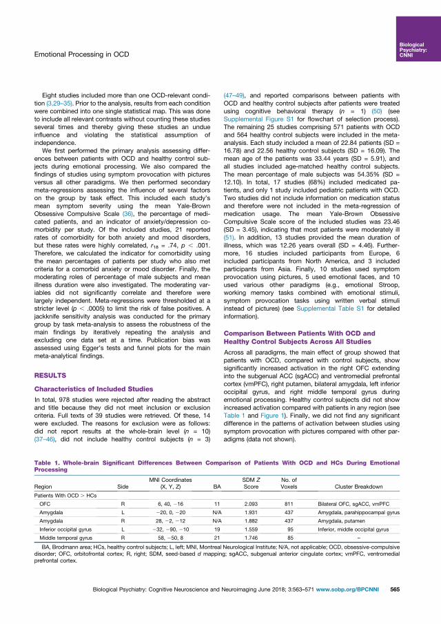
Eight studies included more than one OCD-relevant condi-tion (3,29–35). Prior to the analysis, results from each conditionwere combined into one single statistical map. This was doneto include all relevant contrasts without counting these studiesseveral times and thereby giving these studies an undueinfluence and violating the statistical assumption ofindependence.
We first performed the primary analysis assessing differ-ences between patients with OCD and healthy control sub-jects during emotional processing. We also compared thefindings of studies using symptom provocation with picturesversus all other paradigms. We then performed secondarymeta-regressions assessing the influence of several factorson the group by task effect. This included each study’smean symptom severity using the mean Yale-BrownObsessive Compulsive Scale (36), the percentage of medi-cated patients, and an indicator of anxiety/depression co-morbidity per study. Of the included studies, 21 reportedrates of comorbidity for both anxiety and mood disorders,but these rates were highly correlated, r18 = .74, p , .001.Therefore, we calculated the indicator for comorbidity usingthe mean percentages of patients per study who also metcriteria for a comorbid anxiety or mood disorder. Finally, themoderating roles of percentage of male subjects and meanillness duration were also investigated. The moderating var-iables did not significantly correlate and therefore werelargely independent. Meta-regressions were thresholded at astricter level (p , .0005) to limit the risk of false positives. Ajackknife sensitivity analysis was conducted for the primarygroup by task meta-analysis to assess the robustness of themain findings by iteratively repeating the analysis andexcluding one data set at a time. Publication bias wasassessed using Egger’s tests and funnel plots for the mainmeta-analytical findings.
RESULTS
Characteristics of Included Studies
In total, 978 studies were rejected after reading the abstractand title because they did not meet inclusion or exclusioncriteria. Full texts of 39 studies were retrieved. Of these, 14were excluded. The reasons for exclusion were as follows:did not report results at the whole-brain level (n = 10)(37–46), did not include healthy control subjects (n = 3)
(47–49), and reported comparisons between patients withOCD and healthy control subjects after patients were treatedusing cognitive behavioral therapy (n = 1) (50) (seeSupplemental Figure S1 for flowchart of selection process).The remaining 25 studies comprising 571 patients with OCDand 564 healthy control subjects were included in the meta-analysis. Each study included a mean of 22.84 patients (SD =16.78) and 22.56 healthy control subjects (SD = 16.09). Themean age of the patients was 33.44 years (SD = 5.91), andall studies included age-matched healthy control subjects.The mean percentage of male subjects was 54.35% (SD =12.10). In total, 17 studies (68%) included medicated pa-tients, and only 1 study included pediatric patients with OCD.Two studies did not include information on medication statusand therefore were not included in the meta-regression ofmedication usage. The mean Yale-Brown ObsessiveCompulsive Scale score of the included studies was 23.46(SD = 3.45), indicating that most patients were moderately ill(51). In addition, 13 studies provided the mean duration ofillness, which was 12.26 years overall (SD = 4.46). Further-more, 16 studies included participants from Europe, 6included participants from North America, and 3 includedparticipants from Asia. Finally, 10 studies used symptomprovocation using pictures, 5 used emotional faces, and 10used various other paradigms (e.g., emotional Stroop,working memory tasks combined with emotional stimuli,symptom provocation tasks using written verbal stimuliinstead of pictures) (see Supplemental Table S1 for detailedinformation).
Comparison Between Patients With OCD andHealthy Control Subjects Across All Studies
Across all paradigms, the main effect of group showed thatpatients with OCD, compared with control subjects, showsignificantly increased activation in the right OFC extendinginto the subgenual ACC (sgACC) and ventromedial prefrontalcortex (vmPFC), right putamen, bilateral amygdala, left inferioroccipital gyrus, and right middle temporal gyrus duringemotional processing. Healthy control subjects did not showincreased activation compared with patients in any region (seeTable 1 and Figure 1). Finally, we did not find any significantdifference in the patterns of activation between studies usingsymptom provocation with pictures compared with other par-adigms (data not shown).
Table 1. Whole-brain Significant Differences Between Comparison of Patients With OCD and HCs During EmotionalProcessing
Region SideMNI Coordinates
(X, Y, Z) BASDM ZScore
No. ofVoxels Cluster Breakdown
Patients With OCD . HCs
OFC R 6, 40, 216 11 2.093 811 Bilateral OFC, sgACC, vmPFC
Amygdala L 220, 0, 220 N/A 1.931 437 Amygdala, parahippocampal gyrus
Amygdala R 28, 22, 212 N/A 1.882 437 Amygdala, putamen
Inferior occipital gyrus L 232, 290, 210 19 1.559 95 Inferior, middle occipital gyrus
Middle temporal gyrus R 58, 250, 8 21 1.746 85 –
BA, Brodmann area; HCs, healthy control subjects; L, left; MNI, Montreal Neurological Institute; N/A, not applicable; OCD, obsessive-compulsivedisorder; OFC, orbitofrontal cortex; R, right; SDM, seed-based d mapping; sgACC, subgenual anterior cingulate cortex; vmPFC, ventromedialprefrontal cortex.
Emotional Processing in OCD
Biological Psychiatry: Cognitive Neuroscience and Neuroimaging June 2018; 3:563–571 www.sobp.org/BPCNNI 565
BiologicalPsychiatry:CNNI

Meta-regressions of Factors Influencing theDifference Between Patients With OCD and HealthyControl Subjects
The meta-regression analyses (see Table 2 and Figure 2 fordetails) showed that the percentage of patients per study usingpsychotropic medication, primarily selective serotonin reup-take inhibitors, correlated negatively with activation in the rightamygdala and left inferior occipital gyrus, indicating that theincreased limbic and occipital activation during emotionalprocessing in patients compared with control subjects is mostpronounced in studies with higher percentages of unmedi-cated patients.
Studies including patients with higher symptom severity, asmeasured with the Yale-Brown Obsessive Compulsive Scale,showed significantly increased activation in the right rostralsgACC, the left medial prefrontal cortex, and the right pre-cuneus. Studies with a higher rate of comorbidity with anxietyand mood disorders also found more pronounced activation inthe right putamen, amygdala, and insula as well as less pro-nounced activation in the left amygdala and right vmPFC inpatients compared with control subjects.
Studies with more male patients found significantly lowerdifferences in presupplementary motor area activation.Finally, studies with longer mean duration of illness showedincreased right putamen activation and decreased left tem-poral pole and OFC activation in patients versus controlsubjects.
Sensitivity Analysis and Publication Bias
The whole-brain jackknife sensitivity analysis showed that themain results were replicated in nearly all combinations of
studies. Additional findings, however, appeared in some of thecombinations. Activation of the left inferior frontal gyruswas found to be significantly increased in patients versuscontrol subjects when one of nine studies was removed(5,6,13,31–34,52,53). The removal of one of three studies alsoresulted in significantly decreased activation in the bilateralACC in patients. In addition, the removal of one of two differentstudies increased activation in the left angular gyrus (54) andright precuneus (13) in patients (see Supplemental Table S2 fordetailed information). These jackknife analyses show that thefindings of the main meta-analysis were largely robust, whilehyperactivation in the left inferior frontal gyrus and hypo-activation in the bilateral ACC in patients may have beenunderestimated. However, there was no apparent pattern inthese studies given that these spanned all functional tasks. Inaddition, the meta-regressions did not reveal any relations toany of the explored patient characteristics.
Inspections of Egger’s intercepts and funnel plots did notindicate significant publication bias in any region from the mainresults, with the lowest p value on the Egger’s test being .175.This indicates that there was a low risk of activation beingoverestimated because of studies being withheld or not beingpublished.
DISCUSSION
The current study is the largest meta-analysis of emotionalprocessing in OCD to date, encompassing 25 studies using avariety of emotional tasks, including symptomprovocation usingimagesorwordsaswell asemotional variantsof typical cognitiveparadigms such as the emotional Stroop task and workingmemory tasks with emotional distractors. The results help tointegrate a body of research that has often resulted in inconsis-tent findings that are hard to reconcile, particularly regarding therole of the amygdala in OCD. The main findings were that,compared with healthy control subjects, patients with OCDshowed increased activation in the amygdala, OFC extendinginto the sgACC and vmPFC, putamen, andmiddle temporal andinferior occipital regions during emotional processing.
The meta-regression analyses showed that the findings inthe amygdala are especially sensitive to a number of patientfactors such as medication status and comorbidity. In contrast,the group effects in the amygdala were independent of meansymptom severity of the patient samples. Notably, the left andright amygdala showed opposite activation patterns in themeta-regressions for medication usage and comorbidity withanxiety and mood disorders. The right amygdala showedincreased activation in studies with higher percentages ofunmedicated patients and in studies with more comorbiddisorders. By contrast, activation in the left amygdala was lesspronounced in studies with more comorbidity. Studies withmore male subjects showed lower differences in presupple-mentary motor area activation. Finally, studies with longermean duration of illness showed increased differences in rightputamen activation and lower differences in the left temporalpole and OFC. Unfortunately, the variance in gender was low,and approximately half of the studies did not report duration ofillness, so these effects should be interpreted with caution.These meta-regressions contribute to the understanding of themixed findings on amygdala involvement in OCD in the
Figure 1. Regions of hyperactivation in patients with obsessive-compulsive disorder compared with healthy control subjects duringemotional processing, showing a distributed affective circuit includingfrontal, limbic, striatal, and ventral visual areas. IOC, inferior occipital cortex;MTG, middle temporal gyrus; OFC, orbitofrontal cortex.
Emotional Processing in OCD
566 Biological Psychiatry: Cognitive Neuroscience and Neuroimaging June 2018; 3:563–571 www.sobp.org/BPCNNI
BiologicalPsychiatry:CNNI
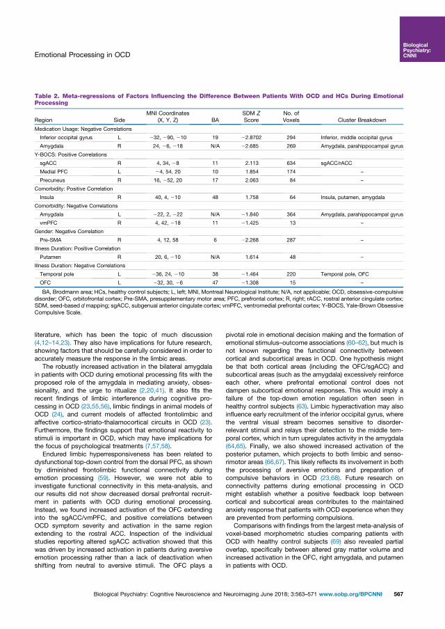
literature, which has been the topic of much discussion(4,12–14,23). They also have implications for future research,showing factors that should be carefully considered in order toaccurately measure the response in the limbic areas.
The robustly increased activation in the bilateral amygdalain patients with OCD during emotional processing fits with theproposed role of the amygdala in mediating anxiety, obses-sionality, and the urge to ritualize (2,20,41). It also fits therecent findings of limbic interference during cognitive pro-cessing in OCD (23,55,56), limbic findings in animal models ofOCD (24), and current models of affected frontolimbic andaffective cortico-striato-thalamocortical circuits in OCD (23).Furthermore, the findings support that emotional reactivity tostimuli is important in OCD, which may have implications forthe focus of psychological treatments (7,57,58).
Endured limbic hyperresponsiveness has been related todysfunctional top-down control from the dorsal PFC, as shownby diminished frontolimbic functional connectivity duringemotion processing (59). However, we were not able toinvestigate functional connectivity in this meta-analysis, andour results did not show decreased dorsal prefrontal recruit-ment in patients with OCD during emotional processing.Instead, we found increased activation of the OFC extendinginto the sgACC/vmPFC, and positive correlations betweenOCD symptom severity and activation in the same regionextending to the rostral ACC. Inspection of the individualstudies reporting altered sgACC activation showed that thiswas driven by increased activation in patients during aversiveemotion processing rather than a lack of deactivation whenshifting from neutral to aversive stimuli. The OFC plays a
pivotal role in emotional decision making and the formation ofemotional stimulus–outcome associations (60–62), but much isnot known regarding the functional connectivity betweencortical and subcortical areas in OCD. One hypothesis mightbe that both cortical areas (including the OFC/sgACC) andsubcortical areas (such as the amygdala) excessively reinforceeach other, where prefrontal emotional control does notdampen subcortical emotional responses. This would imply afailure of the top-down emotion regulation often seen inhealthy control subjects (63). Limbic hyperactivation may alsoinfluence early recruitment of the inferior occipital gyrus, wherethe ventral visual stream becomes sensitive to disorder-relevant stimuli and relays their detection to the middle tem-poral cortex, which in turn upregulates activity in the amygdala(64,65). Finally, we also showed increased activation of theposterior putamen, which projects to both limbic and senso-rimotor areas (66,67). This likely reflects its involvement in boththe processing of aversive emotions and preparation ofcompulsive behaviors in OCD (23,68). Future research onconnectivity patterns during emotional processing in OCDmight establish whether a positive feedback loop betweencortical and subcortical areas contributes to the maintainedanxiety response that patients with OCD experience when theyare prevented from performing compulsions.
Comparisons with findings from the largest meta-analysis ofvoxel-based morphometric studies comparing patients withOCD with healthy control subjects (69) also revealed partialoverlap, specifically between altered gray matter volume andincreased activation in the OFC, right amygdala, and putamenin patients with OCD.
Table 2. Meta-regressions of Factors Influencing the Difference Between Patients With OCD and HCs During EmotionalProcessing
Region SideMNI Coordinates
(X, Y, Z) BASDM ZScore
No. ofVoxels Cluster Breakdown
Medication Usage: Negative Correlations
Inferior occipital gyrus L 232, 290, 210 19 22.8702 294 Inferior, middle occipital gyrus
Amygdala R 24, 26, 218 N/A 22.685 269 Amygdala, parahippocampal gyrus
Y-BOCS: Positive Correlations
sgACC R 4, 34, 28 11 2.113 634 sgACC/rACC
Medial PFC L 24, 54, 20 10 1.854 174 –
Precuneus R 16, 252, 20 17 2.063 84 –
Comorbidity: Positive Correlation
Insula R 40, 4, 210 48 1.758 64 Insula, putamen, amygdala
Comorbidity: Negative Correlations
Amygdala L 222, 2, 222 N/A 21.840 364 Amygdala, parahippocampal gyrus
vmPFC R 4, 42, 218 11 21.425 13 –
Gender: Negative Correlation
Pre-SMA R 4, 12, 58 6 22.268 287 –
Illness Duration: Positive Correlation
Putamen R 20, 6, 210 N/A 1.614 48 –
Illness Duration: Negative Correlations
Temporal pole L 236, 24, 210 38 21.464 220 Temporal pole, OFC
OFC L 232, 30, 26 47 21.308 15 –
BA, Brodmann area; HCs, healthy control subjects; L, left; MNI, Montreal Neurological Institute; N/A, not applicable; OCD, obsessive-compulsivedisorder; OFC, orbitofrontal cortex; Pre-SMA, presupplementary motor area; PFC, prefrontal cortex; R, right; rACC, rostral anterior cingulate cortex;SDM, seed-based dmapping; sgACC, subgenual anterior cingulate cortex; vmPFC, ventromedial prefrontal cortex; Y-BOCS, Yale-Brown ObsessiveCompulsive Scale.
Emotional Processing in OCD
Biological Psychiatry: Cognitive Neuroscience and Neuroimaging June 2018; 3:563–571 www.sobp.org/BPCNNI 567
BiologicalPsychiatry:CNNI

Several studies have investigated whether disorder-specificstimuli elicit different neural responses compared with generalaversive stimuli, with mixed results [e.g., (41,45,59)]. Forinstance, increased activation in the amygdala has been re-ported during disorder-specific stimuli in some studies (34,59),but not other studies, when compared with general aversivestimuli (45). Unfortunately, we were unable to compare theeffects of disorder-specific stimuli with those of general stimulidue to the few studies with comparable paradigms. Becausewe were unable to differentiate between the provocations ofspecific symptom dimensions, we assume homogeneity in ouranalyses, while OCD is a highly heterogeneous disorder notonly in its clinical presentation but also in its etiology (70–72).Different symptom dimensions seem to vary in their limbicinvolvement, being more pronounced in patients with moreaggressive, sexual, or religious symptoms and checking rituals(13,73,74).
Abnormal recruitment of the brain circuits during emotionalprocessing in patients with OCD may represent dynamic cor-relates of the symptom state and not necessarily a statictraitlike marker of vulnerability to OCD. Indeed, several studiesshow that successful treatment with cognitive behavioraltherapy or selective serotonin reuptake inhibitors at least partlynormalizes patients’ provocation-induced response in theOFC, putamen, and parietal cortex (23,75). Less is knownabout the effect of treatment on the limbic response. It is alsopossible that brain abnormalities constitute trait or risk factorsfor the disorder, given that unaffected first-degree relatives ofpatients with OCD also show increased activation in the OFCduring a reversal learning task (76). Longitudinal, geneticallyinformative designs, such as discordant monozygotic twinstudies, are needed to shed further light on the origins of theobserved emotional processing-related activation patternsin OCD.
The current results show notable differences compared withthe findings of the previous smaller meta-analysis (19). Forinstance, we were not able to replicate the authors’ findings ofincreased activation in the medial PFC, bilateral globus pal-lidus, right thalamus, left OFC, or left hippocampus in patientscompared with healthy control subjects. Because we wereable to include nearly three times as many studies as in theprevious meta-analysis and selected only those using whole-brain analyses, the current results could be regarded as lesssensitive to type I and type II errors.
Our study has some limitations that should be considered.We did not have access to patient-level data that may haveprovided additional power. Some of the included studies werequite small (the smallest including only 8 patients and 8 controlsubjects), and smaller studies may have an increased risk ofintroducing noise. The risk of undue noise was also increasedbecause nearly every study used reported foci at uncorrectedp values, which heightens the risk of false positives. Studiesalso varied in their use of statistical packages as well as theiruse of the Montreal Neurological Institute or Talairach co-ordinates, including the transformations used to convert be-tween the coordinate systems. Although we used correctionsfor transforming the foci of each study into Montreal Neuro-logical Institute coordinates using standard SDM procedures,this may have introduced additional noise into our meta-analysis. We chose to include only studies in English, which
Figure 2. Results of meta-regressions indicating factors that are asso-ciated with an increased (red) or decreased (blue) difference betweenpatients with obsessive-compulsive disorder and healthy control subjects.(A) Patient samples with more medicated patients showed less hyper-activation in the right amygdala and left cerebellum. (B) Increased symptomseverity correlated with increased patient hyperactivation in the subgenual/rostral anterior cingulate cortex and medial prefrontal cortex. (C) Patientsamples with more anxiety and mood disorder comorbidity showedincreased activation in the right insula, putamen, and amygdala as well asdecreased activation in the left amygdala and right ventromedial prefrontalcortex. (D) Patient samples with more male subjects showed less activationin the presupplementary motor area. (E) Patient samples with longer meanduration of illness showed increased activation in the right putamen andlower activation in the left temporal pole and orbitofrontal cortex. L, left;Y-BOCS, Yale-Brown Obsessive Compulsive Scale.
Emotional Processing in OCD
568 Biological Psychiatry: Cognitive Neuroscience and Neuroimaging June 2018; 3:563–571 www.sobp.org/BPCNNI
BiologicalPsychiatry:CNNI

may have excluded some studies. However, we are not awareof any relevant high-quality studies in other languages. Finally,although we did not find any significant differences in activa-tion between studies using symptom provocation with picturescompared with all other paradigms, the current literature mightnot provide adequate power or homogeneity to find smallerdifferences. This could also be the case for the variablesexplored using meta-regressions given that the variance waslimited in several of the variables. The field is currently lackingstudies of emotional processing in pediatric OCD, and ourfindings may be seen as more generalizable to adult OCD.Studies that directly compare adults with children, or thatfollow developing children, are needed. The few studiesemploying each paradigm also meant that there would nothave been enough power to adequately analyze them sepa-rately. However, a recent meta-analysis of 90 studies ofobsessive-compulsive symptom induction in clinical andnonclinical samples showed similar results across a range ofinduction procedures (77). This provides some support for ournonsignificant comparison between studies using symptomprovocation with pictures versus other paradigms.
Conclusions
Compared with healthy control subjects, patients with OCDshow increased activation in the frontolimbic circuit, encom-passing the amygdala, OFC/sgACC/vmPFC, occipital andmiddle temporal cortices, and posterior/ventral putamen.Furthermore, the degree to which patients and control subjectsdiffer in their limbic and striatal responses is influenced bymedication status, comorbidity, and symptom severity. Thesefindings help to explain some of the inconsistencies in theliterature and highlight the importance of well-powered meta-and mega-analyses of neuroimaging data.
ACKNOWLEDGMENTS AND DISCLOSURESThe study was supported by Grant Nos. 911754 and 911880 from the HelseVest Health Authority (to GK).
We thank Luke Norman for providing information allowing for a com-parison between the results of his meta-analysis of voxel-based morphologystudies in OCD and our findings.
The authors report no biomedical financial interests or potential conflictsof interest.
ARTICLE INFORMATIONFrom the Obsessive-Compulsive Disorder (OCD) team (ALT, PH, GK, BH,OAvdH), Haukeland University Hospital, and Department of Clinical Psy-chology (ALT, PH. GK, BH), University of Bergen, Bergen, Norway; FIDMAGGermanes Hospitalàries (JR), Centre for Biomedical Research in MentalHealth Network (CIBERSAM), Barcelona, Spain; Department of ClinicalNeuroscience (JR, DM-C), Centre for Psychiatry Research, KarolinskaInstitutet, Stockholm, Sweden; Department of Psychosis Studies (JR),Institute of Psychology, Psychiatry, and Neuroscience, King’s CollegeLondon, London, United Kingdom; Department of Anatomy & Neurosci-ences (OAvdH) and Department of Psychiatry (OAvdH), VU UniversityMedical Center, and Amsterdam Neuroscience (OAvdH), Amsterdam, TheNetherlands.
ALT and PH contributed equally to this work.Address correspondence to Anders Lillevik Thorsen, M.Sc., OCD team,
Haukeland University Hospital, P.O. 1400, 5021 Bergen, Norway; E-mail:[email protected].
Received Dec 24, 2017; accepted Jan 11, 2018.
Supplementary material cited in this article is available online at https://doi.org/10.1016/j.bpsc.2018.01.009.
REFERENCES1. Abramowitz JS, Taylor S, McKay D (2009): Obsessive-compulsive
disorder. Lancet 374:491–499.2. van den Heuvel OA, Veltman DJ, Groenewegen HJ, Dolan RJ,
Cath DC, Boellaard R, et al. (2004): Amygdala activity in obsessive-compulsive disorder with contamination fear: A study with oxygen-15 water positron emission tomography. Psychiatry Res 132:225–237.
3. Mataix-Cols D, Wooderson S, Lawrence N, Brammer MJ, Speckens A,Phillips ML (2004): Distinct neural correlates of washing, checking, andhoarding symptom dimensions in obsessive-compulsive disorder.Arch Gen Psychiatry 61:564–576.
4. Cannistraro PA, Rauch SL (2003): Neural circuitry of anxiety: Evidencefrom structural and functional neuroimaging studies. Psychopharma-col Bull 37:8–25.
5. van den Heuvel OA, Veltman DJ, Groenewegen HJ, Witter MP,Merkelbach J, Cath DC, et al. (2005): Disorder-specific neuroana-tomical correlates of attentional bias in obsessive-compulsive disor-der, panic disorder, and hypochondriasis. Arch Gen Psychiatry62:922–933.
6. Park SE, Yang JC, Jeong GW (2016): Neuroanatomical assessment ofthe impact of negative emotion on implicit memory in patients withobsessive compulsive disorder. Acta Neuropsychiatr 28:206–213.
7. Mataix-Cols D, van den Heuvel OA (2006): Common and distinctneural correlates of obsessive-compulsive and related disorders.Psychiatr Clin North Am 29:391–410.
8. Milad MR, Rauch SL (2012): Obsessive-compulsive disorder: Beyondsegregated cortico-striatal pathways. Trends Cogn Sci 16:43–51.
9. LeDoux JE, Pine DS (2016): Using neuroscience to help understandfear and anxiety: A two-system framework. Am J Psychiatry 173:1083–1093.
10. LeDoux JE (2014): Coming to terms with fear. Proc Natl Acad SciU S A 111:2871–2878.
11. Pessoa L (2017): A network model of the emotional brain. Trends CognSci 21:357–371.
12. Britton JC, Stewart SE, Killgore WD, Rosso IM, Price LM, Gold AL,et al. (2010): Amygdala activation in response to facial expressions inpediatric obsessive-compulsive disorder. Depress Anxiety 27:643–651.
13. Via E, Cardoner N, Pujol J, Alonso P, Lopez-Sola M, Real E, et al.(2014): Amygdala activation and symptom dimensions in obsessive-compulsive disorder. Br J Psychiatry 204:61–68.
14. Cardoner N, Harrison BJ, Pujol J, Soriano-Mas C, Hernández-Ribas R,López-Solá M, et al. (2011): Enhanced brain responsiveness duringactive emotional face processing in obsessive compulsive disorder.World J Biol Psychiatry 12:349–363.
15. Button KS, Ioannidis JP, Mokrysz C, Nosek BA, Flint J, Robinson ES,et al. (2013): Power failure: Why small sample size undermines thereliability of neuroscience. Nat Rev Neurosci 14:365–376.
16. Outhred T, Hawkshead BE, Wager TD, Das P, Malhi GS, Kemp AH(2013): Acute neural effects of selective serotonin reuptake inhibitorsversus noradrenaline reuptake inhibitors on emotion processing: im-plications for differential treatment efficacy. Neurosci Biobehav Rev37:1786–1800.
17. Etkin A, Wager TD (2007): Functional neuroimaging of anxiety: A meta-analysis of emotional processing in PTSD, social anxiety disorder, andspecific phobia. Am J Psychiatry 164:1476–1488.
18. Hamilton JP, Etkin A, Furman DJ, Lemus MG, Johnson RF, Gotlib IH(2012): Functional neuroimaging of major depressive disorder: A meta-analysis and new integration of baseline activation and neuralresponse data. Am J Psychiatry 169:693–703.
19. Rotge J-Y, Guehl D, Dilharreguy B, Cuny E, Tignol J, Bioulac B, et al.(2008): Provocation of obsessive-compulsive symptoms: A quantita-tive voxel-based meta-analysis of functional neuroimaging studies.J Psychiatry Neurosci 33:405–412.
Emotional Processing in OCD
Biological Psychiatry: Cognitive Neuroscience and Neuroimaging June 2018; 3:563–571 www.sobp.org/BPCNNI 569
BiologicalPsychiatry:CNNI

20. McGuire PK, Bench CJ, Frith CD, Marks IM, Frackowiak RS, Dolan RJ(1994): Functional anatomy of obsessive-compulsive phenomena. Br JPsychiatry 164:459–468.
21. Rauch SL, Jenike MA, Alpert NM, Baer L, Breiter HC, Savage CR, et al.(1994): Regional cerebral blood flow measured during symptomprovocation in obsessive-compulsive disorder using oxygen 15-labeled carbon dioxide and positron emission tomography. Arch GenPsychiatry 51:62–70.
22. Rauch SL, Shin LM, Dougherty DD, Alpert NM, Fischman AJ,Jenike MA (2002): Predictors of fluvoxamine response incontamination-related obsessive compulsive disorder: A PET symp-tom provocation study. Neuropsychopharmacology 27:782–791.
23. van den Heuvel OA, van Wingen G, Soriano-Mas C, Alonso P,Chamberlain SR, Nakamae T, et al. (2016): Brain circuitry of compul-sivity. Eur Neuropsychopharmacol 26:810–827.
24. Wood J, Ahmari SE (2015): A framework for understanding theemerging role of corticolimbic-ventral striatal networks in OCD-associated repetitive behaviors. Front Syst Neurosci 9:171.
25. Stroup DF, Berlin JA, Morton SC, Olkin I, Williamson GD, Rennie D,et al. (2000): Meta-analysis of observational studies in epidemiology: Aproposal for reporting. JAMA 283:2008–2012.
26. Radua J, Mataix-Cols D (2009): Voxel-wise meta-analysis of greymatter changes in obsessive-compulsive disorder. Br J Psychiatry195:393–402.
27. Radua J, Mataix-Cols D, Phillips ML, El-Hage W, Kronhaus D,Cardoner N, et al. (2012): A new meta-analytic method for neuro-imaging studies that combines reported peak coordinates and sta-tistical parametric maps. Eur Psychiatry 27:605–611.
28. Radua J, Grau M, van den Heuvel OA, Thiebaut de Schotten M,Stein DJ, Canales-Rodriguez EJ, et al. (2014): Multimodal voxel-basedmeta-analysis of white matter abnormalities in obsessive-compulsivedisorder. Neuropsychopharmacology 39:1547–1557.
29. An S, Mataix-Cols D, Lawrence N, Wooderson S, Giampietro V,Speckens A, et al. (2009): To discard or not to discard: The neuralbasis of hoarding symptoms in obsessive-compulsive disorder. MolPsychiatry 14:318–331.
30. Lawrence NS, An SK, Mataix-Cols D, Ruths F, Speckens A, Phillips ML(2007): Neural responses to facial expressions of disgust but not fearare modulated by washing symptoms in OCD. Biol Psychiatry61:1072–1080.
31. Murayama K, Nakao T, Sanematsu H, Okada K, Yoshiura T, Tomita M,et al. (2013): Differential neural network of checking versus washingsymptoms in obsessive-compulsive disorder. Prog Neuro-psychopharmacol Biol Psychiatry 40:160–166.
32. Phillips ML, Marks IM, Senior C, Lythgoe D, O’Dwyer AM, Meehan O,et al. (2000): A differential neural response in obsessive-compulsivedisorder patients with washing compared with checking symptomsto disgust. Psychol Med 30:1037–1050.
33. Berlin HA, Schulz KP, Zhang S, Turetzky R, Rosenthal D, Goodman W(2015): Neural correlates of emotional response inhibition inobsessive-compulsive disorder: A preliminary study. Psychiatry Res234:259–264.
34. Brennan BP, Tkachenko O, Schwab ZJ, Juelich RJ, Ryan EM,Athey AJ, et al. (2015): An examination of rostral anterior cingulatecortex function and neurochemistry in obsessive-compulsive disorder.Neuropsychopharmacology 40:1866–1876.
35. Thiel A, Thiel J, Oddo S, Langnickel R, Brand M, Markowitsch HJ, et al.(2014): Obsessive-compulsive disorder patients with washing symp-toms show a specific brain network when confronted with aggressive,sexual, and disgusting stimuli. Neuropsychoanalysis 16:83–96.
36. Goodman WK, Price LH, Rasmussen SA, Mazure C, Fleischmann RL,Hill CL, et al. (1989): The Yale-Brown Obsessive Compulsive Scale: I.Development, use, and reliability. Arch Gen Psychiatry 46:1006–1011.
37. Agarwal SM, Jose D, Baruah U, Shivakumar V, Kalmady SV,Venkatasubramanian G, et al. (2013): Neurohemodynamic correlates ofwashingsymptoms inobsessive-compulsivedisorder:Apilot fMRIstudyusing symptom provocation paradigm. Indian J Psychol Med 35:67–74.
38. Simon D, Kaufmann C, Kniesche R, Kischkel E, Kathmann N (2013):Autonomic responses and neural-cardiac coupling during individually
tailored symptom provocation in obsessive-compulsive disorder.J Anxiety Disord 27:635–644.
39. Simon D, Kaufmann C, Musch K, Kischkel E, Kathmann N (2010):Fronto-striato-limbic hyperactivation in obsessive-compulsive disor-der during individually tailored symptom provocation. Psychophysi-ology 47:728–738.
40. Breiter HC, Rauch SL, Kwong KK, Baker JR, Weisskoff RM,Kennedy DN, et al. (1996): Functional magnetic resonance imaging ofsymptom provocation in obsessive-compulsive disorder. Arch GenPsychiatry 53:595–606.
41. Schienle A, Schafer A, Stark R, Walter B, Vaitl D (2005): Neural re-sponses of OCD patients towards disorder-relevant, generally disgust-inducing and fear-inducing pictures. Int J Psychophysiol 57:69–77.
42. Baioui A, Pilgramm J, Merz CJ, Walter B, Vaitl D, Stark R (2013): Neuralresponse in obsessive-compulsive washers depends on individual fitof triggers. Front Hum Neurosci 7:143.
43. Weidt S, Lutz J, Rufer M, Delsignore A, Jakob NJ, Herwig U, et al.(2016): Common and differential alterations of general emotion pro-cessing in obsessive-compulsive and social anxiety disorder. PsycholMed 46:1427–1436.
44. Gilbert AR, Akkal D, Almeida JR, Mataix-Cols D, Kalas C, Devlin B,et al. (2009): Neural correlates of symptom dimensions in pediatricobsessive-compulsive disorder: A functional magnetic resonance im-aging study. J Am Acad Child Adolesc Psychiatry 48:936–944.
45. Simon D, Adler N, Kaufmann C, Kathmann N (2014): Amygdalahyperactivation during symptom provocation in obsessive-compulsivedisorder and its modulation by distraction. NeuroImage Clin 4:549–557.
46. Becker MP, Nitsch AM, Schlosser R, Koch K, Schachtzabel C,Wagner G, et al. (2014): Altered emotional and BOLD responses tonegative, positive and ambiguous performance feedback in OCD. SocCogn Affect Neurosci 9:1127–1133.
47. Shin YW, Kwon JS, Kim JJ, Kang DH, Youn T, Kang KW, et al. (2006):Altered neural circuit for working memory before and after symptomprovocation in patients with obsessive-compulsive disorder. ActaPsychiatr Scand 113:420–429.
48. Nakao T, Nakagawa A, Yoshiura T, Nakatani E, Nabeyama M,Yoshizato C, et al. (2005): Brain activation of patients with obsessive-compulsive disorder during neuropsychological and symptom provo-cation tasks before and after symptom improvement: A functionalmagnetic resonance imaging study. Biol Psychiatry 57:901–910.
49. Schiepek G, Tominschek I, Heinzel S, Aigner M, Dold M, Unger A, et al.(2013): Discontinuous patterns of brain activation in the psychotherapyprocess of obsessive-compulsive disorder: Converging results fromrepeated fMRI and daily self-reports. PLoS One 8:e71863.
50. Baioui A, Pilgramm J, Kagerer S, Walter B, Vaitl D, Stark R (2013):Neural correlates of symptom reduction after CBT in obsessive-compulsive washers—An fMRI symptom provocation study.J Obsessive Compuls Relat Disord 2:322–330.
51. Storch EA, De Nadai AS, do Rosário MC, Shavitt RG, Torres AR,Ferrão YA, et al. (2015): Defining clinical severity in adults withobsessive-compulsive disorder. Compr Psychiatry 63:30–35.
52. Banca P, Voon V, Vestergaard MD, Philipiak G, Almeida I, Pocinho F,et al. (2015): Imbalance in habitual versus goal directed neural systemsduring symptom provocation in obsessive-compulsive disorder. Brain138:798–811.
53. Hennig-Fast K, Michl P, Müller J, Niedermeier N, Coates U, Müller N,et al. (2015): Obsessive-compulsive disorder—A question of con-science? An fMRI study of behavioural and neurofunctional correlatesof shame and guilt. J Psychiatr Res 68:354–362.
54. Harrison BJ, Pujol J, Soriano-Mas C, Hernández-Ribas R, López-Solà M, Ortiz H, et al. (2012): Neural correlates of moral sensitivity inobsessive-compulsive disorder. Arch Gen Psychiatry 69:741–749.
55. van Velzen LS, de Wit SJ, !Curc
ˇ
i!c-Blake B, Cath DC, de Vries FE,Veltman DJ, et al. (2015): Altered inhibition-related frontolimbicconnectivity in obsessive-compulsive disorder. Hum Brain Mapp36:4064–4075.
56. de Vries FE, de Wit SJ, Cath DC, van der Werf YD, van der Borden V,van Rossum TB, et al. (2014): Compensatory frontoparietal activity
Emotional Processing in OCD
570 Biological Psychiatry: Cognitive Neuroscience and Neuroimaging June 2018; 3:563–571 www.sobp.org/BPCNNI
BiologicalPsychiatry:CNNI

during working memory: An endophenotype of obsessive-compulsivedisorder. Biol Psychiatry 76:878–887.
57. Foa EB, McLean CP (2016): The efficacy of exposure therapy foranxiety-related disorders and its underlying mechanisms: The case ofOCD and PTSD. Annu Rev Clin Psychol 12:1–28.
58. Calkins AW, Berman NC, Wilhelm S (2013): Recent advances inresearch on cognition and emotion in OCD: A review. Curr PsychiatryRep 15:357.
59. de Wit SJ, van der Werf YD, Mataix-Cols D, Trujillo JP, van Oppen P,Veltman DJ, et al. (2015): Emotion regulation before and after trans-cranial magnetic stimulation in obsessive compulsive disorder.Psychol Med 45:3059–3073.
60. Remijnse PL, Nielen MM, van Balkom AJ, Cath DC, van Oppen P,Uylings HB, et al. (2006): Reduced orbitofrontal-striatal activity on areversal learning task in obsessive-compulsive disorder. Arch GenPsychiatry 63:1225–1236.
61. Rolls ET, Grabenhorst F (2008): The orbitofrontal cortex and beyond:From affect to decision-making. Prog Neurobiol 86:216–244.
62. Nashiro K, Sakaki M, Nga L, Mather M (2012): Differential brain activityduring emotional versus nonemotional reversal learning. J CognNeurosci 24:1794–1805.
63. Ochsner KN, Silvers JA, Buhle JT (2012): Functional imaging studies ofemotion regulation: A synthetic review and evolving model of thecognitive control of emotion. Ann N Y Acad Sci 1251:E1–E24.
64. Pessoa L, Adolphs R (2010): Emotion processing and the amygdala:From a “low road” to “many roads” of evaluating biological signifi-cance. Nat Rev Neurosci 11:773–783.
65. Vuilleumier P (2005): How brains beware: Neural mechanisms ofemotional attention. Trends Cogn Sci 9:585–594.
66. Jahanshahi M, Obeso I, Rothwell JC, Obeso JA (2015): A fronto-striato-subthalamic-pallidal network for goal-directed and habitualinhibition. Nat Rev Neurosci 16:719–732.
67. Lehéricy S, Ducros M, De Moortele V, Francois C, Thivard L,Poupon C, et al. (2004): Diffusion tensor fiber tracking shows distinctcorticostriatal circuits in humans. Ann Neurol 55:522–529.
68. Pauls DL, Abramovitch A, Rauch SL, Geller DA (2014): Obsessive-compulsive disorder: An integrative genetic and neurobiologicalperspective. Nat Rev Neurosci 15:410–424.
69. Norman LJ, Carlisi C, Lukito S, Hart H, Mataix-Cols D, Radua J, et al.(2016): Structural and functional brain abnormalities in attention-deficit/hyperactivity disorder and obsessive-compulsive disorder: Acomparative meta-analysis. JAMA Psychiatry 73:815–825.
70. Leckman JF, Denys D, Simpson HB, Mataix-Cols D, Hollander E,Saxena S, et al. (2010): Obsessive-compulsive disorder: A review ofthe diagnostic criteria and possible subtypes and dimensional speci-fiers for SDM-V. Depress Anxiety 27:507–527.
71. Mataix-Cols D, Rosario-Campos MC, Leckman JF (2005):A multidimensional model of obsessive-compulsive disorder. Am JPsychiatry 162:228–238.
72. Iervolino AC, Rijsdijk FV, Cherkas L, Fullana MA, Mataix-Cols D (2011):A multivariate twin study of obsessive-compulsive symptomdimensions. Arch Gen Psychiatry 68:637–644.
73. Harrison BJ, Pujol J, Cardoner N, Deus J, Alonso P, Lopez-Sola M,et al. (2013): Brain corticostriatal systems and the major clinicalsymptom dimensions of obsessive-compulsive disorder. Biol Psychi-atry 73:321–328.
74. Pujol J, Soriano-Mas C, Alonso P, et al. (2004): Mapping structuralbrain alterations in obsessive-compulsive disorder. Arch Gen Psychi-atry 61:720–730.
75. Thorsen AL, van den Heuvel OA, Hansen B, Kvale G (2015): Neuro-imaging of psychotherapy for obsessive-compulsive disorder: A sys-tematic review. Psychiatry Res 233:306–313.
76. Chamberlain SR, Menzies L, Hampshire A, Suckling J, Fineberg NA,del Campo N, et al. (2008): Orbitofrontal dysfunction in patients withobsessive-compulsive disorder and their unaffected relatives. Science321:421–422.
77. De Putter LM, Van Yper L, Koster EH (2017): Obsessions andcompulsions in the lab: A meta-analysis of procedures to inducesymptoms of obsessive-compulsive disorder. Clin Psychol Rev52:137–147.
Emotional Processing in OCD
Biological Psychiatry: Cognitive Neuroscience and Neuroimaging June 2018; 3:563–571 www.sobp.org/BPCNNI 571
BiologicalPsychiatry:CNNI
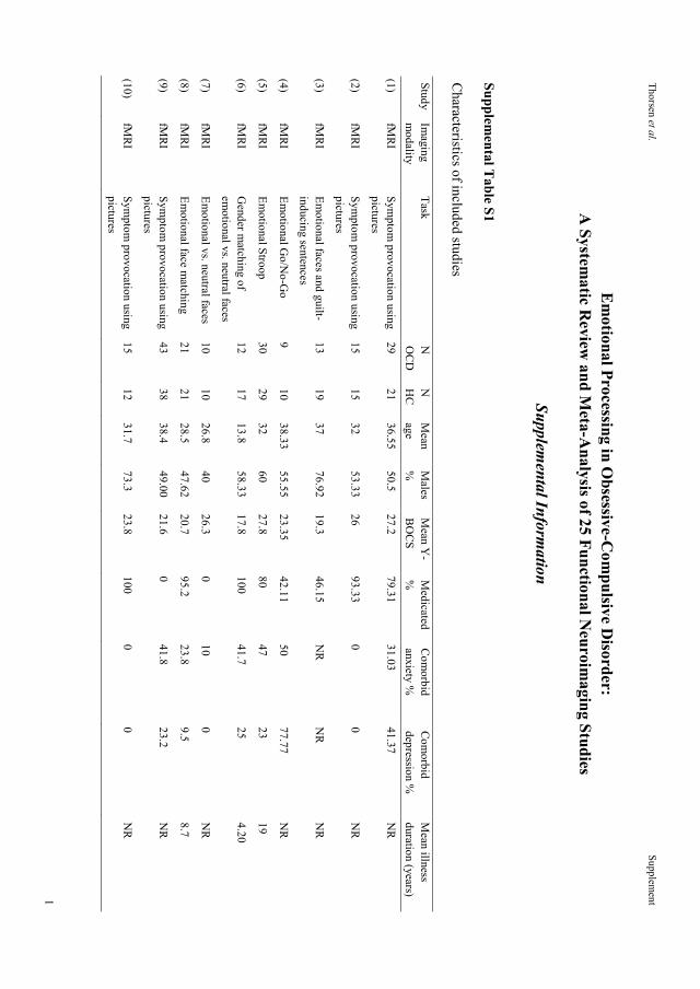
Thorsen et al.
Supplement
1
Em
otional Processing in Obsessive-C
ompulsive D
isorder: A
Systematic R
eview and M
eta-Analysis of 25 Functional N
euroimaging Studies
Supplem
ental Information
Supplemental T
able S1
Characteristics of included studies
Study Im
aging m
odality Task
N
OC
D
N
HC
M
ean age
Males
%
Mean Y
-B
OC
S M
edicated %
C
omorbid
anxiety %
Com
orbid depression %
M
ean illness duration (years)
(1) fM
RI
Symptom
provocation using pictures
29 21
36.55 50.5
27.2 79.31
31.03 41.37
NR
(2) fM
RI
Symptom
provocation using pictures
15 15
32 53.33
26 93.33
0 0
NR
(3) fM
RI
Emotional faces and guilt-
inducing sentences 13
19 37
76.92 19.3
46.15 N
R
NR
N
R
(4) fM
RI
Emotional G
o/No-G
o 9
10 38.33
55.55 23.35
42.11 50
77.77 N
R
(5) fM
RI
Emotional Stroop
30 29
32 60
27.8 80
47 23
19
(6) fM
RI
Gender m
atching of em
otional vs. neutral faces 12
17 13.8
58.33 17.8
100 41.7
25 4.20
(7) fM
RI
Emotional vs. neutral faces
10 10
26.8 40
26.3 0
10 0
NR
(8) fM
RI
Emotional face m
atching 21
21 28.5
47.62 20.7
95.2 23.8
9.5 8.7
(9) fM
RI
Symptom
provocation using pictures
43 38
38.4 49.00
21.6 0
41.8 23.2
NR
(10) fM
RI
Symptom
provocation using pictures
15 12
31.7 73.3
23.8 100
0 0
NR

Thorsen et al.
Supplement
2
Study Im
aging m
odality Task
N
OC
D
N
HC
M
ean age
Males
%
Mean Y
-B
OC
S M
edicated %
C
omorbid
anxiety %
Com
orbid depression %
M
ean illness duration (years)
(11) fM
RI
Working m
emory task w
ith em
otional distractors 20
23 25.5
60 23.9
55 0
15 7.05
(12) fM
RI
Moral dilem
mas
73 73
33.1 57.53
22.1 97.26
14 10
11.5
(13) fM
RI
Shame/guilt-related
sentences 20
20 31.1
50 15.9
NR
0
0 N
R
(14) fM
RI
Emotional vs. neutral faces
17 19
34.9 59
25.53 76.47
23.52 N
R
15.73
(15) fM
RI
Symptom
provocation using pictures
16 17
35.8 50
24.7 75
56.25 29.41
14.2
(16) fM
RI
Symptom
provocation using w
ords 22
19 36.1
36.36 29.9
0 0
0 12.63
(17) fM
RI
Emotional w
orking mem
ory 16
16 31.4
75 25.3
NR
0
0 5.9
(18) fM
RI
Symptom
provocation using pictures
14 14
34 50
28 78.57
NR
N
R
15.5
(19) fM
RI
Symptom
provocation using pictures
8 8
41.8 37.5
25.1 0
NR
N
R
NR
(20) fM
RI
Symptom
provocation using pictures
15 15
43.3 50
24.9 0
NR
N
R
NR
(21) H
2 15O
-PET Sym
ptom provocation using
pictures 11
10 40.5
72.72 23.8
0 0
0 N
R
(22) fM
RI
Emotional Stroop
18 19
33.4 33.33
23.4 0
0 0
NR
(23) fM
RI
Emotional face m
atching 67
67 33.1
56.72 21.8
97.01 16
9 11.42
(24) fM
RI
Symptom
provocation using pictures
42 37
32.5 35.71
17.7 62.3
19.05 32.2
16.23
(25) fM
RI
Olfactory sym
ptom
provocation 15
15 34.07
53.33 17.73
93.3 50
64.89 16.8
Abbreviations: H
C, healthy controls; fM
RI, functional m
agnetic resonance imaging; O
CD
, obsessive-compulsive disorder; PET, positron em
ission tomography; N
R, not
reported; Y-B
OC
S, Yale-B
rown O
bsessive Com
pulsive Scale.
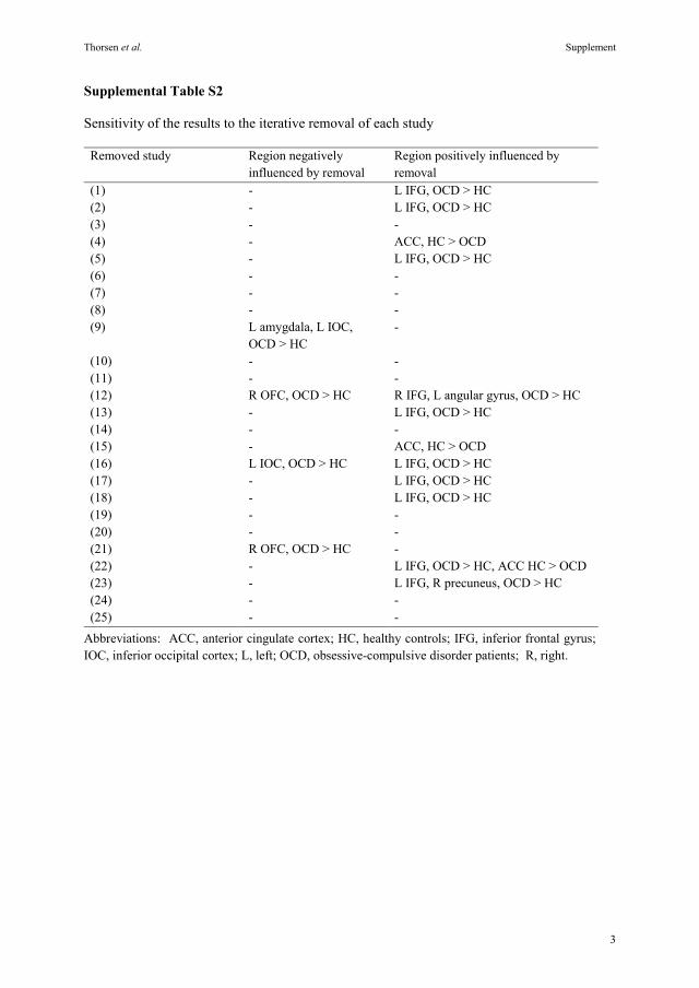
Thorsen et al. Supplement
3
Supplemental Table S2
Sensitivity of the results to the iterative removal of each study
Removed study Region negatively influenced by removal
Region positively influenced by removal
(1) - L IFG, OCD > HC (2) - L IFG, OCD > HC (3) - - (4) - ACC, HC > OCD (5) - L IFG, OCD > HC (6) - - (7) - - (8) - - (9) L amygdala, L IOC,
OCD > HC -
(10) - - (11) - - (12) R OFC, OCD > HC R IFG, L angular gyrus, OCD > HC (13) - L IFG, OCD > HC (14) - - (15) - ACC, HC > OCD (16) L IOC, OCD > HC L IFG, OCD > HC (17) - L IFG, OCD > HC (18) - L IFG, OCD > HC (19) - - (20) - - (21) R OFC, OCD > HC - (22) - L IFG, OCD > HC, ACC HC > OCD (23) - L IFG, R precuneus, OCD > HC (24) - - (25) - -
Abbreviations: ACC, anterior cingulate cortex; HC, healthy controls; IFG, inferior frontal gyrus; IOC, inferior occipital cortex; L, left; OCD, obsessive-compulsive disorder patients; R, right.

Thorsen et al. Supplement
4
Supplemental Figure S1
Flowchart of the results of the systematic search, inclusion, and exclusion of studies.

Thorsen et al. Supplement
5
Supplemental References
1. An S, Mataix-Cols D, Lawrence N, Wooderson S, Giampietro V, Speckens A et al. (2009): To discard or not to discard: the neural basis of hoarding symptoms in obsessive-compulsive disorder. Mol Psychiatry 14:318-331.
2. Banca P, Voon V, Vestergaard MD, Philipiak G, Almeida I, Pocinho F et al. (2015): Imbalance in habitual versus goal directed neural systems during symptom provocation in obsessive-compulsive disorder. Brain 138:798-811.
3. Basile B, Mancini F, Macaluso E, Caltagirone C, Bozzali M. (2014): Abnormal processing of deontological guilt in obsessive–compulsive disorder. Brain Struct Funct 219:1321-1331.
4. Berlin HA, Schulz KP, Zhang S, Turetzky R, Rosenthal D, Goodman W. (2015): Neural correlates of emotional response inhibition in obsessive-compulsive disorder: A preliminary study. Psychiatry Res: Neuroimaging 234:259-264.
5. Brennan BP, Tkachenko O, Schwab ZJ, Juelich RJ, Ryan EM, Athey AJ et al. (2015): An examination of rostral anterior cingulate cortex function and neurochemistry in obsessive–compulsive disorder. Neuropsychopharmacology 40:1866-1876.
6. Britton JC, Stewart SE, Killgore WD, Rosso IM, Price LM, Gold AL et al. (2010): Amygdala activation in response to facial expressions in pediatric obsessive-compulsive disorder. Depress Anxiety 27:643-651.
7. Cannistraro PA, Rauch SL. (2003): Neural circuitry of anxiety: evidence from structural and functional neuroimaging studies. Psychopharmacol Bull 37:8-25.
8. Cardoner N, Harrison BJ, Pujol J, Soriano-Mas C, Hernández-Ribas R, López-Solá M et al. (2011): Enhanced brain responsiveness during active emotional face processing in obsessive compulsive disorder. World J Biol Psychiatry 12:349-363.
9. de Wit SJ, van der Werf YD, Mataix-Cols D, Trujillo JP, van Oppen P, Veltman DJ et al. (2015): Emotion regulation before and after transcranial magnetic stimulation in obsessive compulsive disorder. Psychol Med:1-15.
10. Gonçalves ÓF, Soares JM, Carvalho S, Leite J, Ganho A, Fernandes-Gonçalves A et al. (2015): Brain activation of the defensive and appetitive survival systems in obsessive compulsive disorder. Brain Imaging Behav 9:255-263.
11. Han HJ, Jung WH, Yun JY, Park JW, Cho KK, Hur JW et al. (2016): Disruption of effective connectivity from the dorsolateral prefrontal cortex to the orbitofrontal cortex by negative emotional distraction in obsessive-compulsive disorder. Psychol Med 46:921-932.
12. Harrison BJ, Pujol J, Soriano-Mas C, Hernández-Ribas R, López-Solà M, Ortiz H et al. (2012): Neural correlates of moral sensitivity in obsessive-compulsive disorder. Arch Gen Psychiatry 69:741-749.
13. Hennig-Fast K, Michl P, Müller J, Niedermeier N, Coates U, Müller N et al. (2015): Obsessive-compulsive disorder–A question of conscience? An fMRI study of behavioural and neurofunctional correlates of shame and guilt. J Psychiatr Res 68:354-362.
14. Lawrence NS, An SK, Mataix-Cols D, Ruths F, Speckens A, Phillips ML. (2007): Neural responses to facial expressions of disgust but not fear are modulated by washing

Thorsen et al. Supplement
6
symptoms in OCD. Biol Psychiatry 61:1072-1080.
15. Mataix-Cols D, Wooderson S, Lawrence N, Brammer MJ, Speckens A, Phillips ML. (2004): Distinct neural correlates of washing, checking, and hoarding symptom dimensions in obsessive-compulsive disorder. Arch Gen Psychiatry 61:564-576.
16. Murayama K, Nakao T, Sanematsu H, Okada K, Yoshiura T, Tomita M et al. (2013): Differential neural network of checking versus washing symptoms in obsessive-compulsive disorder. Prog Neuropsychopharmacol Biol Psychiatry 40:160-166.
17. Park SE, Yang JC, Jeong GW. (2016): Neuroanatomical assessment of the impact of negative emotion on implicit memory in patients with obsessive compulsive disorder. Acta Neuropsychiatr 28:206-213.
18. Phillips ML, Marks IM, Senior C, Lythgoe D, O'Dwyer AM, Meehan O et al. (2000): A differential neural response in obsessive-compulsive disorder patients with washing compared with checking symptoms to disgust. Psychol Med 30:1037-1050.
19. Shapira NA, Liu Y, He AG, Bradley MM, Lessig MC, James GA et al. (2003): Brain activation by disgust-inducing pictures in obsessive-compulsive disorder. Biol Psychiatry 54:751-756.
20. Thiel A, Thiel J, Oddo S, Langnickel R, Brand M, Markowitsch HJ et al. (2014): Obsessive-compulsive disorder patients with washing symptoms show a specific brain network when confronted with aggressive, sexual, and disgusting stimuli. Neuropsychoanalysis 16:83-96.
21. van den Heuvel OA, Veltman DJ, Groenewegen HJ, Dolan RJ, Cath DC, Boellaard R et al. (2004): Amygdala activity in obsessive-compulsive disorder with contamination fear: a study with oxygen-15 water positron emission tomography. Psychiatry Res: Neuroimaging 132:225-237.
22. van den Heuvel OA, Veltman DJ, Groenewegen HJ, Witter MP, Merkelbach J, Cath DC et al. (2005): Disorder-specific neuroanatomical correlates of attentional bias in obsessive-compulsive disorder, panic disorder, and hypochondriasis. Arch Gen Psychiatry 62:922-933.
23. Via E, Cardoner N, Pujol J, Alonso P, Lopez-Sola M, Real E et al. (2014): Amygdala activation and symptom dimensions in obsessive-compulsive disorder. Br J Psychiatry 204:61-68.
24. Rus OG, Reess TJ, Wagner G, Zimmer C, Zaudig M, Koch K. (2017): Functional and structural connectivity of the amygdala in obsessive-compulsive disorder. Neuroimage Clin 13:246-255.
25. Berlin HA, Stern ER, Ng J, Zhang S, Rosenthal D, Turetzky R et al. (2017): Altered olfactory processing and increased insula activity in patients with obsessive-compulsive disorder: An fMRI study. Psychiatry Res: Neuroimaging 262:15-24.


100
Paper II
II


Archival Report
Emotion Regulation in Obsessive-CompulsiveDisorder, Unaffected Siblings, and UnrelatedHealthy Control ParticipantsAnders L. Thorsen, Stella J. de Wit, Froukje E. de Vries, Danielle C. Cath, Dick J. Veltman,Ysbrand D. van der Werf, David Mataix-Cols, Bjarne Hansen, Gerd Kvale, andOdile A. van den Heuvel
ABSTRACTBACKGROUND: Functional neuroimaging endophenotypes of obsessive-compulsive disorder (OCD) have beensuggested during executive tasks. The purpose of this study was to investigate whether behavioral and neuralresponses during emotion processing and regulation also represent an endophenotype of OCD.METHODS: Forty-three unmedicated adult OCD patients, 19 of their unaffected siblings, and 38 healthy controlparticipants underwent 3T functional magnetic resonance imaging during an emotion regulation task includingneutral, fear-inducing, and OCD-related visual stimuli. Stimuli were processed during natural appraisal and duringcognitive reappraisal, and distress ratings were collected after each picture. We performed between-groupcomparisons on task behavior and brain activation in regions of interest during emotion provocation and regulation.RESULTS: Siblings reported similar distress as healthy control participants during provocation, and significantly lessthan patients. There was no significant three-group difference in activation during fear provocation or regulation.Three-group comparisons showed that patients had higher amygdala and dorsomedial prefrontal cortex activationduring OCD-related emotion provocation and regulation, respectively, while siblings were intermediate betweenpatients and control participants but not significantly different from either. Siblings showed higher left temporo-occipital activation (compared with both healthy control participants and patients) and higher frontolimbicconnectivity (compared with patients) during OCD-related regulation.CONCLUSIONS: Unaffected siblings do not show the same distress and amygdala activation during emotionalprovocation as OCD patients. Siblings show distinct activation in a temporo-occipital region, possibly related tocompensatory cognitive control. This suggests that emotion regulation is not a strong endophenotype for OCD.When replicated, this contributes to our understanding of familial risk and resilience for OCD.
Keywords: Emotion regulation, Emotional provocation, Endophenotype, Familial risk, fMRI, Obsessive-compulsivedisorder
https://doi.org/10.1016/j.bpsc.2018.03.007
Obsessive-compulsive disorder (OCD) is characterized by dis-tressing obsessive thoughts, urges, or images that patients tryto manage or neutralize through compulsive behaviors (1). Thedisorder affects 1% to 3% of the population (1) and has a largeimpact on social and personal impairment (2,3). OCD is a highlyfamilial disorder (4), with first-degree family members of OCDpatients having a nearly fivefold increase in the odds of devel-oping the disorder compared with family members of non-OCDparticipants (5). Although genetic factors may explain as muchas 47% of the variance of the risk for developing OCD (5), theunderlying genetic factors are not well known (4).
An endophenotype is defined as a measurable trait alongthe path between the phenotype of the disorder and the distalgenotype (6). Because of the considerable heterogeneity in thesymptoms, etiology, and genetic risk markers of OCD, the
diagnosis itself is hard to robustly relate to genetic variation(4,7,8). Endophenotypes may provide simpler clues to thegenetic basis of a disorder than the disorder itself andcontribute to bridging clinical heterogeneity and geneticvulnerability (6). In this context, functional neuroimagingstudies on endophenotypes allow for investigating sharedbehavioral and neural characteristics between OCD patientsand their first-degree unaffected family members, in compari-son with unrelated healthy control participants (9). Previousfunctional neuroimaging studies showed that OCD patientsand their unaffected relatives show similarities in neural re-sponses during response inhibition (10,11), working memory(12), reversal learning (13), and error monitoring (14). Forexample, in previous reports on response inhibition andworking memory involving the same participants as in the
352 ª 2018 Society of Biological Psychiatry. Published by Elsevier Inc. All rights reserved.Biological Psychiatry: Cognitive Neuroscience and Neuroimaging April 2019; 4:352–360 www.sobp.org/BPCNNI ISSN: 2451-9022
BiologicalPsychiatry:CNNI

current study, OCD patients and their unaffected siblingsshared compensatory higher task-related activation in the leftpresupplementary motor area, as well as altered frontolimbicconnectivity, compared with unrelated healthy control partici-pants (10,12,15).
Neural responses during paradigms using emotion provo-cation with disorder-specific stimuli may provide a moredisorder-specific endophenotype for OCD, as deficits in ex-ecutive functioning are not specific to this disorder (16). Whenconfronted with disorder-specific objects, situations, orthoughts, OCD patients often react with negative emotionssuch as anxiety, fear, guilt, or disgust, and they often experi-ence an increased urge to ritualize (17,18). In a recent meta-analysis of 25 whole-brain studies contrasting aversive andneutral stimuli, OCD patients showed higher activation thancontrol participants during emotion provocation in the bilateralamygdala, right putamen, orbitofrontal cortex (extending intothe subgenual anterior cingulate cortex and ventromedialprefrontal cortex [PFC]), middle temporal cortex, and left infe-rior occipital cortex (19).
Emotion regulation involves changing emotional responsesthrough active processes such as altering attentional deploy-ment, cognitive reappraisal (reinterpreting the meaning andone’s connection to a stimulus), or suppression of theexpression or experience of an emotion, and over time theseactive processes are learned and become more automatic (20).Functional magnetic resonance imaging (fMRI) studies haveshown that cognitive reappraisal is associated with higheractivation in the dorsomedial and lateral frontal cortices, dorsalanterior cingulate cortex, and parietal and temporal regions(21,22).
Cognitive behavioral models suggest that the catastrophicinterpretations of obsessions and the use of compulsions asdysfunctional methods of emotion regulation maintain thedisorder (23). Maladaptive emotion regulation strategies, suchas suppression, have also been related to increased distress inresponse to obsessions over time, while acceptance de-creases distress (24). In addition, suppression has been relatedto increased severity of OC symptoms (25). In a previous studywe investigated whether unmedicated OCD patients andhealthy control participants differed in how successfully theyused cognitive reappraisal to regulate distress provoked byfear- and OCD-related pictures, and whether OCD patientsshow altered activation of emotion regulation–related circuitryusing fMRI (26). We showed that distress ratings were higher inOCD patients than in control participants, but that patientswere well able to downregulate distress while viewing fear- andOCD-related pictures within the experimental setting. Howev-er, this was accompanied by aberrant brain activation in pa-tients versus control participants. OCD patients showedgreater emotion provocation-induced amygdala activation andaltered timing, while emotion regulation–related activation waslower in the left dorsolateral PFC (dlPFC) and parietal cortexwhile viewing fear-related pictures, and higher in the dorso-medial PFC (dmPFC) while viewing OCD-related pictures. Inaddition, patients also showed less functional connectivitybetween the dmPFC and bilateral amygdala during regulationof fear-related pictures (26). This suggests that OCD patientsshow frontolimbic and frontoparietal dysfunction duringemotion processing, but whether this dysfunction represents a
consequence of the disorder or an underlying vulnerabilityfactor remains unanswered.
The aim of the present study was to explore the neuralcorrelates of disorder-specific emotion provocation and regu-lation as a potential endophenotype of OCD. Extending ourprevious findings on emotion regulation in OCD, we compareddistress ratings and brain activation during emotion provoca-tion and regulation in a group of unaffected siblings, with thoseof the previously studied OCD patients and healthy controlparticipants (26). Using group comparisons of task-relatedactivation in a priori regions of interest (ROIs), as well asfrontolimbic functional connectivity analyses, shared andnonshared activation patterns during emotion provocation andregulation of general fear– and OCD-related stimuli were pro-bed. Based on the previous endophenotype findings in thissample using executive paradigms (10,12,15), we hypothe-sized that siblings would resemble healthy control participantson the behavioral level (i.e., normal levels of provoked distress)and also show similar amygdala activation during emotionprovocation (i.e., less activation compared with OCD patients).During regulation we expected that siblings would resembleOCD patients on the neural level (i.e., decreased activation indlPFC during fear regulation, increased dmPFC activationduring OCD-related regulation, and reduced frontolimbicconnectivity compared with healthy control participants).
METHODS AND MATERIALS
Participants
Forty-three patients with a primary diagnosis of OCD, 19 oftheir unaffected siblings, and 38 healthy control participantswere included in the study (see the Supplement for recruitmentinformation). All participants were assessed using the Struc-tured Clinical Interview for DSM-IV (27). The Yale-BrownObsessive Compulsive Scale (28) and the Obsessive-Compulsive Inventory–Revised (29) were used to assess theseverity of OC symptoms. Depressive symptoms weremeasured using the Montgomery–Åsberg Depression RatingScale (30). The Emotion Regulation Questionnaire (31) wasused to measure reappraisal and suppression as emotionregulation strategies in daily life. Handedness was assessedusing the Edinburgh Handedness Inventory (32).
All participants had not used psychotropic medication for atleast 4 weeks before inclusion, and had no psychotic symp-toms, major physical or neurological illness, or any MRI con-traindications. All patients had a primary diagnosis of OCDaccording to DSM-IV criteria and did not meet criteria forhoarding disorder. Patients experiencing other comorbiditieswere included as long as OCD was the primary diagnosis.Control participants were excluded if they met criteria for anycurrent DSM-IV diagnosis, while siblings were excluded if theyhad a lifetime history of OCD. The study was in full compliancewith the ethical standards of the local medical ethical reviewboard of VU University Medical Center and with the HelsinkiDeclaration of 1975, revised in 2008, and all participants pro-vided written informed consent.
The results of the comparison between OCD patients andhealthy control participants have been presented previously(26). All subjects also participated in previous endophenotypeanalyses on response inhibition (10) and working memory (12).
Endophenotype of Emotion Regulation in OCD
Biological Psychiatry: Cognitive Neuroscience and Neuroimaging April 2019; 4:352–360 www.sobp.org/BPCNNI 353
BiologicalPsychiatry:CNNI

The data for these reports were all collected during the sameexperiment.
Experimental Task
All participants performed an emotion regulation task [see deWit et al. (26) and the Supplement for detailed procedures] andwere presented with neutral, fearful, and OCD-related pictures.The OCD stimuli included those pertinent to washing, check-ing, and symmetry symptom dimensions, and all the partici-pants watched the same stimuli to capture the symptomheterogeneity of OCD. The participants were instructed toeither simply attend to the presented stimulus (“attend” in-struction) or use cognitive reappraisal techniques to down-regulate negative emotions and cognitions provoked by thestimulus (“regulate” instruction). Stimuli were presented inattend or regulate blocks for each picture type, and eachstimulus was followed by having the participants indicate theircurrent level of distress by moving a cursor to either the left(marked “not distressed”) or the right (“maximally distressed”)along a visual analog scale. Neutral stimuli were presented inthe attend condition only.
Statistical Analysis of Behavioral and ClinicalMeasures
Group comparisons of clinical and demographical variableswere analyzed using one-way analyses of variance (ANOVAs),and followed up by post hoc two-sample t tests. Categoricalvariables were analyzed using c2 tests. Distress scores duringthe attend and regulate conditions were analyzed separatelyfor fear- and OCD-related pictures using repeated-measuresmixed ANOVAs, with group (OCD patients, siblings, controlparticipants) as the between-subjects factor followed by posthoc t tests if the main effects were significant. Within-grouptests of changes in distress during provocation and regula-tion were analyzed with paired-sample t tests. Analyses wereperformed using SPSS statistics version 23 (IBM Corp.,Armonk, NY). The statistical threshold was set at p , .05, withTukey or Games-Howell corrections being used for post hoctests of one-way and repeated-measures ANOVAs.
MRI Acquisition, Processing, and Analysis
Functional gradient echo-planar and structural T1-weightedimaging was performed on a GE Signa HDxt 3.0T MRI scan-ner (GE Healthcare, Chicago, IL). Functional data were pre-processed and analyzed in SPM8 (Wellcome Trust Centre forImaging, London, United Kingdom) (see the Supplement foracquisition parameters and preprocessing steps). Intrasubjectfirst-level analyses included nine regressors of interest con-sisting of the neutral (attend only), general fear, contamination,checking, and symmetry OCD-related pictures during attendand regulate instruction. Regressors of no interest included thetime windows where the participants rated their distress(boxcars of 5 seconds), instruction periods (boxcars of 3seconds), and the participant’s six movement parameters. Thefirst-level analyses were different for emotion provocation andemotion regulation to capture the different timings of theserespective processes [as we described previously in de Witet al. (26)]. In the emotion provocation analysis, regressors ofinterest were modeled as 0-second delta functions and
convolved with the canonical hemodynamic response function(HRF), and its temporal and dispersion derivatives to model theamplitude of the blood oxygen level–dependent response andvariation in its timing and shape. Fear . neutral and OCD .neutral contrast images (collapsed over the attend and regu-late instructions) were then computed per participant. In theemotion regulation analysis, activation during emotion regu-lation was modeled as boxcars of the first 5 seconds of fearand the OCD-related stimuli, and were convolved with thecanonical HRF. Contrast images were then computed for fearregulate . attend and OCD regulate . attend. A high-passfilter with a 128-second cutoff was used to remove low-frequency noise. See the Supplement for all additional infor-mation on MRI acquisition and modeling.
To test whether unaffected siblings also showed alteredfunctional connectivity between the bilateral amygdala anddmPFC during emotion regulation, which was previously re-ported for patients versus control participants (26), we usedthe Generalized Psychophysiological Interaction toolbox (33)(see the Supplement). The dmPFC seed region was set atMontreal Neurological Institute x/y/z of 29/8/64, with a 10-mmsphere, based on the patient-control comparison (26). TheWFU PickAtlas (http://fmri.wfubmc.edu/software/pickatlas)was used to determine the bilateral amygdala ROIs for func-tional connectivity.
Planned group comparisons were performed by enteringthe first-level contrast images into general linear models usingSPM12. Between-group comparisons for emotion provocationwere performed using separate 3 3 3 ANOVAs with group(OCD patients, siblings, control participants) as a between-subject factor and HRF (canonical, temporal, dispersion) as awithin-subjects factor. A 3 3 2 3 3 ANOVA (group by HRF bypicture type [fear- and OCD-related]) was used to test thegroup by picture type interactions. Group comparisons ofemotion regulation and functional connectivity were performedusing separate one-way ANOVAs for the two picture types(fear- and OCD-related stimuli), with group as the between-subjects factor. For all group comparisons we first used an Fcontrast to estimate the main effect of group, and the group byinstruction interaction where appropriate. Post hoc two-groupcomparisons were then performed to follow-up significant ef-fects (two-group ANOVAs for the emotion provocation analysisand t tests for the emotion regulation analysis).
We used an ROI approach, which was derived from thelargest meta-analyses of emotion provocation in OCDcompared with healthy control participants (five ROIs) (19), andemotion regulation in healthy control participants (eight ROIs)(21,22), as no published meta-analysis of emotion regulationcomparing OCD patients and healthy control participants wasavailable (see Supplemental Table S2 for all ROIs, and theSupplement for more information on ROI definitions, place-ment, and extraction). The ROIs were placed at the relevanteffects of task after initial image thresholding of uncorrectedp , .001. Statistical significance was set at p , .05 familywiseerror corrected with small volume correction (pFWE-SVC)with a 10-mm sphere (pFWE-SVC , .05; trends p , .10) (34).Results were corrected for the number of ROIs foreach contrast using a SISA-Bonferroni correction (see http://www.quantitativeskills.com/sisa/calculations/bonhlp.htm) (35).Separate SISA-Bonferroni corrected p values were calculated
Endophenotype of Emotion Regulation in OCD
354 Biological Psychiatry: Cognitive Neuroscience and Neuroimaging April 2019; 4:352–360 www.sobp.org/BPCNNI
BiologicalPsychiatry:CNNI

per analysis because of differences in provocation relateddistress and regulation-related reductions in distress betweenthe picture types (see the Supplement for more information).
RESULTS
Demographic and Clinical Characteristics
The groups were demographically matched. Patients scoredsignificantly higher on every clinical measure compared withboth siblings and control participants, while siblings andcontrol participants did not significantly differ (seeSupplemental Table S1 for all results). For the EmotionRegulation Questionnaire, there was a main effect of group forthe use of reappraisal. Post hoc tests showed that patientsscored lower than control participants, and siblings scoredintermediate (not significantly different from either). Twenty-one OCD patients (49%) had a comorbid DSM-IV diagnosis(details in Supplemental Table S1). One sibling met criteria forspecific phobia, which did not interfere with scanning, while nocontrol participants met criteria for any current mentaldisorder.
Distress Ratings During Emotion Provocation andRegulation
Fear- and OCD-related pictures elicited more distress thanneutral pictures for all three groups, regardless of the task in-struction (all p , .05) (see Table 1 for all behavioral results).There was a significant main effect of group (F contrast: fear,p , .001; OCD, p , .001), with post hoc tests showing thatOCD patients reported more distress in general during bothfear- and OCD-related pictures than healthy control partici-pants and siblings, whereas siblings were not significantlydifferent from control participants (t test: fear related, p = .97;OCD related, p = .86). There was a main effect of regulationacross the three groups (F contrast: fear related, p = .02; OCDrelated, p , .01). For fear stimuli there was no significant groupby instruction interaction (p = .06), whereas the interactionanalysis was significant for OCD stimuli (p , .01). Post hoctwo-sample t tests showed that OCD patients reportedsignificantly larger distress reductions during OCD-relatedregulation compared with both siblings (t43.29 = 23.79, p ,.01) and control participants (t75.16 = 22.59, p = .01), whereassiblings were not different from control participants (t55 = 0.57,p = .57).
Within-group paired-sample t tests revealed a regulationeffect (higher distress ratings during attend instructioncompared with regulation instruction) for OCD patients whileviewing both picture types and in healthy control participantsfor fear-related pictures (all p # .05). In siblings, there was noregulation effect for fear-related (p = .43) or OCD-related(p = .06; trend) pictures.
Neural Response During Emotion Provocation inROIs
The three-group comparison for fear provocation showed nosignificant differences between the groups (SupplementalTable S3). During OCD-related provocation there was a sig-nificant main effect of group in the right amygdala extendinginto the hippocampus, driven by alterations in the timing and
shape of the blood oxygen level–dependent response in OCDpatients compared with healthy control participants, whereassiblings were intermediate and not significantly different fromeither group (Table 2). The group by picture type interactionanalysis was trend significant (p = .02) (Table 2). This wasdriven by higher responses in the right amygdala during OCD-related rather than fear-related provocation in patientscompared with healthy control participants, as we previouslyshowed (26), whereas the siblings were an intermediate groupnot different from either group.
Neural Response During Emotion Regulation inROIs
Three-group comparison showed no significant group differ-ences in activation in any of the ROIs during fear-relatedemotion regulation. For OCD-related regulation a significantmain effect of group was found in the dmPFC and the lefttemporo-occipital cortex (see Table 3 and Figure 1). On posthoc tests, siblings showed significantly higher activation in theleft temporo-occipital cortex compared with healthy controlparticipants and patients. Higher dmPFC activation, as previ-ously found in patients compared with healthy control partici-pants, was also present in siblings compared with controlparticipants, albeit at a trend level.
Functional Connectivity During Emotion Regulationin ROIs
The comparisons of frontolimbic connectivity during fear- andOCD-related regulation did not reveal any significant differ-ences between the three groups (Supplemental Table S6).Exploratory post hoc analyses showed that the previously re-ported decreased frontolimbic connectivity in patientscompared with healthy control participants during fear-relatedregulation was present for the left (Z score = 3.36, pFWE-SVC = .016) and right amygdala (Z score = 3.30, pFWE-SVC =.019) but that siblings were an intermediate group not signifi-cantly different from either group (see Supplemental Figure S1for group-specific parameter estimates). Specifically, pairwisecomparisons of connectivity during OCD-related regulationshowed higher dmPFC-amygdala connectivity in siblingscompared with patients (Z score = 3.22, pFWE-SVC = .023)and right amygdala (Z score = 3.30, pFWE-SVC = .019), whilethere were no other significant between-group differences.
DISCUSSION
The present study assessed whether the neural correlates ofdisorder-specific emotion processing and regulation can serveas a potential endophenotype of OCD. To this aim, we inves-tigated emotion processing during fMRI scanning in unaffectedsiblings of a large sample of unmedicated OCD patients and inunrelated healthy control participants. We found that siblingsresembled control participants in self-reported distress andclinical profile. When assessing brain activation patterns inROIs during emotion provocation and regulation, we mostlyobserved that siblings were an intermediate group betweenpatients and control participants. We also found that siblingsshowed slightly higher dmPFC activation during OCD-relatedregulation compared with OCD patients and healthy controlparticipants, but this difference was not statistically significant.
Endophenotype of Emotion Regulation in OCD
Biological Psychiatry: Cognitive Neuroscience and Neuroimaging April 2019; 4:352–360 www.sobp.org/BPCNNI 355
BiologicalPsychiatry:CNNI

We previously compared the same group of patients andcontrol participants directly (26) and observed in OCD patientsamygdala hyper-responsiveness during OCD-related emotionprovocation, dlPFC hypoactivation and lower dmPFC-amygdala connectivity during fear regulation, and dmPFChyperactivation during OCD-related emotion regulation. Here,we found that prefrontal and amygdala activation were notsignificantly different in siblings compared with either OCDpatients or control participants during either OCD-relatedemotion provocation (amygdala) and regulation (dmPFC) oremotion regulation of fear stimuli (dlPFC activation anddmPFC-amygdala connectivity). A sibling-specific finding wasincreased activation in the temporo-occipital cortex, borderingon the angular gyrus, during OCD-related emotion regulation,
in siblings compared with both control participants and pa-tients. Siblings also showed higher dmPFC-amygdala con-nectivity compared with patients during OCD-related emotionregulation. The difference between the findings in the presentreport and the earlier report is likely due to methodologicaldifferences: 1) the present use of a priori defined ROIs basedon meta-analyses, compared with the earlier use of functionallydefined ROIs (26), and 2) due to performing a three-groupANOVA in which the dlPFC response in the sibling group isintermediate between patients and control participants. This isalso reflected in the Supplemental whole-brain results, showingthe same hypoactivation of the dlPFC during fear-relatedregulation in OCD patients compared with healthy controlparticipants.
Table 1. Group Comparisons of Behavioral Response: Distress During Provocation and Regulation
OCD Patients (n = 43) Siblings (n = 19) HC Participants (n = 38)Neural Pictures Mean SD Mean SD Mean SD
Provocation 2.04 5.24 0 0 0.74 2.78
Fear-Related Pictures Mean SD Mean SD Mean SD
Provocation 41.44 31.15 16.42 14.58 20.74 26.53Regulation 33.74 28.29 14.68 14.20 13.21 15.57
t df p t df p t df p
Provocation vs. regulation 2.02 42 .05a 0.80 18 .43 2.12 37 .04a
F df p
Main effect of group 11.28 1, 97 ,.01a
Main effect of instruction 5.98 1, 97 .02a
Group 3 instruction interaction 0.57 2, 97 .57
t df p
Post hoc tests (provocation)
Siblings vs. OCD 24.31 59.72 ,.01a
Siblings vs. HC participants 20.66 55 .84
Post hoc tests (regulation)
Siblings vs. OCD 23.53 58.89 ,.01a
Siblings vs. HC participants 0.39 55 .97
OCD-Related Pictures Mean SD Mean SD Mean SD
Provocation 22.30 17.95 2.75 4.56 4.88 11.93Regulation 14.36 9.82 2.26 4.51 3.21 7.05
t df p t df p t df p
Provocation vs. regulation 4.08 42 ,.001a 2.02 18 .06 1.15 37 .26
F df p
Main effect of group 25.52 1, 97 ,.01a
Main effect of instruction 9.86 1, 97 ,.01a
Group 3 instruction interaction 5.47 2, 97 ,.01a
t df p
Post hoc tests (provocation)
Siblings vs. OCD 26.67 52.56 ,.01a
Siblings vs. HC participants 20.75 55 .84
Post hoc tests (regulation)
Siblings vs. OCD 26.64 59.85 ,.01a
Siblings vs. HC participants 20.62 55 .91
Responses were rated on a scale of 1 to 100.HC, healthy control; OCD, obsessive-compulsive disorder.aSignificant within- or between-group effect.
Endophenotype of Emotion Regulation in OCD
356 Biological Psychiatry: Cognitive Neuroscience and Neuroimaging April 2019; 4:352–360 www.sobp.org/BPCNNI
BiologicalPsychiatry:CNNI

One explanation for the current findings is that in OCD,altered emotion processing is more state dependent than thepreviously reported traitlike cognitive endophenotypes, whichwere found to be related to altered frontoparietal recruitment,and likely related to a shared genetic vulnerability to the dis-order (10,12,15). Previous task-related fMRI studies in first-degree relatives of OCD patients showed altered recruitmentof frontal and parietal regions including the presupplementarymotor area (10,12), dlPFC (12,13), inferior frontal gyrus (13),parietal cortex (12,13), and precuneus (12) during reversallearning, response inhibition, working memory, and error pro-cessing. During resting state, this sample of unaffected
siblings (compared with healthy control participants) alsoshowed higher connectivity between the frontoparietal controlnetworks and the rostral anterior cingulate cortex and dmPFC,whereas only patients showed alterations in the frontolimbiccircuit (36). Similarly, in this same sample of siblings, frontal-amygdala coupling during executive task we observed tohave small or trend-significant aberrations, whereas findings inpatients compared with control participants were more robust(12,15). In the present study, exploratory post hoc testsshowed that siblings exhibited greater dmPFC-amygdalaconnectivity during disorder-specific emotion regulation thanOCD patients. The increased frontolimbic connectivity in
Table 3. Group Comparisons of Brain Activation During OCD-Related Emotion Regulation
BA Side Ke
MNI Coordinates
Z pFWE-SVCx y z
Fear Regulation: Main Effect of Group (ANOVA) (no significant voxels)
OCD-Related Regulation: Main Effect of Group (ANOVA)
dmPFC 32 Midline 5 0 29 40 3.75 .005a
Temporo-occipital cortex 37 L 4 239 261 13 3.43 .013a
Ke Z Score pFWE-SVC
OCD-Related Regulation: Post Hoc Pairwise Comparisons in dmPFC
Siblings . HC participants 1 3.22 .022b
OCD patients . siblings – – NS
OCD patients . HC participants 10 3.82 .003a
Post Hoc Pairwise Comparisons in Left Temporo-occipital Cortex
Siblings . HC participants 18 3.67 .006a
Siblings . OCD patients 2 3.22 .020a
OCD patients . HC participants – – NS
ANOVA, analysis of variance; BA, Brodmann area; dmPFC, dorsomedial prefrontal cortex; FWE, familywise error; HC, healthy control; Ke, voxelextent of cluster; L, left; MNI, Montreal Neurological Institute; NS, not significant; OCD, obsessive-compulsive disorder; SVC, small volumecorrection.
aSignificant between-group findings.bTrend significant.
Table 2. Group Comparisons of Brain Activation During Emotion Provocation
BA Side Ke
MNI Coordinates
Z pFWE-SVCx y z
Fear Provocation: Main Effect of Group (ANOVA) (no significant voxels)
OCD-Related Provocation: Main Effect of Group (ANOVA)
Amygdala N/A R 12 27 27 217 3.58 .013a
Fear-Related vs. OCD-Related Provocation: Group by Picture Type Interaction (ANOVA)Amygdala N/A R 5 27 27 217 3.45 .020b
Ke Z Score pFWE-SVC
OCD-Related Provocation: Post Hoc Pairwise Comparisons in Right Amygdala
Siblings vs. HC participants NS NS NS
Siblings vs. OCD NS NS NS
OCD patients vs. HC participants 21 4.22 .001a
Fear-Related vs. OCD-Related Provocation: Post Hoc Pairwise Comparisons in Right Amygdala
Siblings vs. HC participants NS NS NS
Siblings vs. OCD patients NS NS NS
OCD patients vs. HC participants 19 4.12 .001a
ANOVA, analysis of variance; BA, Brodmann area; FWE, familywise error; HC, healthy control; Ke, voxel extent of cluster; MNI, MontrealNeurological Institute; N/A, not applicable; NS, not significant; OCD, obsessive-compulsive disorder; R, right; SVC, small volume correction.
aSignificant between-group findings.bTrend significant.
Endophenotype of Emotion Regulation in OCD
Biological Psychiatry: Cognitive Neuroscience and Neuroimaging April 2019; 4:352–360 www.sobp.org/BPCNNI 357
BiologicalPsychiatry:CNNI

siblings may represent a compensatory mechanism. Largersamples are needed, however, to better investigate thisspeculation.
It could also be that blood oxygen level–dependent re-sponses in emotional paradigms in siblings are more variablethan during executive functioning. This potential variabilitytogether with the relatively small sibling group size could havereduced the power to detect differences, resulting in the in-termediate activation patterns in the siblings group. This is incontrast with findings in the same sibling group during inhibi-tion and working memory where frontoparietal responses weremore robust and/or extensive in the siblings compared withpatients (10,12). Alternatively, a more robust provocation ofdistress in siblings and control participants might be needed toavoid floor effects during symptom provocation paradigms.Future research could consider how to evoke distress inhealthy participants that resembles the response typicallyfound in OCD patients.
One criterion for a candidate endophenotype is that the traitis more commonly found in unaffected family members ofpatients than in unrelated healthy control participants (6).Because we did not find that patients and their siblings
showed the same degree of amygdala hyper-responsivenessduring OCD-related emotion provocation, dlPFC hypo-activation during fear regulation, dmPFC hyperactivation dur-ing OCD regulation, or decreased frontolimbic connectivityduring emotion regulation, we conclude that the neural corre-lates of emotion regulation are likely not a strong endophe-notype of OCD.
The siblings showed increased temporo-parieto-occipitalactivation during regulation of disorder-specific stimulicompared with patients and control participants. The temporo-parieto-occipital brain regions are part of the largercross-modal association cortex involved in attentional controland visuospatial representation during emotion processing(37). Taken together with the siblings’ higher frontal-parietalnetwork (36), our results suggest that siblings draw on addi-tional resources compared with OCD patients and healthycontrol participants. This region has previously been reportedas being especially recruited when distancing oneself from astimulus to regulate one’s emotions, while prefrontal dorso-medial and anterior cingulate activation is recruited moreduring reappraisal (20,38). We therefore speculate that siblingsredirect their attention and distance themselves from aversive
Figure 1. Group comparisons of activation during obsessive-compulsive disorder (OCD)–related emotion regulation. Top left: higher activation of the lefttemporo-occipital cortex in unaffected siblings compared with healthy control (HC) participants during OCD-related emotion regulation. Top right: higheractivation of the dorsomedial prefrontal cortex (dmPFC) in OCD patients and siblings (trend) over HC participants during OCD-related emotion regulation.Bottom left and bottom right: parameter estimates of the blood oxygen level–dependent signal for each group during OCD-related regulation in the lefttemporo-occipital cortex and dmPFC, respectively. Parameter estimates are in arbitrary units, with standard errors. L, left.
Endophenotype of Emotion Regulation in OCD
358 Biological Psychiatry: Cognitive Neuroscience and Neuroimaging April 2019; 4:352–360 www.sobp.org/BPCNNI
BiologicalPsychiatry:CNNI
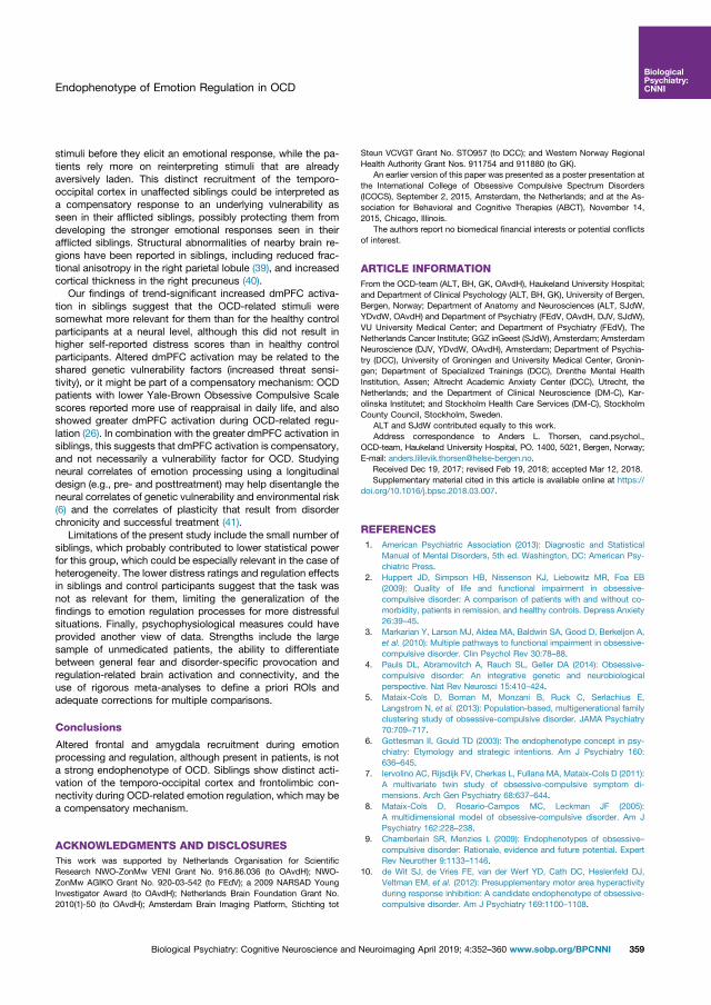
stimuli before they elicit an emotional response, while the pa-tients rely more on reinterpreting stimuli that are alreadyaversively laden. This distinct recruitment of the temporo-occipital cortex in unaffected siblings could be interpreted asa compensatory response to an underlying vulnerability asseen in their afflicted siblings, possibly protecting them fromdeveloping the stronger emotional responses seen in theirafflicted siblings. Structural abnormalities of nearby brain re-gions have been reported in siblings, including reduced frac-tional anisotropy in the right parietal lobule (39), and increasedcortical thickness in the right precuneus (40).
Our findings of trend-significant increased dmPFC activa-tion in siblings suggest that the OCD-related stimuli weresomewhat more relevant for them than for the healthy controlparticipants at a neural level, although this did not result inhigher self-reported distress scores than in healthy controlparticipants. Altered dmPFC activation may be related to theshared genetic vulnerability factors (increased threat sensi-tivity), or it might be part of a compensatory mechanism: OCDpatients with lower Yale-Brown Obsessive Compulsive Scalescores reported more use of reappraisal in daily life, and alsoshowed greater dmPFC activation during OCD-related regu-lation (26). In combination with the greater dmPFC activation insiblings, this suggests that dmPFC activation is compensatory,and not necessarily a vulnerability factor for OCD. Studyingneural correlates of emotion processing using a longitudinaldesign (e.g., pre- and posttreatment) may help disentangle theneural correlates of genetic vulnerability and environmental risk(6) and the correlates of plasticity that result from disorderchronicity and successful treatment (41).
Limitations of the present study include the small number ofsiblings, which probably contributed to lower statistical powerfor this group, which could be especially relevant in the case ofheterogeneity. The lower distress ratings and regulation effectsin siblings and control participants suggest that the task wasnot as relevant for them, limiting the generalization of thefindings to emotion regulation processes for more distressfulsituations. Finally, psychophysiological measures could haveprovided another view of data. Strengths include the largesample of unmedicated patients, the ability to differentiatebetween general fear and disorder-specific provocation andregulation-related brain activation and connectivity, and theuse of rigorous meta-analyses to define a priori ROIs andadequate corrections for multiple comparisons.
Conclusions
Altered frontal and amygdala recruitment during emotionprocessing and regulation, although present in patients, is nota strong endophenotype of OCD. Siblings show distinct acti-vation of the temporo-occipital cortex and frontolimbic con-nectivity during OCD-related emotion regulation, which may bea compensatory mechanism.
ACKNOWLEDGMENTS AND DISCLOSURESThis work was supported by Netherlands Organisation for ScientificResearch NWO-ZonMw VENI Grant No. 916.86.036 (to OAvdH); NWO-ZonMw AGIKO Grant No. 920-03-542 (to FEdV); a 2009 NARSAD YoungInvestigator Award (to OAvdH); Netherlands Brain Foundation Grant No.2010(1)-50 (to OAvdH); Amsterdam Brain Imaging Platform, Stichting tot
Steun VCVGT Grant No. STO957 (to DCC); and Western Norway RegionalHealth Authority Grant Nos. 911754 and 911880 (to GK).
An earlier version of this paper was presented as a poster presentation atthe International College of Obsessive Compulsive Spectrum Disorders(ICOCS), September 2, 2015, Amsterdam, the Netherlands; and at the As-sociation for Behavioral and Cognitive Therapies (ABCT), November 14,2015, Chicago, Illinois.
The authors report no biomedical financial interests or potential conflictsof interest.
ARTICLE INFORMATIONFrom the OCD-team (ALT, BH, GK, OAvdH), Haukeland University Hospital;and Department of Clinical Psychology (ALT, BH, GK), University of Bergen,Bergen, Norway; Department of Anatomy and Neurosciences (ALT, SJdW,YDvdW, OAvdH) and Department of Psychiatry (FEdV, OAvdH, DJV, SJdW),VU University Medical Center; and Department of Psychiatry (FEdV), TheNetherlands Cancer Institute; GGZ inGeest (SJdW), Amsterdam; AmsterdamNeuroscience (DJV, YDvdW, OAvdH), Amsterdam; Department of Psychia-try (DCC), University of Groningen and University Medical Center, Gronin-gen; Department of Specialized Trainings (DCC), Drenthe Mental HealthInstitution, Assen; Altrecht Academic Anxiety Center (DCC), Utrecht, theNetherlands; and the Department of Clinical Neuroscience (DM-C), Kar-olinska Institutet; and Stockholm Health Care Services (DM-C), StockholmCounty Council, Stockholm, Sweden.
ALT and SJdW contributed equally to this work.Address correspondence to Anders L. Thorsen, cand.psychol.,
OCD-team, Haukeland University Hospital, PO. 1400, 5021, Bergen, Norway;E-mail: [email protected].
Received Dec 19, 2017; revised Feb 19, 2018; accepted Mar 12, 2018.Supplementary material cited in this article is available online at https://
doi.org/10.1016/j.bpsc.2018.03.007.
REFERENCES1. American Psychiatric Association (2013): Diagnostic and Statistical
Manual of Mental Disorders, 5th ed. Washington, DC: American Psy-chiatric Press.
2. Huppert JD, Simpson HB, Nissenson KJ, Liebowitz MR, Foa EB(2009): Quality of life and functional impairment in obsessive-compulsive disorder: A comparison of patients with and without co-morbidity, patients in remission, and healthy controls. Depress Anxiety26:39–45.
3. Markarian Y, Larson MJ, Aldea MA, Baldwin SA, Good D, Berkeljon A,et al. (2010): Multiple pathways to functional impairment in obsessive-compulsive disorder. Clin Psychol Rev 30:78–88.
4. Pauls DL, Abramovitch A, Rauch SL, Geller DA (2014): Obsessive-compulsive disorder: An integrative genetic and neurobiologicalperspective. Nat Rev Neurosci 15:410–424.
5. Mataix-Cols D, Boman M, Monzani B, Ruck C, Serlachius E,Langstrom N, et al. (2013): Population-based, multigenerational familyclustering study of obsessive-compulsive disorder. JAMA Psychiatry70:709–717.
6. Gottesman II, Gould TD (2003): The endophenotype concept in psy-chiatry: Etymology and strategic intentions. Am J Psychiatry 160:636–645.
7. Iervolino AC, Rijsdijk FV, Cherkas L, Fullana MA, Mataix-Cols D (2011):A multivariate twin study of obsessive-compulsive symptom di-mensions. Arch Gen Psychiatry 68:637–644.
8. Mataix-Cols D, Rosario-Campos MC, Leckman JF (2005):A multidimensional model of obsessive-compulsive disorder. Am JPsychiatry 162:228–238.
9. Chamberlain SR, Menzies L (2009): Endophenotypes of obsessive–compulsive disorder: Rationale, evidence and future potential. ExpertRev Neurother 9:1133–1146.
10. de Wit SJ, de Vries FE, van der Werf YD, Cath DC, Heslenfeld DJ,Veltman EM, et al. (2012): Presupplementary motor area hyperactivityduring response inhibition: A candidate endophenotype of obsessive-compulsive disorder. Am J Psychiatry 169:1100–1108.
Endophenotype of Emotion Regulation in OCD
Biological Psychiatry: Cognitive Neuroscience and Neuroimaging April 2019; 4:352–360 www.sobp.org/BPCNNI 359
BiologicalPsychiatry:CNNI

11. Menzies L, Achard S, Chamberlain SR, Fineberg N, Chen C-H, delCampo N, et al. (2007): Neurocognitive endophenotypes of obsessive-compulsive disorder. Brain 130:3223–3236.
12. de Vries FE, de Wit SJ, Cath DC, van der Werf YD, van der Borden V,van Rossum TB, et al. (2014): Compensatory frontoparietal activityduring working memory: An endophenotype of obsessive-compulsivedisorder. Biol Psychiatry 76:878–887.
13. Chamberlain SR, Menzies L, Hampshire A, Suckling J, Fineberg NA,del Campo N, et al. (2008): Orbitofrontal dysfunction in patients withobsessive-compulsive disorder and their unaffected relatives. Science321:421–422.
14. Riesel A, Endrass T, Kaufmann C, Kathmann N (2011): Overactiveerror-related brain activity as a candidate endophenotype forobsessive-compulsive disorder: Evidence from unaffected first-degreerelatives. Am J Psychiatry 168:317–324.
15. van Velzen LS, de Wit SJ, !Curc
ˇ
i!c-Blake B, Cath DC, de Vries FE,Veltman DJ, et al. (2015): Altered inhibition-related frontolimbic connec-tivity in obsessive–compulsive disorder. Hum BrainMapp 36:4064–4075.
16. Abramovitch A, Abramowitz JS, Mittelman A (2013): The neuropsy-chology of adult obsessive-compulsive disorder: A meta-analysis. ClinPsychol Rev 33:1163–1171.
17. van den Heuvel OA, Veltman DJ, Groenewegen HJ, Dolan RJ,Cath DC, Boellaard R, et al. (2004): Amygdala activity in obsessive-compulsive disorder with contamination fear: A study with oxygen-15 water positron emission tomography. Psychiatry Res 132:225–237.
18. Schienle A, Schafer A, Stark R, Walter B, Vaitl D (2005): Neural re-sponses of OCD patients towards disorder-relevant, generally disgust-inducing and fear-inducing pictures. Int J Psychophysiol 57:69–77.
19. Thorsen AL, Hagland P, Radua J, Mataix-Cols D, Kvale G, Hansen B,van den Heuvel OA (2018): Emotional processing in obsessive-compulsive disorder: A systematic review and meta-analysis of 25functional neuroimaging studies. Biol Psychiatry Cogn NeurosciNeuroimaging 3:563–571.
20. Ochsner KN, Silvers JA, Buhle JT (2012): Functional imaging studies ofemotion regulation: A synthetic review and evolving model of thecognitive control of emotion. Ann N Y Acad Sci 1251:E1–E24.
21. Buhle JT, Silvers JA, Wager TD, Lopez R, Onyemekwu C, Kober H,et al. (2014): Cognitive reappraisal of emotion: A meta-analysis ofhuman neuroimaging studies. Cereb Cortex 24:2981–2990.
22. Frank D, Dewitt M, Hudgens-Haney M, Schaeffer D, Ball B, Schwarz N,et al. (2014): Emotion regulation: Quantitative meta-analysis of func-tional activation and deactivation. Neurosci Biobehav Rev 45:202–211.
23. Calkins AW, Berman NC, Wilhelm S (2013): Recent advances inresearch on cognition and emotion in OCD: A review. Curr PsychiatryRep 15:357.
24. Najmi S, Riemann BC, Wegner DM (2009): Managing unwantedintrusive thoughts in obsessive–compulsive disorder: Relative effec-tiveness of suppression, focused distraction, and acceptance. BehavRes Ther 47:494–503.
25. Fergus TA, Bardeen JR (2014): Emotion regulation and obsessive–compulsive symptoms: A further examination of associations.J Obsessive Compuls Relat Disord 3:243–248.
26. de Wit SJ, van der Werf YD, Mataix-Cols D, Trujillo JP, van Oppen P,Veltman DJ, et al. (2015): Emotion regulation before and after trans-cranial magnetic stimulation in obsessive compulsive disorder. Psy-chol Med 45:3059–3073.
27. First MB, R.L. S, Gibbon M, Williams JBW (2002): Structured ClinicalInterview for DSM-IV-TR Axis I Disorders, Research Version, PatientEdition (SCID I/P). New York, NY: Biometrics Research, New YorkState Psychiatric Institute.
28. Goodman WK, Price LH, Rasmussen SA, Mazure C, Fleischmann RL,Hill CL, et al. (1989): The Yale-Brown Obsessive Compulsive Scale.I. Development, use, and reliability. Arch Gen Psychiatry 46:1006–1011.
29. Foa EB, Huppert JD, Leiberg S, Langner R, Kichic R, Hajcak G, et al.(2002): The Obsessive-Compulsive Inventory: Development and vali-dation of a short version. Psychol Assess 14:485–496.
30. Montgomery SA, Asberg M (1979): A new depression scale designedto be sensitive to change. Br J Psychiatry 134:382–389.
31. Gross JJ, John OP (2003): Individual differences in two emotionregulation processes: Implications for affect, relationships, and well-being. J Pers Soc Psychol 85:348–362.
32. Oldfield RC (1971): The assessment and analysis of handedness: TheEdinburgh inventory. Neuropsychologia 9:97–113.
33. McLaren DG, Ries ML, Xu G, Johnson SC (2012): A generalized form ofcontext-dependent psychophysiological interactions (gPPI): A com-parison to standard approaches. Neuroimage 61:1277–1286.
34. Worsley KJ, Marrett S, Neelin P, Vandal AC, Friston KJ, Evans AC(1996): A unified statistical approach for determining significant signalsin images of cerebral activation. Hum Brain Mapp 4:58–73.
35. Perneger TV (1998): What’s wrong with Bonferroni adjustments. BMJ316:1236–1238.
36. de Vries FE, de Wit S, van den Heuvel OA, Veltman DJ, Cath DC, vanBalkom AJLM, van der Werf YD (2017): Cognitive control networks inOCD: A resting-state connectivity study in unmedicated patients withobsessive-compulsive disorder and their unaffected relatives [pub-lished online ahead of print Sep 18]. World J Biol Psychiatry.
37. Pessoa L, Adolphs R (2010): Emotion processing and the amygdala:From a ‘low road’ to ‘many roads’ of evaluating biological significance.Nat Rev Neurosci 11:773–783.
38. Morawetz C, Bode S, Derntl B, Heekeren HR (2017): The effect ofstrategies, goals and stimulus material on the neural mechanisms ofemotion regulation: A meta-analysis of fMRI studies. Neurosci Bio-behav Rev 72:111–128.
39. Peng Z, Shi F, Shi C, Miao G, Yang Q, Gao W, et al. (2014): Structuraland diffusion property alterations in unaffected siblings of patients withobsessive-compulsive disorder. PLoS One 9:e85663.
40. Shaw P, Sharp W, Sudre G, Wharton A, Greenstein D, Raznahan A,et al. (2015): Subcortical and cortical morphological anomalies as anendophenotype in obsessive-compulsive disorder. Mol Psychiatry20:224–231.
41. Thorsen AL, van den Heuvel OA, Hansen B, Kvale G (2015): Neuro-imaging of psychotherapy for obsessive-compulsive disorder: A sys-tematic review. Psychiatry Res 233:306–313.
Endophenotype of Emotion Regulation in OCD
360 Biological Psychiatry: Cognitive Neuroscience and Neuroimaging April 2019; 4:352–360 www.sobp.org/BPCNNI
BiologicalPsychiatry:CNNI

Thorsen et al. Supplement
1
Emotion Regulation in Obsessive-Compulsive Disorder, Unaffected Siblings and Unrelated Healthy Controls
Supplemental Information
Participant Recruitment
Patients were recruited through the network of OCD expert clinics within the Netherlands
OCD Association, Altrecht Academic Anxiety Center, and online bulletins, while controls
were recruited through online bulletins and the community.
Design of the Emotion Regulation Task
The emotion regulation task design is part of the backbone of modern research into how
humans can use cognitive reappraisal to increase or decrease their emotional response (1). In
the present study the task involved presenting emotionally relevant or neutral pictures during
several conditions. The “attend” condition involves participants viewing the pictures naturally
and experiencing the emotions they elicit. The “regulate” condition involves having
participants use cognitive reappraisal strategies to decrease any negative emotions elicited by
the stimuli (for example by thinking to oneself “This picture is not that bad, I have handled
worse” or “this is not a real thing”). Neutral stimuli were only presented in the attend
condition, which rendered nine conditions in total: four stimulus types (fear-related images
and contamination/washing, checking/harm, and symmetry/ordering OCD-related images)
presented under two instructions (attend or regulate), as well as the neutral images presented
under the “attend” condition.
Prior to performing the task in the scanner the participants underwent a 20 minute
training session in which reappraisal strategies were practiced. The practice sessions used
stimuli which were not presented in the scanner in order to avoid training effects. After the

Thorsen et al. Supplement
2
scanning session participants were debriefed to ensure that they performed the task as
designed, and that they did not experience any adverse effects. Please see (2) for more details.
Acquisition and Preprocessing of MRI Data
Whole-brain structural images were acquired in a 256x256 matrix; voxel size;
1x0.977x0.977mm; 172 slices. Functional images were acquired in a 64x64 matrix; repetition
time=2100ms; echo time=30ms; field of view=24cm; flip angle=80o; 40 ascending slices per
volume; 3.75x3.75mm in-plane resolution; slice thickness=2.8mm; inter-slice gap=0.2mm.
Functional images were preprocessed using slice-time correction, realignment with
unwarping, co-registration with the structural T1-image, transformation to Montreal
Neurological Institute (MNI) standard space as 3x3x3mm voxels, and smoothed using a 8mm
full-width half-maximum Gaussian kernel.
Functional Connectivity
We used psychophysiological interaction (PPI) to model functional connectivity, which
involves examining how task-related changes in the BOLD signal of a seed region correspond
to the time series of other regions, thereby revealing regional co-activations with the seed
region. We applied the Generalized Psychophysiological Interaction toolbox (gPPI), as this
allows for the simultaneous fitting of multiple task conditions, which provides better model fit
than traditional PPI (3).
Selection of Regions of Interest and SISA Correction
The regions of interest (ROIs) were based on the largest and rigorous meta-analyses of
emotion provocation in OCD patients, and emotion regulation in healthy controls. Thorsen et
al. (4) found in their meta-analysis of 25 studies of tasks contrasting aversive and neutral

Thorsen et al. Supplement
3
stimuli that OCD patients showed increased activation in five regions, which were used as
ROIs for the emotion provocation contrast: the midline orbitofrontal cortex (OFC), bilateral
amygdala, left inferior occipital cortex, and the right middle temporal cortex. The two meta-
analyses of emotion regulation shared eight reported regions that were activated during
cognitive reappraisal (versus attending) of negatively valence stimuli, which were used as
ROIs for the emotion regulation contrast: the bilateral inferior frontal gyri, bilateral lateral
frontal cortices, midline pre-SMA, left dorsomedial frontal cortex, left parietal-temporo-
occipital cortex, and the left middle temporal cortex. The ROIs were functionally placed
within the local clusters for the main effect of provocation and regulation, using the cluster
maxima best corresponding to the those reported by the meta-analyses of emotion provocation
in OCD (4) and emotion regulation in controls (5, 6) (See Supplemental Table S2 for ROI
coordinates).
The main effect of emotion provocation was determined using a 2x3 ANOVA with
picture type (fear, OCD-related) and HRF (canonical, temporal dispersion) as within-subject
factors, with an F-contrast examining the effect of task over both picture types and HRFs. The
main effect of emotion regulation was determined using a one-way ANOVA with picture type
(fear regulate > attend; OCD regulate > attend) as the within-subject factor, using an F-
contrast to determine activation over all participants. Since the right inferior frontal gyrus was
not found in our own main effect of regulation, it therefore had to be excluded as ROI. The
mean correlations for input into SISA were subsequently computed based on beta values per
subject per ROI per analysis, that were obtained in the following way: fear and OCD-related
provocation betas were derived from separate one-way ANOVAs with the three HRFs as the
within-subject factor, using the mean of each participants activation over the three HRFs per
ROI. The betas for the provocation picture type interaction were extracted from a 2x3
ANOVA, with picture type and HRFs as within-subjects factors. For emotion regulation,
Thorsen et al. Supplement
4
separate one-sample t-tests for fear and OCD-related regulation were used to derive the beta
per ROI per subject. The beta values were extracted with MarsBaR
(http://marsbar.sourceforge.net/), using 3mm spheres around the peak voxel of the ROI.
Results were corrected for the number of ROIs for each contrast using a SISA-
Bonferroni correction: the p-values were adjusted for the relatedness of the data by correlating
the ROIs, converting r to Fisher’s z before calculating the mean z, and then converting the
mean z back to Pearson’s r. SISA-Bonferroni corrected p-values were calculated at 0.016 for
fear provocation, 0.017 for the OCD-related provocation and picture type interaction, 0.023
for fear regulation, and 0.021 for OCD-related regulation. The corrected p-value for
functional connectivity between the dmPFC and amygdala was set at 0.025. Whole-brain
group comparisons at uncorrected p<0.001 with a minimum cluster extent of 3 voxels are also
presented below, to allow for their use in future meta-analyses.
Supplemental Results
Separate correlation analyses within each group showed that dmPFC and left temporo-
occipital cortex activation did not correlate with OC symptom severity (Y-BOCS or OCI-R
scores), age, gender, or years of education. MADRS score showed a negative correlation with
temporo-occipital cortex activation in the OCD patient group only (r(41) = -0.41, p < 0.01).
Regression analyses showed that MADRS scores did not moderate the difference between
OCD patients and siblings (B = -.003, t = -.09. p = .93), or healthy controls and siblings (B =
-.009, t = -.14, p = .89). Finally, an ANCOVA including both the effect of group and MADRS
scores showed that the main effect of group on temporo-occipital activation during OCD-
related regulation remained significant when MADRS scores were controlled for (F(2, 96) =
9.83, p < .001).

Thorsen et al. Supplement
5
Supplemental Table S1. Comparisons of demographics and clinical measures in the sample
Patients (N=43) Siblings (N=19) HC (N=38) Analysis
N % N % N % χ2 p
Gender 2.55 0.27
Male 21 49 13 68 18 47
Female 22 51 6 32 20 53
Handedness 1.20 0.55
Right 36 84 15 79 34 89
Left 7 16 4 21 4 11
Any
comorbidity
21 49 1 5 - -
Depressive 10 23 - - - -
Specific phobia 10 23 1 5 - -
Social anxiety 5 12 - - - -
Panic 2 5 - - - -
Eating 2 5 - - - -
Somatoform 2 5 - - - -
Tourette’s 2 5 - - - -
Agoraphobia 1 2 - - - -
M SD Range M SD Range M SD Range F p
Age (years) 37.58 10.00 19-55 37.32 13.10 21-62 39.05 11.27 21-64 0.34 0.97
Education level
(years)
12.72 3.22 5-18 12.88 2.60 9-18 13.00 3.19 9-18 0.08 0.92
Y-BOCS 21.63 6.15 12-35 0.06 0.24 0-1 0.00 0.00 0-1 336.88 < 0.01
OCI-R 24.67 11.79 5-59 3.47 3.16 0-10 3.37 4.71 0-10 66.87 < 0.01
MADRS 11.21 8.10 0-32 2.06 3.60 0-12 0.82 1.41 0-12 37.66 < 0.01
ERQ-
reappraisal
4.12 1.31 1-6.67 4.46 1.31 1.67-7 4.86 1.06 1-7 3.67 0.03
ERQ-
suppression
3.16 1.36 1-6.5 3.34 1.02 1.75-5 2.95 1.09 1-5 0.71 0.50
Significant between-group findings in bold. ERQ = Emotion Regulation Questionnaire; ERQ-reprais = ERQ reappraisal score (mean); ERQ-suppress = ERQ suppression score (mean); MADRS = Montgomery-Åsberg Depression Rating Scale (mean of sum score); OCI-R = Obsessive Compulsive Inventory-Revised (mean of sum score); Y-BOCS = Yale-Brown Obsessive Compulsive Scale (mean of sum score).

Thorsen et al. Supplement
6
Supplemental Table S2. MNI coordinates for the regions-of interests, based on previous meta-analyses (4-6)
MNI coordinates
Region Side X Y Z
Emotion provocation
OFC Midline 0 44 -2
Amygdala R 24 -1 -17
Amygdala L -24 -4 -20
Inferior occipital cortex L -33 -91 -11
Middle temporal cortex R 57 -49 7
Emotion regulation
Pre-SMA Midline -6 8 61
Lateral frontal L -39 2 52
Lateral frontal R 54 2 43
Inferior frontal gyrus L -51 26 4
DmPFC L -6 23 40
Temporo-occipital cortex L -39 -61 22
Middle temporal cortex L -54 -40 -2
DmPFC = dorsomedial prefrontal cortex; MNI = Montreal Neurological Institute; L = left; OFC = orbitofrontal cortex; Pre-SMA = Pre-supplementary motor area; R = right.

Thorsen et al. Supplement
7
Supplemental Table S3. Three-group comparisons of activation during emotion provocation for regions of interest
MNI coordinates
Region BA Side X Y Z Z pFWE-SVC
Fear provocation
OFC 32 Midline 3 44 7 2.75 0.126
Amygdala N/A R 30 -1 -17 2.86 0.098
Amygdala N/A L -27 -1 -14 2.49 0.210
Inferior occipital gyrus 19 L -33 -91 -8 2.92 0.083
Middle temporal gyrus 22 R 54 -40 4 1.89 0.496
OCD provocation
OFC 10 Midline 3 47 4 1.81 0.527
Amygdala N/A R 27 -7 -17 3.58 0.013
Amygdala N/A L -27 -1 -14 2.75 0.122
Inferior occipital gyrus 19 L -36 -88 -11 3.46 0.018*
Middle temporal gyrus 21 R 63 -46 4 1.38 0.702
Picture type interaction
OFC 10 Midline 0 47 7 0.61 0.841
Amygdala N/A R 27 -7 17 3.45 0.020*
Amygdala N/A L -24 -1 -11 2.37 0.264
Inferior occipital gyrus 19 L -36 -88 -11 2.70 0.140
Middle temporal gyrus 21 R 63 -46 4 1.86 0.514
Significant between-group findings in bold. BA = Brodmann's area; FWE = Family-wise error; L = Left; MNI = Montreal Neurological Institute; OCD = Obsessive-compulsive disorder; OFC = Orbitofrontal gyrus; R = Right; SVC = Small volume correction. * = trend significant.
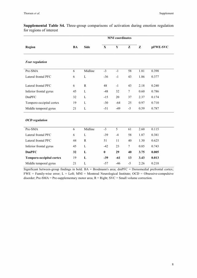
Thorsen et al. Supplement
8
Supplemental Table S4. Three-group comparisons of activation during emotion regulation for regions of interest
MNI coordinates
Region BA Side X Y Z Z pFWE-SVC
Fear regulation
Pre-SMA 6 Midline -3 -1 58 1.81 0.398
Lateral frontal PFC 6 L -36 -1 43 1.86 0.377
Lateral frontal PFC 6 R 48 -1 43 2.18 0.240
Inferior frontal gyrus 45 L -48 32 7 0.60 0.786
DmPFC 32 L -15 20 37 2.37 0.174
Temporo-occipital cortex 19 L -30 -64 25 0.97 0.710
Middle temporal gyrus 21 L -51 -49 -5 0.59 0.787
OCD regulation
Pre-SMA 6 Midline -3 5 61 2.60 0.115
Lateral frontal PFC 6 L -39 -4 58 1.87 0.381
Lateral frontal PFC 44 R 51 11 40 1.30 0.625
Inferior frontal gyrus 45 L -42 23 7 0.85 0.743
DmPFC 32 L 0 29 40 3.75 0.005
Temporo-occipital cortex 19 L -39 -61 13 3.43 0.013
Middle temporal gyrus 21 L -57 -46 -5 2.26 0.218
Significant between-group findings in bold. BA = Brodmann's area; dmPFC = Dorsomedial prefrontal cortex; FWE = Family-wise error; L = Left; MNI = Montreal Neurological Institute; OCD = Obsessive-compulsive disorder; Pre-SMA = Pre-supplementary motor area; R = Right; SVC = Small volume correction.

Thorsen et al. Supplement
9
Supplemental Table S5. Main effect of group for functional connectivity in the bilateral amygdala during emotion regulation
MNI coordinates
Region Side X Y Z Z pFWE-SVC
Fear regulation dmPFC-amygdala connectivity: main effect of group (ANOVA)
Amygdala R 27 -4 -20 2.68 0.124
Amygdala L -30 -4 -20 2.34 0.235
Fear-related regulation: post-hoc pairwise comparisons in right amygdala
Ke Z pFWE-SVC
Siblings > OCD NS
HC > Siblings NS
HC > OCD 3 3.30 0.019
Fear-related regulation: post-hoc pairwise comparisons in left amygdala
Ke Z pFWE-SVC
Siblings > OCD NS
HC > Siblings NS
HC > OCD 14 3.36 0.016
OCD-related regulation dmPFC-amygdala connectivity: main effect of group (ANOVA)
Amygdala L -24 -4 -17 2.21 0.283
Amygdala R 30 -7 -11 1.62 0.561
Fear-related regulation: post-hoc pairwise comparisons in left amygdala
Ke Z pFWE-SVC
Siblings > OCD 3 3.22 0.023
Siblings > HC NS
HC > OCD NS
Fear-related regulation: post-hoc pairwise comparisons in right amygdala
Ke Z pFWE-SVC
Siblings > OCD 2 3.30 0.019
Siblings > HC NS
HC > OCD NS
FWE = Family-wise error; L = Left; MNI = Montreal Neurological Institute; R = Right; SVC = Small volume correction.

Thorsen et al. Supplement
10
Supplemental Table S6. Whole-brain main effect of group on BOLD signal during emotion provocation at uncorrected p < 0.001
MNI coordinates
Region Side Ke X Y Z Z Post-hoc tests of the group differences
Fear-related provocation
Posterior cingulate gyrus R 16 15 -46 31 3.83 Sibs vs HCb
Supramarginal gyrus R 19 63 -28 25 3.70 Sibs vs OCD & HC
Inferior parietal lobule L 6 -45 -37 37 3.57 OCD vs HCa
Middle occipital gyrus L 5 -39 -88 10 3.41 OCD vs HCa
Middle temporal gyrus L 5 -42 -58 -5 3.38 Sibs vs HCb
Inferior occipital gyrus R 6 42 -79 -8 3.35 OCD vs HCa
DlPFC L 5 -36 32 19 3.30 Sibs vs HCb
Fusiform gyrus R 3 36 -64 -17 3.23 NS in post-hoc
OCD-related provocation
Putamen L 27 -24 11 -2 3.84 Sibs vs OCD & HC
Cerebellum/lingual gyrus L 14 -6 -70 -8 3.80 OCD vs Sibs vs HCa
Middle temporal gyrus L 23 -39 -58 -2 3.75 Sibs vs HCb
Hippocampus/amygdala R 15 27 -7 -17 3.58 OCD vs HCa
Inferior occipital gyrus L 8 -36 -88 -11 3.46 OCD vs HCa
Thalamus R 6 6 -16 -2 3.40 Sibs vs HCb
DmPFC L 6 -21 -4 52 3.38 NS in post-hoc
Temporal pole L 4 -42 17 -17 3.30 Sibs vs OCDb
Thalamus L 3 -21 -28 4 3.29 Sibs vs HCb
Cerebellum R 4 9 -73 -20 3.28 OCD vs HCa
Posterior cingulate gyrus R 3 12 -49 31 3.25 Sibs vs HCb
Supramarginal gyrus R 5 60 -28 28 3.16 NS in post-hoc
Picture type interaction
Hippocampus L 10 -36 -22 -11 4.04 OCD vs HCa
Cerebellum L 81 -15 -55 -8 3.88 Sibs & OCD vs HC
Caudate nucleus R 11 6 11 -2 3.88 OCD vs HCa
Putamen L 23 -24 11 -5 3.63 NS in post-hoc
Posterior insula L 8 -39 -13 4 3.49 Sibs vs OCD & HC
Amygdala R 6 27 -7 -17 3.45 OCD vs HCa
Thalamus R 8 6 -13 -2 3.41 Sibs vs HC & OCD
Thalamus L 5 -21 -28 4 3.22 Sibs vs HCb a Siblings vs. OCD and siblings vs. controls: not significant. b Patients vs. siblings and patients vs. controls: not significant. DmPFC = dorsomedial prefrontal cortex; DlPFC = dorsolateral prefrontal cortex; HC = healthy controls; L = left; Ke = voxel extent of cluster; MNI = Montreal Neurological Institute; NS = not significant. OCD = Obsessive-compulsive disorder; R = right; Sibs = unaffected siblings.

Thorsen et al. Supplement
11
Supplemental Table S7. Whole-brain three-group comparisons of BOLD activity during fear and OCD-related emotion regulation at uncorrected p < 0.001
MNI coordinates
Region BA Side Ke X Y Z Z Direction post-hoc tests
Fear regulation
Middle frontal gyrus 9 L 14 -18 38 25 3.37 HC > OCDa
Occipital pole 18 R 8 21 -91 4 3.31 Sibs > OCDb
Superior temporal gyrus 22 R 3 54 -16 -5 3.13 Sibs & HC > OCD
OCD regulation
Superior frontal gyrus 32 R 30 3 32 40 4.01 OCD & Sibs > HCa
Lingual gyrus 17 R 20 3 -82 1 3.58 OCD & Sibs > HC Angular gyrus 21 R 3 57 -58 22 3.44 Sibs > HCc
Middle temporal gyrus 37 L 4 -39 -61 13 3.43 Sibs > HCc
a Siblings vs. OCD and siblings vs. controls: not significant. b Patients vs. siblings and patients vs. controls: not significant. HC = healthy controls; L = left; Ke = voxel extent of cluster; MNI = Montreal Neurological Institute; OCD = Obsessive-compulsive disorder; R = right; Sibs = unaffected siblings. Post-hoc tests: data not shown.

Thorsen et al. Supplement
12
Supplemental Table S8. Whole-brain group comparisons of functional connectivity during fear and OCD-related regulation at uncorrected p < 0.001
MNI coordinates
Region BA Side Ke X Y Z Z Direction of post-hoc
tests
HC > OCD during
fear regulation*
Posterior insula 48 L 8 -42 -16 -2 3.50 HC > OCDa
Right amygdala N/A R 4 24 -7 -20 3.46 HC > OCDa
OCD regulation
Middle temporal gyrus 21 L 29 -42 -52 13 3.76 Sibs > OCD & HC
Middle cingulate gyrus 23 L 17 -6 -16 43 3.57 Sibs > OCD & HC
Supramarginal gyrus 48 R 3 63 -34 22 3.42 Sibs > OCDb
Precuneus 23 L 22 -3 -52 34 3.37 Sibs > OCD & HC
Precuneus 23 L 9 -3 -58 22 3.31 Sibs > OCD & HC
Superior frontal gyrus 10 L 3 -9 56 31 3.20 Sibs > OCD & HC
* Three-group comparison showed no significant voxels at uncorrected p < 0.001. a Siblings vs. OCD and siblings vs. controls: not significant. b Patients vs. siblings and patients vs. controls: not significant. HC = healthy controls; L = left; Ke = voxel extent of cluster; MNI = Montreal Neurological Institute; OCD = Obsessive-compulsive disorder; R = right; Sibs = unaffected siblings. Post-hoc tests: data not shown.

Thorsen et al. Supplement
13
Supplemental Figure S1. Group comparisons of dmPFC-amygdala functional connectivity
during fear and OCD-related emotion regulation.
Panels show BOLD signal parameter estimates of functional connectivity between the left
dmPFC and amygdala for each group during emotion regulation. Panel A and B shows lower
fronto-limbic connectivity in OCD patients compared to healthy controls in the left and right
amygdala during fear-related regulation, while siblings were not significantly different from
either group. Panel C and D shows trend-level higher fronto-limbic connectivity in siblings
compared to patients during OCD-related regulation, while healthy controls were not
significantly different from either group. Parameter estimates are in arbitrary units, with
standard errors.

Thorsen et al. Supplement
14
Supplemental References
1. Ochsner KN, Silvers JA, Buhle JT. (2012): Functional imaging studies of emotion regulation: a synthetic review and evolving model of the cognitive control of emotion. Ann N Y Acad Sci 1251:E1-E24.
2. de Wit SJ, van der Werf YD, Mataix-Cols D, Trujillo JP, van Oppen P, Veltman DJ et al.
(2015): Emotion regulation before and after transcranial magnetic stimulation in obsessive compulsive disorder. Psychol Med 45:3059-3073.
3. McLaren DG, Ries ML, Xu G, Johnson SC. (2012): A generalized form of context-
dependent psychophysiological interactions (gPPI): a comparison to standard approaches. Neuroimage 61:1277-1286.
4. Thorsen AL, Hagland P, Radua J, Mataix-Cols D, Kvale G, Hansen B et al. (In press):
Emotional processing in obsessive-compulsive disorder: A systematic review and meta-analysis of 25 functional neuroimaging studies. Biol Psychiatry Cogn Neurosci Neuroimaging.
5. Buhle JT, Silvers JA, Wager TD, Lopez R, Onyemekwu C, Kober H et al. (2014): Cognitive reappraisal of emotion: a meta-analysis of human neuroimaging studies. Cereb Cortex 24:2981-2990.
6. Frank D, Dewitt M, Hudgens-Haney M, Schaeffer D, Ball B, Schwarz N et al. (2014):
Emotion regulation: quantitative meta-analysis of functional activation and deactivation. Neurosci Biobehav Rev 45:202-211.


I
Doctoral Theses at The Faculty of Psychology, University of Bergen
1980
Allen, Hugh M., Dr. philos. Parent-offspring interactions in willow grouse (Lagopus L. Lagopus).
1981
Myhrer, Trond, Dr. philos. Behavioral Studies after selective disruption of hippocampal inputs in albino rats.
1982
Svebak, Sven, Dr. philos. The significance of motivation for task-induced tonic physiological changes.
1983 Myhre, Grete, Dr. philos. The Biopsychology of behavior in captive Willow ptarmigan.
Eide, Rolf, Dr. philos. PSYCHOSOCIAL FACTORS AND INDICES OF HEALTH RISKS. The relationship of psychosocial conditions to subjective complaints, arterial blood pressure, serum cholesterol, serum triglycerides and urinary catecholamines in middle aged populations in Western Norway.
Værnes, Ragnar J., Dr. philos. Neuropsychological effects of diving.
1984
Kolstad, Arnulf, Dr. philos. Til diskusjonen om sammenhengen mellom sosiale forhold og psykiske strukturer. En epidemiologisk undersøkelse blant barn og unge.
Løberg, Tor, Dr. philos. Neuropsychological assessment in alcohol dependence.
1985
Hellesnes, Tore, Dr. philos. Læring og problemløsning. En studie av den perseptuelle analysens betydning for verbal læring.
Håland, Wenche, Dr. philos. Psykoterapi: relasjon, utviklingsprosess og effekt.
1986
Hagtvet, Knut A., Dr. philos. The construct of test anxiety: Conceptual and methodological issues.
Jellestad, Finn K., Dr. philos. Effects of neuron specific amygdala lesions on fear-motivated behavior in rats.
1987 Aarø, Leif E., Dr. philos. Health behaviour and sosioeconomic Status. A survey among the adult population in Norway.
Underlid, Kjell, Dr. philos. Arbeidsløyse i psykososialt perspektiv.
Laberg, Jon C., Dr. philos. Expectancy and classical conditioning in alcoholics' craving.
Vollmer, Fred, Dr. philos. Essays on explanation in psychology.
Ellertsen, Bjørn, Dr. philos. Migraine and tension headache: Psychophysiology, personality and therapy.
1988 Kaufmann, Astrid, Dr. philos. Antisosial atferd hos ungdom. En studie av psykologiske determinanter.

II
Mykletun, Reidar J., Dr. philos. Teacher stress: personality, work-load and health.
Havik, Odd E., Dr. philos. After the myocardial infarction: A medical and psychological study with special emphasis on perceived illness.
1989 Bråten, Stein, Dr. philos. Menneskedyaden. En teoretisk tese om sinnets dialogiske natur med informasjons- og utviklingspsykologiske implikasjoner sammenholdt med utvalgte spedbarnsstudier.
Wold, Bente, Dr. psychol. Lifestyles and physical activity. A theoretical and empirical analysis of socialization among children and adolescents.
1990 Flaten, Magne A., Dr. psychol. The role of habituation and learning in reflex modification.
1991 Alsaker, Françoise D., Dr. philos.
Global negative self-evaluations in early adolescence.
Kraft, Pål, Dr. philos. AIDS prevention in Norway. Empirical studies on diffusion of knowledge, public opinion, and sexual behaviour.
Endresen, Inger M., Dr. philos. Psychoimmuniological stress markers in working life.
Faleide, Asbjørn O., Dr. philos. Asthma and allergy in childhood. Psychosocial and psychotherapeutic problems.
1992 Dalen, Knut, Dr. philos. Hemispheric asymmetry and the Dual-Task Paradigm: An experimental approach.
Bø, Inge B., Dr. philos. Ungdoms sosiale økologi. En undersøkelse av 14-16 åringers sosiale nettverk.
Nivison, Mary E., Dr. philos. The relationship between noise as an experimental and environmental stressor, physiological changes and psychological factors.
Torgersen, Anne M., Dr. philos. Genetic and environmental influence on temperamental behaviour. A longitudinal study of twins from infancy to adolescence.
1993 Larsen, Svein, Dr. philos. Cultural background and problem drinking.
Nordhus, Inger Hilde, Dr. philos.
Family caregiving. A community psychological study with special emphasis on clinical interventions.
Thuen, Frode, Dr. psychol. Accident-related behaviour among children and young adolescents: Prediction and prevention.
Solheim, Ragnar, Dr. philos. Spesifikke lærevansker. Diskrepanskriteriet anvendt i seleksjonsmetodikk.
Johnsen, Bjørn Helge, Dr. psychol.
Brain assymetry and facial emotional expressions: Conditioning experiments.
1994 Tønnessen, Finn E., Dr. philos. The etiology of Dyslexia.
Kvale, Gerd, Dr. psychol. Psychological factors in anticipatory nausea and vomiting in cancer chemotherapy.

III
Asbjørnsen, Arve E., Dr. psychol.
Structural and dynamic factors in dichotic listening: An interactional model.
Bru, Edvin, Dr. philos. The role of psychological factors in neck, shoulder and low back pain among female hospitale staff.
Braathen, Eli T., Dr. psychol. Prediction of exellence and discontinuation in different types of sport: The significance of motivation and EMG.
Johannessen, Birte F., Dr. philos.
Det flytende kjønnet. Om lederskap, politikk og identitet.
1995 Sam, David L., Dr. psychol. Acculturation of young immigrants in Norway: A psychological and socio-cultural adaptation.
Bjaalid, Inger-Kristin, Dr. philos. Component processes in word recognition.
Martinsen, Øyvind, Dr. philos. Cognitive style and insight.
Nordby, Helge, Dr. philos. Processing of auditory deviant events: Mismatch negativity of event-related brain potentials.
Raaheim, Arild, Dr. philos. Health perception and health behaviour, theoretical considerations, empirical studies, and practical implications.
Seltzer, Wencke J., Dr. philos. Studies of Psychocultural Approach to Families in Therapy.
Brun, Wibecke, Dr. philos. Subjective conceptions of uncertainty and risk.
Aas, Henrik N., Dr. psychol. Alcohol expectancies and socialization: Adolescents learning to drink.
Bjørkly, Stål, Dr. psychol. Diagnosis and prediction of intra-institutional aggressive behaviour in psychotic patients
1996 Anderssen, Norman, Dr. psychol.
Physical activity of young people in a health perspective: Stability, change and social influences.
Sandal, Gro Mjeldheim, Dr. psychol.
Coping in extreme environments: The role of personality.
Strumse, Einar, Dr. philos. The psychology of aesthetics: explaining visual preferences for agrarian landscapes in Western Norway.
Hestad, Knut, Dr. philos. Neuropsychological deficits in HIV-1 infection.
Lugoe, L.Wycliffe, Dr. philos. Prediction of Tanzanian students’ HIV risk and preventive behaviours
Sandvik, B. Gunnhild, Dr. philos.
Fra distriktsjordmor til institusjonsjordmor. Fremveksten av en profesjon og en profesjonsutdanning
Lie, Gro Therese, Dr. psychol. The disease that dares not speak its name: Studies on factors of importance for coping with HIV/AIDS in Northern Tanzania
Øygard, Lisbet, Dr. philos. Health behaviors among young adults. A psychological and sociological approach
Stormark, Kjell Morten, Dr. psychol.
Emotional modulation of selective attention: Experimental and clinical evidence.

IV
Einarsen, Ståle, Dr. psychol. Bullying and harassment at work: epidemiological and psychosocial aspects.
1997 Knivsberg, Ann-Mari, Dr. philos. Behavioural abnormalities and childhood psychopathology: Urinary peptide patterns as a potential tool in diagnosis and remediation.
Eide, Arne H., Dr. philos. Adolescent drug use in Zimbabwe. Cultural orientation in a global-local perspective and use of psychoactive substances among secondary school students.
Sørensen, Marit, Dr. philos. The psychology of initiating and maintaining exercise and diet behaviour.
Skjæveland, Oddvar, Dr. psychol.
Relationships between spatial-physical neighborhood attributes and social relations among neighbors.
Zewdie, Teka, Dr. philos. Mother-child relational patterns in Ethiopia. Issues of developmental theories and intervention programs.
Wilhelmsen, Britt Unni, Dr. philos.
Development and evaluation of two educational programmes designed to prevent alcohol use among adolescents.
Manger, Terje, Dr. philos. Gender differences in mathematical achievement among Norwegian elementary school students.
1998 V
Lindstrøm, Torill Christine, Dr. philos.
«Good Grief»: Adapting to Bereavement.
Skogstad, Anders, Dr. philos. Effects of leadership behaviour on job satisfaction, health and efficiency.
Haldorsen, Ellen M. Håland, Dr. psychol.
Return to work in low back pain patients.
Besemer, Susan P., Dr. philos. Creative Product Analysis: The Search for a Valid Model for Understanding Creativity in Products.
H Winje, Dagfinn, Dr. psychol. Psychological adjustment after severe trauma. A longitudinal study of adults’ and children’s posttraumatic reactions and coping after the bus accident in Måbødalen, Norway 1988.
Vosburg, Suzanne K., Dr. philos.
The effects of mood on creative problem solving.
Eriksen, Hege R., Dr. philos. Stress and coping: Does it really matter for subjective health complaints?
Jakobsen, Reidar, Dr. psychol.
Empiriske studier av kunnskap og holdninger om hiv/aids og den normative seksuelle utvikling i ungdomsårene.
1999 V
Mikkelsen, Aslaug, Dr. philos. Effects of learning opportunities and learning climate on occupational health.
Samdal, Oddrun, Dr. philos. The school environment as a risk or resource for students’ health-related behaviours and subjective well-being.
Friestad, Christine, Dr. philos. Social psychological approaches to smoking.
Ekeland, Tor-Johan, Dr. philos.
Meining som medisin. Ein analyse av placebofenomenet og implikasjoner for terapi og terapeutiske teoriar.

V
H Saban, Sara, Dr. psychol. Brain Asymmetry and Attention: Classical Conditioning Experiments.
Carlsten, Carl Thomas, Dr. philos.
God lesing – God læring. En aksjonsrettet studie av undervisning i fagtekstlesing.
Dundas, Ingrid, Dr. psychol. Functional and dysfunctional closeness. Family interaction and children’s adjustment.
Engen, Liv, Dr. philos.
Kartlegging av leseferdighet på småskoletrinnet og vurdering av faktorer som kan være av betydning for optimal leseutvikling.
2000 V
Hovland, Ole Johan, Dr. philos. Transforming a self-preserving “alarm” reaction into a self-defeating emotional response: Toward an integrative approach to anxiety as a human phenomenon.
Lillejord, Sølvi, Dr. philos. Handlingsrasjonalitet og spesialundervisning. En analyse av aktørperspektiver.
Sandell, Ove, Dr. philos. Den varme kunnskapen.
Oftedal, Marit Petersen, Dr. philos.
Diagnostisering av ordavkodingsvansker: En prosessanalytisk tilnærmingsmåte.
H Sandbak, Tone, Dr. psychol. Alcohol consumption and preference in the rat: The significance of individual differences and relationships to stress pathology
Eid, Jarle, Dr. psychol.
Early predictors of PTSD symptom reporting; The significance of contextual and individual factors.
2001 V
Skinstad, Anne Helene, Dr. philos.
Substance dependence and borderline personality disorders.
Binder, Per-Einar, Dr. psychol. Individet og den meningsbærende andre. En teoretisk undersøkelse av de mellommenneskelige forutsetningene for psykisk liv og utvikling med utgangspunkt i Donald Winnicotts teori.
Roald, Ingvild K., Dr. philos.
Building of concepts. A study of Physics concepts of Norwegian deaf students.
H Fekadu, Zelalem W., Dr. philos. Predicting contraceptive use and intention among a sample of adolescent girls. An application of the theory of planned behaviour in Ethiopian context.
Melesse, Fantu, Dr. philos.
The more intelligent and sensitive child (MISC) mediational intervention in an Ethiopian context: An evaluation study.
Råheim, Målfrid, Dr. philos. Kvinners kroppserfaring og livssammenheng. En fenomenologisk – hermeneutisk studie av friske kvinner og kvinner med kroniske muskelsmerter.
Engelsen, Birthe Kari, Dr. psychol.
Measurement of the eating problem construct.
Lau, Bjørn, Dr. philos. Weight and eating concerns in adolescence.
2002 V
Ihlebæk, Camilla, Dr. philos. Epidemiological studies of subjective health complaints.

VI
Rosén, Gunnar O. R., Dr. philos.
The phantom limb experience. Models for understanding and treatment of pain with hypnosis.
Høines, Marit Johnsen, Dr. philos.
Fleksible språkrom. Matematikklæring som tekstutvikling.
Anthun, Roald Andor, Dr. philos.
School psychology service quality. Consumer appraisal, quality dimensions, and collaborative improvement potential
Pallesen, Ståle, Dr. psychol. Insomnia in the elderly. Epidemiology, psychological characteristics and treatment.
Midthassel, Unni Vere, Dr. philos.
Teacher involvement in school development activity. A study of teachers in Norwegian compulsory schools
Kallestad, Jan Helge, Dr. philos.
Teachers, schools and implementation of the Olweus Bullying Prevention Program.
H Ofte, Sonja Helgesen, Dr. psychol.
Right-left discrimination in adults and children.
Netland, Marit, Dr. psychol. Exposure to political violence. The need to estimate our estimations.
Diseth, Åge, Dr. psychol. Approaches to learning: Validity and prediction of academic performance.
Bjuland, Raymond, Dr. philos.
Problem solving in geometry. Reasoning processes of student teachers working in small groups: A dialogical approach.
2003 V
Arefjord, Kjersti, Dr. psychol. After the myocardial infarction – the wives’ view. Short- and long-term adjustment in wives of myocardial infarction patients.
Ingjaldsson, Jón Þorvaldur, Dr. psychol.
Unconscious Processes and Vagal Activity in Alcohol Dependency.
Holden, Børge, Dr. philos. Følger av atferdsanalytiske forklaringer for atferdsanalysens tilnærming til utforming av behandling.
Holsen, Ingrid, Dr. philos.
Depressed mood from adolescence to ’emerging adulthood’. Course and longitudinal influences of body image and parent-adolescent relationship.
Hammar, Åsa Karin, Dr. psychol.
Major depression and cognitive dysfunction- An experimental study of the cognitive effort hypothesis.
Sprugevica, Ieva, Dr. philos. The impact of enabling skills on early reading acquisition.
Gabrielsen, Egil, Dr. philos. LESE FOR LIVET. Lesekompetansen i den norske voksenbefolkningen sett i lys av visjonen om en enhetsskole.
H Hansen, Anita Lill, Dr. psychol. The influence of heart rate variability in the regulation of attentional and memory processes.
Dyregrov, Kari, Dr. philos.
The loss of child by suicide, SIDS, and accidents: Consequences, needs and provisions of help.
2004 V
Torsheim, Torbjørn, Dr. psychol.
Student role strain and subjective health complaints: Individual, contextual, and longitudinal perspectives.

VII
Haugland, Bente Storm Mowatt Dr. psychol.
Parental alcohol abuse. Family functioning and child adjustment.
Milde, Anne Marita, Dr. psychol.
Ulcerative colitis and the role of stress. Animal studies of psychobiological factors in relationship to experimentally induced colitis.
Stornes, Tor, Dr. philos. Socio-moral behaviour in sport. An investigation of perceptions of sportspersonship in handball related to important factors of socio-moral influence.
Mæhle, Magne, Dr. philos. Re-inventing the child in family therapy: An investigation of the relevance and applicability of theory and research in child development for family therapy involving children.
Kobbeltvedt, Therese, Dr. psychol.
Risk and feelings: A field approach.
2004 H
Thomsen, Tormod, Dr. psychol. Localization of attention in the brain.
Løberg, Else-Marie, Dr. psychol.
Functional laterality and attention modulation in schizophrenia: Effects of clinical variables.
Kyrkjebø, Jane Mikkelsen, Dr. philos.
Learning to improve: Integrating continuous quality improvement learning into nursing education.
Laumann, Karin, Dr. psychol. Restorative and stress-reducing effects of natural environments: Experiencal, behavioural and cardiovascular indices.
Holgersen, Helge, PhD
Mellom oss - Essay i relasjonell psykoanalyse.
2005 V
Hetland, Hilde, Dr. psychol. Leading to the extraordinary? Antecedents and outcomes of transformational leadership.
Iversen, Anette Christine, Dr. philos.
Social differences in health behaviour: the motivational role of perceived control and coping.
2005 H
Mathisen, Gro Ellen, PhD Climates for creativity and innovation: Definitions, measurement, predictors and consequences.
Sævi, Tone, Dr. philos. Seeing disability pedagogically – The lived experience of disability in the pedagogical encounter.
Wiium, Nora, PhD Intrapersonal factors, family and school norms: combined and interactive influence on adolescent smoking behaviour.
Kanagaratnam, Pushpa, PhD Subjective and objective correlates of Posttraumatic Stress in immigrants/refugees exposed to political violence.
Larsen, Torill M. B. , PhD Evaluating principals` and teachers` implementation of Second Step. A case study of four Norwegian primary schools.
Bancila, Delia, PhD
Psychosocial stress and distress among Romanian adolescents and adults.
2006 V
Hillestad, Torgeir Martin, Dr. philos.
Normalitet og avvik. Forutsetninger for et objektivt psykopatologisk avviksbegrep. En psykologisk, sosial, erkjennelsesteoretisk og teorihistorisk framstilling.
VIII
Nordanger, Dag Øystein, Dr. psychol.
Psychosocial discourses and responses to political violence in post-war Tigray, Ethiopia.
Rimol, Lars Morten, PhD Behavioral and fMRI studies of auditory laterality and speech sound processing.
Krumsvik, Rune Johan, Dr. philos.
ICT in the school. ICT-initiated school development in lower secondary school.
Norman, Elisabeth, Dr. psychol. Gut feelings and unconscious thought: An exploration of fringe consiousness in implicit cognition.
Israel, K Pravin, Dr. psychol. Parent involvement in the mental health care of children and adolescents. Emperical studies from clinical care setting.
Glasø, Lars, PhD Affects and emotional regulation in leader-subordinate relationships.
Knutsen, Ketil, Dr. philos. HISTORIER UNGDOM LEVER – En studie av hvordan ungdommer bruker historie for å gjøre livet meningsfullt.
Matthiesen, Stig Berge, PhD Bullying at work. Antecedents and outcomes.
2006 H
Gramstad, Arne, PhD Neuropsychological assessment of cognitive and emotional functioning in patients with epilepsy.
Bendixen, Mons, PhD Antisocial behaviour in early adolescence: Methodological and substantive issues.
Mrumbi, Khalifa Maulid, PhD Parental illness and loss to HIV/AIDS as experienced by AIDS orphans aged between 12-17 years from Temeke District, Dar es Salaam, Tanzania: A study of the children’s psychosocial health and coping responses.
Hetland, Jørn, Dr. psychol. The nature of subjective health complaints in adolescence: Dimensionality, stability, and psychosocial predictors
Kakoko, Deodatus Conatus Vitalis, PhD
Voluntary HIV counselling and testing service uptake among primary school teachers in Mwanza, Tanzania: assessment of socio-demographic, psychosocial and socio-cognitive aspects
Mykletun, Arnstein, Dr. psychol. Mortality and work-related disability as long-term consequences of anxiety and depression: Historical cohort designs based on the HUNT-2 study
Sivertsen, Børge, PhD Insomnia in older adults. Consequences, assessment and treatment.
2007 V
Singhammer, John, Dr. philos. Social conditions from before birth to early adulthood – the influence on health and health behaviour
Janvin, Carmen Ani Cristea, PhD
Cognitive impairment in patients with Parkinson’s disease: profiles and implications for prognosis
Braarud, Hanne Cecilie, Dr.psychol.
Infant regulation of distress: A longitudinal study of transactions between mothers and infants
Tveito, Torill Helene, PhD Sick Leave and Subjective Health Complaints

IX
Magnussen, Liv Heide, PhD Returning disability pensioners with back pain to work
Thuen, Elin Marie, Dr.philos. Learning environment, students’ coping styles and emotional and behavioural problems. A study of Norwegian secondary school students.
Solberg, Ole Asbjørn, PhD Peacekeeping warriors – A longitudinal study of Norwegian peacekeepers in Kosovo
2007 H
Søreide, Gunn Elisabeth, Dr.philos.
Narrative construction of teacher identity
Svensen, Erling, PhD WORK & HEALTH. Cognitive Activation Theory of Stress applied in an organisational setting.
Øverland, Simon Nygaard, PhD Mental health and impairment in disability benefits. Studies applying linkages between health surveys and administrative registries.
Eichele, Tom, PhD Electrophysiological and Hemodynamic Correlates of Expectancy in Target Processing
Børhaug, Kjetil, Dr.philos. Oppseding til demokrati. Ein studie av politisk oppseding i norsk skule.
Eikeland, Thorleif, Dr.philos. Om å vokse opp på barnehjem og på sykehus. En undersøkelse av barnehjemsbarns opplevelser på barnehjem sammenholdt med sanatoriebarns beskrivelse av langvarige sykehusopphold – og et forsøk på forklaring.
Wadel, Carl Cato, Dr.philos. Medarbeidersamhandling og medarbeiderledelse i en lagbasert organisasjon
Vinje, Hege Forbech, PhD Thriving despite adversity: Job engagement and self-care among community nurses
Noort, Maurits van den, PhD Working memory capacity and foreign language acquisition
2008 V
Breivik, Kyrre, Dr.psychol. The Adjustment of Children and Adolescents in Different Post-Divorce Family Structures. A Norwegian Study of Risks and Mechanisms.
Johnsen, Grethe E., PhD Memory impairment in patients with posttraumatic stress disorder
Sætrevik, Bjørn, PhD Cognitive Control in Auditory Processing
Carvalhosa, Susana Fonseca, PhD
Prevention of bullying in schools: an ecological model
2008 H
Brønnick, Kolbjørn Selvåg Attentional dysfunction in dementia associated with Parkinson’s disease.
Posserud, Maj-Britt Rocio Epidemiology of autism spectrum disorders
Haug, Ellen Multilevel correlates of physical activity in the school setting
Skjerve, Arvid Assessing mild dementia – a study of brief cognitive tests.

X
Kjønniksen, Lise The association between adolescent experiences in physical activity and leisure time physical activity in adulthood: a ten year longitudinal study
Gundersen, Hilde The effects of alcohol and expectancy on brain function
Omvik, Siri Insomnia – a night and day problem
2009 V
Molde, Helge Pathological gambling: prevalence, mechanisms and treatment outcome.
Foss, Else Den omsorgsfulle væremåte. En studie av voksnes væremåte i forhold til barn i barnehagen.
Westrheim, Kariane Education in a Political Context: A study of Konwledge Processes and Learning Sites in the PKK.
Wehling, Eike Cognitive and olfactory changes in aging
Wangberg, Silje C. Internet based interventions to support health behaviours: The role of self-efficacy.
Nielsen, Morten B. Methodological issues in research on workplace bullying. Operationalisations, measurements and samples.
Sandu, Anca Larisa MRI measures of brain volume and cortical complexity in clinical groups and during development.
Guribye, Eugene Refugees and mental health interventions
Sørensen, Lin Emotional problems in inattentive children – effects on cognitive control functions.
Tjomsland, Hege E. Health promotion with teachers. Evaluation of the Norwegian Network of Health Promoting Schools: Quantitative and qualitative analyses of predisposing, reinforcing and enabling conditions related to teacher participation and program sustainability.
Helleve, Ingrid Productive interactions in ICT supported communities of learners
2009 H
Skorpen, Aina Øye, Christine
Dagliglivet i en psykiatrisk institusjon: En analyse av miljøterapeutiske praksiser
Andreassen, Cecilie Schou WORKAHOLISM – Antecedents and Outcomes
Stang, Ingun Being in the same boat: An empowerment intervention in breast cancer self-help groups
Sequeira, Sarah Dorothee Dos Santos
The effects of background noise on asymmetrical speech perception
Kleiven, Jo, dr.philos. The Lillehammer scales: Measuring common motives for vacation and leisure behavior
Jónsdóttir, Guðrún Dubito ergo sum? Ni jenter møter naturfaglig kunnskap.
Hove, Oddbjørn Mental health disorders in adults with intellectual disabilities - Methods of assessment and prevalence of mental health disorders and problem behaviour
Wageningen, Heidi Karin van The role of glutamate on brain function
XI
Bjørkvik, Jofrid God nok? Selvaktelse og interpersonlig fungering hos pasienter innen psykisk helsevern: Forholdet til diagnoser, symptomer og behandlingsutbytte
Andersson, Martin A study of attention control in children and elderly using a forced-attention dichotic listening paradigm
Almås, Aslaug Grov Teachers in the Digital Network Society: Visions and Realities. A study of teachers’ experiences with the use of ICT in teaching and learning.
Ulvik, Marit Lærerutdanning som danning? Tre stemmer i diskusjonen
2010 V
Skår, Randi Læringsprosesser i sykepleieres profesjonsutøvelse. En studie av sykepleieres læringserfaringer.
Roald, Knut Kvalitetsvurdering som organisasjonslæring mellom skole og skoleeigar
Lunde, Linn-Heidi Chronic pain in older adults. Consequences, assessment and treatment.
Danielsen, Anne Grete Perceived psychosocial support, students’ self-reported academic initiative and perceived life satisfaction
Hysing, Mari Mental health in children with chronic illness
Olsen, Olav Kjellevold Are good leaders moral leaders? The relationship between effective military operational leadership and morals
Riese, Hanne Friendship and learning. Entrepreneurship education through mini-enterprises.
Holthe, Asle Evaluating the implementation of the Norwegian guidelines for healthy school meals: A case study involving three secondary schools
H Hauge, Lars Johan Environmental antecedents of workplace bullying: A multi-design approach
Bjørkelo, Brita Whistleblowing at work: Antecedents and consequences
Reme, Silje Endresen Common Complaints – Common Cure? Psychiatric comorbidity and predictors of treatment outcome in low back pain and irritable bowel syndrome
Helland, Wenche Andersen Communication difficulties in children identified with psychiatric problems
Beneventi, Harald Neuronal correlates of working memory in dyslexia
Thygesen, Elin Subjective health and coping in care-dependent old persons living at home
Aanes, Mette Marthinussen Poor social relationships as a threat to belongingness needs. Interpersonal stress and subjective health complaints: Mediating and moderating factors.
Anker, Morten Gustav Client directed outcome informed couple therapy
XII
Bull, Torill Combining employment and child care: The subjective well-being of single women in Scandinavia and in Southern Europe
Viig, Nina Grieg Tilrettelegging for læreres deltakelse i helsefremmende arbeid. En kvalitativ og kvantitativ analyse av sammenhengen mellom organisatoriske forhold og læreres deltakelse i utvikling og implementering av Europeisk Nettverk av Helsefremmende Skoler i Norge
Wolff, Katharina To know or not to know? Attitudes towards receiving genetic information among patients and the general public.
Ogden, Terje, dr.philos. Familiebasert behandling av alvorlige atferdsproblemer blant barn og ungdom. Evaluering og implementering av evidensbaserte behandlingsprogrammer i Norge.
Solberg, Mona Elin Self-reported bullying and victimisation at school: Prevalence, overlap and psychosocial adjustment.
2011 V
Bye, Hege Høivik Self-presentation in job interviews. Individual and cultural differences in applicant self-presentation during job interviews and hiring managers’ evaluation
Notelaers, Guy Workplace bullying. A risk control perspective.
Moltu, Christian Being a therapist in difficult therapeutic impasses. A hermeneutic phenomenological analysis of skilled psychotherapists’ experiences, needs, and strategies in difficult therapies ending well.
Myrseth, Helga Pathological Gambling - Treatment and Personality Factors
Schanche, Elisabeth From self-criticism to self-compassion. An empirical investigation of hypothesized change prosesses in the Affect Phobia Treatment Model of short-term dynamic psychotherapy for patients with Cluster C personality disorders.
Våpenstad, Eystein Victor, dr.philos.
Det tempererte nærvær. En teoretisk undersøkelse av psykoterapautens subjektivitet i psykoanalyse og psykoanalytisk psykoterapi.
Haukebø, Kristin Cognitive, behavioral and neural correlates of dental and intra-oral injection phobia. Results from one treatment and one fMRI study of randomized, controlled design.
Harris, Anette Adaptation and health in extreme and isolated environments. From 78°N to 75°S.
Bjørknes, Ragnhild Parent Management Training-Oregon Model: intervention effects on maternal practice and child behavior in ethnic minority families
Mamen, Asgeir Aspects of using physical training in patients with substance dependence and additional mental distress
Espevik, Roar Expert teams: Do shared mental models of team members make a difference
Haara, Frode Olav Unveiling teachers’ reasons for choosing practical activities in mathematics teaching
XIII
2011 H
Hauge, Hans Abraham How can employee empowerment be made conducive to both employee health and organisation performance? An empirical investigation of a tailor-made approach to organisation learning in a municipal public service organisation.
Melkevik, Ole Rogstad Screen-based sedentary behaviours: pastimes for the poor, inactive and overweight? A cross-national survey of children and adolescents in 39 countries.
Vøllestad, Jon Mindfulness-based treatment for anxiety disorders. A quantitative review of the evidence, results from a randomized controlled trial, and a qualitative exploration of patient experiences.
Tolo, Astrid Hvordan blir lærerkompetanse konstruert? En kvalitativ studie av PPU-studenters kunnskapsutvikling.
Saus, Evelyn-Rose Training effectiveness: Situation awareness training in simulators
Nordgreen, Tine Internet-based self-help for social anxiety disorder and panic disorder. Factors associated with effect and use of self-help.
Munkvold, Linda Helen Oppositional Defiant Disorder: Informant discrepancies, gender differences, co-occuring mental health problems and neurocognitive function.
Christiansen, Øivin Når barn plasseres utenfor hjemmet: beslutninger, forløp og relasjoner. Under barnevernets (ved)tak.
Brunborg, Geir Scott Conditionability and Reinforcement Sensitivity in Gambling Behaviour
Hystad, Sigurd William Measuring Psychological Resiliency: Validation of an Adapted Norwegian Hardiness Scale
2012 V
Roness, Dag Hvorfor bli lærer? Motivasjon for utdanning og utøving.
Fjermestad, Krister Westlye The therapeutic alliance in cognitive behavioural therapy for youth anxiety disorders
Jenssen, Eirik Sørnes Tilpasset opplæring i norsk skole: politikeres, skolelederes og læreres handlingsvalg
Saksvik-Lehouillier, Ingvild Shift work tolerance and adaptation to shift work among offshore workers and nurses
Johansen, Venke Frederike Når det intime blir offentlig. Om kvinners åpenhet om brystkreft og om markedsføring av brystkreftsaken.
Herheim, Rune Pupils collaborating in pairs at a computer in mathematics learning: investigating verbal communication patterns and qualities
Vie, Tina Løkke Cognitive appraisal, emotions and subjective health complaints among victims of workplace bullying: A stress-theoretical approach
Jones, Lise Øen Effects of reading skills, spelling skills and accompanying efficacy beliefs on participation in education. A study in Norwegian prisons.

XIV
2012 H
Danielsen, Yngvild Sørebø Childhood obesity – characteristics and treatment. Psychological perspectives.
Horverak, Jøri Gytre Sense or sensibility in hiring processes. Interviewee and interviewer characteristics as antecedents of immigrant applicants’ employment probabilities. An experimental approach.
Jøsendal, Ola Development and evaluation of BE smokeFREE, a school-based smoking prevention program
Osnes, Berge Temporal and Posterior Frontal Involvement in Auditory Speech Perception
Drageset, Sigrunn Psychological distress, coping and social support in the diagnostic and preoperative phase of breast cancer
Aasland, Merethe Schanke Destructive leadership: Conceptualization, measurement, prevalence and outcomes
Bakibinga, Pauline The experience of job engagement and self-care among Ugandan nurses and midwives
Skogen, Jens Christoffer Foetal and early origins of old age health. Linkage between birth records and the old age cohort of the Hordaland Health Study (HUSK)
Leversen, Ingrid Adolescents’ leisure activity participation and their life satisfaction: The role of demographic characteristics and psychological processes
Hanss, Daniel Explaining sustainable consumption: Findings from cross-sectional and intervention approaches
Rød, Per Arne Barn i klem mellom foreldrekonflikter og samfunnsmessig beskyttelse
2013 V
Mentzoni, Rune Aune Structural Characteristics in Gambling
Knudsen, Ann Kristin Long-term sickness absence and disability pension award as consequences of common mental disorders. Epidemiological studies using a population-based health survey and official ill health benefit registries.
Strand, Mari Emotional information processing in recurrent MDD
Veseth, Marius Recovery in bipolar disorder. A reflexive-collaborative exploration of the lived experiences of healing and growth when battling a severe mental illness
Mæland, Silje Sick leave for patients with severe subjective health complaints. Challenges in general practice.
Mjaaland, Thera At the frontiers of change? Women and girls’ pursuit of education in north-western Tigray, Ethiopia
Odéen, Magnus Coping at work. The role of knowledge and coping expectancies in health and sick leave.
Hynninen, Kia Minna Johanna Anxiety, depression and sleep disturbance in chronic obstructive pulmonary disease (COPD). Associations, prevalence and effect of psychological treatment.

XV
Flo, Elisabeth Sleep and health in shift working nurses
Aasen, Elin Margrethe From paternalism to patient participation? The older patients undergoing hemodialysis, their next of kin and the nurses: a discursive perspective on perception of patient participation in dialysis units
Ekornås, Belinda Emotional and Behavioural Problems in Children: Self-perception, peer relationships, and motor abilities
Corbin, J. Hope North-South Partnerships for Health: Key Factors for Partnership Success from the Perspective of the KIWAKKUKI
Birkeland, Marianne Skogbrott Development of global self-esteem: The transition from adolescence to adulthood
2013 H
Gianella-Malca, Camila Challenges in Implementing the Colombian Constitutional Court’s Health-Care System Ruling of 2008
Hovland, Anders Panic disorder – Treatment outcomes and psychophysiological concomitants
Mortensen, Øystein The transition to parenthood – Couple relationships put to the test
Årdal, Guro Major Depressive Disorder – a Ten Year Follow-up Study. Inhibition, Information Processing and Health Related Quality of Life
Johansen, Rino Bandlitz The impact of military identity on performance in the Norwegian armed forces
Bøe, Tormod Socioeconomic Status and Mental Health in Children and Adolescents
2014 V
Nordmo, Ivar Gjennom nåløyet – studenters læringserfaringer i psykologutdanningen
Dovran, Anders Childhood Trauma and Mental Health Problems in Adult Life
Hegelstad, Wenche ten Velden Early Detection and Intervention in Psychosis: A Long-Term Perspective
Urheim, Ragnar Forståelse av pasientaggresjon og forklaringer på nedgang i voldsrate ved Regional sikkerhetsavdeling, Sandviken sykehus
Kinn, Liv Grethe Round-Trips to Work. Qualitative studies of how persons with severe mental illness experience work integration.
Rød, Anne Marie Kinn Consequences of social defeat stress for behaviour and sleep. Short-term and long-term assessments in rats.
Nygård, Merethe Schizophrenia – Cognitive Function, Brain Abnormalities, and Cannabis Use
Tjora, Tore Smoking from adolescence through adulthood: the role of family, friends, depression and socioeconomic status. Predictors of smoking from age 13 to 30 in the “The Norwegian Longitudinal Health Behaviour Study” (NLHB)
Vangsnes, Vigdis The Dramaturgy and Didactics of Computer Gaming. A Study of a Medium in the Educational Context of Kindergartens.

XVI
Nordahl, Kristin Berg Early Father-Child Interaction in a Father-Friendly Context: Gender Differences, Child Outcomes, and Protective Factors related to Fathers’ Parenting Behaviors with One-year-olds
2014 H
Sandvik, Asle Makoto Psychopathy – the heterogenety of the construct
Skotheim, Siv Maternal emotional distress and early mother-infant interaction: Psychological, social and nutritional contributions
Halleland, Helene Barone Executive Functioning in adult Attention Deficit Hyperactivity Disorder (ADHD). From basic mechanisms to functional outcome.
Halvorsen, Kirsti Vindal Partnerskap i lærerutdanning, sett fra et økologisk perspektiv
Solbue, Vibeke Dialogen som visker ut kategorier. En studie av hvilke erfaringer innvandrerungdommer og norskfødte med innvandrerforeldre har med videregående skole. Hva forteller ungdommenes erfaringer om videregående skoles håndtering av etniske ulikheter?
Kvalevaag, Anne Lise Fathers’ mental health and child development. The predictive value of fathers’ psychological distress during pregnancy for the social, emotional and behavioural development of their children
Sandal, Ann Karin Ungdom og utdanningsval. Om elevar sine opplevingar av val og overgangsprosessar.
Haug, Thomas Predictors and moderators of treatment outcome from high- and low-intensity cognitive behavioral therapy for anxiety disorders. Association between patient and process factors, and the outcome from guided self-help, stepped care, and face-to-face cognitive behavioral therapy.
Sjølie, Hege Experiences of Members of a Crisis Resolution Home Treatment Team. Personal history, professional role and emotional support in a CRHT team.
Falkenberg, Liv Eggset Neuronal underpinnings of healthy and dysfunctional cognitive control
Mrdalj, Jelena The early life condition. Importance for sleep, circadian rhythmicity, behaviour and response to later life challenges
Hesjedal, Elisabeth Tverrprofesjonelt samarbeid mellom skule og barnevern: Kva kan støtte utsette barn og unge?
2015 V
Hauken, May Aasebø «The cancer treatment was only half the work!» A Mixed-Method Study of Rehabilitation among Young Adult Cancer Survivors
Ryland, Hilde Katrin Social functioning and mental health in children: the influence of chronic illness and intellectual function
Rønsen, Anne Kristin Vurdering som profesjonskompetanse. Refleksjonsbasert utvikling av læreres kompetanse i formativ vurdering

XVII
Hoff, Helge Andreas Thinking about Symptoms of Psychopathy in Norway: Content Validation of the Comprehensive Assessment of Psychopathic Personality (CAPP) Model in a Norwegian Setting
Schmid, Marit Therese Executive Functioning in recurrent- and first episode Major Depressive Disorder. Longitudinal studies
Sand, Liv Body Image Distortion and Eating Disturbances in Children and Adolescents
Matanda, Dennis Juma Child physical growth and care practices in Kenya: Evidence from Demographic and Health Surveys
Amugsi, Dickson Abanimi Child care practices, resources for care, and nutritional outcomes in Ghana: Findings from Demographic and Health Surveys
Jakobsen, Hilde The good beating: Social norms supporting men’s partner violence in Tanzania
Sagoe, Dominic Nonmedical anabolic-androgenic steroid use: Prevalence, attitudes, and social perception
Eide, Helene Marie Kjærgård Narrating the relationship between leadership and learning outcomes. A study of public narratives in the Norwegian educational sector.
2015 H
Wubs, Annegreet Gera Intimate partner violence among adolescents in South Africa and Tanzania
Hjelmervik, Helene Susanne Sex and sex-hormonal effects on brain organization of fronto-parietal networks
Dahl, Berit Misund The meaning of professional identity in public health nursing
Røykenes, Kari Testangst hos sykepleierstudenter: «Alternativ behandling»
Bless, Josef Johann The smartphone as a research tool in psychology. Assessment of language lateralization and training of auditory attention.
Løvvik, Camilla Margrethe Sigvaldsen
Common mental disorders and work participation – the role of return-to-work expectations
Lehmann, Stine Mental Disorders in Foster Children: A Study of Prevalence, Comorbidity, and Risk Factors
Knapstad, Marit Psychological factors in long-term sickness absence: the role of shame and social support. Epidemiological studies based on the Health Assets Project.
2016 V
Kvestad, Ingrid Biological risks and neurodevelopment in young North Indian children
Sælør, Knut Tore Hinderløyper, halmstrå og hengende snører. En kvalitativ studie av håp innenfor psykisk helse- og rusfeltet.
Mellingen, Sonja Alkoholbruk, partilfredshet og samlivsstatus. Før, inn i, og etter svangerskapet – korrelater eller konsekvenser?
Thun, Eirunn Shift work: negative consequences and protective factors
XVIII
Hilt, Line Torbjørnsen The borderlands of educational inclusion. Analyses of inclusion and exclusion processes for minority language students
Havnen, Audun Treatment of obsessive-compulsive disorder and the importance of assessing clinical effectiveness
Slåtten, Hilde Gay-related name-calling among young adolescents. Exploring the importance of the context.
Ree, Eline Staying at work. The role of expectancies and beliefs in health and workplace interventions.
Morken, Frøydis Reading and writing processing in dyslexia
2016 H
Løvoll, Helga Synnevåg Inside the outdoor experience. On the distinction between pleasant and interesting feelings and their implication in the motivational process.
Hjeltnes, Aslak Facing social fears: An investigation of mindfulness-based stress reduction for young adults with social anxiety disorder
Øyeflaten, Irene Larsen Long-term sick leave and work rehabilitation. Prognostic factors for return to work.
Henriksen, Roger Ekeberg Social relationships, stress and infection risk in mother and child
Johnsen, Iren «Only a friend» - The bereavement process of young adults who have lost a friend to a traumatic death. A mixed methods study.
Helle, Siri Cannabis use in non-affective psychoses: Relationship to age at onset, cognitive functioning and social cognition
Glambek, Mats Workplace bullying and expulsion in working life. A representative study addressing prospective associations and explanatory conditions.
Oanes, Camilla Jensen Tilbakemelding i terapi. På hvilke måter opplever terapeuter at tilbakemeldingsprosedyrer kan virke inn på terapeutiske praksiser?
Reknes, Iselin Exposure to workplace bullying among nurses: Health outcomes and individual coping
Chimhutu, Victor Results-Based Financing (RBF) in the health sector of a low-income country. From agenda setting to implementation: The case of Tanzania
Ness, Ingunn Johanne The Room of Opportunity. Understanding how knowledge and ideas are constructed in multidisciplinary groups working with developing innovative ideas.
Hollekim, Ragnhild Contemporary discourses on children and parenting in Norway. An empirical study based on two cases.
Doran, Rouven Eco-friendly travelling: The relevance of perceived norms and social comparison
2017 V
Katisi, Masego The power of context in health partnerships: Exploring synergy and antagony between external and internal ideologies in implementing Safe Male Circumcision (SMC) for HIV prevention in Botswana
XIX
Jamaludin, Nor Lelawati Binti The “why” and “how” of International Students’ Ambassadorship Roles in International Education
Berthelsen, Mona Effects of shift work and psychological and social work factors on mental distress. Studies of onshore/offshore workers and nurses in Norway.
Krane, Vibeke Lærer-elev-relasjoner, elevers psykiske helse og frafall i videregående skole – en eksplorerende studie om samarbeid og den store betydningen av de små ting
Søvik, Margaret Ljosnes Evaluating the implementation of the Empowering Coaching™ program in Norway
Tonheim, Milfrid A troublesome transition: Social reintegration of girl soldiers returning ‘home’
Senneseth, Mette Improving social network support for partners facing spousal cancer while caring for minors. A randomized controlled trial.
Urke, Helga Bjørnøy Child health and child care of very young children in Bolivia, Colombia and Peru.
Bakhturidze, George Public Participation in Tobacco Control Policy-making in Georgia
Fismen, Anne-Siri Adolescent eating habits. Trends and socio-economic status.
2017 H
Hagatun, Susanne Internet-based cognitive-behavioural therapy for insomnia. A randomised controlled trial in Norway.
Eichele, Heike Electrophysiological Correlates of Performance Monitoring in Children with Tourette Syndrome. A developmental perspective.
Risan, Ulf Patrick Accommodating trauma in police interviews. An exploration of rapport in investigative interviews of traumatized victims.
Sandhåland, Hilde Safety on board offshore vessels: A study of shipboard factors and situation awareness
Blågestad, Tone Fidje Less pain – better sleep and mood? Interrelatedness of pain, sleep and mood in total hip arthroplasty patients
Kronstad, Morten Frå skulebenk til deadlines. Korleis nettjournalistar og journaliststudentar lærer, og korleis dei utviklar journalistfagleg kunnskap
Vedaa, Øystein Shift work: The importance of sufficient time for rest between shifts.
Steine, Iris Mulders Predictors of symptoms outcomes among adult survivors of sexual abuse: The role of abuse characteristics, cumulative childhood maltreatment, genetic variants, and perceived social support.
Høgheim, Sigve Making math interesting: An experimental study of interventions to encourage interest in mathematics
XX
2018 V
Brevik, Erlend Joramo Adult Attention Deficit Hyperactivity Disorder. Beyond the Core Symptoms of the Diagnostic and Statistical Manual of Mental Disorders.
Erevik, Eilin Kristine User-generated alcohol-related content on social media: Determinants and relation to offline alcohol use
Hagen, Egon Cognitive and psychological functioning in patients with substance use disorder; from initial assessment to one-year recovery
Adólfsdóttir, Steinunn Subcomponents of executive functions: Effects of age and brain maturations
Brattabø, Ingfrid Vaksdal Detection of child maltreatment, the role of dental health personnel – A national cross-sectional study among public dental health personnel in Norway
Fylkesnes, Marte Knag Frykt, forhandlinger og deltakelse. Ungdommer og foreldre med etnisk minoritetsbakgrunn i møte med den norske barnevernstjenesten.
Stiegler, Jan Reidar Processing emotions in emotion-focused therapy. Exploring the impact of the two-chair dialogue intervention.
Egelandsdal, Kjetil Clickers and Formative Feedback at University Lectures. Exploring students and teachers’ reception and use of feedback from clicker interventions.
Torjussen, Lars Petter Storm Foreningen av visdom og veltalenhet – utkast til en universitetsdidaktikk gjennom en kritikk og videreføring av Skjervheims pedagogiske filosofi på bakgrunn av Arendt og Foucault. Eller hvorfor menneskelivet er mer som å spille fløyte enn å bygge et hus.
Selvik, Sabreen A childhood at refuges. Children with multiple relocations at refuges for abused women.
2018 H
Leino, Tony Mathias Structural game characteristics, game features, financial outcomes and gambling behaviour
Raknes, Solfrid Anxious Adolescents: Prevalence, Correlates, and Preventive Cogntive Behavioural Interventions
Morken, Katharina Teresa Enehaug
Mentalization-based treatment of female patients with severe personality disorder and substance use disorder
Braatveit, Kirsten Johanne Intellectual disability among in-patients with substance use disorders
Barua, Padmaja Unequal Interdependencies: Exploring Power and Agency in Domestic Work Relations in Contemporary India
Darkwah, Ernest Caring for “parentless” children. An exploration of work-related experiences of caregivers in children’s homes in Ghana.
Valdersnes, Kjersti Bergheim Safety Climate perceptions in High Reliability Organizations – the role of Psychological Capital
XXI
2019 V
Kongsgården, Petter Vurderingspraksiser i teknologirike læringsmiljøer. En undersøkelse av læreres vurderingspraksiser i teknologirike læringsmiljøer og implikasjoner på elevenes medvirkning i egen læringsprosess.
Vikene, Kjetil Complexity in Rhythm and Parkinson’s disease: Cognitive and Neuronal Correlates
Heradstveit, Ove Alcohol- and drug use among adolescents. School-related problems, childhood mental health problems, and psychiatric diagnoses.
Riise, Eili Nygard Concentrated exposure and response prevention for obsessive-compulsive disorder in adolescents: the Bergen 4-day treatment
Vik, Alexandra Imaging the Aging Brain: From Morphometry to Functional Connectivity
Krossbakken, Elfrid Personal and Contextual Factors Influencing Gaming Behaviour. Risk Factors and Prevention of Video Game Addiction.
Solholm, Roar Foreldrenes status og rolle i familie- og nærmiljøbaserte intervensjoner for barn med atferdsvansker
Baldomir, Andrea Margarita Children at Risk and Mothering Networks in Buenos Aires, Argentina: Analyses of Socialization and Law-Abiding Practices in Public Early Childhood Intervention.
Samuelsson, Martin Per Education for Deliberative Democracy. Theoretical assumptions and classroom practices.
Visted, Endre Emotion regulation difficulties. The role in onset, maintenance and recurrence of major depressive disorder.
2019 H
Nordmo, Morten Sleep and naval performance. The impact of personality and leadership.
Sveinsdottir, Vigdis Supported Employment and preventing Early Disability (SEED)
Dwyer, Gerard Eric New approaches to the use of magnetic resonance spectroscopy for investigating the pathophysiology of auditory-verbal hallucinations
Synnevåg, Ellen Strøm Planning for Public Health. Balancing top-down and bottom-up approaches in Norwegian municipalities.

Graphic design: Comm
unication Division, UiB / Print: Skipnes Komm
unikasjon AS
uib.no
ISBN: 9788230859674 (print)9788230856246 (PDF)
Related Documents