1521-0081/73/3/861–896$35.00 https://doi.org/10.1124/pharmrev.120.000090 PHARMACOLOGICAL REVIEWS Pharmacol Rev 73:861–896, July 2021 Copyright © 2021 by The Author(s) This is an open access article distributed under the CC BY Attribution 4.0 International license. ASSOCIATE EDITOR: ERIC BARKER The Emerging Role of the Innate Immune Response in Idiosyncratic Drug Reactions s Samantha Christine Sernoskie, 1 Alison Jee, 1 and Jack Paul Uetrecht Department of Pharmaceutical Sciences, Leslie Dan Faculty of Pharmacy (S.C.S., J.P.U.), and Department of Pharmacology and Toxicology, Temerty Faculty of Medicine, University of Toronto, Toronto, Ontario, Canada (A.J., J.P.U.) Abstract..................................................................................... 863 Significance Statement ...................................................................... 863 I. Introduction ................................................................................. 863 II. Review of Types of Idiosyncratic Drug Reactions ............................................. 864 A. Idiosyncratic Drug-Induced Liver Injury.................................................. 864 1. Hepatocellular Liver Injury ........................................................... 864 2. Autoimmune Liver Injury ............................................................ 864 3. Cholestatic Liver Injury .............................................................. 864 B. Severe Cutaneous Adverse Reactions..................................................... 865 1. Stevens-Johnson Syndrome and Toxic Epidermal Necrolysis ........................... 865 2. Drug Reaction with Eosinophilia and Systemic Symptoms ............................. 865 3. Acute Generalized Exanthematous Pustulosis ......................................... 866 C. Idiosyncratic Drug-Induced Blood Dyscrasias ............................................. 866 1. Idiosyncratic Drug-Induced Agranulocytosis ........................................... 866 2. Idiosyncratic Drug-Induced Hemolytic Anemia ........................................ 866 3. Idiosyncratic Drug-Induced Thrombocytopenia ........................................ 867 D. Other Idiosyncratic Drug Reactions ...................................................... 867 1. Idiosyncratic Drug-Induced Autoimmune Reactions ................................... 867 2. Idiosyncratic Drug-Induced Nephropathy ............................................. 867 E. Rationale for Current Review ............................................................ 868 III. Innate Mechanisms Contributing to Adaptive Immune Activation ............................ 869 A. Cells of the Innate Immune System ...................................................... 869 1. Granulocytes ......................................................................... 869 a. Neutrophils ....................................................................... 869 b. Eosinophils ....................................................................... 870 c. Basophils ......................................................................... 870 d. Mast cells ......................................................................... 870 2. Professional Antigen-Presenting Cells................................................. 871 a. Dendritic cells .................................................................... 871 b. Monocytes ........................................................................ 871 c. Macrophages ...................................................................... 872 3. Innate Lymphoid Cells ............................................................... 872 4. Other Innate Immune Cells .......................................................... 873 5. Nonimmune Cells .................................................................... 873 Address correspondence to: Jack Paul Uetrecht, Leslie Dan Faculty of Pharmacy, 144 College St., 10th Floor, Rm. 1007, University of Toronto, Toronto, ON M5S 3M2, Canada. E-mail: [email protected] This work was supported by the Canadian Institutes of Health Research [Grant 142329]. S.C.S. was supported by an Ontario Graduate Scholarship, a Mitacs Research Training Award, and a University of Toronto Fellowship. A.J. was supported by a Natural Sciences and Engineering Research Council of Canada doctoral scholarship. No author has an actual or perceived conflict of interest with the contents of this article. 1 S.C.S. and A.J. contributed equally to this work. s This article has supplemental material available at pharmrev.aspetjournals.org. https://doi.org/10.1124/pharmrev.120.000090. 861 by guest on February 14, 2022 Downloaded from /content/suppl/2021/07/14/73.3.861.DC2.html /content/suppl/2021/05/30/73.3.861.DC1.html Supplemental Material can be found at:

Welcome message from author
This document is posted to help you gain knowledge. Please leave a comment to let me know what you think about it! Share it to your friends and learn new things together.
Transcript
1521-0081/73/3/861–896$35.00 https://doi.org/10.1124/pharmrev.120.000090PHARMACOLOGICAL REVIEWS Pharmacol Rev 73:861–896, July 2021Copyright © 2021 by The Author(s)This is an open access article distributed under the CC BY Attribution 4.0 International license.
ASSOCIATE EDITOR: ERIC BARKER
The Emerging Role of the Innate Immune Response inIdiosyncratic Drug Reactions s
Samantha Christine Sernoskie,1 Alison Jee,1 and Jack Paul Uetrecht
Department of Pharmaceutical Sciences, Leslie Dan Faculty of Pharmacy (S.C.S., J.P.U.), and Department of Pharmacology and Toxicology,Temerty Faculty of Medicine, University of Toronto, Toronto, Ontario, Canada (A.J., J.P.U.)
Abstract. . . . . . . . . . . . . . . . . . . . . . . . . . . . . . . . . . . . . . . . . . . . . . . . . . . . . . . . . . . . . . . . . . . . . . . . . . . . . . . . . . . . . 863Significance Statement . . . . . . . . . . . . . . . . . . . . . . . . . . . . . . . . . . . . . . . . . . . . . . . . . . . . . . . . . . . . . . . . . . . . . . 863
I. Introduction. . . . . . . . . . . . . . . . . . . . . . . . . . . . . . . . . . . . . . . . . . . . . . . . . . . . . . . . . . . . . . . . . . . . . . . . . . . . . . . . . 863II. Review of Types of Idiosyncratic Drug Reactions . . . . . . . . . . . . . . . . . . . . . . . . . . . . . . . . . . . . . . . . . . . . . 864
A. Idiosyncratic Drug-Induced Liver Injury. . . . . . . . . . . . . . . . . . . . . . . . . . . . . . . . . . . . . . . . . . . . . . . . . . 8641. Hepatocellular Liver Injury. . . . . . . . . . . . . . . . . . . . . . . . . . . . . . . . . . . . . . . . . . . . . . . . . . . . . . . . . . . 8642. Autoimmune Liver Injury . . . . . . . . . . . . . . . . . . . . . . . . . . . . . . . . . . . . . . . . . . . . . . . . . . . . . . . . . . . . 8643. Cholestatic Liver Injury . . . . . . . . . . . . . . . . . . . . . . . . . . . . . . . . . . . . . . . . . . . . . . . . . . . . . . . . . . . . . . 864
B. Severe Cutaneous Adverse Reactions. . . . . . . . . . . . . . . . . . . . . . . . . . . . . . . . . . . . . . . . . . . . . . . . . . . . . 8651. Stevens-Johnson Syndrome and Toxic Epidermal Necrolysis. . . . . . . . . . . . . . . . . . . . . . . . . . . 8652. Drug Reaction with Eosinophilia and Systemic Symptoms. . . . . . . . . . . . . . . . . . . . . . . . . . . . . 8653. Acute Generalized Exanthematous Pustulosis . . . . . . . . . . . . . . . . . . . . . . . . . . . . . . . . . . . . . . . . . 866
C. Idiosyncratic Drug-Induced Blood Dyscrasias . . . . . . . . . . . . . . . . . . . . . . . . . . . . . . . . . . . . . . . . . . . . . 8661. Idiosyncratic Drug-Induced Agranulocytosis . . . . . . . . . . . . . . . . . . . . . . . . . . . . . . . . . . . . . . . . . . . 8662. Idiosyncratic Drug-Induced Hemolytic Anemia . . . . . . . . . . . . . . . . . . . . . . . . . . . . . . . . . . . . . . . . 8663. Idiosyncratic Drug-Induced Thrombocytopenia . . . . . . . . . . . . . . . . . . . . . . . . . . . . . . . . . . . . . . . . 867
D. Other Idiosyncratic Drug Reactions . . . . . . . . . . . . . . . . . . . . . . . . . . . . . . . . . . . . . . . . . . . . . . . . . . . . . . 8671. Idiosyncratic Drug-Induced Autoimmune Reactions . . . . . . . . . . . . . . . . . . . . . . . . . . . . . . . . . . . 8672. Idiosyncratic Drug-Induced Nephropathy . . . . . . . . . . . . . . . . . . . . . . . . . . . . . . . . . . . . . . . . . . . . . 867
E. Rationale for Current Review . . . . . . . . . . . . . . . . . . . . . . . . . . . . . . . . . . . . . . . . . . . . . . . . . . . . . . . . . . . . 868III. Innate Mechanisms Contributing to Adaptive Immune Activation . . . . . . . . . . . . . . . . . . . . . . . . . . . . 869
A. Cells of the Innate Immune System . . . . . . . . . . . . . . . . . . . . . . . . . . . . . . . . . . . . . . . . . . . . . . . . . . . . . . 8691. Granulocytes . . . . . . . . . . . . . . . . . . . . . . . . . . . . . . . . . . . . . . . . . . . . . . . . . . . . . . . . . . . . . . . . . . . . . . . . . 869
a. Neutrophils . . . . . . . . . . . . . . . . . . . . . . . . . . . . . . . . . . . . . . . . . . . . . . . . . . . . . . . . . . . . . . . . . . . . . . . 869b. Eosinophils . . . . . . . . . . . . . . . . . . . . . . . . . . . . . . . . . . . . . . . . . . . . . . . . . . . . . . . . . . . . . . . . . . . . . . . 870c. Basophils . . . . . . . . . . . . . . . . . . . . . . . . . . . . . . . . . . . . . . . . . . . . . . . . . . . . . . . . . . . . . . . . . . . . . . . . . 870d. Mast cells. . . . . . . . . . . . . . . . . . . . . . . . . . . . . . . . . . . . . . . . . . . . . . . . . . . . . . . . . . . . . . . . . . . . . . . . . 870
2. Professional Antigen-Presenting Cells. . . . . . . . . . . . . . . . . . . . . . . . . . . . . . . . . . . . . . . . . . . . . . . . . 871a. Dendritic cells . . . . . . . . . . . . . . . . . . . . . . . . . . . . . . . . . . . . . . . . . . . . . . . . . . . . . . . . . . . . . . . . . . . . 871b. Monocytes . . . . . . . . . . . . . . . . . . . . . . . . . . . . . . . . . . . . . . . . . . . . . . . . . . . . . . . . . . . . . . . . . . . . . . . . 871c. Macrophages. . . . . . . . . . . . . . . . . . . . . . . . . . . . . . . . . . . . . . . . . . . . . . . . . . . . . . . . . . . . . . . . . . . . . . 872
3. Innate Lymphoid Cells . . . . . . . . . . . . . . . . . . . . . . . . . . . . . . . . . . . . . . . . . . . . . . . . . . . . . . . . . . . . . . . 8724. Other Innate Immune Cells . . . . . . . . . . . . . . . . . . . . . . . . . . . . . . . . . . . . . . . . . . . . . . . . . . . . . . . . . . 8735. Nonimmune Cells . . . . . . . . . . . . . . . . . . . . . . . . . . . . . . . . . . . . . . . . . . . . . . . . . . . . . . . . . . . . . . . . . . . . 873
Address correspondence to: Jack Paul Uetrecht, Leslie Dan Faculty of Pharmacy, 144 College St., 10th Floor, Rm. 1007, University ofToronto, Toronto, ON M5S 3M2, Canada. E-mail: [email protected]
This work was supported by the Canadian Institutes of Health Research [Grant 142329]. S.C.S. was supported by an Ontario GraduateScholarship, a Mitacs Research Training Award, and a University of Toronto Fellowship. A.J. was supported by a Natural Sciences andEngineering Research Council of Canada doctoral scholarship.
No author has an actual or perceived conflict of interest with the contents of this article.1S.C.S. and A.J. contributed equally to this work.s This article has supplemental material available at pharmrev.aspetjournals.org.https://doi.org/10.1124/pharmrev.120.000090.
861
by guest on February 14, 2022
Dow
nloaded from
/content/suppl/2021/07/14/73.3.861.DC2.html /content/suppl/2021/05/30/73.3.861.DC1.html Supplemental Material can be found at:
a. Hepatocytes. . . . . . . . . . . . . . . . . . . . . . . . . . . . . . . . . . . . . . . . . . . . . . . . . . . . . . . . . . . . . . . . . . . . . . . 873b. Mesenchymal stem cells . . . . . . . . . . . . . . . . . . . . . . . . . . . . . . . . . . . . . . . . . . . . . . . . . . . . . . . . . . . 873c. Fibroblasts. . . . . . . . . . . . . . . . . . . . . . . . . . . . . . . . . . . . . . . . . . . . . . . . . . . . . . . . . . . . . . . . . . . . . . . . 873
B. Antigen Formation and Cell Damage. . . . . . . . . . . . . . . . . . . . . . . . . . . . . . . . . . . . . . . . . . . . . . . . . . . . . 8741. Hapten Hypothesis . . . . . . . . . . . . . . . . . . . . . . . . . . . . . . . . . . . . . . . . . . . . . . . . . . . . . . . . . . . . . . . . . . . 8742. p-i Concept. . . . . . . . . . . . . . . . . . . . . . . . . . . . . . . . . . . . . . . . . . . . . . . . . . . . . . . . . . . . . . . . . . . . . . . . . . . 8743. Altered Peptide Repertoire Model. . . . . . . . . . . . . . . . . . . . . . . . . . . . . . . . . . . . . . . . . . . . . . . . . . . . . 8744. Danger Hypothesis . . . . . . . . . . . . . . . . . . . . . . . . . . . . . . . . . . . . . . . . . . . . . . . . . . . . . . . . . . . . . . . . . . . 8745. Endoplasmic Reticulum Stress and the Unfolded Protein Response . . . . . . . . . . . . . . . . . . . . 8756. Mitochondrial Toxicity. . . . . . . . . . . . . . . . . . . . . . . . . . . . . . . . . . . . . . . . . . . . . . . . . . . . . . . . . . . . . . . . 875
C. Mediators of Inflammation. . . . . . . . . . . . . . . . . . . . . . . . . . . . . . . . . . . . . . . . . . . . . . . . . . . . . . . . . . . . . . . 8761. Damage-Associated Molecular Patterns . . . . . . . . . . . . . . . . . . . . . . . . . . . . . . . . . . . . . . . . . . . . . . . 8762. Cytokines, Chemokines, and Acute Phase Proteins . . . . . . . . . . . . . . . . . . . . . . . . . . . . . . . . . . . . 876
a. Interleukin-1 cytokines and their activation . . . . . . . . . . . . . . . . . . . . . . . . . . . . . . . . . . . . . . . 8763. Bioactive Lipids . . . . . . . . . . . . . . . . . . . . . . . . . . . . . . . . . . . . . . . . . . . . . . . . . . . . . . . . . . . . . . . . . . . . . . 8774. Pattern Recognition Receptors . . . . . . . . . . . . . . . . . . . . . . . . . . . . . . . . . . . . . . . . . . . . . . . . . . . . . . . . 8775. Transcriptional Regulation of Inflammation. . . . . . . . . . . . . . . . . . . . . . . . . . . . . . . . . . . . . . . . . . . 8786. Other Contributing Factors . . . . . . . . . . . . . . . . . . . . . . . . . . . . . . . . . . . . . . . . . . . . . . . . . . . . . . . . . . . 878
a. Interaction with the microbiome. . . . . . . . . . . . . . . . . . . . . . . . . . . . . . . . . . . . . . . . . . . . . . . . . . . 878b. Communication with the nervous system.. . . . . . . . . . . . . . . . . . . . . . . . . . . . . . . . . . . . . . . . . . 878
D. Antigen Reception/Uptake by Antigen-Presenting Cells . . . . . . . . . . . . . . . . . . . . . . . . . . . . . . . . . . . 8781. Presentation by Major Histocompatibility Complex I: Endogenous Protein,
Crosspresentation . . . . . . . . . . . . . . . . . . . . . . . . . . . . . . . . . . . . . . . . . . . . . . . . . . . . . . . . . . . . . . . . . . . . 8792. Presentation by Major Histocompatibility Complex II: Phagocytosis, Endocytosis,
Macropinocytosis, Autophagy . . . . . . . . . . . . . . . . . . . . . . . . . . . . . . . . . . . . . . . . . . . . . . . . . . . . . . . . . 8793. Crossdressing: Trogocytosis, Extracellular Vesicles, Nanotubes . . . . . . . . . . . . . . . . . . . . . . . . 879
E. Naïve Lymphocyte Activation by Antigen-Presenting Cells. . . . . . . . . . . . . . . . . . . . . . . . . . . . . . . . 8791. Helper T Cells . . . . . . . . . . . . . . . . . . . . . . . . . . . . . . . . . . . . . . . . . . . . . . . . . . . . . . . . . . . . . . . . . . . . . . . 8802. Cytotoxic T Cells . . . . . . . . . . . . . . . . . . . . . . . . . . . . . . . . . . . . . . . . . . . . . . . . . . . . . . . . . . . . . . . . . . . . . 8803. B Cells . . . . . . . . . . . . . . . . . . . . . . . . . . . . . . . . . . . . . . . . . . . . . . . . . . . . . . . . . . . . . . . . . . . . . . . . . . . . . . . 880
F. Fate of the Adaptive Immune Response . . . . . . . . . . . . . . . . . . . . . . . . . . . . . . . . . . . . . . . . . . . . . . . . . . 880IV. Support for Immune Activation Using Model Drugs . . . . . . . . . . . . . . . . . . . . . . . . . . . . . . . . . . . . . . . . . . 881
A. Amodiaquine . . . . . . . . . . . . . . . . . . . . . . . . . . . . . . . . . . . . . . . . . . . . . . . . . . . . . . . . . . . . . . . . . . . . . . . . . . . . 8821. Data from Rodent and Human Studies. . . . . . . . . . . . . . . . . . . . . . . . . . . . . . . . . . . . . . . . . . . . . . . . 882
B. Amoxicillin . . . . . . . . . . . . . . . . . . . . . . . . . . . . . . . . . . . . . . . . . . . . . . . . . . . . . . . . . . . . . . . . . . . . . . . . . . . . . . 8831. Data from Rodent and Human Studies. . . . . . . . . . . . . . . . . . . . . . . . . . . . . . . . . . . . . . . . . . . . . . . . 883
C. Nevirapine . . . . . . . . . . . . . . . . . . . . . . . . . . . . . . . . . . . . . . . . . . . . . . . . . . . . . . . . . . . . . . . . . . . . . . . . . . . . . . 8841. Data from Rodent and Human Studies. . . . . . . . . . . . . . . . . . . . . . . . . . . . . . . . . . . . . . . . . . . . . . . . 885
D. Clozapine . . . . . . . . . . . . . . . . . . . . . . . . . . . . . . . . . . . . . . . . . . . . . . . . . . . . . . . . . . . . . . . . . . . . . . . . . . . . . . . 8851. Data from Rodent and Human Studies. . . . . . . . . . . . . . . . . . . . . . . . . . . . . . . . . . . . . . . . . . . . . . . . 886
E. Summary. . . . . . . . . . . . . . . . . . . . . . . . . . . . . . . . . . . . . . . . . . . . . . . . . . . . . . . . . . . . . . . . . . . . . . . . . . . . . . . . 887V. Conclusions and Perspectives . . . . . . . . . . . . . . . . . . . . . . . . . . . . . . . . . . . . . . . . . . . . . . . . . . . . . . . . . . . . . . . . 888
Acknowledgments . . . . . . . . . . . . . . . . . . . . . . . . . . . . . . . . . . . . . . . . . . . . . . . . . . . . . . . . . . . . . . . . . . . . . . . . . . . 889References . . . . . . . . . . . . . . . . . . . . . . . . . . . . . . . . . . . . . . . . . . . . . . . . . . . . . . . . . . . . . . . . . . . . . . . . . . . . . . . . . . 889
ABBREVIATIONS: AGEP, acute generalized exanthematous pustulosis; AIN, acute interstitial nephritis; ALP, alkaline phosphatase; ALT,alanine aminotransferase; AMPK, AMP-activated protein kinase; APC, antigen-presenting cell; BSEP, bile salt export pump; BSO, buthioninesulphoximine; CCL, chemokine (C-C motif) ligand; cDC, conventional DC; CXCL, C-X-C motif chemokine ligand; DAMP, damage-associatedmolecular pattern; DC, dendritic cell; DIAIN, drug-induced acute interstitial nephritis; DRESS, drug reaction with eosinophilia and systemicsymptoms; ER, endoplasmic reticulum; HIV, human immunodeficiency virus; HLA, human leukocyte antigen; HMGB1, high mobility groupbox 1; IDIAG, idiosyncratic drug-induced agranulocytosis; IDILI, idiosyncratic drug-induced liver injury; IDR, idiosyncratic drug reaction;IFN, interferon; IL, interleukin; ILC, innate lymphoid cell; MHC, major histocompatibility complex; NET, neutrophil extracellular trap; NF-kB, nuclear factor of the k light chain enhancer of B cells; NK, natural killer; NLR, nucleotide-binding oligomerization domain-like receptor;NLRP3, NLR family pyrin domain containing 3; NSAID, nonsteroidal anti‐inflammatory drug; PRR, pattern recognition receptor; Rel, v-relavian reticuloendotheliosis viral oncogene homolog; ROS, reactive oxygen species; SJS, Stevens-Johnson syndrome; Tc cell, cytotoxic T cell;TCR, T-cell receptor; TEN, toxic epidermal necrolysis; Th cell, helper T cell; TLR, Toll-like receptor; TNF, tumor necrosis factor; UPR, unfoldedprotein response.
862 Sernoskie et al.
Abstract——Idiosyncratic drug reactions (IDRs)range from relatively common, mild reactions to rarer,potentially life-threatening adverse effects that posesignificant risks to both human health and successfuldrug discovery. Most frequently, IDRs target the liver,skin, and blood or bone marrow. Clinical data indicatethat most IDRs are mediated by an adaptive immuneresponse against drug-modified proteins, formed whenchemically reactive species of a drug bind to self-proteins, making them appear foreign to the immunesystem. Although much emphasis has been placed oncharacterizing the clinical presentation of IDRs andnoting implicated drugs, limited research has focusedon the mechanisms preceding the manifestations ofthese severe responses. Therefore, we propose thatto address the knowledge gap between drug adminis-tration and onset of a severe IDR, more research isrequired to understand IDR-initiating mechanisms;namely, the role of the innate immune response. Inthis review, we outline the immune processes involvedfromneoantigen formation to the result of the formationof the immunologic synapse and suggest that this
framework be applied to IDR research. Using fourdrugs associated with severe IDRs as examples(amoxicillin, amodiaquine, clozapine, and nevirapine),we also summarize clinical and animal model data thatare supportive of an early innate immune response.Finally, we discuss how understanding the early stepsin innate immune activation in the development of anadaptive IDR will be fundamental in risk assessmentduring drug development.
Significance Statement——Although there is someunderstanding that certain adaptive immune mecha-nisms are involved in the development of idiosyncraticdrug reactions, the early phase of these immuneresponses remains largely uncharacterized. The pre-sented framework refocuses the investigation of IDRpathogenesis from severe clinical manifestations to theinitiating innate immunemechanisms that, in contrast,may be quite mild or clinically silent. A comprehensiveunderstanding of these early influences on IDR onset iscrucial for accurate risk prediction, IDR prevention,and therapeutic intervention.
I. Introduction
Idiosyncratic drug reactions (IDRs) represent a spec-trum of unpredictable adverse drug reactions, rangingfrom mild, more common reactions to potentially life-threatening, less common reactions. IDRs can affect anyorgan, but a common target of IDRs is the liver. This canlead to liver failure and liver transplantation or death.IDRs may affect the skin and can range in presentationfrom a mild rash to toxic epidermal necrolysis (TEN),which has a high mortality rate and leaves survivorswith permanent scars and often blindness. The bonemarrow is also a common target, presenting as agran-ulocytosis, which can lead to sepsis and death. IDRs areresponsible for a substantial burden on patient morbid-ity,mortality, and health care expenses, and becausewecannot predict which drugs may cause IDRs, it alsorepresents a risk to drug development (Suh et al., 2000;Pirmohamed et al., 2004; Breckenridge, 2015).Although their mechanisms are still poorly under-
stood, there is considerable evidence to suggest thatIDRs are immune-mediated. Clinical features such asantidrug or antinuclear antibody detection, humanleukocyte antigen (HLA) associations, delayed reactiononset with rapid onset during rechallenge, and involve-ment of lymphocytes, particularly cytotoxic T cells(identified by histology and by their activation inresponse to drug exposure in vitro) are all highlysuggestive that IDRs are the result of aberrant activa-tion of the adaptive immune response. It is likely thatspecific attributes of the adaptive immune system arewhat make IDRs idiosyncratic. For example, HLAassociations alone often do not accurately predict therisk of developing IDRs. It is possible that the correctcombination of HLA and T-cell receptor (TCR), which
are randomly generated in each individual, is requiredto initiate the adaptive response that leads to the IDR.However, the events that lead up to this, i.e., the innateimmune response that precedes antigen presentation,may not be idiosyncratic.
The postulation that an innate immune response isa necessary initiating mechanism in the progression toa serious IDR has been proposed by a number of groups(Cho and Uetrecht, 2017; Sawalha, 2018; Holman et al.,2019; Ali et al., 2020; Hastings et al., 2020; Yokoi andOda, 2021). However, IDR research to date has pre-dominantly focused on the role of the adaptive immuneresponse and the clinical manifestations of these reac-tions during the IDR itself, but the events leading up tothe clinical manifestation of the IDR remain largelyuncharacterized. Thus, this review aims to encourageprospective research on the mechanisms that are in-volved during the time between commencement of drugadministration and the onset of an adaptive IDR byproviding an overview of the innate immune system andsupporting evidence that drugs that cause IDRs canalso induce an innate response. First, we provide a briefoverview of the major classes of IDRs with reference togeneral characteristics, treatment strategies, and drugsfrequently associated with the reactions. We then in-troduce fundamental principles in innate immunology,as well as mechanisms of adaptive immune activation,that may play a mechanistic role in the subclinicalphase preceding the development of an IDR. Thisincludes the cells and soluble mediators of the innateimmune system in addition to mechanisms of antigenformation, antigen uptake, antigen presentation,and adaptive immune activation. Subsequently, usingfour archetypal IDR-associated drugs (amodiaquine,
The Innate Immune Response in Idiosyncratic Drug Reactions 863
amoxicillin, clozapine, and nevirapine), we summarizethe available clinical and animal model literature thatis supportive of early immune involvement and activa-tion. Patterns and differences among the data for thesedrugs will be discussed, and current knowledge gapswill also be emphasized. Lastly, we suggest the appli-cation of this research to relevant fields in toxicology.
II. Review of Types of IdiosyncraticDrug Reactions
IDRs have been extensively reviewed elsewhere(Uetrecht and Naisbitt, 2013; Böhm et al., 2018), anddescribing these reactions in considerable detail is notthe purpose here. The main presentations of IDRs willbe briefly described, with a focus on the clinical featuresand studies that illustrate the involvement of theimmune system.
A. Idiosyncratic Drug-Induced Liver Injury
Between 1975 and 2007, of 47 drugs that werewithdrawn from the market, 15 were withdrawn be-cause of hepatotoxicity, highlighting the burden of thisadverse event on patient safety and drug develop-ment (Stevens and Baker, 2009). The liver is likelysuch a common target of IDRs because of its role indrug metabolism. The LiverTox website (http://www.livertox.nih.gov/) identifies 12 different types of drug-induced liver injury based on clinical phenotype(Hoofnagle, 2013). Idiosyncratic drug-induced liver in-jury (IDILI) may occur unpredictably after drug admin-istration. To broadly classify the type of IDILI, an Rratio is calculated using alanine aminotransferase(ALT) and alkaline phosphatase (ALP) levels, expressedas a multiple of the upper limit of normal: ALT/ALP# 2indicates cholestatic liver injury, $ 5 indicates hepato-cellular liver injury, and intermediate values indicatea mixed phenotype. Particular HLA class II moleculesmay influence the pattern of liver injury (Andrade et al.,2004).1. Hepatocellular Liver Injury. Hepatocellular liver
injury is caused by hepatocyte death. The time to onsetcan vary widely, with 1–3 months being most common.The severity in presentation also varies, with mild andtransient elevations in liver enzymes presenting morefrequently than severe liver injury that may requireliver transplantation or result in death (Uetrecht,2019a). Symptoms can include allergic features suchas fever or rash (Uetrecht and Naisbitt, 2013). Manydrugs have been associated with causing hepatocellularIDILI, including various anti-infective agents (e.g.,sulfonamides, minocycline, nitrofurantoin, rifampicin,isoniazid, nevirapine), troglitazone, lamotrigine, anddiclofenac; immune checkpoint inhibitors are alsoemerging as a major cause of liver injury (Andradeet al., 2019; Uetrecht, 2019a; Shah et al., 2020).
Histologic examination has revealed the involvementof various cell types, although there is often a mono-nuclear infiltrate, and there may be eosinophils(Zimmerman, 1999). Eosinophilia in peripheral bloodand liver biopsies was correlatedwith a better prognosis(Björnsson et al., 2007). Increases in CD8+ T cells[cytotoxic T cells (Tc cells)] and macrophages have beennoted by immunohistochemical staining (Foureau et al.,2015). An immune response can be a response to injuryrather than its cause; however, the major role of CD8+
T cells is to kill virus-infected cells and cancer cells, notto repair damage. In a mouse model, we showed thatdepletion of CD8+ T cells protected against amodiaquine-induced liver injury, suggesting that these cells do indeedmediate the injury (Mak and Uetrecht, 2015b). Inpatients treated with isoniazid who had a mild increasein liver enzymes, Th17 cells secreting interleukin (IL)-10were also increased in peripheral blood (Metushi et al.,2014).
Various antibodies have also been detected inpatients with IDILI. For instance, a number of casesof anti–cytochrome P450 antibodies have been reportedfor different drugs (Kullak-Ublick et al., 2017), whichsuggests that drug bioactivation is important in pro-ducing the immune response. A recent study found thatanti-mitochondrial antibodies correlated with the se-verity of liver injury better than did anti-nuclear anti-bodies (Weber et al., 2020).
Most genetic associations with the risk of IDILIdevelopment have been related to HLA polymorphisms(Kaliyaperumal et al., 2018). In some cases, otherassociations have been found, such as an associationwith an IL-10-low producing phenotype that correlatedwith an absence of peripheral eosinophilia and moresevere liver injury (Pachkoria et al., 2008), an associa-tion between increased risk of IDILI with a geneticvariant linked to differential expression of interferonregulatory factor-6 in the context of interferon (IFN)-btreatment in multiple sclerosis (Kowalec et al., 2018),and an association between increased risk of IDILI andamissense variant of the gene encoding protein tyrosinephosphatase, nonreceptor type 22 gene (Cirulli et al.,2019).
2. Autoimmune Liver Injury. Certain drugs causea syndrome that closely resembles autoimmune hepa-titis with hypergammaglobulinemia and detectableserum autoantibodies including anti-nuclear antibodiesand smoothmuscle antibodies (de Boer et al., 2017). Thehistology also tends to be consistent with that ofautoimmune hepatitis, such as interface hepatitis andhepatic rosette formation (Hennes et al., 2008). Theonset of autoimmune IDILI is typically later, often afterover a year of drug administration. Nitrofurantoin andminocycline are two of the most commonly implicateddrugs (Björnsson et al., 2010).
3. Cholestatic Liver Injury. Cholestatic liver injuryarises from problems within the biliary system. In some
864 Sernoskie et al.
cases, cholestatic IDILI has beenassociatedwith a lowerrisk of death compared with hepatocellular IDILI(Andrade et al., 2005; Björnsson and Olsson, 2005),but in other cases, the mortality was found to be higher,although the cause of death was not often the liverinjury itself (Chalasani et al., 2008). Such findings maydepend upon the patient population, as cholestaticIDILI is more commonly observed in older patients(Lucena et al., 2009). In terms of the course of the liverinjury, the recovery from cholestatic IDILI tends to bemore prolonged than for hepatocellular IDILI, possiblybecause cholangiocytes regenerate more slowly thanhepatocytes (Abboud and Kaplowitz, 2007). CholestaticIDILI may also lead to ductal injury, such as vanishingbile duct syndrome (Hussaini and Farrington, 2007).Drugs associated with cholestatic drug–induced liverinjury include various anti-infective agents (e.g.,amoxicillin-clavulanate, flucloxacillin, penicillins) andoral contraceptives (Andrade et al., 2019).Bile salt export pump (BSEP) inhibition has been
identified as a possible mechanism that induces chole-static IDILI. The rationale for this hypothesis is basedupon the finding that genetic defects in BSEP activ-ity cause liver failure with a cholestatic pattern(Jacquemin, 2012). Although correlations have beenidentified between in vitro BSEP inhibition and drugsthat cause IDILI (Morgan et al., 2010), there has notbeen convincing evidence that this is the mechanismin vivo. Indeed, many of these drugs cause hepatocellu-lar, rather than cholestatic, liver injury, so this is notconsistent with the proposed mechanism. One groupfound that the in vitro results predict IDILI as well asthe Biopharmaceutics Drug Disposition ClassificationSystem, but because this system is not based uponmechanistic hypotheses of liver injury, BSEP inhibitionas a mechanism cannot be a reliable predictor of drug-induced liver injury (Chan and Benet, 2018). Addition-ally, although it is plausible that BSEP inhibition couldlead to the accumulation of bile salts in the liver andinduce cytotoxicity or cell stress, few clinical studies toexamine bile salt levels in patient sera have beenperformed to further test this hypothesis.Amoxicillin/clavulanic acid is most commonly associ-
ated with cholestatic IDILI, and multiple HLA associ-ations have been identified in different ethnicities(Hautekeete et al., 1999; Lucena et al., 2011; Stephenset al., 2013). HLA associations have also been found forflucloxacillin (Daly et al., 2009; Nicoletti et al., 2019),and a polymorphism in BSEP 1331 has been found forcholestatic IDILI caused by estrogen (Meier et al.,2008).
B. Severe Cutaneous Adverse Reactions
Skin rash is a highly reported adverse effect likelybecause it is visible to the patient, even if it is notusually severe. Additionally, as a barrier between thehost and the environment, the skin has high immune
activity and contains a number of immune cells in-cluding macrophages, Langerhans cells, mast cells, andmultiple lymphocytes (Sharma et al., 2019). Althoughthe skin has very low cytochrome P450 activity relativeto the liver (Rolsted et al., 2008), it contains otherenzymes capable of xenobiotic metabolism, such assulfotransferases and acetyltransferases, which canbioactivate drugs and generate covalently modifiedproteins (Baker et al., 1994; Dooley et al., 2000;Bhaiya et al., 2006; Luu-The et al., 2009). The focus ofthis section will be the severe cutaneous drug reactions,which can be life-threatening skin reactions withsystemic involvement and fever.
1. Stevens-Johnson Syndrome and Toxic EpidermalNecrolysis. Stevens-Johnson syndrome (SJS) andTENare considered to be on the same spectrum of disease,wherein SJS is classified as involving #10% of totalbody surface area, TEN as$30% of body surface area,and SJS-TEN as intermediate involvement (Gerullet al., 2011). TEN is the most severe of the skinreactions and has a mortality rate of 30%. The onsetusually ranges from about 1 to 3 weeks. Drugs witha high risk of causing SJS or TEN include antiepileptics(e.g., carbamazepine, lamotrigine, phenytoin, phenobar-bital), antibiotics (e.g., trimethoprim-sulfamethoxazole,nevirapine), oxicam NSAIDs (e.g., meloxicam, piroxi-cam), allopurinol, and sulfasalazine (Mockenhaupt et al.,2008).
Full-thickness epidermal necrosis, keratinocyte apo-ptosis, and a mild mononuclear infiltrate characterizethe histology (Uetrecht and Naisbitt, 2013). Involve-ment of various inflammatory mediators has beenidentified in the pathology of SJS/TEN, including tumornecrosis factor (TNF)-a (Paquet et al., 1994; Nassifet al., 2004b), soluble Fas ligand (Viard et al., 1998; Abeet al., 2003; Murata et al., 2008), granzyme B andperforin (Posadas et al., 2002; Nassif et al., 2004a), andgranulysin (Chung et al., 2008). These mediators arehighly suggestive of CD8+ T-cell involvement, andindeed these cells have been identified in patient blisterfluid (Chung et al., 2008). In addition, CD8+ T cells frompatients proliferate in response to culprit drugs in vitro(Nassif et al., 2004a; Hanafusa et al., 2012), althoughthis is not always the case (Tang et al., 2012).Monocyteshave also been identified in patient blister fluid (deAraujo et al., 2011; Tohyama and Hashimoto, 2012).
2. Drug Reaction with Eosinophilia and SystemicSymptoms. Drug reaction with eosinophilia and sys-temic symptoms (DRESS) was first identified as beingcaused by anticonvulsant medications and was origi-nally referred to as anticonvulsant hypersensitivitysyndrome (Shear and Spielberg, 1988), but this termis now seldom used (Bocquet et al., 1996; Uetrecht andNaisbitt, 2013). DRESS is characterized by rash, fever,and at least one additional symptom indicating organinvolvement (lymph nodes, liver, kidney, lung, heart,thyroid, or blood) (Peyrière et al., 2006; Walsh and
The Innate Immune Response in Idiosyncratic Drug Reactions 865
Creamer, 2011). However, the presentation of thesyndrome is highly heterogeneous, and diagnosis canbe quite difficult; for example, a rash is not alwayspresent, and if it is, it can vary in its histopathology(Ortonne et al., 2015). The onset of DRESS istypically 2–6 weeks, and its associated mortality rateis about 10% (Cacoub et al., 2011). Moreover, multiplehuman herpesviruses and other viruses have beenfound to be reactivated in patients experiencingDRESS (Kano et al., 2006). Drugs that have beenassociated with DRESS include antiepileptics (e.g.,carbamazepine, phenytoin, lamotrigine), antibiotics(e.g., trimethoprim-sulfamethoxazole, minocycline),allopurinol, and abacavir (Behera et al., 2018).3. Acute Generalized Exanthematous Pustulosis.
Acute generalized exanthematous pustulosis (AGEP)presents as a sterile pustular rash on the trunk, face,axillae, upper extremities, and groin. Neutrophilia andeosinophilia may be present, and systemic symptomsare less common than with the other skin reactions butmay occur (;20% of cases) (Beylot et al., 1980; Sidoroffet al., 2001; Feldmeyer et al., 2016). The onset of AGEPis shorter than with other skin reactions and can be asshort as less than a day or up to 23 days (Roujeau et al.,1991; Choi et al., 2010). Antibiotics (e.g., penicillins,sulfonamides, terbinafine) are the most common causeof AGEP, although other drugs have been implicatedas well (e.g., hydroxychloroquine, diltiazem) (Sidoroff,2012).Both CD4+ T cells (helper T cells, Th cells) and CD8+
T cells have been identified in the dermis and epidermisof patients with AGEP, and neutrophils are observed inthe pustules (Britschgi et al., 2001). These T cells werefound to be drug-reactive and secreted IL-8 [C-X-Cmotifchemokine ligand (CXCL) 8]. Both CD4+ and CD8+
T-cell subsets appear to be activated to a cytotoxic killerphenotype, and perforin, granzyme B, and Fas ligandare involved in the tissue damage (Schmid et al., 2002).Variants in interleukin 36 receptor antagonist (IL-36RN) have been associated with the development ofAGEP (Navarini et al., 2013), as well as HLA-A*31:01(McCormack et al., 2011).
C. Idiosyncratic Drug-Induced Blood Dyscrasias
Several IDRs affect blood cells, possibly by enhancingtheir destruction or impairing their production andmaturation. These blood reactions include agranulocy-tosis, hemolytic anemia, and thrombocytopenia.1. Idiosyncratic Drug-Induced Agranulocytosis.
Agranulocytosis is a deficiency of granulocytes in theperipheral blood, which is classically defined as a neu-trophil count below 500 cells per microliter of blood(Andrès and Maloisel, 2008; Andrès et al., 2011).Agranulocytosis can be the result of a sequestering ofneutrophils in tissue reservoirs, decreased productionof neutrophils in the bone marrow (where there is anabsence of neutrophil precursors beginning at the
promyelocyte stage), and/or increased destruction ofneutrophils or their precursors (Schwartzberg, 2006).Like other IDRs targeting blood and bone marrow, thetime to onset of idiosyncratic drug-induced agranulocy-tosis (IDIAG) is usually delayed, typically between 1and 3 months (Andrès et al., 2017). It can presentclinically as septicemia, septic shock, and/or severeinfection; however, often patients may remain rela-tively asymptomatic, highlighting the need for routinemonitoring of neutrophil counts for high-risk drugs(Palmblad et al., 2016; Andrès et al., 2019). Drugsfrequently associated with this IDR include antibiotics(e.g., cotrimoxazole and amoxicillin 6 clavulanic acid),antithyroid drugs (e.g., carbimazole), psychotropics(e.g., clozapine and carbamazepine), antiviral agents(e.g., valganciclovir), antiaggregants (e.g., ticlopidine),analgesics (e.g., metamizole), disease-modifying anti-rheumatic drugs (e.g., sulfasalazine), and immunecheckpoint inhibitors (e.g., nivolumab and ipilimumab)(Andrès and Mourot-Cottet, 2017; Boegeholz et al.,2020). Some risk factors have been identified, such asthe presence of certain HLA haplotypes. For instance,several HLA-B haplotypes and HLA-DQB1 are associ-ated with an increased risk of agranulocytosis withclozapine (Legge and Walters, 2019).
Rescue of neutrophil counts to baseline levels canusually be achieved by halting treatment with thesuspected drug, and recovery can be assisted with theadministration of granulocyte colony stimulating factoror granulocyte-macrophage-colony stimulating factor,thereby reducing the likelihood of infections or otherfatal complications (Andersohn et al., 2007; Andrès andMourot-Cottet, 2017). Although this treatment is usefulfor patients who have already developed agranulocyto-sis, it does not prevent the onset of this IDR. Overall, theunderlyingmechanism of IDIAG is not well understood,but preclinical and clinical research suggests that thereaction likely involves an immune component linkedwith the formation of reactivemetabolites of the drug bymyeloperoxidase (Johnston and Uetrecht, 2015).
2. Idiosyncratic Drug-Induced Hemolytic Anemia.Hemolytic anemia is characterized by the prematuredestruction of erythrocytes that can occur intra- orextravascularly. Patients may be asymptomatic orpresent with a variety of symptoms, including dyspnea,fatigue, hematuria, tachycardia, and jaundice. Manage-ment simply involves discontinuation of the implicatedagent (Phillips andHenderson, 2018). There is consider-able overlap between drugs that cause agranulocyto-sis or thrombocytopenia and hemolytic anemia, withreports of patients experiencing more than one hema-tologic IDR from a single drug (Garratty, 2012). Fre-quently implicated drugs include the antiarrhythmics(e.g., quinidine, procainamide), antibiotics (e.g., pipera-cillin, minocycline), the antihypertensive a-methyldopa,and the diuretic hydrochlorothiazide (Al Qahtani, 2018).The suggested mechanisms of this IDR include either
866 Sernoskie et al.

drug-dependent or autoimmune antibodies (Gniadeket al., 2018), with some drug-dependent antibodiesdemonstrating potential selectivity for certain bloodgroup antigens (Garratty, 2009).3. Idiosyncratic Drug-Induced Thrombocytopenia.
Thrombocytopenia is a deficiency in circulating plate-lets, typically characterized by a platelet count of lessthan 150,000 cells per microliter of blood, althoughpatients may be asymptomatic until counts fall below50,000 cells per microliter, at which point purpura maybe observed (Gauer and Braun, 2012). With countsbelow 10,000 cells per microliter, spontaneous bleedingmay occur; this constitutes a hematologic emergency(https://www.ncbi.nlm.nih.gov/books/NBK542208/).Typically, treatment involves discontinuation of thecausative agent and allowing counts to recover withoutfurther intervention, although corticosteroids or plate-let transfusions may be administered if the hemorrhageis life-threatening (Andrès et al., 2009). The mostcommon drugs reported in association with immunethrombocytopenia include the anticoagulant heparin;the antimalarial quinine; the antiarrhythmic quinidine;the antibiotics rifampicin, cotrimoxazole, and penicillin;and several oral antidiabetic agents (Andrès et al.,2009). Depending on the offending drug, several mech-anisms responsible for the decrease have been pro-posed, including myelosuppression or the expeditedclearance of platelets caused by anti-platelet or anti-haptenated platelet antibodies or platelet-specific auto-antibodies (Narayanan et al., 2019). One recent exam-ple is a case of moxifloxacin-induced thrombocytopenia,in which IgM and IgG antiplatelet antibodies weredetected in serologic testing and were found to beenhanced in the presence of moxifloxacin, but not withpantoprazole or esomeprazole (Moore et al., 2020).
D. Other Idiosyncratic Drug Reactions
Although reactions targeting the liver, skin, andblood cells are among the most common IDRs, severalother classes exist, including autoimmune reactionsand kidney injury.1. Idiosyncratic Drug-Induced Autoimmune Reactions.
Anumber of drugsmay cause organ-specific autoimmune-type reactions, such as autoimmune hemolytic ane-mia or autoimmune hepatitis, as described above.Frequently, drugs may cause more than one type ofautoimmune reaction, although the pattern of reactionsobserved may be unique for different drugs (Uetrechtand Naisbitt, 2013). Drug-induced vasculitis is anotherexample of a delayed-type autoimmune reaction, wherebypatients may develop antineutrophilic cytoplasmicantibodies against a variety of cytoplasmic neutro-phil antigens, including myeloperoxidase, lactofer-rin, or granule proteins (Guzman and Balagula,2020). Notably, myeloperoxidase can oxidize manydrugs that are associated with autoimmune reactions,and this likely represents a key mechanistic step in
the progression to IDRs (Hofstra and Uetrecht, 1993;Uetrecht, 2005). Drug-induced vasculitis may presentwith morbilliform eruptions but is also manifested byblood vessel wall inflammation and necrosis (Shavitet al., 2018). Medications from a variety of classes havebeen associated with rare cases of vasculitis, includingTNF-a inhibitors such as etanercept (Shavit et al., 2018).
Conversely, the autoimmune reaction induced byhundreds of drugs and herbal medications presentswith systemic lupus erythematosus-like clinical char-acteristics within the first few weeks to months oftreatment (Solhjoo et al., 2020). Although the clinicalmanifestation of different drugs can vary considerably, apositive antinuclear antibody score usually is observed,with autoantibodies including anti-histone antibodies,anti-phospholipid antibodies, and anti-neutrophilic cy-toplasmic antibodies. The necessity of both an innateand adaptive immune response in the onset of drug-induced autoimmunity has also been proposed (Sawalha,2018). One of the earliest drugs reported to havea high incidence of drug-induced lupus during chronictreatment was procainamide, with the majority ofpatients presenting with anti-nuclear antibodies(Uetrecht and Woosley, 1981). Sulfasalazine, a dis-ease-modifying antirheumatic drug, has also beenassociated with a significant number of autoimmunereactions (Atheymen et al., 2013), identified risk factorsfor which include HLA-DR4 and HLA-DR*03:01(Gunnarsson et al., 2000). Resolution of drug-inducedautoimmunity is commonly achieved by discontinuationof the implicated agent.
2. Idiosyncratic Drug-Induced Nephropathy.Drug-induced acute interstitial nephritis (DIAIN) ismost prominent at the corticomedullary junction. Drugtreatment accounts for between 70% and 90% of biopsy-confirmed acute interstitial nephritis (Nast, 2017), andit is the third most common reason for acute kidneyinjury in hospitalized patients (Raghavan and Shawar,2017). Typically, symptoms of DIAIN are nonspecific(e.g., general fatigue,myalgia, and arthralgia) (Perazella,2017), with approximately 50% of cases accompaniedby cutaneous reactions (Raghavan and Eknoyan, 2014).The most accurate diagnosis of interstitial nephritis isachieved with a kidney biopsy, as blood tests aregenerally not useful and various imaging modalities(e.g., computed tomography scans, ultrasounds) andurinary tests (e.g., urine microscopy, eosinophiluria) donot provide highly sensitive and/or specific findings(Perazella, 2017). Key histopathological findings includefocal to diffuse interstitial edema and an inflammatoryinfiltrate of T cells that is frequently accompanied byplasma cells and macrophages but infrequently may beaccompanied by eosinophilia, depending upon the caus-ative drug (Nast, 2017).
More than 250 drugs have been associated with therisk of DIAIN, including NSAIDs (e.g., diclofenac andnaproxen), proton pump inhibitors (e.g., omeprazole
The Innate Immune Response in Idiosyncratic Drug Reactions 867
and esomeprazole), and antibiotics (e.g., penicillins andsulfonamides) (Eddy, 2020), each presenting with dif-fering histology. On average, NSAIDs induce less severeinjury and rarely have infiltrating interstitial eosino-phils, whereas eosinophils are observed in more than80% of proton pump inhibitor–induced acute interstitialnephritis (AIN), which also appears to be a more severereaction and often takes more than 6 months to resolve(Valluri et al., 2015). DRESS may also involve thekidney in approximately 10%–30% of cases caused byantibiotics (Eddy, 2020).The onset of AIN frequently occurs within the first
few weeks of treatment with antibiotics, although casesof NSAID-induced AIN have been reported after 6–18months of treatment (Eddy, 2020).With proton pumpinhibitors, the range of onset is 1–18 months, and inmany cases of drug-induced AIN, the initiating mech-anisms are unclear (Nast, 2017). A possible initiatingmechanism includes the covalent binding of the drug orits metabolites to proteins in the kidney, as may occurwith b-lactam or sulfonamide antibiotics (Raghavanand Shawar, 2017). Resolution of injury often occursafter removal of the offending agent and may be aidedwith corticosteroid treatment; however, in the elderlypopulation, return to baseline kidney function may notbe achieved in up to 50% of patients (Valluri et al.,2015). Moreover, some acute cases of tubulointerstitial
nephritis may progress to chronic kidney disease withinterstitial fibrosis and tubular atrophy (Perazella,2017).
E. Rationale for Current Review
Throughout this section, it is clear that there isinvolvement of the adaptive immune system acrossIDRs affecting different organs. Delayed onset, multiplesymptoms, and HLA-associated risk factors of severeIDRs are most consistent with an adaptive immuneresponse. But cells of the adaptive immune systemrequire activation from the innate immune system, andthe following section outlines how drugs may causeactivation of the innate immune system.Understandingthis process is crucial in understanding the develop-ment of IDRs. Additionally, although the adaptiveresponse appears to be idiosyncratic because of patient-specific factors, the innate response is unlikely to beidiosyncratic, as it is the body’s first and nonspecific lineof defense after the detection of pathogens and otherharmful stimuli. Therefore, thismay represent ameans ofidentifying drug candidates that carry the risk of causingIDRs during drug development and will be discussed inmore detail below.
Ultimately, thegoal of this review is tohighlight theneedfor research on the initiating factors of IDRs to delineatethe events that occur between the commencement of drug
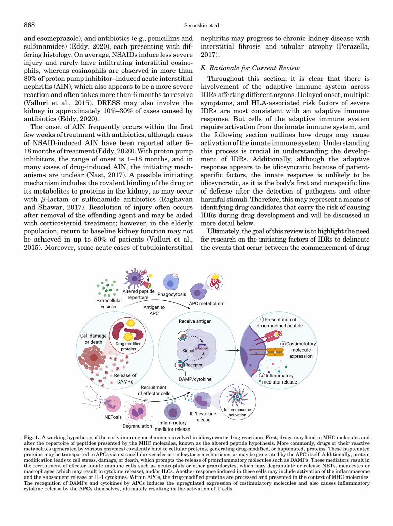
Fig. 1. A working hypothesis of the early immune mechanisms involved in idiosyncratic drug reactions. First, drugs may bind to MHC molecules andalter the repertoire of peptides presented by the MHC molecules, known as the altered peptide hypothesis. More commonly, drugs or their reactivemetabolites (generated by various enzymes) covalently bind to cellular proteins, generating drug-modified, or haptenated, proteins. These haptenatedproteins may be transported to APCs via extracellular vesicles or endocytosis mechanisms, or may be generated by the APC itself. Additionally, proteinmodification leads to cell stress, damage, or death, which prompts the release of proinflammatory molecules such as DAMPs. These mediators result inthe recruitment of effector innate immune cells such as neutrophils or other granulocytes, which may degranulate or release NETs, monocytes ormacrophages (which may result in cytokine release), and/or ILCs. Another response induced in these cells may include activation of the inflammasomeand the subsequent release of IL-1 cytokines. Within APCs, the drug-modified proteins are processed and presented in the context of MHC molecules.The recognition of DAMPs and cytokines by APCs induces the upregulated expression of costimulatory molecules and also causes inflammatorycytokine release by the APCs themselves, ultimately resulting in the activation of T cells.
868 Sernoskie et al.
administration and the development of the IDR. Toguide these investigations, we look to the fundamentalsof immunology to describe how an immune responsemay develop in response to the administration of small-molecule drugs.
III. Innate Mechanisms Contributing to AdaptiveImmune Activation
Overwhelmingly, the adaptive arm of the immunesystem has been the focus of IDR research, as this is themechanism that is likely responsible for clinicallysignificant IDRs. The adaptive immune response islikely also what makes IDRs idiosyncratic: individualspossess unique and dynamic TCR repertoires, formedthrough random somatic recombination events (Krangel,2009), and without the major histocompatibility complex(MHC) presentation of drug-modified peptides to cognateTCRs, adaptive immune activation and subsequent IDRmanifestation cannot occur (Usui and Naisbitt, 2017;Hwang et al., 2020).However, a fundamental dogma of immunology is
that an innate immune response is required to initiatean adaptive immune response, and although progres-sion to a severe IDR may be uncommon, it is likely thata greater proportion of patients experience an innateimmune response that resolves without interventionand without leading to a significant adaptive immuneresponse. Therefore, a more comprehensive under-standing of the subclinical early immune mechanismspreceding IDR onset is necessary to guide futurestrategies in disease management and prevention.Thus, from neoantigen formation to consequences ofimmunologic synapse formation, this section will pro-vide a succinct overview of important principles ininnate immunology as well as mechanisms of adaptiveimmune activation that are potentially involved pre-ceding the development of an IDR. These concepts aresummarized in Fig. 1. Admittedly, the innate immuneresponse is much more complex and nuanced than canbe adequately addressed here; however, these topicsprovide a basic framework to be considered whendesigning future mechanistic studies for drugs associ-ated with the risk of IDRs. For those already familiarwith the innate immune system, this section may beskimmed, skipped, and/or referred to when necessary inaccompaniment with Section IV. Support for ImmuneActivation Using Model Drugs.
A. Cells of the Innate Immune System
Initiation of any inflammatory response is dependentupon the recruitment and activation of innate effectorcells. When this immune response is triggered by thedetection of endogenous danger signals without thedetection of pathogens or pathogen-associated molecu-lar patterns, it is described as sterile inflammation(Chen and Nuñez, 2010). Although the types of innate
immune cells that may participate in this type ofinflammation are generally similar, the function of thesterile response is not to clear an infection but, ulti-mately, to repair the damage caused by chemical orphysical insult; thus, the role of effector cells may varyconsiderably. Responding leukocytes include granulo-cytes (neutrophils, eosinophils, basophils, and mastcells), professional antigen-presenting cells (APCs:monocytes, macrophages, and dendritic cells), and in-nate lymphoid cells (ILC groups 1–3). Other immunecells, including platelets, megakaryocytes, and eryth-rocytes, and nonimmune cells, such as mesenchymalstem cells, fibroblasts, and hepatocytes, may alsofunction during the immune response and are intro-duced briefly. Ultimately, since the function of theinnate immune system is to provide a first line ofdefense against foreign or potentially harmful stimuli,including potential damage caused by binding of drug-reactive metabolites, activation of innate immune cellsrepresents a more universal, non–patient-specific mecha-nism to be studied in the context of IDRs.
1. Granulocytes.a. Neutrophils. Neutrophils are essential for innate
immunity, not only as phagocytes that engulf anddestroy invading pathogens but also as rapid respond-ers during sterile inflammation (McDonald et al., 2010;Lämmermann et al., 2013), and can even possess a re-parative function (Wang et al., 2017). Moreover, in vitro,neutrophils have been demonstrated to function asAPCs under inflammatory conditions, further high-lighting the diverse roles of these cells (Mehrfeldet al., 2018).
Mature neutrophils, derived from common myeloidprogenitors in the bone marrow, are the most abundantleukocyte present in the human circulation, althougha large store of mature cells also exist in the bonemarrow or transiently arrested within blood capillaries(Lawrence et al., 2018). After the detection of any ofan extensive array of inflammatory stimuli [such aschemokines or damage-associated molecular patterns(DAMPs), discussed below], marginated neutrophils arereleased rapidly into the circulation and, through theprocess of chemotaxis, can migrate to the site of in-flammation. Although once considered to be a single,short-lived population, significant neutrophil heteroge-neity has been reported in the steady-state (Fine et al.,2019) as well as in the context of numerous inflamma-tory (Silvestre-Roig et al., 2016; Yang et al., 2017) andcancer models (Hellebrekers et al., 2018; Giese et al.,2019), with extended neutrophil life spans observed inthe presence of inflammation (Filep and Ariel, 2020).Reparative and immunosuppressive phenotypes havealso been described (Rosales, 2020).
Neutrophils contain several types of granules andsecretory vesicles, the contents of which can be releasedin a stimulus-dependent manner, either intracellularlyvia fusion with a phagocytic vacuole or extracellularly
The Innate Immune Response in Idiosyncratic Drug Reactions 869

via degranulation or exocytosis (Giese et al., 2019). Inaddition to the many enzymes, receptors, and cytokinesreleased in granules and secretory vesicles, neutrophilsare also able to generate reactive oxygen species(Sheshachalam et al., 2014; Winterbourn et al., 2016).Together, these components can mediate pathogen de-struction, induce recruitment of additional inflamma-tory cells, or contribute to tissue injury or repair(Silvestre-Roig et al., 2019).Moreover, after stimulation, neutrophils can release
web-like structures called neutrophil extracellulartraps (NETs), composed of histone-linked DNA frag-ments, cathepsin G, elastase, and myeloperoxidase(Brinkmann et al., 2004). Interestingly, NET releasecan occur in a lytic or nonlytic manner, meaningneutrophil lysis and subsequent cell death may or maynot occur during the process (Castanheira and Kubes,2019). Of note, the enzyme myeloperoxidase, which ispresent in both neutrophil granules and NETs, has alsobeen shown to bioactivate a variety of drugs to reactivemetabolites in vitro (Hofstra and Uetrecht, 1993) and tocontribute to the covalent binding of drugs observedin vivo (Lobach and Uetrecht, 2014b). Whereas IDIAGis the result of a delayed, adaptive immune response,paradoxical neutrophilia has been reported in the firstfew weeks of treatment with drugs associated with thisIDR, namely clozapine (Section IV. D. Clozapine). Over-all, neutrophils are integral for coordination and reso-lution of an inflammatory response and for tissuerepair, and they can also play a role in the generationof neoantigens through myeloperoxidase-mediated re-active metabolite formation.b. Eosinophils. Eosinophils are among the rarest
leukocytes in circulation in a healthy state, but theirnumbers can increase up to 20-fold during certainpathologic conditions (Klion et al., 2020). They arefundamental effector cells in innate immunity againsta wide variety of pathogens but also contribute to acuteand chronic inflammatory conditions including asthma,eczema, and different types of autoimmunity and canmediate both tissue damage and repair (Ferrari et al.,2020; Nagata et al., 2020). In addition to granularproteins, eosinophils synthesize more than 40 proin-flammatory mediators, such as TNF-a, IL-1 familycytokines, IL-4, IL-6, IL-8, granulocyte-macrophage-colony stimulating factor, leukotrienes, and reactiveoxygen species (Spencer et al., 2014; Melo and Weller,2018). These mediators can be released via classicexocytosis; through eosinophil cytolysis, whereby intactgranules are liberated directly into target tissues; orthrough piecemeal degranulation, whereby cytokinesare selectively mobilized to vesicles from the maingranules and are then released (Spencer et al., 2014).Much like neutrophils, eosinophils can release extra-cellular traps of DNA and DAMPs, although thesenetworks are more resistant to degradation comparedwith NETs (Ueki et al., 2016). Overall, eosinophils are
a hallmark of allergic inflammation, and as discussed inSection IV. Support for Immune Activation Using ModelDrugs, eosinophilia is frequently reported during theinitial weeks of clozapine therapy in patients, indicativeof an innate immune response. More broadly speaking,eosinophilia is also seen in other IDRs, such as DRESS,AGEP, or liver injury.
c. Basophils. Although they are the rarest andweakest phagocytic granulocyte in circulation, baso-phils play a key role in tissue inflammation; namely,skin, lung, and gastrointestinal tract inflammatoryresponses that are commonly triggered by either aninvading parasite or allergen (Schwartz et al., 2016).Basophils are activated by allergen-induced crosslink-ing of their IgE receptors (Knol, 2006). Indeed, thebasophil activation test is used as a reliable diagnostictool for identifying various allergens. In the context ofdrug allergy, however, the basophil activation test isnot as sensitive as it is in identifying other typesof allergens (Eberlein, 2020). Possibly, this could bebecause the covalent modification of proteins by drugsproduces a range of antigens such that it is notaccurately reproduced in vitro.
Basophils are a source of IL-13 and are known toconstitutively express IL-4, which are cytokines neces-sary for B-cell stimulation and differentiation to plasmacells and also differentiation of naïve helper T cells toTh2 cells (Liang et al., 2011), thus representing animportant bridge between the innate and adaptiveimmune responses. Basophil-derived IL-4 has also beenshown to have an important function in alternativelyactivated (M2) macrophages, which are involved notonly in type 2 immunity but also in tissue repair andphysiologic homeostasis (Yamanishi and Karasuyama,2016). Basophils can quickly migrate to inflamed tis-sues and are among the first responding cells duringskin injury (Chhiba et al., 2017). Activated basophilsrelease a variety of mediators stored in cytoplasmicgranules, including the bioactive lipids leukotrienesand prostaglandins, histamine, chemokines, and othercytokines (Chirumbolo et al., 2018), and also presentwith transcriptional heterogeneity, depending upon thestimuli (Chhiba et al., 2017). Additional innate effectorcells such as eosinophils and ILC2 have also beendemonstrated to be recruited by basophils to inflamma-tory sites (Schwartz et al., 2016). Although basophilswere once considered a redundant counterpart of tissue-resident mast cells, they have more recently beenacknowledged to play many unique roles during theinflammatory response that extend beyond allergy andhypersensitivity reactions.
d. Mast cells. Mast cells share functional and mor-phologic characteristics with basophils but are consid-ered sentinels of the innate immune system, andalthough they are found in most tissues of the body,terminally differentiated mast cells are typically notdetected in circulation. Althoughmast cells have diffuse
870 Sernoskie et al.
cytoplasmic granules comparable to basophils and otherclassic granulocytes, there has been considerable de-bate as to whether the progenitors of the mast celllineage are more closely related to megakaryocyte/erythroid or granulocyte/macrophage progenitors. How-ever, it appears that mast cells are derived indepen-dently from either group and only share the earlycommon myeloid progenitor (Franco et al., 2010). Bothpositive and negative immunoregulatory roles havebeen ascribed to mast cells. They function as a first lineof defense against pathogens, and they are particularlyuseful in degrading venoms and toxins (Dudeck et al.,2019). Additionally, they contribute to allergic inflam-matory responses by recruiting additional innate cellsto the site of inflammation and by activating adaptiveimmune cells, thus promoting chronic responses (Kubo,2018; Olivera et al., 2018). Moreover, excessive andsustained activation ofmast cells can cause anaphylaxisand tissue damage, respectively. These effector func-tions can be attributed to the release of mast cellsecretory granules, which contain proteases, lysosomalenzymes, biogenic amines, and cytokines (TNF, IL-4,IL-5, IL-6, etc.), among numerous other constituents(Wernersson and Pejler, 2014). Mast cells are mostabundant in areas exposed to high levels of antigen,including the skin, other connective tissues, andthe gastrointestinal and respiratory tracts (Krystel-Whittemore et al., 2016), and their roles in innateimmunity can vary depending on the local milieu ofmediators.2. Professional Antigen-Presenting Cells. Professional
APCs are cells that possess constitutive or inducibleexpression of high levels of MHC II molecules, processantigen, and express costimulatory molecules to facili-tate the development of adaptive immunity to specificantigens. Classically, dendritic cells, macrophages, andB cells are considered professional APCs. B cells will notbe discussed here, aside from their antigen presentationfunction, which is briefly described later. Although ithas been recognized that other cell types, referred to asatypical or nonprofessional APCs, can also expressMHC II, there is little evidence that they can activatenaïve T cells (Kambayashi and Laufer, 2014). APCsfacilitate the surveying of antigen by CD4+ T cells toefficiently expand the small subset of T cells expressingthe cognate T-cell receptors to respond to antigenicchallenge.a. Dendritic cells. Dendritic cells (DCs) received
their name from their many branched cellular processes(Steinman and Cohn, 1973). DCs can be categorized asconventional or plasmacytoid, both of which arise froma committed dendritic cell precursor in the bone mar-row. These then diverge, as conventional dendritic cellprecursors leave the bone marrow and seed otherorgans, whereas plasmacytoid dendritic cell precursorsremain. Conventional DCs (cDCs) are the predominantcell type responsible for T-cell activation, and the far
less abundant plasmacytoid DCs are specialized insensing viral RNA and DNA and can produce largeamounts of interferons to drive the antiviral response(Sichien et al., 2017; Musumeci et al., 2019). Histori-cally, cDCs have been further categorized as migratoryor lymphoid-resident; however, more recent studieshave resulted in the classification of cDCs as cDC1and cDC2 based upon surface marker expression andtranscriptomic analyses (Ziegler-Heitbrock et al., 2010;Guilliams et al., 2014, 2016). Langerhans cells wereoriginally presumed to be DCs based upon their func-tion, but based upon their ontogeny, they are residentmacrophages (Doebel et al., 2017), highlighting thecomplexity in classifying these types of cells. Langer-hans cells likely play an important role in mediatingskin IDRs.
DCs are usually found in a resting or immature stateand survey their environment by sampling antigen.Because they have both low surface expression andrapid turnover of MHC II molecules, they are unable toactivate naïve T cells (Drutman et al., 2012). In aninflammatory milieu, DAMPs and pathogen-associatedmolecular patterns are present and engage variouspattern recognition receptors on the DC surface, caus-ing the DC to mature. The cells are then able to expresscytokine and chemokine receptors, facilitating migra-tion to lymph nodes. MHC II turnover decreases whileexpression increases, allowing for presentation of thepeptides found in the inflammatory context (Cella et al.,1997). Additionally, costimulatory molecule expressionand cytokine secretion are induced, and the combina-tion of these changes is sufficient to induce naïve T-cellactivation (Curtsinger and Mescher, 2010). Dependingupon the stimuli received by the DC, it will secretedifferent cytokines and influence the differentiation ofcognate T cells into different subsets of effector T cells(Blanco et al., 2008).
DCs may also be tolerogenic in certain cases. ThymicDC populations appear to be important in maintainingcentral tolerance during T-cell development (Lopeset al., 2015). Peripherally, a small proportion of DCsundergo maturation under homeostatic conditions andupregulate MHC II expression, but this maturationresults in tolerance rather than naïve T-cell activation(Lutz and Schuler, 2002). Indeed, antigen presentationwithout DC priming resulted in antigen-specific toler-ance (Probst et al., 2003). The vast heterogeneity ofDCs, as well as the varied outcomes ofmaturation basedon environmental influences, results in numerous func-tions for DCs.
b. Monocytes. Monocytes are cells of the myeloidlineage that are derived from the bone marrow and arereleased into circulation. Monocytes are phagocytic andscavenge apoptotic cells and toxic macromolecules incirculation. They also function as important orchestra-tors of inflammatory responses by producing cytokinesafter the detection of tissue damage or pathogens.
The Innate Immune Response in Idiosyncratic Drug Reactions 871
Monocyte subsets exist across a spectrum of differenti-ation, initially taking on an “inflammatory” phenotypeupon egress from the bone marrow and then taking ona “patrolling” phenotype over time because of transcrip-tional changes (Mildner et al., 2017). Under steady-state conditions, monocytes can enter tissue, returnto circulation with minimal differentiation, and trafficto lymph nodes to present antigen to T cells (Jakubzicket al., 2013). Although monocytes may themselvesfunction as APCs, upon entering inflamed tissue, theycan also differentiate into macrophages or dendritic cellsto propagate the inflammatory response (Jakubzicket al., 2017).c. Macrophages. Macrophages are phagocytes that
are usually found in tissues, and many have beennamed depending upon the organ in which they reside(e.g., Kupffer cells in the liver, microglia in the centralnervous system, or osteoclasts in bones). The environ-ment in which macrophages are found influences theirphenotype and function; various tissue-resident macro-phage populations express different surface proteins,and even within an organ may have different pheno-types (Hume et al., 2019). Macrophages survey thetissue in which they reside and phagocytose foreignmolecules and debris.Like DCs, macrophages express pattern recognition
receptors, which can stimulate their activation. Macro-phage activation states are highly complex andare often described as polarization to an “M1-like” or“M2-like” phenotype, correlating with Th1 and Th2responses, respectively. As macrophage activation isa dynamic process, different macrophages in the sametissue likely express different mixtures of activationmarkers and perform different functions; this alsoevolves over time (Murray, 2017). For example, inresponse to IFN-g, activated macrophages secreteproinflammatory cytokines (e.g., IL-1b, IL-6, IL-12). Inresponse to IL-4, however, they can secrete insulin-likegrowth factor 1 and resistin-like molecule-a, which canstimulate fibroblast survival and promote extracellularmatrix deposition, respectively (Mosser and Edwards,2008).Macrophages can participate in antigen presentation,
but unlike DCs, they cannot activate naïve T cells. Theirability to present antigen can be influenced by theirenvironment; for example, antigen presentation toT cells and CD40 expression were increased with theuptake of necrotic but not apoptotic cells (Barker et al.,2002). Crosspresentation of antigen by macrophagesfrom dead tumor cells has been shown to be importantin antitumor immunity (Asano et al., 2011). Macro-phages have also been shown to present lipid antigensto invariant natural killer T cells (Barral et al., 2010).Macrophages also have reparative functions and can
secrete growth factors and anti-inflammatory media-tors including IL-10 and TGF-b during tissue repair(Vannella and Wynn, 2017). Overall, macrophages play
varied and dynamic roles in the steady-state andthroughout the inflammatory response.
3. Innate Lymphoid Cells. Over the past decade,ILCs have come to be recognized as fundamentaleffectors of the innate immune response (Moro et al.,2010; Neill et al., 2010; Price et al., 2010), both in healthand in disease states ranging from type 2 inflammatoryconditions (e.g., atopic dermatitis and asthma) toautoimmune diseases (e.g., psoriasis and inflammatorybowel disease) (Ebbo et al., 2017; Kobayashi et al.,2020). Aside from natural killer (NK) cells, which arelocalized in secondary lymphoid organs, ILCs aregenerally under-represented in lymphoid tissues butare predominantly found in the liver, skin, intestine,lungs, adipose tissue, and mesenteric lymph nodes andare most prominent at mucosal barriers (Klose andArtis, 2016). At themost rudimentary level, ILCs can beclassified as group 1, group 2, and group 3, with eachgroup sharing similarities in cytokine production andtranscriptional regulation with a particular T-cell sub-set (although ILC antigenic receptors do not undergothe genetic rearrangement that adaptive lymphocytesundergo) (Spits et al., 2013). Group 1 ILCs include bothILC1s (Th1-like) and natural killer cells (cytotoxic T cell-like) and are characterized by their production of IFN-gand TNF (Spits et al., 2016), whereas group 2 ILCs area single population (Th2-like) that produce amphiregu-lin, IL-4, IL-5, and IL-13 (Klose and Artis, 2016).Group 3 ILCs comprise three populations (Th17-like)—lymphoid tissue inducer cells, natural cytotoxicityreceptor-negative cells, and natural cytotoxicityreceptor-positive cells—that all secrete IL-22, butonly the first two population also secrete IL-17A/IL-17F (Montaldo et al., 2015).
Additionally, some T-cell subsets are “preprog-rammed” and behave like innate cells in that they canrespond rapidly to a limited and conserved antigenicrepertoire. These include invariant natural killerT cells, which differ most prominently from conven-tional T cells in that they recognize lipid-based antigensin the context of CD1d; mucosal-associated invariantT cells, subsets of gd T cells; and certain memory T-cellsubsets (Vivier et al., 2018).
Like many cells, ILCs demonstrate significant plas-ticity, and their functionality is dependent on their localmicroenvironment. Even within specific ILC groups,significant heterogeneity has been reported. For exam-ple, among natural killer cell subsets, some possessmore cytolytic activity and contain high concentrationsof granzyme and perforin, whereas others are morereactive to activation by proinflammatory mediators,and surface receptor expression varies between hepatic,intraepithelial, and other natural killer cell populations(Spits et al., 2016). Natural killer cells, as discussed inthe next section, have also been shown to mediate theinflammatory response induced by amodiaquine, andthey are likely to play fundamental roles in the early
872 Sernoskie et al.
immune responses to other IDR-associated drugs, aswell. Moreover, based on the emerging roles of variousILC subsets in inflammatory conditions, as well as theirlocalization within organs most commonly associatedwith IDRs (e.g., the liver and skin), it is reasonable toquestion whether other members of the ILC family playa crucial part in the innate immune response to drugsassociated with IDRs.4. Other Innate Immune Cells. Other immune cells,
such as megakaryocytes, their platelet derivatives, anderythrocytes, may also be important contributors dur-ing an innate immune response. Although the immu-nologic role of megakaryocytes is not as well defined,their expression is dependent on the demand forplatelets, which can be upregulated during inflamma-tory conditions or infection, vascular damage, andtissue repair (Noetzli et al., 2019). Beyond a role inhemostasis, platelets have significant immunomodula-tory potential: they can release both proinflammatory[e.g., CXCL4, chemokine (C-C motif) ligand (CCL) 5,histamine, epinephrine, and high mobility group box 1(HMGB1)] and proresolving mediators and can formcomplexes with a variety of immune cells, includingneutrophils and monocytes (Margraf and Zarbock,2019). Platelets can release these mediators throughmicrovesicles and exosomes (Heijnen et al., 1999).Likewise, erythrocytes contribute to innate immunityand are more than just oxygen carriers. Interestingly,these cells can bind a variety of chemokines, in turn,either preventing recruitment of effector cells such asneutrophils or possibly extending the half-life of thesemediators to prolong the inflammatory response, re-ferred to as the sink hypothesis and reservoir hypoth-esis, respectively (Anderson et al., 2018).5. Nonimmune Cells. Although the focus of this
section is to introduce the reader to cells of the innateimmune system, it is also worth emphasizing thatcountless nonimmune cells can have inflammatory orimmunoregulatory functions. Naturally, any damagedor dying cells can release DAMPs and proinflammatorymediators that can activate immune cells both locallyand in circulation, resulting in recruitment to thelocation of injury and initiation of an innate immuneresponse; this is not dependent on immune cell status.However, nonimmune cells can also secrete bioactiveand chemotactic molecules in response to the detectionof a stimulus, as well. Such cells include, but are notlimited to, hepatocytes, mesenchymal stem cells, andfibroblasts.a. Hepatocytes. As the predominant parenchymal
cell in the liver, hepatocytes are well known for theirrole in metabolism, xenobiotic detoxification, and pro-tein synthesis, but they are also critical players ininnate immunity (Mehrfeld et al., 2018). The liver ishighly vascularized, receiving 25% of total cardiacoutput (Eipel et al., 2010), and is also responsible forthe production of up to 50% of the lymph collected by the
thoracic duct (Ohtani and Ohtani, 2008). While not indirect contact with the sinusoidal blood flow, hepato-cytes can extend filopodia through fenestrations in theadjacent endothelium to enable interactions with circu-lating leukocytes (Warren et al., 2006). Under steady-state conditions, hepatocytes only express MHC I(Mehrfeld et al., 2018), but they may express MHC IIunder inflammatory conditions (Herkel et al., 2003).Thus, hepatocytes may function as APCs with thepotential to interact with both helper and cytotoxicT cells; indeed, hepatocytes have been shown to activateCD8+ T cells, although they did not promote survival(Bertolino et al., 1998). Like most of the cells discussedthus far, hepatocytes not only share the ability to targetpathogens for destruction but can also secrete a varietyof proinflammatory mediators, such as soluble CD14,soluble myeloid differentiation 2, IL-6, CCL2, andCXCL1 (Zhou et al., 2016). Moreover, among theproteins synthesized and secreted into the blood byhepatocytes are acute phase proteins, such as C-reactiveprotein, serum amyloid A, and serum amyloid P. Theconcentrations of these mediators can dramaticallyincrease after the detection of inflammation, thusacting to amplify the immune response (Schrödl et al.,2016). As most reactive metabolite formation occursin the liver, and the liver is the target of a largeproportion of IDRs, hepatocytes are likely fundamen-tal in the initiation of the innate immune response todrugs that cause IDILI (Uetrecht, 2019b; Ali et al.,2020; Hastings et al., 2020; Mosedale and Watkins,2020; Yokoi and Oda, 2021).
b. Mesenchymal stem cells. Mesenchymal stemcells, also referred to as mesenchymal stromal cells,have been identified in various tissues and have thecapacity to differentiate into chondrocytes, osteoblasts,and adipocytes (Dominici et al., 2006). Moreover, thesemultipotent stem cells help maintain the tissue micro-environment, under both normal and inflamed condi-tions, often promoting an immunosuppressive milieuvia the release of growth factors and anti-inflammatorymolecules (e.g., transforming growth factor-b, IL-1receptor antagonist, IL-10, prostaglandin E2, etc.) afterthe recognition of proinflammatory stimuli (Wang et al.,2014). Exosomes have also been shown to contribute tothe immunomodulatory capabilities of these cells, andeven apoptotic mesenchymal stem cells maintainsuppressive properties (Shi et al., 2018). Mesenchy-mal stem cells can also migrate to the site of tissuedamage to participate in regeneration and can acti-vate or suppress the activation of various innate cells,including neutrophils, macrophages, DCs, and mastcells (Le Blanc and Mougiakakos, 2012; Shi et al.,2018).
c. Fibroblasts. Although a key function of fibroblastsis to maintain connective tissue structural integrity,these sentinel cells also have the capacity to respond topathogens and endogenous danger signals, to secrete
The Innate Immune Response in Idiosyncratic Drug Reactions 873
inflammatory signals, and to initiate tissue repair(Hamada et al., 2019). For instance, intestinal immu-nity is shaped by the secretion of cytokines, chemokines,growth factors, and metalloproteinases by epithelialcells andmyofibroblasts (Curciarello et al., 2019). Thesecells have also been shown to contribute to the persis-tence of inflammation, such as during rheumatoidarthritis, where synovial fibroblasts produce a varietyof proinflammatory and matrix-degrading molecules(Frank-Bertoncelj et al., 2017).
B. Antigen Formation and Cell Damage
Multiple theories have been proposed to explain howdrugs may cause IDRs; these are discussed in detailelsewhere (Cho and Uetrecht, 2017). We will presentthe hypotheses that are relevant to the discussion ofinnate immune system activation. It has long beenunderstood that foreign peptides are recognized by theimmune system. The hapten hypothesis and the alteredpeptide repertoire model describe distinct processes bywhich drug administration may ultimately result in theexposure of the immune system to novel peptides. Theseneoantigens may serve as targets for the immuneresponse, potentially resulting in the development ofan IDR.More recently, it was also recognized that there needs
to be a signal (e.g., a DAMP, discussed below) thatdamage is occurring. This is known as the dangerhypothesis. This hypothesis most likely complementsthe hapten and altered peptide hypotheses. Similarly,in conjunction with the hapten hypothesis, it is plausi-ble that sufficient covalent binding in the cellmay resultin cellular damage. The endoplasmic reticulum (ER)stress and unfolded protein response, as well as mito-chondrial toxicity, may result from the generation ofcovalently modified proteins. These processes may alsoresult in the release of DAMPs. Thus, these hypotheseslikely work together to describe the initiating eventsin IDRs.1. Hapten Hypothesis. Small-molecule drugs are too
small to be detected by the immune system, whichrecognizes larger molecules, such as proteins (Erkesand Selvan, 2014). However, many drugs that causeIDRs have reactive metabolites. The hapten hypothesisposits that drugs are bioactivated to a reactive metab-olite that then covalently binds to endogenous protein,thereby altering the protein and provoking an immuneresponse (Landsteiner and Jacobs, 1935; Faulkneret al., 2014; Cho and Uetrecht, 2017). Although it isvery difficult to prove that reactive metabolites causeIDRs, there are some cases in which they have beenshown to be responsible. For example, penicillin hyper-sensitivity involves IgE, and IgE from hypersensitivepatients has been shown to react to penicillin-modifiedprotein (forming the basis of diagnostic skin tests)(Levine et al., 1967). There have also been studies ofmultiple drugs characterizing drug-protein adducts in
patient samples, although these have not necessarilybeen causally linked to IDR onset. Additionally, nevir-apine is another case in which the reactive metabolitewas identified. Female brown Norway rats developa rash when chronically administered nevirapine. Thefindings that 12-hydroxynevirapine sulfate was cova-lently bound in the skin and that topical application ofa sulfotransferase prevented both the covalent bindingand the rash demonstrates that this reactivemetabolitewas indeed responsible for causing the skin rash(Sharma et al., 2013).
2. p-i Concept. The pharmacological interaction ofdrugs with immune receptors concept (p-i concept)attributes IDRs to the activation of immune receptors,MHC and the TCR specifically, by direct, noncovalentinteraction of the culprit drug (Pichler, 2002). This isbased on the observation that drugs can activatelymphocytes from patients who have experienced anIDR to that drug in the absence of metabolism. How-ever, in the case of nevirapine-induced skin rash, it hasbeen shown that the rash is caused by a reactivemetabolite, and yet cells from rats or humans who havea history of nevirapine-induced skin rash are activatedby the parent drug (Chen et al., 2009). Thus, althoughdirect activation of immune cells by parent drug mayoccur in IDR patients, this mechanism may not playa role in the initiation of the IDR.
3. Altered Peptide Repertoire Model. A mechanismrelated to the p-i concept is the altered peptide reper-toire model, which describes the noncovalent binding ofdrug to the HLA molecule itself, thereby altering itsconformation and the peptide repertoire that it is able topresent. This is illustrated by abacavir, which has beenshown to reversibly bind to the F pocket of the peptide-binding groove of HLA-B*57:01 and alter the repertoireof peptides that it can present (Illing et al., 2012;Norcross et al., 2012; Ostrov et al., 2012).
4. Danger Hypothesis. Although foreign peptide isa requirement for activation of the immune response, itis not usually sufficient; indeed, the body is exposed tonon–self-proteins constantly from food sources and gutmicroflora, for example. It would be detrimental if theimmune system were constantly activated as a result ofthese sources. The danger hypothesis recognizes that itis not necessarily the detection of an entity that appearsforeign but, in fact, an entity that causes damage thatactivates the immune system (Matzinger, 1994). Celldamage causes the release of DAMPs, which signal tothe immune system that there is likely a pathogen thatneeds to be eliminated. Very broadly speaking, celldamage may manifest as cell death, in which intracel-lular contents may be passively released and serve asDAMPs, or the cell may continue to survive, in whichcase DAMPs may be actively secreted.
Additionally, the type of cell death influences thetypes of DAMPs that are released. In apoptosis, cel-lular contents are not necessarily released into the
874 Sernoskie et al.

extracellular milieu as membrane integrity is main-tained; however, ATP is released in a controlledmanneras a “find-me” signal (Elliott et al., 2009). In contrast, incells dying by necrosis, cellular contents are released ascell death is uncontrolled. Necroptosis, a regulated formof necrosis mediated by death receptor activation, andpyroptosis, cell death regulated by inflammasome andcaspase-1 activation, may similarly both result in therelease of a number of DAMPs. Some DAMPs arereleased in the context of both types of programmedcell death (e.g., HMGB1, heat shock protein 90, ATP,IL-1a), whereas some have only been observed innecroptosis (e.g., S100A9, IL-33) or pyroptosis (e.g.,ASC specks) to date (Frank and Vince, 2019).5. Endoplasmic Reticulum Stress and the Unfolded
Protein Response. The ER is the location of proteinfolding and post-translational modification in the cell.Disruption of this process can cause ER stress. A seriesof pathways, termed the unfolded protein response(UPR), maintain quality control of protein synthesisthrough sensing deficiencies in protein folding capacity.Proteins in their proper conformation proceed to theGolgi apparatus as the next step in the secretorypathway, whereas misfolded proteins are retained inthe ER (Schröder and Kaufman, 2005). As cytochromeP450 enzymes tend to localize in the ER (Szczesna-Skorupa and Kemper, 1993), reactive metabolites canbe formed in the ER and adduct to proteins, inducingthe UPR.Unfolded proteins take on a conformation of a higher
free energy than that of their native conformations. Avariety of chaperones can sense this increased freesurface energy as hydrophobic residues are exposed towater. To maintain a balance with the folding capacityof the ER, the UPR employs two strategies: first, toincrease folding capacity by inducing ER-resident mol-ecule chaperones and foldases and by increasing thesize of the ER, and second, to decrease the misfoldedprotein load by downregulating protein synthesis andby increasing clearance of misfolded protein by upregu-lating ER-associated degradation (Schröder andKaufman, 2005).Ultimately, if the unfolded protein burden remains
too great, the UPR response can result in apoptosis. Ithas also been shown that chronic ER stress can lead toinflammation. For example, ER stress has been shownto induce nuclear factor of the k light chain enhancer ofB cells (NF-kB) activation (Deng et al., 2004), NLRfamily pyrin domain containing 3 (NLRP3) inflamma-some activation (Menu et al., 2012), and DAMP secre-tion either freely (Andersohn et al., 2019) or packaged inextracellular vesicles (Collett et al., 2018).Unfolded protein is not the only possible trigger of ER
stress, although it is the most well studied. Aberrationsin lipid homeostasis may also induce ER stress (Songand Malhi, 2019). Although, compared with proteins,changes to lipids have not been studied as extensively in
the context of IDRs, this may be an interesting avenueto explore; for example, lipid-smoothER inclusionswerefound in hepatocytes of brown Norway rats adminis-tered nevirapine (Sastry et al., 2018), which is alsoknown to induce smooth ERhypertrophy (Sharma et al.,2012).
The absolute number of proteins modified by covalentbinding of a drug is quite small (Evans et al., 2004), andcompared with other sources of unfolded protein, drugmodification of protein may not induce sufficient pro-tein unfolding to trigger activation of the UPR. Addi-tionally, a transcriptomic study of primary humanhepatocytes predicted a suppression, rather than in-duction, of pathways related to the UPR (Terelius et al.,2016).
6. Mitochondrial Toxicity. Mitochondrial toxicityhas been identified as an adverse effect of manymedications. Its role in IDRs in particular, however,has been a matter of debate (Boelsterli and Lim, 2007;Cho and Uetrecht, 2017). The mechanisms underlyingmitochondrial toxicity include inhibition of the electrontransport chain, interference with mitochondrial tran-scription and translation, inhibition or uncoupling ofATP synthase, inhibition of enzymes in the citric acidcycle or mitochondrial transporters, and increased re-active oxygen species (ROS) production (Vuda andKamath, 2016; Will et al., 2019). As these mechanismshave been extensively covered elsewhere, we will fo-cus on how mitochondrial toxicity may result ininflammation.
Increased ROS production causes activation of redox-sensitive transcription factors such as NF-kB. It hasalso been shown that autophagy negatively regulatesNLRP3 inflammasome activation, whereas increasedROS positively regulates NLRP3 inflammasome acti-vation and inflammation, at least in part due tocytosolic localization of oxidized mitochondrial DNA(Nakahira et al., 2011; Zhou et al., 2011; Shimada et al.,2012). In addition to mitochondrial DNA, othermitochondrial-derived molecules can function asDAMPs, such as ATP, mitochondrial transcriptionfactor A, N-formyl peptide, succinate, cardiolipin,and cytochrome-c (Nakahira et al., 2015; Grazioliand Pugin, 2018). Cytochrome-c release into thecytosol can induce apoptosis via inducing oligomer-ization of apoptosis-protease activating factor 1 andinitiating caspase activation. Depending upon thecontext and the cleavage products of the caspasesinvolved, this may result in apoptosis, but it may alsoresult in cell differentiation and proliferation(Garrido et al., 2006). Thus, the causes and outcomesof mitochondrial toxicity are varied and complex.
In general, there is little direct evidence that drugsthat cause IDRs do so by inducing mitochondrialdamage. An exception is valproic acid–induced liverinjury, however, which has been associated with var-iants in mitochondrial DNA polymerase g (Stewart
The Innate Immune Response in Idiosyncratic Drug Reactions 875
et al., 2010) and which may present with steatosis andlactic acidosis (Chaudhry et al., 2013; Farinelli et al.,2015; Pham et al., 2015). Acetaminophen also causesmitochondrial damage, but it does not cause IDRs(Jaeschke et al., 2019).
C. Mediators of Inflammation
Depending on the location and severity of the tissueinjury, a variety of factors may be involved in theinitiation and propagation of a sterile inflammatoryresponse, including DAMPs, cytokines, and chemoat-tractants. The transcriptional regulation of many ofthese proinflammatory molecules by NF-kB is also animportant consideration. Moreover, other body systemssuch as the microbiome and the nervous system havethe potential to contribution to inflammation.1. Damage-Associated Molecular Patterns. As dis-
cussed above, the injury or death of a cell may result inthe release of intracellular contents. Once outside oftheir normal subcellular location, these components arereferred to as danger signals or DAMPs, at which pointthey can initiate an inflammatory response (Medzhitov,2008). DAMPs can be classified based on their normallocation in or on the cell and include stimuli such asnuclear and mitochondrial DNA, RNA, ATP, S100, heatshock proteins, HMGB1, and extracellular matrix frag-ments (Chen andNuñez, 2010; Zindel andKubes, 2020).The detection of DAMPs then leads to effector cellrecruitment and propagation of the sterile inflamma-tory response.In the context of efferocytosis, DAMPs such as ATP,
UTP, lysophosphatidylcholine, and sphingosine-1-phos-phate, in addition to adhesion molecules and receptorssuch as intracellular adhesion molecule 3 and CX3Cchemokine receptor, can act as chemotactic find-mesignals, and concurrent with surface expression of eat-me signals such as phosphatidylserine, contribute to theremoval of apoptotic cells by phagocytes (Westmanet al., 2020).The concept of danger signals in the initiation of IDRs
has been proposed several times (Pirmohamed et al.,2002; Li and Uetrecht, 2010; Hassan and Fontana,2019; Uetrecht, 2019b). Although reactive metabolitesof drugs associated with IDRs can covalently bind tocellular proteins that may, in turn, cause cell damageor cell death, the release of DAMPs can also occur inresponse to a wide array of insults, such as UVirradiation, hemorrhagic shock, starvation, and otherforms of injury or trauma (Schaefer, 2014). SinceDAMPs are simply a mechanism by which the immunesystem is alerted that there is tissue injury, they are notidiosyncratic in their release. Therefore, it is possiblethat a similar pattern of DAMPs may be released aftertreatment with a drug associated with IDRs. Thispattern of DAMPs could function as potential bio-markers during preclinical development by indicatingthat a drug candidate may carry the risk of causing
IDRs; however, it is likely too nonspecific to be useful forclinical diagnosis of the early onset of an IDR. Thus,characterization of the specific DAMPs released aftertreatment with different drugs associated with IDRs iscertainly an avenue for future research, as it mayprovide insight into the specific type and target of cellinjury or death that is stimulating the observed innateimmune response.
2. Cytokines, Chemokines, and Acute Phase Proteins.A wide range of classic soluble mediators have alreadybeen highlighted for various functions in innate immu-nity. To reiterate, cytokines such as TNF-a, IL-1b, IL-4,IL-6, and IL-18; chemokines such as CCL2, CCL5,CXCL1, CXCL2, and CXCL8; and acute phase proteinssuch as C-reactive protein, serum amyloid A, and serumamyloid P contribute to effector cell recruitment andactivation and the propagation of the inflammatoryresponse. These mediators are released in coordinatedspatial and temporal responses, the patterns of whichcan influence the types and absolute numbers of innateleukocytes that are recruited to the site of injury. Notyetmentioned are the type I (i.e., a and b) and type II (g)interferons, which act to potentiate proinflammatorysignaling via enhanced cytokine production and antigenpresentation, macrophage priming, and natural killercell function, among numerous other effects (Kopitar-Jerala, 2017). Although additional proinflammatorymolecules are elaborated on elsewhere (Turner et al.,2014; Akdis et al., 2016; Kapurniotu et al., 2019), therole of IL-1 family cytokines is worth emphasizingbecause of their fundamental importance in innateimmunity.
a. Interleukin-1 cytokines and their activation.The IL-1 family of cytokines consists of 11 solublemediators, including proinflammatory IL-1a, IL-1b,IL-18, IL-33, IL-36a, IL-36b, and IL-36g, as well asseveral receptor antagonists and the anti-inflammatorycytokine IL-37 (Dinarello, 2018). IL-1 cytokines are firsttranslated into inactive precursors (except for IL-1a),which then attain functional maturity after enzymaticcleavage in a process mediated predominantly bycaspase-1 and the inflammasome (Mantovani et al.,2019). Fundamentally, the inflammasome is a multi-meric protein complex that, when activated, leads to thematuration of caspase-1, the cleavage and release ofmature IL-1 cytokines, and the induction of additionalinflammatory effector mechanisms (Walsh et al., 2014).Several cytoplasmic pattern recognition receptors canassemble as independent inflammasomes, each respond-ing to specific DAMPs or other stimuli. These patternrecognition receptors (PRRs) are expressed in multiplecells, including neutrophils, monocytes, macrophages,and dendritic cells, and play an important role in innateimmunity (Sharma and Kanneganti, 2016).
The NLRP3 inflammasome may be the most rele-vant for the study of drug-induced immune activation,as it is activated by the widest array of stimulants,
876 Sernoskie et al.
although several other inflammasomes may be in-volved (Schroder and Tschopp, 2010; Latz et al.,2013). Several drugs associated with serious IDRshave also been demonstrated to activate inflamma-somes and increase IL-1b release in vitro (Kato andUetrecht, 2017; Mak et al., 2018; Kato et al., 2019,2020). Additionally, it is known that animals deficientin components of the inflammasome are resistant tocontact hypersensitivity (Watanabe et al., 2007), a re-action that may parallel events in the early immuneresponse to drugs that cause IDRs. Whether inflam-masome activation occurs in humans treated withthese drugs has yet to be clearly demonstrated.3. Bioactive Lipids. In addition to inflammatory
protein mediators, several classes of bioactive lipidsexist and play various roles in inflammation, immuno-regulation, and maintenance of tissue homeostasis(Chiurchiù and Maccarrone, 2016). The main types ofproinflammatory lipids include classic eicosanoids(Dennis and Norris, 2015), certain endocannabinoids(Chiurchiù et al., 2015), lysoglycerophospholipids, andsphingolipids (El Alwani et al., 2006), and specializedproresolving lipid mediators are a relatively new classof lipids that terminate acute inflammation and driveresolution and tissue repair (Serhan, 2014). Thesemolecules are all generated from v-6 or v-3 essentialpolyunsaturated fatty acids precursors (e.g., arach-idonic acid) but demonstrate significant heterogene-ity in structure and function after maturation (Das,2018).Classic eicosanoids (e.g., certain prostaglandins,
prostacyclins, thromboxanes, leukotrienes, hydroxyei-cosatetraenoids, and lipoxins) are typically consideredhighly proinflammatory mediators that are usuallyproduced by innate cells such as neutrophils andmonocytes within the first several hours of an inflam-matory stimulus. Specifically, leukotrienes can act aschemoattractants for neutrophils, macrophages, andeosinophils (De Caterina and Zampolli, 2004), andprostaglandins can function to enhance proinflamma-tory cytokine gene transcription and release and canalso amplify the innate response to DAMPs (Hirata andNarumiya, 2012). However, some eicosanoids can beimmunosuppressive and promote immune tolerance incertain contexts (Obermajer et al., 2012;Wanget al., 2014).The endocannabinoids, such as 2-arachidonoylglycerol,are ubiquitously expressed molecules that have diverseimmunomodulatory effects on monocytes/macrophages,dendritic cells, and granulocytes, and unsurprisingly,perturbations in endocannabinoid homeostasis havebeen shown to contribute to neuroinflammatory andautoimmune diseases (Chiurchiù et al., 2018). Lysogly-cerophospholipids (e.g., lysophosphatidylcholine andlysophosphatidylinositol) and sphingolipids (e.g.,ceramide 1-phosphate and sphingosine 1-phosphate)are key signaling molecules controlling inflammatorycascades, trafficking and activation of immune cells, cell
survival, and apoptosis (Sevastou et al., 2013; Gomez-Muñoz et al., 2016).
NSAIDs are one class of drugs for which the potentialrole of bioactive lipids in the innate immune response isparticularly relevant. Although NSAIDs are the mostfrequently used medications for the management ofpain and inflammation, they are also associated withsome of the highest incidence rates of drug hypersensi-tivity reactions (Conaghan, 2012). Reported reactionsinclude urticaria and other cutaneous reactions, acuteinterstitial nephritis, and hepatotoxicity (Nast, 2017;Yamashita et al., 2017; Wöhrl, 2018). Mechanistically,NSAIDs inhibit the enzymes cyclooxygenase-1 and -2,blocking the synthesis of inflammatory prostanoidssuch as prostaglandin E2. It has even been postulatedthat an innate immune response contributes to theonset of NSAID-mediated adaptive IDRs, potentiallythrough the activation of eosinophil and mast celldegranulation or through the shunting of arachidonicacid precursors to the production of other proinflamma-tory lipid mediators such as leukotrienes (Doña et al.,2020). Based on the fundamental roles of bioactivelipids in the initiation and propagation of an inflamma-tory response, future research to delineate key media-tors in the early immune response to drugs that areassociated with IDRs is necessary.
4. Pattern Recognition Receptors. As part of theinnate immune system, pattern recognition receptorshave evolved to recognize conserved molecular patternsof danger or invading pathogens. Thus, PRRs representa key aspect of the innate immune system that is notlikely to be idiosyncratic, as they are conserved,germline-encoded receptors that are not antigen-specific and respond to a structurally diverse range ofmolecules (Gong et al., 2020), in contrast to an individ-ual’s randomly generated TCR repertoire. PRRs includeToll-like receptors (TLRs), nucleotide-binding oligomer-ization domain-like receptors (NLR), retinoic acid–inducible gene-1–like receptors, C-type lectin receptors,receptor for advanced glycation end products, andscavenger receptors (Gordon, 2002; Xie et al., 2008;Palm and Medzhitov, 2009; Takeuchi and Akira, 2010).Notably, DAMPs are largely recognized by PRRs. Forexample, HMGB1 can signal through TLR4 or receptorfor advanced glycation end products (Lu et al., 2013).TLR signaling can result in NF-kB signaling (discussedbelow) and ultimately the production of proinflamma-tory cytokines (Vidya et al., 2018). PRR signaling mayalso result in cell death (Amarante-Mendes et al., 2018).If signaling through PRRs was directly responsible forthe onset of IDRs, however, it is likely that these severereactions would be observed in most, if not all, patientsgiven a particular drug because of the conserved natureof these receptors, but this is not what is observedclinically. Thus, although likely a necessary first stepfor the development of an IDR, pattern recognitionis not itself sufficient to cause an IDR. Again, we
The Innate Immune Response in Idiosyncratic Drug Reactions 877
emphasize that this innate aspect of the immuneresponse should occur in most patients taking drugsthat are associated with IDRs if they cause cellulardamage and is not idiosyncratic, but other downstreampathways contribute to the development of IDRsthemselves.5. Transcriptional Regulation of Inflammation.
Several families of transcription factors are activated inresponse to inflammatory stimuli, such as signal trans-ducers and activators of transcription, interferon regu-latory factors, and most notably, NF-kB (Smale andNatoli, 2014; Irazoqui, 2020). The NF-kB family con-sists of several inducible transcription factors—NF-kB1(p50), NF-kB2 (p52), RelA (p65), RelB, and c-Rel—thatbind to kB enhancer DNA elements as dimers tomodulate gene transcription (Liu et al., 2017). Activa-tion can occur via the canonical pathway in response toligand binding of various cytokine receptors, PRRs, andTNF receptors, or via the noncanonical pathway inresponse to ligand binding of a specific subset of TNFreceptors (Cildir et al., 2016; Sun, 2017).NF-kB signaling results in the upregulation of target
genes related to cell adhesion, survival and prolifera-tion, dendritic cell maturation, neutrophil recruitment,M1 macrophage polarization, and other inflammatorymediators that function to amplify the detected in-flammatory response (Lambrou et al., 2020). Keyproinflammatory cytokines and chemokines under NF-kB regulation include IL-6, IL-8, TNF-a, CCL2, CCL5,CXCL1, and CXCL2 (Liu et al., 2017). Moreover,activation of NF-kB is necessary for signal 1 (prim-ing) in inflammasome activation, as transcription ofinflammasome-related components such as pro-IL-1b,pro-IL-18, and NLRP3 is upregulated after NF-kBactivation (Latz et al., 2013). If drugs associated withIDRs cause an inflammatory response that is charac-terized by elevated levels of NF-kB–regulated media-tors, then this provides a starting point to determinewhich canonical or noncanonical receptors are activatedafter drug administration and may provide clues asto the types of cell damage or neoantigens formed(i.e., potential receptor ligands) with that drug.6. Other Contributing Factors. In addition to the
multifarious range of activation signals presented thusfar, multiple junctures of interaction have been identi-fied between the innate immune system and both themicrobiome and the nervous system; however, thesewill only be introduced briefly.a. Interaction with the microbiome. Although most
commonly associated with the gut, the human micro-biome refers to the collection of genes of all micro-organisms (e.g., archaea, bacteria, fungi, protists,viruses) that reside on or in all bodily tissues and fluids,including the biliary tract, respiratory tract, and skin(Marchesi and Ravel, 2015). To maintain a commensalrelationship and prevent the initiation of an inappro-priate immune response, extensive crosstalk between
the microbiota and immune cells, particularly ILCs(Thaiss et al., 2016; Negi et al., 2019), must occur. Forinstance, it has been shown that germ-free mice havea significantly altered innate immune system, withattenuated myeloid cell development in the bone mar-row (Khosravi et al., 2014). Although this is an extremeexample that would not be particularly relevant tohumans, it does highlight the potentially profoundimpact of the microbiome on innate immunity.
Commensals are necessary to educate the immunesystem and often promote tolerance (Grice and Segre,2011), but how these microorganism interactions mayshape the metabolism of and subsequent inflammatoryresponse to drugs that cause IDRs has yet to beadequately investigated (Marchesi and Ravel, 2015).One notable exception, however, is immune checkpointinhibitor–induced colitis. A recent investigation dem-onstrated that ipilimumab altered microbiome compo-sition and the subsequent risk of colitis (Dubin et al.,2016). Countless drugs can target components of themicrobiome, the most obvious being the antibiotics;therefore, understanding the reverse of this relation-ship will likely provide novel insights into patient-specific risk factors for IDRs.
b. Communication with the nervous system. Animportant function of the nervous system is to interactwith immune cells. Unsurprisingly, innate immunecells, including neutrophils, macrophages, and den-dritic cells, express receptors for several neurotrans-mitters (e.g., a- and b-adrenergic and acetylcholinergicreceptors), and neurons can express various patternrecognition and cytokine receptors, facilitating effectivecrosstalk between the systems (Chavan et al., 2017).Additionally, at peripheral sites of inflammation andtissue injury, both afferent and efferent neural circuitshave been shown to have immunoregulatory functions(Pavlov and Tracey, 2015). As many drugs associatedwith IDRs have therapeutic effects in the CNS, in-cluding a multitude of anticonvulsant and antischizo-phrenic agents, it is necessary to consider how thesepsychotropics may influence the neuroimmune axis andthe consequential immune activation.
D. Antigen Reception/Uptake by Antigen-Presenting Cells
There are multiple means by which APCs may obtainpeptides or proteins and present them (Avalos andPloegh, 2014; Roche and Furuta, 2015; Allen et al.,2016; Lindenbergh and Stoorvogel, 2018; Li and Hu,2019). Antigen presented by APCs ismost often thoughtto originate from within the cell itself or to be receivedvia uptake from the extracellular environment by pro-cesses such as phagocytosis (Roche and Furuta, 2015;Kotsias et al., 2019). Based on the numerous HLAassociations that have been identified as risk factorsfor different drugs and reactions (Usui and Naisbitt,
878 Sernoskie et al.
2017; Chen et al., 2018), this step is likely to beimportant in the pathogenesis of IDRs.1. Presentation by Major Histocompatibility Complex
I: Endogenous Protein, Crosspresentation. MHC Imolecules are expressed by all nucleated cells, whichallows for CD8+ T cells to survey host tissue foraberrations suggestive of intracellular pathogens ormalignancy (Jongsma et al., 2019). Peptides originatingfromwithin the cell are usually presented in the contextofMHC I (Neefjes et al., 2011). In what is considered theconventional processing route, proteins are digested bythe proteasome to generate shorter peptide fragments,which are then translocated to the ER via the trans-porter associated with antigen processing for loadingonto MHC I molecules after assembly of the peptide-loading complex (Vyas et al., 2008).In some cases, proteins exogenous to the cell may be
presented by MHC I, particularly in the case of DCs;this process is termed crosspresentation (Li and Hu,2019). Peptide loading is described as following eitherthe cytosolic pathway or the vacuolar pathway. Thecytosolic pathway appears to require the proteasome forpeptide processing, and peptide loading may occur inphagosomes or endosomes, whereas the vacuolar path-way is lysosome-dependent and both peptide processingand loading may occur in lysosomes (Embgenbroich andBurgdorf, 2018).2. Presentation by Major Histocompatibility Complex II:
Phagocytosis, Endocytosis, Macropinocytosis, Autophagy.Constitutive expression of MHC II is largely restrictedto professional APCs, although myeloid cells, includingeosinophils, neutrophils, and basophils, as well as ILCs,have been demonstrated to upregulate expression ofMHC II in certain conditions (Kambayashi and Laufer,2014). Peptides originating from exogenous sources aremost often presented in the context of MHC II; however,endogenous peptides may also follow this pathway viaautophagy (Duffy et al., 2017). Exogenous proteins maybe acquired via different methods (Roche and Furuta,2015). Phagocytosis is the internalization of pathogensor particulate antigens and is considered to be the mostimportant mechanism of antigen uptake in vivo (Stuartand Ezekowitz, 2005). This process can be enhanced byopsonins, which are host proteins such as antibodies orcomplement that can coat foreign entities. Clathrin-mediated endocytosis is the internalizing of ligandscomplexed to surface receptors and soluble macromole-cules (Mantegazza et al., 2013). Macropinocytosis isa nonspecific process during which uptake of extracel-lular fluid containing soluble antigens and macromole-cules occurs (Liu and Roche, 2015). Proteins are thenprocessed via the endocytic pathway to peptides inspecialized late endosomes that are enriched withMHC II molecules for antigen presentation (Neefjeset al., 2011).3. Crossdressing: Trogocytosis, Extracellular Vesicles,
Nanotubes. In some cases, preformed MHC-peptide
complexes may be transferred from the surface ofa donor cell to a recipient cell; the process is referredto as crossdressing (Campana et al., 2015). Multiplemechanisms have been proposed to describe the trans-fer of these complexes. Trogocytosis refers to thephenomenon in which patches of the plasma membraneare rapidly transferred from one live cell to anotherupon cell-cell contact. In some cases, phagocytosis maynot be possible if the target cell is too large; thephagocyte may instead ingest smaller pieces of the cellby “nibbling” at the membrane and potentially thecytoplasm (Dance, 2019).
Extracellular vesicles refer to either microvesicles,formed by plasma membrane budding, or exosomes,formed as intraluminal vesicles within endosomalmultivesicular bodies and then released by fusion ofthe multivesicular body with the plasma membrane.Because these two types of vesicles are indistinguish-able after their release, they are collectively termedextracellular vesicles (Groot Kormelink et al., 2018). Inthe context of IDRs, extracellular vesicles have beenshown to be involved in the transport of drug-modifiedantigen to target cells, such as in the case of amoxicillin(Sánchez-Gómez et al., 2017; Ogese et al., 2019).Extracellular vesicles may also transfer proteins thatcan be processed by the recipient cell and presented byMHCmolecules, or they may transfer the MHC-peptidecomplexes themselves. Additionally, extracellular vesiclesderived from multiple cell types including B cells(Raposo et al., 1996) and DCs (Théry et al., 2002) havebeen shown to activate T cells themselves. Con-versely, hepatocyte-derived exosomes have also beenimplicated in the promotion of immune tolerance inthe liver, and dysregulation of this tolerogenic mech-anism may be an important step in the onset of IDILI(Holman et al., 2019).
Tunneling nanotubules are intercellular structuresthat have been shown tomediate the exchange ofMHC Imolecules (Schiller et al., 2013). Such an exchange maybe another means by which crossdressing can occur.
These varied methods of antigen acquisition, process-ing, and presentation describe differentmeans by whichantigen may be presented to the adaptive immunesystem. Understanding how antigen may reach bothAPCs and target T cells will aid in the understanding ofthe pathogenesis of IDRs.
E. Naïve Lymphocyte Activation byAntigen-Presenting Cells
Naïve T cells and B cells are activated by APCs oncethey receive sufficient activation signals (Mak et al.,2014). Classically, the three-signal model is used todescribe this sequence of events: signal one refers to thebinding involving the MHC molecule presenting theantigenic peptide of interest; signal two refers tocostimulatory molecule engagement, which has beenupregulated as a result of exposure to inflammatory
The Innate Immune Response in Idiosyncratic Drug Reactions 879
conditions; and signal three refers to the cytokine helpthat permits the lymphocyte to survive and proliferate.This model describes the activation process for helperT-cell, cytotoxic T-cell, and B-cell activation, althoughthere are some differences between the cell types.1. Helper T Cells. Only mature DCs can activate
naïve T cells. For Th cell activation, the first signalbetween these cells is the binding of cognate TCRs toMHC II-peptide complexes on the DC; a strong in-teraction over several hours results in the signalingcascades that induce cell polarization and forms theimmunologic synapse. T-cell receptor engagement indu-ces NF-kB signaling (Liu et al., 2017). This also inducesCD40L and CD28 expression on the T-cell surface;CD40 engagement on the DC surface by CD40L upre-gulates expression of B7 molecules by the DC.In most cases, costimulatory molecule engagement is
required for T-cell activation, although in some cases,the MHC-peptide complex may deliver a strong enoughsignal to bypass the need for signal two (Wang et al.,2000). The B7molecules on the DC surface interact withCD28 on the T-cell surface. This permits upregulation ofcytokine receptors and induces CD4+ T-cell productionof proinflammatory cytokines such as IL-2 and IFN-g.Signal three is delivered by APCs in the form of
cytokine release. For CD4+ T cells, these cytokinesinclude IL-1, TNF-a, and IL-6 (Pape et al., 1997;Joseph et al., 1998; Curtsinger et al., 1999; Ben-Sasson et al., 2009). This results in activation, pro-liferation, and differentiation to Th effector cells as wellas licensed DCs. Licensed DCs may then proceed toactivate naïve Tc cells.2. Cytotoxic T Cells. Signal one is delivered to the Tc
cell by engagement of MHC I on a licensed DC, theproduct of Th cell activation, to the T-cell receptor(Joffre et al., 2009). Signal two, or costimulation of Tc
cells, is more dependent upon CD28 engagement, asB7 is already upregulated on the DC surface(Curtsinger et al., 2003). Finally, in signal three, thenaïve Tc receives cytokine help, such as IL-12, fromactivated Th cells and APCs, thus allowing for pro-liferation and differentiation to precytotoxic lym-phocytes (Curtsinger et al., 2003; Curtsinger andMescher, 2010). These precytotoxic lymphocytes maythen leave the lymph node and migrate to the site ofinflammation, where signals such as IL-12, IFN-g,and IL-6 induce differentiation to armed cytotoxiclymphocytes (Mescher et al., 2007). Protein synthe-sis for the contents of the cytotoxic granules isinduced. Finally, engagement of the T-cell receptorby antigen presented on MHC I within the tissueinduces targeted cell destruction by the cytotoxic Tc
cell (Groscurth and Filgueira, 1998).3. B Cells. Some antigens are considered to be
T-independent in that the antigens themselves canstimulate the B cell to proliferate without T-cell help(Mond et al., 1995). Most antigens, however, are
T-dependent and require the same three signals forB-cell activation (MacLennan et al., 1997).
Multiple antigens are required to bind to the B cellreceptors on a single cell, termed the B-cell microcluster(Wan and Liu, 2012). This allows for the intracellularsignaling cascades that prepare the B cell to receiveT-cell help. An important distinction from the T-cellactivation process is that the B cell can recognize wholeantigen (Li et al., 2019).
Signal two is provided to B cells by activated Th cells:costimulatory signals are delivered by the Th cell,primarily by the interaction of CD40L, and the receptor,CD40, which is constitutively expressed on the B-cellsurface (Banchereau et al., 1994). This induces theB cell to internalize the antigen engaged by its B-cellreceptors, process the peptides, and present the pep-tides to the T cell. MHC II on the B cell is engaged by theT-cell receptor, which means that both the B and Th
cells must recognize the same antigen, although notnecessarily the same epitopes. This is known as linkedrecognition (Smith, 2012).
Finally, cytokines are also required as signal three forB-cell proliferation (Zubler and Kanagawa, 1982). TheTh cell in contact with the B cell is usually the source ofthese cytokines. IL-4 is critical to induce the primedB cell to proliferate, while other cytokines support thisprocess (Takatsu, 1997).
F. Fate of the Adaptive Immune Response
After the formation of the immunologic synapse,there are several potential outcomes with respect tothe adaptive immune response that are dependentupon the strength of the signals received. At a funda-mental level, the result of synapse formationmay be 1)no adaptive immune activation (if signals are belowthe threshold of activation), 2) the promotion oftolerance via anergy or clonal deletion (if there is anengagement of coinhibitory molecules), or 3) theinitiation of an adaptive immune response, resultingin T- and/or B-cell activation and effector cell matu-ration (after the successful formation of an immuno-logic synapse, complete with the engagement of theMHC-TCR complex and costimulatory receptors)(Finetti and Baldari, 2018). This spectrum of potentialconsequences likely explains why some individualsdevelop IDRs, whereas some develop mild reactionsthat resolve, and others may have no such adverseeffects. Even if an individual has drug-modified pro-teins that have caused cell stress and have stimulatedan innate immune response, it is unlikely that theywill have the specific MHC molecule to present and/orthe specific TCR clone to recognize the neoantigens inthe correct conformation, or the interactionmay not bestrong enough to stimulate T-cell activation andexpansion. There are likely other contributing factorsto the idiosyncrasy of adaptive immune activation that
880 Sernoskie et al.

have yet to be characterized; thus, severe IDRs remaindifficult to predict.
IV. Support for Immune Activation UsingModel Drugs
Hundreds of drugs have been reported to causevarious severe IDRs (Mockenhaupt et al., 2008;Andrès et al., 2009, 2019; Chalasani et al., 2014;Hussaini and Farrington, 2014; Björnsson, 2016; AlQahtani, 2018; Behera et al., 2018; De et al., 2018; Eddy,2020; Solhjoo et al., 2020). Although different drugs areassociated with different IDRs, and many drugs can
cause more than one type of IDR, this section willsummarize the available clinical and animal modelliterature demonstrating early immune involvementusing four archetypal IDR-associated drugs: amodia-quine, amoxicillin, clozapine, and nevirapine (Fig. 2).Together, these IDR-associated drugs provide a repre-sentation of the majority of target organs, encompass-ing liver, skin, and blood reactions.
Using an extensive combination of keywords relatedto the innate immune response, many of which werepresented in Section III. Innate Mechanisms Contribut-ing to Adaptive Immune Activation, we searched theavailable literature for each drug of interest. Reviewed
Fig. 2. Chemical structures of the drugs associated with idiosyncratic drug reactions that are discussed in this review: amodiaquine, amoxicillin,nevirapine, and clozapine. Reactive species of each drug that have been demonstrated or proposed to contribute to the onset of the associatedidiosyncratic reactions are shown in red: (A) amodiaquine can form a quinone imine, (B) amoxicillin is itself a reactive b-lactam, (C) nevirapine canform both a 12-hydroxy species and a quinone methide, and (D) clozapine can form a nitrenium ion.
The Innate Immune Response in Idiosyncratic Drug Reactions 881
studies were only included if the focus of the researchwas on early responses to drug treatment and not on thestudy of an IDR. In vitro studies were largely omitted tofocus on the effect of drugs administered in vivo becauseof the complexity of the immune response, which is notadequately recapitulated using in vitro models. We alsoemphasize studies that focused on the healthy state,rather than disease or injury, to isolate the specificeffects of the drug on the immune system.Although the clinical manifestations of many IDRs
have beenwell documented, research characterizing themechanisms preceding these adaptive immune pro-cesses is limited, particularly for human data. Ofcourse, such mechanistic studies are exceptionallydifficult to undertake, as research in patients is usuallylimited to immune changes observed in blood samples;more detailed studies on organ effects cannot beperformed. Additionally, the timing and duration ofthe innate immune response are likely to vary fordifferent drugs, and the extensive patient monitoringrequired to capture such a response would be quiteexpensive, time-consuming, and generally impractical.Moreover, the characteristics of an early immune re-sponse can diverge greatly depending on the stimuliinvolved, and attempting to encapsulate all potentialbiomarkers of an innate response in clinical testingwould be impossible. Therefore, in addition to data frompatients, relevant studies investigating immune-related changes in experimental animal models are alsodiscussed (refer to Supplemental Data for more detaileddiscussion of the individual studies). Although there areevident species differences and the immune responseobserved in animals may not be identical to thatexperienced by patients in the early weeks of drugtreatment, such studies can provide important mecha-nistic insight into the general cells, pathways, andinflammatory mediators that may be involved in theimmune response.
A. Amodiaquine
The 4-aminoquinolone amodiaquine was introducedas an alternative antimalarial medication to chloro-quine. Although it is still in use in malaria-endemicareas, amodiaquine was withdrawn from most marketsbecause of the occurrence of several serious IDRs,including agranulocytosis (Rouveix et al., 1989) andhepatotoxicity (Neftel et al., 1986).Although the mechanisms of amodiaquine-induced
IDRs are not completely understood, the bioactivationof amodiaquine in both the liver and immune cells hasbeen extensively investigated, providing insights intothe formation of neoantigens and potential immuneactivation. In the liver, amodiaquine is metabolized byCYP2C8 to N-desethylamodiaquine (Li et al., 2002).Both amodiaquine and N-desethylamodiaquine can beoxidized to a reactive quinone imine by cytochromeP450s in the liver and myeloperoxidase in neutrophils,
leading to significant levels of covalent binding (Maggset al., 1987, 1988; Clarke et al., 1990; Tingle et al., 1995;Naisbitt et al., 1997, 1998; Lobach and Uetrecht, 2014b)(Fig. 2A). The sites of reactive metabolite formation,i.e., CYP450 enzymes in the liver and myeloperoxidasein neutrophils and their precursors, are consistent withthe pattern of IDRs caused by amodiaquine, i.e., liverinjury and agranulocytosis.
Moreover, amodiaquine has been found to activateinflammasomes in vitro in a human acute monocyticleukemia cell line (THP-1 cells), with or without priorbioactivation of the drug by human hepatocarcinomafunctional liver cell-4 cells (Kato andUetrecht, 2017). Inan impaired immune tolerance model, treatment offemale programmed cell death protein 1 knockout(PD-12/2) mice with anti–cytotoxic T-lymphocyte-asso-ciated protein 4 (CTLA-4) antibodies and amodiaquinecaused marked liver injury similar to IDILI in humansthat was mediated by Tc cells (Mak et al., 2017).
These data support the role of early antigen formationin the progression to serious hepatotoxicity induced byamodiaquine, and further support of innate immuneinvolvement is discussed below. Notably, no clinicalstudies reviewed reported any relevant data on earlyimmune responses to amodiaquine, and thus, thissection only highlights data obtained from rodentstudies. This is likely reflective of discontinued amo-diaquine use in many countries, although early clinicalmonitoring in areas actively using amodiaquine mayreveal patterns of immune activation that could beleveraged to reduce progression to severe IDRs.
1. Data from Rodent and Human Studies.Several groups have investigated the impact of amo-diaquine on hepatic structure and function. In general,studies characterizing the effects of amodiaquine mono-therapy in the absence of a pre-existing disease haveconsistently demonstrated elevated ALT levels in thefirst few weeks of treatment, which then resolves(Clarke et al., 1990; Shimizu et al., 2009; Mak andUetrecht, 2015a, 2019; Metushi et al., 2015; Liu et al.,2016).
Glutathione-depletion studies using buthionine sul-phoximine (BSO) have been performed to evaluate theeffect of detoxification of amodiaquine. However, thedosing paradigms used differ significantly. In one case,BSO (700 mg/kg intraperitoneally) was administered1 hour before amodiaquine (180 mg/kg orally), and liverinjury was greatly exacerbated in 6–48 hours comparedwith amodiaquine treatment alone (Shimizu et al.,2009). In contrast, BSO (4.4 g/l in drinking water),administered 1 week before amodiaquine (;200 mg/kgper day in rodent meal), in addition to diethyl maleate(4 mmol/kg, intraperitoneally), administered 1 day beforeamodiaquine, prevented liver injury (Liu et al., 2016). It isimportant to note that the liver injury occurredacutely in the former model, which was likely due tothe higher exposure of the mice to amodiaquine by bolus
882 Sernoskie et al.

administration. Acute toxicity represents a differenttype of liver injury compared with what is observedclinically with patients with IDILI, as these drugs donot cause acute toxicity in humans at therapeuticdoses. However, this does not preclude the fact thatthese drugs may cause a clinically silent immuneresponse in patients.Few studies have sought to characterize changes in
inflammatory mediators with amodiaquine treatment.Amodiaquine monotherapy in female mice and malerats caused significant increases in numerous proin-flammatory cytokines and chemokines beginning after1 week of treatment (Metushi et al., 2015; Liu et al.,2016). Interestingly, the addition of amodiaquine wasreported to attenuate increases in some inflammatorycytokines in models of acute tissue injury such ashepatitis or intracerebral hemorrhage (Yokoyamaet al., 2007; Kinoshita et al., 2019). This could be dueto the induction of tolerogenic mechanisms by amodia-quine, which prevents a pathogenic response to amo-diaquine. This illustrates the complexity of immuneresponses.Although the patterns observed are organ-specific
with respect to timing and specific cell types, studiesthat investigated the effect of amodiaquine treatmenton immune cells have consistently reported a decreasein leukocytes in the first several days to weeks oftreatment, followed by an increase around a month oftreatment (Clarke et al., 1990; Ajani et al., 2008; Makand Uetrecht, 2015a, 2019; Metushi et al., 2015; Liuet al., 2016). In studies that undertook phenotyping ofspecific populations, NK cells were demonstrated to bethe most important effector cell in response to amodia-quine treatment, with increased populations observedin the lymph node, spleen, and liver beginning after1 week of treatment (Metushi et al., 2015; Liu et al.,2016; Mak and Uetrecht, 2019).Only two studies, both using male rats, explored
alterations in cell death pathways in response toamodiaquine treatment. Both reported an increase inapoptotic-related processes, either in the seminiferoustubules after 2 weeks (Niu et al., 2016) or in the liverafter 5 weeks (Liu et al., 2016). These data suggest thatamodiaquine-induced cell death may play a role in theactivation of the immune response that ultimatelyresults in severe IDRs. Covalent binding has beendetected in several organs beyond the liver, includingthe kidney, spleen, and gut (Metushi et al., 2015), andthus, similar cell death effects may also occur else-where. Additional work is necessary to characterize themechanisms preceding the onset of apoptosis andwhether this occurs in other organs and, if so, at whattime points.Taken together, amodiaquine has been consistently
shown to induce mild liver injury in rodent models thatresolves spontaneously with continued treatment, andit has been shown that NK cells are important in
mediating this injury. Whether the apoptosis that hasbeen observed is induced by covalent binding of the drugitself or by the subsequent release of DAMPs andrecruitment of NK cells or other immune cells remainsto be determined. However, it is quite clear thatamodiaquine induces an immune response that is notidiosyncratic.
B. Amoxicillin
Amoxicillin is a b-lactam antibiotic often used in thetreatment of multiple bacterial infections. It is some-times administered in combinationwith clavulanic acid,a b-lactamase inhibitor, to prevent the development ofmicrobial resistance. Both of these agents are intrinsi-cally reactive because of the b-lactam ring (Fig. 2B).Amoxicillin on its own is associated with differenthypersensitivity reactions. Hypersensitivity reactionsto b-lactam antibiotics can be classified as immediate ordelayed. Immediate hypersensitivity reactions are IgE-mediated, involving basophil activation, and occurwithin 1 hour of drug administration, whereas delayedhypersensitivity, occurring over 1 hour after adminis-tration, tends to be T cell–mediated (Blanca et al.,2009).
The combination of amoxicillin and clavulanate isalso associated with cholestatic IDILI (de Abajo et al.,2004). As amoxicillin alone is not associated with a highincidence of cholestatic IDILI (https://www.ncbi.nlm.nih.gov/books/NBK547854/), this suggests that clavula-nate is the causative drug; however, as clavulanate isnot used alone, there are no direct data to support this.
Covalent binding of amoxicillin to protein has beenidentified in in vitro studies, and some studies have alsoidentified amoxicillin-modified proteins in exosomes,whichmay represent ameans of transporting antigen tothe immune system, as the exosomes were shown toactivate naïve T cells in vitro in an HLA-A*02:01–dependent manner (Ogese et al., 2017, 2019; Sánchez-Gómez et al., 2017). Additionally, binding of amoxicillinand clavulanate to serum protein was identified inpatients (Ariza et al., 2012; Meng et al., 2016).
1. Data from Rodent and Human Studies.There are few published studies on the immunomodu-latory effects of amoxicillin in uninfected subjects. Astudy in rats administered amoxicillin/clavulanate for30 mg/kg per day intraperitoneally (clavulanate dosenot specified) for 14 days showed some signs of liver celldeath, indicated by increased serum ALT and increasedcaspase expression in the liver. This appeared to havebeen caused by oxidative stress, as evidenced by in-creased malondialdehyde levels, cytochrome-c release,and increased ATPase activity (Oyebode et al., 2019).White blood cell counts were also elevated. However,another study inmice reported a decrease inwhite bloodcell counts only at a higher dose of 500 mg/kg per day byoral gavage (amoxicillin only) for 28 days (Lebrec et al.,
The Innate Immune Response in Idiosyncratic Drug Reactions 883
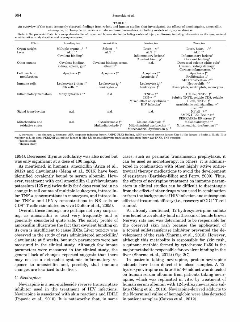
1994). Decreased thymus cellularity was also noted butwas only significant at a dose of 100 mg/kg.As mentioned, in humans, amoxicillin (Ariza et al.,
2012) and clavulanate (Meng et al., 2016) have beenidentified covalently bound to serum albumin. How-ever, treatment with oral amoxicillin (1 g)/clavulanatepotassium (125 mg) twice daily for 5 days resulted in nochange in cell counts of multiple leukocytes, intracellu-lar TNF-a concentrations in monocytes, and intracellu-lar TNF-a and IFN-g concentrations in NK cells orCD8+ T cells stimulated ex vivo (Dufour et al., 2005).Overall, these findings are perhaps not very surpris-
ing, as amoxicillin is used very frequently and isgenerally considered quite safe. The safety profile ofamoxicillin illustrates the fact that covalent binding onits own is insufficient to cause IDRs. Liver toxicity wasobserved in the study of rats administered amoxicillin/clavulanate at 2 weeks, but such parameters were notmeasured in the clinical study. Although few innateparameters were measured in the clinical study, thegeneral lack of changes reported suggests that theremay not be a detectable systemic inflammatory re-sponse to amoxicillin and, possibly, that immunechanges are localized to the liver.
C. Nevirapine
Nevirapine is a non-nucleoside reverse transcriptaseinhibitor used in the treatment of HIV infections.Nevirapine is associated with skin reactions and IDILI(Popovic et al., 2010). It is noteworthy that, in some
cases, such as perinatal transmission prophylaxis, itcan be used as monotherapy; in others, it is adminis-tered in combination with other highly active antire-troviral therapy medications to avoid the developmentof resistance (Bardsley-Elliot and Perry, 2000). Thus,the effects of nevirapine treatment on immune param-eters in clinical studies can be difficult to disentanglefrom the effect of other drugs when used in combinationor from the background of HIV infection and subsequenteffects of treatment efficacy (i.e., recovery of CD4+ T-cellcounts).
As already mentioned, 12-hydroxynevirpine sulfatewas found to covalently bind in the skin of female brownNorway rats and was determined to be responsible forthe observed skin rash because the application ofa topical sulfotransferase inhibitor prevented the de-velopment of the rash (Sharma et al., 2013). However,although this metabolite is responsible for skin rash,a quinone methide formed by cytochrome P450 is themajor metabolite responsible for covalent binding in theliver (Sharma et al., 2012) (Fig. 2C).
In patients taking nevirapine, protein-nevirapineadducts have been detected in blood samples. A 12-hydroxynevirapine sulfate-His146 adduct was detectedon human serum albumin from patients taking nevir-apine, which was replicated in vitro by treatment ofhuman serum albumin with 12-hydroxynevirapine sul-fate (Meng et al., 2013). Nevirapine-derived adducts tothe N-terminal valine of hemoglobin were also detectedin patient samples (Caixas et al., 2012).
TABLE 1An overview of the most commonly observed findings from rodent and human studies that investigated the effects of amodiaquine, amoxicillin,
nevirapine, or clozapine on various innate immune parameters, excluding models of injury or diseaseRefer to Supplemental Data for a comprehensive list of rodent and human studies (including models of injury or disease), including information on the dose, route of
administration, study duration, and primary outcomes.
Effect Amodiaquine Amoxicillin Nevirapine Clozapine
Organ weight Multiple organs ↓/—a Spleen —a Liver —/↑a Liver, heart —/↑a
Liver ALT ↑a
Covalent bindingaALT ↑a ALT ↑a
Inflammatory lesionsa
Covalent bindinga
ALT ↑a
Inflammatory lesionsa
Covalent bindinga
Other organs Covalent binding:kidney, spleen, guta
Covalent binding: serumalbuminb
n.d. Decreased splenic white pulpa
Ovarian, kidney damagea
Cardiac inflammation a,b
Cell death orproliferation
Apoptosis ↑a Apoptosis ↑a Apoptosis ↑a
Apoptosis ↓bApoptosis ↑a
Proliferation ↓a
AIF translocation –a,b
Immune cells Leukocytes ↓ then ↑a
NK cells ↑aLeukocytes ↓/↑a
Leukocytes –bLeukocytes ↓/↑a
Leukocytes ↑bNeutrophils ↑a,b
Eosinophils, neutrophils, monocytes↑b
Inflammatory mediators Many cytokines ↑a n.d. TNF-a ↑a
IFN-g –a
Mixed effect on cytokines 6HIV infectionb
CXCL2, TNF-a ↑a
Soluble TNFR, soluble CD8, solubleIL-2R, TNF-a ↑b
Arachidonic acid signaling ↔a
IL-6 ↑a,b
Signal transduction n.d. n.d. n.d. NF-kB ↑/–a
AMPK-ULK1-Beclin1↑a
PERK/eIF2a ER stress ↑a
Mitochondria andoxidative stress
n.d. Cytochrome-c ↑a
Malondialdehyde ↑aMalondialdehyde ↑a
Mitochondrial dysfunction ↑a
Mitochondrial dysfunction ↑/–b
Malondialdehyde ↑a
Mitochondrial dysfunction ↑a
↑, increase; —, no change; ↓, decrease; AIF, apoptosis-inducing factor; AMPK-ULK1-Beclin1, AMP-activated protein kinase-Unc-51-like kinase 1-Beclin1; IL-2R, IL-2receptor; n.d., no data; PERK/eIF2a, protein kinase R–like ER kinase/eukaryotic translation initiation factor 2A; TNFR, TNF receptor.
aRodent studybHuman study
884 Sernoskie et al.
1. Data from Rodent and Human Studies.Generally, nevirapine administration caused an in-crease in serum ALT levels in male rats and mice(Adaramoye et al., 2012; Sharma et al., 2012; Awodeleet al., 2015), but not in female brown Norway rats(Bekker et al., 2012; Brown et al., 2016), although thisobservation is likely due to the shorter duration of thelatter studies (7 or 14 days). In a clinical study,nevirapine exposure was associated with reduced fibro-sis, although again it is difficult to speculate upon themechanism, as this was in the context of HIV andhepatitis C coinfection (Berenguer et al., 2008).Histologic findings indicative of hepatocyte cell death
were sometimes found in rats and mice (Adaramoyeet al., 2012; Bekker et al., 2012; Sharma et al., 2012).Gene expression changes in the skin of female brownNorway rats 6 hours after 12-hydroxynevirapine treat-ment also appeared to indicate apoptosis or alteredmitochondrial function (Zhang et al., 2013). In rodentstudies, nevirapine caused an increase in malondialde-hyde, although changes in antioxidant enzymes werenot observed (Adaramoye et al., 2012; Awodele et al.,2015). Altogether, the studies in rodents appear tosuggest effects on cell death andmitochondrial function.The effects of nevirapine on mitochondrial function
are less clear in clinical studies. One study showed thatnevirapine (coadministered with stavudine and lami-vudine) increased mitochondrial depolarization andlymphocyte apoptosis (Karamchand et al., 2008). Incontrast, another study showed that switching tonevirapine from nucleoside reverse transcriptase inhib-itors improved mitochondrial parameters, but this maysimply indicate that nevirapine has less of an effect onmitochondria than nucleoside reverse transcriptaseinhibitors, which are known to cause mitochondrialtoxicity (Negredo et al., 2009). Infants treated withnevirapine were not shown to have significant mito-chondrial toxicity (Jao et al., 2017). In terms of oxidativestress, a study measured plasma F2-isoprostane levelsas a measure of lipid peroxidation and found that therewas a trend toward decreased plasma F2-isoprostanelevels with nevirapine treatment (Redhage et al., 2009).Altogether, mitochondrial function may be impaired orunchanged with nevirapine exposure, but any observedeffects do not appear to be as substantial as with otherantiretrovirals.In general, nevirapine did not have a clear impact on
blood cell counts in rodents; depending upon the timingand the dosing, nevirapine was found to decreaseleukocytes (compared with controls, 6 mg/kg per dayorally, 60 days) (Awodele et al., 2015) or increaselymphocytes and platelets (compared with referencerange, 200 mg/kg per day by oral gavage, 21 days)(Bekker et al., 2012) in rats. A low dose of nevirapineacutely increased leukocyte emigration in rats (Ordenet al., 2014). In the female brown Norway rat model ofskin rash, nevirapine treatment appeared to induce
macrophage infiltration in auricular lymph nodes thatpreceded T-cell recruitment (Popovic et al., 2006).Infants exposed to prophylactic nevirapine treatmenthad elevated monocyte counts and percentages andbasophil counts at birth (Schramm et al., 2010).
In rodent models, nevirapine caused some changes incytokine levels, although in most cases, these changesoccurred 3 weeks or longer after initiation of drugtreatment. Serum TNF-a was increased at 24 hours inone study (Bekker et al., 2012), but no changes wereseen with IFN-g up to 3 weeks (Popovic et al., 2006;Bekker et al., 2012). In clinical studies of HIV infection,nevirapine exposure was associated with decreasedserum or plasma cytokines such as CCL3 and IL-8(Shalekoff et al., 2009), IL-6 (Borges et al., 2015), andpotentially soluble CD14 (Allavena et al., 2013). Thelatter two studies compared the effects of multipledrugs, so these results may speak to a nevirapine-specific effect rather than a broad antiviral treatmenteffect. Although there are not many clear or consistentchanges in specific inflammatory markers, nevirapinedoes seem to have modulatory effects on inflammatorymarkers in general, which may even contribute to itsefficacy.
Overall, nevirapine has been shown to cause mildliver injury in otherwise healthy rodents. In some cases,histologic findings of cell death may complement theobservation of liver injury. There are conflicting resultsregarding mitochondrial toxicity, leukocyte changes,and cytokine changes. There is no convincing evidencethat nevirapine mediates mitochondrial injury; if any-thing, it appears less likely to do so than otherantiretrovirals used in the treatment of HIV. Nevira-pine does appear to have effects on peripheral bloodcells, although the differences in study duration, doses,and models used are problematic. It would be informa-tive to determine whether changes observed in femalebrown Norway rats, particularly the macrophage re-cruitment to lymph nodes, are reproduced in otherrodent models that do not develop a skin rash.
D. Clozapine
Clozapine, an atypical antipsychotic, has uniqueefficacy in the treatment of schizophrenia. However, itis infrequently prescribed because of the risks of IDIAGand, more rarely, IDILI (Wu Chou et al., 2014; Li et al.,2020). As mentioned, several HLA haplotypes havebeen associated with an increased risk of clozapine-induced agranulocytosis (Legge and Walters, 2019).
The initiating mechanisms of clozapine-inducedIDRs are poorly understood but are hypothesized toinvolve an aberrant adaptive immune response againstclozapine-modified proteins. Clozapine can be bioacti-vated by cytochromes P450 in the liver and myeloper-oxidase in neutrophils and monocytes to a reactivenitrenium ion that covalently binds to cellular proteinsin vitro and in vivo (Liu and Uetrecht, 1995; Maggs
The Innate Immune Response in Idiosyncratic Drug Reactions 885

et al., 1995; Dragovic et al., 2013; Lobach and Uetrecht,2014b) (Fig. 2D). If this neoantigen formation leads tosignificant cell damage or death, then the proinflamma-tory mediators and DAMPs released could stimulate aninnate immune response that eventually leads to theonset of severe IDRs (Pirmohamed and Park, 1997;Johnston and Uetrecht, 2015).Because of the rigorous hematologic monitoring re-
quired for patients starting clozapine, substantial evi-dence in support of an early innate immune responsehas been reported, as discussed below. Additionally,several animal models, predominantly albino rats, havebeen used to characterize immune changes throughoutthe first few weeks of clozapine treatment.1. Data from Rodent and Human Studies. A review
of the literature revealed more than 50 case reports andcase series that noted the occurrence of an innateimmune response with clozapine, most commonly evi-denced by fever, eosinophilia, neutrophilia, leukocyto-sis, a left shift in blood cells, and increased C-reactiveprotein, ALT, ALP, and aspartate transaminase withinthe first 6 weeks of treatment (Lowe et al., 2007; Røgeet al., 2012; Szota et al., 2013; Fonseka et al., 2016;Bellissima et al., 2018; Verdoux et al., 2019; de Leonet al., 2020). To avoid redundancy, those studies are notpresented here.Although short-term clozapine administration has
been studied in close to 100 rodent studies, the focusof much of this work was to determine how clozapinealters disease and/or injury progression (e.g., inphencyclidine-induced schizophrenia) and not to char-acterize the effects of clozapine alone. Such modelsmake it challenging to delineate a role for clozapine inthe initiation of an innate immune response. Interest-ingly, many of these studies actually reported a pro-tective effect of clozapine, often noting an attenuation ofthe disease model–induced inflammatory response.However, these disease models are physically or chem-ically induced, and such results may not reflect the trueeffects of clozapine monotherapy in patients.Two male rat studies that evaluated clozapine effects
in the liver demonstrated significant increases in injury(e.g., ALT increases, inflammatory cell infiltrates, in-creased liver weight) between 1 and 3 weeks of treat-ment (Jia et al., 2014; Zlatkovi�c et al., 2014). Significantcovalent binding has also been demonstrated in theliver of clozapine-treated rats (Gardner et al., 2005;Ip and Uetrecht, 2008), and it is possible that thehepatic inflammation observed is in response to thishaptenization.The effects of clozapine on various other organs,
including the brain, heart, and kidney, have beeninvestigated using several rodent models. In studiescharacterizing the effects of clozapine in the absence ofinduced injury or disease, decreased splenic white pulpwas observed in both female mice and male rats(Abdelrahman et al., 2014; Mohammed et al., 2020),
and both ovarian and kidney injury were reported inrats (Khalaf et al., 2019; Mohammed et al., 2020).Moreover, significant cardiac inflammation and mor-phologic aberrations were observed during the first fewweeks of treatment (Wang et al., 2008; Abdel-Wahaband Metwally, 2014; Abdel-Wahab et al., 2014; Nikoli�c-Koki�c et al., 2018; Mohammed et al., 2020). Thisparallels what is observed clinically because, in additionto severe IDRs, clozapine has been associated with anincreased risk of myocarditis in patients, which canpresent with fever, eosinophilia, and increased troponinlevels, often during weeks 2 and 3 of treatment (Kilianet al., 1999; Ronaldson et al., 2010; Curto et al., 2015).This is clearly an innate immune response due to theacute onset and effector cells and mediators observed.
The potential for clozapine to trigger cell death hasbeen explored in several organs, including the liver,heart, blood, and brain. The majority of rodent studiesdemonstrated evidence of apoptosis (e.g., increasedterminal deoxynucleotide transferase dUTP nick-endlabeling staining or caspase-3 activation) betweenweeks 1 and 4 of treatment (Wasti et al., 2006;Jarskog et al., 2007; Huang et al., 2012; Abdel-WahabandMetwally, 2014; Abdel-Wahab et al., 2014; Jia et al.,2014; Zlatkovi�c et al., 2014; Hsu and Fu, 2016; Khalafet al., 2019) using doses that would approximatetherapeutic concentrations in patients (Lobach andUetrecht, 2014a). One study also noted that clozapineinduced autophagywithin hours of administration (Kimet al., 2018), and others noted decreased proliferationwithin the first few weeks of treatment (Huang et al.,2012; Hsu and Fu, 2016; Khalaf et al., 2019) as well.Translocation of apoptosis-inducing factor was notobserved in the striatum of clozapine-treated patientsor in rodents after 1 month of treatment (Skoblenicket al., 2006), suggesting against the involvement ofcaspase-independent cell death with clozapine.
In various models of acute injury and disease, cloza-pine was not consistently found to attenuate changes inimmune cell populations. Only a small number ofstudies investigated the effects of clozapine in healthyanimals, almost all of which demonstrated induction ofan immune response by clozapine in the first 3 weeks oftreatment. Most commonly, an increase in neutrophilswas reported in clozapine-treated male and female rats(Wasti et al., 2006; Abdel-Wahab and Metwally, 2014;Lobach and Uetrecht, 2014a; Ng et al., 2014) or rabbits(Iverson et al., 2010). In the only two mouse studies,clozapine caused not only a decrease in several leuko-cyte populations (Abdelrahman et al., 2014; Jiang et al.,2016) but also an increase inmonocytes, suggesting thatthe immunomodulatory effects of clozapine may differacross rodent species. In clinical studies, however,clozapine demonstrated strong evidence of innate im-mune cell activation during the first several weeks oftreatment. Depending on the patient population, stud-ies reported an increased incidence of eosinophilia,
886 Sernoskie et al.
neutrophilia, and/or leukocytosis that typically resolvedwith continued clozapine administration (Banov et al.,1993; Pollmächer et al., 1996; Chatterton, 1997; Thamand Dickson, 2002; Pui-yin Chung et al., 2008; Löffleret al., 2010). One small study also noted an increase incirculating CD34+ hemopoietic stem cells after 2 weeksof clozapine treatment (Löffler et al., 2010).In rodent models in which immunomodulatory effects
were investigated in the context of a disease or injurymodel, clozapine was frequently shown to attenuate themodel-induced inflammation. Contrarily, few studiescharacterized the inflammatory mediator changescaused by clozapine alone. Of these studies, mostdemonstrated an increase in proinflammatory media-tors in either rats or mice, including TNF-a, CXCL2,and heat shock protein 75 (Wang et al., 2008; Abdel-Wahab and Metwally, 2014; Abdel-Wahab et al., 2014;Lobach and Uetrecht, 2014a; Kedracka-Krok et al.,2016; Mohammed et al., 2020). Few studies havereported alterations in bioactive lipids in response tothe drugs reviewed; however, dysregulated arachidonicacid signaling was also noted with clozapine treatment(Kim et al., 2012; Modi et al., 2013). Among the drugsinvestigated for this review, clozapine also provides thestrongest support of innate immune activation inpatients. All but one study reported an increasedincidence of fever and/or increased serum levels ofinflammatory mediators, such as TNF-a, soluble TNFreceptor, soluble CD8, and soluble IL-2 receptor, mostcommonly occurring during the firstmonth of treatment(Pollmächer et al., 1995, 1996, 1997; Maes et al., 1997,2002; Hinze-Selch et al., 1998, 2000; Tham andDickson,2002; Pui-yin Chung et al., 2008; Kluge et al., 2009;Hung et al., 2017).The research focused on the effects of amodiaquine,
amoxicillin, or nevirapine on signal transduction path-ways in rodent models is limited, although this is likelydue to the preference for in vitro work in this area.Contrastingly, the effect of clozapine on a number ofsignaling pathways has been examined in rodents,although many of these observations have yet to beverified in subsequent studies. The reported effects ofclozapine on the regulation of transcription vary greatlyand depend on the timing of the studies, as well as theorgans investigated. Clozapine-induced activation ofhepatic and cardiac NF-kB was demonstrated in tworat models at 3 weeks (Abdel-Wahab and Metwally,2014; Zlatkovi�c et al., 2014), but these changes were notobserved in several brain regions in other models.Other studies have also characterized clozapine-induced activation of other signaling pathways, includ-ing the AMP-activated protein kinase (AMPK)-Unc-51–like kinase 1-Beclin1 pathway (Kim et al., 2018)and the protein kinase R–like ER kinase/eukaryotictranslation initiation factor 2A ER stress axis (Weston-Green et al., 2018), although additional work is neces-sary to confirm the results of these reports. Notably,
AMPK signaling has been shown to play a role in manybiochemical pathways, including autophagy, mitochon-drial biogenesis, and lipid metabolism (Hardie et al.,2016); thus, further investigation of clozapine’s impacton AMPK signaling and its potential role in inflamma-tion should be undertaken.
Several rodent studies have also been conducted toevaluate changes in mitochondrial function due toclozapine. Clozapine often caused attenuatedmitochon-drial function or oxidative stress (e.g., increased malon-dialdehyde levels), which was noted most frequently inmale rats after 3–4 weeks of treatment in various brainregions (Lara et al., 2001; La et al., 2006; Mehler-Wexet al., 2006; Streck et al., 2007; Bullock et al., 2008;Martins et al., 2008; Ji et al., 2009; Bishnoi et al., 2011;Zlatkovi�c et al., 2014; Cai et al., 2017), although cardiac-specific (Nikoli�c-Koki�c et al., 2018) and ovarian-specific(Khalaf et al., 2019) aberrations were also reported.Additionally, another study reported increasedmarkersof ER stress in the liver 1 hour after clozapine treatment(Lauressergues et al., 2012).
Although additional work is clearly needed to char-acterize the mechanisms underlying the findings dis-cussed here, clozapine has frequently been shown tocause innate immune activation, both in patients and invarious animal models. One avenue that should also bepursued moving forward is determining what initiallytriggers the immune response (e.g., triggers of myeloidcell recruitment) and, subsequently, whether inhibitingthis immune response prevents progression to seriousIDRs, effectively reducing the risks associated withclozapine use.
E. Summary
Overall, various early immune-related changes havebeen observed in animal models and human studieswith the drugs presented here (Table 1). In rodents, theincreased serum ALT observed with all drugs is in-dicative of liver damage. Additionally, the induction ofapoptosis in many other organs was also observed witheach of the drugs. A number of changes were describedin various leukocyte populations, with some drugscausing increases in innate immune cells and, in fewerinstances, some drugs causing decreases in leukocytes.In many cases, increases in proinflammatory cytokineswere observed, and with clozapine, activated signaltransduction pathways involved in proinflammatorysignaling were also observed; this has not beenstudied in vivo for the other drugs examined.Markers of mitochondrial dysfunction were alsoreported after administration of amoxicillin, cloza-pine, and nevirapine.
The study of the inflammation caused by amodia-quine is limited to rodent models. However, there isa clear indication of NK cell–mediated liver injury,which spontaneously resolves with continued treat-ment, in addition to other immune cell infiltrates
The Innate Immune Response in Idiosyncratic Drug Reactions 887
detected in the liver, spleen, lymph node, and periph-eral blood. The immune effects of amoxicillin 6clavulanic acid have not been studied in healthysubjects as extensively as some of the other drugspresented here. In general, the data do not suggestthat amoxicillin causes an overt inflammatory re-sponse, but there are certainly changes that suggestsome effects on mitochondria and leukocytes. Nevir-apine treatment appears to cause liver damage buthas variable effects on inflammatory mediators andimmune cell counts. In rodents, clozapine showsthe clearest pattern of a proinflammatory responseof these drugs, which is not surprising, as it hasbeen noted to induce fever, eosinophilia, neutrophilia,and increased proinflammatory cytokine release inpatients.Overall, the effects of each of these drugs are quite
variable depending upon themodels and doses used andthe time points at which different parameters areevaluated. This highlights the complexity of theimmune response and the potential differences thatmay be caused by different drugs, which likely dependupon the conditions in which their reactive metabo-lites are formed and which may also have a bearing onthe types of IDRs that they cause. Studies thatevaluate the time course of drug effects on inflamma-tory pathways are needed to better understandhow drugs that cause IDRs induce innate immuneresponses.
V. Conclusions and Perspectives
Just as the drugs presented here are associated withdifferent IDRs, they have also been demonstrated tocause a variety of immune-related effects during thefirst few weeks of treatment. These differences includethe tissue localization of cellular dysfunction, injury,and death; the responding effector cells; the mecha-nisms contributing to inflammation; and the develop-ment of the immune response over time. These drug-specific observations emphasize the nuances and com-plexity underlying the activation and progression of aninnate immune response. Based on the data presentedhere, it is clear that further research is necessary toexpand our understanding of how drugs that areassociated with severe IDRs canmore frequently inducean early, transient immune response that typicallyresolves with continued treatment. Such research isfundamental to understanding the mechanisms ofIDRs, and it is quite feasible to perform such research.Most drug metabolic pathways and immune responsesshare some similarities in animals and humans, andanimal models are an important tool because theymakeit possible to perform controlled experiments and in-vestigate organs such as the liver and spleen that couldnot be routinely looked at in patients. It is important touse doses in animals that would produce what would be
a therapeutic level in humans because high doses aremore likely to cause overt toxicity that is not involved inthe mechanism of IDRs. However, even though mostfeatures are likely to be similar in rodents and humans,there are clearly important differences between ani-mals and humans; therefore, it is essential to follow upthe animal studies with studies in humans tomake surethat the results in animals correspond to the immuneresponse to drugs in humans.
The innate immune response caused by these IDR-associated drugs is likely to be mild in comparison withthe overt injury induced by disease models and wouldeasily be overlooked in studies not designed to capturethese relatively subtle changes. Moreover, additionalconsideration should be given to how these innateimmune responses resolve with persistent treatment,as this resolution/tolerogenic response, or lack thereof,may provide clues as to why certain individuals even-tually develop severe IDRs while the majority do not.Although certain risk factors for different IDRs havebeen identified, such as particular HLA haplotypes,these factors only account for a small proportion of risk;for most drugs, it remains difficult to predict whichindividuals will develop a severe IDR. An individual’sT-cell receptor repertoire is likely to be a major factor,but it is much more difficult to study than HLAhaplotypes. Many drugs, although highly efficacious inthe treatment of their intended conditions, are thereforelimited in their clinical use because of the risk of IDRs(including amodiaquine, clozapine, and nevirapine).Thus, a better understanding of the mechanisms con-tributing to the early immune response to these drugsmay help predict and prevent or treat their associatedIDRs, enabling the safer use of these agents. Althoughsome work has been done in this area already, asreviewed here, the innate immune response has notbeen systematically studied across drugs that causeIDRs. More work is required to understand whetherdifferent drugs cause different responses, or whetherthere are certain commonalities in the immune changescaused by drugs that cause IDRs. Additionally, it will beimportant to test drugs that do not cause IDRs toensure that they do not have the same effects. Byidentifying alterations in pathways that presage IDRs,these studies will identify potential biomarkers fordrugs that can cause IDRs. These biomarkers could beused to develop a preclinical tool to screen drugcandidates for the potential to cause serious IDRs.Such assays would facilitate the development of saferdrugs and reduce the burden of IDRs on the drugdiscovery process. Additionally, understanding thespecifics of the innate immune response to these drugsmay reveal potential targets to inhibit to prevent thedevelopment of IDRs for drugs in clinical use. Alto-gether, although much work remains in this area, thestudy of the innate immune response is clearly impor-tant in improving drug safety.
888 Sernoskie et al.

Acknowledgments
We are very grateful to Glyneva Bradley-Ridout for assistance withconducting the literature review for Section IV. Support for ImmuneActivation Using Model Drugs.Figures were produced using BioRender.com.
Authorship Contributions
Participated in research design: Sernoskie, Jee, Uetrecht.Performed data analysis: Sernoskie, Jee.Wrote or contributed to the writing of the manuscript: Sernoskie,
Jee, Uetrecht.
ReferencesAbboud G and Kaplowitz N (2007) Drug-induced liver injury. Drug Saf 30:277–294.Abdel-Wahab BA and Metwally ME (2014) Clozapine-induced cardiotoxicity in rats:involvement of tumour necrosis factor alpha, NF-kb and caspase-3. Toxicol Rep 1:1213–1223.
Abdelrahman Y, Fararjeh M, Abdel-Razeq W, MohammadMK, and Bustanji Y (2014)Assessment of possible immunotoxicity of the antipsychotic drug clozapine.J Pharm Pharmacol 66:378–386.
Abdel-Wahab BA, Metwally ME, El-khawanki MM, and Hashim AM (2014) Pro-tective effect of captopril against clozapine-induced myocarditis in rats: role ofoxidative stress, proinflammatory cytokines and DNA damage. Chem Biol Interact216:43–52.
Abe R, Shimizu T, Shibaki A, Nakamura H, Watanabe H, and Shimizu H (2003) Toxicepidermal necrolysis and Stevens-Johnson syndrome are induced by soluble Fasligand. Am J Pathol 162:1515–1520.
Adaramoye OA, Adesanoye OA, Adewumi OM, and Akanni O (2012) Studies on thetoxicological effect of nevirapine, an antiretroviral drug, on the liver, kidney andtestis of male Wistar rats. Hum Exp Toxicol 31:676–685.
Ajani EO, Shallie PD, Adegbesan BO, Salau BA, and Adesanya M (2008) Protectiveeffect of Garcinia kola (kolaviron) extract on predisposition of rats to cardiovasculardiseases following separate administration of amodiaquine and artesunate. AfrJ Tradit Complement Altern Med 5:180–186.
Akdis M, Aab A, Altunbulakli C, Azkur K, Costa RA, Crameri R, Duan S, Eiwegger T,Eljaszewicz A, Ferstl R, et al. (2016) Interleukins (from IL-1 to IL-38), interferons,transforming growth factor b, and TNF-a: receptors, functions, and roles in dis-eases. J Allergy Clin Immunol 138:984–1010.
Ali SE, Waddington JC, Park BK, and Meng X (2020) Definition of the chemical andimmunological signals involved in drug-induced liver injury. Chem Res Toxicol 33:61–76.
Al Qahtani A (2018) Drug-induced megaloblastic, aplastic, and hemolytic anemias:current concepts of pathophysiology and treatment. Int J Clin Exp Med 11:5501–5512.
Allavena C, Rodallec A, Sécher S, Reliquet V, Baffoin S, André-Garnier E, Billaud E,Raffi F, and Ferré V (2013) Evaluation of residual viremia and quantitation ofsoluble CD14 in a large cohort of HIV-infected adults on a long-term non-nucleoside reverse transcriptase inhibitor-based regimen. J Med Virol 85:1878–1882.
Allen F, Tong AA, and Huang AY (2016) Unique transcompartmental bridge:antigen-presenting cells sampling across endothelial and mucosal barriers. FrontImmunol 7:231.
Amarante-Mendes GP, Adjemian S, Branco LM, Zanetti LC, Weinlich R,and Bortoluci KR (2018) Pattern recognition receptors and the host cell deathmolecular machinery. Front Immunol 9:2379.
Andersohn A, Garcia MI, Fan Y, Thompson MC, Akimzhanov AM, Abdullahi A,Jeschke MG, and Boehning D (2019) Aggregated and hyperstable damage-associated molecular patterns are released during ER stress to modulate im-mune function. Front Cell Dev Biol 7:198.
Andersohn F, Konzen C, and Garbe E (2007) Systematic review: agranulocytosisinduced by nonchemotherapy drugs. Ann Intern Med 146:657–665.
Anderson HL, Brodsky IE, and Mangalmurti NS (2018) The evolving erythrocyte: redblood cells as modulators of innate immunity. J Immunol 201:1343–1351.
Andrade RJ, Chalasani N, Björnsson ES, Suzuki A, Kullak-Ublick GA, Watkins PB,Devarbhavi H, Merz M, Lucena MI, Kaplowitz N, et al. (2019) Drug-induced liverinjury. Nat Rev Dis Primers 5:58.
Andrade RJ, Lucena MI, Alonso A, García-Cortes M, García-Ruiz E, Benitez R,Fernández MC, Pelaez G, Romero M, Corpas R, et al. (2004) HLA class II genotypeinfluences the type of liver injury in drug-induced idiosyncratic liver disease.Hepatology 39:1603–1612.
Andrade RJ, Lucena MI, Fernández MC, Pelaez G, Pachkoria K, García-Ruiz E,García-Muñoz B, González-Grande R, Pizarro A, Durán JA, et al.; Spanish Groupfor the Study of Drug-Induced Liver Disease (2005) Drug-induced liver injury: ananalysis of 461 incidences submitted to the Spanish registry over a 10-year period[published correction appears in Gastroenterology (2005) 129:1808]. Gastroenter-ology 129:512–521.
Andrès E, Dali-Youcef N, Serraj K, and Zimmer J (2009) Recognition and manage-ment of drug-induced cytopenias: the example of idiosyncratic drug-inducedthrombocytopenia. Expert Opin Drug Saf 8:183–190.
Andrès E and Maloisel F (2008) Idiosyncratic drug-induced agranulocytosis or acuteneutropenia. Curr Opin Hematol 15:15–21.
Andrès E and Mourot-Cottet R (2017) Non-chemotherapy drug-induced neutropenia -an update. Expert Opin Drug Saf 16:1235–1242.
Andrès E, Mourot-Cottet R, Maloisel F, Séverac F, Keller O, Vogel T, Tebacher M,Weber JC, Kaltenbach G, Gottenberg JE, et al. (2017) Idiosyncratic drug-inducedneutropenia & agranulocytosis. QJM 110:299–305.
Andrès E, Zimmer J, Mecili M, Weitten T, Alt M, and Maloisel F (2011) Clinicalpresentation and management of drug-induced agranulocytosis. Expert Rev Hem-atol 4:143–151.
Andrès E, Villalba NL, Zulfiqar AA, Serraj K, Mourot-Cottet R, and Gottenberg AJ(2019) State of art of idiosyncratic drug-induced neutropenia or agranulocytosis,with a focus on biotherapies. J Clin Med 8:1351.
Ariza A, Garzon D, Abánades DR, de los Ríos V, Vistoli G, Torres MJ, Carini M,Aldini G, and Pérez-Sala D (2012) Protein haptenation by amoxicillin: high reso-lution mass spectrometry analysis and identification of target proteins in serum.J Proteomics 77:504–520.
Asano K, Nabeyama A, Miyake Y, Qiu CH, Kurita A, Tomura M, Kanagawa O,Fujii S, and Tanaka M (2011) CD169-positive macrophages dominate antitu-mor immunity by crosspresenting dead cell-associated antigens. Immunity 34:85–95.
Atheymen R, Affes H, Ksouda K, Mnif L, Sahnoun Z, Tahri N, Zeghal KM,and Hammami S (2013) [Adverse effects of sulfasalazine: discussion of mechanismand role of sulfonamide structure]. Therapie 68:369–373.
Avalos AM and Ploegh HL (2014) Early BCR events and antigen capture, processing,and loading on MHC class II on B cells. Front Immunol 5:92.
Awodele O, Popoola T, Rotimi K, Ikumawoyi V, and Okunowo W (2015) Antioxidantmodulation of nevirapine induced hepatotoxicity in rats. Interdiscip Toxicol 8:8–14.
Baker CA, Uno H, and Johnson GA (1994) Minoxidil sulfation in the hair follicle.Skin Pharmacol 7:335–339.
Banchereau J, Bazan F, Blanchard D, Brière F, Galizzi JP, van Kooten C, Liu YJ,Rousset F, and Saeland S (1994) The CD40 antigen and its ligand. Annu RevImmunol 12:881–922.
Banov MD, Tohen M, and Friedberg J (1993) High risk of eosinophilia in womentreated with clozapine. J Clin Psychiatry 54:466–469.
Bardsley-Elliot A and Perry CM (2000) Nevirapine: a review of its use in the pre-vention and treatment of paediatric HIV infection. Paediatr Drugs 2:373–407.
Barker RN, Erwig LP, Hill KSK, Devine A, Pearce WP, and Rees AJ (2002) Antigenpresentation by macrophages is enhanced by the uptake of necrotic, but not apo-ptotic, cells. Clin Exp Immunol 127:220–225.
Barral P, Polzella P, Bruckbauer A, van Rooijen N, Besra GS, Cerundolo V,and Batista FD (2010) CD169(+) macrophages present lipid antigens to mediateearly activation of iNKT cells in lymph nodes. Nat Immunol 11:303–312.
Behera SK, Das S, Xavier AS, and Selvarajan S (2018) DRESS syndrome: a detailedinsight. Hosp Pract (1995) 46:152–162.
Bekker Z, Walubo A, and du Plessis JB (2012) The role of the immune system innevirapine-induced subclinical liver injury of a rat model. ISRN Pharm 2012:932542.
Bellissima BL, Tingle MD, Cicovi�c A, Alawami M, and Kenedi C (2018) A systematicreview of clozapine-induced myocarditis. Int J Cardiol 259:122–129.
Ben-Sasson SZ, Hu-Li J, Quiel J, Cauchetaux S, Ratner M, Shapira I, DinarelloCA, and Paul WE (2009) IL-1 acts directly on CD4 T cells to enhance theirantigen-driven expansion and differentiation. Proc Natl Acad Sci USA 106:7119–7124.
Berenguer J, Bellón JM, Miralles P, Alvarez E, Castillo I, Cosín J, López JC, SánchezConde M, Padilla B, and Resino S (2008) Association between exposure to nevir-apine and reduced liver fibrosis progression in patients with HIV and hepatitis Cvirus coinfection. Clin Infect Dis 46:137–143.
Bertolino P, Trescol-Biémont MC, and Rabourdin-Combe C (1998) Hepatocytes in-duce functional activation of naive CD8+ T lymphocytes but fail to promote sur-vival. Eur J Immunol 28:221–236.
Beylot C, Bioulac P, and Doutre MS (1980) [Acute generalized exanthematic pustu-loses (four cases) (author’s transl)]. Ann Dermatol Venereol 107:37–48.
Bhaiya P, Roychowdhury S, Vyas PM, Doll MA, Hein DW, and Svensson CK (2006)Bioactivation, protein haptenation, and toxicity of sulfamethoxazole and dapsonein normal human dermal fibroblasts. Toxicol Appl Pharmacol 215:158–167.
Bishnoi M, Chopra K, Rongzhu L, and Kulkarni SK (2011) Protective effect of cur-cumin and its combination with piperine (bioavailability enhancer) againsthaloperidol-associated neurotoxicity: cellular and neurochemical evidence. Neuro-tox Res 20:215–225.
Björnsson E, Kalaitzakis E, and Olsson R (2007) The impact of eosinophilia andhepatic necrosis on prognosis in patients with drug-induced liver injury. AlimentPharmacol Ther 25:1411–1421.
Björnsson E and Olsson R (2005) Outcome and prognostic markers in severe drug-induced liver disease. Hepatology 42:481–489.
Björnsson E, Talwalkar J, Treeprasertsuk S, Kamath PS, Takahashi N, Sanderson S,Neuhauser M, and Lindor K (2010) Drug-induced autoimmune hepatitis: clinicalcharacteristics and prognosis. Hepatology 51:2040–2048.
Björnsson ES (2016) Hepatotoxicity by drugs: the most common implicated agents.Int J Mol Sci 17:224.
Blanca M, Romano A, Torres MJ, Férnandez J, Mayorga C, Rodriguez J, Demoly P,Bousquet PJ, Merk HF, Sanz ML, et al. (2009) Update on the evaluation of hy-persensitivity reactions to betalactams. Allergy 64:183–193.
Blanco P, Palucka AK, Pascual V, and Banchereau J (2008) Dendritic cells andcytokines in human inflammatory and autoimmune diseases. Cytokine GrowthFactor Rev 19:41–52.
Bocquet H, Bagot M, and Roujeau JC (1996) Drug-induced pseudolymphoma anddrug hypersensitivity syndrome (Drug Rash with Eosinophilia and SystemicSymptoms: DRESS). Semin Cutan Med Surg 15:250–257.
Boegeholz J, Brueggen CS, Pauli C, Dimitriou F, Haralambieva E, Dummer R, ManzMG, and Widmer CC (2020) Challenges in diagnosis and management of neu-tropenia upon exposure to immune-checkpoint inhibitors: meta-analysis of a rareimmune-related adverse side effect. BMC Cancer 20:300.
Boelsterli UA and Lim PLK (2007) Mitochondrial abnormalities--a link to idiosyn-cratic drug hepatotoxicity? Toxicol Appl Pharmacol 220:92–107.
The Innate Immune Response in Idiosyncratic Drug Reactions 889
Böhm R, Proksch E, Schwarz T, and Cascorbi I (2018) Drug hypersensitivity. DtschArztebl Int 115:501–512.
Borges AH, O’Connor JL, Phillips AN, Rönsholt FF, Pett S, Vjecha MJ, French MA,Lundgren JD, and Committee IS; INSIGHT SMART and ESPRIT Study Groupsand the SILCAAT Scientific Committee (2015) Factors associated with plasma IL-6levels during HIV infection. J Infect Dis 212:585–595.
Breckenridge A (2015) The burden of adverse drug events. Br J Clin Pharmacol 80:785–787.
Brinkmann V, Reichard U, Goosmann C, Fauler B, Uhlemann Y, Weiss DS, Wein-rauch Y, and Zychlinsky A (2004) Neutrophil extracellular traps kill bacteria.Science 303:1532–1535.
Britschgi M, Steiner UC, Schmid S, Depta JPH, Senti G, Bircher A, Burkhart C,Yawalkar N, and Pichler WJ (2001) T-cell involvement in drug-induced acutegeneralized exanthematous pustulosis. J Clin Invest 107:1433–1441.
Brown HR, Castellino S, Groseclose MR, Elangbam CS, Mellon-Kusibab K, Yoon LW,Gates LD, Krull DL, Cariello NF, Arrington-Brown L, et al. (2016) Drug-inducedliver fibrosis: testing nevirapine in a viral-like liver setting using histopathology,MALDI IMS, and gene expression. Toxicol Pathol 44:112–131.
Bullock WM, Cardon K, Bustillo J, Roberts RC, and Perrone-Bizzozero NI (2008)Altered expression of genes involved in GABAergic transmission and neuro-modulation of granule cell activity in the cerebellum of schizophrenia patients. AmJ Psychiatry 165:1594–1603.
Cacoub P, Musette P, Descamps V, Meyer O, Speirs C, Finzi L, and Roujeau JC(2011) The DRESS syndrome: a literature review. Am J Med 124:588–597.
Cai HL, Jiang P, Tan QY, Dang RL, Tang MM, Xue Y, Deng Y, Zhang BK, FangPF, Xu P, et al. (2017) Therapeutic efficacy of atypical antipsychotic drugs bytargeting multiple stress-related metabolic pathways. Transl Psychiatry 7:e1130.
Caixas U, Antunes AMM, Marinho AT, Godinho ALA, Grilo NM, Marques MM,Oliveira MC, Branco T, Monteiro EC, and Pereira SA (2012) Evidence for nevir-apine bioactivation in man: searching for the first step in the mechanism ofnevirapine toxicity. Toxicology 301:33–39.
Campana S, De Pasquale C, Carrega P, Ferlazzo G, and Bonaccorsi I (2015) Cross-dressing: an alternative mechanism for antigen presentation. Immunol Lett 168:349–354.
Castanheira FVS and Kubes P (2019) Neutrophils and NETs in modulating acuteand chronic inflammation. Blood 133:2178–2185.
Cella M, Engering A, Pinet V, Pieters J, and Lanzavecchia A (1997) Inflammatorystimuli induce accumulation of MHC class II complexes on dendritic cells. Nature388:782–787.
Chalasani N, Fontana RJ, Bonkovsky HL, Watkins PB, Davern T, Serrano J, Yang H,and Rochon J; Drug Induced Liver Injury Network (DILIN) (2008) Causes, clinicalfeatures, and outcomes from a prospective study of drug-induced liver injury in theUnited States. Gastroenterology 135:1924–1934, 1934.e1–1934.e4.
Chalasani NP, Hayashi PH, Bonkovsky HL, Navarro VJ, Lee WM, and Fontana RJ;Practice Parameters Committee of the American College of Gastroenterology(2014) ACG Clinical Guideline: the diagnosis and management of idiosyncraticdrug-induced liver injury. Am J Gastroenterol 109:950–966, quiz 967.
Chan R and Benet LZ (2018) Measures of BSEP inhibition in vitro are not usefulpredictors of DILI. Toxicol Sci 162:499–508.
Chatterton R (1997) Eosinophilia after commencement of clozapine treatment. AustN Z J Psychiatry 31:874–876.
Chaudhry N, Patidar Y, and Puri V (2013) Mitochondrial myopathy, encephalopathy,lactic acidosis, and stroke-like episodes unveiled by valproate. J Pediatr Neurosci 8:135–137.
Chavan SS, Pavlov VA, and Tracey KJ (2017) Mechanisms and therapeutic relevanceof neuro-immune communication. Immunity 46:927–942.
Chen C-B, Abe R, Pan R-Y, Wang C-W, Hung S-I, Tsai Y-G, and Chung W-H (2018)An updated review of the molecular mechanisms in drug hypersensitivity.J Immunol Res 2018:6431694.
Chen GY and Nuñez G (2010) Sterile inflammation: sensing and reacting to damage.Nat Rev Immunol 10:826–837.
Chen X, Tharmanathan T, Mannargudi B, Gou H, and Uetrecht JP (2009) A study ofthe specificity of lymphocytes in nevirapine-induced skin rash. J Pharmacol ExpTher 331:836–841.
Chhiba KD, Hsu C-L, Berdnikovs S, and Bryce PJ (2017) Transcriptional heteroge-neity of mast cells and basophils upon activation. J Immunol 198:4868–4878.
Chirumbolo S, Bjørklund G, Sboarina A, and Vella A (2018) The role of basophils asinnate immune regulatory cells in allergy and immunotherapy. Hum VaccinImmunother 14:815–831.
Chiurchiù V, Battistini L, and Maccarrone M (2015) Endocannabinoid signalling ininnate and adaptive immunity. Immunology 144:352–364.
Chiurchiù V, Leuti A, and Maccarrone M (2018) Bioactive lipids and chronic in-flammation: managing the fire within. Front Immunol 9:38.
Chiurchiù V and Maccarrone M (2016) Bioactive lipids as modulators of immunity,inflammation and emotions. Curr Opin Pharmacol 29:54–62.
Cho T and Uetrecht J (2017) How reactive metabolites induce an immune responsethat sometimes leads to an idiosyncratic drug reaction. Chem Res Toxicol 30:295–314.
Choi MJ, Kim HS, Park HJ, Park CJ, Lee JD, Lee JY, Kim HO, and Park YM (2010)Clinicopathologic manifestations of 36 Korean patients with acute generalizedexanthematous pustulosis: a case series and review of the literature. Ann Dermatol22:163–169.
Chung WH, Hung SI, Yang JY, Su SC, Huang SP, Wei CY, Chin SW, Chiou CC, ChuSC, Ho HC, et al. (2008) Granulysin is a key mediator for disseminated keratino-cyte death in Stevens-Johnson syndrome and toxic epidermal necrolysis. Nat Med14:1343–1350.
Cildir G, Low KC, and Tergaonkar V (2016) Noncanonical NF-kB signaling in healthand disease. Trends Mol Med 22:414–429.
Cirulli ET, Nicoletti P, Abramson K, Andrade RJ, Bjornsson ES, Chalasani N,Fontana RJ, Hallberg P, Li YJ, Lucena MI, et al.; Drug-Induced Liver Injury
Network (DILIN) investigators; International DILI consortium (iDILIC) (2019) Amissense variant in PTPN22 is a risk factor for drug-induced liver injury. Gas-troenterology 156:1707–1716.e2.
Clarke JB, Maggs JL, Kitteringham NR, and Park BK (1990) Immunogenicity ofamodiaquine in the rat. Int Arch Allergy Appl Immunol 91:335–342.
Collett GP, Redman CW, Sargent IL, and Vatish M (2018) Endoplasmic reticulumstress stimulates the release of extracellular vesicles carrying danger-associatedmolecular pattern (DAMP) molecules. Oncotarget 9:6707–6717.
Conaghan PG (2012) A turbulent decade for NSAIDs: update on current concepts ofclassification, epidemiology, comparative efficacy, and toxicity. Rheumatol Int 32:1491–1502.
Curciarello R, Canziani KE, Docena GH, and Muglia CI (2019) Contribution of non-immune cells to activation and modulation of the intestinal inflammation. FrontImmunol 10:647.
Curto M, Comparelli A, Ciavarella GM, Gasperoni C, Lionetto L, Corigliano V,Uccellini A, Mancinelli I, Ferracuti S, Girardi P, et al. (2015) Impairment of leftventricular function early in treatment with clozapine: a preliminary study. IntClin Psychopharmacol 30:282–289.
Curtsinger JM, Lins DC, and Mescher MF (2003) Signal 3 determines toleranceversus full activation of naive CD8 T cells: dissociating proliferation and de-velopment of effector function. J Exp Med 197:1141–1151.
Curtsinger JM and Mescher MF (2010) Inflammatory cytokines as a third signal forT cell activation. Curr Opin Immunol 22:333–340.
Curtsinger JM, Schmidt CS, Mondino A, Lins DC, Kedl RM, Jenkins MK,and Mescher MF (1999) Inflammatory cytokines provide a third signal for activa-tion of naive CD4+ and CD8+ T cells. J Immunol 162:3256–3262.
Daly AK, Donaldson PT, Bhatnagar P, Shen Y, Pe’er I, Floratos A, Daly MJ, Gold-stein DB, John S, Nelson MR, et al.; DILIGEN Study; International SAE Con-sortium (2009) HLA-B*5701 genotype is a major determinant of drug-induced liverinjury due to flucloxacillin. Nat Genet 41:816–819.
Dance A (2019) Core Concept: cells nibble one another via the under-appreciatedprocess of trogocytosis. Proc Natl Acad Sci USA 116:17608–17610.
Das UN (2018) Ageing: is there a role for arachidonic acid and other bioactive lipids?A review. J Adv Res 11:67–79.
De A, Das S, Sarda A, Pal D, and Biswas P (2018) Acute generalised exanthematouspustulosis: an update. Indian J Dermatol 63:22–29.
de Abajo FJ, Montero D, Madurga M, and García Rodríguez LA (2004) Acute andclinically relevant drug-induced liver injury: a population based case-control study.Br J Clin Pharmacol 58:71–80.
de Araujo E, Dessirier V, Laprée G, Valeyrie-Allanore L, Ortonne N, StathopoulosEN, Bagot M, Bensussan A, Mockenhaupt M, Roujeau J-C, et al. (2011) Deathligand TRAIL, secreted by CD1a+ and CD14+ cells in blister fluids, is involved inkilling keratinocytes in toxic epidermal necrolysis. Exp Dermatol 20:107–112.
de Boer YS, Kosinski AS, Urban TJ, Zhao Z, Long N, Chalasani N, Kleiner DE,and Hoofnagle JH; Drug-Induced Liver Injury Network (2017) Features of auto-immune hepatitis in patients with drug-induced liver injury. Clin GastroenterolHepatol 15:103–112.e2.
De Caterina R and Zampolli A (2004) From asthma to atherosclerosis--5-lip-oxygenase, leukotrienes, and inflammation. N Engl J Med 350:4–7.
de Leon J, Ruan C-J, Verdoux H, and Wang C (2020) Clozapine is strongly associatedwith the risk of pneumonia and inflammation. Gen Psychiatr 33:e100183.
Deng J, Lu PD, Zhang Y, Scheuner D, Kaufman RJ, Sonenberg N, Harding HP,and Ron D (2004) Translational repression mediates activation of nuclear factorkappa B by phosphorylated translation initiation factor 2. Mol Cell Biol 24:10161–10168.
Dennis EA and Norris PC (2015) Eicosanoid storm in infection and inflammation.Nat Rev Immunol 15:511–523.
Dinarello CA (2018) Introduction to the interleukin-1 family of cytokines andreceptors: drivers of innate inflammation and acquired immunity. Immunol Rev281:5–7.
Doebel T, Voisin B, and Nagao K (2017) Langerhans cells - the macrophage in den-dritic cell clothing. Trends Immunol 38:817–828.
Dominici M, Le Blanc K, Mueller I, Slaper-Cortenbach I, Marini F, Krause D, DeansR, Keating A, Prockop Dj, and Horwitz E (2006) Minimal criteria for definingmultipotent mesenchymal stromal cells. The International Society for CellularTherapy position statement. Cytotherapy 8:315–317.
Doña I, Pérez-Sánchez N, Eguiluz-Gracia I, Muñoz-Cano R, Bartra J, Torres MJ,and Cornejo-García JA (2020) Progress in understanding hypersensitivity reac-tions to nonsteroidal anti-inflammatory drugs. Allergy 75:561–575.
Dooley TP, Haldeman-Cahill R, Joiner J, and Wilborn TW (2000) Expression pro-filing of human sulfotransferase and sulfatase gene superfamilies in epithelialtissues and cultured cells. Biochem Biophys Res Commun 277:236–245.
Dragovic S, Gunness P, Ingelman-Sundberg M, Vermeulen NPE, CommandeurJNM, Gaertner I, and Breyer-Pfaff U (2013) Characterization of human cyto-chrome P450s involved in the bioactivation of clozapine. Drug Metab Dispos 41:651–658.
Drutman SB, Kendall JC, and Trombetta ES (2012) Inflammatory spleen monocytescan upregulate CD11c expression without converting into dendritic cells.J Immunol 188:3603–3610.
Dubin K, Callahan MK, Ren B, Khanin R, Viale A, Ling L, No D, Gobourne A,Littmann E, Huttenhower C, et al. (2016) Intestinal microbiome analyses identifymelanoma patients at risk for checkpoint-blockade-induced colitis. Nat Commun 7:10391.
Dudeck A, Köberle M, Goldmann O, Meyer N, Dudeck J, Lemmens S, Rohde M,Roldán NG, Dietze-Schwonberg K, Orinska Z, et al. (2019) Mast cells as protectorsof health. J Allergy Clin Immunol 144:S4–S18.
Duffy EB, Drake JR, and Harton JA (2017) Evolving insights for MHC class II an-tigen processing and presentation in health and disease. Curr Pharmacol Rep 3:213–220.
Dufour V, Millon L, Faucher J-F, Bard E, Robinet E, Piarroux R, Vuitton D-A,and Meillet D (2005) Effects of a short-course of amoxicillin/clavulanic acid on
890 Sernoskie et al.
systemic and mucosal immunity in healthy adult humans. Int Immunopharmacol5:917–928.
Ebbo M, Crinier A, Vély F, and Vivier E (2017) Innate lymphoid cells: major playersin inflammatory diseases. Nat Rev Immunol 17:665–678.
Eberlein B (2020) Basophil activation as marker of clinically relevant allergy andtherapy outcome. Front Immunol 11:1815.
Eddy AA (2020) Drug-induced tubulointerstitial nephritis: hypersensitivity andnecroinflammatory pathways. Pediatr Nephrol 35:547–554.
Eipel C, Abshagen K, and Vollmar B (2010) Regulation of hepatic blood flow: thehepatic arterial buffer response revisited. World J Gastroenterol 16:6046–6057.
El Alwani M, Wu BX, Obeid LM, and Hannun YA (2006) Bioactive sphingolipids inthe modulation of the inflammatory response. Pharmacol Ther 112:171–183.
Elliott MR, Chekeni FB, Trampont PC, Lazarowski ER, Kadl A, Walk SF, Park D,Woodson RI, Ostankovich M, Sharma P, et al. (2009) Nucleotides released by ap-optotic cells act as a find-me signal to promote phagocytic clearance. Nature 461:282–286.
Embgenbroich M and Burgdorf S (2018) Current concepts of antigen cross-presentation. Front Immunol 9:1643.
Erkes DA and Selvan SR (2014) Hapten-induced contact hypersensitivity, autoim-mune reactions, and tumor regression: plausibility of mediating antitumor im-munity. J Immunol Res 2014:175265.
Evans DC, Watt AP, Nicoll-Griffith DA, and Baillie TA (2004) Drug-protein adducts:an industry perspective on minimizing the potential for drug bioactivation in drugdiscovery and development. Chem Res Toxicol 17:3–16.
Farinelli E, Giampaoli D, Cenciarini A, Cercado E, and Verrotti A (2015) Valproicacid and nonalcoholic fatty liver disease: a possible association? World J Hepatol 7:1251–1257.
Faulkner L, Meng X, Park BK, and Naisbitt DJ (2014) The importance of hapten-protein complex formation in the development of drug allergy. Curr Opin AllergyClin Immunol 14:293–300.
Feldmeyer L, Heidemeyer K, and Yawalkar N (2016) Acute generalized exanthem-atous pustulosis: pathogenesis, genetic background, clinical variants and therapy.Int J Mol Sci 17:1214.
Ferrari D, Vuerich M, Casciano F, Longhi MS, Melloni E, Secchiero P, Zech A, RobsonSC, Müller T, and Idzko M (2020) Eosinophils and purinergic signaling in healthand disease. Front Immunol 11:1339.
Filep JG and Ariel A (2020) Neutrophil heterogeneity and fate in inflamed tissues:implications for the resolution of inflammation. Am J Physiol Cell Physiol 319:C510–C532.
Fine N, Barzilay O, Sun C, Wellappuli N, Tanwir F, Chadwick JW, Oveisi M,Tasevski N, Prescott D, Gargan M, et al. (2019) Primed PMNs in healthy mouseand human circulation are first responders during acute inflammation. Blood Adv3:1622–1637.
Finetti F and Baldari CT (2018) The immunological synapse as a pharmacologicaltarget. Pharmacol Res 134:118–133.
Fonseka TM, Müller DJ, and Kennedy SH (2016) Inflammatory cytokines andantipsychotic-induced weight gain: review and clinical implications. Mol Neuro-psychiatry 2:1–14.
Foureau DM, Walling TL, Maddukuri V, Anderson W, Culbreath K, Kleiner DE,Ahrens WA, Jacobs C, Watkins PB, Fontana RJ, et al. (2015) Comparativeanalysis of portal hepatic infiltrating leucocytes in acute drug-induced liverinjury, idiopathic autoimmune and viral hepatitis. Clin Exp Immunol 180:40–51.
Franco CB, Chen CC, Drukker M, Weissman IL, and Galli SJ (2010) Distinguishingmast cell and granulocyte differentiation at the single-cell level. Cell Stem Cell 6:361–368.
Frank D and Vince JE (2019) Pyroptosis versus necroptosis: similarities, differences,and crosstalk. Cell Death Differ 26:99–114.
Frank-Bertoncelj M, Trenkmann M, Klein K, Karouzakis E, Rehrauer H, Bratus A,Kolling C, Armaka M, Filer A, Michel BA, et al. (2017) Epigenetically-driven an-atomical diversity of synovial fibroblasts guides joint-specific fibroblast functions.Nat Commun 8:14852.
Gardner I, Popovi�c M, Zahid N, and Uetrecht JP (2005) A comparison of the covalentbinding of clozapine, procainamide, and vesnarinone to human neutrophils in vitroand rat tissues in vitro and in vivo. Chem Res Toxicol 18:1384–1394.
Garratty G (2009) Drug-induced immune hemolytic anemia. Hematology Am SocHematol Educ Program 73–79.
Garratty G (2012) Immune hemolytic anemia caused by drugs. Expert Opin Drug Saf11:635–642.
Garrido C, Galluzzi L, Brunet M, Puig PE, Didelot C, and Kroemer G (2006)Mechanisms of cytochrome c release from mitochondria. Cell Death Differ 13:1423–1433.
Gauer RL and Braun MM (2012) Thrombocytopenia. Am Fam Physician 85:612–622.Gerull R, Nelle M, and Schaible T (2011) Toxic epidermal necrolysis and Stevens-Johnson syndrome: a review. Crit Care Med 39:1521–1532.
Giese MA, Hind LE, and Huttenlocher A (2019) Neutrophil plasticity in the tumormicroenvironment. Blood 133:2159–2167.
Gniadek TJ, Arndt PA, Leger RM, Zydowicz D, Cheng EY, and Zantek ND (2018)Drug-induced immune hemolytic anemia associated with anti-vancomycin com-plicated by a paraben antibody. Transfusion 58:181–188.
Gomez-Muñoz A, Presa N, Gomez-Larrauri A, Rivera IG, Trueba M, and Ordoñez M(2016) Control of inflammatory responses by ceramide, sphingosine 1-phosphateand ceramide 1-phosphate. Prog Lipid Res 61:51–62.
Gong T, Liu L, Jiang W, and Zhou R (2020) DAMP-sensing receptors in sterile in-flammation and inflammatory diseases. Nat Rev Immunol 20:95–112.
Gordon S (2002) Pattern recognition receptors: doubling up for the innate immuneresponse. Cell 111:927–930.
Grazioli S and Pugin J (2018) Mitochondrial damage-associated molecular patterns:from inflammatory signaling to human diseases. Front Immunol 9:832.
Grice EA and Segre JA (2011) The skin microbiome. Nat Rev Microbiol 9:244–253.
Groot Kormelink T, Mol S, de Jong EC, and Wauben MHM (2018) The role of ex-tracellular vesicles when innate meets adaptive. Semin Immunopathol 40:439–452.
Groscurth P and Filgueira L (1998) Killing mechanisms of cytotoxic T lymphocytes.News Physiol Sci 13:17–21.
Guilliams M, Dutertre CA, Scott CL, McGovern N, Sichien D, Chakarov S, VanGassen S, Chen J, Poidinger M, De Prijck S, et al. (2016) Unsupervised high-dimensional analysis aligns dendritic cells across tissues and species. Immunity45:669–684.
Guilliams M, Ginhoux F, Jakubzick C, Naik SH, Onai N, Schraml BU, Segura E,Tussiwand R, and Yona S (2014) Dendritic cells, monocytes and macrophages:a unified nomenclature based on ontogeny. Nat Rev Immunol 14:571–578.
Gunnarsson I, Nordmark B, Hassan Bakri A, Gröndal G, Larsson P, Forslid J,Klareskog L, and Ringertz B (2000) Development of lupus-related side-effects inpatients with early RA during sulphasalazine treatment-the role of IL-10 andHLA. Rheumatology (Oxford) 39:886–893.
Guzman AK and Balagula Y (2020) Drug-induced cutaneous vasculitis andanticoagulant-related cutaneous adverse reactions: insights in pathogenesis, clin-ical presentation, and treatment. Clin Dermatol 38:613–628.
Hamada A, Torre C, Drancourt M, and Ghigo E (2019) Trained immunity carried bynon-immune cells. Front Microbiol 9:3225.
Hanafusa T, Azukizawa H, Matsumura S, and Katayama I (2012) The predominantdrug-specific T-cell population may switch from cytotoxic T cells to regulatoryT cells during the course of anticonvulsant-induced hypersensitivity. J DermatolSci 65:213–219.
Hardie DG, Schaffer BE, and Brunet A (2016) AMPK: an energy-sensing pathwaywith multiple inputs and outputs. Trends Cell Biol 26:190–201.
Hassan A and Fontana RJ (2019) The diagnosis and management of idiosyncraticdrug-induced liver injury. Liver Int 39:31–41.
Hastings KL, Green MD, Gao B, Ganey PE, Roth RA, and Burleson GR (2020) Be-yond metabolism: role of the immune system in hepatic toxicity. Int J Toxicol 39:151–164.
Hautekeete ML, Horsmans Y, Van Waeyenberge C, Demanet C, Henrion J,Verbist L, Brenard R, Sempoux C, Michielsen PP, Yap PSH, et al. (1999) HLAassociation of amoxicillin-clavulanate--induced hepatitis. Gastroenterology 117:1181–1186.
Heijnen HF, Schiel AE, Fijnheer R, Geuze HJ, and Sixma JJ (1999) Activated pla-telets release two types of membrane vesicles: microvesicles by surface sheddingand exosomes derived from exocytosis of multivesicular bodies and alpha-granules.Blood 94:3791–3799.
Hellebrekers P, Vrisekoop N, and Koenderman L (2018) Neutrophil phenotypes inhealth and disease. Eur J Clin Invest 48 (Suppl 2):e12943.
Hennes EM, Zeniya M, Czaja AJ, Parés A, Dalekos GN, Krawitt EL, Bittencourt PL,Porta G, Boberg KM, Hofer H, et al.; International Autoimmune Hepatitis Group(2008) Simplified criteria for the diagnosis of autoimmune hepatitis. Hepatology48:169–176.
Herkel J, Jagemann B, Wiegard C, Lazaro JF, Lueth S, Kanzler S, Blessing M,Schmitt E, and Lohse AW (2003) MHC class II-expressing hepatocytes function asantigen-presenting cells and activate specific CD4 T lymphocyutes. Hepatology 37:1079–1085.
Hinze-Selch D, Becker EW, Stein GM, Berg PA, Mullington J, Holsboer F,and Pollmächer T (1998) Effects of clozapine on in vitro immune parameters:a longitudinal study in clozapine-treated schizophrenic patients. Neuro-psychopharmacology 19:114–122.
Hinze-Selch D, Deuschle M, Weber B, Heuser I, and Pollmächer T (2000) Effect ofcoadministration of clozapine and fluvoxamine versus clozapine monotherapy onblood cell counts, plasma levels of cytokines and body weight. Psychopharmacology(Berl) 149:163–169.
Hirata T and Narumiya S (2012) Prostanoids as regulators of innate and adaptiveimmunity. Adv Immunol 116:143–174.
Hofstra AH and Uetrecht JP (1993) Myeloperoxidase-mediated activation of xeno-biotics by human leukocytes. Toxicology 82:221–242.
Holman NS, Church RJ, Nautiyal M, Rose KA, Thacker SE, Otieno MA, Wolf KK,LeCluyse E, Watkins PB, and Mosedale M (2019) Hepatocyte-derived exosomespromote liver immune tolerance: possible implications for idiosyncratic drug-induced liver injury. Toxicol Sci 170:499–508.
Hoofnagle JH (2013) LiverTox: a website on drug-induced liver injury, in Drug-Induced Liver Disease (Kaplowitz N and DeLeve LD eds) pp 725–732, Elsevier Inc.,London, UK.
Hsu BR-S and Fu S-H (2016) GLP-1 receptor agonist exenatide restores atypicalantipsychotic clozapine treatment-associated glucose dysregulation and damage ofpancreatic islet beta cells in mice. Toxicol Rep 3:458–463.
Huang C-H, Fu S-H, Hsu S, Huang Y-Y, Chen S-T, and Hsu BR-S (2012) High-fat dietaggravates islet beta-cell toxicity in mice treated with clozapine. Chang Gung MedJ 35:318–322.
Hume DA, Irvine KM, and Pridans C (2019) The mononuclear phagocyte system:the relationship between monocytes and macrophages. Trends Immunol 40:98–112.
Hung Y-P, Wang CS-M, Yen C-N, Chang H-C, Chen PS, Lee IH, Chen KC, Yang YK,Lu R-B, and Wang T-Y (2017) Role of cytokine changes in clozapine-induced fever:a cohort prospective study. Psychiatry Clin Neurosci 71:395–402.
Hussaini SH and Farrington EA (2007) Idiosyncratic drug-induced liver injury: anoverview. Expert Opin Drug Saf 6:673–684.
Hussaini SH and Farrington EA (2014) Idiosyncratic drug-induced liver injury: anupdate on the 2007 overview. Expert Opin Drug Saf 13:67–81.
Hwang JR, Byeon Y, Kim D, and Park SG (2020) Recent insights of T cell receptor-mediated signaling pathways for T cell activation and development. Exp Mol Med52:750–761.
Illing PT, Vivian JP, Dudek NL, Kostenko L, Chen Z, Bharadwaj M, Miles JJ, Kjer-Nielsen L, Gras S, Williamson NA, et al. (2012) Immune self-reactivity triggered bydrug-modified HLA-peptide repertoire. Nature 486:554–558.
The Innate Immune Response in Idiosyncratic Drug Reactions 891
Ip J and Uetrecht JP (2008) Testing the hypothesis that selenium deficiency is a riskfactor for clozapine-induced agranulocytosis in rats. Chem Res Toxicol 21:874–878.
Irazoqui JE (2020) Key roles of MiT transcription factors in innate immunity andinflammation. Trends Immunol 41:157–171.
Iverson S, Kautiainen A, Ip J, and Uetrecht JP (2010) Effect of clozapine on neu-trophil kinetics in rabbits. Chem Res Toxicol 23:1184–1191.
Jacquemin E (2012) Progressive familial intrahepatic cholestasis. Clin Res HepatolGastroenterol 36 (Suppl 1):S26–S35.
Jaeschke H, Duan L, Nguyen N, and Ramachandran A (2019) Mitochondrial damageand biogenesis in acetaminophen-induced liver injury. Liver Res 3:150–156.
Jakubzick C, Gautier EL, Gibbings SL, Sojka DK, Schlitzer A, Johnson TE, Ivanov S,Duan Q, Bala S, Condon T, et al. (2013) Minimal differentiation of classical mon-ocytes as they survey steady-state tissues and transport antigen to lymph nodes.Immunity 39:599–610.
Jakubzick CV, Randolph GJ, and Henson PM (2017) Monocyte differentiation andantigen-presenting functions. Nat Rev Immunol 17:349–362.
Jao J, Powis KM, Kirmse B, Yu C, Epie F, Nshom E, Abrams EJ, Sperling RS,Leroith D, Geffner ME, et al. (2017) Lower mitochondrial DNA and altered mito-chondrial fuel metabolism in HIV-exposed uninfected infants in Cameroon. AIDS31:2475–2481.
Jarskog LF, Gilmore JH, Glantz LA, Gable KL, German TT, Tong RI, and LiebermanJA (2007) Caspase-3 activation in rat frontal cortex following treatment withtypical and atypical antipsychotics. Neuropsychopharmacology 32:95–102.
Ji B, La Y, Gao L, Zhu H, Tian N, Zhang M, Yang Y, Zhao X, Tang R, Ma G, et al.(2009) A comparative proteomics analysis of rat mitochondria from the cerebralcortex and hippocampus in response to antipsychotic medications. J Proteome Res8:3633–3641.
Jia LL, Zhong ZY, Li F, Ling ZL, Chen Y, Zhao WM, Li Y, Jiang SW, Xu P, Yang Y,et al. (2014) Aggravation of clozapine-induced hepatotoxicity by glycyrrhetinic acidin rats. J Pharmacol Sci 124:468–479.
Jiang L, Wu X, Wang S, Chen S-H, Zhou H, Wilson B, Jin C-Y, Lu R-B, Xie K, WangQ, et al. (2016) Clozapine metabolites protect dopaminergic neurons through in-hibition of microglial NADPH oxidase. J Neuroinflammation 13:110.
Joffre O, Nolte MA, Spörri R, and Reis e Sousa C (2009) Inflammatory signals indendritic cell activation and the induction of adaptive immunity. Immunol Rev227:234–247.
Johnston A and Uetrecht J (2015) Current understanding of the mechanisms ofidiosyncratic drug-induced agranulocytosis. Expert Opin Drug Metab Toxicol 11:243–257.
Jongsma MLM, Guarda G, and Spaapen RM (2019) The regulatory network behindMHC class I expression. Mol Immunol 113:16–21.
Joseph SB, Miner KT, and Croft M (1998) Augmentation of naive, Th1 and Th2effector CD4 responses by IL-6, IL-1 and TNF. Eur J Immunol 28:277–289.
Kaliyaperumal K, Grove JI, Delahay RM, Griffiths WJH, Duckworth A, and AithalGP (2018) Pharmacogenomics of drug-induced liver injury (DILI): molecular bi-ology to clinical applications. J Hepatol 69:948–957.
Kambayashi T and Laufer TM (2014) Atypical MHC class II-expressing antigen-presenting cells: can anything replace a dendritic cell? Nat Rev Immunol 14:719–730.
Kano Y, Hiraharas K, Sakuma K, and Shiohara T (2006) Several herpesviruses canreactivate in a severe drug-induced multiorgan reaction in the same sequentialorder as in graft-versus-host disease. Br J Dermatol 155:301–306.
Kapurniotu A, Gokce O, and Bernhagen J (2019) The multitasking potential ofalarmins and atypical chemokines. Front Med (Lausanne) 6:3.
Karamchand L, Dawood H, and Chuturgoon AA (2008) Lymphocyte mitochondrialdepolarization and apoptosis in HIV-1-infected HAART patients. J Acquir ImmuneDefic Syndr 48:381–388.
Kato R, Ijiri Y, Hayashi T, and Uetrecht J (2019) The 2-Hydroxyiminostilbene me-tabolite of carbamazepine or the supernatant from incubation of hepatocytes withcarbamazepine activates inflammasomes: implications for carbamazepine-inducedhypersensitivity reactions. Drug Metab Dispos 47:1093–1096.
Kato R, Ijiri Y, Hayashi T, and Uetrecht J (2020) Reactive metabolite of gefitinibactivates inflammasomes: implications for gefitinib-induced idiosyncratic reaction.J Toxicol Sci 45:673–680.
Kato R and Uetrecht J (2017) Supernatant from hepatocyte cultures with drugs thatcause idiosyncratic liver injury activates macrophage inflammasomes. Chem ResToxicol 30:1327–1332.
Kedracka-Krok S, Swiderska B, Jankowska U, Skupien-Rabian B, Solich J,and Dziedzicka-Wasylewska M (2016) Stathmin reduction and cytoskeleton rear-rangement in rat nucleus accumbens in response to clozapine and risperidonetreatment - comparative proteomic study. Neuroscience 316:63–81.
Khalaf HA, Elmorsy E, Mahmoud EM, Aggour AM, and Amer SA (2019) The roleof oxidative stress in ovarian toxicity induced by haloperidol and clozapine-a histological and biochemical study in albino rats. Cell Tissue Res 378:371–383.
Khosravi A, Yáñez A, Price JG, Chow A, Merad M, Goodridge HS, and MazmanianSK (2014) Gut microbiota promote hematopoiesis to control bacterial infection. CellHost Microbe 15:374–381.
Kilian JG, Kerr K, Lawrence C, and Celermajer DS (1999) Myocarditis and cardio-myopathy associated with clozapine. Lancet 354:1841–1845.
Kim H-W, Cheon Y, Modi HR, Rapoport SI, and Rao JS (2012) Effects of chronicclozapine administration on markers of arachidonic acid cascade and synaptic in-tegrity in rat brain. Psychopharmacology (Berl) 222:663–674.
Kim SH, Park S, Yu HS, Ko KH, Park HG, and Kim YS (2018) The antipsychoticagent clozapine induces autophagy via the AMPK-ULK1-Beclin1 signaling path-way in the rat frontal cortex. Prog Neuropsychopharmacol Biol Psychiatry 81:96–104.
Kinoshita K, Matsumoto K, Kurauchi Y, Hisatsune A, Seki T, and Katsuki H (2019) ANurr1 agonist amodiaquine attenuates inflammatory events and neurologicaldeficits in a mouse model of intracerebral hemorrhage. J Neuroimmunol 330:48–54.
Klion AD, Ackerman SJ, and Bochner BS (2020) Contributions of eosinophils tohuman health and disease. Annu Rev Pathol 15:179–209.
Klose CSN and Artis D (2016) Innate lymphoid cells as regulators of immunity,inflammation and tissue homeostasis. Nat Immunol 17:765–774.
Kluge M, Schuld A, Schacht A, Himmerich H, Dalal MA, Wehmeier PM, Hinze-SelchD, Kraus T, Dittmann RW, and Pollmächer T (2009) Effects of clozapine andolanzapine on cytokine systems are closely linked to weight gain and drug-inducedfever. Psychoneuroendocrinology 34:118–128.
Knol EF (2006) Requirements for effective IgE cross-linking on mast cells andbasophils. Mol Nutr Food Res 50:620–624.
Kobayashi T, Ricardo-Gonzalez RR, and Moro K (2020) Skin-resident innate lym-phoid cells - cutaneous innate guardians and regulators. Trends Immunol 41:100–112.
Kopitar-Jerala N (2017) The role of interferons in inflammation and inflammasomeactivation. Front Immunol 8:873.
Kotsias F, Cebrian I, and Alloatti A (2019) Antigen processing and presentation, inInternational Review of Cell and Molecular Biology (Lhuillier C and Galluzzi L eds)pp 69–121, Elsevier Inc., Cambridge, MA.
Kowalec K, Wright GEB, Drögemöller BI, Aminkeng F, Bhavsar AP, Kingwell E,Yoshida EM, Traboulsee A, Marrie RA, Kremenchutzky M, et al. (2018) Commonvariation near IRF6 is associated with IFN-b-induced liver injury in multiplesclerosis. Nat Genet 50:1081–1085.
Krangel MS (2009) Mechanics of T cell receptor gene rearrangement. Curr OpinImmunol 21:133–139.
Krystel-Whittemore M, Dileepan KN, and Wood JG (2016) Mast cell: a multi-functional master cell. Front Immunol 6:620.
Kubo M (2018) Mast cells and basophils in allergic inflammation. Curr OpinImmunol 54:74–79.
Kullak-Ublick GA, Andrade RJ, Merz M, End P, Benesic A, Gerbes AL, and Aithal GP(2017) Drug-induced liver injury: recent advances in diagnosis and risk assess-ment. Gut 66:1154–1164.
La Y, Wan C, Zhu H, Yang Y, Chen Y, Pan Y, Ji B, Feng G, and He L (2006)Hippocampus protein profiling reveals aberration of malate dehydrogenase inchlorpromazine/clozapine treated rats. Neurosci Lett 408:29–34.
Lambrou GI, Hatziagapiou K, and Vlahopoulos S (2020) Inflammation and tissuehomeostasis: the NF-kB system in physiology and malignant progression. Mol BiolRep 47:4047–4063.
Lämmermann T, Afonso PV, Angermann BR, Wang JM, Kastenmüller W, Parent CA,and Germain RN (2013) Neutrophil swarms require LTB4 and integrins at sites ofcell death in vivo. Nature 498:371–375.
Landsteiner K and Jacobs J (1935) Studies on the sensitization of animals withsimple chemical compounds. J Exp Med 61:643–656.
Lara DR, Vianna MR, de Paris F, Quevedo J, Oses JP, Battastini AM, Sarkis JJ,and Souza DO (2001) Chronic treatment with clozapine, but not haloperidol,increases striatal ecto-59-nucleotidase activity in rats. Neuropsychobiology 44:99–102.
Latz E, Xiao TS, and Stutz A (2013) Activation and regulation of the inflammasomes.Nat Rev Immunol 13:397–411.
Lauressergues E, Bert E, Duriez P, Hum D, Majd Z, Staels B, and Cussac D (2012)Does endoplasmic reticulum stress participate in APD-induced hepatic metabolicdysregulation? Neuropharmacology 62:784–796.
Lawrence SM, Corriden R, and Nizet V (2018) The ontogeny of a neutrophil:mechanisms of granulopoiesis and homeostasis. Microbiol Mol Biol Rev 82:e00057-17.
Le Blanc K and Mougiakakos D (2012) Multipotent mesenchymal stromal cells andthe innate immune system. Nat Rev Immunol 12:383–396.
Lebrec H, Blot C, Pequet S, Roger R, Bohuon C, and Pallardy M (1994) Immuno-toxicological investigation using pharmaceutical drugs: in vivo evaluation of im-mune effects. Fundam Appl Toxicol 23:159–168.
Legge SE and Walters JT (2019) Genetics of clozapine-associated neutropenia: recentadvances, challenges and future perspective. Pharmacogenomics 20:279–290.
Levine BB, Redmond AP, Voss HE, and Zolov DM (1967) Prediction of penicillinallergy by immunological tests. Ann N Y Acad Sci 145:298–309.
Li B and Hu L (2019) Cross-presentation of exogenous antigens. Transfus Clin Biol26:346–351.
Li J and Uetrecht JP (2010) The danger hypothesis applied to idiosyncratic drugreactions. Handb Exp Pharmacol 196:493–509.
Li J, Yin W, Jing Y, Kang D, Yang L, Cheng J, Yu Z, Peng Z, Li X, Wen Y, et al. (2019)The coordination between B cell receptor signaling and the actin cytoskeletonduring B cell activation. Front Immunol 9:3096.
Li X-H, Zhong X-M, Lu L, Zheng W, Wang S-B, Rao W-W, Wang S, Ng CH, UngvariGS, Wang G, et al. (2020) The prevalence of agranulocytosis and related death inclozapine-treated patients: a comprehensive meta-analysis of observational stud-ies. Psychol Med 50:583–594.
Li XQ, Björkman A, Andersson TB, Ridderström M, and Masimirembwa CM (2002)Amodiaquine clearance and its metabolism to N-desethylamodiaquine is mediatedby CYP2C8: a new high affinity and turnover enzyme-specific probe substrate.J Pharmacol Exp Ther 300:399–407.
Liang HE, Reinhardt RL, Bando JK, Sullivan BM, Ho IC, and Locksley RM (2011)Divergent expression patterns of IL-4 and IL-13 define unique functions in allergicimmunity. Nat Immunol 13:58–66.
Lindenbergh MFS and Stoorvogel W (2018) Antigen presentation by extracellularvesicles from professional antigen-presenting cells. Annu Rev Immunol 36:435–459.
Liu F, Cai P, Metushi I, Li J, Nakayawa T, Vega L, and Uetrecht J (2016) Exploringan animal model of amodiaquine-induced liver injury in rats and mice.J Immunotoxicol 13:694–712.
Liu T, Zhang L, Joo D, and Sun SC (2017) NF-kB signaling in inflammation. SignalTransduct Target Ther 2:17023.
Liu Z and Roche PA (2015) Macropinocytosis in phagocytes: regulation of MHC class-II-restricted antigen presentation in dendritic cells. Front Physiol 6:1.
892 Sernoskie et al.
Liu ZC and Uetrecht JP (1995) Clozapine is oxidized by activated human neutrophilsto a reactive nitrenium ion that irreversibly binds to the cells. J Pharmacol ExpTher 275:1476–1483.
Lobach AR and Uetrecht J (2014a) Clozapine promotes the proliferation of gran-ulocyte progenitors in the bone marrow leading to increased granulopoiesis andneutrophilia in rats. Chem Res Toxicol 27:1109–1119.
Lobach AR and Uetrecht J (2014b) Involvement of myeloperoxidase and NADPHoxidase in the covalent binding of amodiaquine and clozapine to neutrophils:implications for drug-induced agranulocytosis. Chem Res Toxicol 27:699–709.
Löffler S, Klimke A, Kronenwett R, Kobbe G, Haas R, and Fehsel K (2010) Clozapinemobilizes CD34+ hematopoietic stem and progenitor cells and increases plasmaconcentration of interleukin 6 in patients with schizophrenia. J Clin Psycho-pharmacol 30:591–595.
Lopes N, Sergé A, Ferrier P, and Irla M (2015) Thymic crosstalk coordinates medullaorganization and T-cell tolerance induction. Front Immunol 6:365.
Lowe CM, Grube RRA, and Scates AC (2007) Characterization and clinical man-agement of clozapine-induced fever. Ann Pharmacother 41:1700–1704.
Lu B, Wang H, Andersson U, and Tracey KJ (2013) Regulation of HMGB1 release byinflammasomes. Protein Cell 4:163–167.
Lucena MI, Andrade RJ, Kaplowitz N, García-Cortes M, Fernández MC, Romero-Gomez M, Bruguera M, Hallal H, Robles-Diaz M, Rodriguez-González JF, et al.;Spanish Group for the Study of Drug-Induced Liver Disease (2009) Phenotypiccharacterization of idiosyncratic drug-induced liver injury: the influence of age andsex. Hepatology 49:2001–2009.
Lucena MI, Molokhia M, Shen Y, Urban TJ, Aithal GP, Andrade RJ, Day CP, Ruiz-Cabello F, Donaldson PT, Stephens C, et al.; Spanish DILI Registry; EUDRA-GENE; DILIN; DILIGEN; International SAEC (2011) Susceptibility to amoxicillin-clavulanate-induced liver injury is influenced by multiple HLA class I and IIalleles. Gastroenterology 141:338–347.
Lutz MB and Schuler G (2002) Immature, semi-mature and fully mature dendriticcells: which signals induce tolerance or immunity? Trends Immunol 23:445–449.
Luu-The V, Duche D, Ferraris C, Meunier JR, Leclaire J, and Labrie F (2009) Ex-pression profiles of phases 1 and 2 metabolizing enzymes in human skin and thereconstructed skin models Episkin and full thickness model from Episkin.J Steroid Biochem Mol Biol 116:178–186.
MacLennan ICM, Gulbranson-Judge A, Toellner KM, Casamayor-Palleja M, Chan E,Sze DMY, Luther SA, and Orbea HA (1997) The changing preference of T and B cellsfor partners as T-dependent antibody responses develop. Immunol Rev 156:53–66.
Maes M, Bocchio Chiavetto L, Bignotti S, Battisa Tura G-J, Pioli R, Boin F, Kenis G,Bosmans E, de Jongh R, and Altamura CA (2002) Increased serum interleukin-8and interleukin-10 in schizophrenic patients resistant to treatment with neuro-leptics and the stimulatory effects of clozapine on serum leukemia inhibitory factorreceptor. Schizophr Res 54:281–291.
Maes M, Bosmans E, Kenis G, De Jong R, Smith RS, and Meltzer HY (1997) In vivoimmunomodulatory effects of clozapine in schizophrenia. Schizophr Res 26:221–225.
Maggs JL, Kitteringham NR, Breckenridge AM, and Park BK (1987) Autoxidativeformation of a chemically reactive intermediate from amodiaquine, a myelotoxinand hepatotoxin in man. Biochem Pharmacol 36:2061–2062.
Maggs JL, Tingle MD, Kitteringham NR, and Park BK (1988) Drug-protein con-jugates--XIV. Mechanisms of formation of protein-arylating intermediates fromamodiaquine, a myelotoxin and hepatotoxin in man. Biochem Pharmacol 37:303–311.
Maggs JL, Williams D, Pirmohamed M, and Park BK (1995) The metabolic formationof reactive intermediates from clozapine, a drug associated with agranulocytosis inman. J Pharmacol Exp Ther 275:1463–1475.
Mak A, Johnston A, and Uetrecht J (2017) Effects of immunization and checkpointinhibition on amodiaquine-induced liver injury. J Immunotoxicol 14:89–94.
Mak A, Kato R, Weston K, Hayes A, and Uetrecht J (2018) Editor’s highlight: animpaired immune tolerance animal model distinguishes the potential of troglita-zone/pioglitazone and tolcapone/entacapone to cause IDILI. Toxicol Sci 161:412–420.
Mak A and Uetrecht J (2015a) Immunization with amodiaquine-modified hepaticproteins prevents amodiaquine-induced liver injury. J Immunotoxicol 12:361–367.
Mak A and Uetrecht J (2015b) The role of CD8 T cells in amodiaquine-induced liverinjury in PD1-/- mice cotreated with anti-CTLA-4. Chem Res Toxicol 28:1567–1573.
Mak A and Uetrecht J (2019) Involvement of CCL2/CCR2 macrophage recruitment inamodiaquine-induced liver injury. J Immunotoxicol 16:28–33.
Mak TW, Saunders ME, and Jett BD (2014) Primer to the Immune Response, 2nd ed,Elsevier Inc., Amsterdam.
Mantegazza AR, Magalhaes JG, Amigorena S, and Marks MS (2013) Presentation ofphagocytosed antigens by MHC class I and II. Traffic 14:135–152.
Mantovani A, Dinarello CA, Molgora M, and Garlanda C (2019) Interleukin-1 andrelated cytokines in the regulation of inflammation and immunity. Immunity 50:778–795.
Marchesi JR and Ravel J (2015) The vocabulary of microbiome research: a proposal.Microbiome 3:31.
Margraf A and Zarbock A (2019) Platelets in inflammation and resolution.J Immunol 203:2357–2367.
Martins MR, Petronilho FC, Gomes KM, Dal-Pizzol F, Streck EL, and Quevedo J(2008) Antipsychotic-induced oxidative stress in rat brain. Neurotox Res 13:63–69.
Matzinger P (1994) Tolerance, danger, and the extended family. Annu Rev Immunol12:991–1045.
McCormack M, Alfirevic A, Bourgeois S, Farrell JJ, Kasperavi�ci�ut _e D, Carrington M,Sills GJ, Marson T, Jia X, de Bakker PIW, et al. (2011) HLA-A*3101 andcarbamazepine-induced hypersensitivity reactions in Europeans. N Engl J Med364:1134–1143.
McDonald B, Pittman K, Menezes GB, Hirota SA, Slaba I, Waterhouse CCM, BeckPL, Muruve DA, and Kubes P (2010) Intravascular danger signals guide neu-trophils to sites of sterile inflammation. Science 330:362–366.
Medzhitov R (2008) Origin and physiological roles of inflammation. Nature 454:428–435.
Mehler-Wex C, Grünblatt E, Zeiske S, Gille G, Rausch D, Warnke A, and Gerlach M(2006) Microarray analysis reveals distinct gene expression patterns in the mousecortex following chronic neuroleptic and stimulant treatment: implications for bodyweight changes. J Neural Transm (Vienna) 113:1383–1393.
Mehrfeld C, Zenner S, Kornek M, and Lukacs-Kornek V (2018) The contribution ofnon-professional antigen-presenting cells to immunity and tolerance in the liver.Front Immunol 9:635.
Meier Y, Zodan T, Lang C, Zimmermann R, Kullak-Ublick GA, Meier PJ, Stieger B,and Pauli-Magnus C (2008) Increased susceptibility for intrahepatic cholestasis ofpregnancy and contraceptive-induced cholestasis in carriers of the 1331T.Cpolymorphism in the bile salt export pump. World J Gastroenterol 14:38–45.
Melo RCN and Weller PF (2018) Contemporary understanding of the secretorygranules in human eosinophils. J Leukoc Biol 104:85–93.
Meng X, Earnshaw CJ, Tailor A, Jenkins RE, Waddington JC, Whitaker P, FrenchNS, Naisbitt DJ, and Park BK (2016) Amoxicillin and clavulanate form chemicallyand immunologically distinct multiple haptenic structures in patients. Chem ResToxicol 29:1762–1772.
Meng X, Howarth A, Earnshaw CJ, Jenkins RE, French NS, Back DJ, Naisbitt DJ,and Park BK (2013) Detection of drug bioactivation in vivo: mechanism ofnevirapine-albumin conjugate formation in patients. Chem Res Toxicol 26:575–583.
Menu P, Mayor A, Zhou R, Tardivel A, Ichijo H, Mori K, and Tschopp J (2012) ERstress activates the NLRP3 inflammasome via an UPR-independent pathway. CellDeath Dis 3:e261.
Mescher MF, Agarwal P, Casey KA, Hammerbeck CD, Xiao Z, and Curtsinger JM(2007) Molecular basis for checkpoints in the CD8 T cell response: tolerance versusactivation. Semin Immunol 19:153–161.
Metushi IG, Cai P, Dervovic D, Liu F, Lobach A, Nakagawa T, and Uetrecht J (2015)Development of a novel mouse model of amodiaquine-induced liver injury witha delayed onset. J Immunotoxicol 12:247–260.
Metushi IG, Zhu X, Chen X, Gardam MA, and Uetrecht J (2014) Mild isoniazid-induced liver injury in humans is associated with an increase in Th17 cells andT cells producing IL-10. Chem Res Toxicol 27:683–689.
Mildner A, Schönheit J, Giladi A, David E, Lara-Astiaso D, Lorenzo-Vivas E, Paul F,Chappell-Maor L, Priller J, Leutz A, et al. (2017) Genomic characterization ofmurine monocytes reveals C/EBPb transcription factor dependence of Ly6C- cells.Immunity 46:849–862.e7.
Mockenhaupt M, Viboud C, Dunant A, Naldi L, Halevy S, Bouwes Bavinck JN,Sidoroff A, Schneck J, Roujeau JC, and Flahault A (2008) Stevens-Johnson syn-drome and toxic epidermal necrolysis: assessment of medication risks with em-phasis on recently marketed drugs. The EuroSCAR-study. J Invest Dermatol 128:35–44.
Modi HR, Taha AY, Kim H-W, Chang L, Rapoport SI, and Cheon Y (2013) Chronicclozapine reduces rat brain arachidonic acid metabolism by reducing plasmaarachidonic acid availability. J Neurochem 124:376–387.
Mohammed AT, Khalil SR, Mahmoud FA, Elmowalid GA, Ali HA, El-Serehy HA,and Abdel-Daim MM (2020) The role of sulpiride in attenuating the cardiac, renal,and immune disruptions in rats receiving clozapine: mRNA expression pattern ofthe genes encoding Kim-1, TIMP-1, and CYP isoforms. Environ Sci Pollut Res Int27:25404–25414.
Mond JJ, Vos Q, Lees A, and Snapper CM (1995) T cell independent antigens. CurrOpin Immunol 7:349–354.
Montaldo E, Juelke K, and Romagnani C (2015) Group 3 innate lymphoid cells(ILC3s): origin, differentiation, and plasticity in humans and mice. Eur J Immunol45:2171–2182.
Moore J, Baer MR, Grover BE, Aster RH, and Millstein LS (2020) Moxifloxacin-induced thrombocytopenia mediated by moxifloxacin-dependent IgM and IgGantiplatelet antibodies: a case report. Cureus 12:e10507.
Morgan RE, Trauner M, van Staden CJ, Lee PH, Ramachandran B, Eschenberg M,Afshari CA, Qualls CW Jr, Lightfoot-Dunn R, and Hamadeh HK (2010) In-terference with bile salt export pump function is a susceptibility factor for humanliver injury in drug development. Toxicol Sci 118:485–500.
Moro K, Yamada T, Tanabe M, Takeuchi T, Ikawa T, Kawamoto H, Furusawa J,Ohtani M, Fujii H, and Koyasu S (2010) Innate production of T(H)2 cytokinesby adipose tissue-associated c-Kit(+)Sca-1(+) lymphoid cells. Nature 463:540–544.
Mosedale M and Watkins PB (2020) Understanding idiosyncratic toxicity: lessonslearned from drug-induced liver injury. J Med Chem 63:6436–6461.
Mosser DM and Edwards JP (2008) Exploring the full spectrum of macrophage ac-tivation. Nat Rev Immunol 8:958–969.
Murata J, Abe R, and Shimizu H (2008) Increased soluble Fas ligand levels inpatients with Stevens-Johnson syndrome and toxic epidermal necrolysis precedingskin detachment. J Allergy Clin Immunol 122:992–1000.
Murray PJ (2017) Macrophage polarization. Annu Rev Physiol 79:541–566.Musumeci A, Lutz K, Winheim E, and Krug AB (2019) What makes a PDC: recentadvances in understanding plasmacytoid DC development and heterogeneity.Front Immunol 10:1222.
Nagata M, Nakagome K, and Soma T (2020) Mechanisms of eosinophilic in-flammation. Asia Pac Allergy 10:e14.
Naisbitt DJ, Ruscoe JE, Williams D, O’Neill PM, Pirmohamed M, and Park BK(1997) Disposition of amodiaquine and related antimalarial agents in humanneutrophils: implications for drug design. J Pharmacol Exp Ther 280:884–893.
Naisbitt DJ, Williams DP, O’Neill PM, Maggs JL, Willock DJ, Pirmohamed M,and Park BK (1998) Metabolism-dependent neutrophil cytotoxicity of amodiaquine:a comparison with pyronaridine and related antimalarial drugs. Chem Res Toxicol11:1586–1595.
Nakahira K, Haspel JA, Rathinam VAK, Lee SJ, Dolinay T, Lam HC, Englert JA,Rabinovitch M, Cernadas M, Kim HP, et al. (2011) Autophagy proteins regulate
The Innate Immune Response in Idiosyncratic Drug Reactions 893
innate immune responses by inhibiting the release of mitochondrial DNAmediatedby the NALP3 inflammasome. Nat Immunol 12:222–230.
Nakahira K, Hisata S, and Choi AMK (2015) The roles of mitochondrial damage-associated molecular patterns in diseases. Antioxid Redox Signal 23:1329–1350.
Narayanan PK, Henry S, and Li N (2019) Drug-induced thrombocytopenia:mechanisms and relevance in preclinical safety assessment. Curr Opin Toxicol17:23–30.
Nassif A, Bensussan A, Boumsell L, Deniaud A, Moslehi H, Wolkenstein P, Bagot M,and Roujeau JC (2004a) Toxic epidermal necrolysis: effector cells are drug-specificcytotoxic T cells. J Allergy Clin Immunol 114:1209–1215.
Nassif A, Moslehi H, Le Gouvello S, Bagot M, Lyonnet L, Michel L, Boumsell L,Bensussan A, and Roujeau JC (2004b) Evaluation of the potential role of cytokinesin toxic epidermal necrolysis. J Invest Dermatol 123:850–855.
Nast CC (2017) Medication-induced interstitial nephritis in the 21st century. AdvChronic Kidney Dis 24:72–79.
Navarini AA, Valeyrie-Allanore L, Setta-Kaffetzi N, Barker JN, Capon F, Creamer D,Roujeau JC, Sekula P, Simpson MA, Trembath RC, et al. (2013) Rare variations inIL36RN in severe adverse drug reactions manifesting as acute generalized exan-thematous pustulosis. J Invest Dermatol 133:1904–1907.
Neefjes J, Jongsma MLM, Paul P, and Bakke O (2011) Towards a systems un-derstanding of MHC class I and MHC class II antigen presentation. Nat RevImmunol 11:823–836.
Neftel KA, Woodtly W, Schmid M, Frick PG, and Fehr J (1986) Amodiaquine inducedagranulocytosis and liver damage. Br Med J (Clin Res Ed) 292:721–723.
Negi S, Das DK, Pahari S, Nadeem S, and Agrewala JN (2019) Potential role of gutmicrobiota in induction and regulation of innate immune memory. Front Immunol10:2441.
Negredo E, Miró O, Rodríguez-Santiago B, Garrabou G, Estany C, Masabeu A, ForceL, Barrufet P, Cucurull J, Domingo P, et al.; MULTINEKA Study Group (2009)Improvement of mitochondrial toxicity in patients receiving a nucleoside reverse-transcriptase inhibitor-sparing strategy: results from the Multicenter Study withNevirapine and Kaletra (MULTINEKA). Clin Infect Dis 49:892–900.
Neill DR, Wong SH, Bellosi A, Flynn RJ, Daly M, Langford TKA, Bucks C, Kane CM,Fallon PG, Pannell R, et al. (2010) Nuocytes represent a new innate effector leu-kocyte that mediates type-2 immunity. Nature 464:1367–1370.
Ng W, Kennar R, and Uetrecht J (2014) Effect of clozapine and olanzapine on neu-trophil kinetics: implications for drug-induced agranulocytosis. Chem Res Toxicol27:1104–1108.
Nicoletti P, Aithal GP, Chamberlain TC, Coulthard S, Alshabeeb M, Grove JI,Andrade RJ, Bjornsson E, Dillon JF, Hallberg P, et al.; International Drug-InducedLiver Injury Consortium (iDILIC) (2019) Drug-induced liver injury due to flu-cloxacillin: relevance of multiple human leukocyte antigen alleles. Clin PharmacolTher 106:245–253.
Nikoli�c-Koki�c A, Tatalovi�c N, Nestorov J, Mijovi�c M, Mijuskovi�c A, Miler M,Ore�s�canin-Du�si�c Z, Nikoli�c M, Milo�sevi�c V, Blagojevi�c D, et al. (2018) Clozapine,ziprasidone, and sertindole-induced morphological changes in the rat heart andtheir relationship to antioxidant enzymes function. J Toxicol Environ Health A 81:844–853.
Niu Y-R, Wei B, Chen B, Xu L-H, Jing X, Peng C-L, and Ma T-Z (2016) Amodiaquine-induced reproductive toxicity in adult male rats. Mol Reprod Dev 83:174–182.
Noetzli LJ, French SL, and Machlus KR (2019) New insights into the differentiationof megakaryocytes from hematopoietic progenitors. Arterioscler Thromb Vasc Biol39:1288–1300.
Norcross MA, Luo S, Lu L, Boyne MT, Gomarteli M, Rennels AD, Woodcock J,Margulies DH, McMurtrey C, Vernon S, et al. (2012) Abacavir induces loading ofnovel self-peptides into HLA-B*57: 01: an autoimmune model for HLA-associateddrug hypersensitivity. AIDS 26:F21–F29.
Obermajer N, Wong JL, Edwards RP, Odunsi K, Moysich K, and Kalinski P (2012)PGE(2)-driven induction and maintenance of cancer-associated myeloid-derivedsuppressor cells. Immunol Invest 41:635–657.
Ogese MO, Faulkner L, Jenkins RE, French NS, Copple IM, Antoine DJ, Elmasry M,Malik H, Goldring CE, Park BK, et al. (2017) Characterization of drug-specificsignaling between primary human hepatocytes and immune cells. Toxicol Sci 158:76–89.
Ogese MO, Jenkins RE, Adair K, Tailor A, Meng X, Faulkner L, Enyindah BO,Schofield A, Diaz-Nieto R, Ressel L, et al. (2019) Exosomal transport of hepatocyte-derived drug-modified proteins to the immune system. Hepatology 70:1732–1749.
Ohtani O and Ohtani Y (2008) Lymph circulation in the liver. Anat Rec (Hoboken)291:643–652.
Olivera A, Beaven MA, and Metcalfe DD (2018) Mast cells signal their importance inhealth and disease. J Allergy Clin Immunol 142:381–393.
Orden S, De Pablo C, Rios-Navarro C, Martinez-Cuesta MA, Peris JE, BarrachinaMD, Esplugues JV, and Alvarez A (2014) Efavirenz induces interactions betweenleucocytes and endothelium through the activation of Mac-1 and gp150,95.J Antimicrob Chemother 69:995–1004.
Ortonne N, Valeyrie-Allanore L, Bastuji-Garin S, Wechsler J, de Feraudy S, DuongT-A, Delfau-Larue M-H, Chosidow O, Wolkenstein P, and Roujeau J-C (2015)Histopathology of drug rash with eosinophilia and systemic symptoms syndrome:a morphological and phenotypical study. Br J Dermatol 173:50–58.
Ostrov DA, Grant BJ, Pompeu YA, Sidney J, Harndahl M, Southwood S, Oseroff C,Lu S, Jakoncic J, de Oliveira CAF, et al. (2012) Drug hypersensitivity caused byalteration of the MHC-presented self-peptide repertoire. Proc Natl Acad Sci USA109:9959–9964.
Oyebode OT, Adebiyi OR, and Olorunsogo OO (2019) Toxicity of some broad-spectrum antibacterials in normal rat liver: the role of mitochondrial membranepermeability transition pore. Toxicol Mech Methods 29:128–137.
Pachkoria K, Lucena MI, Crespo E, Ruiz-Cabello F, Lopez-Ortega S, Fernandez MA,Romero-Gomez M, Madrazo A, Durán JA, de Dios AM, et al.; Spanish Group for theStudy of Drug-Induced Liver Disease (Grupo de Estudio para las HepatopatíasAsociadas a Medicamentos (GEHAM)) (2008) Analysis of IL-10, IL-4 and TNF-a
polymorphisms in drug-induced liver injury (DILI) and its outcome [publishedcorrection appears in J Hepatol (2009) 50:636]. J Hepatol 49:107–114.
Palm NW and Medzhitov R (2009) Pattern recognition receptors and control ofadaptive immunity. Immunol Rev 227:221–233.
Palmblad J, Nilsson CC, Höglund P, and Papadaki HA (2016) How we diagnose andtreat neutropenia in adults. Expert Rev Hematol 9:479–487.
Pape KA, Khoruts A, Mondino A, and Jenkins MK (1997) Inflammatory cytokinesenhance the in vivo clonal expansion and differentiation of antigen-activated CD4+T cells. J Immunol 159:591–598.
Paquet P, Nikkels A, Arrese JE, Vanderkelen A, and Piérard GE (1994) Macrophagesand tumor necrosis factor a in toxic epidermal necrolysis. Arch Dermatol 130:605–608.
Pavlov VA and Tracey KJ (2015) Neural circuitry and immunity. Immunol Res 63:38–57.
Perazella MA (2017) Clinical approach to diagnosing acute and chronic tubu-lointerstitial disease. Adv Chronic Kidney Dis 24:57–63.
Peyrière H, Dereure O, Breton H, Demoly P, Cociglio M, Blayac JP, and Hillaire-BuysD; Network of the French Pharmacovigilance Centers (2006) Variability in theclinical pattern of cutaneous side-effects of drugs with systemic symptoms: doesa DRESS syndrome really exist? Br J Dermatol 155:422–428.
Pham AQT, Xu LHR, and Moe OW (2015) Drug-induced metabolic acidosis. F1000Res 4:1460.
Phillips J and Henderson AC (2018) Hemolytic anemia: evaluation and differentialdiagnosis. Am Fam Physician 98:354–361.
Pichler WJ (2002) Pharmacological interaction of drugs with antigen-specific immunereceptors: the p-i concept. Curr Opin Allergy Clin Immunol 2:301–305.
Pirmohamed M, James S, Meakin S, Green C, Scott AK, Walley TJ, Farrar K, ParkBK, and Breckenridge AM (2004) Adverse drug reactions as cause of admission tohospital: prospective analysis of 18 820 patients. BMJ 329:15–19.
Pirmohamed M, Naisbitt DJ, Gordon F, and Park BK (2002) The danger hypothesis--potential role in idiosyncratic drug reactions. Toxicology 181–182:55–63.
Pirmohamed M and Park K (1997) Mechanism of clozapine-induced agranulocytosis:current status of research and implications for drug development. CNS Drugs 7:139–158.
Pollmächer T, Fenzel T, Mullington J, and Hinze-Selch D (1997) The influence ofclozapine treatment on plasma granulocyte colony-stimulating (G-CSF) levels.Pharmacopsychiatry 30:118–121.
Pollmächer T, Hinze-Selch D, and Mullington J (1996) Effects of clozapine onplasma cytokine and soluble cytokine receptor levels. J Clin Psychopharmacol16:403–409.
Pollmächer T, Hinze-Selch D, Mullington J, and Holsboer F (1995) Clozapine-inducedincrease in plasma levels of soluble interleukin-2 receptors. Arch Gen Psychiatry52:877–878.
Popovic M, Caswell JL, Mannargudi B, Shenton JM, and Uetrecht JP (2006) Study ofthe sequence of events involved in nevirapine-induced skin rash in Brown Norwayrats. Chem Res Toxicol 19:1205–1214.
Popovic M, Shenton JM, Chen J, Baban A, Tharmanathan T, Mannargudi B, AbdullaD, and Uetrecht JP (2010) Nevirapine hypersensitivity, in Adverse Drug Reactions,(Uetrecht J ed) pp 437–451, Springer, Berlin, Heidelberg.
Posadas SJ, Padial A, Torres MJ, Mayorga C, Leyva L, Sanchez E, Alvarez J, Ro-mano A, Juarez C, and Blanca M (2002) Delayed reactions to drugs show levels ofperforin, granzyme B, and Fas-L to be related to disease severity. J Allergy ClinImmunol 109:155–161.
Price AE, Liang HE, Sullivan BM, Reinhardt RL, Eisley CJ, Erle DJ, and LocksleyRM (2010) Systemically dispersed innate IL-13-expressing cells in type 2 immu-nity. Proc Natl Acad Sci USA 107:11489–11494.
Probst HC, Lagnel J, Kollias G, and van den Broek M (2003) Inducible transgenicmice reveal resting dendritic cells as potent inducers of CD8+ T cell tolerance.Immunity 18:713–720.
Pui-yin Chung J, Shiu-yin Chong C, Chung KF, Lai-wah Dunn E, Wai-nang Tang O,and Chan WF (2008) The incidence and characteristics of clozapine- induced feverin a local psychiatric unit in Hong Kong. Can J Psychiatry 53:857–862.
Raghavan R and Eknoyan G (2014) Acute interstitial nephritis - a reappraisal andupdate. Clin Nephrol 82:149–162.
Raghavan R and Shawar S (2017) Mechanisms of drug-induced interstitial nephritis.Adv Chronic Kidney Dis 24:64–71.
Raposo G, Nijman HW, Stoorvogel W, Liejendekker R, Harding CV, Melief CJM,and Geuze HJ (1996) B lymphocytes secrete antigen-presenting vesicles. J ExpMed 183:1161–1172.
Redhage LA, Shintani A, Haas DW, Emeagwali N, Markovic M, Oboho I, MwenyaC, Erdem H, Acosta EP, Morrow JD, et al. (2009) Clinical factors associated withplasma F2-isoprostane levels in HIV-infected adults. HIV Clin Trials 10:181–192.
Roche PA and Furuta K (2015) The ins and outs of MHC class II-mediated antigenprocessing and presentation. Nat Rev Immunol 15:203–216.
Røge R, Møller BK, Andersen CR, Correll CU, and Nielsen J (2012) Immunomodu-latory effects of clozapine and their clinical implications: what have we learned sofar? Schizophr Res 140:204–213.
Rolsted K, Kissmeyer AM, Rist GM, and Hansen SH (2008) Evaluation of cytochromeP450 activity in vitro, using dermal and hepatic microsomes from four species andtwo keratinocyte cell lines in culture. Arch Dermatol Res 300:11–18.
Ronaldson KJ, Taylor AJ, Fitzgerald PB, Topliss DJ, Elsik M, and McNeil JJ (2010)Diagnostic characteristics of clozapine-induced myocarditis identified by an anal-ysis of 38 cases and 47 controls. J Clin Psychiatry 71:976–981.
Rosales C (2020) Neutrophils at the crossroads of innate and adaptive immunity.J Leukoc Biol 108:377–396.
Roujeau JC, Bioulac-Sage P, Bourseau C, Guillaume JC, Bernard P, Lok C, PlantinP, Claudy A, Delavierre C, Vaillant L, et al. (1991) Acute generalized exanthem-atous pustulosis. Analysis of 63 cases. Arch Dermatol 127:1333–1338.
Rouveix B, Coulombel L, Aymard JP, Chau F, and Abel L (1989) Amodiaquine-induced immune agranulocytosis. Br J Haematol 71:7–11.
894 Sernoskie et al.
Sánchez-Gómez FJ, González-Morena JM, Vida Y, Pérez-Inestrosa E, Blanca M,Torres MJ, and Pérez-Sala D (2017) Amoxicillin haptenates intracellular proteinsthat can be transported in exosomes to target cells. Allergy 72:385–396.
Sastry J, Mohammed H, Campos MM, Uetrecht J, and Abu-Asab M (2018)Nevirapine-induced liver lipid-SER inclusions and other ultrastructural aberra-tions. Ultrastruct Pathol 42:108–115.
Sawalha AH (2018) Editorial: the innate and adaptive immune response are bothinvolved in drug-induced autoimmunity. Arthritis Rheumatol 70:330–333.
Schaefer L (2014) Complexity of danger: the diverse nature of damage-associatedmolecular patterns. J Biol Chem 289:35237–35245.
Schiller C, Huber JE, Diakopoulos KN, and Weiss EH (2013) Tunneling nanotubesenable intercellular transfer of MHC class I molecules. Hum Immunol 74:412–416.
Schmid S, Kuechler PC, Britschgi M, Steiner UC, Yawalkar N, Limat A, Baltens-perger K, Braathen L, and Pichler WJ (2002) Acute generalized exanthematouspustulosis: role of cytotoxic T cells in pustule formation. Am J Pathol 161:2079–2086.
Schramm DB, Anthony F, Mathebula B, Sherman G, Coovadia A, Gray GE, Kuhn L,and Tiemessen CT (2010) Effect of maternal HIV-1 status and antiretroviral drugson haematological profiles of South African infants in early life. Open AIDS J 4:156–165.
Schroder K and Tschopp J (2010) The inflammasomes. Cell 140:821–832.Schröder M and Kaufman RJ (2005) ER stress and the unfolded protein response.Mutat Res 569:29–63.
Schrödl W, Büchler R, Wendler S, Reinhold P, Muckova P, Reindl J, and Rhode H(2016) Acute phase proteins as promising biomarkers: perspectives and limitationsfor human and veterinary medicine. Proteomics Clin Appl 10:1077–1092.
Schwartz C, Eberle JU, and Voehringer D (2016) Basophils in inflammation. EurJ Pharmacol 778:90–95.
Schwartzberg LS (2006) Neutropenia: etiology and pathogenesis. Clin Cornerstone 8(Suppl 5):S5–S11.
Serhan CN (2014) Pro-resolving lipid mediators are leads for resolution physiology.Nature 510:92–101.
Sevastou I, Kaffe E, Mouratis MA, and Aidinis V (2013) Lysoglycerophospholipids inchronic inflammatory disorders: the PLA(2)/LPC and ATX/LPA axes. BiochimBiophys Acta 1831:42–60.
Shah P, Sundaram V, and Björnsson E (2020) Biologic and checkpoint inhibitor-induced liver injury: a systematic literature review. Hepatol Commun 4:172–184.
Shalekoff S, Meddows-Taylor S, Schramm DB, Gray G, Sherman G, Coovadia A,Kuhn L, and Tiemessen CT (2009) Single-dose nevirapine exposure affects T cellresponse and cytokine levels in HIV type 1-infected women. AIDS Res Hum Ret-roviruses 25:1049–1053.
Sharma A, Saito Y, Hung S-I, Naisbitt D, Uetrecht J, and Bussiere J (2019) The skinas a metabolic and immune-competent organ: implications for drug-induced skinrash. J Immunotoxicol 16:1–12.
Sharma AM, Li Y, Novalen M, Hayes MA, and Uetrecht J (2012) Bioactivation ofnevirapine to a reactive quinone methide: implications for liver injury. Chem ResToxicol 25:1708–1719.
Sharma AM, Novalen M, Tanino T, and Uetrecht JP (2013) 12-OH-nevirapine sul-fate, formed in the skin, is responsible for nevirapine-induced skin rash. Chem ResToxicol 26:817–827.
Sharma D and Kanneganti TD (2016) The cell biology of inflammasomes: mecha-nisms of inflammasome activation and regulation. J Cell Biol 213:617–629.
Shavit E, Alavi A, and Sibbald RG (2018) Vasculitis-what do we have to know? Areview of literature. Int J Low Extrem Wounds 17:218–226.
Shear NH and Spielberg SP (1988) Anticonvulsant hypersensitivity syndrome.in vitro assessment of risk. J Clin Invest 82:1826–1832.
Sheshachalam A, Srivastava N, Mitchell T, Lacy P, and Eitzen G (2014) Granuleprotein processing and regulated secretion in neutrophils. Front Immunol 5:448.
Shi Y, Wang Y, Li Q, Liu K, Hou J, Shao C, and Wang Y (2018) Immunoregulatorymechanisms of mesenchymal stem and stromal cells in inflammatory diseases.NatRev Nephrol 14:493–507.
Shimada K, Crother TR, Karlin J, Dagvadorj J, Chiba N, Chen S, Ramanujan VK,Wolf AJ, Vergnes L, Ojcius DM, et al. (2012) Oxidized mitochondrial DNA activatesthe NLRP3 inflammasome during apoptosis. Immunity 36:401–414.
Shimizu S, Atsumi R, Itokawa K, Iwasaki M, Aoki T, Ono C, Izumi T, Sudo K,and Okazaki O (2009) Metabolism-dependent hepatotoxicity of amodiaquine inglutathione-depleted mice. Arch Toxicol 83:701–707.
Sichien D, Lambrecht BN, Guilliams M, and Scott CL (2017) Development of con-ventional dendritic cells: from common bone marrow progenitors to multiple sub-sets in peripheral tissues. Mucosal Immunol 10:831–844.
Sidoroff A (2012) Acute generalized exanthematous pustulosis, in Chemical Immu-nology and Allergy (French LE ed) pp 139–148, Karger Publishers, Zürich.
Sidoroff A, Halevy S, Bavinck JNB, Vaillant L, and Roujeau JC (2001) Acute gen-eralized exanthematous pustulosis (AGEP)--a clinical reaction pattern. J CutanPathol 28:113–119.
Silvestre-Roig C, Fridlender ZG, Glogauer M, and Scapini P (2019) Neutrophil di-versity in health and disease. Trends Immunol 40:565–583.
Silvestre-Roig C, Hidalgo A, and Soehnlein O (2016) Neutrophil heterogeneity:implications for homeostasis and pathogenesis. Blood 127:2173–2181.
Skoblenick KJ, Castellano JM, Rogoza RM, Dyck BA, Thomas N, Gabriele JP, ChongVZ, and Mishra RK (2006) Translocation of AIF in the human and rat striatumfollowing protracted haloperidol, but not clozapine treatment. Apoptosis 11:663–672.
Smale ST and Natoli G (2014) Transcriptional control of inflammatory responses.Cold Spring Harb Perspect Biol 6:a016261.
Smith KA (2012) Toward a molecular understanding of adaptive immunity: a chro-nology, part I. Front Immunol 3:369.
Solhjoo M, Bansal P, Goyal A, and Chauhan K (2020) Lupus Erythematosus, Drug-Induced, StatPearls Publishing, Treasure Island, FL.
Song MJ and Malhi H (2019) The unfolded protein response and hepatic lipid me-tabolism in non alcoholic fatty liver disease. Pharmacol Ther 203:107401.
Spencer LA, Bonjour K, Melo RCN, and Weller PF (2014) Eosinophil secretion ofgranule-derived cytokines. Front Immunol 5:496.
Spits H, Artis D, Colonna M, Diefenbach A, Di Santo JP, Eberl G, Koyasu S, LocksleyRM, McKenzie ANJ, Mebius RE, et al. (2013) Innate lymphoid cells--a proposal foruniform nomenclature. Nat Rev Immunol 13:145–149.
Spits H, Bernink JH, and Lanier L (2016) NK cells and type 1 innate lymphoid cells:partners in host defense. Nat Immunol 17:758–764.
Steinman RM and Cohn ZA (1973) Identification of a novel cell type in peripherallymphoid organs of mice. I. Morphology, quantitation, tissue distribution. J ExpMed 137:1142–1162.
Stephens C, López-Nevot M-Á, Ruiz-Cabello F, Ulzurrun E, Soriano G, Romero-Gómez M, Moreno-Casares A, Lucena MI, and Andrade RJ (2013) HLA allelesinfluence the clinical signature of amoxicillin-clavulanate hepatotoxicity. PLoSOne 8:e68111.
Stevens JL and Baker TK (2009) The future of drug safety testing: expanding theview and narrowing the focus. Drug Discov Today 14:162–167.
Stewart JD, Horvath R, Baruffini E, Ferrero I, Bulst S, Watkins PB, Fontana RJ, DayCP, and Chinnery PF (2010) Polymerase g gene POLG determines the risk ofsodium valproate-induced liver toxicity. Hepatology 52:1791–1796.
Streck EL, Rezin GT, Barbosa LM, Assis LC, Grandi E, and Quevedo J (2007) Effectof antipsychotics on succinate dehydrogenase and cytochrome oxidase activities inrat brain. Naunyn Schmiedebergs Arch Pharmacol 376:127–133.
Stuart LM and Ezekowitz RAB (2005) Phagocytosis: elegant complexity. Immunity22:539–550.
Suh DC, Woodall BS, Shin SK, and Hermes-De Santis ER (2000) Clinical and eco-nomic impact of adverse drug reactions in hospitalized patients. Ann Pharmac-other 34:1373–1379.
Sun SC (2017) The non-canonical NF-kB pathway in immunity and inflammation.Nat Rev Immunol 17:545–558.
Szczesna-Skorupa E and Kemper B (1993) An N-terminal glycosylation signal oncytochrome P450 is restricted to the endoplasmic reticulum in a luminal orienta-tion. J Biol Chem 268:1757–1762.
Szota A, Ogłodek E, and Araszkiewicz A (2013) Fever development in neurolepticmalignant syndrome during treatment with olanzapine and clozapine. PharmacolRep 65:279–287.
Takatsu K (1997) Cytokines involved in B-cell differentiation and their sites of action.Proc Soc Exp Biol Med 215:121–133.
Takeuchi O and Akira S (2010) Pattern recognition receptors and inflammation. Cell140:805–820.
Tang YH, Mockenhaupt M, Henry A, Bounoua M, Naldi L, Le Gouvello S, BensussanA, and Roujeau JC (2012) Poor relevance of a lymphocyte proliferation assay inlamotrigine-induced Stevens-Johnson syndrome or toxic epidermal necrolysis. ClinExp Allergy 42:248–254.
Terelius Y, Figler RA, Marukian S, Collado MS, Lawson MJ, Mackey AJ, Manka D,Qualls CWJ Jr, Blackman BR, Wamhoff BR, et al. (2016) Transcriptional profilingsuggests that Nevirapine and Ritonavir cause drug induced liver injury throughdistinct mechanisms in primary human hepatocytes. Chem Biol Interact 255:31–44.
Thaiss CA, Zmora N, Levy M, and Elinav E (2016) The microbiome and innateimmunity. Nature 535:65–74.
Tham JC and Dickson RA (2002) Clozapine-induced fevers and 1-year clozapinediscontinuation rate. J Clin Psychiatry 63:880–884.
Théry C, Duban L, Segura E, Véron P, Lantz O, and Amigorena S (2002) Indirectactivation of naïve CD4+ T cells by dendritic cell-derived exosomes. Nat Immunol3:1156–1162.
Tingle MD, Jewell H, Maggs JL, O’Neill PM, and Park BK (1995) The bioactivation ofamodiaquine by human polymorphonuclear leucocytes in vitro: chemical mecha-nisms and the effects of fluorine substitution. Biochem Pharmacol 50:1113–1119.
Tohyama M and Hashimoto K (2012) Immunological mechanisms of epidermaldamage in toxic epidermal necrolysis. Curr Opin Allergy Clin Immunol 12:376–382.
Turner MD, Nedjai B, Hurst T, and Pennington DJ (2014) Cytokines and chemo-kines: at the crossroads of cell signalling and inflammatory disease. BiochimBiophys Acta 1843:2563–2582.
Ueki S, Konno Y, Takeda M, Moritoki Y, Hirokawa M, Matsuwaki Y, Honda K, OhtaN, Yamamoto S, Takagi Y, et al. (2016) Eosinophil extracellular trap cell death-derived DNA traps: their presence in secretions and functional attributes. J AllergyClin Immunol 137:258–267.
Uetrecht J (2005) Current trends in drug-induced autoimmunity. Autoimmun Rev 4:309–314.
Uetrecht J (2019a) Mechanisms of idiosyncratic drug-induced liver injury, inAdvances in Pharmacology (Ramachandran A and Jaeschke H eds) pp 133–163,Elsevier, Cambridge, MA.
Uetrecht J (2019b) Mechanistic studies of idiosyncratic DILI: clinical implications.Front Pharmacol 10:837.
Uetrecht J and Naisbitt DJ (2013) Idiosyncratic adverse drug reactions: currentconcepts. Pharmacol Rev 65:779–808.
Uetrecht JP and Woosley RL (1981) Acetylator phenotype and lupus erythematosus.Clin Pharmacokinet 6:118–134.
Usui T and Naisbitt DJ (2017) Human leukocyte antigen and idiosyncratic adversedrug reactions. Drug Metab Pharmacokinet 32:21–30.
Valluri A, Hetherington L, Mcquarrie E, Fleming S, Kipgen D, Geddes CC, Mack-innon B, and Bell S (2015) Acute tubulointerstitial nephritis in Scotland. QJM 108:527–532.
Vannella KM and Wynn TA (2017) Mechanisms of organ injury and repair by mac-rophages. Annu Rev Physiol 79:593–617.
Verdoux H, Quiles C, and de Leon J (2019) Clinical determinants of fever in clozapineusers and implications for treatment management: a narrative review. SchizophrRes 211:1–9.
Viard I, Wehrli P, Bullani R, Schneider P, Holler N, Salomon D, Hunziker T, SauratJH, Tschopp J, and French LE (1998) Inhibition of toxic epidermal necrolysis byblockade of CD95 with human intravenous immunoglobulin. Science 282:490–493.
The Innate Immune Response in Idiosyncratic Drug Reactions 895
Vidya MK, Kumar VG, Sejian V, Bagath M, Krishnan G, and Bhatta R (2018) Toll-like receptors: significance, ligands, signaling pathways, and functions in mam-mals. Int Rev Immunol 37:20–36.
Vivier E, Artis D, Colonna M, Diefenbach A, Di Santo JP, Eberl G, Koyasu S,Locksley RM, McKenzie ANJ, Mebius RE, et al. (2018) Innate lymphoid cells:10 Years on. Cell 174:1054–1066.
Vuda M and Kamath A (2016) Drug induced mitochondrial dysfunction: mechanismsand adverse clinical consequences. Mitochondrion 31:63–74.
Vyas JM, Van der Veen AG, and Ploegh HL (2008) The known unknowns of antigenprocessing and presentation. Nat Rev Immunol 8:607–618.
Walsh JG, Muruve DA, and Power C (2014) Inflammasomes in the CNS. Nat RevNeurosci 15:84–97.
Walsh SA and Creamer D (2011) Drug reaction with eosinophilia and systemicsymptoms (DRESS): a clinical update and review of current thinking. Clin ExpDermatol 36:6–11.
Wan Z and Liu W (2012) The growth of B cell receptor microcluster is a universalresponse of B cells encountering antigens with different motion features. ProteinCell 3:545–558.
Wang B, Maile R, Greenwood R, Collins EJ, and Frelinger JA (2000) Naive CD8+T cells do not require costimulation for proliferation and differentiation into cy-totoxic effector cells. J Immunol 164:1216–1222.
Wang J, Hossain M, Thanabalasuriar A, Gunzer M, Meininger C, and Kubes P (2017)Visualizing the function and fate of neutrophils in sterile injury and repair. Science358:111–116.
Wang J-F, Min J-Y, Hampton TG, Amende I, Yan X, Malek S, Abelmann WH, GreenAI, Zeind J, and Morgan JP (2008) Clozapine-induced myocarditis: role of cat-echolamines in a murine model. Eur J Pharmacol 592:123–127.
Wang Y, Chen X, Cao W, and Shi Y (2014) Plasticity of mesenchymal stem cells inimmunomodulation: pathological and therapeutic implications. Nat Immunol 15:1009–1016.
Warren A, Le Couteur DG, Fraser R, Bowen DG, McCaughan GW, and Bertolino P(2006) T lymphocytes interact with hepatocytes through fenestrations in murineliver sinusoidal endothelial cells. Hepatology 44:1182–1190.
Wasti A, Ghani R, Manji MA, and Ahmed N (2006) Clozapine induced neutrophilcytotoxicity in rats. J Pak Med Assoc 56:62–65.
Watanabe H, Gaide O, Pétrilli V, Martinon F, Contassot E, Roques S, KummerJA, Tschopp J, and French LE (2007) Activation of the IL-1b-processinginflammasome is involved in contact hypersensitivity. J Invest Dermatol 127:1956–1963.
Weber S, Benesic A, Buchholtz M-L, Rotter I, and Gerbes AL (2020) Anti-mitochondrial rather than antinuclear antibodies correlate with severe drug-induced liver injury. Dig Dis DOI: 10.1159/000511635 [published ahead of print].
Wernersson S and Pejler G (2014) Mast cell secretory granules: armed for battle. NatRev Immunol 14:478–494.
Westman J, Grinstein S, and Marques PE (2020) Phagocytosis of necrotic debris atsites of injury and inflammation. Front Immunol 10:3030.
Weston-Green K, Babic I, de Santis M, Pan B, Montgomery MK, Mitchell T, HuangX-F, and Nealon J (2018) Disrupted sphingolipid metabolism following acute clo-zapine and olanzapine administration. J Biomed Sci 25:40.
Will Y, Shields JE, and Wallace KB (2019) Drug-induced mitochondrial toxicity in thegeriatric population: challenges and future directions. Biology (Basel) 8:32.
Winterbourn CC, Kettle AJ, and Hampton MB (2016) Reactive oxygen species andneutrophil function. Annu Rev Biochem 85:765–792.
Wöhrl S (2018) NSAID hypersensitivity - recommendations for diagnostic work upand patient management. Allergo J Int 27:114–121.
Wu Chou AI, Lu ML, and Shen WW (2014) Hepatotoxicity induced by clozapine:a case report and review of literature. Neuropsychiatr Dis Treat 10:1585–1587.
Xie J, Reverdatto S, Frolov A, Hoffmann R, Burz DS, and Shekhtman A (2008)Structural basis for pattern recognition by the receptor for advanced glycation endproducts (RAGE). J Biol Chem 283:27255–27269.
Yamanishi Y and Karasuyama H (2016) Basophil-derived IL-4 plays versatile roles inimmunity. Semin Immunopathol 38:615–622.
Yamashita YI, Imai K, Mima K, Nakagawa S, Hashimoto D, Chikamoto A, and BabaH (2017) Idiosyncratic drug-induced liver injury: a short review. Hepatol Commun1:494–500.
Yang F, Feng C, Zhang X, Lu J, and Zhao Y (2017) The diverse biological functions ofneutrophils, beyond the defense against infections. Inflammation 40:311–323.
Yokoi T and Oda S (2021) Models of idiosyncratic drug-induced liver injury. Annu RevPharmacol Toxicol 61:247–268.
Yokoyama A, Mori S, Takahashi HK, Kanke T, Wake H, and Nishibori M (2007) Effect ofamodiaquine, a histamine N-methyltransferase inhibitor, on, Propionibacterium acnesand lipopolysaccharide-induced hepatitis in mice. Eur J Pharmacol 558:179–184.
Zhang X, Sharma AM, and Uetrecht J (2013) Identification of danger signals innevirapine-induced skin rash. Chem Res Toxicol 26:1378–1383.
Zhou R, Yazdi AS, Menu P, and Tschopp J (2011) A role for mitochondria in NLRP3inflammasome activation. Nature 469:221–225.
Zhou Z, Xu M-J, and Gao B (2016) Hepatocytes: a key cell type for innate immunity.Cell Mol Immunol 13:301–315.
Ziegler-Heitbrock L, Ancuta P, Crowe S, Dalod M, Grau V, Hart DN, Leenen PJM,Liu Y-J, MacPherson G, Randolph GJ, et al. (2010) Nomenclature of monocytes anddendritic cells in blood. Blood 116:e74–e80.
Zimmerman HJ (1999) Hepatotoxicity: The Adverse Effects of Drugs and OtherChemicals on the Liver, 2nd ed, Lippincott Williams & Wilkins, Philadelphia.
Zindel J and Kubes P (2020) DAMPs, PAMPs, and LAMPs in immunity and sterileinflammation. Annu Rev Pathol 15:493–518.
Zlatkovi�c J, Todorovi�c N, Tomanovi�c N, Bo�skovi�c M, Djordjevi�c S, Lazarevi�c-Pa�sti T,Bernardi RE, Djurdjevi�c A, and Filipovi�c D (2014) Chronic administration of flu-oxetine or clozapine induces oxidative stress in rat liver: a histopathological study.Eur J Pharm Sci 59:20–30.
Zubler RH and Kanagawa O (1982) Requirement for three signals in B cell responses.II. Analysis of antigen- and Ia-restricted T helper cell-B cell interaction. J Exp Med156:415–429.
896 Sernoskie et al.
Related Documents