The Embryology of the Telencephalic Fibre Systems in the Mouse by BENGT KALLEN 1 From the Tornblad Institute for Comparative Embryology, Lund. Head: Professor Gosta Glimstedt WITH ONE PLATE INTRODUCTION BERGQUIST, Kallen, and collaborators, in a series of works summarized by Bergquist & Kallen (1954) have studied the early ontogenesis of the central nervous system in vertebrates including, among other problems, the development of the brain nuclei. As is apparent from these papers, nuclear development starts from so-called 'migration areas', i.e. parts of the ventricular wall with a high migration tendency. From these areas cells migrate either in one or in a number of successive periods, giving rise to migration layers which lie one outside the other. These layers may later become subdivided and in this way form localized cell groups or nuclear anlagen. These studies have also shown that the formation of the nuclei takes place according to a pattern which is very much the same in different vertebrates. The position and the number of migration areas in different brain types is relatively constant. On the other hand, the number of migration layers may vary, and the late subdivisions of the layers into cell groups may also take place differently in different species. In all amniotes which have been studied, however, the number of migration layers is the same in the telencephalon, and the later subdivisions take place in a similar manner. Because of the fact that the forma- tion of the nuclei takes place in so similar a manner in different vertebrates, it is possible to set up a 'Bauplan' for the brain parts investigated and to homo- logize the nuclei of different animals. This procedure has been discussed by Kallen (19516). The questions for discussion in this paper are the following: (1) Can the sequence in the development of the fibre bundles be correlated with their pre- sumed phylogenetic evolution ? (2) Is there any relation between the migration processes and the development of the fibre bundles ? For the morphological studies that are the basis of this paper, the following 1 Author'saddress: Tornblad Institute for Comparative Embryology, Biskopsgatan 7, Lund, Sweden. [J. Embryol. exp. Morph. Vol. 2, Part 2, pp. 87-100, June 1954] H 5584.2

Welcome message from author
This document is posted to help you gain knowledge. Please leave a comment to let me know what you think about it! Share it to your friends and learn new things together.
Transcript

The Embryology of the Telencephalic Fibre Systemsin the Mouse
by BENGT KALLEN1
From the Tornblad Institute for Comparative Embryology, Lund.Head: Professor Gosta Glimstedt
WITH ONE PLATE
INTRODUCTION
BERGQUIST, Kallen, and collaborators, in a series of works summarized byBergquist & Kallen (1954) have studied the early ontogenesis of the centralnervous system in vertebrates including, among other problems, the developmentof the brain nuclei. As is apparent from these papers, nuclear development startsfrom so-called 'migration areas', i.e. parts of the ventricular wall with a highmigration tendency. From these areas cells migrate either in one or in a numberof successive periods, giving rise to migration layers which lie one outside theother. These layers may later become subdivided and in this way form localizedcell groups or nuclear anlagen.
These studies have also shown that the formation of the nuclei takes placeaccording to a pattern which is very much the same in different vertebrates.The position and the number of migration areas in different brain types isrelatively constant. On the other hand, the number of migration layers may vary,and the late subdivisions of the layers into cell groups may also take placedifferently in different species. In all amniotes which have been studied, however,the number of migration layers is the same in the telencephalon, and the latersubdivisions take place in a similar manner. Because of the fact that the forma-tion of the nuclei takes place in so similar a manner in different vertebrates, itis possible to set up a 'Bauplan' for the brain parts investigated and to homo-logize the nuclei of different animals. This procedure has been discussed byKallen (19516).
The questions for discussion in this paper are the following: (1) Can thesequence in the development of the fibre bundles be correlated with their pre-sumed phylogenetic evolution ? (2) Is there any relation between the migrationprocesses and the development of the fibre bundles ?
For the morphological studies that are the basis of this paper, the following1 Author'saddress: Tornblad Institute for Comparative Embryology, Biskopsgatan 7, Lund, Sweden.[J. Embryol. exp. Morph. Vol. 2, Part 2, pp. 87-100, June 1954]
H5584.2
88 KALLEN—TELENCEPHALIC FIBRE SYSTEMS IN THE MOUSE
are of importance: Windle's (1932-5) papers on the ontogenesis of the fibresin cat embryos, Windle & Baxter's (1936) on the rat, and Tello's (1934) on themouse.
MATERIAL AND METHODS
The studies have been carried out on embryos from Mus musculus var. albina.Close stages have been chosen. The age of the embryos was determined in thefollowing way. A male and a female animal were put together for 24 hours whenthe female was in the proestrus phase. The age of the embryo was calculatedfrom the midpoint of this time. The embryos were sectioned and stained by themethod of Palmgren (1948). From each stage at least three embryos have beenused, one sectioned horizontally, one transversally, and one sagittally. Thethickness of the sections was always 10 microns. The brains were reconstructedgraphically and the fibre bundles marked.
DESCRIPTION OF STAGES
The migration areas are named according to the practice of Bergquist &Kallen (1953). The designation of the telencephalic nuclei follows the nomen-clature used by the present author (1951a). It is based on the fact that the sub-pallial telencephalic nuclei develop from three longitudinal columns, called a,b, and c from ventrally to dorsally. Within column a one migration takes place,within columns b and c two migrations. The migration layers are marked as b1,b11, c1, and cu. c1 is divided into an external part (c\xt) and an internal part (c}nt).The former gives rise to the nucleus of the lateral olfactory tract and the nucleuscorticalis amygdalae. From c[nt the globus pallidus is formed rostrally and thenucleus centralis amygdalae caudally. For further details the reader is referredto the papers cited.
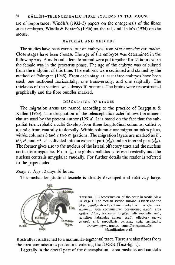
Stage 1. Age 12 days 16 hours.
The medial longitudinal fascicle is already developed and relatively large.
TEXT-FIG. 1. Reconstruction of the brain in medial viewin stage 1. The median section surface is black and thefibre bundles developed are marked with whole lines.
tLtn. a.com.p., area commissurae posterioris; a.opt., areaoptica; f.l.m., fasciculus longitudinalis medialis; hab.,ganglion habenulae anlage; n.olf., olfactory nerve;st.med., stria medullaris; st.term., stria terminalis;
tr.mam.-tegm., tractus mammillo-tegmentalis.Magnification x65.
Rostrally it is attached to a mammillo-tegmental tract. There are also fibres fromthe area commissurae posterioris entering the fascicle (Text-fig. 1).
Laterally in the dorsal part of the diencephalon—area medialis and caudalis

KALLEN—TELENCEPHALIC FIBRE SYSTEMS IN THE MOUSE 89
thalami—there is a thin fibre layer, st. med., which seems to split up in the dorsalpart of the area caudalis, i.e. the habenular anlage. Ventrally the fibres arecollected into a relatively compact bundle, lying dorsal to the foramen Monroiand ending in the area optica. Just rostrally hereto a few fibres, st. term., con-nect the area optica with the caudal part of the &n-layer in the telencephalon.
The olfactory nerve enters the telencephalon rostrally at the limit betweenthe area dorsalis telencephali and the cell column c. No fibres can be seen aroundthe place of entrance, so there are no secondary olfactory tracts developed. Noother telencephalic fibres can be seen.
The nuclear differentiation has proceeded far at this stage. There are twomigration layers developed in the columns b and c.
Stage 2. Age 13 days 6 hours.
A bundle runs caudally from the bulbus olfactorious in this stage, the striaolfactoria lateralis (Text-fig. 2). It lies ventral to the limit between the pallium
TEXT-FIG. 2. Reconstruction of the brain inlateral view in stage 2. The shadowed part is theventral part of the pallium (area dorsalis telen-cephali or d). dn, second migration layer in thepallium; n.olf., olfactory nerve; st.o.l., stria
olfactoria lateralis. Magnification x35.
(d11) and part of the subpallium (clxt) and lateral to the latter structure (Plate,fig. A). Caudally it can be followed to the caudal border of the foramen Monroi.Fibres from it enter the pallial cortex and c\xt. No medial olfactory stria can beseen.
Two bundles begin in c[nt (Text-fig. 3). In the rostral part of it—the futureglobus pallidus—a thick bundle begins and runs caudally to the caudal borderof the foramen Monroi where it enters the diencephalon and spreads over theoptic area and the hypothalamus, the area medialis and caudalis thalami. Itrepresents an ansa lenticularis. From the caudal part of c*nt fibres collect in athin layer, lying dorsal to the ansa lenticularis and crossing this bundle on alevel with the foramen Monroi. Some fibres also come from c11 and b11—i.e.anlagen of the amygdaloid nuclei—and run through b11—i.e. the anlage of thebed of stria terminalis. It must therefore represent a stria terminalis. The positionof the bundle in relation to the ansa lenticularis is seen in Text-figs. 3 and 4.
The relatively diffuse bundle, described as st. med. in stage 1, is now collectedin the rostro-dorsal part of the diencephalon. It still connects the anlage of thehabenular ganglion with the area optica and now also with the area rostralis

90 KALLEN—TELENCEPHALIC FIBRE SYSTEMS IN THE MOUSE
thalami. It thus lies in the same position as the stria medullaris of the adultbrain and is connected with the area optica as is the stria medullaris. It musttherefore represent a stria medullaris. As is apparent from Text-fig. 4, the stria
sl.term.
TEXT-FIG. 3. Reconstruction of the brain in dorsal view instage 2. The hemisphere is cut horizontally and the sectionsurface is black. The shadowed mass is cfnt, i.e. the anlage of,amongst other structures, the globus pallidus. dns.lent., ansalenticularis; b11, second migration layer of the middle subpallialcolumn; cTI, second migration layer of the dorsalmost subpallial
column; st.term., stria terminalis. Magnification x35.
terminalis, the stria medullaris, and the ansa lenticularis cross at the caudallevel of the foramen Monroi. A fibre bundle then connects the stria terminalisand the stria medullaris. Such a connexion has been described in the adult brainby Gurdjian (1925) among others.
.term.
TEXT-FIG. 4. Reconstruction of the brain in medial view in stage2. The median section surface is black, the hidden parts of thehemisphere contour are dotted, ans.lent., ansa lenticularis;st.med., stria medullaris; st.term., stria terminalis; st.term.-med.,
connexion between stria terminalis and medullaris.Magnification x 35.
Stage 3. Age 13 days 19 hours.The lateral olfactory stria has the same position and much the same appear-
ance as in the previous stage, but is a little denser. A very short and indistinctmedial olfactory stria can be seen at this stage.
The stria medullaris has grown bigger but is built up in the same way as inprevious stages. The ansa lenticularis seems to send fibres chiefly to the areacaudalis and medialis thalami, the hypothalamic fibres being rather insignificant(Text-fig. 5).
A new, rather diffuse bundle can be observed in this stage. It begins in thehypothalamus with rather big cells of origin and runs in a rostral directiontowards the area optica and the region immediately rostral to it. This bundlemust represent the first-developed part of the medial forebrain bundle (Text-fig. 5). The stria terminalis is of the same appearance as in earlier stages.

KALLEN—TELENCEPHALIC FIBRE SYSTEMS IN THE MOUSE 91
A commissural bundle is now present in the commissura anterior. It extendsfrom the caudal part of c11, on a level with and caudal to the foramen Monroi,to the corresponding part of the other side, and is situated between the c1 andc11 layers.
TEXT-FIG. 5. Reconstruction of the brain in medialview in stage 3. The median section surface isblack, ans.lent., ansa lenticularis;/././»., fasciculuslongitudinalis medialis; m.f.b., medial forebrainbundle; st.med., stria medullaris; st.term., stria
terminalis. Magnification x 25.
Cortical fibres can now also be seen for the first time, apart from the ascendingfibres from the stria olfactoria lateralis to the lateral part of d11 (the anlage ofthe pyriform cortex) previously described. The new fibres run from the latero-caudal part of d11 into c}nt where they fuse with the ansa lenticular is.
Stage 4. Age 14 days 7 hours.
The lateral olfactory stria is of the same appearance as in the former stage,but the medial stria has grown much bigger. It is, however, diffuse and splits upin the septum, which has grown important here, the b11 part being large. There isno interbulbar component of the medial stria.
A very small tertiary olfactory radiation is present as a bundle between theseptum and the medial part of the cortex. This septo-cortical bundle is rostrallyrather diffuse but caudally densely packed, and it must represent the rudiment ofthe fornix. There are also a few fibres present between the septum and thetuberculum olfactorium-anlage (61), but no diagonal band is developed.
The stria terminalis is better developed than at earlier stages and has a strongerrostral curvature. It ends in the area optica and the area rostralis thalami.
The medial forebrain bundle is also more strongly developed and extends intothe latero-caudal part of the tuberculum olfactorium. Ventral to it lies a smallbut very distinct bundle which connects the rostral part of the hypothalamuswith the supra-optic region.
The cortical fibres form a large association system of transverse bundles,ending in the cortex layer. A large number of descending fibres penetrate thecu formation as a capsula interna, and cross partly in the commissura anterior,the rest lying lateral to the ansa lenticularis as a lateral forebrain fascicle.
92 KALL£N—TELENCEPHALIC FIBRE SYSTEMS IN THE MOUSE
The commissura anterior still contains only one fibre bundle, which now,however, collects fibres from the following parts: the caudal part of c11, d11 (seeabove), and the bed nucleus of the commissura anterior (a derivate from b).
Stage 5. Age 15 days 12 hours.
This stage is very similar to the preceding one. The medial telencephalicfascicle extends up to the rostral part of the tuberculum olfactorium. The lateralfascicle is made up of the ansa lenticularis, which ends in the area caudalis andmedialis thalami and with a few fibres in the area commissurae posterioris, andof the capsula interna, which also runs down into the area fasciculi longitudinalismedialis and area tuberculi posterioris.
Stage 6. Age 16 days 12 hours.
This stage is similar to the previous one. A faintly developed diagonal bandcan be seen: a few fibres run from a in a ventrolateral direction.
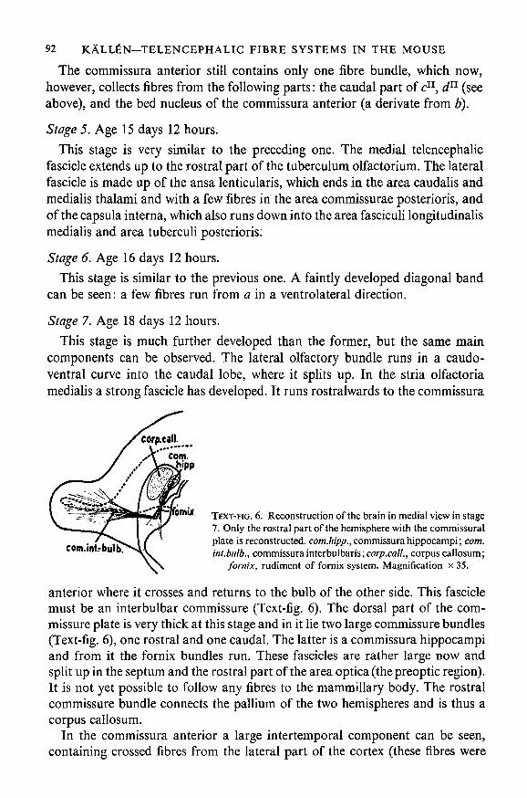
Stage 7. Age 18 days 12 hours.
This stage is much further developed than the former, but the same maincomponents can be observed. The lateral olfactory bundle runs in a caudo-ventral curve into the caudal lobe, where it splits up. In the stria olfactoriamedialis a strong fascicle has developed. It runs rostralwards to the commissura
ornof
com. int.-
TEXT-FIG. 6. Reconstruction of the brain in medial view in stage7. Only the rostral part of the hemisphere with the commissuralplate is reconstructed, com.hipp., commissura hippocampi; com.int.bulb., commissura interbulbaris; corp.cali, corpus callosum;
fornix, rudiment of fornix system. Magnification x 35.
anterior where it crosses and returns to the bulb of the other side. This fasciclemust be an interbulbar commissure (Text-fig. 6). The dorsal part of the com-missure plate is very thick at this stage and in it lie two large commissure bundles(Text-fig. 6), one rostral and one caudal. The latter is a commissura hippocampiand from it the fornix bundles run. These fascicles are rather large now andsplit up in the septum and the rostral part of the area optica (thepreoptic region).It is not yet possible to follow any fibres to the mammillary body. The rostralcommissure bundle connects the pallium of the two hemispheres and is thus acorpus callosum.
In the commissura anterior a large intertemporal component can be seen,containing crossed fibres from the lateral part of the cortex (these fibres were
KALLfiN—TELENCEPHALIC FIBRE SYSTEMS IN THE MOUSE 93
already present in the previous stage) and from c11 in the caudal lobe, an anlageof the amygdaloid nuclei. The interbulbar commissure and, caudally, a bundlefrom the stria terminalis also enter.
The capsula interna is strongly developed in this stage and it now lies rostralto the large ansa lenticularis. The latter is dorsally crossed by the stria termina-lis, which lies in the b11 layer. The stria terminalis is also strongly developed andstill extends from the caudal part of c11, c}nt, and b11 to the preoptic area and thearea rostralis thalami.
RELATION BETWEEN ONTOGENY AND COMPARATIVE ANATOMY OFFIBRE BUNDLES
In a paper on the development of the telencephalic fibres in the pig, Shaner(1936) supposed that a repetition of phylogenetic evolution takes place duringontogeny. He sought an arrangement of the fibres in young stages which re-called the condition in adult fishes. The present author has tried to correlatethe ontogeny of the fibres with their comparative anatomy on the basis of theabove results.
If the fibre anatomy in adult mammals and adult lower vertebrates is com-pared, the following three groups of fibre systems can be distinguished:
1. Bundles which are common to all species, e.g. commissura interbulbaris.2. Bundles present in lower vertebrates but absent in mammals, e.g. secondary
olfactory fibres to the diencephalon.3. Bundles present in mammals but absent in lower vertebrates, e.g. stria
terminalis.If the phylogenetic evolution of the fibres is repeated in ontogeny, it would be
expected that group 1 and, in part at any rate, group 2 would develop beforegroup 3, and that components in group 2 would become completely atrophiedor remain as rudimentary bundles.
The extent of development of the secondary and tertiary olfactory fibres variesmuch in the brains of different species. In fishes there are secondary connexionswith the whole of the telencephalon and with large parts of the diencephalon,especially with the habenular ganglion and the hypothalamus. The conditionsvary considerably in different species, however (see, among others, Holmgren,1920; Backstrom, 1924; Heier, 1948). In urodeles the secondary fibres endchiefly in the telencephalon and are especially well developed in the region closeto the bulb in the nucleus olfactorious anterior (Herrick, 1948). In amniotesthe secondary radiation terminates in the nucleus of the lateral olfactory tract,the medial and lateral amygdaloid nuclei, the tuberculum olfactorium, and thelobus pyriformis (cf. Kappers, Huber, & Crosby, 1936).
During ontogeny in the mouse, the olfactory fibres develop rather early andterminate in the same regions as in the adult brain. On the other hand, nosecondary connexions with the diencephalon have been observed. Windle's
94 KALLEN—TELENCEPHALIC FIBRE SYSTEMS IN THE MOUSE
(1935) results on the ontogeny of the fibres in the cat indicate that such fibresmay exist. He describes a tractus olfacto-subthalamicus (fibres to the arearostralis thalami according to our nomenclature) and a tractus olfacto-hypo-thalamicus. Windle was not sure, however, that they really represent secondaryolfactory fibres. It seems more likely to the present author that they are fascicles,beginning more caudally within the cell column c.
In fishes, most telencephalic nuclei are connected with the thalamus, thehabenular ganglion, and the hypothalamus by both ascending and descendingbundles. In urodeles, too, the greatest part of the hemisphere seems to beconnected with lower centres, even if an accumulation of such connexions seemsto exist in the caudal part of the hemisphere (in Herrick's strio-amygdaloidcomplex). In amniotes the subpallial connexions with lower centres seem to bemainly restricted to the strio-amygdaloid complex, viz. to c11, c]ni, and inmammals also to dv. In addition, the pallium has large and important connexionswith the brain stem in these animals.
During ontogeny in the mouse, the first fibre system to develop in the telen-cephalon is the stria terminalis, i.e. a system which is lacking in fishes, where noamygdaloid complex is developed. The fibres ascending to cell column b arepart of the medial forebrain bundle, a large and important component in thebrains of lower animals. These fibres develop very late in the mouse. The pallio-habenular bundles also develop late—they are not present in the oldest stageinvestigated here—but the pallio-thalamic fibres develop much earlier. In lowervertebrates the former system exists, but the latter acquires importance only inhigher animals.
Similar observations can be made on the commissural system. A commissurecommon to all vertebrates is the commissura interbulbaris. In the mouse itdevelops late—later than the commissure fibres from the amygdaloid anlage(the caudal part of cu and c}Dt) which is lacking in fishes and amphibians.
RELATION BETWEEN FIBRE DEVELOPMENT AND CELL MIGRATIONPROCESSES
Many authors have supposed that an important relation exists between cellmigration processes in the central nervous system and the development of thefibre fascicles. Kappers (1917 and other papers) tried to explain the phylogeneticshifting of nuclei by supposing that neurites which grow past influence theposition of the cell bodies. Many papers have been published on this problem.
From the observations on stage 1 in the present paper it is apparent that thenuclear differentiation has already proceeded far before the fibres start todevelop. In the subpallium a second migration layer is developed both incolumn b and in column c (Plate, fig. B). The formation of migration areas andmigration layers can thus take place before the development of fibre fasciclesin the region in question. The subdivision of the migration layer into parts,nuclear anlagen, seems, however, to take place simultaneously with the ingrowth
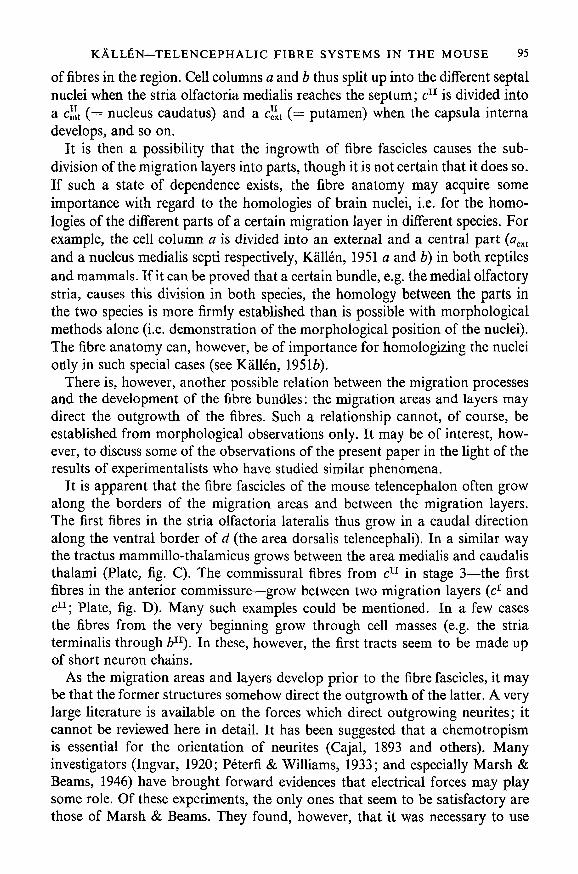
KALL£N—TELENCEPHALIC FIBRE SYSTEMS IN THE MOUSE 95
of fibres in the region. Cell columns a and b thus split up into the different septalnuclei when the stria olfactoria medialis reaches the septum; c11 is divided intoa c"t ( = nucleus caudatus) and a c"t ( = putamen) when the capsula internadevelops, and so on.
It is then a possibility that the ingrowth of fibre fascicles causes the sub-division of the migration layers into parts, though it is not certain that it does so.If such a state of dependence exists, the fibre anatomy may acquire someimportance with regard to the homologies of brain nuclei, i.e. for the homo-logies of the different parts of a certain migration layer in different species. Forexample, the cell column a is divided into an external and a central part (aext
and a nucleus medialis septi respectively, Kallen, 1951 a and b) in both reptilesand mammals. If it can be proved that a certain bundle, e.g. the medial olfactorystria, causes this division in both species, the homology between the parts inthe two species is more firmly established than is possible with morphologicalmethods alone (i.e. demonstration of the morphological position of the nuclei).The fibre anatomy can, however, be of importance for homologizing the nucleionly in such special cases (see Kallen, 19516).
There is, however, another possible relation between the migration processesand the development of the fibre bundles: the migration areas and layers maydirect the outgrowth of the fibres. Such a relationship cannot, of course, beestablished from morphological observations only. It may be of interest, how-ever, to discuss some of the observations of the present paper in the light of theresults of experimentalists who have studied similar phenomena.
It is apparent that the fibre fascicles of the mouse telencephalon often growalong the borders of the migration areas and between the migration layers.The first fibres in the stria olfactoria lateralis thus grow in a caudal directionalong the ventral border of d (the area dorsalis telencephali). In a similar waythe tractus mammillo-thalamicus grows between the area medialis and caudalisthalami (Plate, fig. C). The commissural fibres from c11 in stage 3—the firstfibres in the anterior commissure—grow between two migration layers (c1 andc11; Plate, fig. D). Many such examples could be mentioned. In a few casesthe fibres from the very beginning grow through cell masses (e.g. the striaterminalis through b11). In these, however, the first tracts seem to be made upof short neuron chains.
As the migration areas and layers develop prior to the fibre fascicles, it maybe that the former structures somehow direct the outgrowth of the latter. A verylarge literature is available on the forces which direct outgrowing neurites; itcannot be reviewed here in detail. It has been suggested that a chemotropismis essential for the orientation of neurites (Cajal, 1893 and others). Manyinvestigators (Ingvar, 1920; Peterfi & Williams, 1933; and especially Marsh &Beams, 1946) have brought forward evidences that electrical forces may playsome role. Of these experiments, the only ones that seem to be satisfactory arethose of Marsh & Beams. They found, however, that it was necessary to use

96 KALIAN—TELENCEPHALIC FIBRE SYSTEMS IN THE MOUSE
relatively strong currents to obtain an effect, and they doubted whether suchstrong currents were present during normal development, though according toYoung (1948) this is possible.
Weiss (1934, 1939, 1941, 1950) has brought forward evidence against thegalvanotropic theory of nerve orientation. His experiments and those ofHarrison (1935) and others suggest that mechanical factors, especially ultra-structural conditions in the substrate, may play the most important role.
How do these results fit with the observations made above? That the fibresoften grow along the borders of the migration areas and layers might be ex-plained by the fact that these parts are built up in a somewhat different wayfrom the areas containing cell bodies (e.g. the migration layers). These 'path-ways' between the migration areas might direct the fibre growth, possiblybecause of the existence of orientated ultrastructure (as in Weiss's theory).
If these 'pathways' really are of importance for the outgrowth of the fibres,it would be expected that the bundles of neurites would be less compact inbrains where the 'pathways' are less distinct, e.g. where the cells do not migrateas far as they do in the mammalian brain. In amphibians, dipnoans, elasmo-branchs, and Petromyzon migration in the telencephalon is rather feeble(Kallen, 19516). In these species the secondary olfactory connexions are alsodiffuse, forming a radiatio olfactoria which can, it is true, be divided intodifferent parts, but not into distinct bundles. The author has also observed inPalmgren-stained Rana material that from the very beginning these fibres growvery diffusely. In teleosts and in amniotes, where the migration processes in thetelencephalon are much more pronounced and cells, arranged in migrationlayers, &C, fill the whole of the brain wall, the secondary olfactory projectionis made up of distinct tracti olfactorii.
The fibres formed within a differentiation centre do not, however, growrandomly along all possible 'pathways' formed by the migration layers. Forinstance, the neurites formed within the bulbus olfactorius anlage grow onlyalong the lateral (morphologically caudal) part of the 'pathway' between dand the area ventralis telencephali, forming the lateral olfactory stria. Only inlater stages do neurites grow also in the opposite direction. Similar phenomenacan be observed in the habenulofugal fibres, in the commissural fibres, and soon. These observations indicate that there is some general orientating factoralso present—possibly of a bioelectrical nature such as Marsh & Beams'sresults suggest.
There may be yet another relation present between migration areas and out-growing neurites. According to CoghilPs (1929) observations and Burr's (1932)experiments with transplanted nasal pits, neurites growing in from the peripheryare attracted by every local cell proliferation in the brain. In the same way,neurites growing out from the brain are attracted by cell proliferations in extra-central tissues (Detwiler, 1923,1936; Weiss, 1939, and others). According to Weissalso intracerebral fibres may be attracted by proliferating cells. Observations

KALLEN—TELENCEPHALIC FIBRE SYSTEMS IN THE MOUSE 97
on malformations made by Brodal, Bonnevie & Harkmark (1944), Bonnevie& Brodal (1946), and Brodal (1945, 1946) show that fibres growing into thecerebellum invade 'wrong' regions, if the 'right' regions are lacking. These'wrong' regions are then always in a state of rapid proliferation. Harkmark(1954), however, claims that his later experiments do not support this opinion.
Considered in conjunction with the fact that the nuclei develop from migrationareas, which, according to Bergquist (1932), coincide in position with prolifera-tion centres, this hypothesis explains the fact that fibres grow into the migrationareas they pass; as, for instance, the stria olfactoria lateralis branches into dand c which surround the bundle, and large fascicles branch from the ansa lenti-cularis and enter the area medialis and caudalis thalami, both lying dorsal tothe ansa. Many such examples could be given.
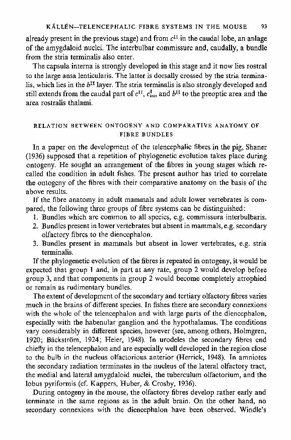
B
TEXT-FIG. 7. Scheme of the mode of developmentof the fibre tracts to the preoptic region. Furtherexplanation in text. A, stage 1. B, stage 2. C,
stage 3.
If this hypothesis is correct, the differentiation centres ought to be connectedwith all the migration areas lying in the direction of the outgrowing neurites.As this is not the case, it might be supposed that some sort of saturation isreached in an area when it has been entered by a certain number of fibres. Thefollowing observations support this suggestion. In the first mouse stage des-cribed above, two systems are developed at the di-telencephalic junction: thestria terminalis and the stria medullaris. The stria terminalis extends from thecaudal part of c in a ventral direction and ends immediately in the rostralpart of the area optica. The stria medullaris extends from the habenular anlagein a ventral direction and ends in the caudal part of the area optica. When athird system, the ansa lenticularis, develops from the rostral part of c, it grows
98 KALLfiN—TELENCEPHALIC FIBRE SYSTEMS IN THE MOUSE
caudally and ends in a part of the area optica not previously reached by anyfibres: caudal to the end-branches of the stria medullaris. This bundle also endsin the area rostralis, medialis, and caudalis thalami, all areas that have notpreviously been reached by fibres. Finally, the medial forebrain bundle grows upfrom the hypothalamus and ends in the part of the area optica not previouslyreached by fibres, i.e. the ventro-rostral part. These facts are schematicallyshown in Text-fig. 7.
The above observations and the experimental results to be found in theprevious literature on the subject thus suggest possible mechanisms for theformation of the first fibre bundles in the brain. Later, when the proliferationprocesses have ceased and the migration layers are dissolved by a scattering ofthe cells, new fibre systems are formed, but it is not impossible that the primaryfibre skeleton then acts as a system of'pioneering fibres' (Weiss, 1950), directingthe outgrowth of the secondary bundles.
SUMMARY
1. The ontogeny of the fibres in the telencephalon of Mus musculus has beenstudied. Reconstructions have been made using seven different stages. Theformation of some telencephalic and diencephalic fascicles is described.
2. The agreement supposed by Shaner (1936) to exist between the phylogenyand ontogeny of the fascicles is not supported by the present investigation.
3. The migration areas and migration layers can develop before the fibrefascicles are formed, and thus without any influence from them. The divisionof the migration layers into nuclei takes place simultaneously with the entranceof the fibres into the layers, and hence a causal relation may exist between thesetwo processes. The fibre anatomy may be of some importance with regard toestablishing the homologies of the late subdivisions of the migration layers.
4. The spaces between the migration areas and layers seem to act as pathwaysfor the outgrowing neurites. The migration areas, which are also proliferationcentres, seem to attract the neurites.
REFERENCES
BACKSTROM, K. (1924). Contributions to the forebrain morphology in selachians. Acta zooi, Stockh.5, 123-270.
BERGQUIST, H. (1932). Zur Morphologie des Zwischenhirns bei niederen Wirbeltieren. Acta zool,Stockh. 13, 57-303.& KALLEN, B. (1953). Studies on the topography of the migration areas in the vertebrate brain.Acta anat. 17, 353-69.
(1954). Notes on the early histogenesis and morphogenesis of the central nervous systemin vertebrates. / . comp. Neurol. In press.
BONNEVIE, K., & BRODAL, A. (1946). Hereditary hydrocephalus in the house mouse. IV. The de-velopment of the cerebellar anomalies during foetal life with notes on the normal developmentof the mouse cerebellum. Skr. norske VidenskAkad. 1 MN. Kl. (IV), pp. 1-60.
BRODAL, A. (1945). Defective development of the cerebellar vermis (partial agenesis) in a child. Skr.norske VidenskAkad. 1 MN. Kl. (Ill), pp. 1-40.
KALLEN—TELENCEPHALIC FIBRE SYSTEMS IN THE MOUSE 99
BRODAL, A., (1946). Correlated changes in nervous tissue in malformations of the central nervoussystem. / . Anat., Lond. 80, 88-93.BONNEVIE, K., & HARKMARK, W. (1944). Hereditary hydrocephalus in the house mouse.
II. The anomalies of the cerebellum. Partial defective development of the vermis. Skr. norskeVidenskAkad. 1 MN. Kl. (VIII), pp. 1-42.
BURR, H. S. (1932). An electrodynamic theory of development suggested by studies of proliferationrates in the brain of Amblystoma. / . comp. Neurol. 56, 347-71.
CAJAL, S. RAMON Y (1893). La r6tine des vert6br6s. Cellule, 9, 121-258.COGHILL, G. E. (1929). Anatomy and the Problem of Behaviour. New York: Macmillan.DETWILER, S. R. (1923). Experiments on the reversal of the spinal cord in Amblystoma embryos at
the level of the anterior limb. J. exp. Zool. 38, 293-321.(1936). Neuroembryology. An Experimental Study. New York: Macmillan.
GURDJIAN, E. S. (1925). Olfactory connections in the albino rat, with special reference to the striamedullaris and the anterior commissure. / . comp. Neurol. 38, 127-63.
HARKMARK, W. (1954). Cell migrations from the rhombic lip to the inferior olive, the nucleus rapheand the pons. A morphological and experimental investigation on chick embryos. / . comp.Neurol. 100, 115-210.
HARRISON, R. G. (1935). The origin and development of the nervous system studied by the methods ofexperimental embryology. Proc. roy. Soc. B, 118, 155-96.
HEIER, P. (1948). Fundamental principles in the structure of the brain. A study of the brain ofPetromyzon fluviatilis. Acta anat. Suppl. VILI.
HERRICK, C. J. (1948). The Brain of the Tiger Salamander. Chicago: The University Press.HOLMGREN, N. (1920). Zur Anatomie und Histologie des Vorderhirns der Teleostier. Acta zool.,
Stockh. 1, 137-315.INGVAR, S. (1920). Reaction of cells to the galvanic current in tissue cultures. Pros. Soc. exp. Biol.,
N.Y. 17, 198-9.KALLEN, B. (1951a). The nuclear development in the mammalian forebrain with special regard to the
subpallium. K. fysiogr. Sa'llsk. Lund Handl. N.F. 61, no. 9, pp. 1-43.(19516). Embryological studies on the nuclei and their homologization in the vertebrate fore-
brain. K. fysiogr. Scillsk. Lund Handl. N.F. 62, no. 5, pp. 1-36.KAPPERS, C. U. ARIENS (1917). Further contributions on neurobiotaxis. IX. An attempt to compare
the phenomenon of neurobiotaxis with other phenomena of taxis and tropism. The dynamicpolarization of the neurone. / . comp. Neurol. 27, 261-98.HUBER, C , & CROSBY, E. C. (1936). The Comparative Anatomy of the Nervous System of Verte-
brates, including Man, vol. ii. New York: Macmillan.MARSH, G., & BEAMS, H. W. (1946). In vitro control of growing chick nerve fibers by applied electric
currents. / . cell. comp. Physiol. 27, 139-57.PALMGREN, A. (1948). A rapid method for selective silver staining of nerve fibres and nerve endings
in mounted paraffin sections. Acta zool., Stockh. 29, 377-92.PETERFI, T., & WILLIAMS, S. C. (1933). Elektrische Reizversuche an geziichteten Gewebezellen.
I. Versuche an Nervenzellen. Arch. exp. Zellforsch. 14, 210-57.SHANER, R. F. (1936). Development of the finer structure and fiber connections of globus pallidus,
corpus of Luys and substantia nigra in the pig. / . comp. Neurol. 64, 213-33.TELLO, J. F. (1934). Les differentiations neurofibrillaires dans le prosence"phale de la souris de 4 a 5
millimetres. Trab. Lab. Invest, biol. Univ. Madr. 29, 339-95.WEISS, P. (1934). In vitro experiments on the factors determining the course of the outgrowing nerve
fiber. / . exp. Zool. 68, 393-448.(1939). Principles of Development. A Text in Experimental Embryology. New York: Henry
Holt & Co.(1941). Nerve patterns. The mechanism of nerve growth. 3rd Symposium on Development
and Growth. Growth, 5, 163-203.(1950). The deplantation of fragments of nervous system in amphibians. I. Central reorganiza-
tion and the formation of nerves. / . exp. Zool. 113, 397-462.WINDLE, W. F. (1932a). The neurofibrillar structure of the 7 mm. cat embryo. / . comp. Neurol. 55,
99-138.(1932Z>). The neurofibrillar structure of the 5-5 mm. cat embryo. / . comp. Neurol. 55, 315-31.

100 KALLfiN—TELENCEPHALIC FIBRE SYSTEMS IN THE MOUSE
WINDLE, W. F. (1933). Neurofibrillar development in the central nervous system of cat embryos between8 and 12 mm. long. / . comp. Neurol. 58, 643-725.(1935). Neurofibrillar development of cat embryos. Extent of development in the telencephalon
and diencephalon up to 15 mm. / . comp. Neurol. 63,139-72.& BAXTER, R. E. (1936). The first neurofibrillar development in albino rat embryos. / . comp.
Neurol. 63, 173-85.YOUNG, J. Z. (1948). Growth and differentiation of nerve fibres. Symp. Soc. exp. Biol. 2, 57-74.
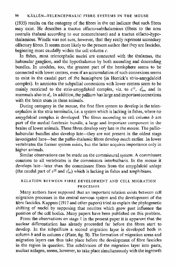
EXPLANATION OF PLATE
FIG. A. Transverse section through the brain in stage 2. The section cuts the hemispheres and thefigure shows its lateroventral part. Stained by Palmgren's method, c1, first migration layer in the dorsal-most subpallial column; c11, second migration layer of the same column; d11, second migration layerof the pallium; st.o.L, stria olfactoria lateralis. Magnification x60.
FIG. B. Transverse section through the brain in stage 1, showing the development of the migrationareas and cell columns in the telencephalon and diencephalon. a.c.th., area caudalis thalami;a.com.post., area commissurae posterioris; a.m.th., area medialis thalami; c\ first migration layer inthe dorsalmost subpallial column; c11, second migration layer in the same column. Magnification X 20.
FIG. C. Sagittal section through the brain in stage 5, showing the position of the mammillo-thalamic tract (tr.mam.-thal.) between area medialis thalami (a.m.th.) and area caudalis thalami(a.c.th.). Stained by Palmgren's method. Magnification x 65.
FIG. D. Transverse section through the telencephalon in stage 4, showing the bundle of the anteriorcommissure (com.ant.) situated between the migration layers c1 and c11 in the dorsalmost subpallialcolumn. Magnification x 65.

J. Embryol. exp. Morph.
st.o.L.
B. KALL^N
Related Documents


