RESEARCH Open Access The effects of hyperoxia on microvascular endothelial cell proliferation and production of vaso-active substances Ilias Attaye 1,2* , Yvo M. Smulders 3 , Monique C. de Waard 1 , Heleen M. Oudemans-van Straaten 1 , Bob Smit 1 , Michiel H. Van Wijhe 2 , Rene J. Musters 2 , Pieter Koolwijk 2† and Angelique M. E. Spoelstra–de Man 1† * Correspondence: [email protected] † Equal contributors 1 Department of Intensive Care, VU University Medical Center, Amsterdam, The Netherlands 2 Department of Physiology, VU University Medical Center, Amsterdam, The Netherlands Full list of author information is available at the end of the article Abstract Background: Hyperoxia, an arterial oxygen pressure of more than 100 mmHg or 13% O 2 , frequently occurs in hospitalized patients due to administration of supplemental oxygen. Increasing evidence suggests that hyperoxia induces vasoconstriction in the systemic (micro)circulation, potentially affecting organ perfusion. This study addresses effects of hyperoxia on viability, proliferative capacity, and on pathways affecting vascular tone in cultured human microvascular endothelial cells (hMVEC). Methods: hMVEC of the systemic circulation were exposed to graded oxygen fractions of 20, 30, 50, and 95% O 2 for 8, 24, and 72 h. These fractions correspond to 152, 228, 380, and 722 mmHg, respectively. Cell proliferation and viability was measured via a proliferation assay, peroxynitrite formation via anti-nitrotyrosine levels, endothelial nitric oxide synthase (eNOS), and endothelin-1 (ET-1) levels via q-PCR and western blot analysis. Results: Exposing hMVEC to 50 and 95% O 2 for more than 24 h impaired cell viability and proliferation. Hyperoxia did not significantly affect nitrotyrosine levels, nor eNOS mRNA and protein levels, regardless of the exposure time or oxygen concentration used. Phosphorylation of eNOS at the serine 1177 (S1177) residue and ET-1 mRNA levels were also not significantly affected. Conclusions: Exposure of isolated human microvascular endothelial cells to marked hyperoxia for more than 24 h decreases cell viability and proliferation. Our results do not support a role of eNOS mRNA and protein or ET-1 mRNA in the potential vasoconstrictive effects of hyperoxia on isolated hMVEC. Keywords: Hyperoxia, Endothelial cells, In vitro, eNOS, ET-1, Peroxynitrite Background Supplemental oxygen (O 2 ) is frequently administered in the hospital, especially in crit- ically ill patients. For years, oxygen therapy focused on avoiding hypoxia, arterial oxy- gen levels below 70 mmHg or 9% O 2, often accepting a state of hyperoxia, arterial oxygen levels of more than 100 mmHg or 13% O 2 [1]. The consequences of hyperoxia, however, remain unclear and have been a topic of debate for several decades in which both beneficial as well as deleterious effects have been reported [2–5]. Intensive Care Medicine Experimental © The Author(s). 2017 Open Access This article is distributed under the terms of the Creative Commons Attribution 4.0 International License (http://creativecommons.org/licenses/by/4.0/), which permits unrestricted use, distribution, and reproduction in any medium, provided you give appropriate credit to the original author(s) and the source, provide a link to the Creative Commons license, and indicate if changes were made. Attaye et al. Intensive Care Medicine Experimental (2017) 5:22 DOI 10.1186/s40635-017-0135-4

Welcome message from author
This document is posted to help you gain knowledge. Please leave a comment to let me know what you think about it! Share it to your friends and learn new things together.
Transcript
RESEARCH Open Access
The effects of hyperoxia on microvascularendothelial cell proliferation andproduction of vaso-active substancesIlias Attaye1,2* , Yvo M. Smulders3, Monique C. de Waard1, Heleen M. Oudemans-van Straaten1, Bob Smit1,Michiel H. Van Wijhe2, Rene J. Musters2, Pieter Koolwijk2† and Angelique M. E. Spoelstra–de Man1†
* Correspondence: [email protected]†Equal contributors1Department of Intensive Care, VUUniversity Medical Center,Amsterdam, The Netherlands2Department of Physiology, VUUniversity Medical Center,Amsterdam, The NetherlandsFull list of author information isavailable at the end of the article
Abstract
Background: Hyperoxia, an arterial oxygen pressure of more than 100 mmHg or13% O2, frequently occurs in hospitalized patients due to administration ofsupplemental oxygen. Increasing evidence suggests that hyperoxia inducesvasoconstriction in the systemic (micro)circulation, potentially affecting organperfusion. This study addresses effects of hyperoxia on viability, proliferative capacity,and on pathways affecting vascular tone in cultured human microvascularendothelial cells (hMVEC).
Methods: hMVEC of the systemic circulation were exposed to graded oxygenfractions of 20, 30, 50, and 95% O2 for 8, 24, and 72 h. These fractions correspond to152, 228, 380, and 722 mmHg, respectively. Cell proliferation and viability wasmeasured via a proliferation assay, peroxynitrite formation via anti-nitrotyrosine levels,endothelial nitric oxide synthase (eNOS), and endothelin-1 (ET-1) levels via q-PCR andwestern blot analysis.
Results: Exposing hMVEC to 50 and 95% O2 for more than 24 h impaired cellviability and proliferation. Hyperoxia did not significantly affect nitrotyrosine levels,nor eNOS mRNA and protein levels, regardless of the exposure time or oxygenconcentration used. Phosphorylation of eNOS at the serine 1177 (S1177) residue andET-1 mRNA levels were also not significantly affected.
Conclusions: Exposure of isolated human microvascular endothelial cells to markedhyperoxia for more than 24 h decreases cell viability and proliferation. Our results donot support a role of eNOS mRNA and protein or ET-1 mRNA in the potentialvasoconstrictive effects of hyperoxia on isolated hMVEC.
Keywords: Hyperoxia, Endothelial cells, In vitro, eNOS, ET-1, Peroxynitrite
BackgroundSupplemental oxygen (O2) is frequently administered in the hospital, especially in crit-
ically ill patients. For years, oxygen therapy focused on avoiding hypoxia, arterial oxy-
gen levels below 70 mmHg or 9% O2, often accepting a state of hyperoxia, arterial
oxygen levels of more than 100 mmHg or 13% O2 [1]. The consequences of hyperoxia,
however, remain unclear and have been a topic of debate for several decades in which
both beneficial as well as deleterious effects have been reported [2–5].
Intensive Care MedicineExperimental
© The Author(s). 2017 Open Access This article is distributed under the terms of the Creative Commons Attribution 4.0 InternationalLicense (http://creativecommons.org/licenses/by/4.0/), which permits unrestricted use, distribution, and reproduction in any medium,provided you give appropriate credit to the original author(s) and the source, provide a link to the Creative Commons license, andindicate if changes were made.
Attaye et al. Intensive Care Medicine Experimental (2017) 5:22 DOI 10.1186/s40635-017-0135-4
Clinically, hyperoxia is associated with negative outcomes such as acute lung injury
[6], increased infarct size in patients with a myocardial or cerebral infarction [7, 8] and
increased mortality in patients after cardiac arrest [9, 10]. In contrast, others report
beneficial effects with improved organ function after cardiac arrest [11] and antimicro-
bial activity with a reduction of surgical site infections [12, 13].
Furthermore, hyperoxia can cause significant hemodynamic alterations [2, 3] and has
been reported to induce vasoconstriction in several (cerebral, coronary, skeletal muscle,
and retinal) [14–17], but not all (renal, mesenteric) [18, 19], vascular beds. This vaso-
constriction appears mainly to occur at microvascular level, since the diameter of large
conduit arteries remains constant [15, 20]. It may lead to heterogeneity of the microcir-
culation with loss of functional capillary density and impaired organ perfusion [3, 21],
but may also stabilize hemodynamics in vasodilatory shock [5]. In addition, other stud-
ies show that hyperoxia is able to redistribute blood flow which can protect hepatos-
planchic organs and the kidneys [22, 23].
Crucial in the pathophysiology of a large variety of clinical conditions, such as
sepsis, trauma, or ischemia/reperfusion injury is microvascular dysfunction. Hyper-
oxia is thought to increase reactive oxygen species (ROS) which can damage the
endothelium and can worsen microvascular dysfunction with increased permeabil-
ity, local inflammation and coagulation, and disturbed hemodynamics [24, 25]. It is
therefore important to gain more insight in the effects of hyperoxia on the micro-
vascular endothelium.
Several studies investigated the direct effects of hyperoxia on cultured endothelial
cells with controversial results. Exposure to hyperoxia exerted a toxic effect with
induction of cell death and reduced cellular proliferation in several cell types, such as
human lung microvascular endothelial cells and bovine adrenal capillary endothelial
cells [26, 27]. Exposure to hyperoxia also led to an inflammatory status by increasing
the expression of inflammatory molecules, such as intercellular adhesion molecule 1
(ICAM-1) in human pulmonary artery endothelial cells and human umbilical vein
endothelial cells [28]. Conversely, other studies showed that hyperoxia decreased the
number of apoptotic cells and had anti-inflammatory effects in a rat intestinal
ischemia-reperfusion and sepsis model [29, 30].
Increased formation of ROS also appear to be of pivotal importance with regard to
the effects of hyperoxia on cell viability and vascular tone [25, 31–33].
The exact underlying mechanisms of vasoconstriction following hyperoxia how-
ever remain unclear. The majority of the studies indicate a central role for the
endothelium [16, 34, 35]. Increased oxidative stress can augment the formation of
peroxynitrite (ONOO−), which is formed when superoxide (O2−) reacts with nitric
oxide (NO), resulting in a reduced bioavailability of NO [36]. NO itself is crucial
for endothelium-driven vasodilation and vascular homeostasis [37]. It has also been
hypothesized that hyperoxia decreases NO levels by affecting endothelial nitric
oxide synthase (eNOS), the main enzyme responsible for the production of NO by
the endothelium [34, 35]. Another potential pathway is a hyperoxia-induced de-
crease of cyclooxygenase (COX) activity in endothelial cells, thereby possibly lower-
ing the levels of vasodilatory prostaglandins [38]. Interestingly enough the effects
of hyperoxia on the protein endothelin-1 (ET-1), which is a potent vasoconstrictor,
remain scarcely investigated [39].
Attaye et al. Intensive Care Medicine Experimental (2017) 5:22 Page 2 of 17
Studies investigating the effects of hyperoxia on vascular smooth muscle cells
(VSMC) showed that hyperoxia can induce vasoconstriction via L-type calcium and by
influencing 20-hydroxyeicosatetraenoic acid (20-HETE) production [40, 41].
Unfortunately, most animal and in vitro studies only investigated extreme hyperoxia
(i.e. 95% O2), often using endothelial cells from the retinal or pulmonary circulation
[16, 33, 34, 38, 42–46]. Studies using endothelial cells of the systemic microcirculation
and milder degrees of hyperoxia are scarce.
Taken together, several lines of evidence suggest that hyperoxia affects endothelial
cell viability and proliferative capacity, but also promotes vasoconstriction primarily via
the endothelium. In this study the effects of hyperoxia on isolated human microvascu-
lar endothelial cells (hMVEC) were investigated, using a clinically relevant scale of oxy-
gen exposure. We investigated the effects of hyperoxia on cell viability and
proliferation. We also addressed potential underlying mechanisms by which hyperoxia
induces toxicity and vasoconstriction by studying peroxynitrite formation via protein
tyrosine nitration capacity, the NO pathway via eNOS mRNA and protein levels and
ET-1 mRNA production.
MethodsCell culture
The study was executed in accordance with the Declaration of Helsinki and was ap-
proved by the University Human Subjects Committee of the VU University Medical
Center. Written informed consent was obtained from all healthy donors in accordance
with the institutional guidelines. All healthy foreskins were collected anonymously and
were kindly provided by the Department of Dermatology (VUmc, Amsterdam). Human
microvascular endothelial cells (hMVEC) were isolated from the foreskins and charac-
terized, as previously described, by the presence of CD31, VE-cadherin, and Von
Willebrand factor [47]. Cells were cultured on gelatin-coated wells (1% w/v) in EBM-2
medium supplemented with EGM-2 MV SingleQuots, 100 U/ml penicillin, 100 mg/ml
streptomycin (p/s), 2 mM L-glutamine, 5 U/ml heparin (all from Lonza, Verviers,
Belgium). The fetal calf serum (FCS) was replaced with 10% human platelet lysate
(hPL) which was acquired as previously described [48]. Cells were cultured in an incuba-
tor kept at 37°C with 95% air, 5% CO2, and 95% humidity. Since the cells were cultured
under 5% CO2, the oxygen exposure changed from atmospheric levels (21% O2) to 20%
O2. Confluent cells were washed with 0.5 mM EDTA (Merck Milipore, MA, USA) in
HBSS and were trypsinized with 0.05% trypsin in EDTA/HBSS (Lonza, Verviers, Belgium).
At least three cell donors from passage 9–11 were used for all the experiments.
Hyperoxia exposure
Cells of a single donor were cultured on four different culture plates. The plates were
placed in airtight acryl boxes with moist tissues to maintain humidity. At the start of
each experiment, the boxes were flushed for 5 min with the indicated oxygen concen-
tration (Table 1) by using two outlets. The boxes were thereafter placed in an incubator
and kept at 37 °C. Oxygen concentration in the boxes was measured at the start and
end of each experiment (Additional file 1: Figure S1) using the Microx 4 (PreSens,
Regensburg, Germany). Experiments were halted if the boxes proved not to be airtight,
as determined by a drop in oxygen concentration.
Attaye et al. Intensive Care Medicine Experimental (2017) 5:22 Page 3 of 17
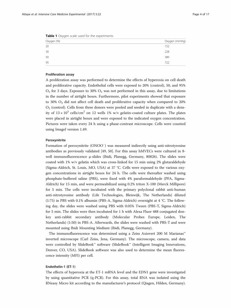
Proliferation assay
A proliferation assay was performed to determine the effects of hyperoxia on cell death
and proliferative capacity. Endothelial cells were exposed to 20% (control), 50, and 95%
O2 for 3 days. Exposure to 30% O2 was not performed in this assay, due to limitations
in the number of airtight boxes. Furthermore, pilot experiments showed that exposure
to 30% O2 did not affect cell death and proliferative capacity when compared to 20%
O2 (control). Cells from three donors were pooled and seeded in duplicate with a dens-
ity of 13 × 103 cells/cm2 on 12 wells 1% w/v gelatin-coated culture plates. The plates
were placed in airtight boxes and were exposed to the indicated oxygen concentration.
Pictures were taken every 24 h using a phase-contrast microscope. Cells were counted
using ImageJ version 1.49.
Peroxynitrite
Formation of peroxynitrite (ONOO−) was measured indirectly using anti-nitrotyrosine
antibodies as previously validated [49, 50]. For this assay hMVECs were cultured in 8-
well immunofluorescence μ-slides (Ibidi, Planegg, Germany, 80826). The slides were
coated with 1% w/v gelatin which was cross-linked for 15 min using 2% glutaraldehyde
(Sigma-Aldrich, St. Louis, MO, USA) at 37 °C. Cells were exposed to the various oxy-
gen concentrations in airtight boxes for 24 h. The cells were thereafter washed using
phosphate-buffered saline (PBS), were fixed with 4% paraformaldehyde (PFA, Sigma-
Aldrich) for 15 min, and were permeabilized using 0.2% triton X-100 (Merck Millipore)
for 5 min. The cells were incubated with the primary polyclonal rabbit anti-human
anti-nitrotyrosine antibody (Life Technologies, Bleiswijk, The Netherlands) diluted
(1:75) in PBS with 0.1% albumin (PBS-A, Sigma-Aldrich) overnight at 4 °C. The follow-
ing day, the slides were washed using PBS with 0.05% Tween (PBS-T, Sigma-Aldrich)
for 5 min. The slides were then incubated for 1 h with Alexa Fluor 488 conjugated don-
key anti-rabbit secondary antibody (Molecular Probes Europe, Leiden, The
Netherlands) (1:50) in PBS-A. Afterwards, the slides were washed with PBS-T and were
mounted using Ibidi Mounting Medium (Ibidi, Planegg, Germany).
The immunofluorescence was determined using a Zeiss Axiovert 200 M Marianas™
inverted microscope (Carl Zeiss, Jena, Germany). The microscope, camera, and data
were controlled by SlideBook™ software (SlideBook™ (Intelligent Imaging Innovations,
Denver, CO, USA). SlideBook software was also used to determine the mean fluores-
cence intensity (MFI) per cell.
Endothelin-1 (ET-1)
The effects of hyperoxia at the ET-1 mRNA level and the EDN1 gene were investigated
by using quantitative PCR (q-PCR). For this assay, total RNA was isolated using the
RNeasy Micro kit according to the manufacturer’s protocol (Qiagen, Hilden, Germany).
Table 1 Oxygen scale used for the experiments
Oxygen (%) Oxygen (mmHg)
20 152
30 228
50 380
95 722
Attaye et al. Intensive Care Medicine Experimental (2017) 5:22 Page 4 of 17
The RNA quality and concentration was determined by Nanodrop (ISOGEN Life
science, De Meern, The Netherlands). 100 μl total copy DNA (cDNA) was synthe-
sized from 1000 ng/ml RNA with the cloned AMV First-Strand cDNA synthesis kit
(Invitrogen). Q-PCR was performed in duplicate using Sybr Green (MESA Green
QPCR Mastermix Plus for Sybr Assay, Eurogentec, Seraing, Belgium) in an ABI7500
RT-PCR machine (Applied Biosystems, Foster City, USA). Primers were self-designed
and synthesized by Invitrogen (Invitrogen, Carlsbad, USA) (Table 2). Primer specifi-
city was tested by homology search with the human genome (BLAST) and was con-
firmed by dissociation curve analysis. Relative expression levels (N-fold differences)
of target genes were calculated using the delta delta Ct method, as previously de-
scribed [51].
Endothelial nitric oxide synthase (eNOS)
NO is difficult to measure directly due to its instability. The effects of hyperoxia were
therefore determined on eNOS, the key enzyme responsible for NO generation in
endothelial cells. The effects were investigated at the mRNA level, using q-PCR as de-
scribed above. Protein expression and phosphorylation status of the serine 1177
(S1177) residue were determined using western blot.
For the western blot assay cell lysates of hMVEC were prepared by scraping the wells
on ice using a rubber policeman and by adding Roche lysis buffer with phosphatase and
protease inhibitors (Roche, Mannheim, Germany). Protein concentrations were deter-
mined using a Bradford assay. The lysates were mixed with Laemmli sample buffer (Bio-
Rad, Hercules, USA) with 5% β-mercaptoethanol and were boiled for 5 min at 95 °C.
Sodium dodecyl sulfate polyacrylamide gel electrophoresis (SDS-PAGE) was performed
using a 6% gel. Proteins were blotted onto nitrocellulose membranes and were blocked
with 5% BSA in tris-buffered saline with 0.05% Tween (TBS-T) for 1 h. Membranes were
probed with the primary rabbit polyclonal antibodies (1:1000 in 5% BSA in TBS-T; eNOS,
ABCAM; peNOS S1177, Cell signaling; ACTN-1, Life Technologies) overnight at 4 °C.
Blots were washed the following day and were probed with a secondary goat-anti rabbit
horseradish peroxidase-conjugated antibody (1:2000 in 5% BSA in TBS-T; Dako, Glostrup,
Denmark) for 90 min at room temperature. The bands were visualized with enhanced
chemiluminescence (GE Healthcare) on a LAS3000 machine (FUJIFILM).
Statistical analysis
Statistical analyses were performed by one-way ANOVA with a post hoc Bonferroni
test. The results were graphed using GraphPad Prism 6.0 for windows (GraphPad
Software Inc, La Jolla, CA, USA). P < 0.05 was considered significant. The results are
given as mean ± standard deviation (SD). 20% O2 was used as a control, since the cells
were isolated and cultured under 20% O2, and an experimental setup using lower and
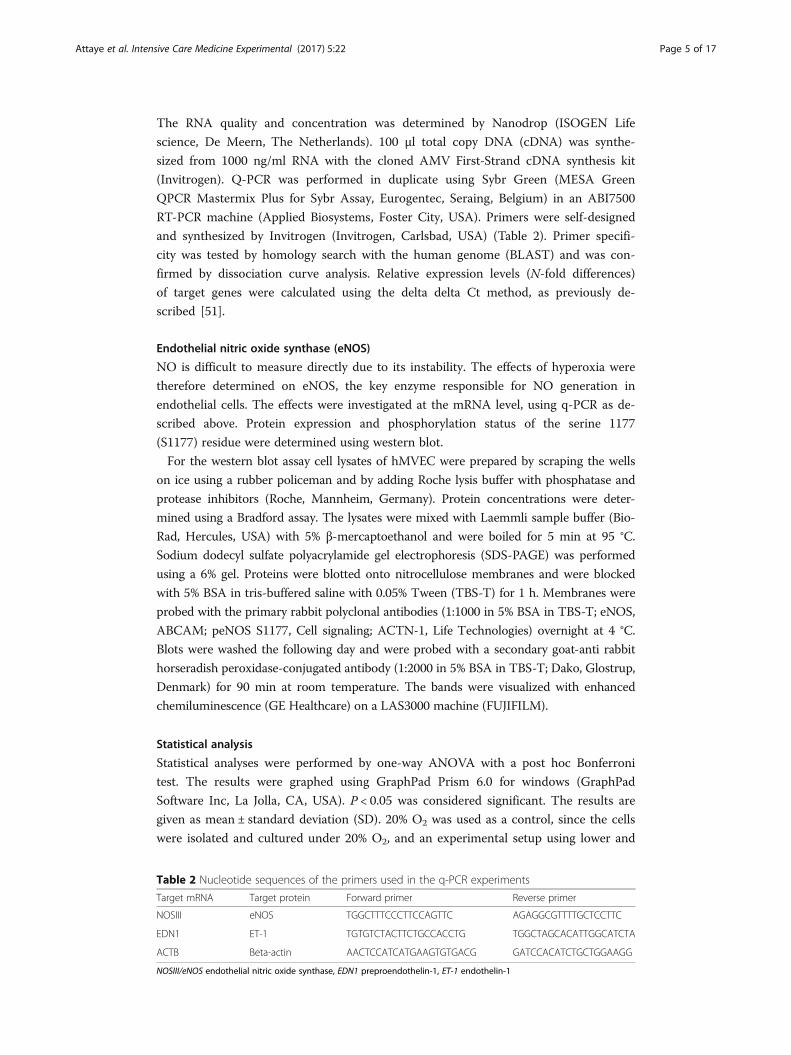
Table 2 Nucleotide sequences of the primers used in the q-PCR experiments
Target mRNA Target protein Forward primer Reverse primer
NOSIII eNOS TGGCTTTCCCTTCCAGTTC AGAGGCGTTTTGCTCCTTC
EDN1 ET-1 TGTGTCTACTTCTGCCACCTG TGGCTAGCACATTGGCATCTA
ACTB Beta-actin AACTCCATCATGAAGTGTGACG GATCCACATCTGCTGGAAGG
NOSIII/eNOS endothelial nitric oxide synthase, EDN1 preproendothelin-1, ET-1 endothelin-1
Attaye et al. Intensive Care Medicine Experimental (2017) 5:22 Page 5 of 17
more physiological levels of oxygen exposure as a control is not feasible in an in vitro
setup. All hyperoxic conditions (30, 50, and 95% O2) were compared to 20% O2.
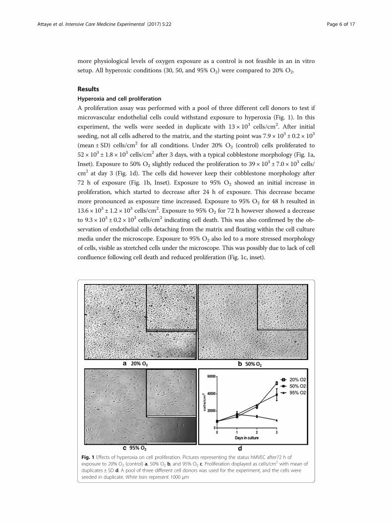
ResultsHyperoxia and cell proliferation
A proliferation assay was performed with a pool of three different cell donors to test if
microvascular endothelial cells could withstand exposure to hyperoxia (Fig. 1). In this
experiment, the wells were seeded in duplicate with 13 × 103 cells/cm2. After initial
seeding, not all cells adhered to the matrix, and the starting point was 7.9 × 10³ ± 0.2 × 10³
(mean ± SD) cells/cm2 for all conditions. Under 20% O2 (control) cells proliferated to
52 × 103 ± 1.8 × 10³ cells/cm2 after 3 days, with a typical cobblestone morphology (Fig. 1a,
Inset). Exposure to 50% O2 slightly reduced the proliferation to 39 × 103 ± 7.0 × 10³ cells/
cm2 at day 3 (Fig. 1d). The cells did however keep their cobblestone morphology after
72 h of exposure (Fig. 1b, Inset). Exposure to 95% O2 showed an initial increase in
proliferation, which started to decrease after 24 h of exposure. This decrease became
more pronounced as exposure time increased. Exposure to 95% O2 for 48 h resulted in
13.6 × 10³ ± 1.2 × 10³ cells/cm2. Exposure to 95% O2 for 72 h however showed a decrease
to 9.3 × 10³ ± 0.2 × 10³ cells/cm2 indicating cell death. This was also confirmed by the ob-
servation of endothelial cells detaching from the matrix and floating within the cell culture
media under the microscope. Exposure to 95% O2 also led to a more stressed morphology
of cells, visible as stretched cells under the microscope. This was possibly due to lack of cell
confluence following cell death and reduced proliferation (Fig. 1c, inset).
Fig. 1 Effects of hyperoxia on cell proliferation. Pictures representing the status hMVEC after72 h ofexposure to 20% O2 (control) a, 50% O2 b, and 95% O2 c. Proliferation displayed as cells/cm2 with mean ofduplicates ± SD d. A pool of three different cell donors was used for the experiment, and the cells wereseeded in duplicate. White bars represent 1000 μm
Attaye et al. Intensive Care Medicine Experimental (2017) 5:22 Page 6 of 17
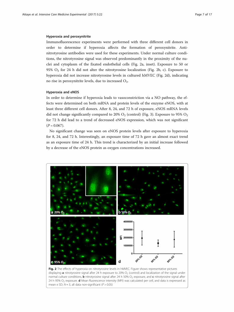
Hyperoxia and peroxynitrite
Immunofluorescence experiments were performed with three different cell donors in
order to determine if hyperoxia affects the formation of peroxynitrite. Anti-
nitrotyrosine antibodies were used for these experiments. Under normal culture condi-
tions, the nitrotyrosine signal was observed predominantly in the proximity of the nu-
clei and cytoplasm of the fixated endothelial cells (Fig. 2a, inset). Exposure to 50 or
95% O2 for 24 h did not alter the nitrotyrosine localization (Fig. 2b, c). Exposure to
hyperoxia did not increase nitrotyrosine levels in cultured hMVEC (Fig. 2d), indicating
no rise in peroxynitrite levels, due to increased O2.
Hyperoxia and eNOS
In order to determine if hyperoxia leads to vasoconstriction via a NO pathway, the ef-
fects were determined on both mRNA and protein levels of the enzyme eNOS, with at
least three different cell donors. After 8, 24, and 72 h of exposure, eNOS mRNA levels
did not change significantly compared to 20% O2 (control) (Fig. 3). Exposure to 95% O2
for 72 h did lead to a trend of decreased eNOS expression, which was not significant
(P = 0.067).
No significant change was seen on eNOS protein levels after exposure to hyperoxia
for 8, 24, and 72 h. Interestingly, an exposure time of 72 h gave an almost exact trend
as an exposure time of 24 h. This trend is characterized by an initial increase followed
by a decrease of the eNOS protein as oxygen concentrations increased.
Fig. 2 The effects of hyperoxia on nitrotyrosine levels in hMVEC. Figure shows representative picturesdisplaying a nitrotyrosine signal after 24 h exposure to 20% O2 (control) and localization of the signal undernormal culture conditions, b nitrotyrosine signal after 24 h 50% O2 exposure, and c nitrotyrosine signal after24 h 95% O2 exposure. d Mean fluorescence intensity (MFI) was calculated per cell, and data is expressed asmean ± SD; N = 3, all data non-significant (P > 0.05)
Attaye et al. Intensive Care Medicine Experimental (2017) 5:22 Page 7 of 17
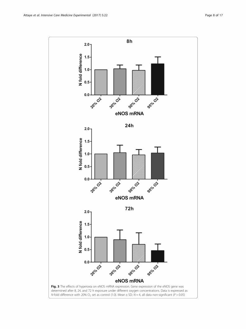
Fig. 3 The effects of hyperoxia on eNOS mRNA expression. Gene expression of the eNOS gene wasdetermined after 8, 24, and 72 h exposure under different oxygen concentrations. Data is expressed asN-fold difference with 20% O2 set as control (1.0). Mean ± SD; N = 4, all data non-significant (P > 0.05)
Attaye et al. Intensive Care Medicine Experimental (2017) 5:22 Page 8 of 17
The effects of hyperoxia were also determined with regards to the ratio of peNOS/
eNOS, which is the ratio of the activated form of eNOS divided by the total eNOS
protein.
Exposure to hyperoxia for 8 h gave a trend of a decreased expression of the ratio
when exposed to different degrees of hyperoxia. Exposure for 24 and 72 h however
gave a trend of increased expression of the ratio when exposed to different degrees of
hyperoxia. These observed trends in the phosphorylation status were not statistically
significant (Fig. 4).
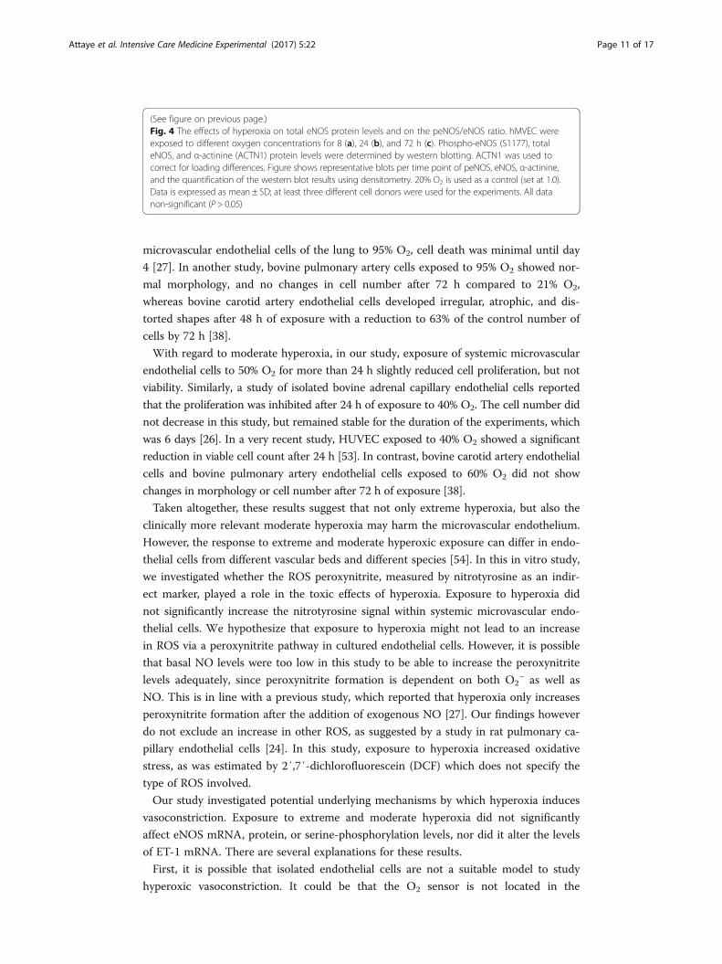
Hyperoxia and ET-1
The effects of hyperoxia on the mRNA levels of the potent vasoconstrictor ET-1 were
also determined with four different cell donors.
Exposure to hyperoxia for 8 h led to a trend of increased expression of ET-1 mRNA
when compared to 20% O2 (control). Exposure for 24 h led to an increase of 1.41 when
the cells were exposed to 95% O2. Exposure for 72 h to 30 and 50% O2 led to a slightly
decreased expression of 0.73 and 0.68 when compared to 20% O2 (control). Exposure
to 95% O2 gave rise to a minor increase of 1.23 in the ET-1 mRNA expression. None of
these changes were statistically significant (Fig. 5).
DiscussionThis cell culture study showed that exposure of isolated human systemic microvascular
endothelial cells to 50% O2 for more than 24 h slightly affected cell proliferation, but
exposure to 95% O2 for more than 24 h markedly reduced cell viability and proliferative
capacity. Exposure to hyperoxia for 8, 24, and 72 h did not lead to a significant change
in protein tyrosine nitration by peroxinitrite. Furthermore, exposure to hyperoxia did
not significantly affect eNOS mRNA, protein, or serine-phosphorylation levels, nor did
it alter the levels of ET-1 mRNA.
To the best of our knowledge, the current study is the first to investigate the role of
hyperoxia on isolated cultured human endothelial cells of the systemic microcirculation
using different degrees of hyperoxia. Systemic microvascular endothelial cells are par-
ticularly relevant, since in critically ill patients (who are most frequently exposed to
hyperoxia during mechanical ventilation) microvascular dysfunction plays a pivotal role
in conditions such as sepsis, trauma, and ischemia/reperfusion injury. An increase of
microvascular endothelial injury, which is the largest endothelial surface of the body,
may worsen organ failure.
Exposure to extreme hyperoxia (95% O2) in our study did not stop microvascular
endothelial cell proliferation in the first day, but after 24 h, cell densities started to de-
crease which became more pronounced as exposure time increased. Exposure for 72 h
decreased total cell numbers back towards seeding densities, indicating cell death. The
toxic effects of extreme hyperoxia were also shown in a recent study which exposed hu-
man umbilical endothelial cells (HUVEC) to 95% O2 [52]. This study showed that the
total number of cells remained equal after 8 h of exposure, whereas the number of
apoptotic and dead cells had increased. After 72 h there was a 15% decrease in alive
cells compared to 21% O2, which was used as a control. The toxic effects of extreme
hyperoxia appear to differ between vascular beds. Pulmonary endothelial cells seem to
be more resistant to the toxic effects of oxygen. In a study exposing isolated human
Attaye et al. Intensive Care Medicine Experimental (2017) 5:22 Page 9 of 17
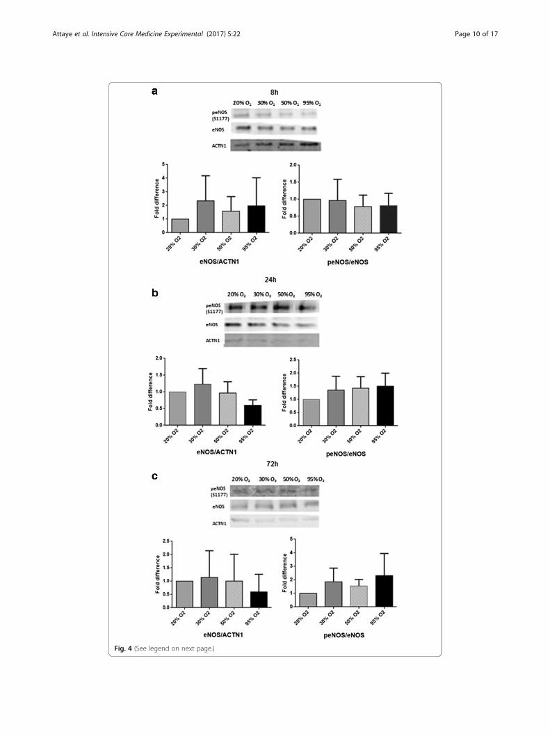
Fig. 4 (See legend on next page.)
Attaye et al. Intensive Care Medicine Experimental (2017) 5:22 Page 10 of 17
microvascular endothelial cells of the lung to 95% O2, cell death was minimal until day
4 [27]. In another study, bovine pulmonary artery cells exposed to 95% O2 showed nor-
mal morphology, and no changes in cell number after 72 h compared to 21% O2,
whereas bovine carotid artery endothelial cells developed irregular, atrophic, and dis-
torted shapes after 48 h of exposure with a reduction to 63% of the control number of
cells by 72 h [38].
With regard to moderate hyperoxia, in our study, exposure of systemic microvascular
endothelial cells to 50% O2 for more than 24 h slightly reduced cell proliferation, but not
viability. Similarly, a study of isolated bovine adrenal capillary endothelial cells reported
that the proliferation was inhibited after 24 h of exposure to 40% O2. The cell number did
not decrease in this study, but remained stable for the duration of the experiments, which
was 6 days [26]. In a very recent study, HUVEC exposed to 40% O2 showed a significant
reduction in viable cell count after 24 h [53]. In contrast, bovine carotid artery endothelial
cells and bovine pulmonary artery endothelial cells exposed to 60% O2 did not show
changes in morphology or cell number after 72 h of exposure [38].
Taken altogether, these results suggest that not only extreme hyperoxia, but also the
clinically more relevant moderate hyperoxia may harm the microvascular endothelium.
However, the response to extreme and moderate hyperoxic exposure can differ in endo-
thelial cells from different vascular beds and different species [54]. In this in vitro study,
we investigated whether the ROS peroxynitrite, measured by nitrotyrosine as an indir-
ect marker, played a role in the toxic effects of hyperoxia. Exposure to hyperoxia did
not significantly increase the nitrotyrosine signal within systemic microvascular endo-
thelial cells. We hypothesize that exposure to hyperoxia might not lead to an increase
in ROS via a peroxynitrite pathway in cultured endothelial cells. However, it is possible
that basal NO levels were too low in this study to be able to increase the peroxynitrite
levels adequately, since peroxynitrite formation is dependent on both O2− as well as
NO. This is in line with a previous study, which reported that hyperoxia only increases
peroxynitrite formation after the addition of exogenous NO [27]. Our findings however
do not exclude an increase in other ROS, as suggested by a study in rat pulmonary ca-
pillary endothelial cells [24]. In this study, exposure to hyperoxia increased oxidative
stress, as was estimated by 2′,7′-dichlorofluorescein (DCF) which does not specify the
type of ROS involved.
Our study investigated potential underlying mechanisms by which hyperoxia induces
vasoconstriction. Exposure to extreme and moderate hyperoxia did not significantly
affect eNOS mRNA, protein, or serine-phosphorylation levels, nor did it alter the levels
of ET-1 mRNA. There are several explanations for these results.
First, it is possible that isolated endothelial cells are not a suitable model to study
hyperoxic vasoconstriction. It could be that the O2 sensor is not located in the
(See figure on previous page.)Fig. 4 The effects of hyperoxia on total eNOS protein levels and on the peNOS/eNOS ratio. hMVEC wereexposed to different oxygen concentrations for 8 (a), 24 (b), and 72 h (c). Phospho-eNOS (S1177), totaleNOS, and α-actinine (ACTN1) protein levels were determined by western blotting. ACTN1 was used tocorrect for loading differences. Figure shows representative blots per time point of peNOS, eNOS, α-actinine,and the quantification of the western blot results using densitometry. 20% O2 is used as a control (set at 1.0).Data is expressed as mean ± SD; at least three different cell donors were used for the experiments. All datanon-significant (P > 0.05)
Attaye et al. Intensive Care Medicine Experimental (2017) 5:22 Page 11 of 17
Fig. 5 The effects of hyperoxia on ET-1 mRNA levels. hMVEC were exposed to different oxygen concentrationsfor 8, 24, and 72 h. Data is expressed as N-fold difference with 20% O2 set as control (1.0). Mean ± SD; N = 4, alldata non-significant (P > 0.05)
Attaye et al. Intensive Care Medicine Experimental (2017) 5:22 Page 12 of 17
endothelium, but in other cells of the arteriolar wall (vascular smooth muscle cells),
intraluminal (red blood cells), or in extravascular cells (parenchymal cells or mast cells).
In addition, interaction with extravascular cells and smooth muscle cells may be neces-
sary to activate the signaling pathway that couples changes in arteriolar oxygen pres-
sure to changes in arteriolar tone [55].
Second, our in vitro model may not be suitable to investigate hyperoxic vasoconstric-
tion, since NO availability is not guaranteed. In our study, no significant effect of
hyperoxia on eNOS mRNA, protein levels, or serine-phosphorylation levels was found.
However, the majority of studies in the literature point in the direction of a reduced
bioavailability of NO as the underlying mechanism of hyperoxic vasoconstriction [32–35].
But for a model to show an effect on NO, the NO availability at the start of the experi-
ment should be comparable to in vivo situations. The isolated endothelial cells were not
exposed to flow, which may have decreased their basal NO level [56, 57].
Furthermore, it is possible that supporting leukocytes are needed in order to increase
the NO levels. Hyperoxia can lead to an inflammatory status of the vascular system
[28]. During this inflammation, NO production can be greatly increased by mainly
macrophages, which possess the enzyme inducible nitric oxide synthase (iNOS). In the
latter situation, an increase in peroxynitrite levels can be expected since both the NO
as well as the O2− will be increased [58]. These results could not be reproduced in our
study, since iNOS regulation is largely controlled via macrophages and not endothelial
cells [59].
However, another possible explanation for our negative results with regard to the NO
pathway can be that hyperoxia does influence NO bioavailability, but that we were not
able to detect it, since it is not possible to measure NO directly.
Third, another reason for the lack of effect of hyperoxia on eNOS and ET-1 in our
study may be that hyperoxia induces vasoconstriction by affecting other vaso-active
mediators. Several studies suggest a role for prostaglandins [16, 38] or 20-
hydroxyeicosatetraenoic acid (20-HETE), a vasoconstrictor which is formed in vascular
smooth muscle cells (VSMC) by the CYP450-4A enzyme system [40, 41, 43, 60]. In
contrast with our ET-1 findings in human endothelial cells of the systemic microcircu-
lation, hyperoxia did increase ET-1 levels in isolated bovine adrenal capillary endothe-
lial cells and bovine retinal endothelial cells [61]. The difference in results can be based
on species variability or upon different vascular origin of the endothelial cells used. Fur-
thermore, we investigated ET-1 only at the mRNA and not the protein level, since gen-
eral consensus within the literature states that the bioavailability of the ET-1 protein is
predominately regulated at the transcriptional level of the EDN1 gene [39]. Therefore,
we cannot exclude the possibility that ET-1 was affected at translation or posttransla-
tional level.
Fourth, the negative results with regard to vasoconstrictive pathways may be caused
by the use of 20% O2 as a control for cultured endothelial cells, because the cells were
isolated and cultured under these conditions. Twenty percent of O2, however, is already
hyperoxic in vivo, especially at tissue level [62]. This may have attenuated the differ-
ences between the groups.
Fifth, hyperoxia might not stimulate vasoconstrictive pathways in these specific micro-
vascular endothelial cells derived from the foreskin, since the effect of hyperoxia on vascu-
lar tone varies between different vascular beds. For example, hyperoxia induced
Attaye et al. Intensive Care Medicine Experimental (2017) 5:22 Page 13 of 17
vasoconstriction in coronary arteries of pigs [34, 63] and in carotid vessels from dogs [64],
but induced vasodilation in renal vessels of the dog [65], whereas no response to hyper-
oxia was observed in the arterioles of the mesentery of the rat and cat [19, 66].
More research is needed exploring the abovementioned pathways in an isolated and
combined way in vitro as well as in vivo. Studies combining cultured endothelial cells,
leukocytes, and vascular smooth muscle cells with flow and inflammatory stimuli can
help to further unravel the mechanisms behind hyperoxic vasoconstriction.
Limitations
Several limitations exist in this study. For all experiments, at least three different cell
donors were used to correct for donor variability. Although this is not uncommon
within in vitro literature, the power of our study is limited. Another possible limitation
is the use of 20% O2 as a control for the experiments, because the endothelial cells
were isolated and cultured under these conditions. 20% O2 is however already hyper-
oxic for endothelial cells in vivo [62]. However, performing experiments using physio-
logical levels of oxygen exposure as a control would require endothelial cells to be
isolated and cultured continuously within a hypoxic chamber. This is not feasible
within an in vitro setup. Performing experiments using 20% O2 as a control is common
practice and a limitation within in vitro literature in general. We did consider this point
and repeated the eNOS and ET-1 experiments by exposing them to 10% O2
(=76 mmHg) (Additional file 1: Figure S2). This did not lead to significantly different
outcomes of the experiments. A limitation of this study was the fact that the cells could
only be cultured under hyperoxia for a maximum of 72 h. After 72 h, the culture media
needed to be refreshed and the airtight boxes opened. This would decrease the oxygen
concentration within the boxes back to atmospheric conditions. Experiments investigat-
ing hyperoxic exposure for short time periods (i.e., <1 h) were also not performed in
this study. In addition, cell proliferation and viability experiments were not performed
under mild hyperoxic (30% O2) conditions. Furthermore, this study investigated ET-1,
via mRNA and eNOS, via mRNA and protein levels and did not investigate down-
stream pathways of NO generation or breakdown. Neither were other pathways investi-
gated such as 20-HETE or prostaglandin production. Finally, our isolated hMVEC
model precluded measuring hyperoxic effects mediated by the interaction between
endothelial cells, leukocytes, and vascular smooth muscle cells and excluded the inter-
action with flow and circulating mediators.
ConclusionsThe present model of isolated and cultured human microvascular endothelial cells sug-
gests that not only extreme hyperoxia (95% O2), but also the clinically more relevant
moderate hyperoxia (50% O2) for more than 24 h may harm the microvascular endo-
thelium. This is especially relevant for critically ill patients, where microvascular dys-
function is frequently present. Hyperoxia may worsen microvascular endothelial injury
and may contribute to multiple organ failure. Because control experiments in vitro
were done under hyperoxic conditions (20% O2) relative to the real tissue conditions in
vivo, already beginning toxic effects in the controls (20% O2) cannot be excluded. Per-
oxynitrite did not seem to play a major role in the reduced viability and proliferation in
vitro. Furthermore, in this model of isolated systemic human microvascular endothelial
Attaye et al. Intensive Care Medicine Experimental (2017) 5:22 Page 14 of 17
cells hyperoxia did not affect several key elements of the vaso-active response, namely,
eNOS mRNA and protein and ET-1 mRNA levels. Hyperoxic vasoconstriction remains
a complex mechanism, and it is possible that isolated in vitro models alone are not suf-
ficient to study this mechanism. To show the full impact of endothelial responses to
hyperoxia, models that allow the interaction between endothelial cells, leukocytes, and
vascular smooth muscle cells in combination with flow and inflammatory stimuli are
likely more appropriate.
Additional file
Additional file 1: Figure S1. A representative example of oxygen stability during the experiments. The otheroxygen percentages used had a similar pattern. Figure S2. The eNOS and the ET-1 experiments of Figs. 3, 4, and 5.displayed as N-fold difference, comparing 10% O2 exposure to 20% O2 (control, set as 1.0) and hyperoxia. Data isexpressed mean ± SD; all data non-significant (P > 0.05). (DOCX 266 kb)
Abbreviations20-HETE: 20-hydroxyeicosatetraenoic acid; BSA: Bovine serum albumin; cDNA: Copy DNA; COX: Cyclooxygenase;eNOS: Endothelial nitric oxide synthase; ET-1: Endothelin-1; FCS: Fetal calf serum; hMVEC: Human MicrovascularEndothelial Cells; hPL: Human platelet lysate; HUVEC: Human umbilical vein endothelial cells; ICAM-1: Intercellularadhesion molecule 1; iNOS: Inducible nitric oxide synthase; MFI: Mean fluorescence intensity; NO: Nitric oxide;O2: Oxygen; O2
−: Superoxide radical; ONOO−: Peroxynitrite; PBS: Phosphate-buffered saline; peNOS: Phospho-eNOS;PFA: Paraformaldehyde; ROS: Reactive oxygen species; S1177: Serine 1177; SDS-PAGE: Sodium dodecyl sulfatepolyacrylamide gel electrophoresis; TBS: Tris-buffered saline
AcknowledgementsProf.dr. Victor van Hinsbergh is greatly thanked for his advice and help when conceiving the experiments.
FundingNot applicable.
Availability of data and materialsThe datasets are available from the corresponding author upon reasonable request.
Authors’ contributionsIA performed the experiments, analyzed the data, and drafted the manuscript. YMS, MCW, HMO, and BS contributedto the design of the studies and wrote the manuscript. MHW supervised the laboratory experiments. RJM contributedto the design and analyses of the immunofluorescence experiments. PK and AMES conceived the study andsupervised the experiments. All authors read and approved the manuscript.
Competing interestsThe authors declare that they have no competing interests.
Consent for publicationNot applicable.
Ethics approval and consent to participateThis study was executed in accordance with the Declaration of Helsinki and was approved by the University HumanSubjects Committee of the VU University Medical Center, Amsterdam, The Netherlands.
Publisher’s NoteSpringer Nature remains neutral with regard to jurisdictional claims in published maps and institutional affiliations.
Author details1Department of Intensive Care, VU University Medical Center, Amsterdam, The Netherlands. 2Department ofPhysiology, VU University Medical Center, Amsterdam, The Netherlands. 3Department of Internal Medicine, VUUniversity Medical Center, Amsterdam, The Netherlands.
Received: 11 October 2016 Accepted: 6 April 2017
References1. de Graaff AE, Dongelmans DA, Binnekade JM, de Jonge E (2011) Clinicians’ response to hyperoxia in ventilated
patients in a Dutch ICU depends on the level of FiO2. Intensive Care Med 37:46–512. Sjöberg F, Singer M (2013) The medical use of oxygen: a time for critical reappraisal. J Intern Med 274:505–528
Attaye et al. Intensive Care Medicine Experimental (2017) 5:22 Page 15 of 17
3. Cornet AD, Kooter AJ, Peters MJ, Smulders YM (2013) The potential harm of oxygen therapy in medicalemergencies. Crit Care 17:313
4. Cornet AD, Kooter AJ, Peters MJSY (2012) Supplemental oxygen therapy in medical emergencies: more harm thanbenefit? Arch Intern Med 172:289–290
5. Calzia E, Asfar P, Matejovic M et al (2010) Hyperoxia may be beneficial. Crit Care Med 38:559–5686. Kallet RH, Matthay MA (2013) Hyperoxic acute lung injury. Respir Care 58:123–1417. Rawles JM, Kenmure AC (1976) Controlled trial of oxygen in uncomplicated myocardial infarction. BMJ 1:1121–11238. Rønning OM, Guldvog B (1999) Should stroke victims routinely receive supplemental oxygen? A quasi-randomized
controlled trial. Stroke 30:2033–20379. Wang C, Chang W, Huang C et al (2014) The effect of hyperoxia on survival following adult cardiac arrest : a
systematic review and meta-analysis of observational studies. Resuscitation 85:1142–114810. Pilcher J, Weatherall M, Shirtcliffe P et al (2012) The effect of hyperoxia following cardiac arrest a systematic
review and meta-analysis of animal trials. Resuscitation 83:417–42211. Elmer J, Scutella M, Pullalarevu R et al (2015) The association between hyperoxia and patient outcomes after
cardiac arrest : analysis of a high-resolution database. Intensive Care Med 41:49–5712. Knighton D, Halliday B, Hunt T (1984) Oxygen as an antibiotic. The effect of inspired oxygen on infection.
Arch Surg 119:19913. Belda JF, Aguilera L, García de la Asunción J et al (2005) Supplemental perioperative oxygen and the risk of
surgical wound infection a randomized controlled trial. JAMA J Am Med Assoc 294:2035–204214. Floyd TF, Clark JM, Gelfand R et al (2003) Independent cerebral vasoconstrictive effects of hyperoxia and
accompanying arterial hypocapnia at 1 ATA. J Appl Physiol 95:2453–246115. Mcnulty PH, Robertson BJ, Tulli MA et al (2007) Effect of hyperoxia and vitamin C on coronary blood flow in
patients with ischemic heart disease. J Appl Physiol 13326:2040–204516. Messina JE, Sun D, Koller A et al (1994) Increases in oxygen tension evoke arteriolar constriction by inhibiting
endothelial prostaglandin synthesis. Microvasc Res 40:151–16017. Kiss B, Polska E, Dorner G et al (2002) Retinal blood flow during hyperoxia in humans revisited: concerted results
using different measurement techniques. Microvasc Res 64:75–8518. Sharkey RA, Mulloy EMT, Neill SJO (1998) Acute effects of hypoxaemia, hyperoxaemia and hypercapnia on renal
blood flow in normal and renal transplant subjects. Eur Respir J 12:653–65719. Lang J, Johnson PC (1988) Elevated ambient oxygen does not affect autoregulation in cat mesentery. Am J
Physiol 255:131–13720. Farquhar H, Weatherall M, Wijesinghe M, Perrin K (2009) Systematic review of studies of the effect of hyperoxia on
coronary blood flow. Am Heart J 158:371–37721. Kamler M, Wendt D, Pizanis N et al (2004) Deleterious effects of oxygen during extracorporeal circulation for the
microcirculation in vivo. Eur J Cardiothorac Surg 26:564–57022. Barth E, Bassi G, Maybauer DM et al (2008) Effects of ventilation with 100% oxygen during early hyperdynamic
porcine fecal peritonitis. Crit Care Med 36:495–50323. Barth E, Bassi G, Simon F, Gro M (2009) Hemodynamic, metabolic, and organ function effects of pure oxygen
ventilation during established fecal peritonitis-induced septic shock. Crit Care Med 37:2465–246924. Brueckl C, Kaestle S, Kerem A et al (2006) Hyperoxia-induced reactive oxygen species formation in pulmonary
capillary endothelial cells in situ. Am J Respir Cell Mol Biol 34:453–46325. Ming L, Ping F, Yao X et al (2006) Reactive oxygen species in vascular wall. Cardiovasc Haematol Disord Targets 6:1–1926. Amore PAD, Sweet E (1987) Effects of hyperoxia on microvascular cells in vitro. Vitr Cell Dev Biol 23:123–12827. Narula P, Xu J, Kazzaz JA et al (1998) Synergistic cytotoxicity from nitric oxide and hyperoxia in cultured lung cells.
Am J Physiol 274:411–41628. Suzuki Y, Aoki T, Takeuchi O et al (1997) Effect of hyperoxia on adhesion molecule expression in human
endothelial cells and neutrophils. Am J Physiol Lung Cell Mol Physiol 16:418–42529. Sukhotnik I, Brod V, Lurie M et al (2009) The effect of 100% oxygen on intestinal preservation and recovery
following ischemia-reperfusion injury in rats. Crit Care Med 37:1054–106130. Waisman D, Brod V, Rahat MA et al (2012) Dose-related effects of hyperoxia on the lung inflammatory response in
septic rats. SHOCK 37:95–10231. Gu X, El-Remessy AB, Brooks SE et al (2003) Hyperoxia induces retinal vascular endothelial cell apoptosis through
formation of peroxynitrite. Am J Physiol Cell Physiol 285:C546–C55432. Chen Z, Wen L, Marin M et al (2015) Oxidative stress activates endothelial innate immunity via sterol regulatory
element binding protein 2 (SREBP2) transactivation of microRNA-92a. Circulation 2:805–81433. Rubanyi GM, Vanhoutte PM (1986) Superoxide anions and hyperoxia inactivate endothelium-derived relaxing
factor. Am J Physiol 250:H822–H82734. Pasgaard T, Stankevicius E, Jørgensen MM et al (2007) Hyperoxia reduces basal release of nitric oxide and
contracts porcine coronary arteries. Acta Physiol 191:285–29635. Pries AR, Heide J, Ley K et al (1995) Effect of oxygen tension on regulation of arteriolar diameter in skeletal muscle
in situ. Microvasc Res 49:289–29936. Gryglewski RJ, Palmer RMJ, Moncada S (1986) Superoxide anion is involved in the breakdown of endothelium-
derived relaxing factor. Nature 320:454–45637. Behrendt DPG (2002) Endothelial function: from vascular biology to clinical applications. Am J Cardiol 9149:40–4838. Ishii Y, Morita I, Murota S, Kitamura S (1993) Hyperoxia decreases cyclooxygenase. Prostaglandins Leukot Essent
Fat Acids 48:455–461.39. Stow LR, Jacobs ME, Wingo CS, Cain BD (2011) Endothelin-1 gene regulation. FASEB J 25:16–2840. Ngo AT, Riemann M, Torp-pedersen C, Jensen LJ (2013) Significance of K ATP channels, L-type Ca 2 + channels
and CYP450-4A enzymes in oxygen sensing in mouse cremaster muscle arterioles In vivo. BMC Physiol 13:1–1141. Welsh DG, Jackson WF, Segal SS (1998) Oxygen induces electromechanical coupling in arteriolar smooth muscle
cells: a role for L-type Ca2+ channels. Am J Physiol 274:H2018–H2024
Attaye et al. Intensive Care Medicine Experimental (2017) 5:22 Page 16 of 17
42. Block RE, Patel MJ, Angelides JK et al (1986) Hyperoxia reduces plasma membrane fluidity : a mechanism forendothelial cell dysfunction. J Appl Physiol 60:826–835
43. Frisbee JC, Krishna UM, Falck JR, Lombard JH (2001) Role of prostanoids and 20-HETE in mediating oxygen-induced constriction of skeletal muscle resistance arteries. Microvasc Res 62:271–283
44. Riemann M, Rai A, Ngo AT et al (2010) Oxygen-dependent vasomotor responses are conducted upstream in themouse cremaster microcirculation. J Vasc Res 48:79–89
45. Clement A, Hubscher U (1985) Effects of hyperoxia on DNA synthesis in cultured porcine aortic endothelial cells.J Appl Physiol 59:1110–1116
46. Willam C, Schindler R, Frei U, Eckardt K (1999) Increases in oxygen tension stimulate expression of ICAM-1 andVCAM-1 on human endothelial cells. Am J Physiol 276:H2044–H2052
47. Van Hinsbergh VW, Sprengers EDKT (1987) Effect of thrombin on the production of plasminogen activators andPA inhibitor-1 by human foreskin microvascular endothelial cells. Thromb Haemost 57:148–153
48. Tasev D, van Wijhe MH, Weijers EM et al (2015) Long-term expansion in platelet lysate increases growth ofperipheral blood-derived endothelial-colony forming cells and their growth factor-induced sprouting capacity.PLoS One 10:e0129935
49. Halliwell B, Zhao K, Whiteman M (1999) Nitric oxide and peroxynitrite. The ugly, the uglier and the not so good: apersonal view of recent controversies. Free Radic Res 31:651–669
50. Sawa T, Akaike T, Maeda H (2000) Tyrosine nitration by peroxynitrite formed from nitric oxide and superoxidegenerated by xanthine oxidase. J Biol Chem 275:32467–32474
51. Wong ML, Medrano JF (2005) One-step versus two-step real-time PCR. Biotechniques 39:75–8552. Wu J, Hafner C, Schramel JP et al (2016) Cyclic and constant hyperoxia cause inflammation, apoptosis and cell
death in human umbilical vein endothelial cells. Acta Anaesthesiol Scand 60:492–50153. Hafner C, Wu J, Soto-gonzalez L et al (2017) Moderate hyperoxia induces inflammation, apoptosis and necrosis in
human umbilical vein endothelial cells: an in-vitro study. Eur J Anaesthesiol 34:141–14954. Lehle K, Straub RH, Morawietz H, Kunz-schughart LA (2010) Relevance of disease- and organ-specific endothelial
cells for in vitro research differences between arterial and venous ECs. Cell Biol Int 34:1231–123855. Jackson WF (2016) Arteriolar oxygen reactivity : where is the sensor and what is the mechanism of action ?
J Physiol 18:5055–507756. Kolluru GK, Siamwala JH, Chatterjee S (2010) eNOS phosphorylation in health and disease. Biochimie 92:1186–119857. Ishibazawa A, Nagaoka T, Takahashi T et al (2011) Effects of shear stress on the gene expressions of endothelial
nitric oxide synthase, endothelin-1, and thrombomodulin in human retinal microvascular endothelial cells. InvestOphthalmol Vis Sci 52:8496–8504
58. Spoelstra-de Man AME, Smit B, Oudemans-van Straaten HM, Smulders YM (2015) Cardiovascular effects ofhyperoxia during and after cardiac surgery. Anaesthesia 70:1307–1319
59. Pautz A, Art J, Hahn S et al (2010) Regulation of the expression of inducible nitric oxide synthase. Nitric Oxide 23:75–9360. Wang J, Schmidt JR, Roman RJ et al (2010) Modulation of vascular O2 responses by cytochrome 450-4A
hydroxylase metabolites in Dahl salt-sensitive rats. Microcirculation 16:345–35461. Higgins DR, Hendricks-Munoz DK, Caines VV et al (1998) Hyperoxia stimulates endothelin-1 secretion from
entohelial cells; modulation by captopril and nifedipine. Curr Eye Res 17:487–49362. Carreau A, El Hafny-Rahbi B, Matejuk A et al (2011) Why is the partial oxygen pressure of human tissues a crucial
parameter? Small molecules and hypoxia. J Cell Mol Med 15:1239–125363. Hedegaard ER, Stankevicius E, Simonsen U, Fröbert O (2011) Non-endothelial endothelin counteracts hypoxic
vasodilation in porcine large coronary arteries. BMC Physiol 11:864. Siegel G, Grote J, Schnalke F, Zimmer K (1989) The significance of the endothelium for hypoxic vasodilatation.
Z Kardiol 78:124–13165. Eskinder H, Harder DR, Lombard JH (1990) Role of the vascular endothelium in regulating the response of small
arteries of the dog kidney to transmural pressure elevation and reduced Po2. Circ Res 66:1427–143666. Van den Bos GC, Westerhof N, Hoogerwerf N, Sicking A (1991) Arteriolar and venular reactivity to superfusate pO2
in tissues with different metabolic capacity. A study in skeletal muscle and mesentery of the rat. Int J MicrocircClin Exp 10:303–316
Submit your manuscript to a journal and benefi t from:
7 Convenient online submission
7 Rigorous peer review
7 Immediate publication on acceptance
7 Open access: articles freely available online
7 High visibility within the fi eld
7 Retaining the copyright to your article
Submit your next manuscript at 7 springeropen.com
Attaye et al. Intensive Care Medicine Experimental (2017) 5:22 Page 17 of 17
Related Documents