The effects of cosolutes on protein dynamics: The reversal of denaturant-induced protein fluctuations by trimethylamine N-oxide VICKY DOAN-NGUYEN 1 AND J. PATRICK LORIA Department of Chemistry Yale University, New Haven, Connecticut 06520, USA (RECEIVED June 8, 2006; FINAL REVISION August 30, 2006; ACCEPTED October 10, 2006) Abstract The protein stabilizing effects of the small molecule osmolyte, trimethylamine N-oxide, against chemical denaturant was investigated by NMR spin-relaxation measurements and model-free analysis. In the presence of 0.7 M guanidine hydrochloride increased picosecond-nanosecond dynamics are observed in the protein ribonuclease A. These increased fluctuations occur throughout the protein, but the most significant increases in flexibility occur at positions believed to be the first to unfold. Addition of 0.35 M trimethylamine N-oxide to this destabilized form of ribonuclease results in significant rigidification of the protein backbone as assessed by 1 H- 15 N order parameters. Statistically, these order parameters are the same as those measured in native ribonuclease indicating that TMAO reduces the amplitude of backbone fluctuations in a destabilized protein. These data suggest that TMAO restricts the bond vector motions on the protein energy landscape to resemble those motions that occur in the native protein and points to a relation between stability and dynamics in this enzyme. Keywords: guanidine; protein dynamics; protein stability; NMR spin-relaxation; TMAO Supplemental material: see www.proteinscience.org Many organisms produce and accumulate small organic solutes to counteract the effects of environmental stresses. Such inhospitable living conditions can include high temperature, pressure, salt, and dehydration. One such solute, trimethylamine N-oxide, (TMAO; Scheme 1) provides protection to proteins against the effects of chemical denaturant, high pressures, and high temper- atures (Yancey et al. 2002; Somero 2003; Yancey 2005). In the enzymes ribonuclease A (RNase A) (Yancey and Somero 1979) and lysozyme (Arakawa and Timasheff 1985), TMAO was shown to increase the midpoint of the thermal unfolding profile. Other protective properties of TMAO include the ability to counteract the deleterious effects of urea as probed by enzyme assays (Yancey and Somero 1979; Palmer et al. 2000) and protein folding studies (Yancey and Somero 1979; Lin and Timasheff 1994). Alexandrescu and coworkers have shown that TMAO increases the protection of amide protons from exchange with solvent by solution NMR hydrogen exchange (HX) experiments (Jaravine et al. 2000). HX experiments on RNase A in urea solutions (Qu and Bolen Scheme 1. Chemical structure of trimethylamine N-oxide. 1 Present address: Department of Chemistry, University of Pennsylvania, Philadelphia, PA 19104, USA. Reprint requests to: J. Patrick Loria, Department of Chemistry Yale University, P.O. Box 208107, New Haven, CT 06520, USA; e-mail: [email protected]; fax: (203) 432-6144. Abbreviations: CD, circular dichroism spectroscopy; Gdn, guanidine hydrochloride; MALDI-TOF, matrix assisted laser desorption ioniza- tion time of flight; RNase A, bovine pancreatic ribonuclease A; TMAO, trimethylamine N-oxide. Article published online ahead of print. Article and publication date are at http://www.proteinscience.org/cgi/doi/10.1110/ps.062393707. 20 Protein Science (2007), 16:20–29. Published by Cold Spring Harbor Laboratory Press. Copyright Ó 2007 The Protein Society

Welcome message from author
This document is posted to help you gain knowledge. Please leave a comment to let me know what you think about it! Share it to your friends and learn new things together.
Transcript
The effects of cosolutes on protein dynamics:The reversal of denaturant-induced protein fluctuationsby trimethylamine N-oxide
VICKY DOAN-NGUYEN1AND J. PATRICK LORIA
Department of Chemistry Yale University, New Haven, Connecticut 06520, USA
(RECEIVED June 8, 2006; FINAL REVISION August 30, 2006; ACCEPTED October 10, 2006)
Abstract
The protein stabilizing effects of the small molecule osmolyte, trimethylamine N-oxide, againstchemical denaturant was investigated by NMR spin-relaxation measurements and model-free analysis.In the presence of 0.7 M guanidine hydrochloride increased picosecond-nanosecond dynamics areobserved in the protein ribonuclease A. These increased fluctuations occur throughout the protein, butthe most significant increases in flexibility occur at positions believed to be the first to unfold. Additionof 0.35 M trimethylamine N-oxide to this destabilized form of ribonuclease results in significantrigidification of the protein backbone as assessed by 1H-15N order parameters. Statistically, these orderparameters are the same as those measured in native ribonuclease indicating that TMAO reduces theamplitude of backbone fluctuations in a destabilized protein. These data suggest that TMAO restricts thebond vector motions on the protein energy landscape to resemble those motions that occur in the nativeprotein and points to a relation between stability and dynamics in this enzyme.
Keywords: guanidine; protein dynamics; protein stability; NMR spin-relaxation; TMAO
Supplemental material: see www.proteinscience.org
Many organisms produce and accumulate small organicsolutes to counteract the effects of environmental stresses.Such inhospitable living conditions can include hightemperature, pressure, salt, and dehydration. One suchsolute, trimethylamine N-oxide, (TMAO; Scheme 1)provides protection to proteins against the effects ofchemical denaturant, high pressures, and high temper-atures (Yancey et al. 2002; Somero 2003; Yancey 2005).
In the enzymes ribonuclease A (RNase A) (Yancey andSomero 1979) and lysozyme (Arakawa and Timasheff 1985), TMAO was shown to increase the midpoint of the
thermal unfolding profile. Other protective properties ofTMAO include the ability to counteract the deleteriouseffects of urea as probed by enzyme assays (Yancey andSomero 1979; Palmer et al. 2000) and protein foldingstudies (Yancey and Somero 1979; Lin and Timasheff1994). Alexandrescu and coworkers have shown thatTMAO increases the protection of amide protonsfrom exchange with solvent by solution NMR hydrogenexchange (HX) experiments (Jaravine et al. 2000). HXexperiments on RNase A in urea solutions (Qu and Bolen
Scheme 1. Chemical structure of trimethylamine N-oxide.
ps0623937 Doan-Nguyen and Loria ARTICLE RA
1Present address: Department of Chemistry, University of Pennsylvania,Philadelphia, PA 19104, USA.
Reprint requests to: J. Patrick Loria, Department of Chemistry YaleUniversity, P.O. Box 208107, New Haven, CT 06520, USA; e-mail:[email protected]; fax: (203) 432-6144.
Abbreviations: CD, circular dichroism spectroscopy; Gdn, guanidinehydrochloride; MALDI-TOF, matrix assisted laser desorption ioniza-tion time of flight; RNase A, bovine pancreatic ribonuclease A;TMAO, trimethylamine N-oxide.
Article published online ahead of print. Article and publication dateare at http://www.proteinscience.org/cgi/doi/10.1110/ps.062393707.
20 Protein Science (2007), 16:20–29. Published by Cold Spring Harbor Laboratory Press. Copyright � 2007 The Protein Society
JOBNAME: PROSCI 16#1 2006 PAGE: 1 OUTPUT: Friday December 8 17:03:00 2006
csh/PROSCI/127806/ps0623937
2003) suggest that TMAO suppresses the native-stateprotein fluctuations, thereby dampening the increasedmotional amplitude caused by denaturant (Wang andBolen 1997; Qu and Bolen 2003). These results suggestthat TMAO directly impacts protein dynamics. In con-trast, phosphorescent lifetime and quenching experimentsindicate that 1.8 M TMAO does not affect the dynamicsof the native protein fold (Gonnelli and Strambini 2001).Thus, there remains some uncertainty as to the role ofTMAO on protein stability and dynamics. The stability ofproteins depends on the difference in free energy betweenthe native and denatured states, and it is thought that theaction of TMAO results in differential stabilizations ofthe native and denatured states (Lin and Timasheff 1994).
Computational and physical methods have also beenused to investigate the mode of action of TMAO on proteinstability. Densimetry studies showed that the chemicalpotential changes of a protein in denaturant alone or indenaturant + TMAO are the same, indicating that TMAOacts independently of denaturant and does not affect thebinding of denaturant to proteins (Lin and Timasheff 1994).Other work has demonstrated that TMAO exerts its effectprimarily through changes in solvent hydrogen bonding andby unfavorable interactions between TMAO and the amidemoiety of proteins (Wang and Bolen 1997; Zou et al. 2002;Bennion and Daggett 2004; Athawale et al. 2005; Espositoand Daggett 2005). However, a somewhat different pictureemerged from recent solution NMR experiments thatsuggest that a hydrated form of TMAO may interact withthe peptide backbone (Hovagimyan and Gerig 2005). Thus,there is not complete consensus on the mechanism ofTMAO action. Because organisms produce TMAO andchemically similar molecules as a general stress response,their effects would be expected to be general and notspecific to any particular protein or nucleic acid.
Here, we use a well-characterized system to probe theeffects of TMAO on protein dynamics using solutionNMR techniques. NMR spin-relaxation experiments canprovide a unique glimpse at protein motions. In particu-lar, analysis of the NH order parameter (S2) can revealimportant information about the distribution of confor-mational states including the amplitude and timescale ofbackbone fluctuations. The backbone dynamics of nativeRNase A in buffer, in the presence of guanidine (Gdn)hydrochloride (RNase A/Gdn) and in the presence of Gdnand TMAO (RNase A/Gdn/TMAO), were investigated bysolution NMR spin-relaxation experiments.
Results
Circular dichroism spectroscopy
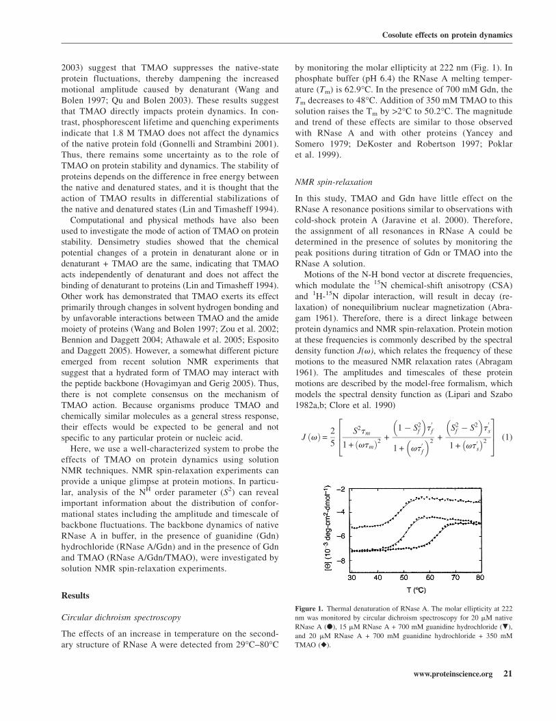
The effects of an increase in temperature on the second-ary structure of RNase A were detected from 29°C–80°C
by monitoring the molar ellipticity at 222 nm (Fig. 1). Inphosphate buffer (pH 6.4) the RNase A melting temper-ature (Tm) is 62.9°C. In the presence of 700 mM Gdn, theTm decreases to 48°C. Addition of 350 mM TMAO to thissolution raises the Tm by >2°C to 50.2°C. The magnitudeand trend of these effects are similar to those observedwith RNase A and with other proteins (Yancey andSomero 1979; DeKoster and Robertson 1997; Poklaret al. 1999).
NMR spin-relaxation
In this study, TMAO and Gdn have little effect on theRNase A resonance positions similar to observations withcold-shock protein A (Jaravine et al. 2000). Therefore,the assignment of all resonances in RNase A could bedetermined in the presence of solutes by monitoring thepeak positions during titration of Gdn or TMAO into theRNase A solution.
Motions of the N-H bond vector at discrete frequencies,which modulate the 15N chemical-shift anisotropy (CSA)and 1H-15N dipolar interaction, will result in decay (re-laxation) of nonequilibrium nuclear magnetization (Abra-gam 1961). Therefore, there is a direct linkage betweenprotein dynamics and NMR spin-relaxation. Protein motionat these frequencies is commonly described by the spectraldensity function J(v), which relates the frequency of thesemotions to the measured NMR relaxation rates (Abragam1961). The amplitudes and timescales of these proteinmotions are described by the model-free formalism, whichmodels the spectral density function as (Lipari and Szabo1982a,b; Clore et al. 1990)
J vð Þ=2
5
S2tm
1 + vtmð Þ2+
1 � S2f
� �t0f
1 + vt0f
� �2+
S2f � S2
� �t0s
1 + vt0s
� �2
264
375 (1)
Figure 1. Thermal denaturation of RNase A. The molar ellipticity at 222
nm was monitored by circular dichroism spectroscopy for 20 mM native
RNase A (d), 15 mM RNase A + 700 mM guanidine hydrochloride (.),
and 20 mM RNase A + 700 mM guanidine hydrochloride + 350 mM
TMAO (r).
Cosolute effects on protein dynamics
www.proteinscience.org 21
JOBNAME: PROSCI 16#1 2006 PAGE: 2 OUTPUT: Friday December 8 17:03:01 2006
csh/PROSCI/127806/ps0623937
in which t0f = tetm= te+tmð Þ; t0
s = tstm= ts+tmð Þ, tm is theisotropic rotational correlation time of the macromole-cule, te is the effective correlation time for fast (<200psec) internal motions, and ts the correlation time forslow (;nsec) motions (te < ts < tm). Corrections fornonisotropic macromolecular tumbling, in most cases, arereadily implemented (Woessner 1962; Lee et al. 1997).S2 = S2
f S2s is the square of the generalized order parameter
that characterizes the amplitude of internal motions, andS2f and S2
s are the order parameters for internal dynamicson the fast and slow time scales, respectively. The orderparameter describes the amplitude of bond vector motionsin the time range from picoseconds to nanoseconds andhas values from 0 for unrestricted motions to 1 indicatinga static bond vector in the molecular reference frame.Consequently, measurement of 15N longitudinal and trans-verse relaxation rates in combination with the 1H-15Nheteronuclear NOE allows sampling of the spectraldensity function and thus amplitudes and time scales ofprotein motion can be determined.
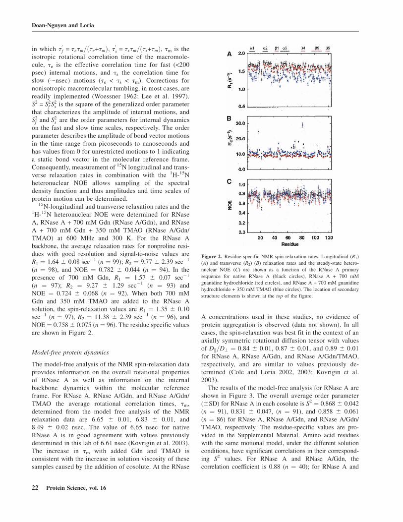
15N-longitudinal and transverse relaxation rates and the1H-15N heteronuclear NOE were determined for RNaseA, RNase A + 700 mM Gdn (RNase A/Gdn), and RNaseA + 700 mM Gdn + 350 mM TMAO (RNase A/Gdn/TMAO) at 600 MHz and 300 K. For the RNase Abackbone, the average relaxation rates for nonproline resi-dues with good resolution and signal-to-noise values areR1 ¼ 1.64 6 0.08 sec�1 (n ¼ 99); R2 ¼ 9.77 6 2.39 sec�1
(n ¼ 98), and NOE ¼ 0.782 6 0.044 (n ¼ 94). In thepresence of 700 mM Gdn, R1 ¼ 1.57 6 0.07 sec�1
(n ¼ 97); R2 ¼ 9.27 6 1.29 sec�1 (n ¼ 93) andNOE ¼ 0.724 6 0.068 (n ¼ 92). When both 700 mMGdn and 350 mM TMAO are added to the RNase Asolution, the spin-relaxation values are R1 ¼ 1.35 6 0.10sec�1 (n ¼ 97), R2 ¼ 11.38 6 2.39 sec�1 (n ¼ 96), andNOE ¼ 0.758 6 0.075 (n ¼ 96). The residue specific valuesare shown in Figure 2.
Model-free protein dynamics
The model-free analysis of the NMR spin-relaxation dataprovides information on the overall rotational propertiesof RNase A as well as information on the internalbackbone dynamics within the molecular referenceframe. For RNase A, RNase A/Gdn, and RNase A/Gdn/TMAO the average rotational correlation times, tm,determined from the model free analysis of the NMRrelaxation data are 6.65 6 0.01, 6.83 6 0.01, and8.49 6 0.02 nsec. The value of 6.65 nsec for nativeRNase A is in good agreement with values previouslydetermined in this lab of 6.61 nsec (Kovrigin et al. 2003).The increase in tm with added Gdn and TMAO isconsistent with the increase in solution viscosity of thesesamples caused by the addition of cosolute. At the RNase
A concentrations used in these studies, no evidence ofprotein aggregation is observed (data not shown). In allcases, the spin-relaxation was best fit in the context of anaxially symmetric rotational diffusion tensor with valuesof Dk=D? ¼ 0.84 6 0.01, 0.87 6 0.01, and 0.89 6 0.01for RNase A, RNase A/Gdn, and RNase A/Gdn/TMAO,respectively, and are similar to values previously de-termined (Cole and Loria 2002, 2003; Kovrigin et al.2003).
The results of the model-free analysis for RNase A areshown in Figure 3. The overall average order parameter(6SD) for RNase A in each cosolute is S2 ¼ 0.868 6 0.042(n ¼ 91), 0.831 6 0.047, (n ¼ 91), and 0.858 6 0.061(n ¼ 86) for RNase A, RNase A/Gdn, and RNase A/Gdn/TMAO, respectively. The residue-specific values are pro-vided in the Supplemental Material. Amino acid residueswith the same motional model, under the different solutionconditions, have significant correlations in their correspond-ing S2 values. For RNase A and RNase A/Gdn, thecorrelation coefficient is 0.88 (n ¼ 40); for RNase A and
Figure 2. Residue-specific NMR spin-relaxation rates. Longitudinal (R1)
(A) and transverse (R2) (B) relaxation rates and the steady-state hetero-
nuclear NOE (C) are shown as a function of the RNase A primary
sequence for native RNase A (black circles), RNase A + 700 mM
guanidine hydrochloride (red circles), and RNase A + 700 mM guanidine
hydrochloride + 350 mM TMAO (blue circles). The location of secondary
structure elements is shown at the top of the figure.
Doan-Nguyen and Loria
22 Protein Science, vol. 16
JOBNAME: PROSCI 16#1 2006 PAGE: 3 OUTPUT: Friday December 8 17:03:09 2006
csh/PROSCI/127806/ps0623937
Fig. 2 live 4/C
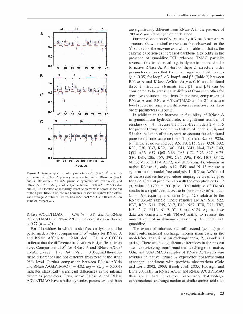
RNase A/Gdn/TMAO, r ¼ 0.76 (n ¼ 51), and for RNaseA/Gdn/TMAO and RNase A/Gdn, the correlation coefficientis 0.77 (n ¼ 43).
For all residues in which model-free analysis could beperformed, a t-test comparison of S2 values for RNase Aand RNase A/Gdn (t ¼ 9.40, dof ¼ 81, p < 0.0001)indicate that the difference in S2 values is significant fromzero. Comparison of S2 for RNase A and RNase A/Gdn/TMAO gives t ¼ 1.97, dof ¼ 78, p ¼ 0.053, and thereforethese differences are not different from zero at the strict95% level. Further comparison between RNase A/Gdnand RNase A/Gdn/TMAO (t ¼ 4.02, dof ¼ 82, p ¼ 0.0001)indicates statistically significant differences in the internaldynamics parameters. Thus, native RNase A and RNaseA/Gdn/TMAO have similar dynamics parameters and both
are significantly different from RNase A in the presence of700 mM guanidine hydrochloride alone.
Further dissection of S2 values by RNase A secondarystructure shows a similar trend as that observed for theS2 values for the enzyme as a whole (Table 1), that is, theenzyme experiences increased backbone flexibility in thepresence of guanidine-HCl, whereas TMAO partiallyreverses this trend, resulting in dynamics more similarto native RNase A. A t-test of these 2° structure orderparameters shows that there are significant differences(p # 0:05) for loop2, a3, loop5, and b6 (Table 2) betweenRNase A and RNase A/Gdn. At p # 0:10 an additionalthree 2° structure elements (a1, b1, and b4) can beconsidered to be statistically different from each other forthese two solution conditions. In contrast, comparison ofRNase A and RNase A/Gdn/TMAO at the 2° structurelevel shows no significant differences from zero for theseorder parameters (Table 2).
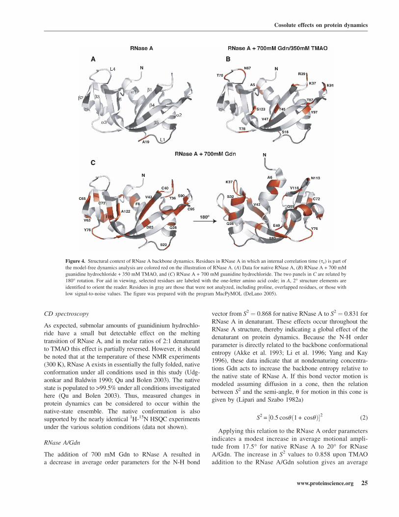
In addition to the increase in flexibility of RNase Ain guanidinium hydrochloride, a significant number ofresidues (n ¼ 41) require the model-free models 2, 4, or 5for proper fitting. A common feature of models 2, 4, and5 is the inclusion of the te term to account for additionalpicosecond time-scale motions (Lipari and Szabo 1982a,b). These residues include A6, F8, S16, S22, Q28, S32,R33, T36, K37, R39, C40, K41, V43, N44, T45, E49,Q55, A56, V57, Q60, V63, C65, C72, Y76, S77, M79,S80, D83, E86, T87, S90, C95, A96, I106, I107, G112,N113, V116, H119, A122, and S123 (Fig. 4), whereas innative RNase A, only A19, E49, and N113 require ate term in the model-free analysis. In RNase A/Gdn, allof these residues have te values ranging between 22 psecfor C65 and 130 psec for S16 with the exception of G112(te value of 1700 6 700 psec). The addition of TMAOresults in a significant decrease in the number of residues(n ¼ 19) requiring a te term (Fig. 4C) relative to theRNase A/Gdn sample. These residues are A5, S16, S22,K37, R39, K41, T45, V47, E49, N67, T70, T78, T87,K91, Y97, G112, N113, Y115, and S123. Again, thesedata are consistent with TMAO acting to reverse thenon-native protein dynamics caused by the denaturant,guanidine.
The extent of microsecond–millisecond (ms–ms) pro-tein conformational exchange motion manifests, in themodel-free analysis as an exchange term, Rex (models 3and 4). There are no significant differences in the proteinsites experiencing conformational exchange in native,Gdn, and Gdn/TMAO samples of RNase A. Twenty-oneresidues in native RNase A experience conformationalexchange, consistent with previous observations (Coleand Loria 2002, 2003; Beach et al. 2005; Kovrigin andLoria 2006a,b). In RNase A/Gdn and RNase A/Gdn/TMAOthere are 17 and 10 residues, respectively, that undergoconformational exchange motion at similar amino acid sites
Figure 3. Residue specific order parameters (S2). (A–C) S2 values as
a function of RNase A primary sequence for native RNase A (black
circles), RNase A + 700 mM guanidine hydrochloride (red circles), and
RNase A + 700 mM guanidine hydrochloride + 350 mM TMAO (blue
circles). The location of secondary structure elements is shown at the top
of the figure. Black, blue, and red horizontal dashed lines show the protein-
wide average S2 value for native, RNaseA/Gdn/TMAO, and RNase A/Gdn
samples, respectively.
Cosolute effects on protein dynamics
www.proteinscience.org 23
JOBNAME: PROSCI 16#1 2006 PAGE: 4 OUTPUT: Friday December 8 17:03:27 2006
csh/PROSCI/127806/ps0623937
Fig. 3 live 4/C

as native RNase A. Many of these seemingly unaccountedfor differences in sites in conformational exchange are theresult of residues in RNase A/Gdn and RNase A/Gdn/TMAOthat are broad and have low signal-to-noise, which ischaracteristic of conformational exchange motion, and weretherefore not analyzed further. Consequently, conformationalexchange motion appears to not be affected by the presenceof these cosolutes.
Discussion
Protein dynamics and stability
There is great interest in the physicochemical factors thatdetermine protein stability and those that modulate pro-tein dynamics. Forces (stabilizing) that protect proteinsfrom denaturation may or may not be distinct from thoseforces that rigidify the protein. For example, some
crystallographic studies have identified thermophilic pro-teins as possessing smaller Debye-Waller factors thantheir mesophilic counterparts, suggesting a link andinverse correlation between thermal stability and proteindynamics (Vihinen 1987). In addition, there have beena number of studies, both experimental and computa-tional, that provide a correlation for (Wagner andWuthrich 1979; Lazaridis et al. 1997; Tang and Dill1998; Zavodszky et al. 1998; Svingor et al. 2001; Tsaiet al. 2001) or show no evidence for (Colombo and Merz1999; Fitter and Heberle 2000; Hernandez et al. 2000;Fitter et al. 2001; Grottesi et al. 2002) a link betweendynamics and stability. In general, the results of this worksupports an inverse correlation between protein dynamicsand stability, though the increase in protein rigidity withTMAO in the presence of guanidine must be consideredin light of the slight (2°C increase) in Tm that is measuredby CD spectroscopy.
Table 1. Average S2 values for secondary structure elements
2° Structure (residues) RNase A nRNase A / 700 mM
Gdn-HCl nRNase A / 700 mM
Gdn-HCl / 350 mM TMAO n
a1 (4–12) 0.894 (0.020) 8 0.866 (0.033) 8 0.876 (0.039) 7
L1 (13–24) 0.888 (0.029) 5 0.822 (0.045) 7 0.852 (0.114) 6
a2 (25–32) 0.896 (0.017) 7 0.878 (0.025) 7 0.886 (0.033) 7
L2 (33–42) 0.876 (0.040) 8 0.828 (0.055) 8 0.848 (0.063) 8
b1 (43–47) 0.885 (0.063) 4 0.834 (0.039) 5 0.852 (0.083) 5
a3 (51–63) 0.910 (0.032) 8 0.853 (0.027) 6 0.896 (0.048) 7
L4 (64–71) 0.864 (0.016) 7 0.847 (0.018) 6 0.850 (0.024) 4
b4 (79–86) 0.856 (0.030) 5 0.829 (0.026) 7 0.857 (0.057) 5
L5 (87–96) 0.836 (0.021) 6 0.798 (0.030) 6 0.822 (0.055) 6
b5 (97–111) 0.853 (0.021) 11 0.841 (0.043) 8 0.847 (0.032) 9
b6 (116–123) 0.868 (0.036) 5 0.820 (0.034) 6 0.846 (0.064) 6
Standard deviations are listed in parentheses.
Table 2. t-Test results for secondary structure elements
2° Structure (residues)
RNase A / RNaseA/Ga RNase A / RNaseA/G+Tb
D dof t p D dof t p
a1 (4–12) 0.028 7 1.90 0.10 0.016 6 1.13 0.30
L1 (13–24) 0.032 3 1.80 0.17 0.020 3 0.45 0.68
a2 (25–32) 0.018 6 1.76 0.13 0.009 6 0.74 0.49
L2 (33–42) 0.047 7 4.68 0.002 0.027 7 1.66 0.14
b1 (43–47) 0.062 3 2.78 0.07 0.028 3 0.50 0.65
a3 (51–63) 0.052 8 4.21 0.03 0.0039 8 0.36 0.73
L4 (64–71) 0.018 5 1.49 0.20 0.0003 3 0.02 0.98
b4 (79–86) 0.031 4 2.59 0.06 0.001 4 0.11 0.92
L5 (87–96) 0.036 4 6.18 0.003 0.003 4 0.14 0.89
b5 (97–111) 0.017 7 1.59 0.16 0.009 8 0.66 0.53
b6 (116–123) 0.043 4 6.29 0.003 0.036 4 2.17 0.11
at-Test results for comparison of native RNase A and RNase A/Gdn.bt-Test results for comparison of native RNase A and RNase A/Gdn/TMAO. D is the mean difference between the data sets,dof are the degrees of freedom, t is the t-value, and p is the probability.
Doan-Nguyen and Loria
24 Protein Science, vol. 16
JOBNAME: PROSCI 16#1 2006 PAGE: 5 OUTPUT: Friday December 8 17:03:41 2006
csh/PROSCI/127806/ps0623937
CD spectroscopy
As expected, submolar amounts of guanidinium hydrochlo-ride have a small but detectable effect on the meltingtransition of RNase A, and in molar ratios of 2:1 denaturantto TMAO this effect is partially reversed. However, it shouldbe noted that at the temperature of these NMR experiments(300 K), RNase A exists in essentially the fully folded, nativeconformation under all conditions used in this study (Udg-aonkar and Baldwin 1990; Qu and Bolen 2003). The nativestate is populated to >99.5% under all conditions investigatedhere (Qu and Bolen 2003). Thus, measured changes inprotein dynamics can be considered to occur within thenative-state ensemble. The native conformation is alsosupported by the nearly identical 1H-15N HSQC experimentsunder the various solution conditions (data not shown).
RNase A/Gdn
The addition of 700 mM Gdn to RNase A resulted ina decrease in average order parameters for the N-H bond
vector from S2 ¼ 0.868 for native RNase A to S2 ¼ 0.831 forRNase A in denaturant. These effects occur throughout theRNase A structure, thereby indicating a global effect of thedenaturant on protein dynamics. Because the N-H orderparameter is directly related to the backbone conformationalentropy (Akke et al. 1993; Li et al. 1996; Yang and Kay1996), these data indicate that at nondenaturing concentra-tions Gdn acts to increase the backbone entropy relative tothe native state of RNase A. If this bond vector motion ismodeled assuming diffusion in a cone, then the relationbetween S2 and the semi-angle, u for motion in this cone isgiven by (Lipari and Szabo 1982a)
S2 = ½0:5 cosuð1 + cosuÞ�2 (2)
Applying this relation to the RNase A order parametersindicates a modest increase in average motional ampli-tude from 17.5° for native RNase A to 20° for RNaseA/Gdn. The increase in S2 values to 0.858 upon TMAOaddition to the RNase A/Gdn solution gives an average
Figure 4. Structural context of RNase A backbone dynamics. Residues in RNase A in which an internal correlation time (te) is part of
the model-free dynamics analysis are colored red on the illustration of RNase A. (A) Data for native RNase A, (B) RNase A + 700 mM
guanidine hydrochloride + 350 mM TMAO, and (C) RNase A + 700 mM guanidine hydrochloride. The two panels in C are related by
180° rotation. For aid in viewing, selected residues are labeled with the one-letter amino acid code; in A, 2° structure elements are
identified to orient the reader. Residues in gray are those that were not analyzed, including proline, overlapped residues, or those with
low signal-to-noise values. The figure was prepared with the program MacPyMOL (DeLano 2005).
Cosolute effects on protein dynamics
www.proteinscience.org 25
JOBNAME: PROSCI 16#1 2006 PAGE: 6 OUTPUT: Friday December 8 17:03:42 2006
csh/PROSCI/127806/ps0623937
Fig. 4 live 4/C

bond vector amplitude of 18°, nearly that of native RNaseA. These data indicate that the amplitude of bond vectormotions in native RNase A and RNase A/Gdn/TMAO arenearly identical.
Though the effects of Gdn on RNase dynamics areglobal, the most statistically significant increases in psec-nsec dynamics occur in loop2, loop5, a3, and b6 (Fig. 5).Moreover, three amino acid residues show large(DS2 ¼ S2
apo – S2Gdn > 0.1) increases in dynamics upon
the addition of Gdn: V43, DS2 ¼ 0.125; V57,DS2 ¼ 0.138; and G112, DS2 ¼ 0.138. Valine 57 is inthe middle of a helix 3 and has its amide nitrogenhydrogen bonded to the carbonyl oxygen of V54. V43is positioned at the N terminus of b1 and G112 is locatedin the loop connecting b5 and b6, with both of theseresidues having solvent-exposed amide nitrogen atoms.Interestingly, G112 is located in the loop that facilitatesformation of the C-domain swapped dimer in RNase A(Liu et al. 2001). These three regions of enhanceddynamics (loop2, loop5, a3, and b6) are solvent exposed,thereby indicating that the primary effects of low con-centrations of denaturant occur on the surface of theprotein, suggesting that backbone fluctuations that lead tolocal unfolding may be the first step in unfolding of theentire enzyme. Several residues in these regions havebeen shown by hydrogen exchange experiments to besome of the first regions to undergo unfolding includingN34, V43, S59, and K91 (Juneja and Udgaonkar 2002). Insomewhat of a contrast, other experiments indicate thatN34, V43, and S59 are weakly protected from exchangein an early folding intermediate (Udgaonkar and Baldwin1990). In addition, the N-terminal, a1 region is shown tobe formed last during folding and is one of the firstregions to become destabilized in the unfolding process(Udgaonkar and Baldwin 1990; Houry and Scheraga1996; Juneja and Udgaonkar 2002). The studies described
in this work, at low denaturant concentration, indicatesmall differences (p ¼ 0.01) in psec-nsec dynamics ina1 between native and RNase A/Gdn and, therefore,may report on motional processes different from thosesampled by hydrogen exchange measurements.
An interesting feature that emerged from these dynam-ics studies is the increased number of amino acid sitesthat experience additional picosecond fluctuations in thepresence of Gdn (Fig. 4). These picosecond motions,characterized by a te term, cluster together in the RNaseA structure, suggesting that the model-free analysis iscapturing real changes in protein dynamics and that thesefluctuations are structurally correlated. For example,residues comprising two of the four disulfide bonds(C95-C40 and C65-C72) in RNase A possess additionalpicosecond dynamics. Residues I106, I107, A122, S123,C72, and C65 are located near each other on adjacentb-sheets (Fig. 4C). Additionally, b5 (97–111) and b6(116–123) pack onto a3 (51–63), and residues in each ofthese secondary structure elements experience additionalpicosecond dynamics. This is also the case of adjacentb strands 1 and 4 and loops 2 and 5. Finally, whereas theS2 values in a1 did not indicate that Gdn had a significanteffect on the amplitude of motion relative to native RNaseA (vide supra), several residues in a1 for the RNase A/Gdn sample also require te in the model-free fitting.These Gdn-induced effects observed here may indicatea correlation between instability and increased dynamics.In fact, previous studies suggest that thisN-terminal region is a site of instability and potentiallyone of the first regions of RNase A to unfold (Udgaonkarand Baldwin 1990; Houry and Scheraga 1996; Juneja andUdgaonkar 2002). These additional picosecond motionsobserved in the presence of Gdn suggest a hierarchicalmechanism in which very fast native-state bond vectormotions increase in amplitude and begin to sampleadditional conformational substates.
RNase A/Gdn/TMAO
As noted, osmolytes such as TMAO should exert theirstabilizing effects in a general way because their role is tostabilize many distinct cellular proteins (Yancey 2001). Inorganisms that produce TMAO to counteract the effectsof chemical denaturant, the ratio of denaturant to TMAOis typically 2:1 and the absolute concentrations are foundto be <1 M (Yancey 1994, 2001; Yancey et al. 2001).Therefore, in this NMR work similar conditions wereused. As shown in Tables 1 and 2 and Figure 3, TMAOeffects changes in protein dynamics caused by Gdn torender them statistically indistinguishable from nativeRNase A. This is somewhat at odds with the work ofGonnelli and Strambini, in which, based on phosphores-cence lifetime measurements with apoazurin, alcohol
Figure 5. Guanidine-induced changes in protein backbone dynamics.
Regions of RNase A in which backbone dynamics show the largest
statistical difference from native RNase A (by t-test,Table 2) are colored
black. Statistically significant differences are those in which p < 0.05
(Devore 2000). The figure was prepared with the program MacPyMOL
(DeLano 2005).
Doan-Nguyen and Loria
26 Protein Science, vol. 16
JOBNAME: PROSCI 16#1 2006 PAGE: 7 OUTPUT: Friday December 8 17:04:09 2006
csh/PROSCI/127806/ps0623937
dehydrogenase, alkaline phosphatase, and glyceraldehyde-3-phosphate dehydrogenase, they observe no effect ofTMAO on the internal dynamics of these proteins (Gonnelliand Strambini 2001). This contrasting view of the effects ofTMAO may be due to the much different time scale that isprobed by the phosphorescence and NMR techniques or bythe limited site resolution of phosphorescence measure-ments. Moreover, our results clearly show significant effectson protein dynamics caused by TMAO and are in agreementwith other experimental and theoretical studies, indicatingthat TMAO reverses the effects of chemical denaturants bydecreasing the fluctuations of the native state (Jaravine et al.2000; Qu and Bolen 2003; Bennion and Daggett 2004; Gahlet al. 2004). However, the NMR spin-relaxation experimentsdescribed here provide two additional important pieces ofinformation that complement and add to the experimentalstudies noted above. First, these NMR spin-relaxationexperiments typically allow analysis of significantly moreresidues than other methods and thus result in a morecomplete dynamics picture. Second, motions over a largetime window (psec-msec) can be examined by these experi-ments, thereby providing additional insight on the types ofprotein motions involved.
For example, as demonstrated in Figure 4C, in thepresence of guanidine the dynamics description of 41residues requires the inclusion of an internal correlationtime te, whereas in native RNase A there are only threete requiring residues and in the RNase A/Gdn/TMAOsample this number is 19. te in the model-free formalismsuggests the presence of additional internal motionoccurring on a timescale from 20 to 200 psec. Of these41 sites in both the native and Gdn/TMAO RNase Asamples, all but nine (K37, R39, K41, T45, E49, T87,G112, N113, and S123) are best described by models notcontaining te, indicating that for these 32 residues theinternal correlation time is very fast (<20 psec) in thenative and Gdn/TMAO RNase A forms. TMAO reversesthe effects of guanidine by reducing the amplitude offluctuations as measured by the NMR order parametersand causes a reduction in the number of residuesexperiencing internal motions on the te time scale (20psec < te < tm).
These studies demonstrate that TMAO restricts theincrease in conformational space sampled by the N-Hbond vectors in the presence of guanidine hydrochloridealone. Thus, TMAO causes more restricted, native-likeprotein fluctuations, possibly limiting access to higherenergy conformational substates that would ultimatelylead to protein denaturation. Several of the protein sitesexperiencing this TMAO-induced reversal of dynamicsreflect those identified by hydrogen exchange experi-ments (Idiyatullin et al. 2003), which occurs on a muchslower time scale, suggesting that NMR spin-relaxationexperiments are a useful and complementary tool for
studying protein stability. The nearly uniform nature ofthe effects on protein dynamics and NMR chemicalshifts caused by TMAO argues against specific proteinbinding (Lin and Timasheff 1994; Zou et al. 2002).Finally, these NMR experiments are independent of themechanism of amide hydrogen exchange with solventand, therefore, their interpretation is often more straight-forward.
Materials and methods
Protein preparation
The plasmid (pBXR) encoding RNase A was a gift fromProfessor Ronald T. Raines (University of Wisconsin-Madison).15N-labeled RNase A was expressed and purified from Escher-ichia coli strain BL21 (DE3) as described previously (Cole andLoria 2002). The 15N-labeled RNase A sample used here wasprovided by Dr. Evgenii Kovrigin (Yale University). RNase Awas judged pure by SDS-PAGE and MALDI-TOF mass spec-trometry. The concentration of RNase A was determined usinga molar extinction coefficient, e278 ¼ 9800 M�1 cm�1 (Ander-son et al. 1968). Trimethylamine N-oxide (TMAO) and guani-dine hydrochloride (Gdn) were purchased from Sigma-Aldrichand were of the highest purity available. Concentrations of thesecosolutes were determined gravimetrically. All reference to theRNase A three-dimensional structures are based on the X-raycrystal structure 7RSA (Wlodawer et al. 1988).
Circular dichroism spectroscopy
For CD experiments, [RNase A] ¼ 20 or 15 mM in 10 mMpotassium phosphate buffer (pH 6.4). In the temperature-dependent studies, guanidine hydrochloride (Gdn) and/or trimeth-ylamine N-oxide (TMAO) were included in this buffer at thenoted concentrations. In all cases, blanks were run with identicalbuffer components excluding RNase A. Melting profiles weredetermined by monitoring the change in molar ellipticity at 222nm between 302 and 353 K (29°C–81°C). The temperature wasincreased at a rate of 1°C/min with data acquisition for 5 sec ateach temperature on an Aviv CD spectrometer. All meltingcurves were performed in triplicate and the values wereaveraged.
NMR spectroscopy
For NMR experiments, [RNase A] ¼ 0.65 mM in 5 mM MESbuffer (pH 6.4) and 10% D2O. NMR experiments were performedon either a Varian Inova 600 or Varian Unity+ 600 MHz instru-ments. The temperature was calibrated prior to each experiment at300 K using 100% methanol as a standard. The 1H carrierfrequency was set to the water resonance and the 15N frequencywas set to 120 ppm. All experiments were collected using gradient-selected sensitivity-enhanced pulse sequences (Kay et al. 1992;Kordel et al. 1992; Palmer et al. 1992; Skelton et al. 1993) with 128t1 increments and 2k points in the direct dimension with spectralwidths of 1800 and 9000 Hz, respectively. The data were zero filledprior to Fourier transformation. A Kaiser window (Cavanagh et al.1996) was applied to the t1 dimension with Lorentz-to-Gaussresolution enhancement in t2. All NMR were processed using
Cosolute effects on protein dynamics
www.proteinscience.org 27
JOBNAME: PROSCI 16#1 2006 PAGE: 8 OUTPUT: Friday December 8 17:04:14 2006
csh/PROSCI/127806/ps0623937
NMRPipe (Delaglio et al. 1995) and visualized with Sparky (T.D.Goddard and D.G. Kneller, University of California, San Fran-cisco). Peak heights were determined from 3 3 3 grids centered onthe peak maximum.
For the R1 and R2 relaxation rate measurements, eachexperiment was preceded by a 2.7-sec recycle delay. Relaxationdelays for the R1 experiments were 2 (32), 102, 222, 357, 512,692, 912 (32), and 1202 msec. The R2 experiments wereacquired with relaxation delays of 0 (32), 10, 22, 38 (32),70, 92, 122, and 162 msec. All relaxation data was fit with asingle exponential decay function to obtain the relaxation ratesusing Curvefit (http://cpmcnet.columbia.edu/dept/gsas/biochem/labs/palmer/software.html). 1H(15N) 90° pulse lengths (microsec-onds) for the RNase A, RNase A/700 mM Gdn-HCl, and RNaseA/700mM Gdn-HCl/350 mM TMAO samples were 9.5 (37.5),14.0 (37.3), and 12.5 (38.0), respectively. The heteronuclear NOEeffect was measured using interleaved experiments with andwithout proton saturation pulses with 10-sec recycle delays.
Model-free dynamics analysis
Amide backbone dynamics were characterized by fitting theexperimentally determined R1, R2, and NOE to each of fivespectral density models with the corresponding free parameters(Lipari and Szabo 1982a,b; Clore et al. 1990).
model 1, S2; model 2, S2, te; model 3, S2, Rex; model 4, S2, te,
Rex; model 5, S2f , S2, te (3)
in which S2 is the generalized order parameter, S2f is the order
parameter for motion with a correlation time faster than 10–20 psec, Rex is the contribution due to conformational exchangemotion (tc ; msec-msec), and te is the internal correlation time(20 psec < te < 200 psec). Motional parameters were determinedusing the program FAST-model free (Cole and Loria 2003)interfaced with Model-Free 4.1 (http://cpmcnet.columbia.edu/dept/gsas/biochem/labs/palmer/software.html). The statisticalcriteria used for model selection is as described by Mandelet al. (1995). Prior to model fitting, the RNase A rotationaldiffusion tensor was estimated from R2/R1 ratios and theprogram pdbinertia (http://cpmcnet.columbia.edu/dept/gsas/biochem/labs/palmer/software.html) using the crystal structureof RNase A (7RSA), in which hydrogens had been added and themolecule minimized to <0.1 kcal energy difference using theminimize routine in the program TINKER v4.2. For the model-free analysis, the N–H bond length was assumed to be 1.02 Aand the 15N chemical-shift anisotropy was assumed to be axiallysymmetric with a value of –160 ppm. Other aspects of themodel-free fitting are as described previously (Cole and Loria2003; Kovrigin et al. 2003).
Electronic supplemental material
Supplemental material contains the NMR spin-relaxation ratesand the results of model free analysis.
Acknowledgments
We thank Professor Andrew Hamilton (Yale University) for theuse of his CD spectrometer and Dr. Evgenii Kovrigin (Yale
University) for the RNase A sample and for critical reading ofthis manuscript. J.P.L. acknowledges funding from an NSF-CAREER award and a fellowship from the Alfred P. SloanFoundation. This work, in part, fulfilled the undergraduatechemistry degree requirements for V.D.N. V.D.N. was part ofthe STARS undergraduate research program at Yale that waspartially funded by the Howard Hughes Medical Institute andBoehringer-Ingelheim Pharmaceuticals.
References
Abragam, A. 1961. Principles of nuclear magnetism. Clarendon Press, Oxford,UK.
Akke, M., Bruschweiler, R., and Palmer, A.G. 1993. NMR order parametersand free energy: An analytic approach and application to cooperative Ca2+
binding by calbindin D9k. J. Am. Chem. Soc. 115: 9832–9833.Anderson, D.G., Hammes, G.G., and Walz Jr., F.G. 1968. Binding of phosphate
ligands to ribonuclease A. Biochemistry 7: 1637–1645.Arakawa, T. and Timasheff, S.N. 1985. The stabilization of proteins by
osmolytes. Biophys. J. 47: 411–414.Athawale, M.V., Dordick, J.S., and Garde, S. 2005. Osmolyte trimethylamine-
N-oxide does not affect the strength of hydrophobic interactions: Origin ofosmolyte compatibility. Biophys. J. 89: 858–866.
Beach, H., Cole, R., Gill, M., and Loria, J.P. 2005. Conservation of ms-msenzyme motions in the apo- and substrate-mimicked state. J. Am. Chem.Soc. 127: 9167–9176.
Bennion, B.J. and Daggett, V. 2004. Counteraction of urea-induced proteindenaturation by trimethylamine N-oxide: A chemical chaperone at atomicresolution. Proc. Natl. Acad. Sci. 101: 6433–6438.
Cavanagh, J., Fairbrother, W.J., Palmer, A.G., and Skelton, N.J. 1996. ProteinNMR spectroscopy: Principles and practice. Academic Press, San Diego,CA.
Clore, G.M., Szabo, A., Bax, A., Kay, L.E., Driscoll, P.C., andGronenborn, A.M. 1990. Deviations from the simple two-parametermodel-free approach to the interpretation of nitrogen-15 nuclear magneticrelaxation of proteins. J. Am. Chem. Soc. 112: 4989–4991.
Cole, R. and Loria, J.P. 2002. Evidence for flexibility in the function ofribonuclease A. Biochemistry 41: 6072–6081.
Cole, R. and Loria, J.P. 2003. FAST-Modelfree: A program for rapid automatedanalysis of solution NMR spin-relaxation data. J. Biomol. NMR 26: 203–213.
Colombo, G. and Merz, K.M. 1999. Stability and activity of mesophilicsubtilisin E and its thermophilic homologue: Insights from moleculardynamics simulations. J. Am. Chem. Soc. 121: 6895–6903.
DeKoster, G.T. and Robertson, A.D. 1997. Calorimetrically-derived parametersfor protein interactions with urea and guanidine-HCl are not consistent withdenaturant m values. Biophys. Chem. 64: 59–68.
Delaglio, F., Grzesiek, S., Vuister, G.W., Zhu, G., Pfeifer, J., and Bax, A. 1995.NMRPipe: A multidimensional spectral processing system based on UNIXpipes. J. Biomol. NMR 6: 277–293.
DeLano, W.L. 2005. MacPyMOL: A PyMOL-based molecular graphicsapplication for MacOS X. DeLano Scientific LLC, South San Francisco,CA.
Devore, J. 2000. Probability and statistics for engineering and the sciences.Brooks/Cole Publishing Company, Monterey, CA.
Esposito, L. and Daggett, V. 2005. Insight into ribonuclease A domainswapping by molecular dynamics unfolding simulations. Biochemistry44: 3358–3368.
Fitter, J. and Heberle, J. 2000. Structural equilibrium fluctuations in mesophilicand thermophilic a-amylase. Biophys. J. 79: 1629–1636.
Fitter, J., Herrmann, R., Dencher, N.A., Blume, A., and Hauss, T. 2001. Activityand stability of a thermostable a-amylase compared to its mesophilichomologue: Mechanisms of thermal adaptation. Biochemistry 40: 10723–10731.
Gahl, R.F., Narayan, M., Xu, G., and Scheraga, H.A. 2004. Trimethylamine-N-oxide modulates the reductive unfolding of onconase. Biochem. Biophys.Res. Commun. 325: 707–710.
Gonnelli, M. and Strambini, G.B. 2001. No effect of trimethylamine N-oxideon the internal dynamics of the protein native fold. Biophys. Chem. 89:77–85.
Grottesi, A., Ceruso, M.A., Colosimo, A., and Di Nola, A. 2002. Moleculardynamics study of a hyperthermophilic and a mesophilic rubredoxin.Proteins 46: 287–294.
Doan-Nguyen and Loria
28 Protein Science, vol. 16
JOBNAME: PROSCI 16#1 2006 PAGE: 9 OUTPUT: Friday December 8 17:04:15 2006
csh/PROSCI/127806/ps0623937
Hernandez, G., Jenney Jr., F.E., Adams, M.W., and LeMaster, D.M. 2000.Millisecond time scale conformational flexibility in a hyperther-mophile protein at ambient temperature. Proc. Natl. Acad. Sci. 97: 3166–3170.
Houry, W.A. and Scheraga, H.A. 1996. Structure of a hydrophobicallycollapsed intermediate on the conformational folding pathway of ribonu-clease A probed by hydrogen- deuterium exchange. Biochemistry 35:11734–11746.
Hovagimyan, K.G. and Gerig, J.T. 2005. Interactions of trimethylamineN-oxide and water with cyclo-alanylglycine. J. Phys. Chem. B. 109:24142–24151.
Idiyatullin, D., Nesmelova, I., Daragan, V.A., and Mayo, K.H. 2003. Heatcapacities and a snapshot of the energy landscape in protein GB1 from thepre-denaturation temperature dependence of backbone NH nanosecondfluctuations. J. Mol. Biol. 325: 149–162.
Jaravine, V.A., Rathgeb-Szabo, K., and Alexandrescu, A.T. 2000. Microscopicstability of cold shock protein A examined by NMR native state hydrogenexchange as a function of urea and trimethylamine N-oxide. Protein Sci.9: 290–301.
Juneja, J. and Udgaonkar, J.B. 2002. Characterization of the unfolding ofribonuclease a by a pulsed hydrogen exchange study: Evidence forcompeting pathways for unfolding. Biochemistry 41: 2641–2654.
Kay, L.E., Keifer, P., and Saarinen, T. 1992. Pure absorption gradient enhancedheteronuclear single quantum correlation spectroscopy with improvedsensitivity. J. Am. Chem. Soc. 114: 10663–10665.
Kordel, J., Skelton, N.J., Akke, M., Palmer, A.G., and Chazin, W.J. 1992.Backbone dynamics of calcium-loaded calbindin D9k studied by two-dimensional proton-detected NMR spectroscopy. Biochemistry 31: 4856–4866.
Kovrigin, E.L. and Loria, J.P. 2006a. Characterization of the transition state offunctional enzyme dynamics. J. Am. Chem. Soc. 128: 7724–7725.
Kovrigin, E.L. and Loria, J.P. 2006b. Enzyme dynamics along the reactioncoordinate: Critical role of a conserved residue. Biochemistry 45: 2636–2647.
Kovrigin, E.L., Cole, R., and Loria, J.P. 2003. Temperature dependence of thebackbone dynamics of Ribonuclease A in the ground state and bound to theinhibitor 59-phosphothymidine (39-59) pyrophosphate adenosine 39-phosphate.Biochemistry 42: 5279–5291.
Lazaridis, T., Lee, I., and Karplus, M. 1997. Dynamics and unfolding pathwaysof a hyperthermophilic and a mesophilic rubredoxin. Protein Sci. 6: 2589–2605.
Lee, L.K., Rance, M., Chazin, W.J., and Palmer, A.G. 1997. Rotationaldiffusion anisotropy of proteins from simultaneous analysis of 15N and13Ca nuclear spin relaxation. J. Biomol. NMR 9: 287–298.
Li, Z., Raychaudhuri, S., and Wand, A.J. 1996. Insights into the local residualentropy of proteins provided by NMR relaxation. Protein Sci. 5: 2647–2650.
Lin, T.Y. and Timasheff, S.N. 1994. Why do some organisms use a urea-methylamine mixture as osmolyte? Thermodynamic compensation of ureaand trimethylamine N-oxide interactions with protein. Biochemistry 33:12695–12701.
Lipari, G. and Szabo, A. 1982a. Model-free approach to the interpretation ofnuclear magnetic resonance relaxation in macromolecules. 1. Theory andrange of validity. J. Am. Chem. Soc. 104: 4546–4559.
Lipari, G. and Szabo, A. 1982b. Model-free approach to the interpretation ofnuclear magnetic resonance relaxation in macromolecules. 2. Analysis ofexperimental results. J. Am. Chem. Soc. 104: 4559–4570.
Liu, Y., Gotte, G., Libonati, M., and Eisenberg, D. 2001. A domain-swappedRNase A dimer with implications for amyloid formation. Nat. Struct. Biol.8: 211–214.
Mandel, A.M., Akke, M., and Palmer, A.G. 1995. Backbone dynamics ofEscherichia coli ribonuclease HI: Correlations with structure and functionin an active enzyme. J. Mol. Biol. 246: 144–163.
Palmer, A.G., Skelton, N.J., Chazin, W.J., Wright, P.E., and Rance, M. 1992.Suppression of the effects of cross-correlation between dipolar and
anisotropic chemical shift relaxation mechanisms in the measurement ofspin-spin relaxation rates. Mol. Phys. 75: 699–711.
Palmer, H.R., Bedford, J.J., Leader, J.P., and Smith, R.A. 2000. 31P and 1HNMR studies of the effect of the counteracting osmolyte trimethylamine-N-oxide on interactions of urea with ribonuclease A. J. Biol. Chem. 275:27708–27711.
Poklar, N., Petrovcic, N., Oblak, M., and Vesnaver, G. 1999. Thermodynamicstability of ribonuclease A in alkylurea solutions and preferential solvationchanges accompanying its thermal denaturation: A calorimetric andspectroscopic study. Protein Sci. 8: 832–840.
Qu, Y. and Bolen, D.W. 2003. Hydrogen exchange kinetics of RNase A and theurea:TMAO paradigm. Biochemistry 42: 5837–5849.
Skelton, N.J., Palmer, A.G., Akke, M., Kordel, J., Rance, M., and Chazin, W.J.1993. Practical aspects of two-dimensional proton-detected 15N spinrelaxation measurements. J. Magn. Reson. B 102: 253–264.
Somero, G.N. 2003. Protein adaptations to temperature and pressure: Comple-mentary roles of adaptive changes in amino acid sequence and internalmilieu. Comp. Biochem. Physiol. B Biochem. Mol. Biol. 136: 577–591.
Svingor, A., Kardos, J., Hajdu, I., Nemeth, A., and Zavodszky, P. 2001. A betterenzyme to cope with cold. Comparative flexibility studies on psychrotrophic,mesophilic, and thermophilic IPMDHs. J. Biol. Chem. 276: 28121–28125.
Tang, K.E. and Dill, K.A. 1998. Native protein fluctuations: The conformational-motion temperature and the inverse correlation of protein flexibility withprotein stability. J. Biomol. Struct. Dyn 16: 397–411.
Tsai, A.M., Udovic, T.J., and Neumann, D.A. 2001. The inverse relationshipbetween protein dynamics and thermal stability. Biophys. J. 81: 2339–2343.
Udgaonkar, J.B. and Baldwin, R.L. 1990. Early folding intermediate ofribonuclease A. Proc. Natl. Acad. Sci. 87: 8197–8201.
Vihinen, M. 1987. Relationship of protein flexibility to thermostability. ProteinEng. 1: 477–480.
Wagner, G. and Wuthrich, K. 1979. Correlation between the amide protonexchange rates and the denaturation temperatures in globular proteinsrelated to the basic pancreatic trypsin inhibitor. J. Mol. Biol. 130: 31–37.
Wang, A. and Bolen, D.W. 1997. A naturally occurring protective system inurea-rich cells: Mechanism of osmolyte protection of proteins against ureadenaturation. Biochemistry 36: 9101–9108.
Wlodawer, A., Svensson, L.A., Sjolin, L., and Gilliland, G.L. 1988. Structure ofphosphate-free ribonuclease A refined at 1.26 A. Biochemistry 27: 2705–2717.
Woessner, D.E. 1962. Spin relaxation processes in a two-proton systemundergoing anisotropic reorientation. J. Chem. Phys. 36: 1–4.
Yancey, P.H. 1994. Compatible and counteracting solutes. In Cellular andmolecular physiology of cell volume regulation (ed. K. Strange), pp. 81–109. CRC Press, Boca Raton, FL.
Yancey, P.H. 2001. Protein, osmolytes and water stress. Am. Zool. 41: 699–709.Yancey, P.H. 2005. Organic osmolytes as compatible, metabolic and counter-
acting cytoprotectants in high osmolarity and other stresses. J. Exp. Biol.208: 2819–2830.
Yancey, P.H. and Somero, G.N. 1979. Counteraction of urea destabilization ofprotein structure by methylamine osmoregulatory compounds of elasmo-branch fishes. Biochem. J. 183: 317–323.
Yancey, P.H., Fyfe-Johnson, A.L., Kelly, R.H., Walker, V.P., and Aunon, M.T.2001. Trimethylamine oxide counteracts effects of hydrostatic pressure onproteins of deep-sea teleosts. J. Exp. Zool. 289: 172–176.
Yancey, P.H., Blake, W.R., and Conley, J. 2002. Unusual organic osmolytes indeep-sea animals: Adaptations to hydrostatic pressure and other pertur-bants. Comp. Biochem. Physiol. A Mol. Integr. Physiol. 133: 667–676.
Yang, D. and Kay, L.E. 1996. Contributions to conformational entropy arisingfrom bond vector fluctuations measured from NMR-derived order param-eters: Application to protein folding. J. Mol. Biol. 263: 369–382.
Zavodszky, P., Kardos, J., Svingor, A., and Petsko, G.A. 1998. Adjustment ofconformational flexibility is a key event in the thermal adaptation ofproteins. Proc. Natl. Acad. Sci. 95: 7406–7411.
Zou, Q., Bennion, B.J., Daggett, V., and Murphy, K.P. 2002. The molecularmechanism of stabilization of proteins by TMAO and its ability tocounteract the effects of urea. J. Am. Chem. Soc. 124: 1192–1202.
Cosolute effects on protein dynamics
www.proteinscience.org 29
JOBNAME: PROSCI 16#1 2006 PAGE: 10 OUTPUT: Friday December 8 17:04:17 2006
csh/PROSCI/127806/ps0623937
Related Documents