REVIEW THE EFFECTS OF ABUSED DRUGS ON ADOLESCENT DEVELOPMENT OF CORTICOLIMBIC CIRCUITRY AND BEHAVIOR J. M. GULLEY * AND J. M. JURASKA * Department of Psychology and Neuroscience Program, University of Illinois at Urbana-Champaign, USA Abstract—Adolescence is a period of significant neurobio- logical change that occurs as individuals transition from childhood to adulthood. Because the nervous system is in a relatively labile state during this stage of development, it may be especially sensitive to experience-induced plastic- ity. One such experience that is relatively common to ado- lescents is the exposure to drugs of abuse, particularly alcohol and psychostimulants. In this review, we highlight recent findings on the long-lasting effects of exposure to these drugs during adolescence in humans as well as in ani- mal models. Whenever possible, our focus is on studies that use comparison groups of adolescent- and adult-exposed subjects as this is a more direct test of the hypothesis that adolescence represents a period of enhanced vulnerability to the effects of drug-induced plasticity. Lastly, we suggest areas of future investigation that are needed and methodo- logical concerns that should be addressed. Ó 2013 IBRO. Published by Elsevier Ltd. All rights reserved. Key words: adolescent, young adult, neuroanatomy, neuro- physiology, psychostimulants. Contents Introduction 00 Adolescence: The last developmental phase of PFC maturation 00 Neuron number in the medial prefrontal cortex (mPFC) 00 Connectivity changes 00 The basolateral amygdala 00 Alcohol and adolescence 00 Effects of alcohol on brain structure 00 Effects of alcohol on neurophysiology 00 Effects of alcohol on neurochemistry 00 Behavioral effects of alcohol during adolescence 00 Psychostimulants and adolescence 00 Effects of psychostimulants on brain structure 00 Effects of psychostimulants on neurophysiology 00 Effects of psychostimulants on neurochemistry 00 Behavioral effects of psychostimulants during adolescence 00 Future challenges 00 Acknowledgments 00 References 00 INTRODUCTION Adolescence, the transition from the juvenile period to adulthood, is marked by puberty and numerous physical and neural changes. In humans, adolescence begins at approximately 12 years of age and may extend to the mid-twenties (Dahl, 2004). In rats, adolescence has been conservatively defined as beginning around postnatal day (P) 28 and extending to P42 (Spear, 2000) or perhaps as late as P60 (Tirelli et al., 2003; Brenhouse and Andersen, 2011). This is based, in part, on the rise of pubertal hormones which leads to the vaginal opening in female rats between P29 and P37 (Castellano et al., 2011) and preputial separation in male rats between P39–47 (Korenbrot et al., 1977). During this time, there is substantial behavioral and neural development (Spear, 2000; Sisk and Foster, 2004), with corticolimbic brain regions such as the prefrontal cortex (PFC), nucleus accumbens (NAc), and basolateral amygdala (BLA) being among the last brain circuits to fully mature in both humans and rodents (Casey et al., 2000; Brenhouse and Andersen, 2011). Because the brain is undergoing this programed period of dramatic change, it might be especially sensitive to outside influences that have the ability to induce plasticity in the nervous system. One such influence that is pervasive during human adolescence is exposure to drugs such as alcohol and psychostimulants. Recent data from the nationwide Monitoring the Future study (Johnston et al., 2012), which sampled from over 46,000 eighth to 12th grade students, suggests that approximately 70% of young people have consumed alcohol by the end of the 12th grade and 33% have been intoxicated within the last month. Nicotine (via cigarette smoking) is consumed at 0306-4522/13 $36.00 Ó 2013 IBRO. Published by Elsevier Ltd. All rights reserved. http://dx.doi.org/10.1016/j.neuroscience.2013.05.026 * Corresponding authors. Addresses: Department of Psychology and Neuroscience Program, University of Illinois at Urbana-Champaign, 731 Psychology Building MC-716, 603 E Daniel Street, Champaign, IL 61820, USA. Tel: +1-217-265-6413; fax: +1-217-244-5876 (J. M. Gulley), Department of Psychology and Neuroscience Program, University of Illinois at Urbana-Champaign, 735 Psychology Building MC-716, 603 E Daniel Street, Champaign, IL 61820, USA. Tel: +1- 217-333-8546; fax: +1-217-244-5876 (J. M. Juraska). E-mail addresses: [email protected] (J. M. Gulley), jjuraska@ illinois.edu (J. M. Juraska). Abbreviations: BLA, basolateral amygdala; CRH, corticotropin- releasing hormone; EEG, electroencephalography; LTD, long-term depression; LTP, long-term potentiation; mPFC, medial prefrontal cortex; MRI, magnetic resonance imaging; NAc, nucleus accumbens; NMDA, N-methyl-D-aspartate; P, postnatal day; PFC, prefrontal cortex. Neuroscience xxx (2013) xxx–xxx Please cite this article in press as: Gulley JM, Juraska JM. The effects of abused drugs on adolescent development of corticolimbic circuitry and behav- ior. Neuroscience (2013), http://dx.doi.org/10.1016/j.neuroscience.2013.05.026 1

Welcome message from author
This document is posted to help you gain knowledge. Please leave a comment to let me know what you think about it! Share it to your friends and learn new things together.
Transcript
Please cite this article in press as: Gulley JM, Juraska JM. The effects of abused drugs on adolescent development of corticolimbic circuitry and behav-
ior. Neuroscience (2013), http://dx.doi.org/10.1016/j.neuroscience.2013.05.026
Neuroscience xxx (2013) xxx–xxx
REVIEW
THE EFFECTS OF ABUSED DRUGS ON ADOLESCENT DEVELOPMENT OFCORTICOLIMBIC CIRCUITRY AND BEHAVIOR
J. M. GULLEY * AND J. M. JURASKA *
Department of Psychology and Neuroscience Program, University
of Illinois at Urbana-Champaign, USA
Abstract—Adolescence is a period of significant neurobio-
logical change that occurs as individuals transition from
childhood to adulthood. Because the nervous system is in
a relatively labile state during this stage of development, it
may be especially sensitive to experience-induced plastic-
ity. One such experience that is relatively common to ado-
lescents is the exposure to drugs of abuse, particularly
alcohol and psychostimulants. In this review, we highlight
recent findings on the long-lasting effects of exposure to
these drugs during adolescence in humans as well as in ani-
mal models. Whenever possible, our focus is on studies that
use comparison groups of adolescent- and adult-exposed
subjects as this is a more direct test of the hypothesis that
adolescence represents a period of enhanced vulnerability
to the effects of drug-induced plasticity. Lastly, we suggest
areas of future investigation that are needed and methodo-
logical concerns that should be addressed.
� 2013 IBRO. Published by Elsevier Ltd. All rights reserved.
Key words: adolescent, young adult, neuroanatomy, neuro-
physiology, psychostimulants.
Contents
Introduction 00
Adolescence: The last developmental phase of
PFC maturation 00
Neuron number in the medial prefrontal cortex (mPFC) 00
Connectivity changes 00
The basolateral amygdala 00
Alcohol and adolescence 00
Effects of alcohol on brain structure 00
Effects of alcohol on neurophysiology 00
0306-4522/13 $36.00 � 2013 IBRO. Published by Elsevier Ltd. All rights reservehttp://dx.doi.org/10.1016/j.neuroscience.2013.05.026
*Corresponding authors. Addresses: Department of Psychology andNeuroscience Program, University of Illinois at Urbana-Champaign,731 Psychology Building MC-716, 603 E Daniel Street, Champaign,IL 61820, USA. Tel: +1-217-265-6413; fax: +1-217-244-5876 (J. M.Gulley), Department of Psychology and Neuroscience Program,University of Illinois at Urbana-Champaign, 735 Psychology BuildingMC-716, 603 E Daniel Street, Champaign, IL 61820, USA. Tel: +1-217-333-8546; fax: +1-217-244-5876 (J. M. Juraska).
E-mail addresses: [email protected] (J. M. Gulley), [email protected] (J. M. Juraska).Abbreviations: BLA, basolateral amygdala; CRH, corticotropin-releasing hormone; EEG, electroencephalography; LTD, long-termdepression; LTP, long-term potentiation; mPFC, medial prefrontalcortex; MRI, magnetic resonance imaging; NAc, nucleus accumbens;NMDA, N-methyl-D-aspartate; P, postnatal day; PFC, prefrontal cortex.
1
Effects of alcohol on neurochemistry 00
Behavioral effects of alcohol during adolescence 00
Psychostimulants and adolescence 00
Effects of psychostimulants on brain structure 00
Effects of psychostimulants on neurophysiology 00
Effects of psychostimulants on neurochemistry 00
Behavioral effects of psychostimulants during adolescence 00
Future challenges 00
Acknowledgments 00
References 00
INTRODUCTION
Adolescence, the transition from the juvenile period to
adulthood, is marked by puberty and numerous physical
and neural changes. In humans, adolescence begins at
approximately 12 years of age and may extend to the
mid-twenties (Dahl, 2004). In rats, adolescence has
been conservatively defined as beginning around
postnatal day (P) 28 and extending to P42 (Spear,
2000) or perhaps as late as P60 (Tirelli et al., 2003;
Brenhouse and Andersen, 2011). This is based, in part,
on the rise of pubertal hormones which leads to the
vaginal opening in female rats between P29 and P37
(Castellano et al., 2011) and preputial separation in
male rats between P39–47 (Korenbrot et al., 1977).
During this time, there is substantial behavioral and
neural development (Spear, 2000; Sisk and Foster,
2004), with corticolimbic brain regions such as the
prefrontal cortex (PFC), nucleus accumbens (NAc), and
basolateral amygdala (BLA) being among the last brain
circuits to fully mature in both humans and rodents
(Casey et al., 2000; Brenhouse and Andersen, 2011).
Because the brain is undergoing this programed period
of dramatic change, it might be especially sensitive to
outside influences that have the ability to induce
plasticity in the nervous system.
One such influence that is pervasive during human
adolescence is exposure to drugs such as alcohol and
psychostimulants. Recent data from the nationwide
Monitoring the Future study (Johnston et al., 2012),
which sampled from over 46,000 eighth to 12th grade
students, suggests that approximately 70% of young
people have consumed alcohol by the end of the 12th
grade and 33% have been intoxicated within the last
month. Nicotine (via cigarette smoking) is consumed at
d.

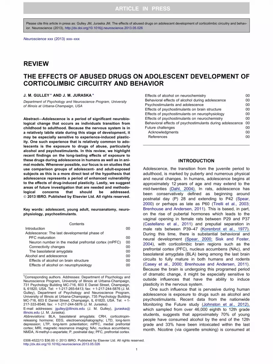
Fig. 1. (A) The number of neurons in the ventral portion of the rat
mPFC at periadolescence (P35) and adulthood (P90) in both sexes.
Adapted from Markham et al. (2007). (B) The number of neurons in
the basolateral amygdalar nucleus at periadolescence and adulthood.
Adapted from Rubinow and Juraska (2009). ⁄p< 0.04.
2 J. M. Gulley, J. M. Juraska /Neuroscience xxx (2013) xxx–xxx
least once by about 40% of adolescents by the 12th
grade, and nearly 20% of 12th graders report being
current smokers. Nearly 1% and 3% of adolescents
report they are current users of cocaine and
amphetamines, respectively. These relatively high levels
of use are of concern because these drugs are known
to produce significant and long-lasting changes in brain
structure and function (Luscher and Malenka, 2011) that
have been linked to long-term changes in cognitive
functioning and the development of addiction (Goldstein
and Volkow, 2002). In this review, we will highlight
recent findings on the long-lasting effects of alcohol and
psychostimulant exposure during periadolescence.
Although some evidence from humans is available, we
will primarily focus on studies in animal models. In doing
so, we will place particular emphasis on those that
utilize both adolescent and adult exposure groups since
these directly assess the potential for age of exposure-
dependent effects.
ADOLESCENCE: THE LAST DEVELOPMENTALPHASE OF PFC MATURATION
The scientific community and the general public were
startled when human structural magnetic resonance
imaging (MRI) studies showed that the cortex, including
the PFC, decreases in size between 11and 22 years of
age (Giedd et al., 1999; Sowell et al., 1999). Previous to
this finding, most neural development in humans was
thought to be completed by 12 years of age, which is
approximately when overall brain volume is at adult
levels (Courchesne et al., 2000). The continuation of
development during adolescence suggests greater
vulnerability than adults to many of the effects of
environmental influences, including those produced by
exposure to drugs of abuse. This is a major problem
because adolescence is also a time of high novelty and
sensation seeking (Spear, 2000), which often includes
experimentation with drugs.
Prior to the advent of MRI studies, there were
indications of cellular changes in the PFC during
adolescence. Periadolescent anatomical refinement of
circuitry in the primate PFC was described in both
excitatory and inhibitory circuits (reviewed by Lewis,
1997; Woo et al., 1997). Synaptic density in this area
was also found to decline during adolescence in both
monkeys (Bourgeois et al., 1994; Anderson et al.,
1995) and humans (Huttenlocher, 1979; Huttenlocher
and Dabholkar, 1997); however, this decrease in
synaptic number would have a small effect on cortical
volume, as was noted by Bourgeois and Rakic (1993).
Here, we suggest that a loss of neurons could readily
account for the MRI finding of volume loss in human
adolescence.
Neuron number in the medial prefrontal cortex(mPFC)
The number of neurons (density � volume) during
development and adolescence has not been examined
in the frontal cortex or any other cortical region in
primates. This is due in part to the technical difficulties
Please cite this article in press as: Gulley JM, Juraska JM. The effects of abuse
ior. Neuroscience (2013), http://dx.doi.org/10.1016/j.neuroscience.2013.05.026
of parcellating regions of the cortex in large brains that
have variable gyri. However, subtle declines in neuronal
density between 2 and 16 years of age in human frontal
cortex have been noted (Huttenlocher, 1979). The rat
PFC is a more practical model than that of the primate
for exploring the cellular basis for pruning during
adolescence because the rat PFC is less differentiated
and segregated. Furthermore, on the basis of the
reciprocity of specific thalamic as well as other
connections, embryological development, and
electrophysiological and behavioral characteristics, rats
do have a PFC that is homologous to that of the primate
(Brown and Bowman, 2002; see Uylings et al., 2003 for
an extensive review). Interestingly, like humans, the rat
PFC undergoes a decrease in volume during the
periadolescent period (van Eden and Uylings, 1985;
Markham et al., 2007).
To investigate the possibility of cell loss, we (Markham
et al., 2007) quantified the number of neurons in the
mPFC of male and female rats that were either
peripubertal (P35) or adults (P90). No sex differences
were found in the number of neurons at P35, but sex
differences did appear at P90. This was because
females had lost more neurons between these ages
d drugs on adolescent development of corticolimbic circuitry and behav-
J. M. Gulley, J. M. Juraska /Neuroscience xxx (2013) xxx–xxx 3
(19%) than did males (5%) (Fig. 1A). The mosaic of
changes characteristic of volume changes in the
adolescent human cortex (Giedd et al., 1999) can also
be found in the rat, where the cortex adjacent to the
mPFC, the anterior cingulate, does not exhibit
differences in volume or number of neurons between
P35 and P90 (Markham et al., 2007).
Connectivity changes
There is considerable restructuring of neurotransmitters
and neural connections during adolescence in the rat
mPFC. Both N-methyl-D-aspartate (NMDA) and
dopamine receptor (D1- and D2-like) density is higher in
the periadolescent period than in adulthood (Insel et al.,
1990; Andersen et al., 2000). Koss, Hristov and Juraska
(unpublished data) have found that dendritic spines on
the basilar dendrites of lower layer mPFC are higher
during adolescence than they are before adolescence or
in adulthood. In contrast to the losses, there appears to
be a progressive increase in dopaminergic and
serotonergic fiber density in the mPFC during
adolescence (Benes et al., 2000). In fact, there are
dissociations between a peak/fall in D1 receptors on the
projecting neurons from the mPFC to the NAc and the
steady increase in the cortical projection to the NAc
(Brenhouse et al., 2008). There is also an increase in
glutaminergic fibers from the BLA to the mPFC, at least
through P65 (Cunningham et al., 2002), while the
projection from the mPFC to the BLA undergoes late
pruning between P45 and P90 (Cressman et al., 2010).
These late changes in connectivity between the mPFC
and the BLA indicate that the BLA may be changing
during adolescence as well.
The basolateral amygdala
Based on its embryonic origins and cellular structure, the
BLA is considered the cortical-like portion of the many
nuclei of the amygdalar complex (Carlsen and Heimer,
1988; McDonald, 1998). In addition to the late
adolescent growth and pruning of axons between the
BLA and mPFC, increases in cholinergic innervation
have been noted in the BLA until P60 (Berdel et al.,
1996). While not as striking as in the mPFC, there are
also indications that NMDA receptors peak in the BLA
during adolescence (Insel et al., 1990). Given the sex
differences in neuronal loss in the mPFC, it is
unfortunate that all of these studies used only male rats.
In a study that examined both males and females,
Rubinow and Juraska (2009) investigated the possibility
that neuronal loss in the BLA might mirror the loss of
neurons in the mPFC. The number of neurons remained
stable between P20 and P35, whereas 13% were lost
between P35 and P90 and significantly more loss was
seen in females compared to males (Fig. 1B). In
conclusion, the cerebral cortex and associated neural
regions are changing during adolescence with losses in
the number of neurons and alterations in connectivity.
These cellular changes may result in increased
vulnerability to the adverse consequences of repeated
drug exposure, which include deleterious effects on
Please cite this article in press as: Gulley JM, Juraska JM. The effects of abuse
ior. Neuroscience (2013), http://dx.doi.org/10.1016/j.neuroscience.2013.05.026
cognitive development and an increased susceptibility to
the development of addiction.
ALCOHOL AND ADOLESCENCE
It is estimated that approximately 85% of people within
the United States have had their first drink by the legal
drinking age of 21 (Grant and Dawson, 1997).
Furthermore, binge-drinking rates steadily increase from
7% to 34% in both males and females between the
ages of 14 and 20 years (Johnston et al., 2012). The
laboratory rat can serve as a particularly useful model
for the effects of exposure to high doses of alcohol
during adolescence. One of the major contributors to
this endeavor, particularly in the behavioral realm, is Dr.
Linda Spear (see contribution in this issue). In this
review, we will concentrate on the changes in brain
structure following adolescent alcohol exposure, as well
as our own contribution to studies of behavior using rats
of both sexes.
Effects of alcohol on brain structure
Exposure to high levels of alcohol during adolescence
results in neural damage. For example, binge drinking in
adolescent male rats (P28–42) results in fewer neurons
expressing corticotrophin releasing factor (CRF) in the
lateral portion of the central amygdala in adulthood
(Gilpin et al., 2012). This could be due to a loss of
neurons or to a phenotypic change in existing neurons.
Alcohol also compromises neurogenesis in the rat
hippocampal dentate gyrus in adolescents to a much
greater degree than in adults (Crews and Nixon, 2003;
Crews et al., 2006), as well as in adolescent rhesus
monkeys (Taffe et al., 2010). Both the proliferation and
survival of new dentate neurons are adversely affected
by alcohol (Morris et al., 2010; Taffe et al., 2010).
However, it is not clear how these alcohol-induced
changes in neurogenesis relate to the drug’s long-
lasting effects on cognitive function, since these were
dissociated in at least one study where performance on
the radial arm maze only correlated with the number of
surviving hippocampal dentate neurons when both
alcohol and 3,4-methylenedioxy-N-methylamphetamine
(MDMA) were co-administered, not alcohol alone
(Hernandez-Rabaza et al., 2010). The immune
response to alcohol during adolescence, which is
evident in activated microglia in the dentate gyrus
(McClain et al., 2011) as well as increased levels of
inflammatory cytokines in many neural areas (Pascual
et al., 2007), may contribute to this vulnerability. Given
that neurogenesis in the rat dentate gyrus peaks
between P8–10 and continues to decrease throughout
the lifespan (Schlessinger et al., 1975; Bayer et al.,
1982), it is logical that the neurogenesis in the dentate
gyrus would be more vulnerable during adolescence
than in later adulthood when it is comparatively lower.
Along with these alcohol-induced changes in
neurogenesis, silver staining, a marker of cellular stress
that may lead to cell death, is increased in the olfactory
tubercle, hippocampal dentate gyrus, and the piriform,
perirhinal, and entorhinal cortices in rats given 4
d drugs on adolescent development of corticolimbic circuitry and behav-

4 J. M. Gulley, J. M. Juraska /Neuroscience xxx (2013) xxx–xxx
consecutive days of exposure to a very high dose of
alcohol (9–10 g/kg/day over four infusions/day) during
adolescence (Crews et al., 2000). Aside from the
dentate gyrus that was investigated with the same high
dose (Obernier et al., 2002), no one has established if
the silver stains inevitably indicate cell death, so that the
extent of neuronal death following alcohol exposure
cannot be assessed from these studies. Pascual et al.
(2007) found rats exposed to 3 g/kg/day alcohol (2 out
of every 3 days) from the juvenile into the adolescent
period (P25–38) had evidence of increased cell death
through changes in DNA fragmentation and capsase-3
activity in the neocortex, hippocampus, and cerebellum.
These markers of cell death could indicate death of
neurons, glia or both.
The continued loss of neurons in the cerebral cortex
and BLA during adolescence might also result in these
structures being particularly vulnerable to alcohol-
induced neurotoxicity. During the prenatal and early
postnatal period, alcohol sharply increases naturally
occurring neuronal death throughout the rat brain (Miller
and Potempa, 1990; Goodlett and Eilers, 1997;
Ikonomidou et al., 2000), including in the mPFC
(Mihalick et al., 2001). This may account for the
vulnerability of the dentate granule cells, virtually all of
which are generated postnatally (Schlessinger et al.,
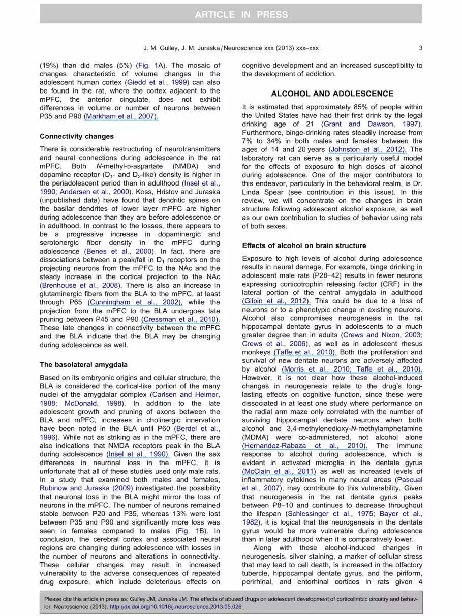
Fig. 2. The number of neurons and glia in the mPFC and BLA in adults that
There were no effects of alcohol on the number of neurons in the mPFC in e
males (⁄p< 0.02), but not in females compared to saline-exposed controls. (B
Adapted from Koss et al. (2012).
Please cite this article in press as: Gulley JM, Juraska JM. The effects of abuse
ior. Neuroscience (2013), http://dx.doi.org/10.1016/j.neuroscience.2013.05.026
1975). Furthermore, alcohol has effects on many types
of glia, including astrocytes and microglia (Evrard et al.,
2006; McClain et al., 2011). The normal occurrence of
neuronal death and alterations in the number of glia
during adolescence (Markham et al., 2007; Rubinow
and Juraska, 2009) may render the mPFC and BLA
particularly vulnerable to alcohol exposure.
We investigated this by giving male and female rats
3 g/kg alcohol (i.p.) for 2 out of every 3 days during
adolescence (P35–45; Koss et al., 2012). This age range
encompasses puberty and the time of neural losses for
both sexes and thus is clearly within the adolescent time
period. To assess the long-term impact on the number of
neurons and glia, we waited until the rats were adults
(P100) before performing stereological quantification of
cell numbers in both the ventral portion of the mPFC and
the BLA. As shown in Fig. 2, the number of neurons was
not altered in either of these neural areas in either sex.
The number of glia, however, was reduced in the mPFC
in male rats that had been exposed to alcohol as
adolescents. This was not found in females or in the BLA
of either sex. Markham et al. (2007) found that the
number of glia in the mPFC increased between P35 and
P90 in males but not in females and that there were no
indications of glial proliferation in the BLA (Rubinow and
Juraska, 2009). Thus, only the dividing cells in the mPFC
have been exposed to binge levels of alcohol during adolescence. (A)
ither sex, but exposure to alcohol resulted in fewer glial cells (14%) in
) There were no differences in neurons or glia in either sex in the BLA.
d drugs on adolescent development of corticolimbic circuitry and behav-

J. M. Gulley, J. M. Juraska /Neuroscience xxx (2013) xxx–xxx 5
and BLA were vulnerable to the effects of alcohol, as in the
hippocampal dentate gyrus (Nixon and Crews, 2002) and
in the adult olfactory bulb where neuronal proliferation
continues through adulthood (Hansson et al., 2010). It is
also possible that the increasing levels of estrogen during
adolescence may be neuroprotective, which is true in the
case of hypoxia–ischemic episodes (Wise and Dubal,
2000; Zhu et al., 2006). The functional implications of the
loss of glia remain to be elucidated. Since glia are now
known to be involved in synaptic stabilization (Eroglu and
Barres, 2010), the loss of these cells might have
implications for the synaptic connectivity changes in the
mPFC during male adolescence.
Effects of alcohol on neurophysiology
In light of these effects of alcohol exposure during
adolescence on brain structure, it is not surprising that
multiple influences on neurophysiology have also been
reported. Much of this work has focused on the
hippocampus, especially with regard to lasting
alterations in synaptic excitability, though some studies
also report effects in the frontal cortex and amygdala.
For example, early work suggested that exposing
adolescent male rats to alcohol vapor for either 5 or
10 days from P30 to P40 induced changes in the
electroencephalography (EEG) of the parietal cortex and
hippocampus that persisted up to 7 weeks (Slawecki
et al., 2001). In a follow-up study (Slawecki, 2002), rats
exposed to alcohol vapor from P35 to P40 were less
sensitive to subsequent challenges with 1.5 g/kg ethanol
as adults. Specifically, ethanol-exposed rats failed to
exhibit the increases in 4–6 Hz power in the
hippocampus and parietal cortex that were evident in
control rats given the ethanol challenge. These rats
were also rated by observers as less intoxicated
compared to controls, suggesting that the lack of
change in EEG power after ethanol is indicative of a
reduced sensitivity to the sedative properties of the
drug. The mechanism for these effects of alcohol
exposure was not identified, but subsequent studies
suggested a potential role for changes in NMDA
receptor expression and/or function. Rats exposed to
ethanol vapor for 12 h/day as juveniles through late
adolescence/young adulthood (P24–60) exhibited
significant changes in both cortical and hippocampal
EEG following a challenge with the NMDA receptor
antagonist MK-801 (Criado et al., 2008). Additionally,
expression of NR1 and NR2A subunits in the
hippocampus was increased and decreased following
24-h and 2-week withdrawal, respectively, from alcohol
vapor exposure that occurred for 14 h/day starting in the
juvenile period (P23–37) (Pian et al., 2010). This study
also showed no change in subunit expression in frontal
cortex after either withdrawal period.
The effects of alcohol on synaptic excitability have
been well documented in studies of in vitro brain slice
preparations, though most of this work has been done in
rodents exposed during adulthood. For example, acute
exposure to ethanol in vitro typically reduces long-term
potentiation (LTP) in the adult hippocampus (Sinclair
Please cite this article in press as: Gulley JM, Juraska JM. The effects of abuse
ior. Neuroscience (2013), http://dx.doi.org/10.1016/j.neuroscience.2013.05.026
and Lo, 1986; Taube and Schwartzkroin, 1986; Blitzer
et al., 1990) and mPFC (Kroener et al., 2012). In the
adult NAc, acute ethanol decreases long-term
depression (LTD), whereas ethanol pre-exposure leads
to an absence of LTD and an emergence of LTP
(Jeanes et al., 2011). In the adolescent brain, where
studies focusing on alcohol’s effects on excitability have
been much less numerous, certain drug effects are
enhanced while others are diminished compared to
those seen in adults. For example, LTP in hippocampal
slices taken from rats at P30 was enhanced relative to
that observed in slices taken from adults (P90).
Subsequent application of ethanol (10–30 mM) resulted
in a blockade of LTP in the adolescent, but not the
adult, hippocampus (Pyapali et al., 1999). Contributing
factors to the acute effects of ethanol on hippocampal
LTP in adolescents relative to adults include a greater
sensitivity to ethanol’s inhibitory effects on NMDA-
mediated excitation (Swartzwelder et al., 1995), its
ability to enhance GABA-receptor mediated inhibition
(Fleming et al., 2007), and its ability to enhance the
activity of GABAergic interneurons (Yan et al., 2009,
2010). In contrast to this enhanced sensitivity to ethanol
in the adolescent hippocampus, there appears to be a
decreased sensitivity in the cerebellum. Using in vivoelectrophysiology, Van Skike et al. (2010) showed that
the inhibitory effect of 1.5 g/kg (i.p.) ethanol on the
activity of cerebellar Purkinje neurons was evident in
adult, but not adolescent, rats. Together, the relatively
limited number of studies that have directly compared
the acute effects of ethanol on synaptic excitability
suggest a heightened sensitivity of adolescents to
ethanol-induced decreases in LTP in the hippocampus
but a decreased sensitivity to this effect the cerebellum.
Repeated alcohol exposure beginning in adolescence
(Roberto et al., 2002) or adulthood (Durand and Carlen,
1984; Fujii et al., 2008) also reduces hippocampal LTP,
and this effect persists for up to 2 months of withdrawal
(Durand and Carlen, 1984). In rats exposed to alcohol
during early (P28–36), but not late (P45–50),
adolescence, there is an enhancement of a unique,
NMDA receptor-independent form of hippocampal LTP
(Sabeti and Gruol, 2008). This effect, which was
dependent on activation of sigma receptors, was
observed when brain slices were taken 24 h following
the last ethanol exposure (i.e., during acute withdrawal).
Sabeti (2011) further found these changes in LTP
following early adolescent exposure to ethanol are
accompanied by changes in the intrinsic excitability of
CA1 pyramidal neurons that likely develop during
withdrawal. Using a binge-like method for chronic
ethanol exposure, Fleming et al. (2012) recently
demonstrated that the GABA receptor-mediated
inhibitory tone is reduced in the hippocampus of
adolescent-exposed adult rats compared to saline-
treated controls. Thus, the emerging picture from these
studies of ethanol effects on the adolescent brain is that
many, though certainly not all, of the effects of drug
exposure on neurophysiology are greater and,
potentially, longer lasting than those seen when
exposure occurs during adulthood.
d drugs on adolescent development of corticolimbic circuitry and behav-
6 J. M. Gulley, J. M. Juraska /Neuroscience xxx (2013) xxx–xxx
Effects of alcohol on neurochemistry
Several neurochemical systems, and their associated
receptors, are altered by alcohol exposure during
adolescence. Both voluntary alcohol intake (Sahr et al.,
2004) and exposure through i.p. injection (Badanich
et al., 2007; Pascual et al., 2009; Philpot et al., 2009)
induce changes in NAc dopamine in adulthood.
Specifically, rats exposed to alcohol in adolescence
have higher basal levels of dopamine in the NAc
compared to unexposed controls (Sahr et al., 2004;
Badanich et al., 2007) and adult-exposed rats (Pascual
et al., 2009). However, adolescents exhibit attenuations
in ethanol-induced increases in dopamine overflow
following a challenge injection (Philpot et al., 2009).
Using in vivo voltammetry to measure rapid changes in
dopamine concentrations as rats engaged in a risk-
based decision-making task, Nasrallah et al. (2011)
showed that adult rats who self-administered ethanol
from P30 to P49 had a greater release of NAc
dopamine following presentation of a lever associated
with a risky choice. These rats also made more risky
choices compared to controls.
Glutamate in the NAc is also affected as mice pre-
exposed to ethanol during adolescence respond to an
ethanol challenge (1.8 g/kg, i.p.) with 200% increases in
glutamate concentrations (Carrara-Nascimento et al.,
2011). Mice in this study that were pre-exposed to
ethanol in adulthood, in contrast, had 50% reductions in
NAc glutamate in response following the challenge. The
mechanism for this age of exposure-dependent
divergence in glutamate response to ethanol challenge
is not apparent, though it has been shown that NMDA
receptors are upregulated (Sircar and Sircar, 2006),
whereas the function of the NR2B subunit is reduced
(Pascual et al., 2009), in the PFC of adolescent-
exposed rats. In addition, a recent whole-brain analysis
of ethanol-induced changes in neurotransmitter-related
gene expression revealed widespread downregulation in
the brains of adolescent-exposed mice, compared to
controls (Coleman et al., 2011). This included significant
changes in the expression of genes coding for peptides,
GABAA receptor subunits, nicotinic acetylcholine
receptors, and dopamine receptors. It is not clear if
alcohol-induced decreases in NMDA receptor
expression and function or changes in neurotransmitter-
related gene expression are directly responsible for the
changes in neurotransmitter levels demonstrated in
studies of adolescent alcohol exposure, but it is notable
that many of the effects reported by Coleman et al.
were in addition to the changes in gene expression
evident in control mice assessed in adolescence
compared to adulthood. Thus, alcohol’s ability to alter
the normal trajectory of development in neurotransmitter
systems appears to play an important role in its long-
lasting effects on neurochemistry. Interestingly, some of
the effects of alcohol exposure may be more prominent
when exposure occurs in adulthood. For example,
alcohol-induced decreases in mRNAs for corticotropin-
releasing hormone (CRH), GABAA and a1-adrenergic
receptors were decreased in the BLA of adult-, but not
adolescent-, exposed rats (Falco et al., 2009).
Please cite this article in press as: Gulley JM, Juraska JM. The effects of abuse
ior. Neuroscience (2013), http://dx.doi.org/10.1016/j.neuroscience.2013.05.026
Given the important role of experience in the
development of the hypothalamo–pituitary–adrenal
(HPA) axis and the response to stress (Romeo, 2010), it
is not surprising that exposure to alcohol during
adolescence has long-lasting consequences on
glucocorticoids, For example, in adolescent rats, ethanol
significantly elevates plasma corticosterone levels
relative to untreated or saline-injected controls, though
this effect is attenuated with repeated exposure and is
more significant in females compared to males
(Przybycien-Szymanska et al., 2010). This change in
corticosterone response is associated with an increase
in CRH gene expression in the hypothalamus of male,
but not female, rats exposed repeatedly to ethanol.
Subsequent studies revealed this sex difference was
due to a protective effect of estradiol on ethanol-induced
upregulation of gene expression in females (Przybycien-
Szymanska et al., 2012). When tested during adulthood,
male rats pre-exposed to ethanol during adolescence,
compared to saline-treated controls, exhibit reductions
in basal corticosterone levels and exaggerated
increases in corticosterone and CRH gene expression
following ethanol challenge (Przybycien-Szymanska
et al., 2011). In male rats given alcohol vapor exposure
during adolescence and challenged with 4.5 g/kg
ethanol during late adolescence/early adulthood, CRH
gene expression and c-fos expression was shown to be
reduced and unaffected, respectively, compared to air-
exposed controls (Allen et al., 2011a,b). A significant
reduction in CRH immunoreactive cells in the central
nucleus of the amygdala has also been found in rats
that self-administered alcohol from P27 through sacrifice
at P42 (Allen et al., 2011a,b). Although more
investigations of the interacting effects of stress and
alcohol exposure during adolescence are needed, it is
clear from the currently available studies that alcohol
can have a long-lasting impact on the neurochemistry of
the stress response system and that some of these
effects may contribute to sex differences in alcohol-
induced behavior.
Behavioral effects of alcohol during adolescence
Alcohol exposure during adolescence has been shown to
have consequences for behavior later in life, with alcohol
self-administration receiving a significant amount of
research attention. The literature is somewhat
equivocal, however, as there is published evidence for
both increases and decreases in drinking behavior when
exposure begins in adolescence. On the one hand,
chronic exposure to alcohol via systemic injection
(Pascual et al., 2007, 2009; Maldonado-Devincci et al.,
2010), voluntary drinking (Rodd-Henricks et al., 2002;
Walker and Ehlers, 2009; Strong et al., 2010; O’Tousa
et al., 2013) or forced consumption (Blizard et al., 2004)
has been shown to increase drinking behavior in
adulthood. Using similar methods of ethanol pre-
exposure, however, the opposite or no consistent effect
on alcohol drinking in adulthood has been reported
(Lancaster et al., 1996; Slawecki and Betancourt, 2002;
Slawecki et al., 2004; Siegmund et al., 2005;
Broadwater et al., 2011; Gilpin et al., 2012). In our own
d drugs on adolescent development of corticolimbic circuitry and behav-
J. M. Gulley, J. M. Juraska /Neuroscience xxx (2013) xxx–xxx 7
lab (Sherrill et al., 2011b), we found that alcohol drinking
was modestly increased in both male and female rats who
were pre-exposed in a ‘‘binge-like’’ fashion to 3 g/kg
ethanol (i.p.) during adolescence. Interestingly, pre-
pubertal gonadectomy potentiated the pre-exposure
effect, but only in females. Thus, estrogen appeared to
protect females from the long-term effects of alcohol
exposure during adolescence. In a parallel study
(Sherrill et al., 2011a), we found that adult males
develop a more robust ethanol-induced conditioned
taste aversion compared to females. In addition, males,
but not females, exhibited an attenuated taste aversion
in adulthood following pre-exposure to ethanol during
adolescence. Changes in alcohol’s aversive properties
might contribute to the ability of adolescent alcohol
exposure to modulate alcohol drinking in adulthood. The
reasons for the equivocal findings in these studies of
alcohol pre-exposure during adolescence are not
entirely clear, although differences in exposure methods
and procedures for assessing drinking behavior certainly
play a role.
Investigations of the effects of adolescent alcohol
exposure on cognitive behavior typically demonstrate
adverse consequences. For example, ethanol-induced
impairments in auditory fear conditioning (Bergstrom
et al., 2006) and memory (Markwiese et al., 1998; White
et al., 2000; Silvers et al., 2003, 2006; Land and Spear,
2004) are more pronounced in adolescent compared to
adult rats. Interestingly, this is true even though
adolescents are known to be less sensitive to ethanol’s
hypothermic, anxiolytic, and motor-impairing effects
(Spear and Varlinskaya, 2005). Following chronic,
intermittent exposure to alcohol vapor during
adolescence (P35–40) (Slawecki et al., 2004) or free
access to 10% ethanol for 5 weeks starting in the
juvenile period (3–8 weeks of age) (Salimov et al.,
1996), rats exhibit increases in anxiety- and depression-
like behaviors when they are tested in adulthood. A
recent study found that only those rats with prior binge
drinking experience restricted to adolescence, rather
than earlier in adulthood, showed increases in open-arm
entries on an elevated plus maze (Gilpin et al., 2012).
This result was interpreted as being consistent with
either decreased anxiety or increased impulsivity. The
latter possibility is consistent with data from adult
humans performing delayed reward-discounting tasks,
which assess impulsive choice behavior. In these
studies, those with a history of adolescent alcohol
exposure have higher levels of impulsivity than controls
(Rogers et al., 2010). Data from laboratory animals are
inconsistent with this interpretation, however. In adult
rats trained to resist the impulse to respond during a
premature phase of a five-choice serial reaction time
task, there were no observed effects of repeated
exposure to 5 g/kg (i.g.) ethanol every 8 h from P33 to
P36 on baseline measures of attention, impulsivity or
cognitive flexibility. Somewhat surprisingly, adolescent-
exposed rats appeared to maintain a lasting tolerance to
ethanol as they were less sensitive to ethanol-induced
disruptions of task performance following a challenge
with 1.5 or 3.0 g/kg ethanol (Semenova, 2012).
Please cite this article in press as: Gulley JM, Juraska JM. The effects of abuse
ior. Neuroscience (2013), http://dx.doi.org/10.1016/j.neuroscience.2013.05.026
Additional evidence for alcohol-induced changes in
decision making comes from studies in rats that were
allowed to consume a sweetened gelatin and ethanol
mixture from P30 to P49 and were then tested in a
probability-discounting task when they were adults. In
this task, where rats are given the choice between small
and certain or large but uncertain rewards, those
exposed to alcohol during adolescence behaved in a
more risky fashion than controls (Nasrallah et al., 2009).
This preference for risk seems to be due to an
imbalance in learning of better-than-expected outcomes
over those that are worse than expected (Clark et al.,
2012). Together, studies of the effects of alcohol
exposure during adolescence have often, though not
always, suggested a long-lasting impairment in cognitive
function that is evident well into adulthood. Much of this
work has not included comparison groups of adult-
exposed subjects, however, so it is not yet clear if
adolescents are uniquely sensitive compared to adults.
PSYCHOSTIMULANTS AND ADOLESCENCE
The use of the psychostimulant drugs nicotine, cocaine
and the amphetamines is relatively high among
adolescents, particularly in comparison to other age
groups. The most recent National Survey of Drug Use
and Health (SAMHSA, 2012) suggests that individuals
18–25 years old have the highest rates of current
tobacco use (39.5%) compared to those 12–17 (10.0%)
and adults who are 26 or older (26.3%). For individuals
in the 12–25-year-old range, that represents
approximately 21 million individuals. Although
considerably lower, the use of more difficult to obtain
drugs like cocaine and the amphetamines is
nonetheless significant in young people (Johnston et al.,
2012). Cocaine use by 12th grade students, which
peaked in the mid-1980s at about 13%, is now
estimated to be at about 3%. The non-therapeutic use
of amphetamines, which are more widely available due
in part to diversion of prescriptions for medical
conditions such as attention deficit hyperactivity disorder
(ADHD), is nearly three times higher, with approximately
9% of 12th graders reporting use in the previous year.
The use of these drugs is estimated to be even higher
in those who are in the latter stages of adolescence. For
those 18–25, it is estimated that over 1.5 million have
used cocaine in the previous year, whereas nearly 1.3
million used amphetamines and other non-tobacco
stimulants (SAMHSA, 2012). Clearly, adolescents are
using these drugs and it is therefore critical to develop a
full understanding of the potential for psychostimulants
to induce adaptations in the brain and behavior that may
persist and lead to adverse consequences, even after
drug taking has ceased.
Effects of psychostimulants on brain structure
Similar to what has been seen in adult-exposed rats
(Robinson and Kolb, 1997, 1999; Brown and Kolb,
2001), psychostimulant exposure during adolescence
has been reported to alter neuronal morphology, with
d drugs on adolescent development of corticolimbic circuitry and behav-

8 J. M. Gulley, J. M. Juraska /Neuroscience xxx (2013) xxx–xxx
most of the changes reported to date in the PFC for
amphetamine, cocaine and nicotine, along with
additional effects reported for nicotine in the NAc,
hippocampus and BLA. For example, the length and
branching of basilar dendrites of pyramidal neurons in
the mPFC of 48-day-old rats was increased by twice
daily injections of 0.5 mg/kg amphetamine from P22 to
P34 (Diaz Heijtz et al., 2003). This study also showed
that these structural changes were associated with an
upregulation in the expression of calcium/calmodulin-
dependent protein kinase II (CaMKII) and tyrosine
hydroxylase in slices of the mPFC taken from a
separate group of rats given the same treatments.
Long-lasting changes in the response of dendritic spines
in the orbitofrontal cortex have also been reported. In
this study (Gourley et al., 2012), mice were pre-exposed
to 10 mg/kg cocaine, once per day from P31 to P36 and
were then given a challenge injection of 10 mg/kg
cocaine on P63. When spine morphology was assessed
24 h later, it was discovered that there was decreased
spine density, but increased spine head size, in cocaine
pre-exposed compared to saline pre-exposed mice.
Nicotine-induced structural plasticity has been
demonstrated in the NAc, BLA, hippocampus and
mPFC, with some effects appearing to be dependent on
the age of exposure. In rats that were sacrificed for
anatomical analysis on P144 following continuous
exposure to nicotine from the juvenile period to young
adulthood (P22–69) via an osmotic minipump (2 mg/kg/
day), medium spiny neurons of the NAc had more and
longer dendritic segments compared to saline-exposed
controls (McDonald et al., 2005). Subsequently, this
same group demonstrated that nicotine-induced
increases in dendritic length and branch number in
medium spiny neurons was selectively increased in rats
exposed from P29 to P43, but not in those exposed
from P80 to P94 (McDonald et al., 2007). In the mPFC,
and more specifically pyramidal cells of the prelimbic
cortex, continuous exposure to nicotine during
adolescence (P29–43) increases the length of basilar
dendrites in cells classified as ‘‘complex’’ because of
their large dendritic arbor. In rats exposed to nicotine in
adulthood (P80–94), there was an increase in dendritic
length and number of branches, but only in cells
classified as ‘‘simple’’ because of their relatively small
arbors (Bergstrom et al., 2008). This effect of nicotine
on mPFC might be somewhat selective for the prelimbic
region as nicotine failed to alter dendrites in the
infralimbic region when it was given to adolescent (P32–
46) and adult (P61–75) rats via an intermittent exposure
method (six subcutaneous injections of 0.5 mg/kg;
Bergstrom et al., 2010). This intermittent-exposure study
also showed that nicotine increased dendritic length in
principal neurons of the BLA, though this effect was
dependent on both age of exposure and on brain
hemisphere. Specifically, adult nicotine exposure
induced an increase in dendritic complexity and, in the
right hemisphere only, an increase in dendritic length. In
adolescent-exposed rats, there was also an increase in
complexity relative to saline-injected controls, but the
increase in length was not observed (Bergstrom et al.,
Please cite this article in press as: Gulley JM, Juraska JM. The effects of abuse
ior. Neuroscience (2013), http://dx.doi.org/10.1016/j.neuroscience.2013.05.026
2010). An important issue that remains to be fully
addressed is the duration of these effects of nicotine on
brain structure and whether or not they are fully
reversed following withdrawal. A recent study in
adolescent mice (P30–45) that were chronically
exposed to nicotine in their drinking water suggests
most observed effects were reversed by the time
animals reached young adulthood (Oliveira-da-Silva
et al., 2010).
Effects of psychostimulants on neurophysiology
Numerous studies have used neurophysiological
measures to investigate psychostimulant-induced
plasticity in adolescent-exposed rodents, though most of
these have only assessed adaptations following
relatively short withdrawal periods – a few days to
3 weeks. For example, studies in juvenile to adolescent
rats or mice (typically between P21 and P41) that were
sacrificed soon after their last injection of nicotine,
cocaine or amphetamine have demonstrated drug-
induced changes in synaptic excitability in the ventral
tegmental area (Mansvelder and McGehee, 2000;
Ungless et al., 2001; Saal et al., 2003), hippocampus
(Perez et al., 2010), amygdala (Huang et al., 2003;
Pollandt et al., 2006) and mPFC (Huang et al., 2007;
Goriounova and Mansvelder, 2012). In the hippocampus
of late adolescent/young adult mice (P63), the induction
of LTP was enhanced by once daily exposure to 3 mg/
kg amphetamine from P28 to P37, compared to that
observed in saline-treated controls (Gramage et al.,
2013). This neural adaptation was associated with a
significant, albeit transient, increase in anxiety-related
behavior and memory impairments, as measured by
deficits in passive avoidance and Y-maze performance,
respectively.
In the NAc, pre-exposure to cocaine during
adolescence tends to decrease excitability (Thomas
et al., 2001), though this effect may be dependent on
the method of drug exposure (experimenter- vs. self-
administered; see Jacobs et al., 2003) and the duration
of withdrawal (Mu et al., 2010). In addition, a recent
study (Huang et al., 2011) suggested the effects of
cocaine on accumbal LTD may be dependent on the
subregion that is analyzed. In this report, 5 days of
exposure to cocaine (15 mg/kg/day) starting between
P26 and P28 led to a decrease in LTD in the NAc shell,
but not core, that lasted for up to 28 days following
withdrawal (P56). Kourrich and Thomas (2009) also
showed that unique effects in the shell compared to the
core may depend on the duration of withdrawal from
cocaine or amphetamine. In at least one study (Li and
Kauer, 2004), amphetamine exposure during
adolescence had no effect on the induction of LTP by
high-frequency activation of glutamatergic afferents.
However, when brain slices were subsequently exposed
to amphetamine, which causes a dopamine-dependent
attenuation of LTP induction in saline pre-treated rats,
there was a reduced sensitivity to the inhibitory effect of
acute amphetamine exposure on LTP (Li and Kauer,
2004). An important, and as yet unanswered question,
d drugs on adolescent development of corticolimbic circuitry and behav-

J. M. Gulley, J. M. Juraska /Neuroscience xxx (2013) xxx–xxx 9
is whether or not these effects differ between males and
females. Sex differences were not assessed in any of
these studies and nearly all of them utilized male rats or
mice (or sex was not specified).
Although the neurophysiological effects of repeated
psychostimulant exposure in adulthood have been
extensively studied (for recent reviews, see Bowers
et al., 2010; Luscher and Malenka, 2011), the paucity of
studies using comparison groups of adolescent- and
adult-exposed subjects makes it difficult to make direct
comparisons and, in turn, assess whether adolescents
are differentially sensitive to drug-induced plasticity. In
the small number of studies that have used comparison
groups of different ages, there is evidence that the
adolescent brain is more sensitive to some drug effects.
In one study of repeated nicotine exposure, rats given
nicotine (0.4 mg/kg, three times per day) from P34 to
P43 had decreases in short-term depression of evoked
excitatory postsynaptic currents (eEPSCs) in layer V
pyramidal cells of the mPFC. This effect, which was
measured 5 weeks after the last nicotine injection
(starting at P78), was not observed in saline-treated
controls or in rats exposed to nicotine during late
adolescence/young adulthood (P60–69) (Counotte et al.,
2011). Additionally, parallel studies revealed that these
long-lasting changes in synaptic function were
associated with decreases in mGluR2 expression and
function in the mPFC, as well as deficits in behavioral
measures of attention and inhibitory control (Counotte
et al., 2009, 2011). Additional support for the important
role of nicotine-induced adaptations in mGluR2 comes
from a report showing alternations in LTP in the mPFC
of adult rats exposed to nicotine from P34 to P43
(Goriounova and Mansvelder, 2012). In this study,
decreases in LTP were observed in the adolescent
mPFC following bath application of nicotine (10 lM) and
during the first 4 days following the last nicotine injection
(on P43). However, when LTP was assessed following a
5-week withdrawal period (after P78), nicotine pre-
exposed rats exhibited enhanced LTP compared to
saline-treated controls. These short- and long-term
adaptations, which were both linked to impairments in
mGluR2 signaling in the mPFC, were not present in rats
exposed to nicotine from P60 to P69 (Goriounova and
Mansvelder, 2012).
Recently, we have found that repeated exposure to
amphetamine during adolescence leads to changes in
the function of mPFC neurons that persist for
�3 months following the last drug injection (Paul, Kang,
Cox, and Gulley, unpublished observations). In this
study, rats were given saline or 3 mg/kg amphetamine
(i.p.), every other day during adolescence (P27–45) or
adulthood (P85–103). When rats were between P125
and P140, we prepared brain slices at the level of the
mPFC and performed whole-cell recordings of excitatory
layer V pyramidal cells and fast-spiking inhibitory
interneurons using methods similar to those described
previously (Paul and Cox, 2013). We found no
differences in the basic cellular properties of pyramidal
neurons between the controls and those exposed to
amphetamine. However, application of amphetamine
Please cite this article in press as: Gulley JM, Juraska JM. The effects of abuse
ior. Neuroscience (2013), http://dx.doi.org/10.1016/j.neuroscience.2013.05.026
(25 lm) or dopamine (50 lm) increased the frequency
of spontaneous inhibitory postsynaptic currents (sIPSC)
in controls while having no significant effects in
amphetamine pre-exposed rats. These effects, which
were not dependent on age of exposure, were also
apparent in a comparison group of rats that was studied
at approximately P66 (i.e., the same �3-weekwithdrawal period used in the adult-exposed rats).
Furthermore, we found that the excitability of
interneurons, as measured by number and frequency of
action potentials to depolarizing pulses, was significantly
reduced in amphetamine-exposed compared to control
rats. Thus, it appears that that the reduction of
spontaneous inhibitory activity on layer V pyramidal
neurons in amphetamine-exposed animals is due to the
reduced excitability of fast-spiking interneurons.
Moreover, the effects of amphetamine exposure during
adolescence are measurable at both short and long
withdrawal periods.
Effects of psychostimulants on neurochemistry
Given their potent pharmacological effects and the
continuing development of monoamine systems during
adolescence, it is not surprising that psychostimulants
have the potential to induce unique changes in
dopamine system function in the young brain. Following
a single injection, cocaine and amphetamine have been
shown to increase extracellular dopamine
concentrations in the dorsal striatum to a greater extent
in adolescent compared to adult male rats (Walker and
Kuhn, 2008; Walker et al., 2010) while rats in the
juvenile period have a significantly lower dopamine
response to cocaine (Chen et al., 2010). Additionally,
inconsistent results have been found in the NAc, where
cocaine-induced dopamine overflow was found to be
either greater in adolescents (Badanich et al., 2006) or
not different as a function of age following cocaine
(Frantz et al., 2007) or amphetamine (Silvagni et al.,
2008). A likely mechanism for the age-dependent
differences that have been observed is differences in
the expression and/or function of dopamine
transporters. Indeed, both are increased in the striatum
of adolescents compared to adults (Volz et al., 2008).
The function of the vesicular monoamine transporter,
which is responsible for sequestering monoamines into
vesicles, is also higher in adolescents compared to
adults (Volz et al., 2008). These developmental
differences in dopamine transporters, along with those
reported for D1 and D2 receptor expression (Andersen
et al., 2000; Brenhouse et al., 2008) and tyrosine
hydroxylase (Mathews et al., 2009), provide an
opportunity for enhanced vulnerability to the effects of
repeated drug exposure.
Chronic treatment with nicotine, cocaine or
amphetamine leads to enhanced responsiveness, or
sensitization, to the behavioral and neurobiological
effects of these drugs (Robinson and Berridge, 1993;
Vanderschuren and Kalivas, 2000). Some evidence
suggests this drug-induced plasticity may be enhanced
when exposure occurs during adolescence. For
d drugs on adolescent development of corticolimbic circuitry and behav-

10 J. M. Gulley, J. M. Juraska /Neuroscience xxx (2013) xxx–xxx
example, in rats given 2 or 10 mg/kg amphetamine daily
for 3 days and challenged with 2 mg/kg amphetamine
after a 5-day withdrawal period, it was shown that
amphetamine-stimulated dopamine release in the
striatum was significantly higher in those exposed during
adolescence (P33–43) compared to young adulthood
(P61–71; Laviola et al., 2001). Following a similar
treatment protocol, but a significantly longer withdrawal
period of 28 days, adolescent-exposed rats continue to
exhibit a sensitized behavioral response that was
associated with heightened neural activation in the
striatum and amygdala (McPherson and Lawrence,
2006). A potential mechanism for these effects is a
lasting change in the responsiveness of monoamine
neurons to subsequent drug challenges. Labonte et al.
(2012) used in vivo electrophysiology to demonstrate
that daily exposure to amphetamine from P30 to P50
led to significant increases in the firing rate of dopamine
neurons in the ventral tegmental area and 5-HT neurons
in the dorsal raphe, compared to neurons recorded from
these brain regions in saline-exposed controls.
Nicotine’s effects on dopamine, which are indirect via
the drug’s activation of nicotinic acetylcholine receptors
(nAChRs) located on dopamine terminals (Livingstone
and Wonnacott, 2009), have generally been shown to
be different in adolescents compared to adults, but the
direction of this effect has varied. In the NAc, a single
injection of 0.3 mg/kg nicotine was reported to elevate
dopamine levels to a greater extent in adolescent
(�P28) compared to adult (P63–84) rats (Shearman
et al., 2008), whereas 0.6 mg/kg nicotine reportedly
increased dopamine in adults (P60), but not adolescents
(P35 or P45; Badanich and Kirsteina, 2004). Consistent
with the former result is a study showing elevations in
nicotine-stimulated [3H]dopamine from NAc
synaptosomes prepared from adolescent rats, compared
to those taken from adults (Azam et al., 2007). With
repeated exposure, nicotine induces adaptations in the
dopamine system such that dopamine concentrations in
the NAc are decreased during precipitated withdrawal in
both adolescents and adults. Interestingly, the
magnitude of this decrease is significantly lower in
adolescent-exposed rats (Natividad et al., 2010). This
effect was shown to be mediated by age-dependent
differences in nicotine-induced adaptations in the
interacting glutamatergic and GABAergic systems of the
ventral tegmental area (Natividad et al., 2012).
Stimulated dopamine release and receptor-mediated
signaling are also known to be elevated in adult rats
exposed to nicotine during adolescence (Trauth et al.,
2001; Abreu-Villaca et al., 2003; Dickson et al., 2011).
Although many of these studies of chronic nicotine
effects have utilized continuous exposure techniques
that result in high-dose exposure for relatively long
periods of time, intermittent exposure to low or
moderate doses during adolescence also has the
potential to produce lasting effects. For example, daily
injections of 0.4 mg/kg nicotine from P30 to P36 results
in increases and decreases in the expression of
dopamine and serotonin transporters, respectively, that
were not evident in rats exposed from P60 to P66
Please cite this article in press as: Gulley JM, Juraska JM. The effects of abuse
ior. Neuroscience (2013), http://dx.doi.org/10.1016/j.neuroscience.2013.05.026
(Collins et al., 2004). After only 4 days of intravenous
injections with 0.06 mg/kg nicotine, serotonin
transporters are elevated in the PFC and the BLA of
adolescent- (P32) but not adult-exposed (P90) rats (Dao
et al., 2011).
Dopamine and other monoamines are not the only
neurochemical systems to exhibit age-dependent
differences in psychostimulant-induced adaptations. In
the adolescent brain, cholinergic systems are a primary
target for nicotine-induced plasticity (O’Dell, 2009). This
may be due in part to the numerous changes in
expression and function of the nicotinic receptor during
normal adolescent development (Slotkin, 2002). Recent
studies have also highlighted a potential role for
adaptations in cannabinoid and opiate receptors in the
long-lasting behavioral effects of adolescent nicotine
exposure (Marco et al., 2007; Mateos et al., 2011).
Behavioral effects of psychostimulants duringadolescence
Much of the work on the behavioral effects of adolescent
psychostimulant exposure has focused on long-lasting
changes in either the locomotor stimulant properties of
the drugs or their ability to influence reward-seeking
behavior. With regard to the former, some studies report
greater amphetamine- or cocaine-induced sensitization
in adolescent-exposed rodents (Adriani et al., 1998;
Schramm-Sapyta et al., 2004; Caster et al., 2005;
Mathews et al., 2010, 2011; Kameda et al., 2011),
whereas others indicate greater effects in adults (Frantz
et al., 2007; Zakharova et al., 2009; Good and Radcliffe,
2011; Richetto et al., 2013; Sherrill et al., 2013) or no
difference between age groups (Niculescu et al., 2005;
Good and Radcliffe, 2011). Studies with nicotine have
also reported inconsistent results for age-dependent
differences in locomotor sensitization (Belluzzi et al.,
2004; Schochet et al., 2004; Cruz et al., 2005).
Between- and within-study methodological differences
contribute to some of these discrepant findings, with key
factors being age of exposure (e.g., early vs. late
adolescence), drug dose and the aspect of drug-induced
behavior that is measured (e.g., locomotion or
stereotypy). For example, at lower doses of
amphetamine (<1.5 mg/kg), adolescents tend to show
an attenuated response to the first injection but
enhanced locomotor sensitization relative to adults
(Bolanos et al., 1998; Mathews and McCormick, 2007;
Mathews et al., 2009; Zakharova et al., 2009). With
higher doses (>2 mg/kg), however, age-dependent
differences in initial responsiveness diminish and
repeated exposure produces robust stereotypy and
reduced locomotor activity, particularly in adults (Adriani
et al., 1998; Adriani and Laviola, 2002; Sherrill et al.,
2013). Thus, adolescents appear to have a higher
threshold for the psychomotor-activating effects of
cocaine and amphetamine, but once activated their
response is similar to that seen in adults.
Studies of age-dependent differences in the rewarding
properties of psychostimulants have generally been more
consistent – rodents exposed during adolescence tend to
d drugs on adolescent development of corticolimbic circuitry and behav-

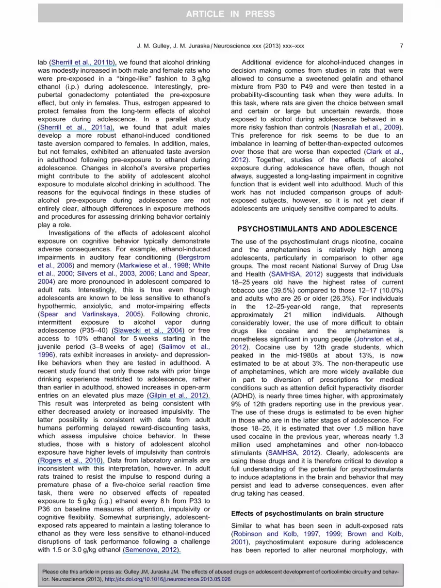
Fig. 3. The effects of pre-exposure to amphetamine on working
memory in a delay matching to position (DMTP) and delay non-match
to position (DNMTP) task. (A) Mean choice accuracy (% correct)
within each delay block averaged across the first two training
sessions. (B) Mean number of sessions to reach a performance
criterion (STC) of P85% correct choices for two consecutive
sessions. Matching letters indicate p< 0.001; ⁄⁄⁄p< 0.001 vs.
control and adult-exposed groups within delay; ###p< 0.001 vs.
DNMTP, collapsed across exposure group. Adapted from Sherrill
et al. (2013).
J. M. Gulley, J. M. Juraska /Neuroscience xxx (2013) xxx–xxx 11
exhibit heightened reward-related behavior later in life
compared to those exposed during adulthood. For
example, nicotine-induced conditioned place preference
is enhanced in adolescents compared to adults (Vastola
et al., 2002; Belluzzi et al., 2004; Torrella et al., 2004;
Shram et al., 2006; Brielmaier et al., 2007; Kota et al.,
2008, 2011; Torres et al., 2008, 2009; Shram and Le,
2010). Adolescent rats also exhibit reduced conditioned
place aversion to nicotine compared to adults (O’Dell
et al., 2007). A similar increase in sensitivity to cocaine
and amphetamine reward in adolescents has been
demonstrated (Badanich et al., 2006; Brenhouse and
Andersen, 2008; Brenhouse et al., 2008; Zakharova
et al., 2009), with adolescents exhibiting a potent
resistance to extinction of drug-environment
associations (Brenhouse et al., 2010). There are,
however, several studies showing that adults are
relatively more sensitive (Adriani and Laviola, 2003;
Aberg et al., 2007; Vidal-Infer et al., 2012) or no age-
dependent differences (Campbell et al., 2000;
Schramm-Sapyta et al., 2004; Mathews and McCormick,
2007; Mathews et al., 2010). Moreover, there are robust
sex differences in these effects. For example, in a study
of cocaine-induced conditioned place preference
(Zakharova et al., 2009), it was demonstrated that the
female sex and adolescent exposure independently
resulted in higher sensitivity to the rewarding effects of
cocaine in adults. Studies showing changes in
psychostimulant self-administration behavior following
pre-exposure to the drugs in adolescence or adulthood,
which have been reviewed in detail elsewhere
(Shahbazi et al., 2008; Schramm-Sapyta et al., 2009;
Anker et al., 2011), suggest that adolescents have a
more ‘‘addiction vulnerable’’ phenotype. Specifically,
they exhibit heightened motivation for the drug, reduced
extinction and greater relapse of drug-seeking behavior,
and have reduced withdrawal responses.
Of great clinical interest is the potential for long-lasting
changes in cognition that might result from adolescent
exposure to psychostimulant drugs. Human stimulant
abusers, who usually initiate drug use in adolescence,
have been shown to exhibit significant cognitive deficits
that vary in magnitude depending on the duration of
drug exposure (Bolla et al., 1998; Verdejo-Garcia et al.,
2006). In adult rats exposed to psychostimulants during
adolescence, there is evidence of enduring deficits in
cognitive tasks that assess attention, fear learning,
memory, decision making, and impulse control (Harvey
et al., 2009; Santucci and Rabidou, 2011; Sillivan et al.,
2011; Hankosky and Gulley, 2012). However, in these
studies, it is difficult to ascertain if adolescents are
relatively more sensitive to these effects because
comparison groups of adult-exposed subjects were
rarely utilized.
In studies that directly assess the age-dependence of
psychostimulant-induced cognitive deficits, there is
evidence that adolescents may be more vulnerable. For
example, nicotine exposure during adolescence, but not
adulthood, increases impulsive action and enhances
electrically stimulated dopamine release in vitro from
slices of the mPFC (Counotte et al., 2009). In addition,
Please cite this article in press as: Gulley JM, Juraska JM. The effects of abuse
ior. Neuroscience (2013), http://dx.doi.org/10.1016/j.neuroscience.2013.05.026
there are unique effects of cocaine on memory
processes when the onset of drug exposure occurs in
adolescence compared to adulthood (Harvey et al.,
2009). Recently, we found that adult rats that were
exposed to 3 mg/kg amphetamine during adolescence
displayed delay-dependent deficits in choice accuracy in
an operant-based working memory task (Sherrill et al.,
2013). In addition, they required more sessions to
optimize performance and learn task rules, and they
were more susceptible to proactive interference,
compared to control and adult-exposed groups (Fig. 3).
Interestingly, we also found that amphetamine-induced
locomotor sensitization was enhanced in adult-
compared to adolescent-exposed rats, suggesting that
drug-induced changes in cognition were dissociable
from amphetamine’s lasting effects on sensitivity to its
motor activating effects. These findings of heightened
vulnerability of adolescents to the psychostimulant-
induced cognitive deficits are not without exception,
however. Repeated cocaine exposure has been
reported to have no specificity or even greater effects in
adults on an amygdala-sensitive maze task (Kerstetter
and Kantak, 2007).
FUTURE CHALLENGES
There is mounting evidence from the rat model that
adolescence is a particularly vulnerable time for both
behavioral and neural effects of alcohol and
d drugs on adolescent development of corticolimbic circuitry and behav-

12 J. M. Gulley, J. M. Juraska /Neuroscience xxx (2013) xxx–xxx
psychostimulant exposure. Nevertheless, more studies
are needed that directly compare adolescent and adult
exposures to firmly establish the unique sensitivity of
the adolescent to drug-induced plasticity and its
associated consequences. Until recently, the majority of
the work in this area has focused on one age group or
the other (McCutcheon and Marinelli, 2009). More
studies are also needed to understand the
discrepancies in results that exist. Many of the
inconsistencies are at least partially attributable to
obvious factors such as drug dose and method of
exposure. These are aspects of experimental design
that influence the generality of findings and ultimately
contribute to the translational impact of observed
results. There are other factors, however, that need to
be more closely controlled since their variation can lead
to results that are confounded and otherwise difficult to
interpret. For example, the ages that are considered
adolescence are often defined too broadly. If a rise in
gonadal hormones is an essential marker of
adolescence, which it is in humans at approximately
12 years old, then the earliest age at which rats should
be considered adolescent is P28. This is especially true
for male rats where overt signs of puberty are not found
until after P38. Exposures that are started before this
age are modeling drug intake during the juvenile period
that continues into adolescence. Observed effects could
thus be due to the juvenile exposure, per se. Moreover,
including juveniles in these studies is addressing
separate issues since it is not modeling the
experimentation with drugs and alcohol that occur
during human adolescence in our society.
The inclusion of females in experimental designs is
also needed. As we have discussed above, there are
numerous studies in the literature and from our own
laboratories that demonstrate different effects of drug
exposure in male compared to female adolescents.
Clearly, the inclusion of both sexes adds complexity to
experimental design, analysis, and interpretation of
results, but the lack of female subjects in most
experiments to date limits their generalizability.
Another key factor that has been largely ignored is the
effect of differential rearing environments and early-life
stress that are introduced when experimental animals
are shipped from commercial vendors to research
facilities when they are in utero or around weaning
(�P22). This is of particular concern for many of the
studies discussed in this review as rearing environment
and early-life stress have been shown to have
significant effects on PFC development and cognitive
processes mediated by this brain region (Liston et al.,
2006; Cerqueira et al., 2007; Green et al., 2012; Yuen
et al., 2012). Moreover, there are numerous reports
demonstrating the long-lasting effects of shipping stress
and rearing conditions on behavior and neurobiology
(Prager et al., 2011), including the response to drugs
(Bardo et al., 2001; Adriani and Laviola, 2002; Ogawa
et al., 2007; Wiley and Evans, 2009; Martini and
Valverde, 2012; Mogi et al., 2012). There is also ample
evidence that the effects of stress during adolescence
are different and often more prolonged in their duration
Please cite this article in press as: Gulley JM, Juraska JM. The effects of abuse
ior. Neuroscience (2013), http://dx.doi.org/10.1016/j.neuroscience.2013.05.026
compared to those seen when stress is induced in
adulthood (see Romeo in this volume). It could be
argued that all subjects (i.e., both ‘‘experimental’’ and
‘‘control’’) experience the same early-life stress.
However, it is likely that these effects can interact with
later drug exposures. Such interactions, along with the
additional influence of the animal’s sex, have been
demonstrated previously (Ogawa et al., 2007; Wiley and
Evans, 2009; Mogi et al., 2012). All of these potential
interactions can be avoided if animals are bred and
housed within the research facility where experiments
will occur. This approach has the additional advantage
of allowing for the control of litter effects that are
prevalent in multiparous research animals like rats and
mice (Zorrilla, 1997), which would make the distinction
between effects in adolescents and adults clearer.
Acknowledgments—Studies from the authors’ laboratories were
supported in part by grants from National Institute on Alcohol
Abuse and Alcoholism (AA017354) and the National Institute
on Drug Abuse (DA029815). We thank Wendy Koss for assis-
tance with manuscript preparation.
REFERENCES
Aberg M, Wade D, Wall E, Izenwasser S (2007) Effect of MDMA
(ecstasy) on activity and cocaine conditioned place preference in
adult and adolescent rats. Neurotoxicol Teratol 29:37–46.
Abreu-Villaca Y, Seidler FJ, Slotkin TA (2003) Impact of adolescent
nicotine exposure on adenylyl cyclase-mediated cell signaling:
enzyme induction, neurotransmitter-specific effects, regional
selectivities, and the role of withdrawal. Brain Res 988:164–172.
Adriani W, Laviola G (2002) Spontaneous novelty seeking and
amphetamine-induced conditioning and sensitization in adult
mice: evidence of dissociation as a function of age at weaning.
Neuropsychopharmacology 27:225–236.
Adriani W, Laviola G (2003) Elevated levels of impulsivity and
reduced place conditioning with d-amphetamine: two behavioral
features of adolescence in mice. Behav Neurosci 117:695–703.
Adriani W, Chiarotti F, Laviola G (1998) Elevated novelty seeking and
peculiar d-amphetamine sensitization in periadolescent mice
compared with adult mice. Behav Neurosci 112:1152–1166.
Allen CD, Lee S, Koob GF, Rivier C (2011a) Immediate and
prolonged effects of alcohol exposure on the activity of the
hypothalamic–pituitary–adrenal axis in adult and adolescent rats.
Brain Behav Immun 25(Suppl. 1):S50–S60.
Allen CD, Rivier CL, Lee SY (2011b) Adolescent alcohol exposure
alters the central brain circuits known to regulate the stress
response. Neuroscience 182:162–168.
Andersen SL, Thompson AT, Rutstein M, Hostetter JC, Teicher MH
(2000) Dopamine receptor pruning in prefrontal cortex during the
periadolescent period in rats. Synapse 37:167–169.
Anderson SA, Classey JD, Conde F, Lund JS, Lewis DA (1995)
Synchronous development of pyramidal neuron dendritic spines
and parvalbumin-immunoreactive chandelier neuron axon
terminals in layer III of monkey prefrontal cortex. Neuroscience
67:7–22.
Anker JJ, Zlebnik NE, Navin SF, Carroll ME (2011) Responding
during signaled availability and nonavailability of iv cocaine and
food in rats: age and sex differences. Psychopharmacology
215:785–799.
Azam L, Chen Y, Leslie FM (2007) Developmental regulation of
nicotinic acetylcholine receptors within midbrain dopamine
neurons. Neuroscience 144:1347–1360.
d drugs on adolescent development of corticolimbic circuitry and behav-

J. M. Gulley, J. M. Juraska /Neuroscience xxx (2013) xxx–xxx 13
Badanich KA, Kirsteina CL (2004) Nicotine administration
significantly alters accumbal dopamine in the adult but not in the
adolescent rat. Ann N Y Acad Sci 1021:410–417.
Badanich KA, Adler KJ, Kirstein CL (2006) Adolescents differ from
adults in cocaine conditioned place preference and cocaine-
induced dopamine in the nucleus accumbens septi. Eur J
Pharmacol 550:95–106.
Badanich KA, Maldonado AM, Kirstein CL (2007) Chronic ethanol
exposure during adolescence increases basal dopamine in the
nucleus accumbens septi during adulthood. Alcohol Clin Exp Res
31:895–900.
Bardo MT, Klebaur JE, Valone JM, Deaton C (2001) Environmental
enrichment decreases intravenous self-administration of
amphetamine in female and male rats. Psychopharmacology
(Berl) 155:278–284.
Bayer SA, Yackel JW, Puri PS (1982) Neurons in the rat dentate
gyrus granular layer substantially increase during juvenile and
adult life. Science 216:890–892.
Belluzzi J, Lee A, Oliff H, Leslie F (2004) Age-dependent effects of
nicotine on locomotor activity and conditioned place preference in
rats. Psychopharmacology (Berl) 174:389–395.
Benes FM, Taylor JB, Cunningham MC (2000) Convergence and
plasticity of monoaminergic systems in the medial prefrontal
cortex during the postnatal period: implications for the
development of psychopathology. Cereb Cortex 10:1014–1027.
Berdel B, Morys J, Maciejewska B, Narkiewicz O (1996)
Acetylcholinesterase activity as a marker of maturation of the
basolateral complex of the amygdaloid body in the rat. Int J Dev
Neurosci 14:543–549.
Bergstrom HC, McDonald CG, Smith RF (2006) Alcohol exposure
during adolescence impairs auditory fear conditioning in adult
Long-Evans rats. Physiol Behav 88:466–472.
Bergstrom HC, McDonald CG, French HT, Smith RF (2008)
Continuous nicotine administration produces selective, age-
dependent structural alteration of pyramidal neurons from
prelimbic cortex. Synapse 62:31–39.
Bergstrom HC, Smith RF, Mollinedo NS, McDonald CG (2010)
Chronic nicotine exposure produces lateralized, age-dependent
dendritic remodeling in the rodent basolateral amygdala. Synapse
64:754–764.
Blitzer RD, Gil O, Landau EM (1990) Long-term potentiation in rat
hippocampus is inhibited by low concentrations of ethanol. Brain
Res 537:203–208.
Blizard DA, Vandenbergh DJ, Jefferson AL, Chatlos CD, Vogler GP,
McClearn GE (2004) Effects of periadolescent ethanol exposure
on alcohol preference in two BALB substrains. Alcohol
34:177–185.
Bolanos CA, Glatt SJ, Jackson D (1998) Subsensitivity to
dopaminergic drugs in periadolescent rats: a behavioral and
neurochemical analysis. Brain Res Dev Brain Res 111:25–33.
Bolla KI, Cadet JL, London ED (1998) The neuropsychiatry of chronic
cocaine abuse. J Neuropsychiatry Clin Neurosci 10:280–289.
Bourgeois JP, Rakic P (1993) Changes of synaptic density in the
primary visual cortex of the macaque monkey from fetal to adult
stage. J Neurosci 13:2801–2820.
Bourgeois JP, Goldman-Rakic PS, Rakic P (1994) Synaptogenesis in
the prefrontal cortex of rhesus monkeys. Cereb Cortex 4:78–96.
Bowers MS, Chen BT, Bonci A (2010) AMPA receptor synaptic
plasticity induced by psychostimulants: the past, present, and
therapeutic future. Neuron 67:11–24.
Brenhouse HC, Andersen SL (2008) Delayed extinction and stronger
reinstatement of cocaine conditioned place preference in
adolescent rats, compared to adults. Behav Neurosci
122:460–465.
Brenhouse HC, Andersen SL (2011) Developmental trajectories
during adolescence in males and females: a cross-species
understanding of underlying brain changes. Neurosci Biobehav
Rev 35:1687–1703.
Brenhouse HC, Sonntag KC, Andersen SL (2008) Transient D1
dopamine receptor expression on prefrontal cortex projection
Please cite this article in press as: Gulley JM, Juraska JM. The effects of abuse
ior. Neuroscience (2013), http://dx.doi.org/10.1016/j.neuroscience.2013.05.026
neurons: relationship to enhanced motivational salience of drug
cues in adolescence. J Neurosci 28:2375–2382.
Brenhouse HC, Dumais K, Andersen SL (2010) Enhancing the
salience of dullness: behavioral and pharmacological strategies to
facilitate extinction of drug-cue associations in adolescent rats.
Neuroscience 169:628–636.
Brielmaier JM, McDonald CG, Smith RF (2007) Immediate and long-
term behavioral effects of a single nicotine injection in adolescent
and adult rats. Neurotoxicol Teratol 29:74–80.
Broadwater M, Varlinskaya EI, Spear LP (2011) Chronic intermittent
ethanol exposure in early adolescent and adult male rats: effects
on tolerance, social behavior, and ethanol intake. Alcohol Clin Exp
Res 35:1392–1403.
Brown VJ, Bowman EM (2002) Rodent models of prefrontal cortical
function. Trends Neurosci 25:340–343.
Brown RW, Kolb B (2001) Nicotine sensitization increases dendritic
length and spine density in the nucleus accumbens and cingulate
cortex. Brain Res 899:94–100.
Campbell JO, Wood RD, Spear LP (2000) Cocaine and morphine-
induced place conditioning in adolescent and adult rats. Physiol
Behav 68:487–493.
Carlsen J, Heimer L (1988) The basolateral amygdaloid complex as a
cortical-like structure. Brain Res 441:377–380.
Carrara-Nascimento PF, Griffin 3rd WC, Pastrello DM, Olive MF,
Camarini R (2011) Changes in extracellular levels of glutamate in
the nucleus accumbens after ethanol-induced behavioral
sensitization in adolescent and adult mice. Alcohol 45:451–460.
Casey BJ, Giedd JN, Thomas KM (2000) Structural and functional
brain development and its relation to cognitive development. Biol
Psychol 54:241–257.
Castellano JM, Bentsen AH, Sanchez-Garrido MA, Ruiz-Pino F,
Romero M, Garcia-Galiano D, Aguilar E, Pinilla L, Dieguez C,
Mikkelsen JD, Tena-Sempere M (2011) Early metabolic
programming of puberty onset: impact of changes in postnatal
feeding and rearing conditions on the timing of puberty and
development of the hypothalamic kisspeptin system.
Endocrinology 152:3396–3408.
Caster JM, Walker QD, Kuhn CM (2005) Enhanced behavioral
response to repeated-dose cocaine in adolescent rats.
Psychopharmacology 183:218–225.
Cerqueira JJ, Mailliet F, Almeida OF, Jay TM, Sousa N (2007) The
prefrontal cortex as a key target of the maladaptive response to
stress. J Neurosci 27:2781–2787.
Chen YI, Choi JK, Xu H, Ren J, Andersen SL, Jenkins BG (2010)
Pharmacologic neuroimaging of the ontogeny of dopamine
receptor function. Dev Neurosci 32:125–138.
Clark JJ, Nasrallah NA, Hart AS, Collins AL, Bernstein IL, Phillips PE
(2012) Altered risk-based decision making following adolescent
alcohol use results from an imbalance in reinforcement learning in
rats. PLoS One 7:e37357.
Coleman Jr LG, He J, Lee J, Styner M, Crews FT (2011) Adolescent
binge drinking alters adult brain neurotransmitter gene
expression, behavior, brain regional volumes, and
neurochemistry in mice. Alcohol Clin Exp Res 35:671–688.
Collins SL, Wade D, Ledon J, Izenwasser S (2004) Neurochemical
alterations produced by daily nicotine exposure in periadolescent
vs. adult male rats. Eur J Pharmacol 502:75–85.
Counotte DS, Spijker S, Van de Burgwal LH, Hogenboom F,
Schoffelmeer AN, De Vries TJ, Smit AB, Pattij T (2009) Long-
lasting cognitive deficits resulting from adolescent nicotine
exposure in rats. Neuropsychopharmacology 34:299–306.
Counotte DS, Goriounova NA, Li KW, Loos M, van der Schors RC,
Schetters D, Schoffelmeer AN, Smit AB, Mansvelder HD, Pattij T,
Spijker S (2011) Lasting synaptic changes underlie attention
deficits caused by nicotine exposure during adolescence. Nat
Neurosci 14:417–419.
Courchesne E, Chisum HJ, Townsend J, Cowles A, Covington J,
Egaas B, Harwood M, Hinds S, Press GA (2000) Normal brain
development and aging: quantitative analysis at in vivo MR
imaging in healthy volunteers. Radiology 216:672–682.
d drugs on adolescent development of corticolimbic circuitry and behav-
14 J. M. Gulley, J. M. Juraska /Neuroscience xxx (2013) xxx–xxx
Cressman VL, Balaban J, Steinfeld S, Shemyakin A, Graham P,
Parisot N, Moore H (2010) Prefrontal cortical inputs to the basal
amygdala undergo pruning during late adolescence in the rat. J
Comp Neurol 518:2693–2709.
Crews FT, Nixon K (2003) Alcohol, neural stem cells, and adult
neurogenesis. Alcohol Res Health 27:197–204.
Crews FT, Braun CJ, Hoplight B, Switzer 3rd RC, Knapp DJ (2000)
Binge ethanol consumption causes differential brain damage in
young adolescent rats compared with adult rats. Alcohol Clin Exp
Res 24:1712–1723.
Crews FT, Mdzinarishvili A, Kim D, He J, Nixon K (2006)
Neurogenesis in adolescent brain is potently inhibited by
ethanol. Neuroscience 137:437–445.
Criado JR, Wills DN, Walker BM, Ehlers CL (2008)
Electrophysiological effects of dizocilpine (MK-801) in adult rats
exposed to ethanol during adolescence. Alcohol Clin Exp Res
32:1752–1762.
Cruz FC, Delucia R, Planeta CS (2005) Differential behavioral and
neuroendocrine effects of repeated nicotine in adolescent and
adult rats. Pharmacol Biochem Behav 80:411–417.
Cunningham MG, Bhattacharyya S, Benes FM (2002) Amygdalo-
cortical sprouting continues into early adulthood: implications for
the development of normal and abnormal function during
adolescence. J Comp Neurol 453:116–130.
Dahl RE (2004) Adolescent brain development: a period of
vulnerabilities and opportunities. Keynote address. Ann N Y
Acad Sci 1021:1–22.
Dao JM, McQuown SC, Loughlin SE, Belluzzi JD, Leslie FM (2011)
Nicotine alters limbic function in adolescent rat by a 5-HT1A
receptor mechanism. Neuropsychopharmacology 36:1319–1331.
Diaz Heijtz R, Kolb B, Forssberg H (2003) Can a therapeutic dose of
amphetamine during pre-adolescence modify the pattern of
synaptic organization in the brain? Eur J Neurosci 18:
3394–3399.
Dickson PE, Rogers TD, Lester DB, Miller MM, Matta SG, Chesler
EJ, Goldowitz D, Blaha CD, Mittleman G (2011) Genotype-
dependent effects of adolescent nicotine exposure on dopamine
functional dynamics in the nucleus accumbens shell in male and
female mice: a potential mechanism underlying the gateway effect
of nicotine. Psychopharmacology 215:631–642.
Durand D, Carlen PL (1984) Impairment of long-term potentiation in
rat hippocampus following chronic ethanol treatment. Brain Res
308:325–332.
Eroglu C, Barres BA (2010) Regulation of synaptic connectivity by
glia. Nature 468:223–231.
Evrard SG, Duhalde-Vega M, Tagliaferro P, Mirochnic S, Caltana LR,
Brusco A (2006) A low chronic ethanol exposure induces
morphological changes in the adolescent rat brain that are not
fully recovered even after a long abstinence: an
immunohistochemical study. Exp Neurol 200:438–459.
Falco AM, Bergstrom HC, Bachus SE, Smith RF (2009) Persisting
changes in basolateral amygdala mRNAs after chronic ethanol
consumption. Physiol Behav 96:169–173.
Fleming RL, Wilson WA, Swartzwelder HS (2007) Magnitude and
ethanol sensitivity of tonic GABAA receptor-mediated inhibition in
dentate gyrus changes from adolescence to adulthood. J
Neurophysiol 97:3806–3811.
Fleming RL, Acheson SK, Moore SD, Wilson WA, Swartzwelder HS
(2012) In the rat, chronic intermittent ethanol exposure during
adolescence alters the ethanol sensitivity of tonic inhibition in
adulthood. Alcohol Clin Exp Res 36:279–285.
Frantz KJ, O’Dell LE, Parsons LH (2007) Behavioral and
neurochemical responses to cocaine in periadolescent and adult
rats. Neuropsychopharmacology 32:625–637.
Fujii S, Yamazaki Y, Sugihara T, Wakabayashi I (2008) Acute and
chronic ethanol exposure differentially affect induction of
hippocampal LTP. Brain Res 1211:13–21.
Giedd JN, Blumenthal J, Jeffries NO, Castellanos FX, Liu H,
Zijdenbos A, Paus T, Evans AC, Rapoport JL (1999) Brain
development during childhood and adolescence: a longitudinal
MRI study. Nat Neurosci 2:861–863.
Please cite this article in press as: Gulley JM, Juraska JM. The effects of abuse
ior. Neuroscience (2013), http://dx.doi.org/10.1016/j.neuroscience.2013.05.026
Gilpin NW, Karanikas CA, Richardson HN (2012) Adolescent binge
drinking leads to changes in alcohol drinking, anxiety, and
amygdalar corticotropin releasing factor cells in adulthood in
male rats. PLoS One 7:e31466.
Goldstein RZ, Volkow ND (2002) Drug addiction and its underlying
neurobiological basis: neuroimaging evidence for the involvement
of the frontal cortex. Am J Psychiatry 159:1642–1652.
Good RL, Radcliffe RA (2011) Methamphetamine-induced locomotor
changes are dependent on age, dose and genotype. Pharmacol
Biochem Behav 98:101–111.
Goodlett CR, Eilers AT (1997) Alcohol-induced Purkinje cell loss with
a single binge exposure in neonatal rats: a stereological study of
temporal windows of vulnerability. Alcohol Clin Exp Res
21:738–744.
Goriounova NA, Mansvelder HD (2012) Nicotine exposure during
adolescence leads to short- and long-term changes in spike
timing-dependent plasticity in rat prefrontal cortex. J Neurosci
32:10484–10493.
Gourley SL, Olevska A, Warren MS, Taylor JR, Koleske AJ (2012)
Arg kinase regulates prefrontal dendritic spine refinement and
cocaine-induced plasticity. J Neurosci 2012:2314–2323.
Gramage E, Del Olmo N, Fole A, Martin YB, Herradon G (2013)
Periadolescent amphetamine treatment causes transient
cognitive disruptions and long-term changes in hippocampal
LTP depending on the endogenous expression of pleiotrophin.
Addict Biol 18:19–29.
Grant BF, Dawson DA (1997) Age at onset of alcohol use and its
association with DSM-IV alcohol abuse and dependence: results
from the National Longitudinal Alcohol Epidemiologic Survey. J
Subst Abuse 9:103–110.
Green MR, Barnes B, McCormick CM (2012) Social instability stress
in adolescence increases anxiety and reduces social interactions
in adulthood in male Long–Evans rats. Dev Psychobiol. http://
dx.doi.org/10.1002/dev.21077.
Hankosky ER, Gulley JM (2012) Performance on an impulse control
task is altered in adult rats exposed to amphetamine during
adolescence. Dev Psychobiol 54. http://dx.doi.org/10.1002/
dev.21067.
Hansson AC, Nixon K, Rimondini R, Damadzic R, Sommer WH,
Eskay R, Crews FT, Heilig M (2010) Long-term suppression of
forebrain neurogenesis and loss of neuronal progenitor cells
following prolonged alcohol dependence in rats. Int J
Neuropsychopharmacol 13:583–593.
Harvey RC, Dembro KA, Rajagopalan K, Mutebi MM, Kantak KM
(2009) Effects of self-administered cocaine in adolescent and
adult male rats on orbitofrontal cortex-related neurocognitive
functioning. Psychopharmacology 206:61–71.
Hernandez-Rabaza V, Navarro-Mora G, Velazquez-Sanchez C,
Ferragud A, Marin MP, Garcia-Verdugo JM, Renau-Piqueras J,
Canales JJ (2010) Neurotoxicity and persistent cognitive deficits
induced by combined MDMA and alcohol exposure in adolescent
rats. Addict Biol 15:413–423.
Huang YC, Wang SJ, Chiou LC, Gean PW (2003) Mediation of
amphetamine-induced long-term depression of synaptic
transmission by CB1 cannabinoid receptors in the rat amygdala.
J Neurosci 23:10311–10320.
Huang CC, Lin HJ, Hsu KS (2007) Repeated cocaine administration
promotes long-term potentiation induction in rat medial prefrontal
cortex. Cereb Cortex 17:1877–1888.
Huang CC, Yeh CM, Wu MY, Chang AY, Chan JY, Chan SH, Hsu KS
(2011) Cocaine withdrawal impairs metabotropic glutamate
receptor-dependent long-term depression in the nucleus
accumbens. J Neurosci 31:4194–4203.
Huttenlocher PR (1979) Synaptic density in human frontal cortex –
developmental changes and effects of aging. Brain Res
163:195–205.
Huttenlocher PR, Dabholkar AS (1997) Regional differences in
synaptogenesis in human cerebral cortex. J Comp Neurol
387:167–178.
Ikonomidou C, Bittigau P, Ishimaru MJ, Wozniak DF, Koch C, Genz
K, Price MT, Stefovska V, Horster F, Tenkova T, Dikranian K,
d drugs on adolescent development of corticolimbic circuitry and behav-

J. M. Gulley, J. M. Juraska /Neuroscience xxx (2013) xxx–xxx 15
Olney JW (2000) Ethanol-induced apoptotic neurodegeneration
and fetal alcohol syndrome. Science 287:1056–1060.
Insel TR, Miller LP, Gelhard RE (1990) The ontogeny of excitatory
amino acid receptors in rat forebrain – I. N-Methyl-D-aspartate and
quisqualate receptors. Neuroscience 35:31–43.
Jacobs EH, Smit AB, de Vries TJ, Schoffelmeer AN (2003)
Neuroadaptive effects of active versus passive drug
administration in addiction research. Trends Pharmacol Sci
24:566–573.
Jeanes ZM, Buske TR, Morrisett RA (2011) In vivo chronic
intermittent ethanol exposure reverses the polarity of synaptic
plasticity in the nucleus accumbens shell. J Pharmacol Exp Ther
336:155–164.
Johnston LD, O’Malley PM, Bachman JG, Schulenberg JE (2012)
Monitoring the Future national survey results on drug use, 1975–
2011. Volume II: college students and adults ages 19–50, 314 p.
Kameda SR, Fukushiro DF, Trombin TF, Procopio-Souza R, Patti CL,
Hollais AW, Calzavara MB, Abilio VC, Ribeiro RA, Tufik S,
D’Almeida V, Frussa-Filho R (2011) Adolescent mice are more
vulnerable than adults to single injection-induced behavioral
sensitization to amphetamine. Pharmacol Biochem Behav
98:320–324.
Kerstetter KA, Kantak KM (2007) Differential effects of self-
administered cocaine in adolescent and adult rats on stimulus-
reward learning. Psychopharmacology 194:403–411.
Korenbrot CC, Huhtaniemi IT, Weiner RI (1977) Preputial separation
as an external sign of pubertal development in the male rat. Biol
Reprod 17:298–303.
Koss WA, Sadowski RN, Sherrill LK, Gulley JM, Juraska JM (2012)
Effects of ethanol during adolescence on the number of neurons
and glia in the medial prefrontal cortex and basolateral amygdala
of adult male and female rats. Brain Res 1466:24–32.
Kota D, Martin BR, Damaj MI (2008) Age-dependent differences in
nicotine reward and withdrawal in female mice.
Psychopharmacology 198:201–210.
Kota D, Sanjakdar S, Marks MJ, Khabour O, Alzoubi K, Damaj MI
(2011) Exploring behavioral and molecular mechanisms of
nicotine reward in adolescent mice. Biochem Pharmacol
82:1008–1014.
Kourrich S, Thomas MJ (2009) Similar neurons, opposite
adaptations: psychostimulant experience differentially alters
firing properties in accumbens core versus shell. J Neurosci
29:12275–12283.
Kroener S, Mulholland PJ, New NN, Gass JT, Becker HC, Chandler
LJ (2012) Chronic alcohol exposure alters behavioral and
synaptic plasticity of the rodent prefrontal cortex. PLoS One
7:e37541.
Labonte B, McLaughlin RJ, Dominguez-Lopez S, Bambico FR,
Lucchino I, Ochoa-Sanchez R, Leyton M, Gobbi G (2012)
Adolescent amphetamine exposure elicits dose-specific effects
on monoaminergic neurotransmission and behaviour in
adulthood. Int J Neuropsychopharmacol 15:1319–1330.
Lancaster FE, Brown TD, Coker KL, Elliott JA, Wren SB (1996) Sex
differences in alcohol preference and drinking patterns emerge
during the early postpubertal period. Alcohol Clin Exp Res
20:1043–1049.
Land C, Spear NE (2004) Ethanol impairs memory of a simple
discrimination in adolescent rats at doses that leave adult memory
unaffected. Neurobiol Learn Mem 81:75–81.
Laviola G, Pascucci T, Pieretti S (2001) Striatal dopamine
sensitization to D-amphetamine in periadolescent but not in
adult rats. Pharmacol Biochem Behav 68:115–124.
Lewis DA (1997) Development of the prefrontal cortex during
adolescence: insights into vulnerable neural circuits in
schizophrenia. Neuropsychopharmacology 16:385–398.
Li Y, Kauer JA (2004) Repeated exposure to amphetamine disrupts
dopaminergic modulation of excitatory synaptic plasticity and
neurotransmission in nucleus accumbens. Synapse 51:1–10.
Liston C, Miller MM, Goldwater DS, Radley JJ, Rocher AB, Hof PR,
Morrison JH, McEwen BS (2006) Stress-induced alterations in
prefrontal cortical dendritic morphology predict selective
Please cite this article in press as: Gulley JM, Juraska JM. The effects of abuse
ior. Neuroscience (2013), http://dx.doi.org/10.1016/j.neuroscience.2013.05.026
impairments in perceptual attentional set-shifting. J Neurosci
26:7870–7874.
Livingstone PD, Wonnacott S (2009) Nicotinic acetylcholine receptors
and the ascending dopamine pathways. Biochem Pharmacol
78:744–755.
Luscher C, Malenka RC (2011) Drug-evoked synaptic plasticity in
addiction: from molecular changes to circuit remodeling. Neuron
69:650–663.
Maldonado-Devincci AM, Alipour KK, Michael LA, Kirstein CL (2010)
Repeated binge ethanol administration during adolescence
enhances voluntary sweetened ethanol intake in young
adulthood in male and female rats. Pharmacol Biochem Behav
96:476–487.
Mansvelder HD, McGehee DS (2000) Long-term potentiation of
excitatory inputs to brain reward areas by nicotine. Neuron
27:349–357.
Marco EM, Granstrem O, Moreno E, Llorente R, Adriani W, Laviola G,
Viveros MP (2007) Subchronic nicotine exposure in adolescence
induces long-term effects on hippocampal and striatal
cannabinoid-CB1 and mu-opioid receptors in rats. Eur J
Pharmacol 557:37–43.
Markham JA, Morris JR, Juraska JM (2007) Neuron number
decreases in the rat ventral, but not dorsal, medial prefrontal
cortex between adolescence and adulthood. Neuroscience
144:961–968.
Markwiese BJ, Acheson SK, Levin ED, Wilson WA, Swartzwelder HS
(1998) Differential effects of ethanol on memory in adolescent and
adult rats. Alcohol Clin Exp Res 22:416–421.
Martini M, Valverde O (2012) A single episode of maternal deprivation
impairs the motivation for cocaine in adolescent mice.
Psychopharmacology 219:149–158.
Mateos B, Borcel E, Loriga R, Luesu W, Bini V, Llorente R, Castelli
M, Viveros MP (2011) Adolescent exposure to nicotine and/or
the cannabinoid agonist CP 55,940 induces gender-dependent
long-lasting memory impairments and changes in brain nicotinic
and CB1 cannabinoid receptors. J Psychopharmacol
25:1676–1690.
Mathews IZ, McCormick CM (2007) Female and male rats in late
adolescence differ from adults in amphetamine-induced
locomotor activity, but not in conditioned place preference for
amphetamine. Behav Pharmacol 18:641–650.
Mathews IZ, Waters P, McCormick CM (2009) Changes in
hyporesponsiveness to acute amphetamine and age differences
in tyrosine hydroxylase immunoreactivity in the brain over
adolescence in male and female rats. Dev Psychobiol
51:417–428.
Mathews IZ, Morrissey MD, McCormick CM (2010) Individual
differences in activity predict locomotor activity and conditioned
place preference to amphetamine in both adolescent and adult
rats. Pharmacol Biochem Behav 95:63–71.
Mathews IZ, Kelly H, McCormick CM (2011) Low doses of
amphetamine lead to immediate and lasting locomotor
sensitization in adolescent, not adult, male rats. Pharmacol
Biochem Behav 97:640–646.
McClain JA, Morris SA, Deeny MA, Marshall SA, Hayes DM, Kiser
ZM, Nixon K (2011) Adolescent binge alcohol exposure induces
long-lasting partial activation of microglia. Brain Behav Immun
25(Suppl. 1):S120–S128.
McCutcheon JE, Marinelli M (2009) Age matters. Eur J Neurosci
29:997–1014.
McDonald AJ (1998) Cortical pathways to the mammalian amygdala.
Prog Neurobiol 55:257–332.
McDonald CG, Dailey VK, Bergstrom HC, Wheeler TL, Eppolito AK,
Smith LN, Smith RF (2005) Periadolescent nicotine administration
produces enduring changes in dendritic morphology of medium
spiny neurons from nucleus accumbens. Neurosci Lett
385:163–167.
McDonald CG, Eppolito AK, Brielmaier JM, Smith LN, Bergstrom HC,
Lawhead MR, Smith RF (2007) Evidence for elevated nicotine-
induced structural plasticity in nucleus accumbens of adolescent
rats. Brain Res 1151:211–218.
d drugs on adolescent development of corticolimbic circuitry and behav-
16 J. M. Gulley, J. M. Juraska /Neuroscience xxx (2013) xxx–xxx
McPherson CS, Lawrence AJ (2006) Exposure to amphetamine in
rats during periadolescence establishes behavioural and
extrastriatal neural sensitization in adulthood. Int J
Neuropsychopharmacol 9:377–392.
Mihalick SM, Crandall JE, Langlois JC, Krienke JD, Dube WV (2001)
Prenatal ethanol exposure, generalized learning impairment, and
medial prefrontal cortical deficits in rats. Neurotoxicol Teratol
23:453–462.
Miller MW, Potempa G (1990) Numbers of neurons and glia in mature
rat somatosensory cortex: effects of prenatal exposure to ethanol.
J Comp Neurol 293:92–102.
Mogi K, Shimokawa Y, Nagasawa M, Kikusui T (2012) Effects of sex
and rearing environment on imipramine response in mice.
Psychopharmacology 224:201–208.
Morris SA, Eaves DW, Smith AR, Nixon K (2010) Alcohol inhibition of
neurogenesis: a mechanism of hippocampal neurodegeneration
in an adolescent alcohol abuse model. Hippocampus 20:596–607.
Mu P, Moyer JT, Ishikawa M, Zhang Y, Panksepp J, Sorg BA,
Schluter OM, Dong Y (2010) Exposure to cocaine dynamically
regulates the intrinsic membrane excitability of nucleus
accumbens neurons. J Neurosci 30:3689–3699.
Nasrallah NA, Yang TW, Bernstein IL (2009) Long-term risk
preference and suboptimal decision making following adolescent
alcohol use. Proc Natl Acad Sci U S A 106:17600–17604.
Nasrallah NA, Clark JJ, Collins AL, Akers CA, Phillips PE, Bernstein
IL (2011) Risk preference following adolescent alcohol use is
associated with corrupted encoding of costs but not rewards by
mesolimbic dopamine. Proc Natl Acad Sci U S A 108:5466–5471.
Natividad LA, Tejeda HA, Torres OV, O’Dell LE (2010) Nicotine
withdrawal produces a decrease in extracellular levels of
dopamine in the nucleus accumbens that is lower in adolescent
versus adult male rats. Synapse 64:136–145.
Natividad LA, Buczynski MW, Parsons LH, Torres OV, O’Dell LE
(2012) Adolescent rats are resistant to adaptations in excitatory
and inhibitory mechanisms that modulate mesolimbic dopamine
during nicotine withdrawal. J Neurochem 123:578–588.
Niculescu M, Ehrlich ME, Unterwald EM (2005) Age-specific
behavioral responses to psychostimulants in mice. Pharmacol
Biochem Behav 82:280–288.
Nixon K, Crews FT (2002) Binge ethanol exposure decreases
neurogenesis in adult rat hippocampus. J Neurochem
83:1087–1093.
Obernier JA, Bouldin TW, Crews FT (2002) Binge ethanol exposure
in adult rats causes necrotic cell death. Alcohol Clin Exp Res
26:547–557.
O’Dell LE (2009) A psychobiological framework of the substrates that
mediate nicotine use during adolescence. Neuropharmacology
56:263–278.
O’Dell LE, Torres OV, Natividad LA, Tejeda HA (2007) Adolescent
nicotine exposure produces less affective measures of withdrawal
relative to adult nicotine exposure in male rats. Neurotoxicol
Teratol 29:17–22.
Ogawa T, Kuwagata M, Hori Y, Shioda S (2007) Valproate-induced
developmental neurotoxicity is affected by maternal conditions
including shipping stress and environmental change during early
pregnancy. Toxicol Lett 174:18–24.
Oliveira-da-Silva A, Manhaes AC, Cristina-Rodrigues F, Filgueiras
CC, Abreu-Villaca Y (2010) Hippocampal increased cell death
and decreased cell density elicited by nicotine and/or ethanol
during adolescence are reversed during drug withdrawal.
Neuroscience 167:163–173.
O’Tousa DS, Matson LM, Grahame NJ (2013) Effects of intoxicating
free-choice alcohol consumption during adolescence on drinking
and impulsivity during adulthood in selectively bred high-alcohol
preferring mice. Alcohol Clin Exp Res 37:141–149.
Pascual M, Blanco AM, Cauli O, Minarro J, Guerri C (2007)
Intermittent ethanol exposure induces inflammatory brain
damage and causes long-term behavioural alterations in
adolescent rats. Eur J Neurosci 25:541–550.
Pascual M, Boix J, Felipo V, Guerri C (2009) Repeated alcohol
administration during adolescence causes changes in the
Please cite this article in press as: Gulley JM, Juraska JM. The effects of abuse
ior. Neuroscience (2013), http://dx.doi.org/10.1016/j.neuroscience.2013.05.026
mesolimbic dopaminergic and glutamatergic systems and
promotes alcohol intake in the adult rat. J Neurochem
108:920–931.
Paul K, Cox CL (2013) Age-dependent actions of dopamine on
inhibitory synaptic transmission in superficial layers of mouse
prefrontal cortex. J Neurophysiol. 109:1323–1332.
Perez MF, Gabach LA, Almiron RS, Carlini VP, De Barioglio SR,
Ramirez OA (2010) Different chronic cocaine administration
protocols induce changes on dentate gyrus plasticity and
hippocampal dependent behavior. Synapse 64:742–753.
Philpot RM, Wecker L, Kirstein CL (2009) Repeated ethanol exposure
during adolescence alters the developmental trajectory of
dopaminergic output from the nucleus accumbens septi. Int J
Dev Neurosci 27:805–815.
Pian JP, Criado JR, Milner R, Ehlers CL (2010) N-Methyl-D-aspartate
receptor subunit expression in adult and adolescent brain
following chronic ethanol exposure. Neuroscience 170:645–654.
Pollandt S, Liu J, Orozco-Cabal L, Grigoriadis DE, Vale WW,
Gallagher JP, Shinnick-Gallagher P (2006) Cocaine withdrawal
enhances long-term potentiation induced by corticotropin-
releasing factor at central amygdala glutamatergic synapses via
CRF, NMDA receptors and PKA. Eur J Neurosci 24:1733–1743.
Prager EM, Bergstrom HC, Grunberg NE, Johnson LR (2011) The
importance of reporting housing and husbandry in rat research.
Front Behav Neurosci 5:38.
Przybycien-Szymanska MM, Rao YS, Pak TR (2010) Binge-pattern
alcohol exposure during puberty induces sexually dimorphic
changes in genes regulating the HPA axis. Am J Physiol
Endocrinol Metab 298:E320–E328.
Przybycien-Szymanska MM, Mott NN, Paul CR, Gillespie RA, Pak TR
(2011) Binge-pattern alcohol exposure during puberty induces
long-term changes in HPA axis reactivity. PLoS One 6:e18350.
Przybycien-Szymanska MM, Gillespie RA, Pak TR (2012) 17beta-
Estradiol is required for the sexually dimorphic effects of repeated
binge-pattern alcohol exposure on the HPA axis during
adolescence. PLoS One 7:e32263.
Pyapali GK, Turner DA, Wilson WA, Swartzwelder HS (1999) Age
and dose-dependent effects of ethanol on the induction of
hippocampal long-term potentiation. Alcohol 19:107–111.
Richetto J, Feldon J, Riva MA, Meyer U (2013) Comparison of the
long-term consequences of withdrawal from repeated
amphetamine exposure in adolescence and adulthood on
information processing and locomotor sensitization in mice. Eur
Neuropsychopharmacol 23:160–170.
Roberto M, Nelson TE, Ur CL, Gruol DL (2002) Long-term
potentiation in the rat hippocampus is reversibly depressed by
chronic intermittent ethanol exposure. J Neurophysiol
87:2385–2397.
Robinson TE, Berridge KC (1993) The neural basis of drug craving:
an incentive-sensitization theory of addiction. Brain Res Brain Res
Rev 18:247–291.
Robinson TE, Kolb B (1997) Persistent structural modifications in
nucleus accumbens and prefrontal cortex neurons produced by
previous experience with amphetamine. J Neurosci
17:8491–8497.
Robinson TE, Kolb B (1999) Alterations in the morphology of
dendrites and dendritic spines in the nucleus accumbens and
prefrontal cortex following repeated treatment with amphetamine
or cocaine. Eur J Neurosci 11:1598–1604.
Rodd-Henricks ZA, Bell RL, Kuc KA, Murphy JM, McBride WJ,
Lumeng L, Li TK (2002) Effects of ethanol exposure on
subsequent acquisition and extinction of ethanol self-
administration and expression of alcohol-seeking behavior in
adult alcohol-preferring (P) rats: I. Periadolescent exposure.
Alcohol Clin Exp Res 26:1632–1641.
Rogers RD, Moeller FG, Swann AC, Clark L (2010) Recent research
on impulsivity in individuals with drug use and mental health
disorders: implications for alcoholism. Alcohol Clin Exp Res
34:1319–1333.
Romeo RD (2010) Adolescence: a central event in shaping stress
reactivity. Dev Psychobiol 52:244–253.
d drugs on adolescent development of corticolimbic circuitry and behav-
J. M. Gulley, J. M. Juraska /Neuroscience xxx (2013) xxx–xxx 17
Rubinow MJ, Juraska JM (2009) Neuron and glia numbers in the
basolateral nucleus of the amygdala from preweaning through old
age in male and female rats: a stereological study. J Comp Neurol
512:717–725.
Saal D, Dong Y, Bonci A, Malenka RC (2003) Drugs of abuse and
stress trigger a common synaptic adaptation in dopamine
neurons. Neuron 37:577–582.
Sabeti J (2011) Ethanol exposure in early adolescence inhibits
intrinsic neuronal plasticity via sigma-1 receptor activation in
hippocampal CA1 neurons. Alcohol Clin Exp Res 35:885–904.
Sabeti J, Gruol DL (2008) Emergence of NMDAR-independent long-
term potentiation at hippocampal CA1 synapses following early
adolescent exposure to chronic intermittent ethanol: role for
sigma-receptors. Hippocampus 18:148–168.
Sahr AE, Thielen RJ, Lumeng L, Li TK, McBride WJ (2004) Long-
lasting alterations of the mesolimbic dopamine system after
periadolescent ethanol drinking by alcohol-preferring rats. Alcohol
Clin Exp Res 28:702–711.
Salimov RM, McBride WJ, McKinzie DL, Lumeng L, Li TK (1996)
Effects of ethanol consumption by adolescent alcohol-preferring P
rats on subsequent behavioral performance in the cross-maze
and slip funnel tests. Alcohol 13:297–300.
Santucci AC, Rabidou D (2011) Residual performance impairments in
adult rats trained on an object discrimination task subsequent to
cocaine administration during adolescence. Addict Biol 16:30–42.
Schlessinger AR, Cowan WM, Gottlieb DI (1975) An autoradiographic
study of the time of origin and the pattern of granule cell migration
in the dentate gyrus of the rat. J Comp Neurol 159:149–175.
Schochet TL, Kelley AE, Landry CF (2004) Differential behavioral
effects of nicotine exposure in adolescent and adult rats.
Psychopharmacology 175:265–273.
Schramm-Sapyta NL, Pratt AR, Winder DG (2004) Effects of
periadolescent versus adult cocaine exposure on cocaine
conditioned place preference and motor sensitization in mice.
Psychopharmacology 173:41–48.
Schramm-Sapyta NL, Walker QD, Caster JM, Levin ED, Kuhn CM
(2009) Are adolescents more vulnerable to drug addiction than
adults? Evidence from animal models. Psychopharmacology
206:1–21.
Semenova S (2012) Attention, impulsivity, and cognitive flexibility in
adult male rats exposed to ethanol binge during adolescence as
measured in the five-choice serial reaction time task: the effects of
task and ethanol challenges. Psychopharmacology 219:433–442.
Shahbazi M, Moffett AM, Williams BF, Frantz KJ (2008) Age- and
sex-dependent amphetamine self-administration in rats.
Psychopharmacology 196:71–81.
Shearman E, Fallon S, Sershen H, Lajtha A (2008) Nicotine-induced
monoamine neurotransmitter changes in the brain of young rats.
Brain Res Bull 76:626–639.
Sherrill LK, Berthold C, Koss WA, Juraska JM, Gulley JM (2011a)
Sex differences in the effects of ethanol pre-exposure during
adolescence on ethanol-induced conditioned taste aversion in
adult rats. Behav Brain Res 225:104–109.
Sherrill LK, Koss WA, Foreman ES, Gulley JM (2011b) The effects of
pre-pubertal gonadectomy and binge-like ethanol exposure during
adolescence on ethanol drinking in adult male and female rats.
Behav Brain Res 216:569–575.
Sherrill LK, Stanis JJ, Gulley JM (2013) Age-dependent effects of
repeated amphetamine exposure on working memory in rats.
Behav Brain Res 242:84–94.
Shram MJ, Le AD (2010) Adolescent male Wistar rats are more
responsive than adult rats to the conditioned rewarding effects of
intravenously administered nicotine in the place conditioning
procedure. Behav Brain Res 206:240–244.
Shram MJ, Funk D, Li Z, Le AD (2006) Periadolescent and adult rats
respond differently in tests measuring the rewarding and aversive
effects of nicotine. Psychopharmacology (Berl) 186:201–208.
Siegmund S, Vengeliene V, Singer MV, Spanagel R (2005) Influence
of age at drinking onset on long-term ethanol self-administration
with deprivation and stress phases. Alcohol Clin Exp Res
29:1139–1145.
Please cite this article in press as: Gulley JM, Juraska JM. The effects of abuse
ior. Neuroscience (2013), http://dx.doi.org/10.1016/j.neuroscience.2013.05.026
Sillivan SE, Black YD, Naydenov AV, Vassoler FR, Hanlin RP,
Konradi C (2011) Binge cocaine administration in adolescent rats
affects amygdalar gene expression patterns and alters anxiety-
related behavior in adulthood. Biol Psychiatry 70:583–592.
Silvagni A, Barros VG, Mura C, Antonelli MC, Carboni E (2008)
Prenatal restraint stress differentially modifies basal and
stimulated dopamine and noradrenaline release in the nucleus
accumbens shell: an ‘in vivo’ microdialysis study in adolescent
and young adult rats. Eur J Neurosci 28:744–758.
Silvers JM, Tokunaga S, Mittleman G, Matthews DB (2003) Chronic
intermittent injections of high-dose ethanol during adolescence
produce metabolic, hypnotic, and cognitive tolerance in rats.
Alcohol Clin Exp Res 27:1606–1612.
Silvers JM, Tokunaga S, Mittleman G, O’Buckley T, Morrow AL,
Matthews DB (2006) Chronic intermittent ethanol exposure during
adolescence reduces the effect of ethanol challenge on
hippocampal allopregnanolone levels and Morris water maze
task performance. Alcohol 39:151–158.
Sinclair JG, Lo GF (1986) Ethanol blocks tetanic and calcium-induced
long-term potentiation in the hippocampal slice. Gen Pharmacol
17:231–233.
Sircar R, Sircar D (2006) Repeated ethanol treatment in adolescent
rats alters cortical NMDA receptor. Alcohol 39:51–58.
Sisk CL, Foster DL (2004) The neural basis of puberty and
adolescence. Nat Neurosci 7:1040–1047.
Slawecki CJ (2002) Altered EEG responses to ethanol in adult rats
exposed to ethanol during adolescence. Alcohol Clin Exp Res
26:246–254.
Slawecki CJ, Betancourt M (2002) Effects of adolescent ethanol
exposure on ethanol consumption in adult rats. Alcohol 26:
23–30.
Slawecki CJ, Betancourt M, Cole M, Ehlers CL (2001) Periadolescent
alcohol exposure has lasting effects on adult neurophysiological
function in rats. Brain Res Dev Brain Res 128:63–72.
Slawecki CJ, Thorsell A, Ehlers CL (2004) Long-term
neurobehavioral effects of alcohol or nicotine exposure in
adolescent animal models. Ann N Y Acad Sci 1021:448–452.
Slotkin TA (2002) Nicotine and the adolescent brain: insights from an
animal model. Neurotoxicol Teratol 24:369–384.
Sowell ER, Thompson PM, Holmes CJ, Batth R, Jernigan TL, Toga
AW (1999) Localizing age-related changes in brain structure
between childhood and adolescence using statistical parametric
mapping. Neuroimage 9:587–597.
Spear LP (2000) The adolescent brain and age-related behavioral
manifestations. Neurosci Biobehav Rev 24:417–463.
Spear LP, Varlinskaya EI (2005) Adolescence. Alcohol sensitivity,
tolerance, and intake. Recent Dev Alcohol 17:143–159.
Strong MN, Yoneyama N, Fretwell AM, Snelling C, Tanchuck MA,
Finn DA (2010) ‘‘Binge’’ drinking experience in adolescent mice
shows sex differences and elevated ethanol intake in adulthood.
Horm Behav 58:82–90.
SAMHSA - Substance Abuse and Mental Health Services
Administration (2012) Results from the 2011 National Survey on
Drug Use and Health: Summary of National Findings, NSDUH
Series H-44, HHS Publication No. (SMA) 12-4713. Rockville, MD:
Substance Abuse and Mental Health Services Administration.
Swartzwelder HS, Wilson WA, Tayyeb MI (1995) Differential
sensitivity of NMDA receptor-mediated synaptic potentials to
ethanol in immature versus mature hippocampus. Alcohol Clin
Exp Res 19:320–323.
Taffe MA, Kotzebue RW, Crean RD, Crawford EF, Edwards S,
Mandyam CD (2010) Long-lasting reduction in hippocampal
neurogenesis by alcohol consumption in adolescent nonhuman
primates. Proc Natl Acad Sci U S A 107:11104–11109.
Taube JS, Schwartzkroin PA (1986) Ineffectiveness of organic
calcium channel blockers in antagonizing long-term potentiation.
Brain Res 379:275–285.
Thomas MJ, Beurrier C, Bonci A, Malenka RC (2001) Long-term
depression in the nucleus accumbens: a neural correlate of
behavioral sensitization to cocaine. Nat Neurosci
4:1217–1223.
d drugs on adolescent development of corticolimbic circuitry and behav-

18 J. M. Gulley, J. M. Juraska /Neuroscience xxx (2013) xxx–xxx
Tirelli E, Laviola G, Adriani W (2003) Ontogenesis of behavioral
sensitization and conditioned place preference induced by
psychostimulants in laboratory rodents. Neurosci Biobehav Rev
27:163–178.
Torrella TA, Badanich KA, Philpot RM, Kirstein CL, Wecker L (2004)
Developmental differences in nicotine place conditioning. Ann N Y
Acad Sci 1021:399–403.
Torres OV, Tejeda HA, Natividad LA, O’Dell LE (2008) Enhanced
vulnerability to the rewarding effects of nicotine during the
adolescent period of development. Pharmacol Biochem Behav
90:658–663.
Torres OV, Natividad LA, Tejeda HA, Van Weelden SA, O’Dell LE
(2009) Female rats display dose-dependent differences to the
rewarding and aversive effects of nicotine in an age-, hormone-,
and sex-dependent manner. Psychopharmacology 206:303–312.
Trauth JA, Seidler FJ, Ali SF, Slotkin TA (2001) Adolescent nicotine
exposure produces immediate and long-term changes in CNS
noradrenergic and dopaminergic function. Brain Res
892:269–280.
Ungless MA, Whistler JL, Malenka RC, Bonci A (2001) Single cocaine
exposure in vivo induces long-term potentiation in dopamine
neurons. Nature 411:583–587.
Uylings HB, Groenewegen HJ, Kolb B (2003) Do rats have a
prefrontal cortex? Behav Brain Res 146:3–17.
Van Eden CG, Uylings HB (1985) Postnatal volumetric development
of the prefrontal cortex in the rat. J Comp Neurol 241:268–274.
Van Skike CE, Botta P, Chin VS, Tokunaga S, McDaniel JM, Venard
J, Diaz-Granados JL, Valenzuela CF, Matthews DB (2010)
Behavioral effects of ethanol in cerebellum are age dependent:
potential system and molecular mechanisms. Alcohol Clin Exp
Res 34:2070–2080.
Vanderschuren LJ, Kalivas PW (2000) Alterations in dopaminergic
and glutamatergic transmission in the induction and expression of
behavioral sensitization: a critical review of preclinical studies.
Psychopharmacology (Berl) 151:99–120.
Vastola BJ, Douglas LA, Varlinskaya EI, Spear LP (2002) Nicotine-
induced conditioned place preference in adolescent and adult
rats. Physiol Behav 77:107–114.
Verdejo-Garcia A, Bechara A, Recknor EC, Perez-Garcia M (2006)
Executive dysfunction in substance dependent individuals during
drug use and abstinence: an examination of the behavioral,
cognitive and emotional correlates of addiction. J Int
Neuropsychol Soc 12:405–415.
Vidal-Infer A, Arenas MC, Daza-Losada M, Aguilar MA, Minarro J,
Rodriguez-Arias M (2012) High novelty-seeking predicts greater
sensitivity to the conditioned rewarding effects of cocaine.
Pharmacol Biochem Behav 102:124–132.
Volz TJ, Farnsworth SJ, Rowley SD, Hanson GR, Fleckenstein AE
(2008) Age-dependent differences in dopamine transporter and
Please cite this article in press as: Gulley JM, Juraska JM. The effects of abuse
ior. Neuroscience (2013), http://dx.doi.org/10.1016/j.neuroscience.2013.05.026
vesicular monoamine transporter-2 function and their implications
for methamphetamine neurotoxicity. Synapse 63:147–151.
Walker BM, Ehlers CL (2009) Appetitive motivational experience
during adolescence results in enhanced alcohol consumption
during adulthood. Behav Neurosci 123:926–935.
Walker QD, Kuhn CM (2008) Cocaine increases stimulated dopamine
release more in periadolescent than adult rats. Neurotoxicol
Teratol 30:412–418.
Walker QD, Morris SE, Arrant AE, Nagel JM, Parylak S, Zhou G,
Caster JM, Kuhn CM (2010) Dopamine uptake inhibitors but not
dopamine releasers induce greater increases in motor behavior
and extracellular dopamine in adolescent rats than in adult male
rats. J Pharmacol Exp Ther 335:124–132.
White AM, Ghia AJ, Levin ED, Swartzwelder HS (2000) Binge pattern
ethanol exposure in adolescent and adult rats: differential impact
on subsequent responsiveness to ethanol. Alcohol Clin Exp Res
24:1251–1256.
Wiley JL, Evans RL (2009) To breed or not to breed? Empirical
evaluation of drug effects in adolescent rats. Int J Dev Neurosci
27:9–20.
Wise PM, Dubal DB (2000) Estradiol protects against ischemic brain
injury in middle-aged rats. Biol Reprod 63:982–985.
Woo TU, Pucak ML, Kye CH, Matus CV, Lewis DA (1997)
Peripubertal refinement of the intrinsic and associational
circuitry in monkey prefrontal cortex. Neuroscience
80:1149–1158.
Yan H, Li Q, Fleming R, Madison RD, Wilson WA, Swartzwelder HS
(2009) Developmental sensitivity of hippocampal interneurons to
ethanol: involvement of the hyperpolarization-activated current,
Ih. J Neurophysiol 101:67–83.
Yan H, Li Q, Madison R, Wilson WA, Swartzwelder HS (2010)
Differential sensitivity of hippocampal interneurons to ethanol in
adolescent and adult rats. J Pharmacol Exp Ther 335:51–60.
Yuen EY, Wei J, Liu W, Zhong P, Li X, Yan Z (2012) Repeated stress
causes cognitive impairment by suppressing glutamate receptor
expression and function in prefrontal cortex. Neuron 73:962–977.
Zakharova E, Leoni G, Kichko I, Izenwasser S (2009) Differential
effects of methamphetamine and cocaine on conditioned place
preference and locomotor activity in adult and adolescent male
rats. Behav Brain Res 198:45–50.
Zhu C, Xu F, Wang X, Shibata M, Uchiyama Y, Blomgren K, Hagberg
H (2006) Different apoptotic mechanisms are activated in male
and female brains after neonatal hypoxia–ischaemia. J
Neurochem 96:1016–1027.
Zorrilla EP (1997) Multiparous species present problems (and
possibilities) to developmentalists. Developmental
Psychobiology 30:141–150.
(Accepted 16 May 2013)(Available online xxxx)
d drugs on adolescent development of corticolimbic circuitry and behav-
Related Documents