The Effect of Naturally Occurring Chronic Kidney Disease on the Micro-Structural and Mechanical Properties of Bone Anna Shipov 1 * . , Gilad Segev 1. , Hagar Meltzer 1 , Moran Milrad 1 , Ori Brenner 2 , Ayelet Atkins 1 , Ron Shahar 1 1 Koret School of Veterinary Medicine, Hebrew University of Jerusalem, Rehovot, Israel, 2 Department of Veterinary Resources, Weizmann Institute, Rehovot, Israel Abstract Chronic kidney disease (CKD) is a growing public health concern worldwide, and is associated with marked increase of bone fragility. Previous studies assessing the effect of CKD on bone quality were based on biopsies from human patients or on laboratory animal models. Such studies provide information of limited relevance due to the small size of the samples (biopsies) or the non-physiologic CKD syndrome studied (rodent models with artificially induced CKD). Furthermore, the type, architecture, structure and biology of the bone of rodents are remarkably different from human bones; therefore similar clinicopathologic circumstances may affect their bones differently. We describe the effects of naturally occurring CKD with features resembling human CKD on the skeleton of cats, whose bone biology, structure and composition are remarkably similar to those of humans. We show that CKD causes significant increase of resorption cavity density compared with healthy controls, as well as significantly lower cortical mineral density, cortical cross-sectional area and cortical cross- sectional thickness. Young’s modulus, yield stress, and ultimate stress of the cortical bone material were all significantly decreased in the skeleton of CKD cats. Cancellous bone was also affected, having significantly lower trabecular thickness and bone volume over total volume in CKD cats compared with controls. This study shows that naturally occurring CKD has deleterious effects on bone quality and strength. Since many similarities exist between human and feline CKD patients, including the clinicopathologic features of the syndrome and bone microarchitecture and biology, these results contribute to better understanding of bone abnormalities associated with CKD. Citation: Shipov A, Segev G, Meltzer H, Milrad M, Brenner O, et al. (2014) The Effect of Naturally Occurring Chronic Kidney Disease on the Micro-Structural and Mechanical Properties of Bone. PLoS ONE 9(10): e110057. doi:10.1371/journal.pone.0110057 Editor: Luc Malaval, Universite ´ Jean Monnet, France Received June 12, 2014; Accepted September 9, 2014; Published October 15, 2014 Copyright: ß 2014 Shipov et al. This is an open-access article distributed under the terms of the Creative Commons Attribution License, which permits unrestricted use, distribution, and reproduction in any medium, provided the original author and source are credited. Data Availability: The authors confirm that all data underlying the findings are fully available without restriction. All relevant data are within the paper. Funding: This work was supported by the Israel Science Foundation (Grant No 151/08) to RS. The funders had no role in study design, data collection and analysis, decision to publish, or preparation of the manuscript. Competing Interests: The authors have declared that no competing interests exist. * Email: [email protected] . These authors contributed equally to this work. Introduction Chronic kidney disease (CKD) is a growing public health concern worldwide, with increasing incidence in all age groups. The prevalence of moderate to severe CKD in the general population is reported to be as high as 8.5% [1,2]. The disease is irreversible and progressive in nature, and as it progresses, metabolic derangements worsen. This is particularly true in the ageing population, where CKD has become a major cause of morbidity and mortality. CKD-associated bone diseases include several different types of bone pathologies, such as adynamic bone disease and osteomalacia which are characterized by low bone turnover, osteitis fibrosa cystica which is characterized by high bone turnover (due to secondary hyperparathyroidism) and mixed uremic osteodystro- phy which is characterized by either high or low turnover and abnormal mineralization [3]. One of the inevitable metabolic consequences of CKD is secondary renal hyperparathyroidism (SRH) [4]. The pathophys- iology of SRH is complex and involves phosphorus retention leading to hyperphosphatemia, ionized hypocalcemia, decreased circulating 1,25-dihydroxyvitamin D (calcitriol) concentration and increased concentrations of parathyroid hormone (PTH) and fibroblast growth factor 23 [FGF23, [5]]. FGF23, which has been shown to have a pivotal role in mineral homeostasis, is produced mainly by osteocytes and osteoblasts [6]. Serum levels of FGF23 increase already in the early stages of CKD, when patients are still normo-phosphatemic and have normal PTH levels [7–9]. When PTH levels increase, they promote bone resorption, and persis- tently high PTH concentrations, as documented in CKD patients, eventually lead to, osteopenia, and increased risk of pathological fractures [10]. It is widely recognized that bone fragility increases markedly in patients with CKD, and that fracture risk increases with progression of the disease [11–13]. The risk of pathological fractures has been reported to increase by 9% with each 200-pg/ mL increase in PTH concentration, and by 72% with PTH concentrations above 900 pg/mL (reference range, 150–300 pg/ mL), [10], Furthermore, the United States Renal Data System PLOS ONE | www.plosone.org 1 October 2014 | Volume 9 | Issue 10 | e110057

Welcome message from author
This document is posted to help you gain knowledge. Please leave a comment to let me know what you think about it! Share it to your friends and learn new things together.
Transcript

The Effect of Naturally Occurring Chronic Kidney Diseaseon the Micro-Structural and Mechanical Properties ofBoneAnna Shipov1*., Gilad Segev1., Hagar Meltzer1, Moran Milrad1, Ori Brenner2, Ayelet Atkins1,
Ron Shahar1
1 Koret School of Veterinary Medicine, Hebrew University of Jerusalem, Rehovot, Israel, 2 Department of Veterinary Resources, Weizmann Institute, Rehovot, Israel
Abstract
Chronic kidney disease (CKD) is a growing public health concern worldwide, and is associated with marked increase of bonefragility. Previous studies assessing the effect of CKD on bone quality were based on biopsies from human patients or onlaboratory animal models. Such studies provide information of limited relevance due to the small size of the samples(biopsies) or the non-physiologic CKD syndrome studied (rodent models with artificially induced CKD). Furthermore, thetype, architecture, structure and biology of the bone of rodents are remarkably different from human bones; thereforesimilar clinicopathologic circumstances may affect their bones differently. We describe the effects of naturally occurring CKDwith features resembling human CKD on the skeleton of cats, whose bone biology, structure and composition areremarkably similar to those of humans. We show that CKD causes significant increase of resorption cavity density comparedwith healthy controls, as well as significantly lower cortical mineral density, cortical cross-sectional area and cortical cross-sectional thickness. Young’s modulus, yield stress, and ultimate stress of the cortical bone material were all significantlydecreased in the skeleton of CKD cats. Cancellous bone was also affected, having significantly lower trabecular thicknessand bone volume over total volume in CKD cats compared with controls. This study shows that naturally occurring CKD hasdeleterious effects on bone quality and strength. Since many similarities exist between human and feline CKD patients,including the clinicopathologic features of the syndrome and bone microarchitecture and biology, these results contributeto better understanding of bone abnormalities associated with CKD.
Citation: Shipov A, Segev G, Meltzer H, Milrad M, Brenner O, et al. (2014) The Effect of Naturally Occurring Chronic Kidney Disease on the Micro-Structural andMechanical Properties of Bone. PLoS ONE 9(10): e110057. doi:10.1371/journal.pone.0110057
Editor: Luc Malaval, Universite Jean Monnet, France
Received June 12, 2014; Accepted September 9, 2014; Published October 15, 2014
Copyright: � 2014 Shipov et al. This is an open-access article distributed under the terms of the Creative Commons Attribution License, which permitsunrestricted use, distribution, and reproduction in any medium, provided the original author and source are credited.
Data Availability: The authors confirm that all data underlying the findings are fully available without restriction. All relevant data are within the paper.
Funding: This work was supported by the Israel Science Foundation (Grant No 151/08) to RS. The funders had no role in study design, data collection andanalysis, decision to publish, or preparation of the manuscript.
Competing Interests: The authors have declared that no competing interests exist.
* Email: [email protected]
. These authors contributed equally to this work.
Introduction
Chronic kidney disease (CKD) is a growing public health
concern worldwide, with increasing incidence in all age groups.
The prevalence of moderate to severe CKD in the general
population is reported to be as high as 8.5% [1,2]. The disease is
irreversible and progressive in nature, and as it progresses,
metabolic derangements worsen. This is particularly true in the
ageing population, where CKD has become a major cause of
morbidity and mortality.
CKD-associated bone diseases include several different types of
bone pathologies, such as adynamic bone disease and osteomalacia
which are characterized by low bone turnover, osteitis fibrosa
cystica which is characterized by high bone turnover (due to
secondary hyperparathyroidism) and mixed uremic osteodystro-
phy which is characterized by either high or low turnover and
abnormal mineralization [3].
One of the inevitable metabolic consequences of CKD is
secondary renal hyperparathyroidism (SRH) [4]. The pathophys-
iology of SRH is complex and involves phosphorus retention
leading to hyperphosphatemia, ionized hypocalcemia, decreased
circulating 1,25-dihydroxyvitamin D (calcitriol) concentration and
increased concentrations of parathyroid hormone (PTH) and
fibroblast growth factor 23 [FGF23, [5]]. FGF23, which has been
shown to have a pivotal role in mineral homeostasis, is produced
mainly by osteocytes and osteoblasts [6]. Serum levels of FGF23
increase already in the early stages of CKD, when patients are still
normo-phosphatemic and have normal PTH levels [7–9]. When
PTH levels increase, they promote bone resorption, and persis-
tently high PTH concentrations, as documented in CKD patients,
eventually lead to, osteopenia, and increased risk of pathological
fractures [10].
It is widely recognized that bone fragility increases markedly in
patients with CKD, and that fracture risk increases with
progression of the disease [11–13]. The risk of pathological
fractures has been reported to increase by 9% with each 200-pg/
mL increase in PTH concentration, and by 72% with PTH
concentrations above 900 pg/mL (reference range, 150–300 pg/
mL), [10], Furthermore, the United States Renal Data System
PLOS ONE | www.plosone.org 1 October 2014 | Volume 9 | Issue 10 | e110057

identified a 4-fold greater risk of hip fractures in human dialysis
patients as compared to the general population [11].
The precise structural and compositional changes in the
skeleton that occur in CKD patients, are not entirely clear. Data
regarding the nature of these changes is crucial to the
understanding of the skeletal consequences of CKD, since they
determine the quality of bone material and the quantitative
deterioration of material’s mechanical properties. Most previous
studies were based on data collected from human patients or from
rodent models. For obvious reasons, studies conducted on human
patients are subjected to severe inherent limitations, primarily the
need to rely on non-invasive or minimally invasive (biopsy)
methods. Biopsies, while a valuable diagnostic tool, by their nature
provide data relevant to a very small region in a bone, and
therefore can provide only limited information. Noninvasive
methods used in human patient studies, such as determination of
bone mass (or apparent mineral density) by dual x-ray absorpti-
ometry (DEXA) provide imprecise information because of
technical limitations, as described eloquently by Parfitt [14]. The
main limitations of DEXA include its reliance on a 2-D proxy of
mineral density (g/cm2) measurement rather than true 3-D
density, and the inability to obtain separate data for cortical and
trabecular bone. Peripheral quantitative computed tomography
(pQCT) provides true 3-D information and is therefore a valuable
tool, however its resolution is in the mm range.
On the other hand, use of model animals (almost exclusively
mice and rats), while allowing the use of a wide array of testing
methods, is hindered by the fact that in most studies, CKD is
induced by non-physiologic means, mostly partial nephrectomy
[15]. This obviously does not mimic with precision the disease in
human patients, and may affect the skeleton in ways which are
subtly (or even substantially) different from those caused by the
natural course of the disease in humans. Moreover, the structure
and architecture of rat and mouse cortical bone differs greatly
from that of human cortical bone, as shown recently by Shipov etal and Bach-Gansmo et al [16,17]. Therefore, the ability to
directly extend the observed effects of artificially-induced CKD in
the rat skeleton to the effects of naturally-occurring CKD in the
human skeleton is limited.
The course, pathology and pathophysiology, diagnosis and
treatment of feline CKD mirror those of the human disease very
closely, and the disease is very prevalent in the feline population
[18]. Another major advantage of studying the effects of CKD in
cats is that the bones of mature cats are structurally and
compositionally very similar to those of humans, both consisting
mostly of remodeled secondary osteons [19].
Here we present a detailed study of the skeletal changes, both
structural and mechanical, in cats with naturally occurring CKD.
In this study we compare the femora and vertebra of cats
diagnosed with CKD and those of age-matched cats with normally
functioning kidneys.
Materials and Methods
2.1 Animals and data collectionThe study was prospective, based on the patient population of
the Veterinary Teaching Hospital of the Hebrew University of
Jerusalem, and was approved by the institutional animal care and
use committee. Cats considered for the study either died or were
euthanized at their owners’ request after medical management had
failed. Euthanasia was performed using 200 mg/kg pentobarbital
(CTS chemical industries LTD, Israel) administered intravenously.
Cats were enrolled only after their owners had signed an informed
consent form and donated the body to science. The study group
consisted of 13 cats diagnosed with Stage III or IV CKD, based on
the classification scheme of the International Renal Interest
Society guidelines [20], for at least 6 months prior to death or
euthanasia. These criteria included documentation of persistent
azotemia (3 occasions, at least 2 weeks interval, serum creatinine
concentration .2.8 mg/dL), urine specific gravity ,1.020 and
ultrasonographic changes consistent with CKD. CKD was
additionally confirmed in all cats by histopathological examination
showing moderate to severe interstitial nephritis accompanied by
moderate to severe fibrosis.
The control group included 13 healthy cats without any
clinicopathologic signs of CKD (e.g., normal creatinine, concen-
trated urine) that died or were euthanized in the Veterinary
Medical Teaching Hospital due to reasons unrelated to diseases of
the urinary system. Cats with concurrent metabolic diseases that
could potentially affect the skeleton were excluded, as were cats
that were treated for more than 2 weeks during the 6 months prior
to their death with medications that could alter bone metabolism
(e.g., vitamin D derivatives, corticosteroids).
Figure 1. Bone cavity analysis. (a) Light microscopy image of a typical transverse cross section of the bone created by stitching together of manyindividual images. Classification of voids was performed on based on their size. (b) An individual image from the cross sectional image (marked by awhite rectangle in image a). (c) Each image was first binarized, separating it into ‘bone’ (white) and ‘void’ (black) (right image). Cavities within therange of 9–50 mm2 were considered to be lacunae (long arrows), cavities within the range of 151–2000 mm2 were considered to be Haversian canal(short arrows) and cavities larger than 2000 mm2 were considered resorptive lesions (arrowheads).doi:10.1371/journal.pone.0110057.g001
The Effect of Chronic Kidney Disease on Bone
PLOS ONE | www.plosone.org 2 October 2014 | Volume 9 | Issue 10 | e110057
2.2 Sample collection and preparationBlood and serum samples from all cats were collected
antemortem for complete blood count and biochemical analysis.
Sera were stored at 280uC for determination of PTH and vitamin
D levels.
Tissue sample collection was performed within 12 hours of
death. The right femora and lumbar vertebrae were carefully
removed and cleaned of all soft tissue, wrapped in saline-soaked
gauze, placed in a sealed plastic bag and stored at 220uC until
testing. Kidney samples were harvested and stored in 10%
formalin for histologic evaluation.
2.3 Light microscopyThin transverse slices (400 microns thick) of the mid-diaphyseal
region of all right femora were cut by a water-cooled slow-speed
diamond saw (Buehler Isomet low Speed saw, USA). The slices
were then polished by increasingly fine grit (Buehlet Minimet
Polisher, USA), from 320 grit to 1 mm diamond paste. Transverse
cross-sections of all cortical samples were viewed by reflective light
microscopy (Olympus BX-51) and their detailed architecture
characterized by analysis of images captured by a dedicated high-
resolution camera (Olympus DP 71, 12 MegaPixels).
Quantitative analysis of the transverse cross-sectional images,
particularly quantification of voids and their classification, was
performed with a public domain image processing software
(ImageJ, NIH, v. 1.44p). Several microstructural parameters were
measured, such as the number, size and density of the osteocytic
lacunae, Haversian canals and resorption cavities within each
cross section [21]. Specifically, each cross-sectional image was first
binarized by selection of an appropriate threshold, separating it
into ‘bone’ (white) and ‘void’ (black) entities [22], (Fig. 1 a, b).
Next, the ImagJ ‘analyze particles’ command was applied to each
cross-section to identify all individual voids within it. This
command analyzes each void by its size and reports the results
in a tabular form (see Figure 1) [23]. Based on sizes of osteocytic
lacuna and Haversian canals reported in the literature [24–27]
voids with an area in the range of 9–150 mm2 were considered to
be osteocytic lacunae, voids with an area in the range of 151–
2000 mm2 were considered to be Haversian canals, while voids
larger than 2000 mm2 were considered resorption cavities which
are in the process of remodeling [21]. Voids smaller than 9 mm2
were considered to be artifacts. Images were also visually
examined by two of the authors (AS and HM) and the results of
thresholding and void categorization were manually corrected if
indicated. Overall porosity was calculated as the ratio of total void
area (i.e. resorption cavities, lacunae and blood vessels) to total
bone area.
2.4 Mechanical testingMechanical properties of cortical bone were evaluated using
four-point bending tests performed on bone beams prepared from
the cranial aspect of the mid-diaphyseal cortical region of the right
femora. Beam sizes were 20 mm61.5 mm61 mm (long dimen-
sion along the bone axis).
Mechanical testing was performed with the samples immersed
in saline, using a custom-built micromechanical-testing device as
previously described [28]. All samples were thawed immediately
before testing for one hour at room temperature. The beams were
placed within a saline-containing testing chamber that had a
stationary anvil attached to its wall [28]. This anvil consisted of
two supports which were 15 mm apart. A movable double-
pronged loading anvil was attached to a load-cell (model 31,
Honeywell Sensotec, Colombus, OH, USA), which was in turn
attached to a high-precision linear motor (PI GmbH, Karlsruhe,
Germany). The loading anvil had a span of 5 mm between its two
prongs, which were centered between the two supports of the
stationary anvil.
The upper prongs were brought into contact with the tested
beams at a predetermined preload (2N), the chamber was filled
with physiologic saline solution at room temperature until the
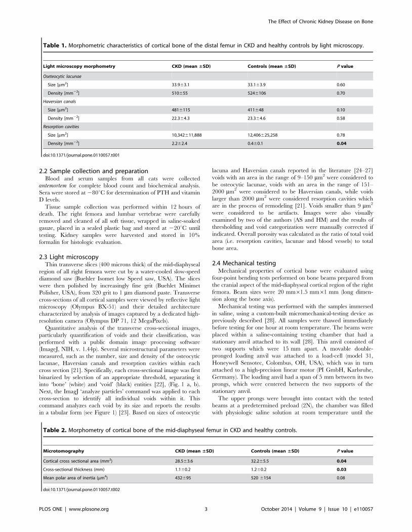
Table 1. Morphometric characteristics of cortical bone of the distal femur in CKD and healthy controls by light microscopy.
Light microscopy morphometry CKD (mean ±SD) Controls (mean ±SD) P value
Oseteocytic lacunae
Size [mm2] 33.963.1 33.163.9 0.60
Density [mm22] 510655 5246106 0.70
Haversian canals
Size [mm2] 4816115 411648 0.10
Density [mm22] 22.364.3 23.364.6 0.58
Resorption cavities
Size [mm2] 10,342611,888 12,406625,258 0.78
Density [mm22] 2.262.4 0.460.1 0.04
doi:10.1371/journal.pone.0110057.t001
Table 2. Morphometry of cortical bone of the mid-diaphyseal femur in CKD and healthy controls.
Microtomography CKD (mean ±SD) Controls (mean ±SD) P value
Cortical cross sectional area (mm2) 28.563.6 32.265.5 0.04
Cross-sectional thickness (mm) 1.160.2 1.260.2 0.03
Mean polar area of inertia (mm4) 432695 520 6154 0.08
doi:10.1371/journal.pone.0110057.t002
The Effect of Chronic Kidney Disease on Bone
PLOS ONE | www.plosone.org 3 October 2014 | Volume 9 | Issue 10 | e110057

samples were fully immersed, and bending tests were conducted
under displacement control at a rate of 500 mm/180 seconds up to
failure. Force-displacement data were collected by custom-written
software (LabView, National Instruments, Texas, USA) at 50 Hz.
Load and displacement values were converted to stress and strain,
respectively, based on beam theory [29]. The stress-strain curves
were used to estimate Young’s modulus of the beam material, as
well as yield and failure stresses and strains. It should be noted that
Figure 2. Light microscopy images of three transverse cross-sections of the femoral mid-diaphysis of (a, c, e) cats with CKD and (b,d, f) healthy cats. Note dramatic increase in unfilled resorption cavities in CKD cats compared to the healthy cats.doi:10.1371/journal.pone.0110057.g002
The Effect of Chronic Kidney Disease on Bone
PLOS ONE | www.plosone.org 4 October 2014 | Volume 9 | Issue 10 | e110057
care was taken to minimize shear deformation at the supports by
maintaining a ratio of distance between supports/beam depth of
15:1 [30,31]. Yield point was determined for each beam as the
point at which a line parallel with the linear portion of the stress-
strain curve and offset by 0.03% strain intersected with this curve
[32].
2.5 Microstructural characterization by Micro-CTAll cortical beams, the right femur and the 6th and 7th lumbar
vertebrae were scanned by microCT (Skyscan 1174 compact
micro-CT scanner, Belgium), with the beams scanned prior to
mechanical testing. Analyses were performed on the entire beam,
the mid-diaphyseal femoral cortex (cortical bone analysis), and in
the distal femoral metaphyses and vertebral bodies (cancellous
bone analysis).
The X-ray source was set at 50 kVp and 800 mA. A total of 450
projections were acquired over an angular range of 180u. The
samples were scanned with an isotropic voxel size of 11.1 mm for
the cortical bone beams and 19.6 mm for the femoral cortex and
cancellous bone of both the femora and vertebrae. Integration
time for all scans was 4500 ms, and a 0.25 mm aluminum filter
was used. Scans were reconstructed and analyzed using commer-
cial software (NRecon Skyscan software, version 1.6.1.2 and CT
analyser Skyscan software, version 1.9.3.2, respectively). Cortical
bone mineral density (BMD) of the beams was determined based
on calibration with 2 phantoms of known mineral density (0.25 g/
cm3 and 0.75 g/cm3) supplied by SkyScan, which were scanned
under exactly the same condition as were the bone specimens.
2.6 Statistical analysisThe distribution of continuous parameters (normal vs. non-
normal) was assessed using the Shapiro-Wilk’s test. Normally and
non-normally distributed continuous parameters were compared
between the study and the control group using Student’s t-test and
Mann-Whitney U test, respectively. Gender proportion between
the study group and the control groups was compared using the
Fischer Exact test. Correlations between continuous parameters
(e.g., biomechanical parameters and PTH concentration) were
performed using the Pearson or the Spearman Rank correlation
test, according to data distribution. For all tests P,0.05 was
considered statistically significant. All calculations were performed
using a statistical software (SPSS 17.0 for Windows, SPSS Inc;
Chicago, IL, USA).
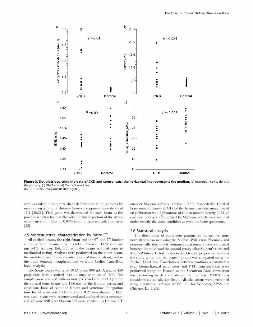
Figure 3. Dot plots depicting the data of CKD and control cats; the horizontal line represents the median. (a) resorption cavity density(b) porosity, (c) BMD and (d) Young’s modulus.doi:10.1371/journal.pone.0110057.g003
The Effect of Chronic Kidney Disease on Bone
PLOS ONE | www.plosone.org 5 October 2014 | Volume 9 | Issue 10 | e110057

Results
3.1 AnimalsThe study population included 26 cats, of which 13 were
diagnosed with CKD and 13 were healthy controls. There were
eight males and five females in the study group and six males and
seven females in the control group, with no gender proportion
differences between the study groups. Mean body weight was
significantly lower in cats with CKD compared with healthy
controls (2.860.6 kg vs. 3.760.9 kg, respectively; P = 0.01). There
was no statistically significant difference in mean age between the
study and control groups (10.565.6 years compared to 9.763.9
years, respectively; P = 0.7).
3.2 Clinical pathologyMedian serum creatinine concentration within the CKD group
was 8.2 mg/dL (range 3.5–16.0 mg/dL) compared with a median
of 0.9 mg/dL (range 0.6–1.2 mg/dL) [reference interval (RI), 0.5–
1.6 mg/dL] of the control group. Three cats in the study group
(23%) were classified as Stage III CKD, and the rest (77%) were
classified as Stage IV CKD. Median phosphorous concentration in
the study group was 8.8 mg/dL (range 5.3–21.7 mg/dL; RI, 3.0–
6.2 mg/dL). Median concentration of ionized calcium in the study
group was 0.80 mmol/L (range 0.65–1.01 mmol/L; RI, 0.9–
1.4 mmol/L). PTH concentration, available for five cats of the
study group, had a median concentration of 15.70 pmol/L (range,
0.9–32.9 pmol/L; RI, 0.4–2.5 pmol/L). Vitamin D concentration
was below normal in five out of the seven cats in which it was
measured (median 63 nmol/L, range 35–143 nmol/L; RI 65–
170 nmol/L).
3.3 Cortical bone architectureThe results of the architectural analysis are presented in Table 1
(microscopy) and Table 2 (microCT). CKD-affected cats had
significantly higher density of resorption cavities compared to
healthy controls (Table 1, Figures 2, 3a). Other structural
parameters were not significantly different between the groups.
Porosity tended to be higher in the CKD group, however the
difference between groups did not reach statistical significance
(P = 0.084, Figure 3b).
Micro-CT analysis of cortical bone of the femoral diaphysis
showed significantly lower cortical cross-sectional area and cross-
sectional thickness in CKD cats (Table 2). Mean polar area
moment of inertia tended to be lower in the CKD group, but the
differences did not reach statistical significance (P = 0.083)
(Table 2).
3.4 Cortical mineral densityCortical mineral density of CKD cats was lower by 4.8%
compared to controls, (P = 0.02, as shown in Figure 3c).
3.5 Mechanical properties of cortical boneTable 3 and Figure 3 present a comparison of several
mechanical properties of cortical bone material between the
CKD and control groups. Bones from the CKD group were shown
to have inferior mechanical properties compared to the control
group, in particular lower stiffness (Young’s modulus), lower yield
stress, and lower ultimate stress (Table 3, Figure 3).
Correlation could not be demonstrated between PTH levels and
any of the mechanical properties of the cortical bone.
3.6 Architecture of trabecular boneAnalysis of cancellous bone in the 6th and 7th lumbar vertebrae
and in the distal femur revealed significantly lower trabecular
thickness and bone volume over total volume (BV/TV) in bones
belonging to the CKD group, compared with control cats
(Figure 4).
Discussion
This study demonstrates that advanced CKD in cats results in
deterioration of bone quality, in particular a dramatic increase of
resorption cavities and decreased bone mineral density. These
results provide insight into skeletal changes occurring in human
CKD due to the similarity between cats and humans in terms of
the pathophysiology of the syndrome and the type of bone
architecture.
To the best of our knowledge, this is the first study to measure
the detailed mechanical and structural effects of CKD on the
skeleton of an animal model with naturally occurring CKD. Feline
and human CKD have very similar clinicopathologic features and
progression, therefore cats are superior models compared to
rodents in which pharmaceutical and surgical interventions are
usually employed to induce kidney disease [33–36]. Furthermore,
the cat skeleton shows great similarities to human bone, in
particular because it remodels continuously throughout life and
therefore consists mostly of secondary osteons. Rodent bones on
the other hand are remarkably different from those of human
bones in terms of type, architecture, structure and biology. Most
dramatically, rodent cortical bone does not remodel [16,17].
Therefore similar clinicopathologic circumstances, such as those
occurring due to CKD, may affect rodent bones differently from
human (or cat) bones.
We found that naturally occurring CKD results in several
alterations to the architecture and morphology of the bones of the
skeleton. Cortical thickness was found to be decreased by
approximately 17% in CKD cats compared with controls. This
change compromises the mechanical performance of long bones,
by reducing their flexural stiffness and is likely to lead to increased
fracture risk. Previous studies conducted in human patients with
CKD showed similar tendencies, however with much smaller
changes. For instance, a recent study documented 4.2% increase
Table 3. Mechanical properties of the cortical bone of CKD and control groups.
Parameter CKD (mean ±SD) Controls (mean ±SD) P value
Young’s modulus (GPa) 23.5±2.9 27.1±2.8 0.01
Yield stress (MPa) 151±18 166±14 0.04
Yield strain (millistrain) 5.660.8 5.660.4 .95
Ultimate stress (MPa) 185±16 205±15 0.01
Ultimate strain (millistrain) 8.661.0 8.160.4 0.37
doi:10.1371/journal.pone.0110057.t003
The Effect of Chronic Kidney Disease on Bone
PLOS ONE | www.plosone.org 6 October 2014 | Volume 9 | Issue 10 | e110057

in porosity, 2.9% decrease in cortical area and 2.8% decrease in
cortical thickness, indicating progressive loss of cortical bone [37].
These results, like those of other human studies, were based on
DEXA and high resolution peripheral quantitative CT. These
methods have been shown to be limited in precision in terms of
bone volume quantification due to inability to separate cortical
from cancellous bone (DEXA), and low-resolution volumetric
measurements, compared to microCT and whole bone sampling
[38].
Overall cortical porosity in cats with advanced CKD tended to
be somewhat higher compared with controls (Figure 3b), but this
difference did not reach statistical significance, most likely due to
small group size and biological variation. However, the density
(number per unit area) of resorption cavities in CKD patients is
greatly increased (5-fold, 2.22/mm2 vs. 0.41/mm2, P = 0.04).
Such a difference is expected to affect mechanical behavior of long
bones radically, and is likely to play an important role in the
increased fragility of CKD patients. Previous studies in a rat model
demonstrated that persistently elevated PTH concentrations result
in high bone turnover, exhibited as elevated numbers and size of
osteoclasts, increased osteoblastic activity and enhanced bone
resorption [33]. Consequently, these studies found extensive
endocortical, intracortical and periosteal resorption, resulting in
a dramatic increase in cortical porosity (9.75% compared to 0%).
It should be noted however that these results were observed in rat
bone, which normally does not remodel, as oppose to cat (and
human) bone.
Figure 4. Trabecular bone analysis: Trabecular thickness (a) and bone volume/total volume (BV/TV, b) in the cancellous bone ofCKD and control cats for both the femora (F) and the vertebrae (V). Data are presented as dot plots. The horizontal line represents themedian. Data from the femur and vertebra are similar within the study groups; however, there is a significant difference for both parameters betweenthe study and the control groups. Micro-CT scans of cancellous bone of a control (c) and CKD (d) cat.doi:10.1371/journal.pone.0110057.g004
The Effect of Chronic Kidney Disease on Bone
PLOS ONE | www.plosone.org 7 October 2014 | Volume 9 | Issue 10 | e110057
Bone mineral density is another major determinant of bone
quality, and the current clinical standard for prediction of fracture
risk in osteoporotic patients [39]. Therefore, determining the
influence of CKD on BMD was a major objective of this study.
The cortical mineral density of cats with CKD was significantly
lower compared to controls. It should be noted that despite
appearing small, this decrease (4.8%) is clinically significant, as
even a small decrease in BMD substantially decreases the stiffness
of the bone and increases fracture risk [40], since the relationship
between them is exponential [41]. A decrease in BMD was also
reported in various studies in humans CKD patients, with a wide
range of values [1.3% to 17.5%; [37,42,43]]. However, these
studies were based on areal BMD (g/cm2, using DEXA),
histomorphometry or pQCT, while the current study measured
volumetric BMD at high resolution and precision using microCT
[38,44].
Reliable measurement of material properties requires precise
and accurate mechanical testing. Such testing is difficult to achieve
in rodents due to the small size of their bones, which are often
tested by bending tests of whole bones, using the 3-point bending
technique. Results are dependent upon the geometry of the bones
and mechanical properties of the material, often leading to
underestimation of Young’s modulus [45,46]. The size of cat
bones allowed us to prepare cortical bone beams, enabling
accurate and reliable assessment of the material properties using
four-point bending testing. Furthermore, four-point bending tests
of beams allowed us also to measure other material properties, like
yield and strength, and to document significant decrease in yield
stress and failure stress in the cortical bone of the CKD cats
compared to controls.
This study demonstrates that the cortical bone material of CKD
cats is less stiff and more prone to micro-damage which occurs at
lower loads, and will result in increased bone fragility. In
particular, Young’s modulus of the cortical bone, which reflects
the stiffness of the material, was shown here to be significantly
lower in the CKD group (23.5 GPa vs 27.1 GPa, respectively,
P = 0.008). The lower Young’s modulus in CKD cats compared
with controls is most likely due to a combination of higher porosity
and lower BMD.
Cancellous bone is also significantly affected by CKD, as
reflected by decreased trabecular thickness and lower bone volume
(BV/TV). Furthermore, the effect on cancellous bone was multi-
site, shown to occur both in the vertebral bodies as well as in the
long bones. Both changes (trabecular thickness and BV/TV)
negatively affect bone quality and were shown to be associated
with increased risk for fracture [47].
Since this study was based on cats with naturally occurring
kidney disease, some variability existed among them in terms of
the severity and the chronicity of the disease. Furthermore, the
number of cats included in this study was relatively small, though
comparable to numbers typically seen in published rodent model
studies. It should also be noted that cats with advanced CKD often
exhibit decreased appetite and thus might fail to consume enough
food to meet their caloric requirements. Such nutritional
deficiencies, as in human patients, might contribute to their
decreased bone quality. Additionally, all of the cats in this study
had a single etiology (interstitial nephritis, which is the etiology in
more than 70% of cases with CKD in cats [18]), whereas human
patients with CKD have multiple etiologies (e.g., diabetic
nephropathy, transplant patients, etc.). Nevertheless, slowly
progressive natural disease represents the human syndrome and
its consequences, including renal osteopathy, much better than
artificially-induced rodent models.
In conclusion, the current study demonstrates the deleterious
effects of CKD on remodeled (secondary osteonal) bone quality
and strength, including increased bone resorption, decreased
BMD, and inferior mechanical and structural properties. Due to
these changes, the bones of CKD patients become more fragile.
Since many similarities have been demonstrated between human
and feline CKD patients, in terms of the clinic-pathologic features
of the syndrome, as well as bone-associated effects, cats are an
extremely suitable and relevant animal model for studying the
development of bone abnormalities in humans suffering from
CKD.
Author Contributions
Conceived and designed the experiments: AS GS HM MM RS. Performed
the experiments: AS GS HM MM AA OB RS. Analyzed the data: AS GS
HM MM AA RS. Contributed reagents/materials/analysis tools: RS OB.
Wrote the paper: AS HM GS MM OB AA RS.
References
1. Lora CM, Daviglus ML, Kusek JW, Porter A, Ricardo AC, et al. (2009) Chronic
kidney disease in United States Hispanics: a growing public health problem.
Ethn Dis 19: 466–472.
2. McClellan WM, Plantinga LC (2013) A public health perspective on CKD and
obesity. Nephrol Dial Transplant 28 Suppl 4: iv37–iv42.
3. Spasovski GB, Bervoets AR, Behets GJ, Ivanovski N, Sikole A, et al. (2003)
Spectrum of renal bone disease in end-stage renal failure patients not yet on
dialysis. Nephrol Dial Transplant 18: 1159–1166.
4. Levin A, Bakris GL, Molitch M, Smulders M, Tian J, et al. (2007) Prevalence of
abnormal serum vitamin D, PTH, calcium, and phosphorus in patients with
chronic kidney disease: results of the study to evaluate early kidney disease.
Kidney Int 71: 31–38.
5. Cozzolino M, Ciceri P, Volpi EM, Olivi L, Messa PG (2009) Pathophysiology of
calcium and phosphate metabolism impairment in chronic kidney disease. Blood
Purif 27: 338–344.
6. Nabeshima Y (2008) The discovery of alpha-Klotho and FGF23 unveiled new
insight into calcium and phosphate homeostasis. Cell Mol Life Sci 65: 3218–
3230.
7. Saito H, Kusano K, Kinosaki M, Ito H, Hirata M, et al. (2003) Human
fibroblast growth factor-23 mutants suppress Na+ -dependent phosphate co-
transport activity and 1 alpha,25-dihydroxyvitamin D-3 production. J Biol
Chem 278: 2206–2211.
8. Shimada T, Hasegawa H, Yamazaki Y, Muto T, Hino R, et al. (2004) FGF-23 is
a potent regulator of vitamin D metabolism and phosphate homeostasis. J Bone
Miner Res 19: 429–435.
9. Ketteler M, Biggar PH, Liangos O (2013) FGF23 antagonism: the thin line
between adaptation and maladaptation in chronic kidney disease. Nephrol DialTransplant 28: 821–825.
10. Danese MD, Kim J, Doan QV, Dylan M, Griffiths R, et al. (2006) PTH and the
risks for hip, vertebral, and pelvic fractures among patients on dialysis.
Am J Kidney Dis 47: 149–156.
11. Alem AM, Sherrard DJ, Gillen DL, Weiss NS, Beresford SA, et al. (2000)
Increased risk of hip fracture among patients with end-stage renal disease.Kidney Int 58: 396–399.
12. Coco M, Rush H (2000) Increased incidence of hip fractures in dialysis patients
with low serum parathyroid hormone. Am J Kidney Dis 36: 1115–1121.
13. Stehman-Breen CO, Sherrard DJ, Alem AM, Gillen DL, Heckbert SR, et al.
(2000) Risk factors for hip fracture among patients with end-stage renal disease.Kidney Int 58: 2200–2205.
14. Parfitt AM (1998) A structural approach to renal bone disease. J Bone MinerRes 13: 1213–1220.
15. Iwasaki Y, Kazama JJ, Yamato H, Shimoda H, Fukagawa M (2013)Accumulated uremic toxins attenuate bone mechanical properties in rats with
chronic kidney disease. Bone 57: 477–483.
16. Bach-Gansmo FL, Irvine SC, Bruel A, Thomsen JS, Birkedal H (2013) Calcified
Cartilage Islands in Rat Cortical Bone. Calcif Tissue Int 92: 330–338.
17. Shipov A, Zaslansky P, Riesemeier H, Segev G, Atkins A, et al. (2013)
Unremodeled endochondral bone is a major architectural component of thecortical bone of the rat (Rattus norvegicus). J Struct Biol 183: 132–140.
18. Polzin DJ (2010) Chronic kidney disease. In: Ettinger SJ, Feldman EC,
editors.Textbook of Veterinary Internal Medicine. 7th ed. St. Louis: Saunders.
pp. 1990–2020.
The Effect of Chronic Kidney Disease on Bone
PLOS ONE | www.plosone.org 8 October 2014 | Volume 9 | Issue 10 | e110057
19. Hillier ML, Bell LS (2007) Differentiating human bone from animal bone: A
review of histological methods. J Forensic Sci 52: 249–263.20. Segev G, Palm C, LeRoy B, Cowgill LD, Westropp JL (2013) Evaluation of
neutrophil gelatinase-associated lipocalin as a marker of kidney injury in dogs.
J Vet Intern Med 27: 1362–1367.21. Zebaze RM, Ghasem-Zadeh A, Bohte A, Iuliano-Burns S, Mirams M, et al.
(2010) Intracortical remodelling and porosity in the distal radius and post-mortem femurs of women: a cross-sectional study. Lancet 375: 1729–1736.
22. Bousson V, Meunier A, Bergot C, Vicaut E, Rocha MA, et al. (2001)
Distribution of intracortical porosity in human midfemoral cortex by age andgender. J Bone Miner Res 16: 1308–1317.
23. Montanari S, Brusatte SL, De Wolf W, Norell MA (2011) Variation of osteocytelacunae size within the tetrapod skeleton: implications for palaeogenomics. Biol
Lett 7: 751–754.24. Urbanova P, Novotny V (2005) Distinguishing between human and non-human
bones: Histometric method for forensic anthropology. Anthropologie 43: 77–85.
25. Currey JD, Shahar R (2013) Cavities in the compact bone in tetrapods and fishand their effect on mechanical properties. J Struct Biol 183: 107–122.
26. Kuchler U, Pfingstner G, Busenlechner D, Dobsak T, Reich K, et al. (2013)Osteocyte lacunar density and area in newly formed bone of the augmented
sinus. Clin Oral Implants Res 24: 285–289.
27. Tazawa K, Hoshi K, Kawamoto S, Tanaka M, Ejiri S, et al. (2004) Osteocyticosteolysis observed in rats to which parathyroid hormone was continuously
administered. J Bone Miner Metab 22: 524–529.28. Cohen L, Dean M, Shipov A, Atkins A, Monsonego-Ornan E, et al. (2012)
Comparison of structural, architectural and mechanical aspects of cellular andacellular bone in two teleost fish. J Exp Biol 215: 1983–1993.
29. Sharir A, Barak MM, Shahar R (2008) Whole bone mechanics and mechanical
testing. Vet J 177: 8–17.30. Spatz HC, O’Leary EJ, Vincent JF (1996) Young’s moduli and shear moduli in
cortical bone. Proc Biol Sci 263: 287–294.31. Draper ER, Goodship AE (2003) A novel technique for four-point bending of
small bone samples with semi-automatic analysis. J Biomech 36: 1497–1502.
32. Turner CH, Burr DB (1993) Basic biomechanical measurements of bone: atutorial. Bone 14: 595–608.
33. Miller MA, Chin J, Miller SC, Fox J (1998) Disparate effects of mild, moderate,and severe secondary hyperparathyroidism on cancellous and cortical bone in
rats with chronic renal insufficiency. Bone 23: 257–266.34. Cao HH, Nazarian A, Ackerman JL, Snyder BD, Rosenberg AE, et al. (2010)
Quantitative P-31 NMR spectroscopy and H-1 MRI measurements of bone
mineral and matrix density differentiate metabolic bone diseases in rat models.
Bone 46: 1582–1590.35. Iwasaki Y, Kazama JJ, Yamato H, Fukagawa M (2011) Changes in chemical
composition of cortical bone associated with bone fragility in rat model with
chronic kidney disease. Bone 48: 1260–1267.36. Jokihaara J, Jarvinen TLN, Jolma P, Koobi P, Kalliovalkama J, et al. (2006)
Renal insufficiency-induced bone loss is associated with an increase in bone sizeand preservation of strength in rat proximal femur. Bone 39: 353–360.
37. Nickolas TL, Stein EM, Dworakowski E, Nishiyama KK, Komandah-Kosseh
M, et al. (2013) Rapid cortical bone loss in patients with chronic kidney disease.J Bone Miner Res 28: 1811–1820.
38. Barou O, Valentin D, Vico L, Tirode C, Barbier A, et al. (2002) High-resolutionthree-dimensional micro-computed tomography detects bone loss and changes
in trabecular architecture early: comparison with DEXA and bone histomor-phometry in a rat model of disuse osteoporosis. Invest Radiol 37: 40–46.
39. Nickolas TL, Leonard MB, Shane E (2008) Chronic kidney disease and bone
fracture: a growing concern. Kidney Int 74: 721–731.40. Yenchek RH, Ix JH, Shlipak MG, Bauer DC, Rianon NJ, et al. (2012) Bone
mineral density and fracture risk in older individuals with CKD. Clin J Am SocNephrol 7: 1130–1136.
41. Wasnich RD, Ross PD, Davis JW, Vogel JM (1989) A Comparison of Single and
Multi-Site Bmc Measurements for Assessment of Spine Fracture Probability.J Nucl Med 30: 1166–1171.
42. Balon BP, Hojs R, Zavratnik A, Kos M (2002) Bone mineral density in patientsbeginning hemodialysis treatment. Am J Nephrol 22: 14–17.
43. Rix M, Andreassen H, Eskildsen P, Langdahl B, Olgaard K (1999) Bone mineraldensity and biochemical markers of bone turnover in patients with predialysis
chronic renal failure. Kidney Int 56: 1084–1093.
44. Leonard MB (2009) A Structural Approach to Skeletal Fragility in ChronicKidney Disease. Semin Nephrol 29: 133–143.
45. van Lenthe GH, Voide R, Boyd SK, Muller R (2008) Tissue modulus calculatedfrom beam theory is biased by bone size and geometry: implications for the use
of three-point bending tests to determine bone tissue modulus. Bone 43: 717–
723.46. Torcasio A, Van Oosterwyck H, van Lenthe GH (2008) The systematic errors in
tissue modulus of murine bones when estimated from three-point bending.J Biomech 41: S14.
47. Nickolas TL, Stein E, Cohen A, Thomas V, Staron RB, et al. (2010) Bone massand microarchitecture in CKD patients with fracture. J Am Soc Nephrol 21:
1371–1380.
The Effect of Chronic Kidney Disease on Bone
PLOS ONE | www.plosone.org 9 October 2014 | Volume 9 | Issue 10 | e110057
Related Documents


