The Effect of Intravenously Paratyroid Cell Xenotransplantation in Sheep: As an Animal Model Emrah YUCESAN 1,a Beyza GONCU 2,b Oguz IDIZ 3,c Ramazan UCAK 4,d Burcu OZDEMIR 2,e Ebru KANIMDAN 2,f Yeliz Emine ERSOY 5,g Erhan AYSAN 6,h 1 Bezmialem Vakif University, Faculty of Medicine, Department of Medical Biology, TR-34093 Istanbul - TURKEY 2 Bezmialem Vakif University, Experimental Research Center, TR-34093 Istanbul - TURKEY 3 Istanbul Training and Research Hospital, Department of General Surgery, TR-34098 Istanbul - TURKEY 4 Istanbul Sisli Hamidiye Etfal Training and Research Hospital, Department of Pathology, TR-34360 Istanbul - TURKEY 5 Bezmialem Vakif University, Faculty of Medicine, Department of General Surgery, TR-34093 Istanbul - TURKEY 6 Yeditepe University Faculty of Medicine, Department of General Surgery, TR-34734 Istanbul - TURKEY ORCIDs: a 0000-0003-4512-8764; b 0000-0001-6026-8218; c 0000-0002-8462-7809; d 0000-0002-2999-7495; e 0000-0003-0288-0470 f 0000-0002-7123-4600; g 0000-0002-5028-6436; h 0000-0002-9563-3761 Article ID: KVFD-2020-24309 Received: 12.04.2020 Accepted: 10.08.2020 Published Online: 10.08.2020 How to Cite This Article Yucesan E, Goncu B, Idiz O, Ucak R, Ozdemir B, Kanimdan E, Ersoy YE, Aysan E: The effect of intravenously paratyroid cell xenotransplantation in sheep: As an animal model. Kafkas Univ Vet Fak Derg, 26 (6): 765-770, 2020. DOI: 10.9775/kvfd.2020.24309 Abstract Parathyroid cell transplantation is an effective approach for the treatment of permanent hypoparathyroidism. Intramuscular and intraperitoneal routes were performed previously but intravenous adminstration has not been conducted previous studies. Our aim is to determine proper homing site for parathyroid cells, therefore we administered parathyroid cells intravenously and observed results. This study is carried out on nine sheep. According to the given substance the sheep were divided into two groups; seven were parathyroid cell injected group and two were isotonic saline solution injected control group. Parathyroid cells were obtained from a patient with chronic kidney failure and were injected intravenously into sheep after cell isolation.: 20x10 6 cells for two animals, 50x10 6 cells for two animals, 100x10 6 cells for two animals, and 200x10 6 cells for one animal respectively, with no post injection immunosuppresive therapy. Immunosuppresion was not administered. Physical conditions of animals and blood biochemical tests were observed for three months. After sacrifications, kidneys and livers were evaluated histopathologically. In 20x10 6 and 50x10 6 cell transplanted groups serum PTH levels increased in the first seven days but in the other groups remained stable. Histopathological evaluations of kidneys and livers revealed fibrosis related to the number of infused cells, however biochemical functional differentiations were not detected. Intravenous parathyroid cell transplantation is considered as an effective and useful technique to perform without immunosuppression. However, further and long term studies need to have more acceptable results in future for clinical purpose. Keywords: Xenotransplantation, Parathyroid cell, İntravenous injection, Permanent hypoparathyroidism, Sheep Bir Hayvan Modeli Olarak: Koyunlarda Intravenöz Paratiroid Hücre Zenonaklinin Etkisi Öz Paratiroid hücre nakli, kalıcı hipoparatiroidizmin tedavisinde etkili bir yaklaşımdır. İntramüsküler ve intraperitoneal yollar daha önce yapılmış, ancak intravenöz uygulama daha önce yapılmamıştır. Amacımız, paratiroid hücreleri için uygun yerleşim bölgesini belirlemekti, bu amaçla paratiroid hücrelerini intravenöz olarak uyguladık ve sonuçları gözlemledik. Bu çalışma dokuz koyun üzerinde gerçekleştirildi. Verilen maddeye göre koyunlar iki gruba ayrıldı; yedisi paratiroid hücresi enjekte edilen grup, ikisi iztonik tuz çözeltisi enjekte edilen kontrol grubudur. Paratiroid hücreleri kronik böbrek yetmezliği olan bir hastadan elde edildi ve hücre izolasyonu sonrası intravenöz olarak koyunlara enjekte edildi. Sırasıyla; 20x10 6 hücre iki hayvana, 50x10 6 hücre iki hayvana, 100x10 6 hücre iki hayvana ve 200x106 hücre bir hayvana hayvana uygulandı ve enjeksiyon sonrası immünsupresyon uygulanmadı. Üç ay süresince hayvanların fiziksel kondisyonları ve kan biyokimyasal testleri gözlemlendi. Sakrifikasyon sonrası, karaciğerler ve böbrekler histopatolojik olarak incelendi. 20x10 6 ve 50x10 6 hücre nakledilen gruplarda serum PTH seviyeleri ilk yedi gün yükseldi ancak diğer gruplarda sabit kaldı. Böbreklerin ve karaciğerlerin histopatolojik incelemelerinde, infüze edilen hücre sayısıyla ilişkili fibrozis saptandı, ancak biyokimyasal fonksiyonlarda herhangi bir farklılık tespit edilmedi. İntravenöz paratiroid hücre naklinin, immünsupresyonsuz uygulanacak etkili ve kullanışlı bir teknik olduğu değerlendirildi. Bununla birlikte, klinik amaçla kullanım öncesi ilerleyen dönemde daha kabul edilebilir sonuçları olan uzun süreli çalışmalara ihtiyaç vardır. Anahtar sözcükler: Zenonakil, Paratiroid hücresi, İntravenöz enjeksiyon, Kalıcı hipoparatiroidizm, Koyun Correspondence +90 212 5232288/ext.1465 Fax: +90 212 4531870 [email protected]; [email protected] Kafkas Univ Vet Fak Derg 26 (6): 765-770, 2020 DOI: 10.9775/kvfd.2020.24309 Kafkas Universitesi Veteriner Fakultesi Dergisi ISSN: 1300-6045 e-ISSN: 1309-2251 Journal Home-Page: http://vetdergikafkas.org Online Submission: http://submit.vetdergikafkas.org Research Article

Welcome message from author
This document is posted to help you gain knowledge. Please leave a comment to let me know what you think about it! Share it to your friends and learn new things together.
Transcript
The Effect of Intravenously Paratyroid Cell Xenotransplantation in Sheep: As an Animal Model
Emrah YUCESAN 1,a Beyza GONCU 2,b Oguz IDIZ 3,c Ramazan UCAK 4,d Burcu OZDEMIR 2,e Ebru KANIMDAN 2,f Yeliz Emine ERSOY 5,g Erhan AYSAN 6,h
1 Bezmialem Vakif University, Faculty of Medicine, Department of Medical Biology, TR-34093 Istanbul - TURKEY2 Bezmialem Vakif University, Experimental Research Center, TR-34093 Istanbul - TURKEY 3 Istanbul Training and Research Hospital, Department of General Surgery, TR-34098 Istanbul - TURKEY 4 Istanbul Sisli Hamidiye Etfal Training and Research Hospital, Department of Pathology, TR-34360 Istanbul - TURKEY5 Bezmialem Vakif University, Faculty of Medicine, Department of General Surgery, TR-34093 Istanbul - TURKEY6 Yeditepe University Faculty of Medicine, Department of General Surgery, TR-34734 Istanbul - TURKEY ORCIDs: a 0000-0003-4512-8764; b 0000-0001-6026-8218; c 0000-0002-8462-7809; d 0000-0002-2999-7495; e 0000-0003-0288-0470 f 0000-0002-7123-4600; g 0000-0002-5028-6436; h 0000-0002-9563-3761
Article ID: KVFD-2020-24309 Received: 12.04.2020 Accepted: 10.08.2020 Published Online: 10.08.2020
How to Cite This Article
Yucesan E, Goncu B, Idiz O, Ucak R, Ozdemir B, Kanimdan E, Ersoy YE, Aysan E: The effect of intravenously paratyroid cell xenotransplantation in sheep: As an animal model. Kafkas Univ Vet Fak Derg, 26 (6): 765-770, 2020. DOI: 10.9775/kvfd.2020.24309
AbstractParathyroid cell transplantation is an effective approach for the treatment of permanent hypoparathyroidism. Intramuscular and intraperitoneal routes were performed previously but intravenous adminstration has not been conducted previous studies. Our aim is to determine proper homing site for parathyroid cells, therefore we administered parathyroid cells intravenously and observed results. This study is carried out on nine sheep. According to the given substance the sheep were divided into two groups; seven were parathyroid cell injected group and two were isotonic saline solution injected control group. Parathyroid cells were obtained from a patient with chronic kidney failure and were injected intravenously into sheep after cell isolation.: 20x106 cells for two animals, 50x106 cells for two animals, 100x106 cells for two animals, and 200x106 cells for one animal respectively, with no post injection immunosuppresive therapy. Immunosuppresion was not administered. Physical conditions of animals and blood biochemical tests were observed for three months. After sacrifications, kidneys and livers were evaluated histopathologically. In 20x106 and 50x106 cell transplanted groups serum PTH levels increased in the first seven days but in the other groups remained stable. Histopathological evaluations of kidneys and livers revealed fibrosis related to the number of infused cells, however biochemical functional differentiations were not detected. Intravenous parathyroid cell transplantation is considered as an effective and useful technique to perform without immunosuppression. However, further and long term studies need to have more acceptable results in future for clinical purpose.
Keywords: Xenotransplantation, Parathyroid cell, İntravenous injection, Permanent hypoparathyroidism, Sheep
Bir Hayvan Modeli Olarak: Koyunlarda Intravenöz Paratiroid Hücre Zenonaklinin Etkisi
ÖzParatiroid hücre nakli, kalıcı hipoparatiroidizmin tedavisinde etkili bir yaklaşımdır. İntramüsküler ve intraperitoneal yollar daha önce yapılmış, ancak intravenöz uygulama daha önce yapılmamıştır. Amacımız, paratiroid hücreleri için uygun yerleşim bölgesini belirlemekti, bu amaçla paratiroid hücrelerini intravenöz olarak uyguladık ve sonuçları gözlemledik. Bu çalışma dokuz koyun üzerinde gerçekleştirildi. Verilen maddeye göre koyunlar iki gruba ayrıldı; yedisi paratiroid hücresi enjekte edilen grup, ikisi iztonik tuz çözeltisi enjekte edilen kontrol grubudur. Paratiroid hücreleri kronik böbrek yetmezliği olan bir hastadan elde edildi ve hücre izolasyonu sonrası intravenöz olarak koyunlara enjekte edildi. Sırasıyla; 20x106 hücre iki hayvana, 50x106 hücre iki hayvana, 100x106 hücre iki hayvana ve 200x106 hücre bir hayvana hayvana uygulandı ve enjeksiyon sonrası immünsupresyon uygulanmadı. Üç ay süresince hayvanların fiziksel kondisyonları ve kan biyokimyasal testleri gözlemlendi. Sakrifikasyon sonrası, karaciğerler ve böbrekler histopatolojik olarak incelendi. 20x106 ve 50x106 hücre nakledilen gruplarda serum PTH seviyeleri ilk yedi gün yükseldi ancak diğer gruplarda sabit kaldı. Böbreklerin ve karaciğerlerin histopatolojik incelemelerinde, infüze edilen hücre sayısıyla ilişkili fibrozis saptandı, ancak biyokimyasal fonksiyonlarda herhangi bir farklılık tespit edilmedi. İntravenöz paratiroid hücre naklinin, immünsupresyonsuz uygulanacak etkili ve kullanışlı bir teknik olduğu değerlendirildi. Bununla birlikte, klinik amaçla kullanım öncesi ilerleyen dönemde daha kabul edilebilir sonuçları olan uzun süreli çalışmalara ihtiyaç vardır.
Anahtar sözcükler: Zenonakil, Paratiroid hücresi, İntravenöz enjeksiyon, Kalıcı hipoparatiroidizm, Koyun
Correspondence +90 212 5232288/ext.1465 Fax: +90 212 4531870 [email protected]; [email protected]
Kafkas Univ Vet Fak Derg26 (6): 765-770, 2020DOI: 10.9775/kvfd.2020.24309
Kafkas Universitesi Veteriner Fakultesi DergisiISSN: 1300-6045 e-ISSN: 1309-2251
Journal Home-Page: http://vetdergikafkas.orgOnline Submission: http://submit.vetdergikafkas.org
Research Article
766Intravenous Parathyroid Cells Injection
INTRODUCTIONPermanent hypoparathyroidism (PH) is a clinical condition accompanied by hypocalcemia, hyperphosphatemia, and low parathormone (PTH) levels. The most common etiologic factor of PH is thyroid surgery [1]. The current standard treatment of PH is vitamin D and calcium supplementation. Standard treatment only relieves the symptoms temporarily and may causes several side effects [2]. Recombinant parathormone drugs reveal better results however balancing the dosage, efficacy and safety is not clear. In addition, recombinant parathormone drugs are more expensive than standard treatment [3]. Parathyroid cell transplantation is the most promising technique for the treatment of PH [4]. In the literature, several trans-plantation approaches have been reported such as auto- transplantation [5], allo-transplantation [6], and xenotrans-plantation [7] (XT). These approaches are used with different methods e.g. direct tissue injection [8], non-treated cultivated cells injection [9], cultured parathyroid cells treated with IFNγ [10], macroencapsulation [11], and microencapsulation [12]
Different experimental animal models for the assessment of parathyroid function, morphology, and disease progression have been investigated including, dog [13], rat [14], rabbit [15], and sheep [16]. Among them parathyroid transplantation models were assessed by intramuscular [17] and intra-peritoneal routes [7], respectively. Till the time of the research was planned, intravenous parathyroid cell trans-plantation has not been tried. In the present study, we injected human parathyroid cells to the sheep, and observed the functionality of the cells and their effects on the kidney and liver.
MATERIAL and METHODSThe study was approved by the Bezmialem Vakif University, Local Experimental Animals Ethics Committee (approval number: 2018/12). In the power analysis, the number of subjects was determined in 85% confidence interval and 95% significance level. We studied on nine sheep (12 months-old, mean weight 27.05 kg, weight range 24.5-35.5). The animals were housed and fed ad libitum throughout the study. The animals were divided into two groups; intravenous parathyroid cells injected group (n=7) and intravenous isotonic saline solution injected group (n=2). Peripheral blood samples were obtained before XT and continued biweekly after XT for 90 days. Weights and other physical conditions of the animals were examined weekly. After 90 days, the animals were sacrificed. Excisional biopsies were taken from the kidney and liver for histopathological evaluations. Resected tissues were immersed in neutral-buffered 10% formalin and fixed for at least 24 h. Formalin-fixed tissues were processed within paraffin wax and sectioned at 5 μm, mounted on positively charged glass slides, and air-dried overnight. Slides were rehydrated and stained by hematoxylin and eosin. Three to four transects of each slide were counted.
Tissues were evaluated by one expert pathologist who blinded the groups, according to fibrosis scoring (Grade 0: No fibrosis for liver and no congestion/fibrosis for kidney, Grade 1: Mild fibrosis for liver and mild congestion/fibrosis for kidney, Grade 2: Moderate fibrosis for liver and moderate congestion/fibrosis for kidney, and Grade 3: Severe fibrosis for liver and severe congestion/fibrosis for kidney) as previously reported by Idiz et al.[18]. Before the XT procedure the Local Human Ethics Committee approval was received (approval number: 71306642-050.01.04). All of the protocols were confirmed according to the ethical guidelines of the Helsinki Declaration and written informed consent was obtained from the donor. The donor patient was a 34 year old man with parathyroid hyperplasia resistant to drug therapy due to chronic renal failure who was referred from the nephrology outpatient clinic to the general surgery department for surgical intervention. Standard subtotal parathyroidectomy procedure was performed and half of each of the resected glands were delivered to the pathology laboratory for histopathological evaluation. The remaining parts of the glands were snap frozen. After the histopathological evaluations were reported as benign parathyroid hyperplasia, the tissues were prepared for the XT process.
Cell Preparation Procedure for XT
In laboratory conditions, the tissue was cut and washed with 1% Phosphate-buffered solution (Thermo Fisher Scientific, MA) and minced in a petri dish on ice. The minced preparation was combined with 2 mL for a total 100 mg/mL bovine serum albumin (Merck Millipore, Germany), 215 mmol collagenase type II (Thermo Fisher Scientific), 0.32 mM DNase I (AppliChem, Gatersleben, Germany), and 1 mL Ham’s F10 Supplement (Thermo Fisher Scientific). Samples were transferred to an incubator (CCL-170B-8; ESCO, Singapore) at 37°C with humidified atmosphere containing 5% CO2, where they were incubated overnight. Each sample was filtered into a 15-mL conical Falcon tube using a sterile cell strainer (70 mm, Falcon; BD Biosciences, NJ). The solution was centrifuged at 306 g for 15 min to obtain a pellet. Cells were suspended in 1 mL of culture medium. Parathyroid cell viability was assessed before cryopreservation using a Muse Cell Analyzer (Merck Millipore) with a Muse Count & Viability Assay Kit (Merck Millipore) [19]. The cells were mixed with 10% DMSO (dimethyl sulfoxide), 10% FBS, and suspended in cryotubes kept at -80°C, and then transferred to a liquid nitrogen tank for storage. The day before XT, cells were removed from the nitrogen tank and cultivated in flasks with McCoy’s 5A (Modified) Medium (Thermo Fisher Scientific) with 1% sodium pyruvate, 1% penicillin-streptomycin, and 10% FBS and placed in an incubator (CCL-170B-8; ESCO) at 37°C with 5% CO2 humidified atmosphere, where they were kept overnight before XT.
XT Procedure
Prepared xenograft cells were administered to seven animals
767
YUCESAN, GONCU, IDIZ, UCAKOZDEMIR, KANIMDAN, ERSOY, AYSAN
by a decreased count system: 100x106 cells for two animals, 50x106 cells for two animals, 20x106 cells for two animals, and 200x106 cells for one animal into the 10 mL isotonic saline solution. Injections were performed via external jugular vein catheterization. In the control group, 10 mL isotonic saline solution physiologic was injected intravenously via the external jugular vein.
Blood Biochemical Tests
Serum sheep-PTH and human-PTH levels were measured using Sheep Parathyroid Hormone ELISA Kit® (MyBiSource, CA, USA) and Architect Intact hu-PTH Assay Kit® (Abbott, IL, USA), respectively. A complete blood count (CBC) was measured by Hematology Analyzer Abacus Junior Vet® (Diatron, Budapest, Hungary). Aspartate amino-transferase (AST), alanine aminotransferase (ALT), gamma-glutamyl transpeptidase (GGT), alkaline phosphatase (ALP), lactate dehydrogenase (LDH), troponin, creatine kinase (CK), amylase, lipase, urea, total bilirubin, calcium, phosphorus, albumin, and creatinine were measured by Chemistry Analyzer IDEXX VetTest® (IDEXX Laboratories, Maine, USA).
Statistics
All data during the given period were compared with pre-op values. Statistical analyses were performed using SPSS software v22.0 (IBM, Armonk, NY, USA). Data was not normally distributed, thus we used Friedman test as a non-parametric test, and P<0.05 was considered statistically significant.
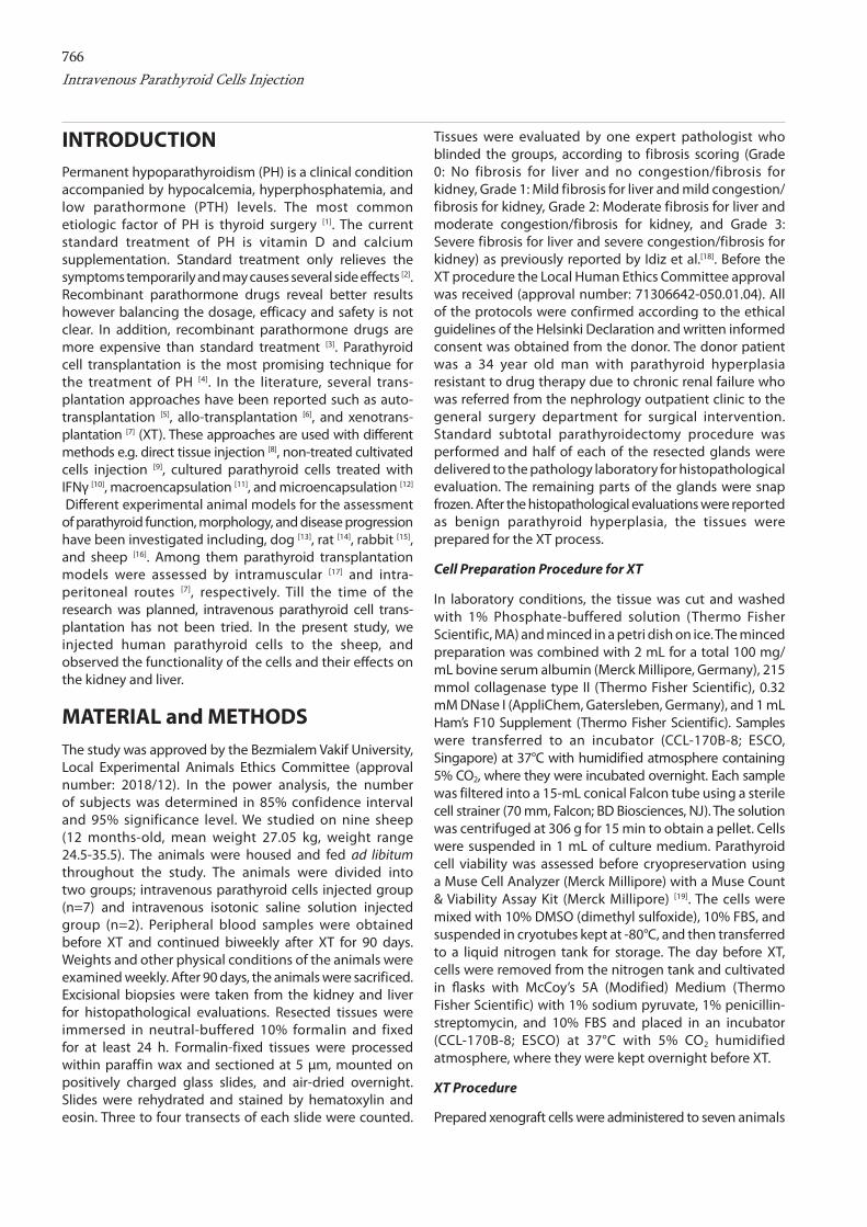
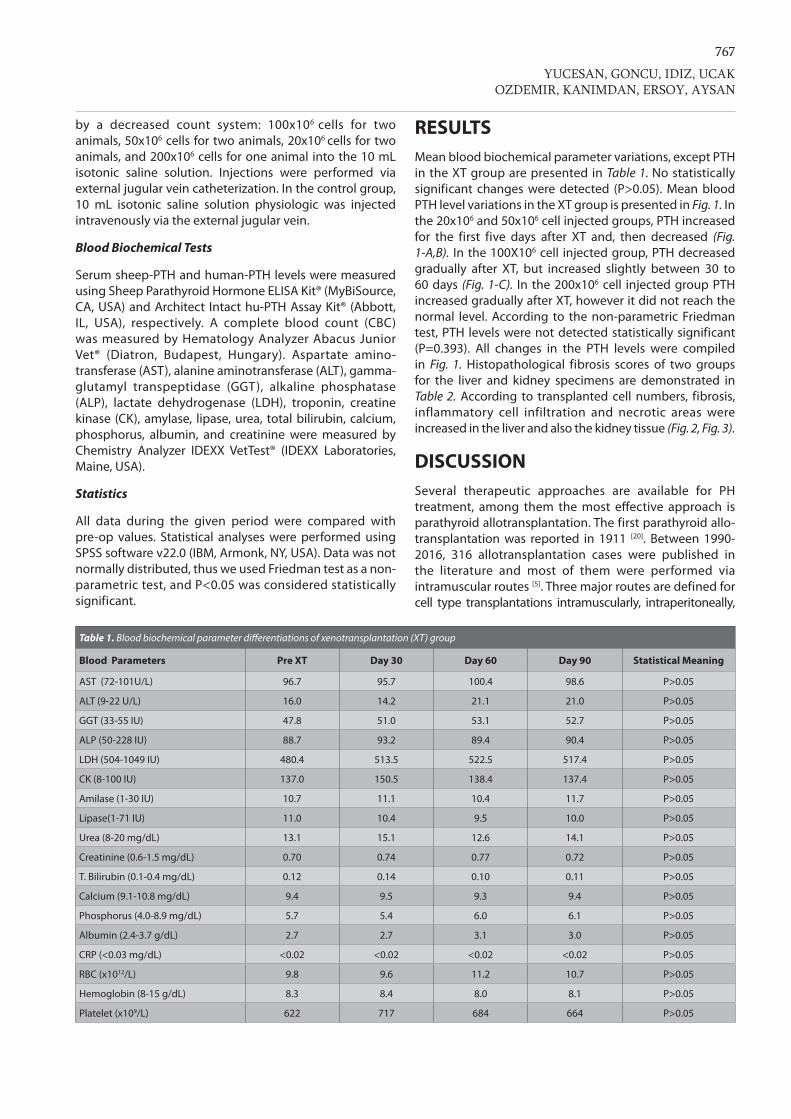
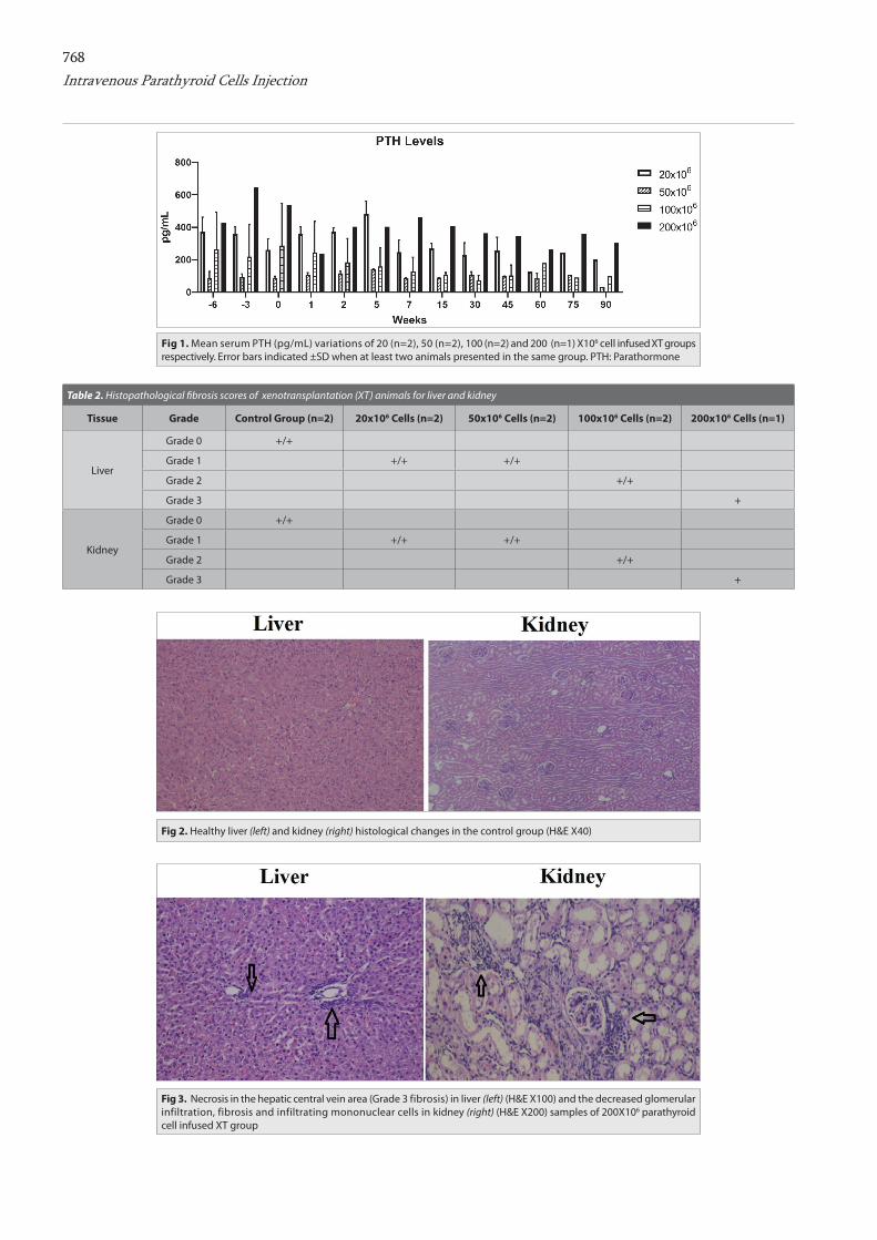
RESULTSMean blood biochemical parameter variations, except PTH in the XT group are presented in Table 1. No statistically significant changes were detected (P>0.05). Mean blood PTH level variations in the XT group is presented in Fig. 1. In the 20x106 and 50x106 cell injected groups, PTH increased for the first five days after XT and, then decreased (Fig. 1-A,B). In the 100X106 cell injected group, PTH decreased gradually after XT, but increased slightly between 30 to 60 days (Fig. 1-C). In the 200x106 cell injected group PTH increased gradually after XT, however it did not reach the normal level. According to the non-parametric Friedman test, PTH levels were not detected statistically significant (P=0.393). All changes in the PTH levels were compiled in Fig. 1. Histopathological fibrosis scores of two groups for the liver and kidney specimens are demonstrated in Table 2. According to transplanted cell numbers, fibrosis, inflammatory cell infiltration and necrotic areas were increased in the liver and also the kidney tissue (Fig. 2, Fig. 3).
DISCUSSIONSeveral therapeutic approaches are available for PH treatment, among them the most effective approach is parathyroid allotransplantation. The first parathyroid allo- transplantation was reported in 1911 [20]. Between 1990-2016, 316 allotransplantation cases were published in the literature and most of them were performed via intramuscular routes [5]. Three major routes are defined for cell type transplantations intramuscularly, intraperitoneally,
Table 1. Blood biochemical parameter differentiations of xenotransplantation (XT) group
Blood Parameters Pre XT Day 30 Day 60 Day 90 Statistical Meaning
AST (72-101U/L) 96.7 95.7 100.4 98.6 P>0.05
ALT (9-22 U/L) 16.0 14.2 21.1 21.0 P>0.05
GGT (33-55 IU) 47.8 51.0 53.1 52.7 P>0.05
ALP (50-228 IU) 88.7 93.2 89.4 90.4 P>0.05
LDH (504-1049 IU) 480.4 513.5 522.5 517.4 P>0.05
CK (8-100 IU) 137.0 150.5 138.4 137.4 P>0.05
Amilase (1-30 IU) 10.7 11.1 10.4 11.7 P>0.05
Lipase(1-71 IU) 11.0 10.4 9.5 10.0 P>0.05
Urea (8-20 mg/dL) 13.1 15.1 12.6 14.1 P>0.05
Creatinine (0.6-1.5 mg/dL) 0.70 0.74 0.77 0.72 P>0.05
T. Bilirubin (0.1-0.4 mg/dL) 0.12 0.14 0.10 0.11 P>0.05
Calcium (9.1-10.8 mg/dL) 9.4 9.5 9.3 9.4 P>0.05
Phosphorus (4.0-8.9 mg/dL) 5.7 5.4 6.0 6.1 P>0.05
Albumin (2.4-3.7 g/dL) 2.7 2.7 3.1 3.0 P>0.05
CRP (<0.03 mg/dL) <0.02 <0.02 <0.02 <0.02 P>0.05
RBC (x1012/L) 9.8 9.6 11.2 10.7 P>0.05
Hemoglobin (8-15 g/dL) 8.3 8.4 8.0 8.1 P>0.05
Platelet (x109/L) 622 717 684 664 P>0.05
768Intravenous Parathyroid Cells Injection
Table 2. Histopathological fi brosis scores of xenotransplantation (XT) animals for liver and kidney
Tissue Grade Control Group (n=2) 20x106 Cells (n=2) 50x106 Cells (n=2) 100x106 Cells (n=2) 200x106 Cells (n=1)
Liver
Grade 0 +/+
Grade 1 +/+ +/+
Grade 2 +/+
Grade 3 +
Kidney
Grade 0 +/+
Grade 1 +/+ +/+
Grade 2 +/+
Grade 3 +
Fig 1. Mean serum PTH (pg/mL) variations of 20 (n=2), 50 (n=2), 100 (n=2) and 200 (n=1) X106 cell infused XT groups respectively. Error bars indicated ±SD when at least two animals presented in the same group. PTH: Parathormone
Fig 2. Healthy liver (left) and kidney (right) histological changes in the control group (H&E X40)
Fig 3. Necrosis in the hepatic central vein area (Grade 3 fibrosis) in liver (left) (H&E X100) and the decreased glomerular infiltration, fibrosis and infiltrating mononuclear cells in kidney (right) (H&E X200) samples of 200X106 parathyroid cell infused XT group
769
and intravenously [7,21]. The acceptable route for parathyroid cell allotransplantation has not been determined with the best results yet [22]. Intramuscular route is routine way for cell type transplantations because it is easy and quick to perform, but the rate of success varies between the centers [8,23]. Different results could be due to technical preparation of details related to cell manupulation or immunological responses.
On the other hand, according to the Kimura et al.[24], PTH enhances myocyte differentiation by stimulating myotubes and accelerated muscle strength may increase mechanical stress for the transplanted parathyroid allo-graft. Therefore, intramuscular transplantation may increase mechanical stress on transplanted parathyroid cells by muscle strength. Intraperitoneal cell transplantation is a traditional technique but has been popular nowadays [25] and some clinicians use the route for therapy. Several types of cells such as islet cells [26] and Sertoli cells [27] were transplanted via the intraperitoneal route. The main advantages of intraperitoneal adminstration are low intraabdominal pressure and rich vascular structures, as omentum. A disadvantage of the route is that surgical intervention is required under general anesthesia [28]. Intravenous cell transplantation mainly utilize hematopoetic stem cells for bone marrow related hematologic disorders [29]. In addition islet cells [30] and mesechymal stem cells [31] were trans- planted intravenously as well. The portal vein is the routine access for transplantation of islet and mesenchymal stem cells [27] but the portal vein route is associated with cell loss and poor engraftment due to instant blood-mediated inflammatory reaction (IBMIR). Once transplanted cells trigger IBMIR, a significant amount of injected cells die or lose their functions [32]. In intravenous transplantation, homing site of transplanted cells is the optimal way for determination of success rate. Whether transplanted cells are positive for CD-34 surface protein, they migrate to the bone marrow directly. CD surface proteins of parathyroid cells have not been detected yet therefore their homing site is uncertain. In this study, we evaluated histopathologically liver and kidney tissue for potential homing sites of parathyroid cells. We postulated that transplanted parathyroid cells may be homing to the liver and kidney due to the membranous protein similarity or joined with their microcapilleries. We detected inflammatory reactions lead to fibrous tissue related to the number of transplanted cells in the liver and kidney, and no parathyroid cells in their tissue histopathologically. So any functional changes were not seen in profile levels of liver and kidney. Serum PTH levels increased during the first seven days only the 20x106 and 50X106 cells in animals recevied XT. Serum calcium levels did not change of all XT animals. The study revealed that, intravenous parathyroid cell administration has not have functional impairement in majority of organs such as the liver and kidney for three months follow-up. We also revealed that, despite human cells being infused in to the sheep without
any immunusuppressive therapy, PTH levels increased during the first seven days in the 20X106 and 50X106 cell XT animals. Allotransplantation instead of XT with less number of cells may cause higher serum PTH levels with a longer period of time without histopathological damage to the major organs such as the liver and kidneys. As a conclusion, our results are promising as a new treatment option for the treatment of PH. However, long term follow-up studies with a different number of cells are in need before clinical trial.
Acknowledgement
We highly appreciate the efforts of Monica Ann Ozkan, MSN, RN, and CPAN in English editing of this paper. We also thank Ali Toprak and Ozlem Toluk for statistical contribution and data analysis.
declArAtion of interest stAtement
The authors declare no conflict of interest
Author contributions
OI, YEE, EA were carried out in animal experiments. EY, BG, BO, EK were conducted most of the wet lab experiments. RU was evaluated the pathological specimens. EY, BG, YEE, EA critically read the manuscript. YEE, EA supervised the study.
REFERENCES
1. Aysan E, Altug B, Ercan C, Kesgin Toka C, Idiz UO, Muslumanoglu M: Parathyroid allotransplant with a new technique: A prospective clinical trial. Exp Clin Transplant, 14 (4): 431-435, 2016. DOI: 10.6002/ect.2014.0294
2. Mantovani G, Elli FM, Corbetta S: Hypothyroidism associated with parathyroid disorders. Best Pract Res Clin Endocrinol Metab, 31 (2): 161-173, 2017. DOI: 10.1016/j.beem.2017.04.004
3. Chomsky-Higgins KH, Rochefort HM, Seib CD, Gosnell JE, Shen WT, Duh QY, Suh I: Recombinant parathyroid hormone versus usual care: Do the outcomes justify the cost? World J Surg, 42, 431-436, 2018. DOI: 10.1007/s00268-017-4248-4
4. Barczyński M, Gołkowski F, Nawrot I: Parathyroid transplantation in thyroid surgery. Gland Surg, 6 (5): 530-536, 2017. DOI: 10.21037/gs.2017.06.07
5. Aysan E, Kilic U, Gok O, Altug B, Ercan C, Idiz UO, Kesgin C, Muslumanoglu M: A novel non-surgical, minimally invasive technique for parathyroid autotransplantation: A case report. Pediatr Transplant, 19 (2): E37-E40, 2015. DOI: 10.1111/petr.12410
6. Yucesan E, Goncu B, Basoglu H, Ozten Kandas N, Ersoy YE, Akbas F, Aysan E: Fresh tissue parathyroid allotransplantation with short-term immunosuppression: 1-year follow-up. Clin Transplant, 31 (11):e13086, 2017. DOI: 10.1111/ctr.13086
7. Nawrot I, Woźniewicz B, Szmidt J, Śladowski D, Zając K, Chudziński W: Xenotransplantation of human cultured parathyroid progenitor cells into mouse peritoneum does not induce rejection reaction. Centr Eur J Immunol, 39 (3): 279-284, 2014. DOI: 10.5114/ceji.2014.45937
8. Agarwal A, Waghray A, Gupta S, Sharma R, Milas M: Cryopreservation of parathyroid tissue: An illustrated technique using the cleveland clinic protocol. J Am Coll Surg, 216 (1): E1-E9, 2013. DOI: 10.1016/j.jamcollsurg.2012.09.021
9. Aysan E, Kilic U, Gok O, Altug B, Ercan C, Kesgin Toka C, Idiz UO, Muslumanoglu M: Parathyroid allotransplant for persistent hypocalcaemia: A new technique ınvolving short-term culture. Exp Clin Transplant, 14 (2):
YUCESAN, GONCU, IDIZ, UCAKOZDEMIR, KANIMDAN, ERSOY, AYSAN
770
238-241, 2016. DOI: 10.6002/ect.2014.0110
10. Nawrot I, Woźniewicz B, Tołłoczko T, Sawicki A, Górski A, Chudziński W, Wojtaszek M, Grzesiuk W, Sladowski D, Karwacki J, Zawitkowska T, Szmidt J: Allotransplantation of cultured parathyroid progenitor cells without immunosuppression: clinical results. Transplantation, 83 (6): 734-740, 2007. DOI: 10.1097/01.tp.0000258601.17505.9d
11. Khryshchanovich V, Ghoussein Y: Allotransplantation of macro-encapsulated parathyroid cells as a treatment of severe postsurgical hypoparathyroidism: Case report. Ann Saudi Med, 36 (2): 143-147, 2016. DOI: 10.5144/0256-4947.2016.21.3.1130
12. Hasse C, Zielke A, Klöck G, Schlosser A, Zimmermann U, Rothmund M: Isotransplantation of microencapsulated parathyroid tissue in rats. Exp Clin Endocrinol Diabetes, 105 (1): 53-56, 1997. DOI: 10.1055/s-0029-1211727
13. Copp DH, Davidson AGF: Direct humoral control of parathyroid function in the dog. Proc Soc Exp Biol Med,107, 342-344, 1961. DOI: 10.3181/00379727-107-26619
14. Rodríguez-Ortiz ME, Canalejo A, Herencia C, Martinez-Moreno JM, Peralta-Ramirez A, Perez-Martinez P, Navarro-González JF, Rodríguez M, Peter M, Gundlach K, Steppan S, Passlick-Deetjen J, Muñoz-Castañeda JR, Almaden Y: Magnesium modulates parathyroid hormone secretion and upregulates parathyroid receptor expression at moderately low calcium concentration. Nephrol Dial Transplant, 29 (2): 282-289, 2014. DOI: 10.1093/ndt/gft400
15. Bai RJ, Cheng XG, Yan D, Qian ZH, Li XM, Qu H, Tian W: Rabbit model of primary hyperparathyroidism induced by high-phosphate diet. Domest Anim Endocrinol, 42 (1): 20-30, 2012. DOI: 10.1016/j.domaniend.2011.09.001
16. Herm G, Muscher-Banse AS, Breves G, Schröder B, Wilkens MR: Renal mechanisms of calcium homeostasis in sheep and goats. J Anim Sci, 93 (4): 1608-1621, 2015. DOI: 10.2527/jas.2014-8450
17. Can I, Aysan E, Yucesan E, Sayitoglu M, Ozbek U, Ercivan M, Atasoy H, Buyukpinarbasili N, Muslumanoglu M: Parathyroid allotransplantation in rabbits without cultivation. Int J Clin Exp Med, 7 (1): 280-284, 2014.
18. Idiz O, Aysan E, Firat D, Ersoy YE, Cengiz MB, Akbulut H, Isik A, Muslumanoglu M: Efficacy of glycerol and flax seed oil as anti-adhesive barriers after thyroidectomy. Med Sci Monit, 20, 1090-1094, 2014. DOI: 10.12659/MSM.890460
19. Goncu B, Yucesan E, Ozdemir B, Basoglu H, Kandas NO, Akbas F, Aysan E: A new transport solution for parathyroid allotransplantation: effects on cell viability and calcium-sensing receptors. Biopreserv Biobank, 16 (4): 278-284, 2018. DOI: 10.1089/bio.2018.0008
20. Brown WH: Parathyroid implantation in the treatment of tetania parathyreopriva. Ann Surg, 53 (3): 305-317, 1911. DOI: 10.1097/00000658-191103000-00001
21. Heath RD, Ertem F, Romana BS, Ibdah JA, Tahan V: Hepatocyte transplantation: Consider infusion before incision. World J Transplant, 7 (6): 317-323, 2017. DOI: 10.5500/wjt.v7.i6.317
22. Soto-Gutierrez A, Yagi H, Uygun BE, Navarro-Alvarez N, Uygun K, Kobayashi N, Yang YG, Yarmush ML: Cell delivery: From cell transplantation to organ engineering. Cell Transplant, 19, 655-665, 2010. DOI: 10.3727/096368910X508753
23. Flechner SM, Berber E, Askar M, Stephany B, Agarwal A, Milas M: Allotransplantation of cryopreserved parathyroid tissue for severe hypocalcemia in a renal transplant recipient. Am J Transplant, 10 (9): 2061-2065, 2010. DOI: 10.1111/j.1600-6143.2010.03234.x
24. Kimura S, Yoshioka K: Parathyroid hormone and parathyroid hormone type-1 receptor accelerate myocyte differentiation. Sci Rep, 4:5066, 2014. DOI: 10.1038/srep05066
25. Sato H, Essner E, Belkin M: Experiments on an ascites hepatoma. II. Intraperitoneal transplantation of free tumor cells separated from islands of the rat ascites hepatoma. Exp Cell Res, 9 (3): 381-392, 1955. DOI: 10.1016/0014-4827(55)90068-9
26. Nishimura M, Iizuka N, Fujita Y, Sawamoto O, Matsumoto S: Effects of encapsulated porcine islets on glucose and C-peptide concentrations in diabetic nude mice 6 months after intraperitoneal transplantation. Xenotransplantation, 24 (4):e12313, 2017. DOI: 10.1111/xen.12313
27. Chiappalupi S, Luca G, Mancuso F, Madaro L, Fallarino F, Nicoletti C, Calvitti M, Arato I, Falabella G, Salvadori L, Di Meo A, Bufalari A, Giovagnoli S, Calafiore R, Donato R, Sorci G: Intraperitoneal injection of microencapsulated sertoli cells restores muscle morphology and performance in dystrophic mice. Biomaterials, 75, 313-326, 2016. DOI: 10.1016/j.biomaterials.2015.10.029
28. Stice MJ, Dunn TB, Bellin MD, Skube ME, Beilman GJ: Omental pouch technique for combined site ıslet autotransplantation following total pancreatectomy. Cell Transplant, 27 (10): 1561-1568, 2018. DOI: 10.1177/0963689718798627
29. Copelan EA: Hematopoietic stem-cell transplantation. N Engl J Med, 354, 1813-1826, 2006. DOI: 10.1056/NEJMra052638
30. Robertson RP: Islet transplantation for type 1 diabetes, 2015: What have we learned from alloislet and autoislet successes? Diabetes Care, 38 (6): 1030-1035, 2015. DOI: 10.2337/dc15-0079
31. Tanrıverdi AK, Polat O, Elçin AE, Ahlat O, Gürman G, Günalp, Oğuz AB, Genç S, Elçin YM: Mesenchymal stem cell transplantation in polytrauma: Evaluation of bone and liver healing response in an experimental rat model. Eur J Trauma Emerg Surg, 46 (1): 53-64, 2020. DOI: 10.1007/s00068-019-01101-9
32. Griesemer A, Yamada K, Sykes M: Xenotransplantation: Immunological hurdles and progress toward tolerance. Immunol Rev, 258 (1): 241-258, 2014. DOI: 10.1111/imr.12152
Intravenous Parathyroid Cells Injection
Related Documents