545 Personal non-commercial use only. EJH copyright © 2021. All rights served DOI: 10.21608/ejh.2020.31682.1306 Original Article The Effect of Induction of Maternal Hypothyroidism on Postnatal Cerebellar Cortex Development in Albino Rat Offspring and the Role of Thyroxin Replacement Therapy: Histological, Immunohistochemical and Genetic Study Wael B. El-kholy, Marwa A. Omar, Mustafa M. El-Habiby and Marwa A. Al-Gholam Department of Anatomy and Embryology, Faculty of Medicine, Menoufia University, Egypt ABSTRACT Background: Maternal thyroid hormones are necessary for the growth of the central nervous system before birth and their shortage can delay cerebellum development. Aim: This study aimed to evaluate the effect of maternal hypothyroidism induction on the development of the cerebellar cortex postnatally in offspring and to compare thyroxin replacement to mothers and postnatally to offspring. Material and Methods: Rat offspring were divided into 3 groups; group I (control), group II (hypothyroid); 15 offspring whose mothers received carbimazole (20 mg/kg/day orally) from the 1st gestational day to the 21st day of lactation. Group III (thyroid hormone replacement) included subgroup IIIa (15 rats) their mothers received carbimazole as group II and Levothyroxine (20 μg/kg/day subcutaneously) from the 10th day of gestation to 21 st day of lactation, and subgroup IIIb (15 rats), their mothers received carbimazole as group II and offspring received Levothyroxine (20μg/kg/day subcutaneously) from day 1 postnatally. At the end of 1st, 2nd, and 3rd postnatal weeks, serum Thyroid-stimulating hormone, Free triiodothyronine, and thyroxin were estimated. Cerebellar cortex sections were stained with hematoxylin and eosin, Neurofilament, Myelin basic protein, and Bcl2 immunohistochemical stains. The real-time polymerase chain reaction was done for the reelin gene. Results: Group II showed a significantly reduced Free triiodothyronine, thyroxin, and increased Thyroid-stimulating hormone. Vacuolation in the external granular layer and delayed its disappearance and degeneration of Purkinje cells that increased with age were observed. Reduced myelination, neurofilament content, and Reelin gene expression in the offspring were also detected. Replacement therapy (group III) especially to the mothers (subgroup IIIa) revealed amelioration of these changes. Conclusion: Maternal hypothyroidism impaired development of the offspring cerebellar cortex. However, thyroxin replacement for mothers was more effective than the treatment of offspring. Therefore, treatment of hypothyroid mothers during pregnancy is essential to ensure adequate cerebellar cortex development. Received: 03 June 2020, Accepted: 20 August 2020 Key Words: Cerebellar cortex; hypothyroidism; offspring; reelin; thyroxin. Corresponding Author: Marwa A. Omar, MSc, Department of Anatomy and Embryology, Faculty of Medicine, Menoufia University, Menoufia, Egypt, Tel.: +20 1062023766, E-mail: [email protected] ISSN: 1110-0559, Vol. 44, No.2 INTRODUCTION Thyroid diseases are considered the second commonest gestational endocrinal disorder [1] . Hypothyroidism results from thyroid gland under-activity but hyperthyroidism results from its over-activity [2] . In both humans and rats, the thyroid hormone (TH) of maternal origin reaches the fetus before the onset of the function of the fetal thyroid gland. Hypothyroidism develops mental retardation and delays in cognitive functions in early fetal life [3] . Also, other neurological disorders, including ataxia, abnormal muscle tone, and motor incoordination can result from congenital hypothyroidism [4] . Carbimazole is an anti-thyroid drug used for hyperthyroidism treatment [5] . It can pass the barrier of the placenta and is excreted in the milk so; it is used for the induction of hypothyroidism in rats [6] . The cerebellum is one of the targets of THs. It is used for studying the effect of THs on the central nervous system (CNS) [7] . TH replacement is very effective in treating hypothyroidism. Levothyroxine (L-thyroxine) is a manufactured TH, thyroxine (T4) [8] . It is the most effective treatment for hypothyroidism [9] . Reelin is an extracellular glycoprotein known to be involved in various cellular events in the development of the CNS [10] . Under conditions of complete Reelin deficiency, many brain structures develop abnormally. The most severe malformation is observed in the cerebellum, which is affected by both neuronal disorganization and marked hypoplasia [11] . This study aimed to evaluate the effect of maternal hypothyroidism induction on the development of the cerebellar cortex postnatally in offspring and to compare between thyroxin replacement to mothers and postnatally to the offspring.

Welcome message from author
This document is posted to help you gain knowledge. Please leave a comment to let me know what you think about it! Share it to your friends and learn new things together.
Transcript
545
Personal non-commercial use only. EJH copyright © 2021. All rights served DOI: 10.21608/ejh.2020.31682.1306
Original Article
The Effect of Induction of Maternal Hypothyroidism on Postnatal Cerebellar Cortex Development in Albino Rat Offspring and the Role of Thyroxin Replacement Therapy: Histological, Immunohistochemical and Genetic Study
Wael B. El-kholy, Marwa A. Omar, Mustafa M. El-Habiby and Marwa A. Al-Gholam
Department of Anatomy and Embryology, Faculty of Medicine, Menoufia University, Egypt
ABSTRACTBackground: Maternal thyroid hormones are necessary for the growth of the central nervous system before birth and their shortage can delay cerebellum development.Aim: This study aimed to evaluate the effect of maternal hypothyroidism induction on the development of the cerebellar cortex postnatally in offspring and to compare thyroxin replacement to mothers and postnatally to offspring. Material and Methods: Rat offspring were divided into 3 groups; group I (control), group II (hypothyroid); 15 offspring whose mothers received carbimazole (20 mg/kg/day orally) from the 1st gestational day to the 21st day of lactation. Group III (thyroid hormone replacement) included subgroup IIIa (15 rats) their mothers received carbimazole as group II and Levothyroxine (20 μg/kg/day subcutaneously) from the 10th day of gestation to 21st day of lactation, and subgroup IIIb (15 rats), their mothers received carbimazole as group II and offspring received Levothyroxine (20μg/kg/day subcutaneously) from day 1 postnatally. At the end of 1st, 2nd, and 3rd postnatal weeks, serum Thyroid-stimulating hormone, Free triiodothyronine, and thyroxin were estimated. Cerebellar cortex sections were stained with hematoxylin and eosin, Neurofilament, Myelin basic protein, and Bcl2 immunohistochemical stains. The real-time polymerase chain reaction was done for the reelin gene. Results: Group II showed a significantly reduced Free triiodothyronine, thyroxin, and increased Thyroid-stimulating hormone. Vacuolation in the external granular layer and delayed its disappearance and degeneration of Purkinje cells that increased with age were observed. Reduced myelination, neurofilament content, and Reelin gene expression in the offspring were also detected. Replacement therapy (group III) especially to the mothers (subgroup IIIa) revealed amelioration of these changes. Conclusion: Maternal hypothyroidism impaired development of the offspring cerebellar cortex. However, thyroxin replacement for mothers was more effective than the treatment of offspring. Therefore, treatment of hypothyroid mothers during pregnancy is essential to ensure adequate cerebellar cortex development.
Received: 03 June 2020, Accepted: 20 August 2020
Key Words: Cerebellar cortex; hypothyroidism; offspring; reelin; thyroxin. Corresponding Author: Marwa A. Omar, MSc, Department of Anatomy and Embryology, Faculty of Medicine, Menoufia University, Menoufia, Egypt, Tel.: +20 1062023766, E-mail: [email protected]: 1110-0559, Vol. 44, No.2
INTRODUCTION
Thyroid diseases are considered the second commonest gestational endocrinal disorder[1]. Hypothyroidism results from thyroid gland under-activity but hyperthyroidism results from its over-activity[2].
In both humans and rats, the thyroid hormone (TH) of maternal origin reaches the fetus before the onset of the function of the fetal thyroid gland. Hypothyroidism develops mental retardation and delays in cognitive functions in early fetal life[3]. Also, other neurological disorders, including ataxia, abnormal muscle tone, and motor incoordination can result from congenital hypothyroidism[4].
Carbimazole is an anti-thyroid drug used for hyperthyroidism treatment[5]. It can pass the barrier of the placenta and is excreted in the milk so; it is used for the induction of hypothyroidism in rats[6].
The cerebellum is one of the targets of THs. It is used for studying the effect of THs on the central nervous system (CNS)[7]. TH replacement is very effective in treating hypothyroidism. Levothyroxine (L-thyroxine) is a manufactured TH, thyroxine (T4)[8]. It is the most effective treatment for hypothyroidism[9].
Reelin is an extracellular glycoprotein known to be involved in various cellular events in the development of the CNS[10]. Under conditions of complete Reelin deficiency, many brain structures develop abnormally. The most severe malformation is observed in the cerebellum, which is affected by both neuronal disorganization and marked hypoplasia[11].
This study aimed to evaluate the effect of maternal hypothyroidism induction on the development of the cerebellar cortex postnatally in offspring and to compare between thyroxin replacement to mothers and postnatally to the offspring.

546
HYPOTHYROIDISM ON CEREBELLUM DEVELOPMENT
MATERIALS AND METHODS
Drugs and immune markersCarbimazole: was a product of GlaxoSmithKline
Company, Cairo, Egypt in the form of tablets (5 mg)
Levothyroxine (Eltroxin): was a product of GlaxoSmithKline Company, Cairo, Egypt in the form of tablets (50µg),
Kits for Neurofilament (NF; 200 kDa&68 kDa), Myelin basic protein (MBP) & BCL 2: were obtained from Midco Trade Company, Giza, Egypt.
Experimental animalsTwenty-four sexually mature female and twelve male
albino rats (for mating) of Sprague-Dawley strain, weighing between 200-250g were obtained from Tanta, Egypt. They were housed in the animal house of the Faculty of Medicine, Menoufia University, and kept in metallic cages at a constant temperature of 25 ± 2◦C, a relative humidity of approximately 50%, and illumination (12 h light/dark) throughout the experiment.
Animals had free access to standard diet and tap water. Strict care and hygiene were taken to maintain a normal and healthy environment for all animals all the time. This work was approved by the ethics committee on the animal experiment of the Faculty of Medicine, Menoufia University following the National Institutes of Health guide for the care and use of laboratory animals.
Each two of the females were housed overnight with a sexually mature male for mating, and every morning smears from the vagina were taken and microscopically examined for the detection of sperms. The day of sperms detection in smears was estimated as day one of gestation.
Experimental design
The study was done on offspring at the end of 1st, 2nd, and 3rd postnatal weeks.
Seventy-five newly born rat offspring were divided randomly into three groups as follows:
Group I (Control group): 30 offspring were equally subdivided into:
• Subgroup Ia (negative control): were kept without any treatment.
• Subgroup Ib (positive control): received 1ml of 0.9% saline/rat once subcutaneously.
Group II (Hypothyroid group): fifteen offspring, their mothers were rendered hypothyroid by the administration of carbimazole at a dose of 20 mg/kg /day dissolved in distilled water by gastric tube from day one of pregnancy to the 21st
day of lactation[12]. The mothers’ sera were taken 10 days after the beginning of the administration of carbimazole to estimate TSH, FT3, and FT4 levels[13].
Group III (Thyroid hormone replacement group): was divided into:
• Subgroup IIIa: fifteen offspring, their mothers received carbimazole as group II and after confirmation of occurrence of hypothyroidism at day 10 of gestation by measuring TSH, FT3 and FT4[13], Levothyroxine (T4) (20 μg/ kg B.W/day) was injected subcutaneously (S.C.) from the 10th day of gestation to 21st day of lactation[14].
• Subgroup IIIb: fifteen offspring, their mothers received carbimazole 20 mg/ kg B.W/day by gastric tube from 1st day of pregnancy to the 21st day of lactation. Following birth, the offspring received daily thyroxin replacement therapy at day 1 at a dose of 20μg/kg B.W. subcutaneously until the targeted period[15]. This replacement dose showed restoration of the euthyroid condition in hypothyroid rat neonates[15].
Methods Morphological: The crown-rump length was measured
in newborns.
BiochemicalTSH, FT3, and FT4 levels were estimated for newborns
at the termination of the postnatal weeks 1, 2, and 3 at the Biochemistry Department, Faculty of Medicine, Menoufia University.
Histological study
Haematoxylin and eosin stain (Hx & E)Under mild diethyl ether anesthesia, offspring were
decapitated. The skull of each animal was splitted, opened and the cerebellum was dissected out. Some Specimens were fixed for 24 hours in neutral buffered formol (10%) then dehydrated in ascending grades of alcohol. They were then cleared and embedded in paraffin. After deparaffinizing the 5 microns thick tissue sections were cut by microtome and stained with the routine hematoxylin and eosin (Hx & E)[16].
Immunohistochemical studiesa. Anti-NF: for staining intermediate filaments of
neurons and their processes
b. Anti-MBP: for staining myelin sheath protein.
c. Anti-BCL2: for staining the perinuclear membrane brown. It indicates positive non-apoptotic neurocytes.
Paraffin sections of the cerebellum were cut at 5μm thickness on positive-charged glass slides. They were incubated in 42°C oven for 24 hours. Sections were deparaffinized in xylene for one hour. They were hydrated in descending grades of alcohol and then rinsed in distilled water. They were then incubated in hydrogen peroxide block

547
El-kholy et al.,
for five minutes, to reduce non-specific background staining. They were washed two times in phosphate-buffered saline for five minutes each. Ultra v block was applied for five minutes to block non-specific background staining. Two drops of ready to use a primary antibody to the target antigen (the neurofilament, myelin basic protein, and BCL2 were applied to each section, and then they were incubated for one hour at room temperature. Sections were washed twice in phosphate-buffered saline five minutes each. Two drops of biotinylated secondary antibody were applied to each section and then incubated for 15 minutes at room temperature. Sections were washed two times in phosphate-buffered saline five minutes each. Two drops of streptavidin-biotin-peroxidase were applied to each section & they were incubated for 15 minutes at room temperature. Sections were washed 2 times in phosphate-buffered saline, five minutes each. Two drops of DAB solution were applied to all sections, which were incubated for 10 minutes. Sections were washed in distilled water then counterstained with Mayer’s hematoxylin for two minutes. They were then dehydrated in absolute alcohol, cleared and mounted by DPX[17].
Genetic studySome cerebellar specimens were preserved in 0.9%
saline and sent to the Central laboratory at the Faculty of Medicine Menoufia University. Real-time PCR (RT-PCR) was used for the detection of the Reelin gene; extracellular glycoprotein that helps to regulate neuronal positioning and migration during brain development by controlling cell-cell interactions.
Tissue samples were prepared for total RNA isolation using Qiagen RN easy plus Universal Kit from, USA. RNA was stored in -80ºC till used, then first step-PCR or cDNA synthesis (reverse transcription step) using QuantiTect Reverse Transcription Kit, Qiagen from the USA, using Applied Biosystems 2720 thermal cycler (Singapore) for only one cycle as follows: 10 min at 42°C then, 5 min at 95°C to inactivate Reverse Transcriptase and finally for 5 min at 4°C. GAPDH primers were used in RT-PCR reaction as the RNA loading control. Second step- PCR or cDNA amplification (real-time PCR step): The cDNA was used in SYBR green-based quantitative real-time PCR for Relative Quantification (RQ) of Reelin gene expression by SensiFASTTMSYBR Lo-ROX Kit, USA, using the following designed primers (Midland, Texas):
-For reelin assay, the forward primer sequence was 5`ATACGTGGATCCCTG TATCTACTTGCTGTGTTGC3`, and the reverse primer was 5`ATACGTCTAGACA AGTCACTTTGTTACCACAG3`.
- For glyceraldehyde phosphate dehydrogenase (GAPDH) assay, the forward primer was 5`GGCCCCTCTGGAAAGCTGTGG, and the reverse primer was CCTTGGAG GCCATGTAGGCCAT3`. Lastly, data analysis with the Applied Biosystems 7500 software version 2.0.1 was done. The RQ of Reelin gene expression was performed using comparative ΔΔCt method where the amount of the target (Reelin) mRNA, is normalized to
an endogenous reference gene (GAPDH) and relative to control[18].
Morphometric studyFive images were randomly taken from each section on
a light microscope (Olympus BX-40, Olympus Optical Co. Ltd., Japan), using a colored video camera (Panasonic Color CCTV camera, Matsushita Communication Industrial Co. Ltd., Japan) at 400 X magnification and 2.6 zooms. Images were then analyzed using Image J analyzer program (Image analyzer, Maryland, USA) for detection of The thickness of the external granular (EGL) and molecular (ML) layers, The number of degenerated Purkinje cells (DPC) and the surface area of the brown color of NF, MBP and BCL2 immunohistochemistry.
Statistical analysisStatistical Analysis was performed for the morphological,
biochemical, morphometric, and genetic results. The data were collected, tabulated, and statistically analyzed by graph pad 4 instant software and graph pad prism version 6.03 (San Diego, CA, U.S.A) using a personal computer. The results expressed as mean (x) ± standard deviation (SD). Analysis of variance (ANOVA) test: was a test of significance used for the analysis of statistics of the different groups[19]. A P-value of >0.05 was considered statistically significant. A P-value of <0.05 was considered statistically non-significant.
RESULTS
Concerning all results, there were no differences between the negative control group (subgroup Ia) and positive control group (subgroup Ib) so; the negative subgroup Ia was considered the reference control group I.
Crown-rump length
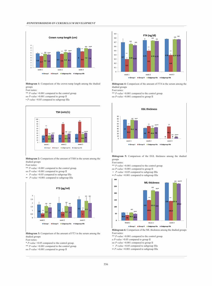
At all corresponding ages, Group II showed a significant decrease when compared to the control group (P>0.001). Subgroups IIIa and IIIb showed a significant increment in comparison with group II (P>0.001). No significant difference was detected between subgroup IIIa and the control group (P>0.05) but subgroup IIIb showed a significant decrease in comparing it with the control group and subgroup IIIa (P>0.05). The crown-rump length of the control group increased with age (Histogram 1).
Biochemical results
Serum TSH
At all corresponding ages, Group II showed a significant increment when compared to the control group (P>0.001). Subgroups IIIa and IIIb showed a significant decrease in comparison with group II (P>0.001). No significant difference was detected between subgroup IIIa and the control group (P>0.05) but subgroup IIIb showed a significant increment when compared to the control and subgroup IIIa in 1st, 2nd
and 3rd weeks (P>0.05, P<0.05, P<0.001 respectively) (Histogram 2).
548
HYPOTHYROIDISM ON CEREBELLUM DEVELOPMENT
Serum FT3At all corresponding ages, Group II showed a significant
decrease when compared to the control (P>0.001). Subgroup IIIa showed a significant increment in comparison with group II (P>0.001). Subgroup IIIb showed a significant increment in comparison with group II (P<0.001, P<0.001, P>0.05) in the 1st and 2nd weeks and the 3rd week respectively. Subgroups IIIa and IIIb showed no significant difference in comparison with the control (P>0.05). Also, there was no significant difference between subgroup IIIb and subgroup IIIa (P>0.05) (Histogram 3).
Serum FT4At all corresponding ages, Group II showed a significant
decrease in comparing it with the control (P>0.001). Subgroups IIIa and IIIb showed a significant increment in comparison with group II (P>0.001). On comparing subgroups IIIa, IIIb with the control, and with each other; no significant difference was detected (P>0.05) (Histogram 4).
Histological results
HX and E
At 1st weekH & E stained sections of a control rat showed that
the cerebellar cortex consisted of four layers: the external granular layer (EGL), the molecular layer (ML), the Purkinje cell layer (PCL) and the internal granular layer (IGL).
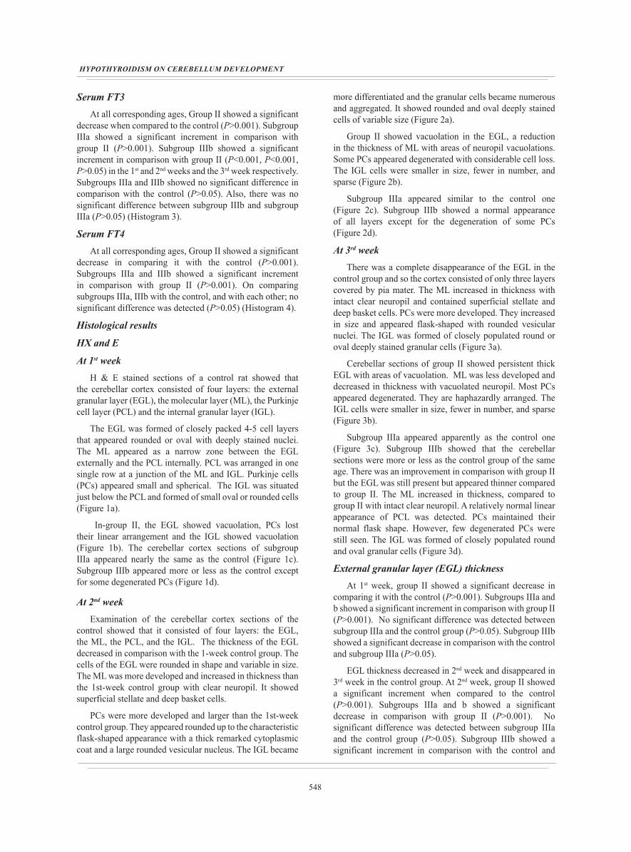
The EGL was formed of closely packed 4-5 cell layers that appeared rounded or oval with deeply stained nuclei. The ML appeared as a narrow zone between the EGL externally and the PCL internally. PCL was arranged in one single row at a junction of the ML and IGL. Purkinje cells (PCs) appeared small and spherical. The IGL was situated just below the PCL and formed of small oval or rounded cells (Figure 1a).
In-group II, the EGL showed vacuolation, PCs lost their linear arrangement and the IGL showed vacuolation (Figure 1b). The cerebellar cortex sections of subgroup IIIa appeared nearly the same as the control (Figure 1c). Subgroup IIIb appeared more or less as the control except for some degenerated PCs (Figure 1d).
At 2nd weekExamination of the cerebellar cortex sections of the
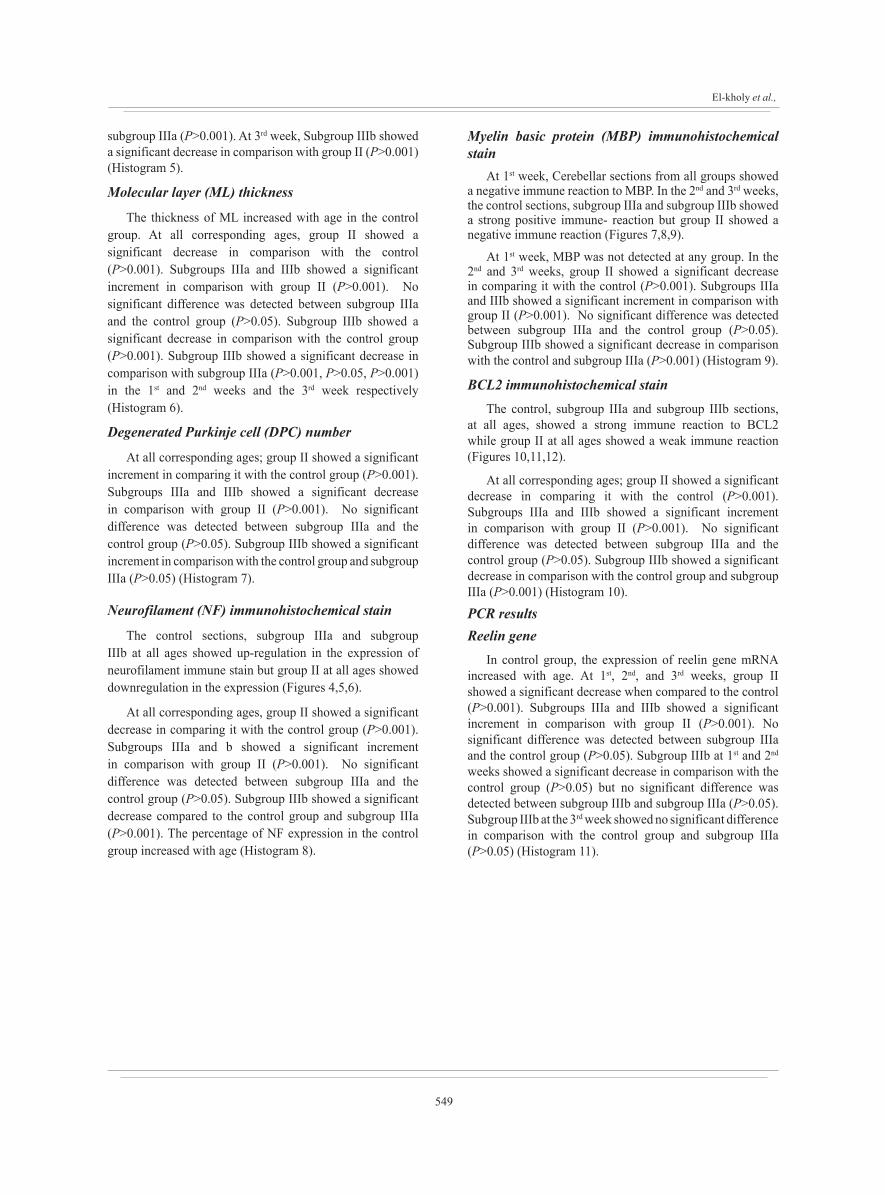
control showed that it consisted of four layers: the EGL, the ML, the PCL, and the IGL. The thickness of the EGL decreased in comparison with the 1-week control group. The cells of the EGL were rounded in shape and variable in size. The ML was more developed and increased in thickness than the 1st-week control group with clear neuropil. It showed superficial stellate and deep basket cells.
PCs were more developed and larger than the 1st-week control group. They appeared rounded up to the characteristic flask-shaped appearance with a thick remarked cytoplasmic coat and a large rounded vesicular nucleus. The IGL became
more differentiated and the granular cells became numerous and aggregated. It showed rounded and oval deeply stained cells of variable size (Figure 2a).
Group II showed vacuolation in the EGL, a reduction in the thickness of ML with areas of neuropil vacuolations. Some PCs appeared degenerated with considerable cell loss. The IGL cells were smaller in size, fewer in number, and sparse (Figure 2b).
Subgroup IIIa appeared similar to the control one (Figure 2c). Subgroup IIIb showed a normal appearance of all layers except for the degeneration of some PCs (Figure 2d).
At 3rd weekThere was a complete disappearance of the EGL in the
control group and so the cortex consisted of only three layers covered by pia mater. The ML increased in thickness with intact clear neuropil and contained superficial stellate and deep basket cells. PCs were more developed. They increased in size and appeared flask-shaped with rounded vesicular nuclei. The IGL was formed of closely populated round or oval deeply stained granular cells (Figure 3a).
Cerebellar sections of group II showed persistent thick EGL with areas of vacuolation. ML was less developed and decreased in thickness with vacuolated neuropil. Most PCs appeared degenerated. They are haphazardly arranged. The IGL cells were smaller in size, fewer in number, and sparse (Figure 3b).
Subgroup IIIa appeared apparently as the control one (Figure 3c). Subgroup IIIb showed that the cerebellar sections were more or less as the control group of the same age. There was an improvement in comparison with group II but the EGL was still present but appeared thinner compared to group II. The ML increased in thickness, compared to group II with intact clear neuropil. A relatively normal linear appearance of PCL was detected. PCs maintained their normal flask shape. However, few degenerated PCs were still seen. The IGL was formed of closely populated round and oval granular cells (Figure 3d).
External granular layer (EGL) thickness At 1st week, group II showed a significant decrease in
comparing it with the control (P>0.001). Subgroups IIIa and b showed a significant increment in comparison with group II (P>0.001). No significant difference was detected between subgroup IIIa and the control group (P>0.05). Subgroup IIIb showed a significant decrease in comparison with the control and subgroup IIIa (P>0.05).
EGL thickness decreased in 2nd week and disappeared in 3rd week in the control group. At 2nd week, group II showed a significant increment when compared to the control (P>0.001). Subgroups IIIa and b showed a significant decrease in comparison with group II (P>0.001). No significant difference was detected between subgroup IIIa and the control group (P>0.05). Subgroup IIIb showed a significant increment in comparison with the control and
549
El-kholy et al.,
subgroup IIIa (P>0.001). At 3rd week, Subgroup IIIb showed a significant decrease in comparison with group II (P>0.001) (Histogram 5).
Molecular layer (ML) thickness
The thickness of ML increased with age in the control group. At all corresponding ages, group II showed a significant decrease in comparison with the control (P>0.001). Subgroups IIIa and IIIb showed a significant increment in comparison with group II (P>0.001). No significant difference was detected between subgroup IIIa and the control group (P>0.05). Subgroup IIIb showed a significant decrease in comparison with the control group (P>0.001). Subgroup IIIb showed a significant decrease in comparison with subgroup IIIa (P>0.001, P>0.05, P>0.001) in the 1st and 2nd weeks and the 3rd week respectively (Histogram 6).
Degenerated Purkinje cell (DPC) number
At all corresponding ages; group II showed a significant increment in comparing it with the control group (P>0.001). Subgroups IIIa and IIIb showed a significant decrease in comparison with group II (P>0.001). No significant difference was detected between subgroup IIIa and the control group (P>0.05). Subgroup IIIb showed a significant increment in comparison with the control group and subgroup IIIa (P>0.05) (Histogram 7).
Neurofilament (NF) immunohistochemical stain
The control sections, subgroup IIIa and subgroup IIIb at all ages showed up-regulation in the expression of neurofilament immune stain but group II at all ages showed downregulation in the expression (Figures 4,5,6).
At all corresponding ages, group II showed a significant decrease in comparing it with the control group (P>0.001). Subgroups IIIa and b showed a significant increment in comparison with group II (P>0.001). No significant difference was detected between subgroup IIIa and the control group (P>0.05). Subgroup IIIb showed a significant decrease compared to the control group and subgroup IIIa (P>0.001). The percentage of NF expression in the control group increased with age (Histogram 8).
Myelin basic protein (MBP) immunohistochemical stain
At 1st week, Cerebellar sections from all groups showed a negative immune reaction to MBP. In the 2nd and 3rd weeks, the control sections, subgroup IIIa and subgroup IIIb showed a strong positive immune- reaction but group II showed a negative immune reaction (Figures 7,8,9).
At 1st week, MBP was not detected at any group. In the 2nd and 3rd weeks, group II showed a significant decrease in comparing it with the control (P>0.001). Subgroups IIIa and IIIb showed a significant increment in comparison with group II (P>0.001). No significant difference was detected between subgroup IIIa and the control group (P>0.05). Subgroup IIIb showed a significant decrease in comparison with the control and subgroup IIIa (P>0.001) (Histogram 9).
BCL2 immunohistochemical stainThe control, subgroup IIIa and subgroup IIIb sections,
at all ages, showed a strong immune reaction to BCL2 while group II at all ages showed a weak immune reaction (Figures 10,11,12).
At all corresponding ages; group II showed a significant decrease in comparing it with the control (P>0.001). Subgroups IIIa and IIIb showed a significant increment in comparison with group II (P>0.001). No significant difference was detected between subgroup IIIa and the control group (P>0.05). Subgroup IIIb showed a significant decrease in comparison with the control group and subgroup IIIa (P>0.001) (Histogram 10).
PCR resultsReelin gene
In control group, the expression of reelin gene mRNA increased with age. At 1st, 2nd, and 3rd weeks, group II showed a significant decrease when compared to the control (P>0.001). Subgroups IIIa and IIIb showed a significant increment in comparison with group II (P>0.001). No significant difference was detected between subgroup IIIa and the control group (P>0.05). Subgroup IIIb at 1st and 2nd
weeks showed a significant decrease in comparison with the control group (P>0.05) but no significant difference was detected between subgroup IIIb and subgroup IIIa (P>0.05). Subgroup IIIb at the 3rd week showed no significant difference in comparison with the control group and subgroup IIIa (P>0.05) (Histogram 11).
550
HYPOTHYROIDISM ON CEREBELLUM DEVELOPMENT
Fig. 1 : Photomicrographs of sagittal sections of the cerebellar cortex of rats aged 1 week showing normal EGL, ML, PCL and IGL in control and subgroup IIIa (a, c) respectively. The EGL is formed of 4-5 cell layers that appeared rounded or oval in shape with deeply stained nuclei. The ML appeared as a narrow zone between the EGL and the PCL. PCL shows PCs that are arranged in a single row. PCs appeared small rounded or spherical in shape. The IGL is formed of small oval or rounded granular cells. Group II shows vacuolation in EGL (asterisks), loss of the linear arrangement of PCs (arched arrows) and vacuolation in the IGL (arrows) (b). Subgroup IIIb appears more or less similar to that of the control except for some degenerated PCs (arched arrows) (d). (HX& E X 400)
Fig.2: Photomicrographs of sagittal sections of the cerebellar cortex of rats aged 2 weeks showing decreased thickness of EGL, increased thickness of ML with superficial stellate(s) and deep basket (B) cells, with intact clear neuropil (N). PCL showed increased size of PCs and appear flask shape with thick remarked cytoplasmic coat and large rounded vesicular nucleus. The IGL shows prominent, numerous and aggregated granular cells in control and subgroup IIIa (a, c) respectively. Group II shows vacuolation in the EGL (asterisks), reduction in size of ML (black up-down arrow) with areas of neuropil vacuolations (N). Some PCs are degenerated (arched arrows), with considerable cell loss (arrows). The IGL cells are fewer in number, smaller in size and sparse (b). Subgroup IIIb shows nearly normal appearance of all layers except for degeneration of some PCs (arched arrows) (d). (HX& E X 400)
551
El-kholy et al.,
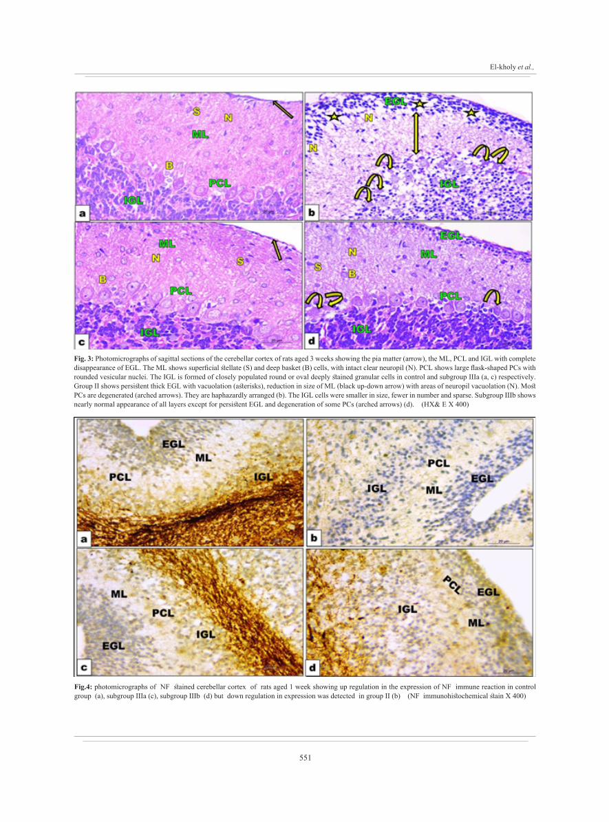
Fig. 3: Photomicrographs of sagittal sections of the cerebellar cortex of rats aged 3 weeks showing the pia matter (arrow), the ML, PCL and IGL with complete disappearance of EGL. The ML shows superficial stellate (S) and deep basket (B) cells, with intact clear neuropil (N). PCL shows large flask-shaped PCs with rounded vesicular nuclei. The IGL is formed of closely populated round or oval deeply stained granular cells in control and subgroup IIIa (a, c) respectively. Group II shows persistent thick EGL with vacuolation (asterisks), reduction in size of ML (black up-down arrow) with areas of neuropil vacuolation (N). Most PCs are degenerated (arched arrows). They are haphazardly arranged (b). The IGL cells were smaller in size, fewer in number and sparse. Subgroup IIIb shows nearly normal appearance of all layers except for persistent EGL and degeneration of some PCs (arched arrows) (d). (HX& E X 400)
Fig.4: photomicrographs of NF stained cerebellar cortex of rats aged 1 week showing up regulation in the expression of NF immune reaction in control group (a), subgroup IIIa (c), subgroup IIIb (d) but down regulation in expression was detected in group II (b) (NF immunohistochemical stain X 400)
552
HYPOTHYROIDISM ON CEREBELLUM DEVELOPMENT
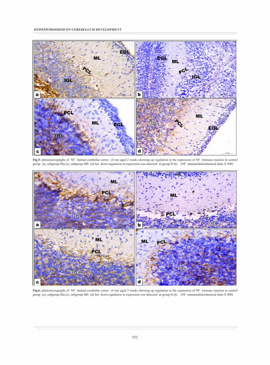
Fig.5: photomicrographs of NF stained cerebellar cortex of rats aged 2 weeks showing up regulation in the expression of NF immune reaction in control group (a), subgroup IIIa (c), subgroup IIIb (d) but down regulation in expression was detected in group II (b) (NF immunohistochemical stain X 400)
Fig.6: photomicrographs of NF stained cerebellar cortex of rats aged 3 weeks showing up regulation in the expression of NF immune reaction in control group (a), subgroup IIIa (c), subgroup IIIb (d) but down regulation in expression was detected in group II (b) (NF immunohistochemical stain X 400)

553
El-kholy et al.,
Fig. 7: photomicrographs of MBP stained cerebellar cortex of rats aged 1 week showing: a negative immune reaction in group I (a), group II (b), subgroup IIIa (c)and subgroup IIIb (d) (MBP immunohistochemical stain X 400)
Fig. 8: photomicrographs of MBP stained cerebellar cortex of rats aged 2 weeks showing a strong positive reaction in control group (a), subgroup IIIa (c) and subgroup IIIb (d ) but a negative immune reaction in group II (b) (MBP immunohistochemical stain X 400)

554
HYPOTHYROIDISM ON CEREBELLUM DEVELOPMENT
Fig. 9: photomicrographs of MBP stained cerebellar cortex of rats aged 3 weeks showing a strong positive reaction in control group (a), subgroup IIIa (c) and subgroup IIIb (d ) but a negative immune reaction in group II (b) (MBP immunohistochemical stain X 400)
Fig. 10: photomicrographs of BCL2 stained cerebellar cortex of rats aged 1 week showing: a strong immune reaction in group I (a), subgroup IIIa ( c) , subgroup IIIb ( d) but the reaction is weak in group II (b) (BCL2 immunohistochemical stain X 400)

555
El-kholy et al.,
Fig. 11: photomicrographs of BCL2 stained cerebellar cortex of rats aged 2 weeks showing: a strong immune reaction in group I (a), subgroup IIIa ( c) , subgroup IIIb ( d) but the reaction is weak in group II (b) (BCL2 immunohistochemical stain X 400)
Fig.12: photomicrographs of BCL2 stained cerebellar cortex of rats aged 3 weeks showing: a strong immune reaction in group I (a), subgroup IIIa ( c) , subgroup IIIb ( d) but the reaction is weak in group II (b) (BCL2 immunohistochemical stain X 400)
556
HYPOTHYROIDISM ON CEREBELLUM DEVELOPMENT
Histogram 1: Comparison of the crown-rump length among the studied groupsFoot notes:** P-value >0.001 compared to the control group.ᴏᴏ P-value >0.001 compared to group II • P-value >0.05 compared to subgroup IIIa
Histogram 2: Comparison of the amount of TSH in the serum among the studied groupsFoot notes:** P-value >0.001 compared to the control group.ᴏᴏ P-value >0.001 compared to group II • P-value >0.05 compared to subgroup IIIa •• P-value >0.001 compared to subgroup IIIa
Histogram 3: Comparison of the amount of FT3 in the serum among the studied groupsFoot notes:* P-value >0.05 compared to the control group.** P-value >0.001 compared to the control group.ᴏᴏ P-value >0.001 compared to group II
Histogram 4: Comparison of the amount of FT4 in the serum among the studied groups Foot notes: ** P-value >0.001 compared to the control group.ᴏᴏ P-value >0.001 compared to group II
Histogram 5: Comparison of the EGL thickness among the studied groups Foot notes: ** P-value >0.001 compared to the control group.ᴏᴏ P-value >0.001 compared to group II • P-value >0.05 compared to subgroup IIIa •• P-value >0.001 compared to subgroup IIIa
Histogram 6: Comparison of the ML thickness among the studied groups Foot notes:** P-value >0.001 compared to the control group.ᴏ P-value >0.05 compared to group II ᴏᴏ P-value >0.001 compared to group II • P-value >0.05 compared to subgroup IIIa •• P-value >0.001 compared to subgroup IIIa

557
El-kholy et al.,
Histogram 7: Comparison of number of DPC among the studied groupsFoot notes: ** P-value >0.001 compared to the control group.ᴏᴏ P-value >0.001 compared to group II • P-value >0.05 compared to subgroup IIIa
Histogram 8: Comparison of area percentage of NF among the studied groups Foot notes: ** P-value >0.001 compared to the control group.ᴏᴏ P-value >0.001 compared to group II •• P-value >0.001 compared to subgroup IIIa
Histogram 9: Comparison of area percentage of MBP among the studied groupsFoot notes:** P-value >0.001 compared to the control group.ᴏᴏ P-value >0.001 compared to group II •• P-value >0.001 compared to subgroup IIIa
Histogram 10: Comparison of area percentage of BCL2 among the studied groups Foot notes: ** P-value >0.001 compared to the control group.ᴏᴏ P-value >0.001 compared to group II •• P-value >0.001 compared to subgroup IIIa
Histogram 11: Comparison of the amount of reelin in the cerebellum among the studied groupsFoot notes: ** P-value >0.001 compared to the control group.ᴏᴏ P-value >0.001 compared to group II
DISCUSSION
The physiological functioning of the body systems is highly affected by intrauterine conditions in which the fetus develops. Insufficient maternal oxygen, hormones, and nutrients can change the developmental planning of fetal tissue growth[20]. Changes in TH levels affect the development of the cerebellum[7], and the consequences of hypothyroidism result in numerous alterations[21].
Carbimazole was the drug chosen for inducing hypothyroidism as it crosses the placental barrier, and is also excreted in the milk[6].
An extended methodology was done in this study including biochemical, histological, immunohistochemical, genetic, morphometric, and statistical methods to detect the effect of maternal hypothyroidism induction on the development of the cerebellar cortex in rat offspring postnatally. Also, this study compared the effect of thyroxin replacement to mothers and offspring.

558
HYPOTHYROIDISM ON CEREBELLUM DEVELOPMENT
The current study showed that the crown-rump length of control increased as the age progress, which was in line with a previous study[22]. Some authors reported that bone turnover increased during lactation in rats[23]. Besides, THs altered pituitary growth hormone secretion and effects[24].
Group II showed a decrease in the crown-rump in comparison with the control as previously reported by some authors[22].
Subgroup IIIa was similar to the control. Subgroup IIIb showed a significant increment in comparison with group II but a significant decrease when compared to the control. This might indicate that the dose used was not sufficient.
Concerning thyroid functions of control rat offspring, a gradual increase of serum FT3, FT4, and TSH levels as the age progress was detected in this study. A similar result was reported in another study, which mentioned that after the beginning of the secretion of fetal THs, fetal T3, and T4 increased steadily until the complete development of the thyroid gland[23].
In this study, exposure of pregnant female rats to carbimazole reduced serum levels of FT3 and FT4 in offspring while, serum TSH increased. This meant that hypothyroidism has occurred in offspring indicating that maternal thyroid state during pregnancy and lactation affected the thyroid status of rat offspring[25]. TSH, FT3, and FT4 in subgroup IIIa showed no significant difference in comparison with the control. In subgroup IIIb, FT3 and FT4 showed no significant difference in comparison with subgroup IIIa and the control but TSH levels showed a significant increment.
On the histological level, the disappearance of the EGL at the 3rd week in the control group was probably due to the migration of the cells from it towards the ML, PCL, and IGL. The EGL was the origin of the migrating cells that gave rise to many cellular elements of the cerebellar cortex[26].
In the hypothyroid group at 1st week, the cerebellar cortex showed vacuolation in the EGL, loss of linear arrangement of PCs, and vacuolation in IGL. At 2nd week, the cerebellar cortex showed vacuolation in the EGL. The ML showed a reduction in its thickness with areas of neuropil vacuolation. PCs showed degeneration with considerable cell loss. The IGL cells were smaller in size, fewer in number, and sparse.
Cerebellar sections at the 3rd week showed persistent thick EGL with areas of vacuolation and reduction of the size of the ML with areas of neuropil vacuolation. Most PCs appeared degenerated. They were haphazardly arranged. The IGL cells were smaller in size, fewer in number, and sparse.
In this study, the thickness of the EGL showed a significant decrease in comparison with the control in the 1st week as hypothyroidism decreased the proliferation of cells but a significant increment was detected at weeks 2 and 3 as hypothyroidism reduced the rate of migration of cells from the EGL.
Our previous results were consistent with some authors who reported that TH deficiency produced extensive cell loss, decreased proliferation, and delayed migration of cells from the external to the germinal layer[27,28]. Besides, It has been previously documented that hypothyroidism during the fetal period resulted in several histological changes in the neonatal rat brain including PC degeneration and more intercellular spaces in IGL[25].
Subgroups IIIa appeared nearly as the control group because TH levels had reached the control one. It was documented that the correction of gestational hypothyroxinemia at the early stages with T4 prevented the impairment of neurodevelopment in the offspring[29]. Also, some authors[30] observed that early maternal treatment with T4 improved cell migration in the developing brain.
In subgroup IIIb, administration of thyroxin to neonates produced nearly normal levels of FT3 and FT4 but TSH remained high. Besides, an improvement was observed in the histological results that appeared nearly similar to that of the control group except for the degeneration of some PCs. Also, the EGL was still present in the 3rd week but showed a significant decrease when compared to group II. This improvement might be due to the rapid replacement by T4 to neonates immediately after birth. This was in line with the results of some authors[28] who mentioned that abnormal development of the cerebellum could be rescued if TH is replaced within the first 2 weeks following birth in rodents. Also, T4 replacement initiated at birth partially restored the development of the cerebellum[15].
THs are important for the normal development of the cytoskeletal system that includes intermediate filaments, specific for neurons (Neurofilaments)[31].
Regarding neurofilament stain results, this study showed a strong immune reaction in control that increased with age in line with a study, which stated that NF expression increased gradually as the brain, develops[32]. A negative reaction was detected in group II. This was supported by other authors[33]. Subgroup IIIa showed a strong positive immune reaction to NF. Subgroup IIIb showed a significant increment in expression when compared to group II but a significant decrease in comparison with the control and subgroup IIIa. TH exerted a positive effect on NF expression and transport involved in axonal regeneration[34].
Our study revealed negative reactions to MBP in the 1st week. However, the expression of MBP started in the 2nd week and increased in the 3rd week. It was previously documented that myelination in rats began at around postnatal day10 and reached its peak around postnatal day 20[35].
In this study, group II showed a weak positive immune reaction to MBP. It was reported that the differentiation of oligodendrocytes, the glial cells that perform myelination of the axons in the CNS, is strongly TH-dependent. TH deficiency decreased major myelin proteins including MBP expression and delayed myelination[36]. Also, a reduction in myelination in the hypothyroid neonatal brain was previously detected[37].

559
El-kholy et al.,
Subgroups IIIa showed improvement of myelination due to return of TH levels toward normal but subgroup IIIb showed a significant decrease in expression when compared to the control and subgroup IIIa. TH promotes CNS repair through CNS re-myelination and genomic and non-genomic effects on mitochondrial biogenesis and function[21].
Regarding BCL2 immune stain results, the control group of this work showed a strong reaction while group II showed a negative reaction. This study suggested apoptosis as a mechanism of cerebellum damage in neonates born to hypothyroid mothers. These results were consistent with some studies, which demonstrated that TH deficiency during cerebellar development enhanced DNA fragmentation, down-regulated the expression of antiapoptotic genes; Bcl-2 and Bcl-xL, and increased the pro-apoptotic molecules leading to increased apoptosis[27].
Subgroups IIIa showed a strong immune reaction to Bcl-2 immune stain as TH returned to normal levels and so decreasing apoptosis. Also, subgroups IIIb showed a strong immune reaction due to the rapid replacement of TH after birth that helped increasing the levels of THs in this group.
Reelin is a secretory protein essential for embryogenesis and also during adult life. The Reelin gene is regulated by TH. It is essential for cell migration and neural plasticity during neural development[38].
In this study, reelin gene expression was age-dependent among the control rat but was lower in group II meaning that hypothyroidism reduced reelin gene expression. It was reported that in the cerebellum, reelin expression was down-regulated in perinatally hypothyroid rat[32]. Our study showed an increased number of degenerated Purkinje cells correlating with a reduction in reelin gene expression and this was supported by other authors[39].
TH replacement in subgroup IIIa returned reelin gene expression near normal values while subgroup IIIb showed a significant increment in reelin gene expression when compared to group II. The up-regulation of reelin expression in the replacement group may suggest compensation for retarded development.
Developmental hypothyroidism is known to result in permanent disorders in cerebellar development. Based on the results presented, our study detected that maternal hypothyroidism during pregnancy and lactation evoked developmental defects in the cerebellar cortex of their offspring. The underlying molecular mechanisms were largely unknown. Reelin is an important molecule affecting neuronal migration and development of the cerebellum Thus, our study might confirm an important complement to the existing literature regarding molecular mechanisms involved in impairments of cerebellar development.
CONCLUSION
Maternal hypothyroidism during pregnancy and lactation impaired the maturation and development of the cerebellar cortex in albino rat offspring. Hypothyroidism induced its
neurotoxic effect on the cerebellar cortex through enhancing apoptosis, demyelination, and alteration of NF contents and Reelin gene expression. Thyroxin replacement to mothers was more effective than the treatment of offspring.
RECOMMENDATIONS
The results of the present study trigger an interest in routine screening for hypothyroid mothers during pregnancy and their early treatment to ensure adequate cerebellar cortex development.
ACKNOWLEDGMENT
The authors are highly thankful to the Department of Anatomy and Embryology, Faculty of Medicine, Menoufia University.
ABBREVIATIONS
ANOVA: analysis of variance; BCL2: B-cell lymphoma 2; CNS: central nervous system; EGL: external granular layer; FT3: free triiodothyronine, FT4: Free thyroxin, Hx & E: Haematoxylin and eosin; IGL: internal granular layer; MBP: Myelin basic protein; ML: molecular layer; NF: Neurofilament; PCL: Purkinje cell layer; PCR: polymerase chain reaction; TH: thyroid hormone; TSH: thyroid-stimulating hormone.
CONFLICT OF INTERESTS
There are no Conflicts of Interest.
REFERENCES
1. Youssef S, Abd-El- Aty1 OA and Morgan EN: S100 Expression in Astrocytes of the Developing Cerebella of the Offspring Rats of Hyperthyroidism Mothers (Biochemical, Histological and Immunohistochemical Studies) in Swift Journal of Medicine and Medical Sciences. (2016) 2 (3): 25-38.
2. Fabrizio M: Classification of thyroid diseases: suggestions for a revision in J Clin Endocrinol Metab. (2003) 88: 1428–1432.
3. Dirar AM and Kalhan A: Hypothyroidism during pregnancy: Controversy over screening and intervention in World J Obstet Gynecol. (2018) 7(1):1-16
4. Nandi-Munshi D and Taplin CE: Thyroid-Related Neurological Disorders and Complications in Children in Pediatr Neurol. (2015) 52 (4):373-382
5. Humar M, Dohrmann H, Stein P, Andriopoulos N, Goebel U, Roesslein M, Schmidt R, Schwer C I, Loop T, Geiger K K, Pahl H L and Pannen BH: Thionamides inhibit the transcription factor nuclear factor-kappaB by suppression of Rac1 and inhibitor of kappaB kinase alpha in J Pharmacol Exp Ther. (2008) 324 (3):1037-1044.
6. Shehata MR, Mohamed DA, El-Meligy MM and Bastwrous AE: The effect of maternal hypothyroidism on the postnatal development of the pituitary–thyroid axis in albino rats: a histological, morphometric, and immunohistochemical study in J Curr Med Res Pract. (2017) 2: 79–97.
560
HYPOTHYROIDISM ON CEREBELLUM DEVELOPMENT
7. Faustino LS and Ortiga-Carvalho TM: Thyroid hormone role on cerebellar development and maintenance: a perspective based on transgenic mouse models in Frontiers in Endocrinology. (2014) 5 (75): 1-8.
8. King TL and Brucker MC: Pharmacology for Women's Health. Jones & Bartlett Publishers. (2010) Pp: 544.
9. Roberts CG and Ladenson PW: Hypothyroidism in Lancet. (2004) 363 (9411): 793–803.
10. Hirota Y, Nakajima K: Neuronal Migration and Aggregation by Reelin Signaling in the Developing Cerebral Cortex in Front Cell Dev Biol. (2017) 5(40):1-8.
11. Hevner RF: Reelin and the Cerebellum. Springer Science+ Business Media, LLC, 233 Spring Street, New York, NY 10013, USA. (2008) P. 141-58.
12. Zaidi TM, Khan AA, Hasan BM and Faruqi AN: Carbimazole Induced Thyroid Histopathy in Albino Rats during Development in J. Anat. Soc. India. (2004) 53 (2): 14-17.
13. Ahmed OM, Abd El-Tawab S M, and Ahmed R.G: Effects of experimentally induced maternal hypothyroidism and hyperthyroidism on the development of rat offspring: I. The development of the thyroid hormones-neurotransmitters and adenosinergic system interactions in Int J Dev Neurosci. (2010) 28(6): 437-54.
14. Hua X, Cao X, Wang X, Sun P and Chen L: Exposure of Pregnant Mice to Triclosan Causes Insulin Resistance via Thyroxine Reduction in Toxicological Sciences. (2017) 160 (1): 150–160.
15. Li G, Post J, Koibuchi N and Sajdel-Sulkowska EM: Impact of thyroid hormone deficiency on the developing CNS: cerebellar glial and neuronal protein expression in rat neonates exposed to antithyroid drug propylthiouracil in The Cerebellum. (2004) 3: 100–106.
16. Bancroft JD and Gamble A. Theory and practice of histological techniques. 6th edition, Churchill Livingstone, New York, London. (2008) pp: 165-175.
17. Bancroft J and Gamble M. Immunohistochemical techniques in: Theory and practice of histological techniques. 6th ed. Chapter 21. Elsevier Health Sciences. Churchill Livingstone. (2008) pp: 433.
18. Dorak M. Real time PCR. Clinical Chemistry. Taylor & Francis Group, 270 Madison Avenue New York, N Y 10016. (2004) 50: 1680-82.
19. Saunders PM and Roberts GT: Basic of clinical biostatistics. 2nd ed., Lang medical book. (1994) Pp: 220-229.
20. Sedaghat K, Zahedias S and Ghasemi A: Gestational hypothyroidism-induced changes in L-type calcium channels of rat aorta smooth muscle and their impact on the responses to vasoconstrictors in Iran J Basic Med Sci. (2015) 18: 172-179.
21. Mohamed DA and Ahmed SM: Donepezil Improves Histological and Biochemical Changes in the Hippocampus of Adult Hypothyroid Male Rats in EJH. (2018) 41(4): 445-458.
22. Al-Gholam MA, Nooh H Z, El-Mehi AE, El-Barbary AA and Zo El Fokar AS: Protective effect of rosemary on acrylamide motor neurotoxicity in spinal cord of rat offspring: postnatal follow-up study in Anat Cell Biol. (2016) 49:34-49.
23. El-Bakry AM, El-Gareib AW and Ahmed RG: Comparative study of the effects of experimentally induced hypothyroidism and hyperthyroidism in some brain regions in albino rats in Int J Dev Neurosci. (2010) 28 (5): 371–389.
24. Saranac L, Stamenkovic H, Stankovic T, Markovic I, Zivanovic S and Djuric Z: Growth in children with thyroid dysfunction. The Current Topics in Hypothyroidism with Focus on Development. (2013):119-134.
25. El-Ghareeb A, El-Bakry A, Ahmed RG and Asmaa G: Effects of Zinc Supplementation in Neonatal Hypothyroidism and Cerebellar Distortion Induced by Maternal Carbimazole in Asian Journal of Applied Sciences. (2016) 4 (4):1030-1040.
26. El-Shall E B: Effect of oxcarbazepine (Tripleptal on the cerebellar cortex of the albino rat. M.D. Thesis, Anatomy dept. Faculty of Medicine for girls, Al- Azhar University. Egypt (2004).
27. Singh R, Upadhyay G, Kumar S, Kapoor A, Kumar A, Tiwari M and Godbole MM: Hypothyroidism alters the expression of Bcl-2 family genes to induce enhanced apoptosis in the developing cerebellum in Journal of Endocrinology. (2003) 176: 39–46.
28. Koibuchi N and Ikeda Y: Hormones and cerebellar development. In: Manto M, Gruol D, Schmahmann JD, Koibuchi N, editors., Handbook of the Cerebellum and Cerebellar Disorders. New York, NY: Springer. (2013) pp: 319–339.
29. Ausó E, Lavado-Autric R, Cuevas E. A moderate and transient deficiency of maternal thyroid function at the beginning of fetal neocorticogenesis alters neuronal migration. Endocrinology. (2004) 145: 4037-47.
30. Lu L, Yu X, Teng W, Shan Z. Treatment with levothyroxine in pregnant rats with subclinical hypothyroidism improves cell migration in the developing brain of the progeny. J Endocrinol Invest. (2012) 35: 490–96.
31. Ahmed RG: Perinatal Hypothyroidism and Cytoskeleton Dysfunction in Endocrinol Metab Syndr. (2017) 6(4): 1-3.
32. White JJ, Sillitoe RV. Postnatal development of cerebellar zones revealed by neurofilament heavy chain protein expression, Front Neuroanat. (2013) 7 (9):1-10.
561
El-kholy et al.,
33. Leclerc, N Gravel C, Plioplys A and Hawkes R: Basket cell development in the normal and hypothyroid rat cerebellar cortex studied with a monoclonal anti-neurofilament antibody in Can. J. Bioc-hem. Cell Biol. (1985) 63: 564-576.
34. Schenker M, Riederer BM, Kuntzer T, Barakat-Walter I. Thyroid hormones stimulate expression and modification of cytoskeletal protein during rat sciatic nerve regeneration. Brain Res. (2002). 957(2):259-70.
35. Downes N and Mullins P: The Development of Myelin in the Brain of the Juvenile Rat in Toxicologic Pathology. (2014) 42(5): 913-922.
36. Moog NK, Entringera S, Heima C, Wadhwab PD, Kathmannf N and Bussa C: Influence of maternal thyroid hormones during gestation on fetal brain
development in Neuroscience. (2017) 7(342): 68–100.
37. Howdeshell KL: A Model of the Development of the Brain as a Construct of the Thyroid System Environmental Health Perspectives. (2002) 110 (3): 337-342.
38. Suia L and Li B: Effects of perinatal hypothyroidism on regulation of reelin and brain-derived neurotrophic factor gene expression in rat hippocampus: Role of DNA methylation and histone acetylation in Steroids. (2010) 75: 988–997.
39. Maloku E, Covelo IR, Hanbauer I, Guidotti A, Kadriu B, Hu Q, Davis JM and Costa E: Lower number of cerebellar Purkinje neurons in psychosis is associated with reduced reelin expression in Neuroscience. (2010) 107 (9): 4407–4411.
562
HYPOTHYROIDISM ON CEREBELLUM DEVELOPMENT
الملخص العربى
تأثير قصور نشاط الغدة الدرقية الأمومى المستحث على نمو قشرة مخيخ مواليد الجرذان البيضاء ودور استبدال الثيروكسين
دراسة نسيجية وهيستوكيميائية مناعية ووراثيةوائل بدر الخولى، مروه عبد الرسول عمر، مصطفى محمود الحبيبى، مروه عبد الصمد الغلام
قسم التشريح وعلى الأجنة - كلية الطب - جامعة المنوفية - مصر
مقدمة: تعتبر هرمونات الغدة الدرقية الأمومية ضرورية للنمو الطبيعي للجهاز العصبي المركزي قبل الولادة حيث أن نقص هذه الهرمونات يمكن أن يؤخر من تطور المخيخ.
الهدف: يهدف هذا البحث إلى دراسة تأثير قصور نشاط الغدة الدرقية الأمومي المستحث على تطور ما بعد الولادة في قشرة مخيخ مواليد الجرذان البيضاء والمقارنة بين استبدال الثيروكسين للأمهات وللمواليد بعد الولادة .
المواد والطرق: لقد تم تقسيم المواليد إلى ثلاث مجموعات، المجموعة الأولى (الضابطه)، المجموعة الثانية قصور الغدة الدرقية) المكونة من 15 جرذ والتى تم بها اعطاء الأم الكاربيمازول (20 ميللى جرام/ كجم بالفم) من اليوم الأول للحمل وحتى اليوم الحادى والعشرين من الرضاعة، المجموعة الثالثة استبدال الثيروكسين التى تم تقسيمها الى المجموعه الفرعية الأولى التى تتكون من 15 جرذ تم اعطاء الأم الكاربيمازول كما فى المجموعة الثانية بالاضافه الى اعطائها من والعشرين الحادي اليوم إلى للحمل العاشر اليوم من الجلد) تحت كجم ميكرو جرام/ 20) الليفوثيروكسين مادة الرضاعة، المجموعه الفرعية الثانية التى تتكون من 15 جرذ والتى تم بها اعطاء الأم الكاربيمازول كما فى المجموعة
الثانية وتم اعطاء المواليد الثيروكسين (20 ميكرو جرام/ كجم تحت الجلد) منذ اليوم الأول بعد الولادة.الدم، في الدرقية لللغدة المحفز الهرمون مستوى قياس تم الولادة، بعد والثالثة والثانية الأولى الأسابيع نهاية وفي والثيروكسين الثلاثى الحر والرباعى الحر كما تم أخذ عينات من القشرة المخيخية وقد جهزت للدراسة الهستولوجية وصبغت بالهيماتوكسلين والأيوسين، وللدراسة الهستوكيميائية المناعية باستخدام صبغه هستوكيميائية للخيوط العصبيه،
وللبروتين الأساسي للميلين، صبغة بي سي ال 2 كما تم إجراء تفاعل سلسلة البلمرة لجين الريلين.الحر الحر والرباعى الثلاثى الثيروكسين ذا دلالة احصائية في هرمون انخفاضًا الثانية المجموعة النتائج: أظهرت وزيادة فى الهرمون المحفز للغدة الدرقية كما لوحظ وجود تجاويف في الطبقة الحبيبية الخارجية وتأخر فى اختفائها وتدهور فى خلايا بركنجى والتى زادت مع تقدم العمر كما تم الكشف أيضًا عن انخفاض فى الميالين، ومحتوى الخيوط العصبية، وتعبير جين الريلين في النسل ولقد أظهر العلاج البديل فى المجموعة الثالثة وخاصة للأمهات فى المجموعة
الفرعية الأولى تحسن فى هذه التغيرات.الخلاصة: يسبب قصور الغدة الدرقية لدى الأمهات خلل في تطور قشرة مخيخ المواليد ولقد كان استبدال الثيروكسين للأمهات أكثر فعالية من علاج المواليد لذلك، يعد علاج الأمهات المصابات بقصور الغدة الدرقية أثناء الحمل ضرورياً
لضمان التطور الأمثل لقشرة المخيخ.
Related Documents