BioMed Central Page 1 of 8 (page number not for citation purposes) Reproductive Biology and Endocrinology Open Access Research The effect of follicular fluid hormones on oocyte recovery after ovarian stimulation: FSH level predicts oocyte recovery Mitchell P Rosen*, A Musa Zamah, Shehua Shen, Anthony T Dobson, Charles E McCulloch, Paolo F Rinaudo, Julie D Lamb and Marcelle I Cedars Address: UCSF Medical Center, Department of Obstetrics, Gynecology and Reproductive Sciences, 2356 Sutter Street, 7th Floor, San Francisco, CA 94115, USA Email: Mitchell P Rosen* - [email protected]; A Musa Zamah - [email protected]; Shehua Shen - [email protected]; Anthony T Dobson - [email protected]; Charles E McCulloch - [email protected]; Paolo F Rinaudo - [email protected]; Julie D Lamb - [email protected]; Marcelle I Cedars - [email protected] * Corresponding author Abstract Background: Ovarian stimulation for assisted reproductive technology (ART) overcomes the physiologic process to develop a single dominant follicle. However, following stimulation, egg recovery rates are not 100%. The objective of this study is to determine if the follicular fluid hormonal environment is associated with oocyte recovery. Methods: This is a prospective study involving patients undergoing ART by standard ovarian stimulation protocols at an urban academic medical center. A total of 143 follicular fluid aspirates were collected from 80 patients. Concentrations of FSH, hCG, estradiol, progesterone, testosterone and prolactin were determined. A multivariable regression analysis was used to investigate the relationship between the follicular fluid hormones and oocyte recovery. Results: Intrafollicular FSH was significantly associated with oocyte recovery after adjustment for hCG (Adjusted odds ratio (AOR) = 1.21, 95%CI 1.03–1.42). The hCG concentration alone, in the range tested, did not impact the odds of oocyte recovery (AOR = 0.99, 95%CI 0.93–1.07). Estradiol was significantly associated with oocyte recovery (AOR = 0.98, 95% CI 0.96–0.99). After adjustment for progesterone, the strength of association between FSH and oocyte recovery increased (AOR = 1.84, 95%CI 1.45–2.34). Conclusion: The relationship between FSH and oocyte recovery is significant and appears to work through mechanisms independent of the sex hormones. FSH may be important for the physiologic event of separation of the cumulus-oocyte complex from the follicle wall, thereby influencing oocyte recovery. Current methods for inducing the final stages of oocyte maturation, with hCG administration alone, may not be optimal. Modifications of treatment protocols utilizing additional FSH may enhance oocyte recovery. Published: 23 April 2009 Reproductive Biology and Endocrinology 2009, 7:35 doi:10.1186/1477-7827-7-35 Received: 24 November 2008 Accepted: 23 April 2009 This article is available from: http://www.rbej.com/content/7/1/35 © 2009 Rosen et al; licensee BioMed Central Ltd. This is an Open Access article distributed under the terms of the Creative Commons Attribution License (http://creativecommons.org/licenses/by/2.0 ), which permits unrestricted use, distribution, and reproduction in any medium, provided the original work is properly cited.

Welcome message from author
This document is posted to help you gain knowledge. Please leave a comment to let me know what you think about it! Share it to your friends and learn new things together.
Transcript
BioMed Central
Reproductive Biology and Endocrinology
ss
Open AcceResearchThe effect of follicular fluid hormones on oocyte recovery after ovarian stimulation: FSH level predicts oocyte recoveryMitchell P Rosen*, A Musa Zamah, Shehua Shen, Anthony T Dobson, Charles E McCulloch, Paolo F Rinaudo, Julie D Lamb and Marcelle I CedarsAddress: UCSF Medical Center, Department of Obstetrics, Gynecology and Reproductive Sciences, 2356 Sutter Street, 7th Floor, San Francisco, CA 94115, USA
Email: Mitchell P Rosen* - [email protected]; A Musa Zamah - [email protected]; Shehua Shen - [email protected]; Anthony T Dobson - [email protected]; Charles E McCulloch - [email protected]; Paolo F Rinaudo - [email protected]; Julie D Lamb - [email protected]; Marcelle I Cedars - [email protected]
* Corresponding author
AbstractBackground: Ovarian stimulation for assisted reproductive technology (ART) overcomes thephysiologic process to develop a single dominant follicle. However, following stimulation, eggrecovery rates are not 100%. The objective of this study is to determine if the follicular fluidhormonal environment is associated with oocyte recovery.
Methods: This is a prospective study involving patients undergoing ART by standard ovarianstimulation protocols at an urban academic medical center. A total of 143 follicular fluid aspirateswere collected from 80 patients. Concentrations of FSH, hCG, estradiol, progesterone,testosterone and prolactin were determined. A multivariable regression analysis was used toinvestigate the relationship between the follicular fluid hormones and oocyte recovery.
Results: Intrafollicular FSH was significantly associated with oocyte recovery after adjustment forhCG (Adjusted odds ratio (AOR) = 1.21, 95%CI 1.03–1.42). The hCG concentration alone, in therange tested, did not impact the odds of oocyte recovery (AOR = 0.99, 95%CI 0.93–1.07). Estradiolwas significantly associated with oocyte recovery (AOR = 0.98, 95% CI 0.96–0.99). Afteradjustment for progesterone, the strength of association between FSH and oocyte recoveryincreased (AOR = 1.84, 95%CI 1.45–2.34).
Conclusion: The relationship between FSH and oocyte recovery is significant and appears to workthrough mechanisms independent of the sex hormones. FSH may be important for the physiologicevent of separation of the cumulus-oocyte complex from the follicle wall, thereby influencingoocyte recovery. Current methods for inducing the final stages of oocyte maturation, with hCGadministration alone, may not be optimal. Modifications of treatment protocols utilizing additionalFSH may enhance oocyte recovery.
Published: 23 April 2009
Reproductive Biology and Endocrinology 2009, 7:35 doi:10.1186/1477-7827-7-35
Received: 24 November 2008Accepted: 23 April 2009
This article is available from: http://www.rbej.com/content/7/1/35
© 2009 Rosen et al; licensee BioMed Central Ltd. This is an Open Access article distributed under the terms of the Creative Commons Attribution License (http://creativecommons.org/licenses/by/2.0), which permits unrestricted use, distribution, and reproduction in any medium, provided the original work is properly cited.
Page 1 of 8(page number not for citation purposes)
Reproductive Biology and Endocrinology 2009, 7:35 http://www.rbej.com/content/7/1/35
BackgroundIn order to maximize in vitro fertilization (IVF) success,ovarian stimulation is performed to generate multiplemature oocytes. This process overrides the physiologicmechanisms that normally select a single dominant folli-cle. Ovarian stimulation most typically describes the useof exogenous gonadotropins to rescue smaller antral folli-cles and stimulate the growth of multiple follicles simul-taneously. This process yields numerous follicles ofvarying size with variable rates of oocyte recovery [1].
The regulation of selection, growth, and ovulation of thedominant follicle is a complex process that involves folli-cle stimulating hormone (FSH), luteinizing hormone(LH) and modulation by an intra-ovarian network of fac-tors [2]. Surges in both FSH and LH precede spontaneousovulation, characterized by detachment of the cumulus-oocyte complex from the follicular wall and subsequentexpulsion from the ovary [3,4]. It is well accepted that LHis obligatory for oocyte nuclear maturation and has a fun-damental role in ovulation. However, pure FSH has beenshown to stimulate plasminogen activator within thegranulosa cells and induce ovulation in hypophysect-omized rats [5,6]. Plasminogen activator converts plas-minogen to the active protease plasmin, which ispresumably involved in dissociating the oocyte from thefollicular wall and weakening the wall to facilitate rupture[7,8].
Several studies have suggested follicular diameter corre-lates with oocyte recovery. We hypothesize oocyte recov-ery also has a biological basis, which is dependent uponthe follicular fluid hormonal milieu. Specifically, wehypothesize that FSH promotes oocyte recovery by stimu-lating follicle growth while simultaneously initiating fol-licular hormone production. Then, FSH facilitates thepartitioning of the oocyte from the follicular wall eitherdirectly or indirectly. Administration of human chorionicgonadotropin (hCG) results in luteinization of the granu-losa cells, affecting hormone production and directly orindirectly triggering the detachment of the cumulus-oocyte complex from the follicular wall. The aim of thisstudy was to determine whether any of the intrafollicularhormones we examined would be correlated with oocyterecovery and thereby presumably be important for the stillpoorly understood process of cumulus-oocyte complexdetachment.
MethodsStudy populationFrom April 2005 to Sept 2005, 80 patients undergoingART by standard ovarian stimulation protocols wererecruited to collect follicular fluid. Three stimulation pro-tocols were utilized; down regulated (n = 62), microdoseflare (n = 10), and antagonist (n = 8). Each patient had 1
or 2 individual follicle aspirates collected at the time ofretrieval. A total of 143 follicular fluid aspirates wereobtained. One hundred and eleven follicular fluid sam-ples were used to measure hormones. The remaining 32samples were excluded due to blood contamination orexcessive dehydration. This study was approved by theinstitutional review board at University of California, SanFrancisco.
Follicular size measurement and egg retrievalIndividuals were monitored with transvaginal ultrasound(Shimadzu SDU-450XL, Kyoto, Japan) and follicles weremeasured to obtain a two-dimensional mean diameter.HCG was administrated once two follicles measured 18mm in mean diameter. A transvaginal ultrasound guidedfollicular aspiration was conducted 36 hours post hCGadministration. Individual follicles of varying size wererandomly selected and measured before aspiration. Thefollicles were categorized into 5 groups according to follic-ular size at the time of retrieval (the mean of the two-dimension measurements: A, ≥ 18 mm; B, 16–18 mm; C,13–15 mm; D, 10–12 mm; E, <10 mm).
Prior to aspiration, the collection system (needle and tub-ing) was flushed to allow for direct correlation of follicu-lar size and fluid volume. The first follicle from either oneor both ovaries was aspirated and collected. Each folliclewas pierced with a single lumen needle and completelyaspirated. The tubing was then flushed and the contentscollected in a separate tube to ensure capture of the corre-sponding oocyte if it was retrieved from the follicle. Inorder to minimize technical differences, all egg retrievalswere performed by one of two physicians. In each patienta maximum of two follicles were aspirated, one from eachovary. Follicles were chosen for our study based on theirrelative location within the ovary.
For each follicle, the presence or absence of an egg wasrecorded immediately and the follicular fluid was placedinto a 15 ml sterile Falcon conical tube. The follicular sizewas corrected to the volume of follicular fluid, with a var-iation between the two methods of measurement averag-ing 14.6%. The volumes corresponding to each folliclesize (<10 mm, <0.5 mL; 10–12 mm, 0.6–1 mL; 13–15mm, 1.2–2 mL; 16–18 mm, 2.1–3 mL, and >18 mm, >3.1mL) were similar to those previously described by Witt-maack et al [1]. If the follicular fluid contained blood orwas notably dehydrated, the sample was excluded fromthe study. The follicular fluid was cleared by centrifuga-tion at room temperature for 10 minutes at 1500 × g, aliq-uoted into 2 ml cryovials, and placed at -80 degreesCelsius for later analysis.
Page 2 of 8(page number not for citation purposes)
Reproductive Biology and Endocrinology 2009, 7:35 http://www.rbej.com/content/7/1/35
Follicular fluid hormonesThe following hormone concentrations were quantified induplicate, and measured with commercially availableautomated chemiluminescent immunoassays on theDPC-Immulite 2000 (Diagnostic Products, Los Angeles,CA): estradiol (E2), progesterone (P), total testosterone(T), prolactin (PRL), FSH and hCG. Prior to running sam-ples, follicular fluid assays were validated by dilution test-ing and confirmation of linearity. These hormones wereselected, guided by published literature, because of eithertheir possible or established association with folliculardevelopment or to each other. Prior to each test theImmulite 2000 was calibrated with 3 controls of low,medium, and high concentrations of the appropriate hor-mone. Dilutions were performed prior to measurement ofE2 (1:1000) and P (1:500) depending on the calibrationrange. The intraassay coefficient of variations were: E215%, P 16%, T 13%, PRL 6.8%, and FSH 2.6%. The inte-rassay coefficient of variations were: E2 16%, P 16%, T16.4%, PRL 9.6%, and FSH 5.8%.
Statistical analysisStudent t-tests were performed on baseline characteristics,and stimulation parameters to compare follicles withoocytes recovered versus not recovered. Repeated meas-ures ANOVA was performed to compare levels of FSH inrelation to oocyte recovery. Logistic regression analyseswere used to determine if AFC or any of the hormonestested were associated with oocyte recovery after adjustingfor follicle size. Then, multivariate models were con-structed to determine the independent effects of FSH andhCG by adding one hormone at a time back to the modelto assess whether the effect of FSH on oocyte recovery wasmodified. A final model was constructed adding all hor-mones that achieved significance to determine the directeffect of FSH on oocyte recovery. Repeated measures lin-ear regression was used to identify the associationbetween follicular fluid gonadotropin and sex steroid con-centrations. Both estrogen/progesterone and estrogen/tes-tosterone ratios were also considered as hormonevariables. Effect modification of hormones was assessedwith an adjustment for follicle size. All analyses were per-
formed using Stata version 9.0 (Stata Corporation, Col-lege Station, TX). Tests were declared statisticallysignificant for a two-sided p-value <0.05.
ResultsThe characteristics of patients recruited into the study andcorresponding stimulation parameters are shown in Table1. The positive oocyte recovery group included patientswhere each follicle aspirated contained an egg. If one eggwas not recovered from each aspirated follicle they wereincluded in the negative oocyte recovery group. Analysesto determine whether these parameters were associatedwith oocyte recovery revealed that the total antral folliclecount was inversely associated with the odds of oocytecapture (Adjusted odds ratio (AOR) = 0.94, P = 0.016,95%CI 0.89–0.98). The infertility diagnoses are shown inTable 2. In comparison to an unexplained diagnosis, thereis no difference in oocyte recovery with different infertilitydiagnoses. The average levels of intrafollicular FSH andoocyte recovery separated by stimulation protocol aredepicted in Table 3.
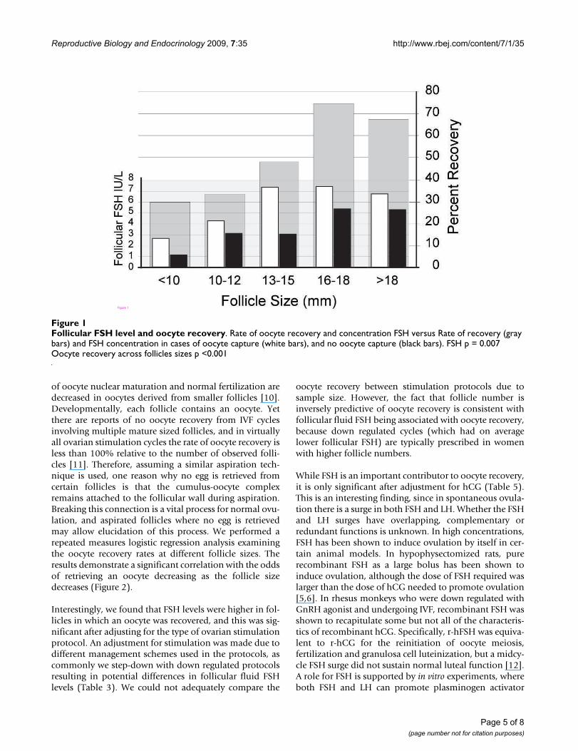
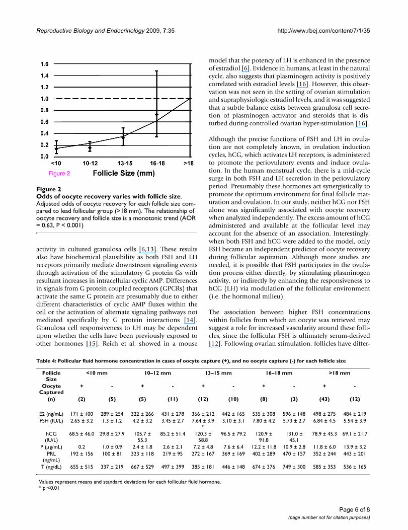
The oocyte recovery rate was significantly associated withfollicular size. The raw results are illustrated in Figure 1.The odds of oocyte recovery generally decreased withdecreasing size (OR: 0.63, P < 0.001, Figure 1, Figure 2).The lead follicular group (Reference, Odds Ratio = 1) isdefined as greater than 18 mm in size. The odds of retriev-ing an egg from follicles of 16–18 mm size was 73% (P =0.655) compared to the lead follicular group. Oocyterecovery from follicles that were 13–15 mm was 26% (P =0.020) compared to the lead follicular group, and withsmaller sized follicles the chance was further decreased(10–12 mm, 18% (P = 0.001); <10 mm, 14% (P =0.010)). These findings are consistent with previous workand led us to adjust the potential hormonal effects ofoocyte recovery for follicle size [1,9].
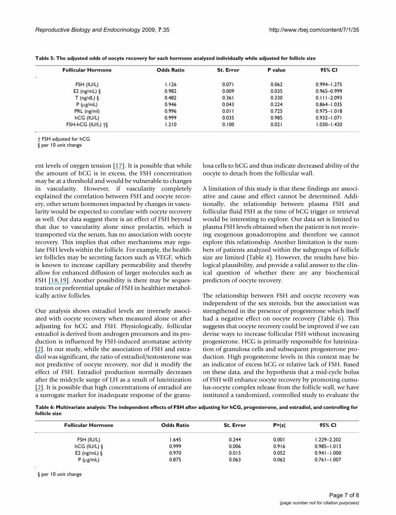
The mean follicular fluid hormone concentrations withcorresponding standard deviations according to follicularsize are summarized in Table 4. The concentrations forFSH (P = 0.01), hCG (P = 0.006), estradiol (P = 0.03), pro-
Table 1: Patient characteristics and stimulation parameters grouped by oocyte recovery
100% oocyte recoveryN = 70
< 100% oocyte recoveryN = 41
Mean SD Mean SD OR P value CI
Age 35.53 5.96 34.81 5.17 1.07 0.221 0.95,1.20Day 3 FSH 6.87 2.75 6.34 1.64 1.17 0.180 0.92,1.48Start Dose 4.67 1.18 4.46 1.07 1.23 0.298 0.82,1.85Total Dose 42.00 13.84 38.45 12.61 1.02 0.196 0.98,1.06Antral follicle count 15.40 8.67 19.35 9.26 0.94 0.016 0.89,0.99Stimulation days 11.07 1.14 11.14 1.19 0.98 0.939 0.65,1.48Peak estradiol 2287.64 1252.57 2711.70 1335.41 0.94 0.264 0.95,1.02
Page 3 of 8(page number not for citation purposes)
Reproductive Biology and Endocrinology 2009, 7:35 http://www.rbej.com/content/7/1/35
gesterone (P = .0001), and prolactin (P = 0.004) were sig-nificantly different between the follicle sizes. Thetestosterone concentration did not differ (P = 0.16).
Gonadotropins (FSH, hCG) were significantly associatedwith the follicular fluid hormone production. Intrafollic-ular FSH concentration was positively associated withestradiol concentration (P < 0.001) and progesterone con-centration (P < 0.001) within the follicle. The FSH con-centration did not correlate with testosteroneconcentration. The hCG concentration correlated withestrogen (P = 0.002) and progesterone concentrations (P< 0.001), but not with the testosterone concentration.
The analyses assessing the impact of each hormone onoocyte recovery adjusting for follicle size are shown (Table5). Estradiol concentrations were negatively correlatedwith oocyte recovery, but only slightly (AOR = 0.98, 95%CI 0.96–0.99). The odds of oocyte recovery is 2% less foreach 10 ng/dl increase in estradiol.
The intrafollicular FSH concentration was significantlyhigher when an oocyte was retrieved than when one wasnot retrieved (P = 0.007, Figure 1 white versus black barsat each follicle size). In a regression analysis, whenadjusted for follicle size, follicular FSH concentrationsshowed a trend toward positive association with oocyterecovery (AOR = 1.13, 95%CI 0.99–1.27, Table 5). Thisassociation between follicular FSH and oocyte recoverywas strengthened and statistically significant after adjust-ment for hCG (AOR = 1.21, 95%CI 1.03–1.42, Table 5);i.e., the odds of retrieving an oocyte increased by 21% forevery unit (IU/L) increase of FSH in the follicular fluidwhen controlled for hCG. In the range tested, hCG con-
centration alone did not impact the odds of oocyte recov-ery (AOR = 0.99, 95%CI 0.93–1.07).
When testosterone, estradiol, progesterone, and prolactinwere each added to the FSH and hCG model, only proges-terone influenced the impact of FSH on the rate of oocyterecovery. It did so by strengthening the FSH and oocyterelationship (AOR = 1.84, 95%CI 1.45–2.34). With pro-gesterone in the model, the odds of oocyte recoveryincreased by 84% for every IU/L increase in FSH. How-ever, the independent effect of progesterone concentra-tion was negatively associated with oocyte recovery (AOR= 0.79, 95%CI 0.70–0.91); i.e., for every 1 ug/mL increasein progesterone production in the presence of both FSHand hCG, the odds of oocyte recovery decreased by 21%.Follicular estradiol also remained an independent nega-tive predictor of oocyte recovery in the presence of bothFSH and hCG (AOR = 0.97, 95%CI 0.95–0.99); i.e., forevery 10 ng/dl increase in estradiol, the likelihood ofoocyte recovery decreased by 3%. However, estradiol didnot modify the FSH effect on oocyte recovery. After adjust-ing for estradiol and progesterone, while controlling forhCG and follicle size, FSH remained an independent pre-dictor of oocyte recovery (AOR = 1.65, 95%CI 1.22–2.20,Table 6); i.e., the odds of oocyte recovery increased by65% for every IU/L increase in follicular FSH.
The association of hormones and oocyte recovery previ-ously observed was similar after adjusting for stimulationprotocol. For FSH, after adjusting for follicle size, theeffect on oocyte recovery as shown in Table 5 was AOR =1.13 (95%CI 0.994–1.275). This is essentially the sameafter also adjusting for stimulation protocol (AOR = 1.16;95%CI 1.03–1.30). The majority of our follicle aspirateswere obtained from down regulated cycles, and evensolely within this group the association of FSH and oocyterecovery was similar (AOR = 1.22; 95%CI 1.07–1.39).
DiscussionWhile the decision to go to egg retrieval is based largelyupon the size of the lead follicular group, an attempt isusually made to retrieve oocytes regardless of follicularsize. Several studies have suggested that the chance ofrecovering an oocyte decreases with smaller follicular size,and our findings are consistent with this [1,9]. A recentpublication from our group has also shown that the odds
Table 2: Infertility diagnosis for study patients
Diagnosis Number (%)
Unexplained 37 (46)Diminished Ovarian Reserve 12 (15)Tubal factor 11 (14)Ovulatory dysfunction 2 (3)Other/Combined 18 (22)
χ2 = no difference in oocyte recovery.
Table 3: Effect of stimulation protocol on follicular FSH levels and oocyte recovery
Stimulation Protocol n Avg follicular [FSH] SD [FSH] Proportion oocyte recovery
Down Regulated 62 4.8 * 3.8 0.63Microdose Flare 10 12.0 *ξ 3.2 0.81Antagonist 8 7.3 ξ 3.1 0.81
* p <0.001, ξp < 0.01
Page 4 of 8(page number not for citation purposes)
Reproductive Biology and Endocrinology 2009, 7:35 http://www.rbej.com/content/7/1/35
of oocyte nuclear maturation and normal fertilization aredecreased in oocytes derived from smaller follicles [10].Developmentally, each follicle contains an oocyte. Yetthere are reports of no oocyte recovery from IVF cyclesinvolving multiple mature sized follicles, and in virtuallyall ovarian stimulation cycles the rate of oocyte recovery isless than 100% relative to the number of observed folli-cles [11]. Therefore, assuming a similar aspiration tech-nique is used, one reason why no egg is retrieved fromcertain follicles is that the cumulus-oocyte complexremains attached to the follicular wall during aspiration.Breaking this connection is a vital process for normal ovu-lation, and aspirated follicles where no egg is retrievedmay allow elucidation of this process. We performed arepeated measures logistic regression analysis examiningthe oocyte recovery rates at different follicle sizes. Theresults demonstrate a significant correlation with the oddsof retrieving an oocyte decreasing as the follicle sizedecreases (Figure 2).
Interestingly, we found that FSH levels were higher in fol-licles in which an oocyte was recovered, and this was sig-nificant after adjusting for the type of ovarian stimulationprotocol. An adjustment for stimulation was made due todifferent management schemes used in the protocols, ascommonly we step-down with down regulated protocolsresulting in potential differences in follicular fluid FSHlevels (Table 3). We could not adequately compare the
oocyte recovery between stimulation protocols due tosample size. However, the fact that follicle number isinversely predictive of oocyte recovery is consistent withfollicular fluid FSH being associated with oocyte recovery,because down regulated cycles (which had on averagelower follicular FSH) are typically prescribed in womenwith higher follicle numbers.
While FSH is an important contributor to oocyte recovery,it is only significant after adjustment for hCG (Table 5).This is an interesting finding, since in spontaneous ovula-tion there is a surge in both FSH and LH. Whether the FSHand LH surges have overlapping, complementary orredundant functions is unknown. In high concentrations,FSH has been shown to induce ovulation by itself in cer-tain animal models. In hypophysectomized rats, purerecombinant FSH as a large bolus has been shown toinduce ovulation, although the dose of FSH required waslarger than the dose of hCG needed to promote ovulation[5,6]. In rhesus monkeys who were down regulated withGnRH agonist and undergoing IVF, recombinant FSH wasshown to recapitulate some but not all of the characteris-tics of recombinant hCG. Specifically, r-hFSH was equiva-lent to r-hCG for the reinitiation of oocyte meiosis,fertilization and granulosa cell luteinization, but a midcy-cle FSH surge did not sustain normal luteal function [12].A role for FSH is supported by in vitro experiments, whereboth FSH and LH can promote plasminogen activator
Follicular FSH level and oocyte recoveryFigure 1Follicular FSH level and oocyte recovery. Rate of oocyte recovery and concentration FSH versus Rate of recovery (gray bars) and FSH concentration in cases of oocyte capture (white bars), and no oocyte capture (black bars). FSH p = 0.007 Oocyte recovery across follicles sizes p <0.001
Figure 1
Page 5 of 8(page number not for citation purposes)
Reproductive Biology and Endocrinology 2009, 7:35 http://www.rbej.com/content/7/1/35
activity in cultured granulosa cells [6,13]. These resultsalso have biochemical plausibility as both FSH and LHreceptors primarily mediate downstream signaling eventsthrough activation of the stimulatory G protein Gs withresultant increases in intracellular cyclic AMP. Differencesin signals from G protein coupled receptors (GPCRs) thatactivate the same G protein are presumably due to eitherdifferent characteristics of cyclic AMP fluxes within thecell or the activation of alternate signaling pathways notmediated specifically by G protein interactions [14].Granulosa cell responsiveness to LH may be dependentupon whether the cells have been previously exposed toother hormones [15]. Reich et al, showed in a mouse
model that the potency of LH is enhanced in the presenceof estradiol [6]. Evidence in humans, at least in the naturalcycle, also suggests that plasminogen activity is positivelycorrelated with estradiol levels [16]. However, this obser-vation was not seen in the setting of ovarian stimulationand supraphysiologic estradiol levels, and it was suggestedthat a subtle balance exists between granulosa cell secre-tion of plasminogen activator and steroids that is dis-turbed during controlled ovarian hyper-stimulation [16].
Although the precise functions of FSH and LH in ovula-tion are not completely known, in ovulation inductioncycles, hCG, which activates LH receptors, is administeredto promote the periovulatory events and induce ovula-tion. In the human menstrual cycle, there is a mid-cyclesurge in both FSH and LH secretion in the periovulatoryperiod. Presumably these hormones act synergistically topromote the optimum environment for final follicle mat-uration and ovulation. In our study, neither hCG nor FSHalone was significantly associated with oocyte recoverywhen analyzed independently. The excess amount of hCGadministered and available at the follicular level mayaccount for the absence of an association. Interestingly,when both FSH and hCG were added to the model, onlyFSH became an independent predictor of oocyte recoveryduring follicular aspiration. Although more studies areneeded, it is possible that FSH participates in the ovula-tion process either directly, by stimulating plasminogenactivity, or indirectly by enhancing the responsiveness tohCG (LH) via modulation of the follicular environment(i.e. the hormonal milieu).
The association between higher FSH concentrationswithin follicles from which an oocyte was retrieved maysuggest a role for increased vascularity around these folli-cles, since the follicular FSH is ultimately serum-derived[12]. Following ovarian stimulation, follicles have differ-
Odds of oocyte recovery varies with follicle sizeFigure 2Odds of oocyte recovery varies with follicle size. Adjusted odds of oocyte recovery for each follicle size com-pared to lead follicular group (>18 mm). The relationship of oocyte recovery and follicle size is a monotonic trend (AOR = 0.63, P < 0.001)
Figure 2
Table 4: Follicular fluid hormone concentration in cases of oocyte capture (+), and no oocyte capture (-) for each follicle size
Follicle Size
<10 mm 10–12 mm 13–15 mm 16–18 mm >18 mm
Oocyte Captured
+ - + - + - + - + -
(n) (2) (5) (5) (11) (12) (10) (8) (3) (43) (12)
E2 (ng/mL) 171 ± 100 289 ± 254 322 ± 266 431 ± 278 366 ± 212 442 ± 165 535 ± 308 596 ± 148 498 ± 275 484 ± 219FSH (IU/L) 2.65 ± 3.2 1.3 ± 1.2 4.2 ± 3.2 3.45 ± 2.7 7.64 ± 3.9
*3.10 ± 3.1 7.80 ± 4.2 5.73 ± 2.7 6.84 ± 4.5 5.54 ± 3.9
hCG (IU/L)
68.5 ± 46.0 29.8 ± 27.9 105.7 ± 55.3
85.2 ± 51.4 120.3 ± 58.8
96.5 ± 79.2 120.9 ± 91.8
131.0 ± 45.1
78.9 ± 45.3 69.1 ± 21.7
P (μg/mL) 0.2 1.0 ± 0.9 2.4 ± 1.8 2.6 ± 2.1 7.2 ± 4.8 7.6 ± 6.4 12.2 ± 11.8 10.9 ± 2.8 11.8 ± 6.0 13.9 ± 3.2PRL
(ng/mL)192 ± 156 100 ± 81 323 ± 118 219 ± 95 272 ± 167 369 ± 169 402 ± 289 470 ± 157 352 ± 244 443 ± 201
T (ng/dL) 655 ± 515 337 ± 219 667 ± 529 497 ± 399 385 ± 181 446 ± 148 674 ± 376 749 ± 300 585 ± 353 536 ± 165
Values represent means and standard deviations for each follicular fluid hormone.* p <0.01
Page 6 of 8(page number not for citation purposes)
Reproductive Biology and Endocrinology 2009, 7:35 http://www.rbej.com/content/7/1/35
ent levels of oxygen tension [17]. It is possible that whilethe amount of hCG is in excess, the FSH concentrationmay be at a threshold and would be vulnerable to changesin vascularity. However, if vascularity completelyexplained the correlation between FSH and oocyte recov-ery, other serum hormones impacted by changes in vascu-larity would be expected to correlate with oocyte recoveryas well. Our data suggest there is an effect of FSH beyondthat due to vascularity alone since prolactin, which istransported via the serum, has no association with oocyterecovery. This implies that other mechanisms may regu-late FSH levels within the follicle. For example, the health-ier follicles may be secreting factors such as VEGF, whichis known to increase capillary permeability and therebyallow for enhanced diffusion of larger molecules such asFSH [18,19]. Another possibility is there may be seques-tration or preferential uptake of FSH in healthier metabol-ically active follicles.
Our analysis shows estradiol levels are inversely associ-ated with oocyte recovery when measured alone or afteradjusting for hCG and FSH. Physiologically, follicularestradiol is derived from androgen precursors and its pro-duction is influenced by FSH-induced aromatase activity[2]. In our study, while the association of FSH and estra-diol was significant, the ratio of estradiol/testosterone wasnot predictive of oocyte recovery, nor did it modify theeffect of FSH. Estradiol production normally decreasesafter the midcycle surge of LH as a result of luteinization[2]. It is possible that high concentrations of estradiol area surrogate marker for inadequate response of the granu-
losa cells to hCG and thus indicate decreased ability of theoocyte to detach from the follicular wall.
A limitation of this study is that these findings are associ-ative and cause and effect cannot be determined. Addi-tionally, the relationship between plasma FSH andfollicular fluid FSH at the time of hCG trigger or retrievalwould be interesting to explore. Our data set is limited toplasma FSH levels obtained when the patient is not receiv-ing exogenous gonadotropins and therefore we cannotexplore this relationship. Another limitation is the num-bers of patients analyzed within the subgroups of folliclesize are limited (Table 4). However, the results have bio-logical plausibility, and provide a valid answer to the clin-ical question of whether there are any biochemicalpredictors of oocyte recovery.
The relationship between FSH and oocyte recovery wasindependent of the sex steroids, but the association wasstrengthened in the presence of progesterone which itselfhad a negative effect on oocyte recovery (Table 6). Thissuggests that oocyte recovery could be improved if we candevise ways to increase follicular FSH without increasingprogesterone. HCG is primarily responsible for luteiniza-tion of granulosa cells and subsequent progesterone pro-duction. High progesterone levels in this context may bean indicator of excess hCG or relative lack of FSH. Basedon these data, and the hypothesis that a mid-cycle bolusof FSH will enhance oocyte recovery by promoting cumu-lus-oocyte complex release from the follicle wall, we haveinstituted a randomized, controlled study to evaluate the
Table 5: The adjusted odds of oocyte recovery for each hormone analyzed individually while adjusted for follicle size
Follicular Hormone Odds Ratio St. Error P value 95% CI
FSH (IU/L) 1.126 0.071 0.062 0.994–1.275E2 (ng/mL) § 0.982 0.009 0.035 0.965–0.999T (ng/dL) § 0.482 0.361 0.330 0.111–2.093P (μg/mL) 0.946 0.043 0.224 0.864–1.035
PRL (ng/ml) 0.996 0.011 0.725 0.975–1.018hCG (IU/L) 0.999 0.035 0.985 0.932–1.071
FSH-hCG (IU/L) †§ 1.210 0.100 0.021 1.030–1.420
† FSH adjusted for hCG§ per 10 unit change
Table 6: Multivariate analysis: The independent effects of FSH after adjusting for hCG, progesterone, and estradiol, and controlling for follicle size
Follicular Hormone Odds Ratio St. Error P>|z| 95% Cl
FSH (IU/L) 1.645 0.244 0.001 1.229–2.202hCG (IU/L) § 0.999 0.006 0.916 0.985–1.013E2 (ng/mL) § 0.970 0.015 0.052 0.941–1.000P (μg/mL) 0.875 0.063 0.062 0.761–1.007
§ per 10 unit change
Page 7 of 8(page number not for citation purposes)
Reproductive Biology and Endocrinology 2009, 7:35 http://www.rbej.com/content/7/1/35
Publish with BioMed Central and every scientist can read your work free of charge
"BioMed Central will be the most significant development for disseminating the results of biomedical research in our lifetime."
Sir Paul Nurse, Cancer Research UK
Your research papers will be:
available free of charge to the entire biomedical community
peer reviewed and published immediately upon acceptance
cited in PubMed and archived on PubMed Central
yours — you keep the copyright
Submit your manuscript here:http://www.biomedcentral.com/info/publishing_adv.asp
BioMedcentral
effects of FSH administration at the time of the hCG trig-ger during IVF. More studies are needed to determine thebiological basis for the impact of FSH on oocyte recoveryand to determine whether modifying gonadotropinadministration can improve oocyte recovery rates.
Competing interestsThe authors declare that they have no competing interests.
Authors' contributionsMPR conceived of the study, and participated in its designand coordination and helped to draft the manuscript.AMZ participated in data analysis and interpretation andhelped to draft the manuscript. SS participated in studydesign and data collection and ran immunoassays. ATDparticipated in study design and coordination and helpedto draft the manuscript. CEM performed the statisticalanalyses and helped to draft the manuscript. PR partici-pated in study design and data analysis and interpreta-tion. JDL helped to draft the manuscript. MIC participatedin study design and coordination and helped to draft themanuscript. All authors read and approved the final man-uscript.
References1. Wittmaack FM, Kreger DO, Blasco L, Tureck RW, Mastroianni L Jr,
Lessey BA: Effect of follicular size on oocyte retrieval, fertili-zation, cleavage, and embryo quality in in vitro fertilizationcycles: a 6-year data collection. Fertil Steril 1994, 62:1205-1210.
2. Gougeon A: Regulation of ovarian follicular development inprimates: facts and hypotheses. Endocr Rev 1996, 17:121-155.
3. Hoff JD, Quigley ME, Yen SS: Hormonal dynamics at midcycle: areevaluation. J Clin Endocrinol Metab 1983, 57:792-796.
4. Thorneycroft IH, Mishell DR Jr, Stone SC, Kharma KM, NakamuraRM: The relation of serum 17-hydroxyprogesterone andestradiol-17-beta levels during the human menstrual cycle.Am J Obstet Gynecol 1971, 111:947-951.
5. Galway AB, Lapolt PS, Tsafriri A, Dargan CM, Boime I, Hsueh AJ:Recombinant follicle-stimulating hormone induces ovulationand tissue plasminogen activator expression in hypophysec-tomized rats. Endocrinology 1990, 127:3023-3028.
6. Reich R, Miskin R, Tsafriri A: Follicular plasminogen activator:involvement in ovulation. Endocrinology 1985, 116:516-521.
7. Morioka N, Zhu C, Brannstrom M, Woessner JF, LeMaire WJ: Mech-anism of mammalian ovulation. Prog Clin Biol Res 1989,294:65-85.
8. Strickland S, Beers WH: Studies on the role of plasminogen acti-vator in ovulation. In vitro response of granulosa cells togonadotropins, cyclic nucleotides, and prostaglandins. J BiolChem 1976, 251:5694-5702.
9. Scott RT, Hofmann GE, Muasher SJ, Acosta AA, Kreiner DK,Rosenwaks Z: Correlation of follicular diameter with oocyterecovery and maturity at the time of transvaginal follicularaspiration. J In Vitro Fert Embryo Transf 1989, 6:73-75.
10. Rosen MP, Shen S, Dobson AT, Rinaudo PF, McCulloch CE, CedarsMI: A quantitative assessment of follicle size on oocyte devel-opmental competence. Fertil Steril 2008, 90:684-690.
11. Stevenson TL, Lashen H: Empty follicle syndrome: the reality ofa controversial syndrome, a systematic review. Fertil Steril2008, 90:691-698.
12. Christenson LK, Stouffer RL: Follicle-stimulating hormone andluteinizing hormone/chorionic gonadotropin stimulation ofvascular endothelial growth factor production by macaquegranulosa cells from pre- and periovulatory follicles. J ClinEndocrinol Metab 1997, 82:2135-2142.
13. Wang C, Leung A: Gonadotropins regulate plasminogen acti-vator production by rat granulosa cells. Endocrinology 1983,112:1201-1207.
14. Shenoy SK, Lefkowitz RJ: Seven-transmembrane receptor sign-aling through beta-arrestin. Sci STKE 2005, 2005:cm10.
15. Canipari R, Strickland S: Studies on the hormonal regulation ofplasminogen activator production in the rat ovary. Endocrinol-ogy 1986, 118:1652-1659.
16. Weimer SL, Campeau JD, Marrs RP, Dizerega GS: Alteration ofhuman follicular fluid plasminogen activator activity by ovar-ian hyperstimulation. J In Vitro Fert Embryo Transf 1984, 1:263-266.
17. Van Blerkom J, Antczak M, Schrader R: The developmental poten-tial of the human oocyte is related to the dissolved oxygencontent of follicular fluid: association with vascular endothe-lial growth factor levels and perifollicular blood flow charac-teristics. Hum Reprod 1997, 12:1047-1055.
18. Gutman G, Barak V, Maslovitz S, Amit A, Lessing JB, Geva E: Regula-tion of vascular endothelial growth factor-A and its solublereceptor sFlt-1 by luteinizing hormone in vivo: implicationfor ovarian follicle angiogenesis. Fertil Steril 2008, 89:922-926.
19. Monteleone P, Giovanni Artini P, Simi G, Casarosa E, Cela V, Genaz-zani AR: Follicular fluid VEGF levels directly correlate withperifollicular blood flow in normoresponder patients under-going IVF. J Assist Reprod Genet 2008, 25:183-186.
Page 8 of 8(page number not for citation purposes)
http://www.ncbi.nlm.nih.gov/entrez/query.fcgi?cmd=Retrieve&db=PubMed&dopt=Abstract&list_uids=7957985
http://www.ncbi.nlm.nih.gov/entrez/query.fcgi?cmd=Retrieve&db=PubMed&dopt=Abstract&list_uids=7957985
http://www.ncbi.nlm.nih.gov/entrez/query.fcgi?cmd=Retrieve&db=PubMed&dopt=Abstract&list_uids=7957985
http://www.ncbi.nlm.nih.gov/entrez/query.fcgi?cmd=Retrieve&db=PubMed&dopt=Abstract&list_uids=8706629
http://www.ncbi.nlm.nih.gov/entrez/query.fcgi?cmd=Retrieve&db=PubMed&dopt=Abstract&list_uids=8706629
http://www.ncbi.nlm.nih.gov/entrez/query.fcgi?cmd=Retrieve&db=PubMed&dopt=Abstract&list_uids=6411753
http://www.ncbi.nlm.nih.gov/entrez/query.fcgi?cmd=Retrieve&db=PubMed&dopt=Abstract&list_uids=6411753
http://www.ncbi.nlm.nih.gov/entrez/query.fcgi?cmd=Retrieve&db=PubMed&dopt=Abstract&list_uids=5118032
http://www.ncbi.nlm.nih.gov/entrez/query.fcgi?cmd=Retrieve&db=PubMed&dopt=Abstract&list_uids=5118032
http://www.ncbi.nlm.nih.gov/entrez/query.fcgi?cmd=Retrieve&db=PubMed&dopt=Abstract&list_uids=2123446
http://www.ncbi.nlm.nih.gov/entrez/query.fcgi?cmd=Retrieve&db=PubMed&dopt=Abstract&list_uids=2123446
http://www.ncbi.nlm.nih.gov/entrez/query.fcgi?cmd=Retrieve&db=PubMed&dopt=Abstract&list_uids=2123446
http://www.ncbi.nlm.nih.gov/entrez/query.fcgi?cmd=Retrieve&db=PubMed&dopt=Abstract&list_uids=3917902
http://www.ncbi.nlm.nih.gov/entrez/query.fcgi?cmd=Retrieve&db=PubMed&dopt=Abstract&list_uids=3917902
http://www.ncbi.nlm.nih.gov/entrez/query.fcgi?cmd=Retrieve&db=PubMed&dopt=Abstract&list_uids=2657783
http://www.ncbi.nlm.nih.gov/entrez/query.fcgi?cmd=Retrieve&db=PubMed&dopt=Abstract&list_uids=2657783
http://www.ncbi.nlm.nih.gov/entrez/query.fcgi?cmd=Retrieve&db=PubMed&dopt=Abstract&list_uids=2723509
http://www.ncbi.nlm.nih.gov/entrez/query.fcgi?cmd=Retrieve&db=PubMed&dopt=Abstract&list_uids=2723509
http://www.ncbi.nlm.nih.gov/entrez/query.fcgi?cmd=Retrieve&db=PubMed&dopt=Abstract&list_uids=2723509
http://www.ncbi.nlm.nih.gov/entrez/query.fcgi?cmd=Retrieve&db=PubMed&dopt=Abstract&list_uids=9215284
http://www.ncbi.nlm.nih.gov/entrez/query.fcgi?cmd=Retrieve&db=PubMed&dopt=Abstract&list_uids=9215284
http://www.ncbi.nlm.nih.gov/entrez/query.fcgi?cmd=Retrieve&db=PubMed&dopt=Abstract&list_uids=9215284
http://www.ncbi.nlm.nih.gov/entrez/query.fcgi?cmd=Retrieve&db=PubMed&dopt=Abstract&list_uids=6299687
http://www.ncbi.nlm.nih.gov/entrez/query.fcgi?cmd=Retrieve&db=PubMed&dopt=Abstract&list_uids=6299687
http://www.ncbi.nlm.nih.gov/entrez/query.fcgi?cmd=Retrieve&db=PubMed&dopt=Abstract&list_uids=3081331
http://www.ncbi.nlm.nih.gov/entrez/query.fcgi?cmd=Retrieve&db=PubMed&dopt=Abstract&list_uids=3081331
http://www.ncbi.nlm.nih.gov/entrez/query.fcgi?cmd=Retrieve&db=PubMed&dopt=Abstract&list_uids=6443121
http://www.ncbi.nlm.nih.gov/entrez/query.fcgi?cmd=Retrieve&db=PubMed&dopt=Abstract&list_uids=6443121
http://www.ncbi.nlm.nih.gov/entrez/query.fcgi?cmd=Retrieve&db=PubMed&dopt=Abstract&list_uids=6443121
http://www.ncbi.nlm.nih.gov/entrez/query.fcgi?cmd=Retrieve&db=PubMed&dopt=Abstract&list_uids=9194664
http://www.ncbi.nlm.nih.gov/entrez/query.fcgi?cmd=Retrieve&db=PubMed&dopt=Abstract&list_uids=9194664
Related Documents


