RESEARCH ARTICLE Open Access The effect of dexamethasone on labor induction: a systematic review Zaynab Mohaghegh 1 , Shahla Faal Siahkal 2* , Hadis Bahmaei 3 , Foruzan Sharifipour 4 , Ehsan Kazemnejad Leyli 5 and Maryam Zahedian 6 Abstract Background: To evaluate the effect of dexamethasone administration on the interval between initiation of labor induction and active phase of labor. Methods: The databases including PubMed, Cochrane Library, Embase, Scopus and Web of Science were searched for studies published up to June 27, 2021. Two types of articles were included: a) full-text articles published in English or any other languages, and b) Randomized Controlled Trials (RCTs). Participants were primi- or multigravida women with term or post-term pregnancy. The intervention group received parenteral or extra-amniotic dexamethasone whereas the control group received normal saline or no treatment before initiation of labor induction. All data were analyzed using Review Manager 5.3. Results: Seventeen studies involving 1879 patients were included in the meta-analysis. Administration of dexamethasone reduced the interval between the initiation of labor induction and the beginning of active phase by about 70 min [MD: - 1.17 (- 1.37, - 1.00); P < 0.00001]. Duration of the first stage of labor in the dexamethasone group was about 88 min shorter than that in the control. There were no maternal and fetal adverse effects. Conclusions: Dexamethasone could significantly reduce the length of induction-active phase interval, and length of the first stage of labor, with no difference in maternal or fetal adverse effects. Keywords: Dexamethasone, Labor induction, Corticosteroid, Cervical ripening Background Induction of labor refers to the process of stimulating contractions before the spontaneous onset of labor, ei- ther with or without ruptured membrane. Labor induc- tion can be considered as a therapeutic intervention when the benefits of precipitous delivery to either the mother or the fetus are greater than the risks of preg- nancy continuation [1]. Induction of labor is the most common obstetric procedure and the fastest-growing medical technique in the United States [2]. Most re- cently, the prevalence of induction has been estimated to be 23.3% in the United States, 22.4% in Ethiopia, and 9% in Sudan [3–5]. Rupture of the membranes without labor, gestational hypertension, oligohydramnios, non- reassuring fetal status, post-term pregnancy, and various maternal medical conditions such as chronic hyperten- sion and diabetes are the common cause of start termin- ation of pregnancy [6]. Several medical and non-medical methods are used for the induction of labor [7, 8]. While oxytocin is the most conventional method, there are other effective methods including the use of prostaglan- dins such as misoprostol and dinoprostone as well as the application of some mechanical methods like stripping of membranes, artificial rupture of membranes, extra- amniotic saline infusion, trans cervical balloons, and hygroscopic cervical dilators [9]. The success of © The Author(s). 2021 Open Access This article is licensed under a Creative Commons Attribution 4.0 International License, which permits use, sharing, adaptation, distribution and reproduction in any medium or format, as long as you give appropriate credit to the original author(s) and the source, provide a link to the Creative Commons licence, and indicate if changes were made. The images or other third party material in this article are included in the article's Creative Commons licence, unless indicated otherwise in a credit line to the material. If material is not included in the article's Creative Commons licence and your intended use is not permitted by statutory regulation or exceeds the permitted use, you will need to obtain permission directly from the copyright holder. To view a copy of this licence, visit http://creativecommons.org/licenses/by/4.0/. The Creative Commons Public Domain Dedication waiver (http://creativecommons.org/publicdomain/zero/1.0/) applies to the data made available in this article, unless otherwise stated in a credit line to the data. * Correspondence: [email protected] 2 Department of Midwifery, Marand Branch, Islamic Azad University, Marand, Iran Full list of author information is available at the end of the article Mohaghegh et al. BMC Pregnancy and Childbirth (2021) 21:563 https://doi.org/10.1186/s12884-021-04010-1

Welcome message from author
This document is posted to help you gain knowledge. Please leave a comment to let me know what you think about it! Share it to your friends and learn new things together.
Transcript

RESEARCH ARTICLE Open Access
The effect of dexamethasone on laborinduction: a systematic reviewZaynab Mohaghegh1, Shahla Faal Siahkal2* , Hadis Bahmaei3, Foruzan Sharifipour4, Ehsan Kazemnejad Leyli5 andMaryam Zahedian6
Abstract
Background: To evaluate the effect of dexamethasone administration on the interval between initiation of laborinduction and active phase of labor.
Methods: The databases including PubMed, Cochrane Library, Embase, Scopus and Web of Science were searchedfor studies published up to June 27, 2021. Two types of articles were included: a) full-text articles published inEnglish or any other languages, and b) Randomized Controlled Trials (RCTs). Participants were primi- or multigravidawomen with term or post-term pregnancy. The intervention group received parenteral or extra-amnioticdexamethasone whereas the control group received normal saline or no treatment before initiation of laborinduction. All data were analyzed using Review Manager 5.3.
Results: Seventeen studies involving 1879 patients were included in the meta-analysis. Administration ofdexamethasone reduced the interval between the initiation of labor induction and the beginning of active phaseby about 70 min [MD: - 1.17 (− 1.37, − 1.00); P < 0.00001]. Duration of the first stage of labor in the dexamethasonegroup was about 88 min shorter than that in the control. There were no maternal and fetal adverse effects.
Conclusions: Dexamethasone could significantly reduce the length of induction-active phase interval, and lengthof the first stage of labor, with no difference in maternal or fetal adverse effects.
Keywords: Dexamethasone, Labor induction, Corticosteroid, Cervical ripening
BackgroundInduction of labor refers to the process of stimulatingcontractions before the spontaneous onset of labor, ei-ther with or without ruptured membrane. Labor induc-tion can be considered as a therapeutic interventionwhen the benefits of precipitous delivery to either themother or the fetus are greater than the risks of preg-nancy continuation [1]. Induction of labor is the mostcommon obstetric procedure and the fastest-growingmedical technique in the United States [2]. Most re-cently, the prevalence of induction has been estimated to
be 23.3% in the United States, 22.4% in Ethiopia, and 9%in Sudan [3–5]. Rupture of the membranes withoutlabor, gestational hypertension, oligohydramnios, non-reassuring fetal status, post-term pregnancy, and variousmaternal medical conditions such as chronic hyperten-sion and diabetes are the common cause of start termin-ation of pregnancy [6]. Several medical and non-medicalmethods are used for the induction of labor [7, 8]. Whileoxytocin is the most conventional method, there areother effective methods including the use of prostaglan-dins such as misoprostol and dinoprostone as well as theapplication of some mechanical methods like strippingof membranes, artificial rupture of membranes, extra-amniotic saline infusion, trans cervical balloons, andhygroscopic cervical dilators [9]. The success of
© The Author(s). 2021 Open Access This article is licensed under a Creative Commons Attribution 4.0 International License,which permits use, sharing, adaptation, distribution and reproduction in any medium or format, as long as you giveappropriate credit to the original author(s) and the source, provide a link to the Creative Commons licence, and indicate ifchanges were made. The images or other third party material in this article are included in the article's Creative Commonslicence, unless indicated otherwise in a credit line to the material. If material is not included in the article's Creative Commonslicence and your intended use is not permitted by statutory regulation or exceeds the permitted use, you will need to obtainpermission directly from the copyright holder. To view a copy of this licence, visit http://creativecommons.org/licenses/by/4.0/.The Creative Commons Public Domain Dedication waiver (http://creativecommons.org/publicdomain/zero/1.0/) applies to thedata made available in this article, unless otherwise stated in a credit line to the data.
* Correspondence: [email protected] of Midwifery, Marand Branch, Islamic Azad University, Marand,IranFull list of author information is available at the end of the article
Mohaghegh et al. BMC Pregnancy and Childbirth (2021) 21:563 https://doi.org/10.1186/s12884-021-04010-1

induction and progression of labor depends on severalfactors including multiparity, low body mass index(BMI), ruptured membranes, tall height, low estimatedfetal weight, and absence of comorbidities associatedwith placental insufficiency (e.g., preeclampsia) [10].According to a study by Kawakita et al., of nulliparous
women who underwent induction of labor in their study,77.4% had a vaginal delivery [11]. The time required forthe induction of labor, especially when there is an un-favorable cervix, increases the risk of cesarean delivery[10]. Some studies have reported the role of administer-ing glucocorticoids such as dexamethasone in cervicalripening [12]. Although the role of glucocorticoids in theinitiation of labor is still unknown, there is evidence sup-porting the role of glucocorticosteroid receptors in thefetal membranes at the beginning of the labor process[13, 14].Corticotropin-releasing hormone (CRH) in the female
reproductive system is the main regulator of thehypothalamic-pituitary-adrenal axis [12, 15]. Duringpregnancy, the placenta and fetal membranes secretelarge amounts of CRH. Also, CRH levels in plasma in-crease during labor, with the maximum value at vaginaldelivery [16]. There is considerable evidence for the ef-fect of dexamethasone on the duration of labor stages.The aim of the current systematic review was to exam-ine the effect of dexamethasone on the length of theinterval between the initiation of labor induction and thebeginning of the active phase of labor. Moreover, thelength of different stages of labor and maternal and neo-natal outcomes were investigated.
MethodsSystematic Reviews and Meta-Analyses of RandomizedControlled Trials (RCTs) were used to conduct this sys-tematic review [17].
Search strategiesThe authors searched promulgated studies publisheduntil July 8. 2020 in databases and publishing servicesincluding Cochrane Library, Scopus, ISI Web of ScienceCore Collection, PubMed, and EMBASE. It is updatedon June.27.2021.The keywords that were searched in-cluded “Corticosteroids”, “Dexamethasone”, “Deoxone”,“Dexpak”, “Induction of Labor”, “Cervical Ripening”.(Supplementary material).
Inclusion and exclusion criteriaTwo types of articles were included in this review: a)full-text articles published in English or any other lan-guages, and b) Randomized Controlled Trials (RCTs).Abstracts, comments, letters to editor, and observationalstudies were excluded.
ParticipantsThe criteria for including the participants were as fol-lows: an unfavorable cervix with a Bishop score of ≤4;singleton pregnancy with a duration of at least 37 weeksaccording to a reliable date for the last menstrual periodand a first trimester ultrasound evaluation; cephalicpresentation and intact membrane; and normal amnioticfluid. Women with any of the following conditions wereexcluded from the study: uterine malfunction, macroso-mia, placenta previa or placenta abruption risk, historyof surgery on uterus, uterine contractions, fetal distress,and fetal occiput posterior position.
Types of interventionsThe included studies involved intervention groups re-ceiving dexamethasone before initiation of labor induc-tion by any route of administration (intramuscular,intravenous or extra-amniotic) compared with controlgroups (placebo or no intervention).
Types of outcome measuresThe primary outcomes were as follows: The interval be-tween initiation of induction of labor and active phase oflabor, length of different stages of labor, the interval be-tween initiation of induction of labor and delivery andBishop Score after induction. Two types of secondaryoutcomes were as follows: Maternal and neonatal out-comes. The maternal outcomes were Caesarean sectionrate, and adverse events. Neonatal outcomes includedApgar score at the 1st and 5th minutes after birth, fetaldistress, meconium-stained liquid, and admission toneonatal intensive care unit (NICU).
Study selectionFollowing the initial search in the targeted databases,two of the authors (ZM) and (SH F), independentlyscreened titles and abstracts of the search results. Fulltext screening was conducted by the same two authors.Covidence was used for all screening, data extraction,and quality assessment. Any probable disagreement wasresolved by discussion or asking assistance from thethird author (E.K).
Data extractionThe authors used Covidence for data extraction and en-tered the data into Review Manager Software (RevMan5.3). Details of the studies including the design of thestudy, inclusion and exclusion criteria, baseline charac-teristics, interventions, and outcomes were extracted bytwo of the authors (ZM and SH F), independently.
Assessment risk of bias in included studiesThe risk of bias for each study was independentlyassessed by two reviewing authors (ZM and SH F) who
Mohaghegh et al. BMC Pregnancy and Childbirth (2021) 21:563 Page 2 of 16

used seven criteria suggested by Cochrane for the qualityassessment of randomized controlled trials. These cri-teria included selection bias, performance bias, detectionbias, attrition bias, selective reporting, and other risks ofbias. If the authors had any discrepancy, they deliberatedan issue to resolve it.
Statistical analysisDifferent statistical procedures were taken for continu-ous and dichotomous data. Mean and standard deviationwith 95% CIs were used for continuous data such as theinterval between initiation of induction of labor and ac-tive phase of labor, the length of different stages of labor,Apgar score at the 1st and 5th minutes, and BishopScore after induction. For dichotomous data, the resultswere presented as summary risk ratio or odds ratio (OR)with 95% confidence intervals. Outcome measurementin all trials was similar. To demonstrate the effect sizeand CI, Forest plots were used. Moreover, heterogeneitybetween the included studies was assessed by I2. By de-fault, we used fixed effects for all pooled studies. If I2 >50%, the random effect model was used for the primaryresults of heterogeneity. Furthermore, sensitivity analyseswere conducted to discover the potential source of het-erogeneity if it was statistically significant across thestudies. The authors performed sensitivity analyses bysequentially omitting one single study each time to testthe robustness of uncertainty in the meta-analysis. Fi-nally, all the data were analyzed using Review Manager(RevMan 5.3) statistical software from the Cochranegroup. The significance level was set at 0.05 for randomeffects and fixed effects.
Subgroup analysis and investigation of heterogeneityThe following subgroup analyses were carried out: par-enteral versus extra-amniotic administration of dexa-methasone. Some outcomes were also used includingthe length of the interval between initiation of inductionand delivery, mode of delivery, fetal distress, and admis-sion to NICU.
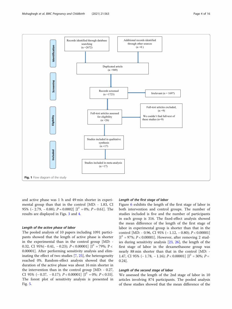
ResultsLiterature searchFigure 1 Shows the flowchart of the selection process ofstudies. In our search of databases, 2672 articles wereobtained as follows: Cochrane Library (n = 80), Scopus(n = 493), Web of Science (n = 178), PubMed (n = 1119),and EMBASE (n = 802). After removing duplicates (n =949) using Covidence, 1723 papers were screened ofwhich, 26 eligible articles were selected for full-textscreening, but we could not have access to the full-textof nine papers because these papers were published be-fore 2000. Therefore, 17 articles were finally included inthe study
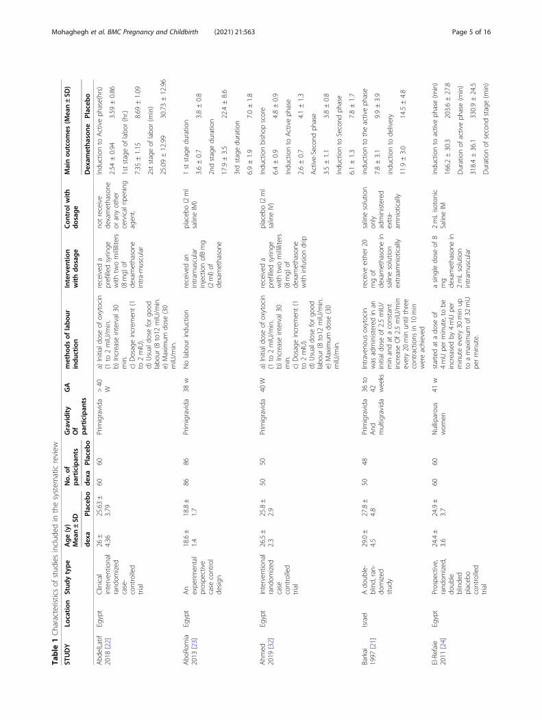
Characteristics and quality assessment of studiesOut of the 17 studies included in this review, four inves-tigated the effect of dexamethasone as an extra-amnioticinfusion with a Foley catheter [18–21], and the restassessed the effect of parenteral injection of dexametha-sone on the duration of labor induction. We examinedthe outcomes of these two groups separately. Amongthese articles, eight studies were published in Egypt [12,22–28], seven in Iran [7, 19, 20, 29–32], one in Iraq [18],and one in Israel [21]. All of them had an RCT designwith fourteen using placebo and three having no inter-vention in their control groups. All of the studiesassessed the effect of dexamethasone on nulliparouswomen except four studies that examined multiparousand nulliparous women [19–21, 29]. The range ofwomen’s age was between 18 and 35 years, and the ges-tational age was 37–42 weeks. All papers except one [23]used oxytocin for labor induction about 6 to 12 h afterdexamethasone injection. Two papers did not mentiontheir induction protocol [19, 30]. The number of partici-pants in studies differed from 31 to 86 in each group.The characteristics of the studies are demonstrated inTable 1.Quality assessment of papers was conducted by two
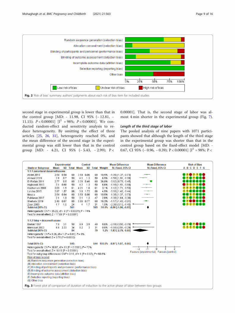
reviewers (ZM, SHF) according to Cochran Risk of Biastool. The result of the assessment is presented in Fig. 2.The lowest biases were related selection bias, reportingbias, and attrition bias, respectively. However, most ofthe detection bias and allocation concealment signifiesin the unclear risk. In terms of other types of bias, 50 %of papers were in the low-risk zone and the others werein the high-risk zone.
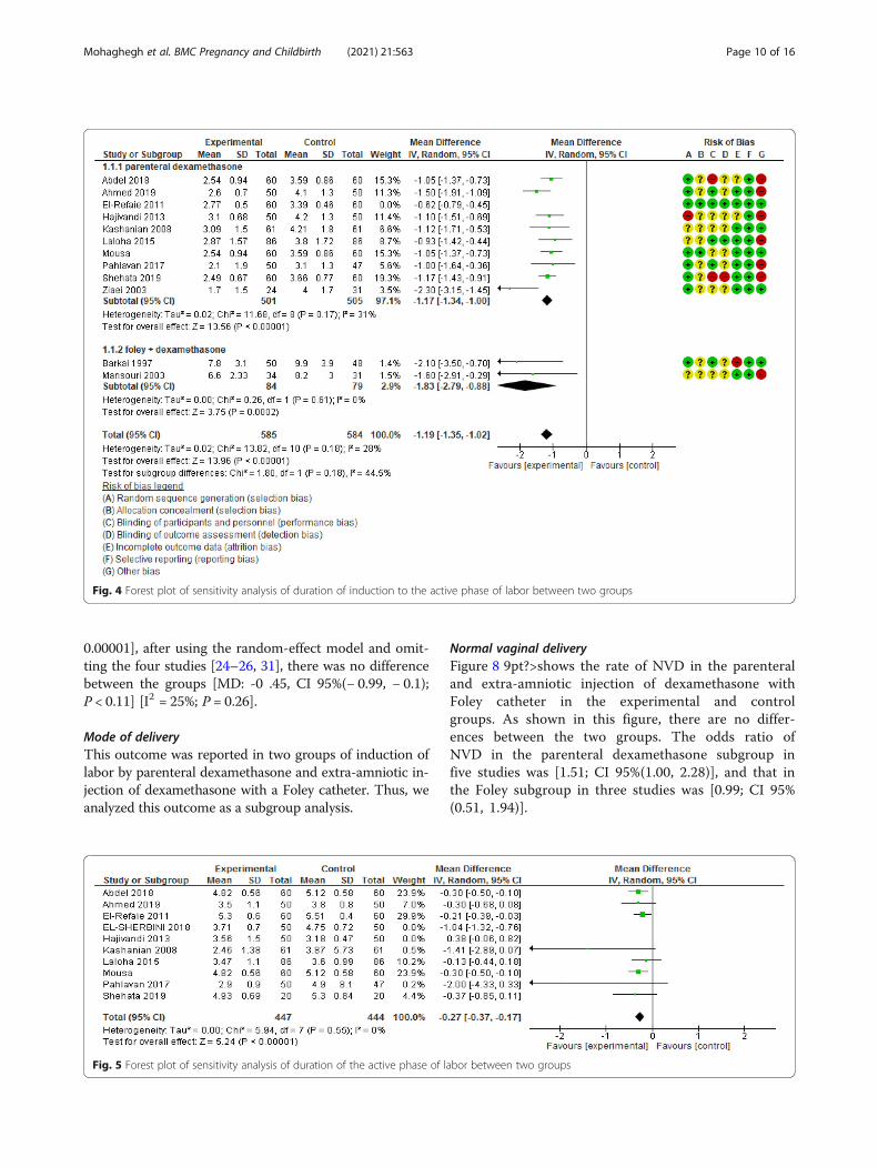
Overall meta-analysisThe interval between induction of labor and active phase oflaborTen articles including 1126 participants were analyzedin order to assess the effect of dexamethasone on theinterval between the initiation of induction of labor andthe active phase of labor. The result of the pooled ana-lysis showed that the mean difference of this intervalwas shorter in the experimental group compared withthe control group [MD: - 0.96, CI 95% (− 1.06, − 0.85);P < 0.00001]. Because of high heterogeneity (I2 = 74%;P < 0.0001), sensitivity analysis and random-effect ana-lysis were done. By omitting one study [24], the hetero-geneity reached 31%. However, this interval was about70 min shorter in the intervention group compared withthe control group [MD: - 1.17, CI 95% (− 1.37, − 1.00);P < 0.00001].In the subgroup of extra-amniotic administration of
dexamethasone with Foley catheter, two papers reportedthis outcome. There were 163 participants. The analysisshowed that the length of the interval between induction
Mohaghegh et al. BMC Pregnancy and Childbirth (2021) 21:563 Page 3 of 16
and active phase was 1 h and 49min shorter in experi-mental group than that in the control [MD: - 1.83, CI95% (− 2.79, − 0.88); P = 0.0002] [I2 = 0%; P = 0.61]. Theresults are displayed in Figs. 3 and 4.
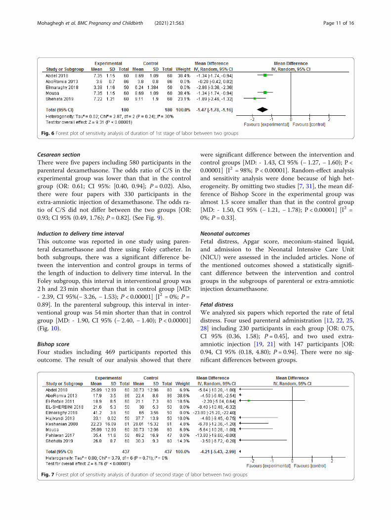
Length of the active phase of laborThe pooled analysis of 10 papers including 1091 partici-pants showed that the length of active phase is shorterin the experimental than in the control group [MD: -0.32, CI 95%(− 0.41, − 0.23); P < 0.00001] [I2 = 79%; P <0.00001]. After performing sensitivity analysis and elim-inating the effect of two studies [7, 25], the heterogeneityreached 0%. Random-effect analysis showed that theduration of the active phase was about 16 min shorter inthe intervention than in the control group [MD: - 0.27,CI 95% (− 0.37, − 0.17); P < 0.00001] [I2 = 0%; P < 0.55].The forest plot of sensitivity analysis is presented inFig. 5.
Length of the first stage of laborFigure 6 exhibits the length of the first stage of labor inboth intervention and control groups. The number ofstudies included is five and the number of participantsin each group is 316. The fixed-effect analysis showedthe mean difference of the length of the first stage oflabor in experimental group is shorter than that in thecontrol [MD: - 0.96, CI 95% (− 1.12, − 0.80); P < 0.00001][I2 = 97%; P < 0.00001]. However, after removing 2 stud-ies during sensitivity analysis [23, 26], the length of thefirst stage of labor in the dexamethasone group wasnearly 88 min shorter than that in the control [MD: -1.47, CI 95% (− 1.78, − 1.16); P < 0.00001] [I2 = 30%; P =0.24].
Length of the second stage of laborWe assessed the length of the 2nd stage of labor in 10articles involving 874 participants. The pooled analysisof these studies showed that the mean difference of the
Fig. 1 Flow diagram of the study
Mohaghegh et al. BMC Pregnancy and Childbirth (2021) 21:563 Page 4 of 16
Table
1Characteristicsof
stud
iesinclud
edin
thesystem
aticreview
STUDY
Location
Stud
ytype
Age(y)
Mea
n±SD
No.
ofparticipan
tsGravidity
Of
participan
ts
GA
metho
dsof
labou
rinduc
tion
Interven
tion
withdosag
eCon
trol
with
dosag
eMainou
tcom
es(M
ean±SD
)
dexa
Placeb
odexa
Placeb
oDexam
etha
sone
Placeb
o
Abd
elLatif
2018
[22]
Egypt
Clinical
interven
tional
rand
omized
case-
controlled
trial
26±
4.36
25.63±
3.79
6060
Prim
igravida
>40
Wa)
Initialdo
seof
oxytocin
(1to
2mIU/m
in.
b)Increase
interval30
min.
c)Dosageincrem
ent(1
to2mIU).
d)Usualdo
seforgo
odlabo
ur(8
to12
mIU/m
in.
e)Maxim
umdo
se(30
mIU/m
in.
received
aprefilled
syrin
gewith
twomilliliters
(8mg)
ofde
xamethasone
intra-muscular
notreceive
dexamethasone
oranyothe
rcervicalrip
ening
agen
t.
Indu
ctionto
Activeph
ase(hrs)
2.54
±0.94
3.59
±0.86
1ststageof
labo
r(hr.)
7.35
±1.15
8.69
±1.09
2ststageof
labo
r(m
in)
25.09±12.99
30.73±12.96
Abo
Romia
2013
[23]
Egypt
An
expe
rimen
tal
prospe
ctive
case
control
design
18.6±
1.4
18.8±
1.7
8686
Prim
igravida
38w
Nolabo
urindu
ction
received
anintram
uscular
injectionof8mg
(2ml)of
dexamethasone
placeb
o(2ml
salineIM)
1ststagedu
ratio
n
3.6±0.7
3.8±0.8
2ndstagedu
ratio
n
17.9±3.5
22.4±8.6
3rdstagedu
ratio
n
6.9±1.9
7.0±1.8
Ahm
ed2019
[32]
Egypt
Interven
tional
rand
omized
case
controlled
trial
26.5±
2.3
25.8±
2.9
5050
Prim
igravida
40W
a)Initialdo
seof
oxytocin
(1to
2mIU/m
in.
b)Increase
interval30
min.
c)Dosageincrem
ent(1
to2mIU).
d)Usualdo
seforgo
odlabo
ur(8
to12
mIU/m
in.
e)Maxim
umdo
se(30
mIU/m
in.
received
aprefilled
syrin
gewith
twomilliliters
(8mg)
ofde
xamethasone
with
infusion
drip
placeb
o(2ml
salineIV)
Indu
ctionbishop
score
6.4±0.9
4.8±0.9
Indu
ctionto
Activeph
ase
2.6±0.7
4.1±1.3
Active-Second
phase
3.5±1.1
3.8±0.8
Indu
ctionto
Second
phase
6.1±1.3
7.8±1.7
Barkai
1997
[21]
Israel
Ado
uble-
blind,
ran-
domized
stud
y
29.0±
4.5
27.8±
4.8
5048
Prim
igravida
And
multig
ravida
36to
42 weeks
Intraven
ousoxytocin
was
administeredin
aninitialdo
seof
2.5mIU/
min
andat
aconstant
increase
Of2.5mIU/m
inevery20
min
until
three
contractions
in10
min
wereachieved
receiveeither
20mgof
dexamethasone
insalinesolutio
nextraamniotically
salinesolutio
non
lyadministered
extra-
amniotically
indu
ctionto
theactiveph
ase
7.8±3.1
9.9±3.9
indu
ctionto
delivery
11.9±3.0
14.5±4.8
El-Refaie
2011
[24]
Egypt
Prospe
ctive,
rand
omized
,do
uble
blinde
dplaceb
ocontrolled
trial
24.4±
3.6
24.9±
3.7
6060
Nulliparou
swom
en41
wstartedat
ado
seof
4mUpe
rminute,to
beincreasedby
4mUpe
rminuteevery30
min
upto
amaxim
umof
32mU
perminute.
asing
ledo
seof
8mg
dexamethasone
in2mLsolutio
nintram
uscular
2mLisoton
icSalineIM
indu
ctionto
activeph
ase(m
in)
166.2±30.3
203.6±27.8
Durationof
activeph
ase(m
in)
318.4±36.1
330.9±24.5
Durationof
second
stage(m
in)
Mohaghegh et al. BMC Pregnancy and Childbirth (2021) 21:563 Page 5 of 16
Table
1Characteristicsof
stud
iesinclud
edin
thesystem
aticreview
(Con
tinued)
STUDY
Location
Stud
ytype
Age(y)
Mea
n±SD
No.
ofparticipan
tsGravidity
Of
participan
ts
GA
metho
dsof
labou
rinduc
tion
Interven
tion
withdosag
eCon
trol
with
dosag
eMainou
tcom
es(M
ean±SD
)
dexa
Placeb
odexa
Placeb
oDexam
etha
sone
Placeb
o
18.9±8.5
21.1±7.3
EL-
SHERBINI
2018
[25]
Egypt
Prospe
ctive
rand
omized
sing
le-blinde
dplaceb
o-controlled
stud
y
27.3±
3.8
27.1±
4.3
5050
prim
igravida
38-
40w
2.5m
IU/m
inof
oxytocin
intraven
ousdrip
(in500
mLlactated
Ring
er’s
solutio
n),w
iththedo
seincreasedby
2.5m
IU/m
levery20
min
until
labo
rwas
establishe
d
received
IMde
xamethasone
(8mg)
6hbe
fore
IOLthen
IM2ml
salineat
the
beginn
ingof
activeph
ase
placeb
o(2ml
saline)
IM6h
before
IOLand
atthe
beginn
ingof
theactive
phase.
Activeph
ase(hr)
3.71
±0.70
4.75
±0.72
2ndstage(m
in)
21.6±5.3
30.0±5.3
Elmaraghy
2018
[26]
Egypt
Dou
ble
blinde
drand
omized
,controlled
trial
Not
repo
rted
Not
repo
rted
5050
Nulliparou
s38–42
wStartedby
5drop
s/m
inuteof
500cc
saline
5un
itsof
oxytocin
with
thedo
seincreasedby
5–10
drop
s/minuteevery
30min
tillo
ptim
alcontractions
arereache
d
Eigh
tmg(2ml)
dexamethasone
IM atleasthalfan
hour
and
maxim
ally6h
before
labo
rindu
ction.
2mlo
fdistilled
water
IMat
asa
sameway
1ststage(hr.)
3.38
±1.16
6.24
±1.384
2ststage(m
in)
41.2±36.3
65±35.66
Hajivandi
2013
[7]
Iran
One
blinde
drand
omized
,controlled
trial
23.5±
3.83
22.8±
3.89
5050
Prim
igravida
40–42
wOxytocinstartedat
10un
itspe
r1000
ccof
ringe
rat
arate
of2.5mU
/min
andincreasedby
thesameam
ount
every
15min
until
regu
lar
contractions
continued.
Eigh
tmg(2ml)of
dexamethasone
IMat
12hbe
fore
labo
rindu
ction
2mlo
fno
rmal
salineIM
at12
hbe
fore
initiation
oflabo
rindu
ction
Bishop
score
7.2±1.32
2.98
±0.89
indu
ctionto
activeph
ase(hrs)
3.1±0.68
4.2±1.3
Kashanian
2008
[20]
Iran
Ado
uble-
blind,
ran-
domized
,con
-trolledtrial
24.38±
4.5
22.85±
3.5
6161
Nulliparou
sStartedwith
2.5mU/m
inof
oxytocin,w
iththe
dose
increasedby
2.5
mU/m
inevery20
min.
8mg(2mL)
oftheprod
uctIM
6hbe
fore
initiation
oflabo
rindu
ction
2mLof
distilled
water
IM6h
before
initiation
oflabo
rindu
ction
indu
ctionto
theactiveph
ase,h
3.09
±1.5
4.21
±1.8
Activeph
ase,h
2.46
±1.38
3.87
±5.73
second
stage,min
22.23±16.09
29.01±15.32
Kashanian
2008
[20]
Iran
Ado
uble-
blindrand
om-
ized
clinical
trial
28.22±
5.85
26.58±
7.31
4143
Prim
igravida
And
multig
ravida
>40
windu
ctionwith
oxytocin
atado
seof
2.5mIu/m
inwas
initiated
inbo
thgrou
ps,and
was
increasedat
ado
seof
2.5
mIu/m
inevery20
min
until
thewom
enen
tered
theactiveph
aseof
labo
randcontinuedun
tilde
livery
dexamethasone
20mgmixed
with
norm
alsalineto
achievea20
mL
volumewas
infusedextra-
amnioticspace
for6h.
20mLof
norm
alsalinewas
infusedextra-
amniotic
similarto
the
previous
grou
p
indu
ctionto
delivery(h)
7.25
±2.86
9.76
±3.91
Mohaghegh et al. BMC Pregnancy and Childbirth (2021) 21:563 Page 6 of 16

Table
1Characteristicsof
stud
iesinclud
edin
thesystem
aticreview
(Con
tinued)
STUDY
Location
Stud
ytype
Age(y)
Mea
n±SD
No.
ofparticipan
tsGravidity
Of
participan
ts
GA
metho
dsof
labou
rinduc
tion
Interven
tion
withdosag
eCon
trol
with
dosag
eMainou
tcom
es(M
ean±SD
)
dexa
Placeb
odexa
Placeb
oDexam
etha
sone
Placeb
o
Laloha
2015
[30]
Iran
A rand
omized
,clinical,and
doub
le–
blindtrial
21.7±
0.67
22.4±
0.67
8686
prim
parous,
40w
Itwas
used
butthe
metho
dwas
not
men
tione
d
2mlinjectedwith
Dexam
ethasone
(IV)fou
rho
urs
before
labo
rindu
ction
2mlinjected
with
distilled
water
(IV)fou
rho
ursbe
fore
thestartof
labo
rindu
ction.
indu
ctionto
activeph
ase(hrs)
2.87
±1.57
3.8±1.72
Activeph
aseto
second
stage
3.47
±1.1
3.6±0.99
Mansouri
2003
[19]
Iran
Ado
uble
blind
rand
omized
stud
y
26±
7.07
25±
5.54
3431
Prim
igravida
And
multig
ravida
39-
41w
Itwas
used
butthe
metho
dwas
not
men
tione
d
20mLof
norm
alsalinecontaining
20mgof
dexamethasone
wereinfusedinto
theextra-am
niotic
space
20mLof
norm
alsaline,were
infusedinto
the
extra-am
niotic
space.
indu
ctionto
activeph
ase(hrs)
6.6±2.33
8.2±3
indu
ctionto
delivery(hrs)
8.4±2.62
10.5±3.35
Mou
sa2014
[28]
Egypt
Dou
ble
blinde
drand
omized
,controlled
trial
26±
4.36
25.63±
3.79
6060
Nulliparou
swom
en> 41w
Startin
gby
infusion
of5
drop
s/minuteof
500cc
Ring
er’ssolutio
n+5un
itsof
oxytocin
with
the
dose
increased5drop
s/minutes
every30
min.
2ml
dexamethasone
was
administrated
IMat
6hbe
fore
labo
rindu
ction
2mld
istilled
water
(IM)six
hoursbe
fore
thestartof
labo
rindu
ction
indu
ctionto
activeph
ase(hrs)
2.54
±0.94
3.59
±0.86
Durationof
activeph
ase(hrs)
4.82
±0.56
5.12
±0.58
second
stage,min
25.09±12.99
30.73±12.96
Pahlavan
2017
[31]
Iran
Arand
omized
doub
le-blind
clinicaltrial
24.2±
3.9
23.9±
4.1
6160
nulliparous
40–42
wTheaugm
entatio
nof
labo
rwith
theuseof
intraven
ousoxytocin
infusion
(2.5m
units/pe
rminute)
beganin
both
grou
ps.
2ml
dexamethasone
intram
uscular4
mg/mLbe
fore
startin
goxytocin
infusion
2mlsterile
water
IMbe
fore
startin
goxytocin
infusion
indu
ctionto
activeph
ase(hrs)
2.1±1.9
3.1±1.3
Durationof
activeph
ase(hrs)
2.9±0.9
4.9±8.1
second
stage(m
in)
35.4±11.6
49.2±16.9
Salm
an2017
[18]
Iraq
Ado
uble
blind
rand
omized
case-control
stud
y
28.53±
5.38
28.8±
5.71
5841
nulliparous
40w
After
extraam
niotic
Cathe
terexpe
lled,
intraven
ousoxytocin
administeredas
aninitial
dose
until
three
contractions
perten
minutewereachieved
.themetho
dwas
not
men
tione
d.
20mg
dexamethasone
mixed
with
20cc
ofsterile
saline
solutio
ninfused
into
theextra-
amnioticspace
500mlo
fpu
resalinesolutio
n,with
arate
of5
drop
/min
throug
hthe
Cathe
terinto
theextra-
amnioticspace.
1ststage(m
in)
184.53
±44.6
222.0±47.62
2ststage(m
in)
33.25±9.14
44.02±7.0
Sheh
ata
2019
[12]
Egypt
rand
omized
controlled
clinicaltrial
18–35
18–35
6060
Prim
igravida.>41
wAfter
sixho
ursof
the
initialdo
se,the
labo
rindu
ctionwas
startedvia
oxytocin
a.Initialdo
seof
oxytocin
(1to
2mIU/
min.)b.
Increase
interval
prefilled
syrin
gewith
twomilliliters
Dexam
ethasone
.be
fore
sixho
urs
labo
rindu
ction
didno
treceive
dexamethasone
oranyothe
rcervicalrip
ening
agen
t.
Indu
ctionto
activeph
ase(hrs)
2.49
±0.67
3.66
±0.77
1ststageof
labo
ur(hrs)
7.22
±1.21
9.11
±1.9
Mohaghegh et al. BMC Pregnancy and Childbirth (2021) 21:563 Page 7 of 16
Table
1Characteristicsof
stud
iesinclud
edin
thesystem
aticreview
(Con
tinued)
STUDY
Location
Stud
ytype
Age(y)
Mea
n±SD
No.
ofparticipan
tsGravidity
Of
participan
ts
GA
metho
dsof
labou
rinduc
tion
Interven
tion
withdosag
eCon
trol
with
dosag
eMainou
tcom
es(M
ean±SD
)
dexa
Placeb
odexa
Placeb
oDexam
etha
sone
Placeb
o
30Minutes.c.D
osage
increm
ent1to
2mIU.d
.Usualdo
seforgo
odlabo
ur8to12
mIU/m
in.e.
Maxim
umdo
se30
mIU/
min.
second
stage(m
in)
26.8±8.7
30.3±9.3
Ziaei2003
[29]
Iran
RCT
23.66±
5.02
24.21±
5.09
3333
Prim
igravida
And
multig
ravida
>41
w24
hfro
mthebe
ginn
ing
ofthefirstdo
se,the
injectionof
oxytocin
startedby
2mUpe
rminute.Ifne
cessary,it
was
increasedby
2mu
perminuteevery15
min,
notexceed
ing32
mupe
rminute
10mgof
dexamethasone
IMtw
odo
ses,at
anintervalof
12h,24
hfro
mthe
beginn
ingof
the
firstdo
se,the
intraven
ous
oxytocin
was
started.
thecontrol
grou
p,who
received
only
intraven
ous
oxytocin
24h’
afteren
rolling
.
indu
ctionto
activeph
ase(hrs)
1.7±1.5
4±1.7
Mohaghegh et al. BMC Pregnancy and Childbirth (2021) 21:563 Page 8 of 16
second stage in experimental group is lower than that inthe control group [MD: - 11.98, CI 95% (− 12.81, −11.15); P < 0.00001] [I2 = 98%; P < 0.00001]. We con-ducted random-effect and sensitivity analysis to re-duce heterogeneity. By omitting the effect of threearticles [25, 26, 31], heterogeneity reached 0%, andthe mean difference of the second stage in the experi-mental group was still lower than that in the controlgroup [MD: - 4.21, CI 95% (− 5.43, − 2.99); P <
0.00001]. That is, the second stage of labor was al-most 4 min shorter in the experimental group (Fig. 7).
Length of the third stage of laborThe pooled analysis of nine papers with 1071 partici-pants showed that although the length of the third stagein the experimental group was shorter than that in thecontrol group based on the fixed-effect model [MD: -0.67, CI 95% (− 0.96, − 0.39); P < 0.00001] [I2 = 98%; P <
Fig. 2 ‘Risk of bias’ summary: authors’ judgments about each risk of bias item for included studies
Fig. 3 Forest plot of comparison of duration of induction to the active phase of labor between two groups
Mohaghegh et al. BMC Pregnancy and Childbirth (2021) 21:563 Page 9 of 16
0.00001], after using the random-effect model and omit-ting the four studies [24–26, 31], there was no differencebetween the groups [MD: -0 .45, CI 95%(− 0.99, − 0.1);P < 0.11] [I2 = 25%; P = 0.26].
Mode of deliveryThis outcome was reported in two groups of induction oflabor by parenteral dexamethasone and extra-amniotic in-jection of dexamethasone with a Foley catheter. Thus, weanalyzed this outcome as a subgroup analysis.
Normal vaginal deliveryFigure 8 9pt?>shows the rate of NVD in the parenteraland extra-amniotic injection of dexamethasone withFoley catheter in the experimental and controlgroups. As shown in this figure, there are no differ-ences between the two groups. The odds ratio ofNVD in the parenteral dexamethasone subgroup infive studies was [1.51; CI 95%(1.00, 2.28)], and that inthe Foley subgroup in three studies was [0.99; CI 95%(0.51, 1.94)].
Fig. 4 Forest plot of sensitivity analysis of duration of induction to the active phase of labor between two groups
Fig. 5 Forest plot of sensitivity analysis of duration of the active phase of labor between two groups
Mohaghegh et al. BMC Pregnancy and Childbirth (2021) 21:563 Page 10 of 16
Cesarean sectionThere were five papers including 580 participants in theparenteral dexamethasone. The odds ratio of C/S in theexperimental group was lower than that in the controlgroup (OR: 0.61; CI 95%: [0.40, 0.94]; P = 0.02). Also,there were four papers with 330 participants in theextra-amniotic injection of dexamethasone. The odds ra-tio of C/S did not differ between the two groups [OR:0.93; CI 95% (0.49, 1.76); P = 0.82]. (See Fig. 9).
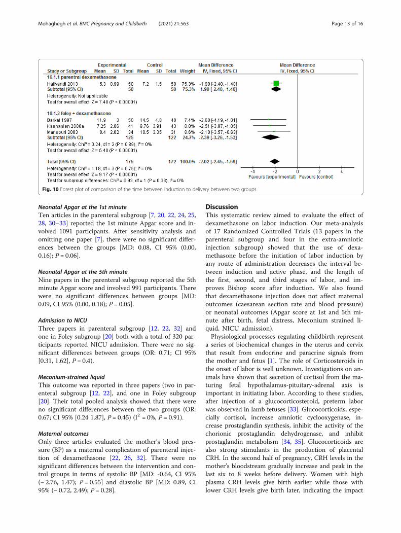
Induction to delivery time intervalThis outcome was reported in one study using paren-teral dexamethasone and three using Foley catheter. Inboth subgroups, there was a significant difference be-tween the intervention and control groups in terms ofthe length of induction to delivery time interval. In theFoley subgroup, this interval in interventional group was2 h and 23min shorter than that in control group [MD:- 2.39, CI 95%(− 3.26, − 1.53); P < 0.00001] [I2 = 0%; P =0.89]. In the parenteral subgroup, this interval in inter-ventional group was 54min shorter than that in controlgroup [MD: - 1.90, CI 95% (− 2.40, − 1.40); P < 0.00001](Fig. 10).
Bishop scoreFour studies including 469 participants reported thisoutcome. The result of our analysis showed that there
were significant difference between the intervention andcontrol groups [MD: - 1.43, CI 95% (− 1.27, − 1.60); P <0.00001] [I2 = 98%; P < 0.00001]. Random-effect analysisand sensitivity analysis were done because of high het-erogeneity. By omitting two studies [7, 31], the mean dif-ference of Bishop Score in the experimental group wasalmost 1.5 score smaller than that in the control group[MD: - 1.50, CI 95% (− 1.21, − 1.78); P < 0.00001] [I2 =0%; P = 0.33].
Neonatal outcomesFetal distress, Apgar score, meconium-stained liquid,and admission to the Neonatal Intensive Care Unit(NICU) were assessed in the included articles. None ofthe mentioned outcomes showed a statistically signifi-cant difference between the intervention and controlgroups in the subgroups of parenteral or extra-amnioticinjection dexamethasone.
Fetal distressWe analyzed six papers which reported the rate of fetaldistress. Four used parenteral administration [12, 22, 25,28] including 230 participants in each group [OR: 0.75,CI 95% (0.36, 1.58); P = 0.45], and two used extra-amniotic injection [19, 21] with 147 participants [OR:0.94, CI 95% (0.18, 4.80); P = 0.94]. There were no sig-nificant differences between groups.
Fig. 6 Forest plot of sensitivity analysis of duration of 1st stage of labor between two groups
Fig. 7 Forest plot of sensitivity analysis of duration of second stage of labor between two groups
Mohaghegh et al. BMC Pregnancy and Childbirth (2021) 21:563 Page 11 of 16
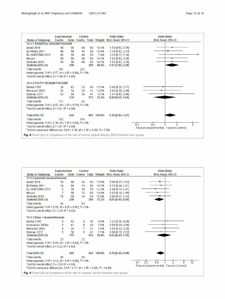
Fig. 8 Forest plot of comparison of the rate of normal vaginal delivery (NVD) between two groups
Fig. 9 Forest plot of comparison of the rate of cesarean section between two groups
Mohaghegh et al. BMC Pregnancy and Childbirth (2021) 21:563 Page 12 of 16
Neonatal Apgar at the 1st minuteTen articles in the parenteral subgroup [7, 20, 22, 24, 25,28, 30–33] reported the 1st minute Apgar score and in-volved 1091 participants. After sensitivity analysis andomitting one paper [7], there were no significant differ-ences between the groups [MD: 0.08, CI 95% (0.00,0.16); P = 0.06].
Neonatal Apgar at the 5th minuteNine papers in the parenteral subgroup reported the 5thminute Apgar score and involved 991 participants. Therewere no significant differences between groups [MD:0.09, CI 95% (0.00, 0.18); P = 0.05].
Admission to NICUThree papers in parenteral subgroup [12, 22, 32] andone in Foley subgroup [20] both with a total of 320 par-ticipants reported NICU admission. There were no sig-nificant differences between groups (OR: 0.71; CI 95%[0.31, 1.62], P = 0.4).
Meconium-strained liquidThis outcome was reported in three papers (two in par-enteral subgroup [12, 22], and one in Foley subgroup[20]. Their total pooled analysis showed that there wereno significant differences between the two groups (OR:0.67; CI 95% [0.24 1.87], P = 0.45) (I2 = 0%, P = 0.91).
Maternal outcomesOnly three articles evaluated the mother’s blood pres-sure (BP) as a maternal complication of parenteral injec-tion of dexamethasone [22, 26, 32]. There were nosignificant differences between the intervention and con-trol groups in terms of systolic BP [MD: -0.64, CI 95%(− 2.76, 1.47); P = 0.55] and diastolic BP [MD: 0.89, CI95% (− 0.72, 2.49); P = 0.28].
DiscussionThis systematic review aimed to evaluate the effect ofdexamethasone on labor induction. Our meta-analysisof 17 Randomized Controlled Trials (13 papers in theparenteral subgroup and four in the extra-amnioticinjection subgroup) showed that the use of dexa-methasone before the initiation of labor induction byany route of administration decreases the interval be-tween induction and active phase, and the length ofthe first, second, and third stages of labor, and im-proves Bishop score after induction. We also foundthat dexamethasone injection does not affect maternaloutcomes (caesarean section rate and blood pressure)or neonatal outcomes (Apgar score at 1st and 5th mi-nute after birth, fetal distress, Meconium strained li-quid, NICU admission).Physiological processes regulating childbirth represent
a series of biochemical changes in the uterus and cervixthat result from endocrine and paracrine signals fromthe mother and fetus [1]. The role of Corticosteroids inthe onset of labor is well unknown. Investigations on an-imals have shown that secretion of cortisol from the ma-turing fetal hypothalamus-pituitary-adrenal axis isimportant in initiating labor. According to these studies,after injection of a glucocorticosteroid, preterm laborwas observed in lamb fetuses [33]. Glucocorticoids, espe-cially cortisol, increase amniotic cyclooxygenase, in-crease prostaglandin synthesis, inhibit the activity of thechorionic prostaglandin dehydrogenase, and inhibitprostaglandin metabolism [34, 35]. Glucocorticoids arealso strong stimulants in the production of placentalCRH. In the second half of pregnancy, CRH levels in themother’s bloodstream gradually increase and peak in thelast six to 8 weeks before delivery. Women with highplasma CRH levels give birth earlier while those withlower CRH levels give birth later, indicating the impact
Fig. 10 Forest plot of comparison of the time between induction to delivery between two groups
Mohaghegh et al. BMC Pregnancy and Childbirth (2021) 21:563 Page 13 of 16

of CRH production as an important factor in the onsetof labor.CRH does not have a direct inotropic effect on the hu-
man myometrium, but it does cause uterine vasodilationby affecting the placenta locally. It can also stimulate thesecretion of dehydroepiandrosterone sulfate (DHEA-S)in fetal adrenal cortex cells. CRH enhances the effects ofestrogen on these tissues of the uterus and cervix, in-creases prostaglandins in the amniotic sac, chorion, anddecidua, and enhances the effect of oxytocin [35]. Sev-eral studies have reported that the injection of cortico-steroids provokes successful induction of labor in lambsand humans. Since glucocorticoid receptors are presentin the amniotic sac, glucocorticosteroids could carry outa possible role in parturition through paracrine or auto-crine mechanisms [36].Kalantaridou et al. (2007) reported that the
corticotrophin-releasing hormone (CRH) is the maincorrector of the hypothalamic-pituitary-adrenal axis.Circulating placental CRH is responsible for thephysiologic hypercortisolism of the second half ofpregnancy and plays a role in the commencement oflabor [15].In humans, the production of CRH by the placenta
and the increase of this hormone in maternal plasma areassociated with the timing of parturition [37]. Recently,it has been shown that CRH stimulates the placenta forthe production of estrogens and inhibits the productionof progesterone [38]. Increasing the ratio of estrogen toprogesterone in the mother’s serum may progress theexpression of contraction-associated proteins in themyometrium, thus facilitating the beginning of partur-ition [1, 39]. Furthermore, glucocorticoids induce theproduction of CRH by the placenta and the productionof prostaglandins (PGF2 and PGE2) by fetal membranes[40].
Limitations of this studySeveral limitations existed in this meta-analysis: 1) publi-cation bias was not been measured; 2) Although moststudies included in this review had examined the effectof dexamethasone on nulliparous women, some had notexamined this effect on a parity basis.; 3) Most studieswere conducted in Iran and Egypt, and the number ofarticles from other countries was small; 3) Maternal andneonatal outcomes had not been reported in all includedstudies completely. These limitations could have con-tributed to heterogeneity. On the other hand, given thepossible effect of dexamethasone on neonatal outcomes[41, 42], none of the studies examined other outcomessuch as neonatal hypoglycemia and the consequences ofneurological and behavioral development in neonatesborn to these mothers.
ConclusionDexamethasone could significantly reduce the length ofthe time interval between induction and active phaseand length of the first stage of labor with no differencein maternal or fetal adverse effects. Considering the highheterogeneity and quality of the studies included in thisreview, high-quality double-blind clinical trials areneeded to be included in future reviews in order to drawmore solid conclusion in this regard.
AbbreviationsRCT: Randomized controlled trial; CRH: Corticotrophin-releasing hormone;BMI: Body mass index; MD: Mean difference; OR: Odds ratio; CI: ConfidenceInterval; AD: Anno Domini; NICU: Neonatal intensive care unit; NVD: NormalVaginal Delivery; C/S: Cesarean Section; BP: Blood Pressure
Supplementary InformationThe online version contains supplementary material available at https://doi.org/10.1186/s12884-021-04010-1.
Additional file 1.
AcknowledgementsNot applicable.
Authors’ contributionsZM, SHF were responsible for the design of the study. MZ was responsiblefor the searching process. ZM, SHF and EKL were involved in data screeningand data extraction. ZM, SHF, HB and FSH wrote the manuscript. All authorsread and approved the final version of the manuscript.
FundingThis study was not funded by any funding resource.
Availability of data and materialsNot Applicable.
Declarations
Ethics approval and consent to participateNot Applicable.
Consent for publicationNot Applicable.
Competing interestsNone.
Author details1Family Health Department, Health Deputy, Tehran University of MedicalSciences, Tehran, Iran. 2Department of Midwifery, Marand Branch, IslamicAzad University, Marand, Iran. 3Department of Midwifery, Ahvaz JundishapurUniversity of Medical Sciences, Ahvaz, Iran. 4Department of Midwifery, Schoolof Nursing and Midwifery, Kermanshah University of Medical Sciences,Kermanshah, Iran. 5Guilan Road Trauma Research Center, BiostatisticsDepartment, Guilan University of Medical Sciences, Rasht, Iran. 6Liberian ofNursing and Midwifery School, Ahvaz Jundishapur University of MedicalScience, Ahvaz, Iran.
Received: 2 September 2020 Accepted: 19 July 2021
References1. Cunningham FGGN, Leveno KJ, Gilstrap LC III, Hauth JC, Wenstrom KD.
Williams obstetrics(23rd ed). New York: McGraw-Hill professional; 2018. ISBN:978-0-07-170285-0
Mohaghegh et al. BMC Pregnancy and Childbirth (2021) 21:563 Page 14 of 16

2. WHO. WHO Recommendations for Induction of labour 2011. Available from:http://apps.who.int/iris/bitstream/10665/44531/1/9789241501156_eng.pdf.ISBN 978 92 4 150115 6.
3. Centers for Disease Control and Prevention. Recent declines in induction oflabor. 2014 march 1:http://www.cdc.gov/nchs/data/databriefs/db155.htm.
4. Asefa D, Ma G, Tolessa D, Abdulkadir Y. Induction of Labor Prevalence andAssociated Factors for Its Outcome at Wolliso St. Luke, Catholic Hospital,South West Shewa, Oromia. Intern Med. 2017;7(5). Open Access. https://doi.org/10.4172/2165-8048.1000255.
5. Lueth GD, Kebede A, Medhanyie AA. Prevalence, outcomes andassociated factors of labor induction among women delivered at publichospitals of MEKELLE town-(a hospital based cross sectional study). BMCPregnancy Childbirth. 2020;20:1–10. https://doi.org/10.1186/s12884-020-02862-7.
6. ACOG Practice Bulletin No. 107: Induction of labor. Obstet Gynecol. 2009;114(2 Pt 1):386–97. https://doi.org/10.1097/AOG.0b013e3181b48ef5.
7. Hajivandi L, Montazeri S, Iravani M, Dawoodi M, Haghighizade MH. Effect ofintramuscular dexamethasone on onset of labor in postdate pregnancy. JBabol Univ Med Sci. 2013;15:24–9 URL: https://www.researchgate.net/publication/289020036_Effect_of_Intramuscular_Dexamethasone_on_Onset_of_Labor_in_Postdate_Pregnancy.
8. Alfirevic Z, Keeney E, Dowswell T, Welton NJ, Medley N, Dias S, et al.Methods to induce labour: a systematic review, network meta-analysis andcost-effectiveness analysis. BJOG. 2016;123(9):1462–70. https://doi.org/10.1111/1471-0528.13981.
9. Alfirevic Z, Keeney E, Dowswell T, Welton NJ, Medley N, Dias S, et al. Whichmethod is best for the induction of labour? A systematic review, networkmeta-analysis and cost-effectiveness analysis. Health Technol Assess. 2016;20(65):1–583. https://doi.org/10.3310/hta20650.
10. Grobman W, Lockwood CJ, Barss VA. Induction of labor with oxytocin. 2018.Uptodate; URL: https://www.uptodate.com/contents/induction-of-labor-with-oxytocin
11. Kawakita T, Reddy U, Huang C, Auguste T, Bauer D, Overcash R. Predictingvaginal delivery in nulliparous women undergoing induction of labor atterm. Am J Perinatol. 2018;35(07):660–8. https://doi.org/10.1055/s-0037-1608847.
12. Shehata EE, Zaitoun MM, Azzam MN, Radwan ME. Effect of IntramuscularAdministration of Dexamethasone on Labour Outcome in Induction ofPrimigravida at Late-Term Pregnancy. 2019;74(1):184–92. https://doi.org/10.21608/EJHM.2019.22649.
13. Campbell E, Linton E, Wolfe C, Scraggs P, Jones M, Lowry P. Plasmacorticotropin-releasing hormone concentrations during pregnancy andparturition. J Clin Endocrinol Metab. 1987;64(5):1054–9. https://doi.org/10.1210/jcem-64-5-1054.
14. Meyer E, Nenke M, Rankin W, Lewis J, Torpy DJ. Corticosteroid-bindingglobulin: a review of basic and clinical advances. Horm Metab Res. 2016;48(06):359–71. https://doi.org/10.1055/s-0042-108071.
15. Kalantaridou S, Makrigiannakis A, Zoumakis E, Chrousos GP. Peripheralcorticotropin-releasing hormone is produced in the immune andreproductive systems: actions, potential roles and clinical implications. FrontBiosci. 2007;12(1):572–80. https://doi.org/10.2741/2083.
16. Stjernholm TV, Nyberg A, Cardell M, Höybye C. Circulating maternalcortisol levels during vaginal delivery and elective cesarean section.Arch Gynecol Obstet. 2016;294(2):267–71. https://doi.org/10.1007/s00404-015-3981-x.
17. Yao X, Florez ID, Zhang P, Zhang C, Zhang Y, Wang C, et al. Clinical researchmethods for treatment, diagnosis, prognosis, etiology, screening, andprevention: A narrative review. J Evid-Based Med. 2020;13(2):130–6. https://doi.org/10.1111/jebm.12384.
18. Salman ST. Cervical ripening by using extra-amniotic dexamethasoneinfusion versus extra-amniotic saline infusion. J Facult Med. 2017;59(4):299–02. https://doi.org/10.32007/jfacmedbagdad.59468.
19. Mansouri M, Pourjavad A, Panahi G. Induction of labor with use of a Foleycatheter and extraamniotic corticosteroids. Med J Islam Repub Iran. 2003;17(2):97–100.
20. Kashanian M, Fekrat M, Naghghash S, Ansari NS. Evaluation of the effect ofextra-amniotic normal saline infusion alone or in combination withdexamethasone for the induction of labor. J Obstet Gynaecol Res. 2008;34(1):47–50. https://doi.org/10.1111/j.1447-0756.2007.00703.x.
21. Barkai G, Cohen SB, Kees S, Lusky A, Margalit V, Mashiach S, et al. Inductionof labor with use of a Foley catheter and extraamniotic corticosteroids. Am
J Obstet Gynecol. 1997;177(5):1145–8. https://doi.org/10.1016/s0002-9378(97)70031-0.
22. Abdel Latif EM, Taha WS, Ahmed AARA. Effect of Intra-MuscularAdministration of Dexamethasone on the Duration of Induction of Labor inPrimigravida Full-Term Pregnancy. Egyptian J Hosp Med. 2018;73(6):6973–82.https://doi.org/10.21608/EJHM.2018.17213.
23. AboRomia F, El-Adham AF. Influence of intramuscular dexamethasone onlabor duration in primigravidas. Tanta Sci Nurs J. 2013;4(1):143–54. https://doi.org/10.21608/tsnj.2013.74465.
24. El-Refaie T, Yehia M, Younan R. Effect of dexamethasone on the duration oflabor among women undergoing labor induction: a randomized controlledtrial. J Egypt Soc Gynaecol Obstet. 2011;37(1):595–9.
25. El-Sherbini M, Helal O, Zaki MD, Sherif S. The effect of intramuscularAdministration of Dexamethasone Alone or in combination with hyoscineButylbromide in shortening the duration of induced labor in Primigravidas.Med J Cairo Univ. 2018;86(September):2335–40. https://doi.org/10.21608/mjcu.2018.57534.
26. Elmaraghy MA-A, El Refaie T, Labib KM, Mohamed MG. Effect ofintramuscular administration of dexamethasone on the duration of laborinduction. Evid Based Womens Health J. 2018;8(4):311–7. en. https://doi.org/10.21608/EBWHJ.2018.20075
27. Ahmad A, Webb SS, Early B, Sitch A, Khan K, MacArthur C. Associationbetween fetal position at onset of labor and mode of delivery: aprospective cohort study. Ultrasound Obstet Gynecol. 2014;43(2):176–82.https://doi.org/10.1002/uog.13189.
28. Mousa K, Awadalla A, Ezat T. Effect of Intramuscular Administration ofDexamethasone on the Duration of Labor in Postterm Induced Delivery. AinShams Med J. 2014;65(1,2&3). https://doi.org/10.12816/001342.
29. Ziaei S, Rosebehani N, Kazeminejad A, Zafarghandi S. The effects ofintramuscular administration of corticosteroids on the induction ofparturition. J Perinat Med. 2003;31(2):134–9. https://doi.org/10.1515/JPM.2003.018.
30. Fatemeh L, Negin A, Ameneh B, Farideh M, Ezzatossadat Haj Seyed J. Effectof Intravenous Dexamethasone on Preparing the Cervix and LaborInduction. Acta Medica Iranica. 2015;53(9):568–72.
31. Pahlavan Z, Barat S, Esmaeilzadeh S, Ghanbarpour A, Naeimirad M,Golsorkhtabaramiri M. The effect of dexamethasone therapy on prolongedlatent phase of labor: a randomized double-blind clinical trial. Caspian JReprod Med. 2017;3(1):33–8 URL: http://caspjrm.ir/article-1-117-en.html.
32. Ahmed AEM, Deif OM, El-Saftawy MA. Potency of Dexamethasone in LaborInduction. Egypt J Hosp Med. 2019;74(7):1544–51. en. https://doi.org/10.21608/EJHM.2019.27861.
33. Kavanagh J, Kelly AJ, Thomas J. Corticosteroids for cervical ripening andinduction of labour. Cochrane Database Syst Rev. 2006;(2):CD003100.https://doi.org/10.1002/14651858.CD003100.pub2.
34. Petraglia F, Benedetto C, Florio P, D'Ambrogio G, Genazzani AD, Marozio L,et al. Effect of corticotropin-releasing factor-binding protein onprostaglandin release from cultured maternal decidua and on contractileactivity of human myometrium in vitro. J Clin Endocrinol Metab. 1995;80(10):3073–6. https://doi.org/10.1210/jcem.80.10.7559899.
35. Errol R Norwitz, Charles J Lockwood, Vanessa A Barss. Physiology ofparturition at term [Internet]. 2021. UpToDate. Available from: https://www2.utdos.ir/contents/physiology-of-parturition-at term?search=Physiology%20of%20parturition%20at%20term&source=search_result&selectedTitle=1~150&usage_type=default&display_rank=1.
36. Pushpalatha K. A Comparative Study of Extra-Amniotic Infusion ofCorticosteroids versus Normal Saline for Induction of Labour. Internet JThird World Med. 2008;8(1).
37. McLean M, Bisits A, Davies J, Woods R, Lowry P, Smith R. A placental clockcontrolling the length of human pregnancy. Nat Med. 1995;1(5):460–3.https://doi.org/10.1038/nm0595-460.
38. Yang R, You X, Tang X, Gao L, Ni X. Corticotropin-releasing hormone inhibitsprogesterone production in cultured human placental trophoblasts. J MolEndocrinol. 2006;37(3):533–40. https://doi.org/10.1677/jme.1.02119.
39. Mastorakos G, Ilias I. Maternal and fetal hypothalamic-pituitary-adrenal axesduring pregnancy and postpartum. Ann N Y Acad Sci. 2003;997(1):136–49.https://doi.org/10.1196/annals.1290.016.
40. Cheng YH, Nicholson RC, King B, Chan EC, Fitter JT, Smith R. Corticotropin-releasing hormone gene expression in primary placental cells is modulated bycyclic adenosine 3′,5′-monophosphate. J Clin Endocrinol Metab. 2000;85(3):1239–44. PubMed PMID: 10720069. https://doi.org/10.1210/jcem.85.3.6420.
Mohaghegh et al. BMC Pregnancy and Childbirth (2021) 21:563 Page 15 of 16

41. Zipori Y, Zidan R, Lauterbach R, Hagag A, Ginsberg Y, Solt I, et al. Antenatalbetamethasone and the risk of neonatal hypoglycemia: it's all about timing.Arch Gynecol Obstet. 2021;303(3):695–701. https://doi.org/10.1007/s00404-020-05785-y.
42. Franks, AL, Berry, KJ, DeFranco, DB. Prenatal drug exposure andneurodevelopmental programming of glucocorticoid signalling. JNeuroendocrinol. 2020; 32:e12786. https://doi.org/10.1111/jne.12786.
Publisher’s NoteSpringer Nature remains neutral with regard to jurisdictional claims inpublished maps and institutional affiliations.
Mohaghegh et al. BMC Pregnancy and Childbirth (2021) 21:563 Page 16 of 16
Related Documents


![CP.PHAR.310 Daratumumab (Darzalex) · Table 2 In combination with bortezomib, thalidomide and dexamethasone ([VTd]; 4 - week cycle dosing regimen) Induction Weeks 1 to 8: 16 mg/kg](https://static.cupdf.com/doc/110x72/5f2ffb030555a776ff621103/cpphar310-daratumumab-darzalex-table-2-in-combination-with-bortezomib-thalidomide.jpg)


