CASE REPORT Open Access The effect of autologous adipose derived mesenchymal stem cell therapy in the treatment of a large osteochondral defect of the knee following unsuccessful surgical intervention of osteochondritis dissecans – a case study Julien Freitag 1* , Kiran Shah 2 , James Wickham 1 , Richard Boyd 3 and Abi Tenen 4 Abstract Background: A prospective analysis of the effect of autologous adipose derived mesenchymal stem cell (MSC) therapy in the treatment of an osteochondral defect of the knee with early progressive osteoarthritis following unsuccessful surgical intervention of osteochondritis dissecans (OCD). Case presentation: After failed conventional management of OCD a patient undergoes intra-articular MSC therapy. Patient outcome measures included the Numeric Pain Rating Scale (NPRS), the Western Ontario and McMaster Universities Arthritis Index (WOMAC) and the Knee Injury and Osteoarthritis Outcome Score (KOOS). Structural outcome was assessed using MRI with the novel technique of T2 mapping used to indicate cartilage quality. Following MSC therapy the patient reported improvement in pain and function as measured by NPRS, WOMAC and KOOS. Repeat MRI analysis showed regeneration of cartilage. MRI T2 mapping indicated hyaline like cartilage regrowth. Conclusion: In this report, the use of MSCs, after unsuccessful conventional OCD management, resulted in structural, functional and pain improvement. These results highlight the need to further study the regenerative potential of MSC therapy. Trial registration: Australian and New Zealand Clinical Trial Registry Number - ACTRN12615000258550 (Date registered 19/03/2015 – retrospectively registered). Keywords: Osteochondritis dissecans, Osteochondral defect, Osteoarthritis, Arthritis, Mesenchymal stem cells, Case study Background Osteochondritis dissecans (OCD) was a term first used to describe the development of loose bodies within a joint with no prior evidence of significant trauma [1]. Whilst the pathophysiology of OCD remains uncertain, it is accepted that it is a subchondral lesion characterized by variable degrees of bone resorption, collapse, sequestra- tion and overlying articular cartilage disruption [2, 3]. Commonly held belief is that OCD may result from inflammation, vascular deficiency, genetic predisposition or repetitive microtrauma. Interestingly, OCD was initially termed osteochondritis due to a belief that it resulted from an inflammatory process [4]. Histological studies have, however, demonstrated areas of necrosis within OCD lesions rather than an inflammatory process. Repetitive microtrauma has become an accepted preceding stimulus due to the observed increased incidence of OCD amongst the athletic population [5–7]. OCD typically presents as activity related knee pain without prior history of trauma [8, 9]. Whilst it may * Correspondence: [email protected] 1 Charles Sturt University, NSW, Australia Full list of author information is available at the end of the article © The Author(s). 2017 Open Access This article is distributed under the terms of the Creative Commons Attribution 4.0 International License (http://creativecommons.org/licenses/by/4.0/), which permits unrestricted use, distribution, and reproduction in any medium, provided you give appropriate credit to the original author(s) and the source, provide a link to the Creative Commons license, and indicate if changes were made. The Creative Commons Public Domain Dedication waiver (http://creativecommons.org/publicdomain/zero/1.0/) applies to the data made available in this article, unless otherwise stated. Freitag et al. BMC Musculoskeletal Disorders (2017) 18:298 DOI 10.1186/s12891-017-1658-2

Welcome message from author
This document is posted to help you gain knowledge. Please leave a comment to let me know what you think about it! Share it to your friends and learn new things together.
Transcript
CASE REPORT Open Access
The effect of autologous adipose derivedmesenchymal stem cell therapy in thetreatment of a large osteochondral defectof the knee following unsuccessful surgicalintervention of osteochondritis dissecans –a case studyJulien Freitag1* , Kiran Shah2, James Wickham1, Richard Boyd3 and Abi Tenen4
Abstract
Background: A prospective analysis of the effect of autologous adipose derived mesenchymal stem cell (MSC) therapyin the treatment of an osteochondral defect of the knee with early progressive osteoarthritis following unsuccessful surgicalintervention of osteochondritis dissecans (OCD).
Case presentation: After failed conventional management of OCD a patient undergoes intra-articular MSC therapy. Patientoutcome measures included the Numeric Pain Rating Scale (NPRS), the Western Ontario and McMaster Universities ArthritisIndex (WOMAC) and the Knee Injury and Osteoarthritis Outcome Score (KOOS). Structural outcome was assessed using MRIwith the novel technique of T2 mapping used to indicate cartilage quality. Following MSC therapy the patientreported improvement in pain and function as measured by NPRS, WOMAC and KOOS. Repeat MRI analysis showedregeneration of cartilage. MRI T2 mapping indicated hyaline like cartilage regrowth.
Conclusion: In this report, the use of MSCs, after unsuccessful conventional OCD management, resulted in structural,functional and pain improvement. These results highlight the need to further study the regenerative potential ofMSC therapy.
Trial registration: Australian and New Zealand Clinical Trial Registry Number - ACTRN12615000258550 (Date registered19/03/2015 – retrospectively registered).
Keywords: Osteochondritis dissecans, Osteochondral defect, Osteoarthritis, Arthritis, Mesenchymal stem cells, Case study
BackgroundOsteochondritis dissecans (OCD) was a term first used todescribe the development of loose bodies within a jointwith no prior evidence of significant trauma [1]. Whilstthe pathophysiology of OCD remains uncertain, it isaccepted that it is a subchondral lesion characterized byvariable degrees of bone resorption, collapse, sequestra-tion and overlying articular cartilage disruption [2, 3].
Commonly held belief is that OCD may result frominflammation, vascular deficiency, genetic predispositionor repetitive microtrauma. Interestingly, OCD was initiallytermed osteochondritis due to a belief that it resulted froman inflammatory process [4]. Histological studies have,however, demonstrated areas of necrosis within OCDlesions rather than an inflammatory process. Repetitivemicrotrauma has become an accepted preceding stimulusdue to the observed increased incidence of OCD amongstthe athletic population [5–7].OCD typically presents as activity related knee pain
without prior history of trauma [8, 9]. Whilst it may
* Correspondence: [email protected] Sturt University, NSW, AustraliaFull list of author information is available at the end of the article
© The Author(s). 2017 Open Access This article is distributed under the terms of the Creative Commons Attribution 4.0International License (http://creativecommons.org/licenses/by/4.0/), which permits unrestricted use, distribution, andreproduction in any medium, provided you give appropriate credit to the original author(s) and the source, provide a link tothe Creative Commons license, and indicate if changes were made. The Creative Commons Public Domain Dedication waiver(http://creativecommons.org/publicdomain/zero/1.0/) applies to the data made available in this article, unless otherwise stated.
Freitag et al. BMC Musculoskeletal Disorders (2017) 18:298 DOI 10.1186/s12891-017-1658-2
affect several joints it is commonly described in the kneewhere it is most commonly seen in the lateral aspect ofthe medial femoral condyle (51%). Other areas of theknee include the central medial femoral condyle (19%),lateral femoral condyle (17%), medial edge of the medialfemoral condyle (7%) and the patella (7%), with up to20% of lesions being bilateral [7, 10, 11].OCD lesions are often described as stable or unstable
with stable lesions considered to be suitable for non-operative conservative management [7, 12]. De Smet andcolleagues, using a T2 weighted MRI technique, notedfour criteria with an observed correlation with instability– 1) a high signal line beneath the lesion, 2) a focal area ofoverlying articular cartilage defect, 3) a fracture of thearticular cartilage, 4) subchondral cyst formation [13, 14].Using these criteria Kijowski and colleagues showed that ifall four criteria are present this correlated with a high sur-gical observation of instability in adult OCD, thoughsurprisingly this had low sensitivity in predicting insta-bility in childhood OCD [15].It is generally accepted that first line treatment for
radiologically stable OCD lesions in children shouldconsist of a non-operative management strategy [16, 17].Protocols for non-operative management are howeverinconsistent and may include non-weight bearing,casting, protected weight bearing, bracing and / or activ-ity modification. Rates of radiological healing with non-operative management have been reported to vary from50 to 94% [18].Surgical management of stable lesions that fail conserva-
tive management can include drilling, bone grafting andfixation [18–20]. Whilst arthroscopic drilling has becomea commonly accepted practice and has a healing responseof 82–98%, extra-articular trans-epiphyseal drilling hasbeen proposed as an alternative as it does not violate theoverlying intact articular cartilage [21–23].Surgical fixation of unstable lesions may include curet-
tage and drilling of the underlying bone, bone grafting ifnecessary and fixation of the OCD using compressionscrews or bio-absorbable implant/nails [24]. In the casewhere the OCD lesion is fragmented and unable toundergo surgical fixation then the lesion/fragment shouldbe removed. Whilst removal of the loose body may resultin short term pain improvement, up to 79% of patients willhave degenerative findings on plain film at 11 years follow-up [25]. This is in keeping with observed progression ofosteochondral defects to osteoarthritis [26, 27]. Theobserved progressive joint degeneration has seen theexploration of additional interventions to promote healing.Whilst microfracture remains a technique commonly
used for osteochondral defects, its use in OCD is less welldefined and outcome may arguably be influenced by thepathology of the subchondral bone that is observed inOCD. A randomised study comparing microfracture to
osteochondral autologous transplantation in OCD demon-strated similar short-term results. The microfracturegroup, however, deteriorated with time and exhibited afailure rate of 41% at 4 years post operatively. Research byBritterg and colleagues, using autologous chondrocyte im-plantation, has shown excellent outcome in the manage-ment of chondral defects with success seen inosteochondral lesions as deep as 9 mm [28].The observed progression of osteochondral defects to
early degenerative osteoarthritis poses a particular prob-lem with patients with OCD who fail surgical fixation oralternative treatment of the defect following removal of anunstable fragment. The role of mesenchymal stem cells(MSCs), with their inherent ability to differentiate alongdifferent cell lines of mesodermal lineages (includingosteoblasts and chondrocytes) coupled with their observedparacrine expression of several growth factors and cyto-kines which promote tissue repair, has seen a renewedfocus on their potential in orthopaedic regenerative tech-niques [29–32]. Within both pre-clinical and clinicalliterature there is a growing indication of the possibleefficacy of MSC therapy in the management of osteochon-dral defects and similarly in osteoarthritis.Preclinical trials using techniques similar to autolo-
gous chondrocyte transplantation but replacing chon-drocytes with MSCs have shown positive outcomes withnoted formation of hyaline like cartilage at the site ofrepair [33, 34]. Wakitani and colleagues have used thissimilar technique in a limited clinical trial to successfullytreat isolated cartilage defects [35].The use of microdrilling (a technique similar to micro-
fracture) in combination with intra-articular MSC injec-tions in the treatment of osteochondral defects has showninitial promise with later biopsy showing histopathologyconsistent with type II collagen and hyaline like cartilageformation [36]. The authors of this case report are alsoinvolved in a concurrent randomised controlled trialassessing a similar microfracture technique in com-bination with autologous adipose derived stem cells [37].Whether stem cell therapies have similar observable
benefits in the treatment of osteochondritis dissecans isyet to be formally studied.
Case presentationMedical history and assessmentA 26 year old male presented with increasing right kneepain and functional debility with a history of osteochon-dritis dissecans and having had multiple past surgicalinterventions. He was otherwise well, though was cur-rently unable to perform any activities that involved pro-longed weight bearing and found it increasingly difficultto continue his occupation within allied health care.The patient noted an initial diagnosis of OCD at age
13. This was appropriately treated conservatively with a
Freitag et al. BMC Musculoskeletal Disorders (2017) 18:298 Page 2 of 11

period of reduced load and protected weight bearing. Hewas later able to return to sport - which included AustralianRules football.Due to recurrence of pain he underwent arthroscopic
examination at age 14. Arthroscopic probing of theOCD lesion indicated that it was stable and conserva-tive management and further unloading was pursued.The patient failed conservative management and 1 yearlater underwent a repeat arthroscopy at which time anunstable 3 cm × 3 cm lesion involving his medial fe-moral condyle was removed. It was felt that as the frag-ment had little subchondral bone that direct repair wasnot achievable. A further arthroscopy was performed8 months later and a chondral biopsy was taken forlater autologous chondrocyte transplantation. At thetime it was noted that no healing had occurred at thesite of the defect.Two months later, the treating surgeon, using a lateral
arthrotomy approach, patched the defect using a matrixinduced autologous chondrocyte implant (MACI). Thepatch was fixed in place using fibrin glue. One year later,and due to persistent discomfort and swelling of theknee, the patient underwent a further arthroscopy. Itwas noted that the outer rim of the MACI patch hadincorporated well, yet an inner area of 1.5 cm × 1.5 cmhad failed to fill completely.At age 23 and due to troubling discomfort and me-
chanical features of instability the patient underwentanother arthroscopy. Numerous loose bodies within boththe medial and lateral compartments were washed out.The area of the past OCD and MACI graft had failed withexposure of underlying subchondral bone and formationof a large subchondral cyst. Unstable edges of the area ofMACI graft were debrided but as the entrance to thesubchondral cyst was small a surgical decision was madenot to further expose nor debride the cavity.Failing to symptomatically improve, the patient under-
went a further arthrotomy with debridement of the sub-chondral cyst, after which it was filled with bone graftsubstitute cortico-cancellous crunch granules. A perios-teal flap taken from the medial border of the tibia wasthen applied over the lesion. The patient underwent afinal arthroscopy at age 25 for debridement of an un-stable fibro-cartilaginous cap at the site of the past peri-osteal flap.In total the patient underwent seven separate operations.Initial examination upon presentation showed evidence
of a moderate right knee effusion. The patient had fullrange of motion and the knee was stable.Radiological examination included both X-Ray and
MRI. X-Ray confirmed early degenerative change withintercondylar notch osteophytes consistent with Kellgren-Lawrence Grade II. There was noted concavity to thelateral aspect of the medial femoral condyle consistent
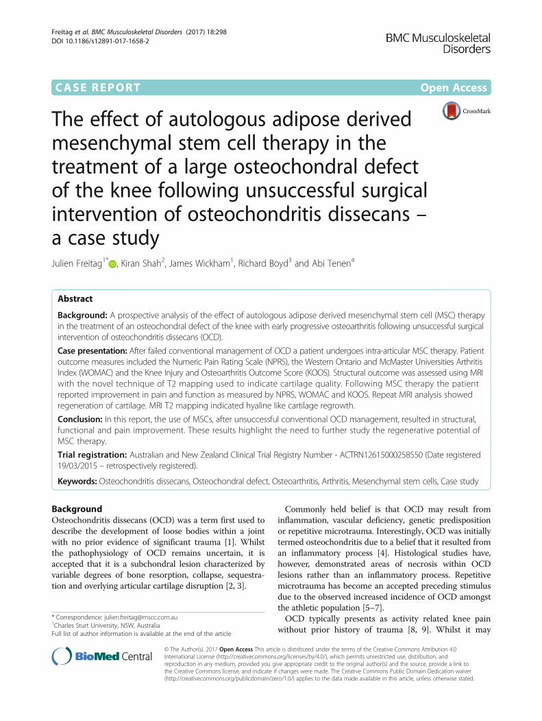
with the previous area of OCD (Fig. 1). MRI showedmarked abnormality over the weight bearing portion ofthe medial femoral condyle measuring 2.3 cm × 1.5 cm.The subchondral cyst had successfully been debrided andgrafted and was no longer present on MRI. A thin layer ofhyper-intense tissue incompletely covered the area ofabnormal cortex (at site of past bone substitute grafting)(Fig. 2). The area of pathology had a modified Inter-national Cartilage Repair Society (ICRS) score of 4. Inaddition to routine MRI protocols, the method of T2-relaxation time cartilage mapping was used. T2 mappingindicated significantly elevated values within the thin layeroverlying the osteochondral defect but also elevated valueswithin the surrounding cartilage (Fig. 3).The patient sought advice regarding further interven-
tions that may improve both his current symptoms butalso his long-term prognosis as he wished to avoidprogressive degeneration and early joint arthroplasty.After careful consideration and as he now had featuresof early degenerative change he was assessed as suitablefor inclusion in an ethics approved, registered case serieson the use of adipose derived MSC therapy in thetreatment of osteoarthritis.The patient was given written information regarding
the use of MSC therapy, including relative risks of MSCtherapy and also relevant treatment alternatives thatcould otherwise be explored. Formal written informedconsent was obtained prior to commencing therapy.
Fig. 1 X-ray of the knee showing flattening of the medial femoralcondyle and intercondylar notch osteophyte formation – KellgrenLawrence Grade II
Freitag et al. BMC Musculoskeletal Disorders (2017) 18:298 Page 3 of 11
Source of mesenchymal stem cellsAdipose tissue was chosen as a source of MSCs due toease of harvest, abundance of MSCs and the observedchondrogenic potential of adipose derived MSCs. Bonemarrow aspirate was considered an alternative source orMSCs but surprisingly has a relative paucity of MSCs –comprising .001–.02% of the mono-nucleated cell popu-lation in comparison to ~1–7% found within adiposetissue [38–40]. Many studies have indicated similarchondrogenic potential between bone marrow derivedand adipose derived MSCs [41].
Autologous adipose derived mesenchymal stem cellpreparationThe patient underwent an abdominal liposuction har-vest procedure. Using a lateral abdominal approach, the
subcutaneous fat was infiltrated with an anaesthetictumescent fluid preparation comprising of 30 mls of 2%lignocaine, 1 ml of 1:1000 adrenaline, 1 ml of 8.4%bicarbonate suspended in a normal saline solution (total1000 ml). Using a 4 mm lipo-aspirate cannula, 60 mls ofadipose tissue and tumescent fluid was successfully har-vested and collected within a sterile medical grade singleuse Shippert Tissu-Trans Collection filter (ShippertMedical, Colorado, USA). The sample was then trans-ferred directly from the theatre via an air lock system to alaboratory clean room facility operated by Magellan StemCells (Magellan Stem Cells, Melbourne, Australia).Autologous MSCs were isolated and expanded from
the harvested adipose tissue using previously publishedprotocols [42]. The processing of the lipo-aspirate wasperformed within the environment of a Biological SafetyCabinet (BSC) Class II using strict sterile techniques.Stromal Vascular Fraction (SVF) obtained from the
lipo-aspirate was culture purified using standard growthmedia containing Minimum Essential Media Eagle(MEM) supplemented with 2 mM glutamate and 10%Fetal Bovine Serum (FBS) (HyClone – GE Healthcare,USA). The cells were cultured under hypoxic conditionswith 5% CO2 at 37 °C. Non-adherent cells were removedby washing with sterile phosphate buffered saline (PBS).The adherent cells were cultured until 80% confluencyin freshly added growth media. The cultured purifiedcells were then harvested at passage 0 (P0). These cellswere further plated to tissue culture flasks and expandedup to passage 2 (P2). The cells were harvested andanalyzed for cell count and viability using a Muse CellAnalyzer (Merck Millipore, USA). The harvested cellswere washed three times to remove FBS and cryopre-served in cryovials using clinical grade qualified MSCcryoprotectant media and following a previouslyvalidated control rate freezing technique of 1 °C/min[43, 44]. The cryovials were transferred to liquidnitrogen storage until use.At completion of isolation and expansion, the cells
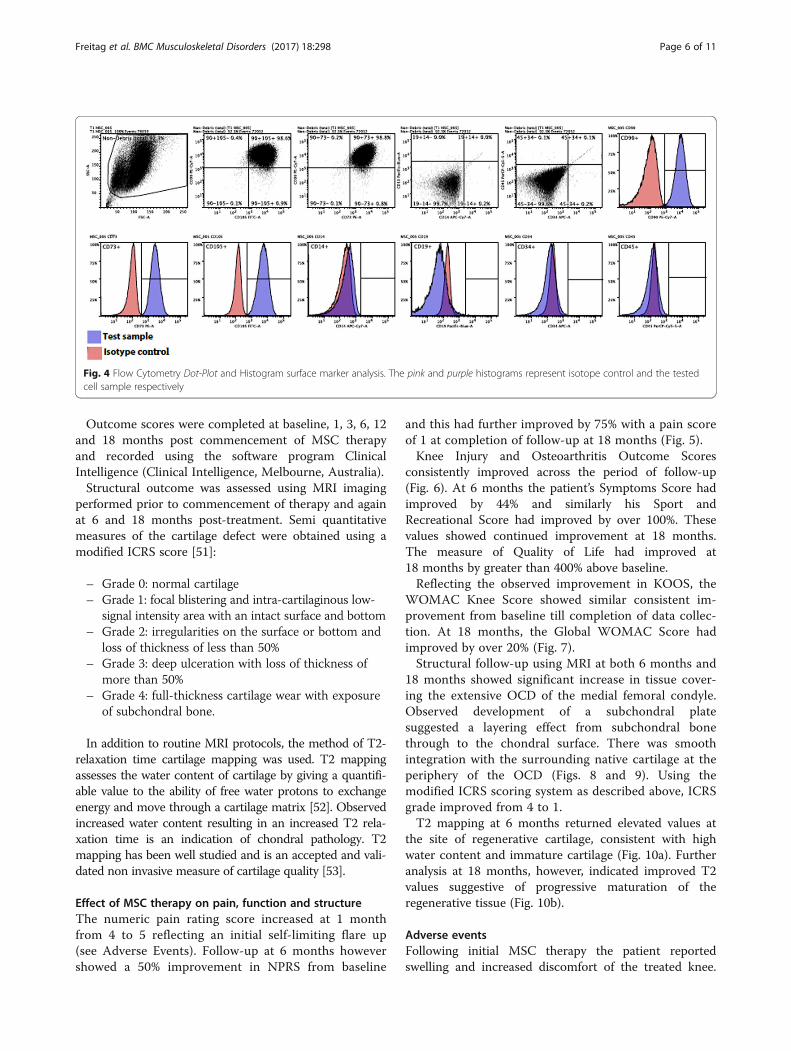
underwent independent phenotypic analysis at MonashUniversity and were characterised by flow cytometryusing Florescent Activated Cell Sorting (FACS). Usingstandards established by the Internal Society of CellularTherapy, the cells were assessed for the presence of posi-tive surface markers CD 90, CD 73 and CD 105 andabsence of hematopoetic surface markers CD14, CD19,CD34 and CD45 [45] (Tables 1-2 and Fig. 4). A furthersample was sent for independent sterility testing formicrobial growth.
Injection methodPrior to the intra-articular injection the cryopreservedcells were retrieved from the liquid nitrogen Dewar andthawed quickly within a sterile 37 °C water bath. The
Fig. 2 Pre-Treatment Proton Density (PD) weighted Coronal andSagittal MRI images of the knee showing the isolated chondraldefect involving the central weight bearing area of the medialfemoral condyle
Fig. 3 Pre-Treatment MRI T2 mapping of the medial compartmentof the knee indicating elevated hydration of the cartilage in the areasurrounding the OCD and also of the thin layer of tissue whichpartially covered the defect
Freitag et al. BMC Musculoskeletal Disorders (2017) 18:298 Page 4 of 11

thawed cells were then washed in chilled PBS to removecryoprotectant media, centrifuged and the resultant cellpellet re-suspended in clinical grade 0.9% normal saline.The cells were again analyzed for cell count and viabilitypost thaw using the Muse Cell Analyzer. The cells wereinjected within 30 min of thawing.The patient received a total of 118 million MSCs (viability
98%) suspended in 3mls of normal saline at commence-ment of therapy. A second injection of 50 million MSCs(viability 95%) at 6 months was also given.At both injection time points the patient’s right knee
was prepped using a chlorhexidine solution and draped.2 mls of 1% lignocaine was infiltrated subcutaneously atthe site of injection. Using a supero-lateral approach tothe patella, and under sterile conditions and ultrasoundguidance, the MSCs suspended in 3 mls of normal salinewere injected into the intra-articular space.
Post injection rehabilitationAfter the initial injection the patient was fitted with amedial compartment customised unloading knee braceto allow protected but full weight-bearing. This wasachieved using an Ossur OA Unloader One brace(Ossur, Reykjavic, Iceland).The patient was given post injection instructions,
which included range of motion exercises and lowerlimb muscle activation exercises, and encouraged to per-form repetitive low impact and low load exercisesagainst minimal resistance on a stationary bike (continu-ous active motion versus continuous passive motion).This protocol was developed due to previously publishedevidence which has indicated the benefit of controlledload on cartilage health [46].
Potential side effects / risksImportantly, recent systematic review and meta-analysisof previous clinical papers investigating the use ofmesenchymal stem cell therapy – both intravascular andintra-articular applications – and including autologous,allogeneic and expanded MSC preparations has indicated
that MSC therapy is safe. No adverse events including in-fection, death or malignancy have been recorded.Saw and colleagues have documented a self-limited
flare up with discomfort and swelling following intra-articular MSC therapy [36]. This was not considered aserious adverse event.Relative risks of the liposuction harvest procedure
include infection, bruising and post operative discom-fort. Whilst the risk of infection is low, the patientreceived a single dose of prophylactic antibioticsprior to the procedure as part of accepted routineclinical practice [47].
Analysis methods and outcome measuresProspective analysis of patient outcome to intra-articularMSC therapy included the following measures:
1. The Knee Injury and Osteoarthritis OutcomeScore (KOOS). This is a validated scoring systemintended for the assessment of knee injury thatmay result in post-traumatic knee osteoarthritis.The score consists of 5 subscales - pain, othersymptoms, function in daily living, function insport and recreation and knee related quality oflife. Standardised answers to questions are given(5 Likert scale) and each question is assigned ascore of 0–4. A normalized score is calculated foreach subscale (100 indicates no symptoms and 0indicates maximum symptoms) [48].
2. The Western Ontario and McMaster UniversitiesArthritis Index (WOMAC Index 3.0). This score is avalidated quality of life score and quantitativelyassesses the pain, stiffness and physical function inpatients with symptomatic osteoarthritis [49].
3. The Numeric Pain Rating Scale (NPRS). Thepatient rates their knee pain intensity over theprevious week on a scale of 0–10. The NPRS hasbeen validated for use in people with kneeosteoarthritis [50].
Table 1 Flow Cytometry Dot-Plot surface marker analysis showing results consistent with mesenchymal stem cells as per theInternational Society of Cellular Therapy guidelines
Dotplots - Positive Markers Dotplots - Negative Markers
CD90 & CD105 + ve CD90 & CD73 + ve CD14 & CD19 + ve CD34 & CD45 + ve
Percentage 98.64 98.83 0.04 0.14
Table 2 Flow Cytometry Histogram surface marker analysis showing results consistent with mesenchymal stem cells as per theInternational Society of Cellular Therapy guidelines
Histograms - Positive Markers Histograms - Negative Markers
CD90 + ve CD73 + ve CD105 + ve CD14 + ve CD19 + ve CD34 + ve CD45 + ve
Percentage 99.01 99.67 99.69 0.28 0.07 0.32 0.29
Freitag et al. BMC Musculoskeletal Disorders (2017) 18:298 Page 5 of 11
Outcome scores were completed at baseline, 1, 3, 6, 12and 18 months post commencement of MSC therapyand recorded using the software program ClinicalIntelligence (Clinical Intelligence, Melbourne, Australia).Structural outcome was assessed using MRI imaging
performed prior to commencement of therapy and againat 6 and 18 months post-treatment. Semi quantitativemeasures of the cartilage defect were obtained using amodified ICRS score [51]:
– Grade 0: normal cartilage– Grade 1: focal blistering and intra-cartilaginous low-
signal intensity area with an intact surface and bottom– Grade 2: irregularities on the surface or bottom and
loss of thickness of less than 50%– Grade 3: deep ulceration with loss of thickness of
more than 50%– Grade 4: full-thickness cartilage wear with exposure
of subchondral bone.
In addition to routine MRI protocols, the method of T2-relaxation time cartilage mapping was used. T2 mappingassesses the water content of cartilage by giving a quantifi-able value to the ability of free water protons to exchangeenergy and move through a cartilage matrix [52]. Observedincreased water content resulting in an increased T2 rela-xation time is an indication of chondral pathology. T2mapping has been well studied and is an accepted and vali-dated non invasive measure of cartilage quality [53].
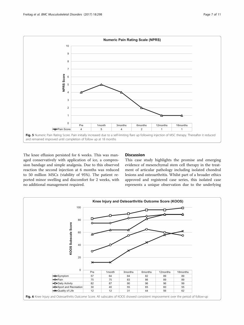
Effect of MSC therapy on pain, function and structureThe numeric pain rating score increased at 1 monthfrom 4 to 5 reflecting an initial self-limiting flare up(see Adverse Events). Follow-up at 6 months howevershowed a 50% improvement in NPRS from baseline
and this had further improved by 75% with a pain scoreof 1 at completion of follow-up at 18 months (Fig. 5).Knee Injury and Osteoarthritis Outcome Scores
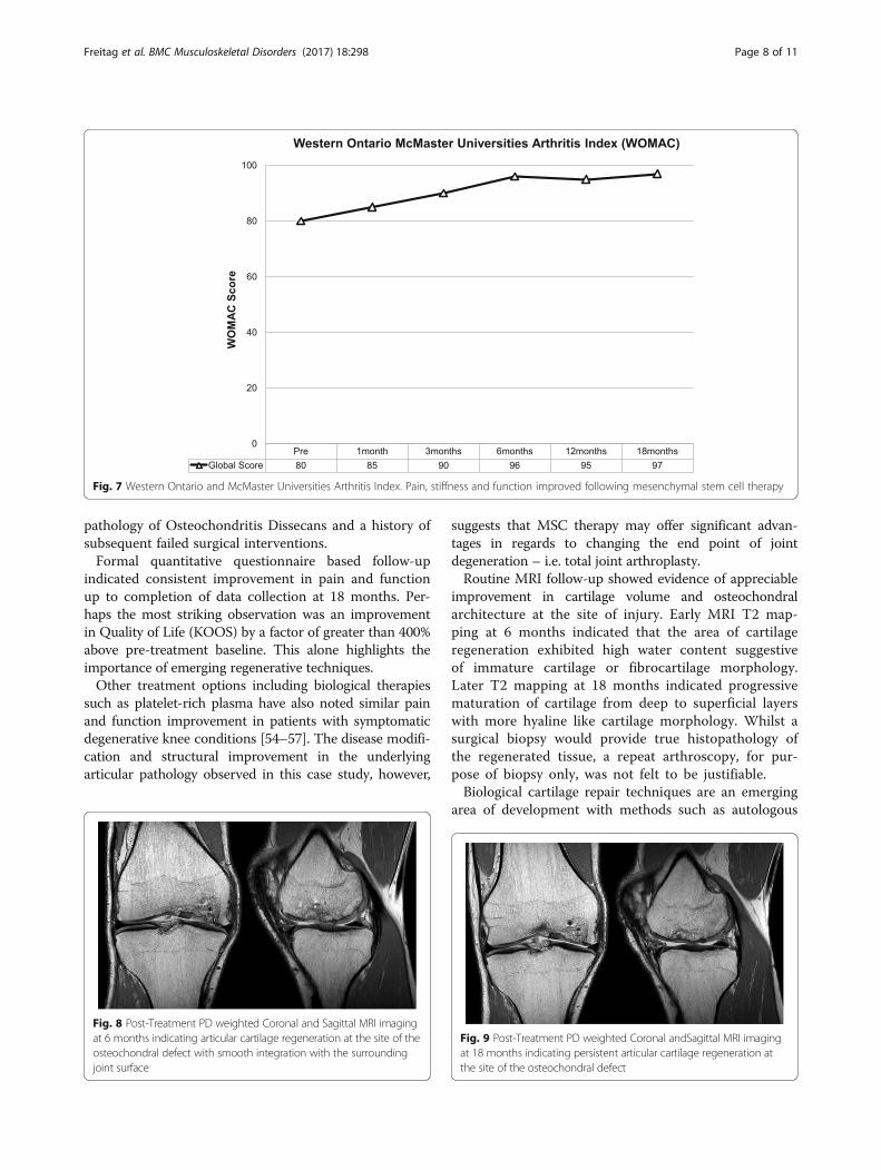
consistently improved across the period of follow-up(Fig. 6). At 6 months the patient’s Symptoms Score hadimproved by 44% and similarly his Sport andRecreational Score had improved by over 100%. Thesevalues showed continued improvement at 18 months.The measure of Quality of Life had improved at18 months by greater than 400% above baseline.Reflecting the observed improvement in KOOS, the
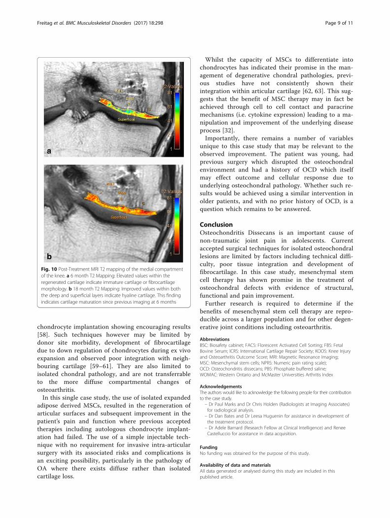
WOMAC Knee Score showed similar consistent im-provement from baseline till completion of data collec-tion. At 18 months, the Global WOMAC Score hadimproved by over 20% (Fig. 7).Structural follow-up using MRI at both 6 months and
18 months showed significant increase in tissue cover-ing the extensive OCD of the medial femoral condyle.Observed development of a subchondral platesuggested a layering effect from subchondral bonethrough to the chondral surface. There was smoothintegration with the surrounding native cartilage at theperiphery of the OCD (Figs. 8 and 9). Using themodified ICRS scoring system as described above, ICRSgrade improved from 4 to 1.T2 mapping at 6 months returned elevated values at
the site of regenerative cartilage, consistent with highwater content and immature cartilage (Fig. 10a). Furtheranalysis at 18 months, however, indicated improved T2values suggestive of progressive maturation of theregenerative tissue (Fig. 10b).
Adverse eventsFollowing initial MSC therapy the patient reportedswelling and increased discomfort of the treated knee.
Fig. 4 Flow Cytometry Dot-Plot and Histogram surface marker analysis. The pink and purple histograms represent isotope control and the testedcell sample respectively
Freitag et al. BMC Musculoskeletal Disorders (2017) 18:298 Page 6 of 11
The knee effusion persisted for 6 weeks. This was man-aged conservatively with application of ice, a compres-sion bandage and simple analgesia. Due to this observedreaction the second injection at 6 months was reducedto 50 million MSCs (viability of 95%). The patient re-ported minor swelling and discomfort for 2 weeks, withno additional management required.
DiscussionThis case study highlights the promise and emergingevidence of mesenchymal stem cell therapy in the treat-ment of articular pathology including isolated chondrallesions and osteoarthritis. Whilst part of a broader ethicsapproved and registered case series, this isolated caserepresents a unique observation due to the underlying
Fig. 5 Numeric Pain Rating Score. Pain initially increased due to a self-limiting flare up following injection of MSC therapy. Thereafter it reducedand remained improved until completion of follow up at 18 months
Fig. 6 Knee Injury and Osteoarthritis Outcome Score. All subscales of KOOS showed consistent improvement over the period of follow-up
Freitag et al. BMC Musculoskeletal Disorders (2017) 18:298 Page 7 of 11
pathology of Osteochondritis Dissecans and a history ofsubsequent failed surgical interventions.Formal quantitative questionnaire based follow-up
indicated consistent improvement in pain and functionup to completion of data collection at 18 months. Per-haps the most striking observation was an improvementin Quality of Life (KOOS) by a factor of greater than 400%above pre-treatment baseline. This alone highlights theimportance of emerging regenerative techniques.Other treatment options including biological therapies
such as platelet-rich plasma have also noted similar painand function improvement in patients with symptomaticdegenerative knee conditions [54–57]. The disease modifi-cation and structural improvement in the underlyingarticular pathology observed in this case study, however,
suggests that MSC therapy may offer significant advan-tages in regards to changing the end point of jointdegeneration – i.e. total joint arthroplasty.Routine MRI follow-up showed evidence of appreciable
improvement in cartilage volume and osteochondralarchitecture at the site of injury. Early MRI T2 map-ping at 6 months indicated that the area of cartilageregeneration exhibited high water content suggestiveof immature cartilage or fibrocartilage morphology.Later T2 mapping at 18 months indicated progressivematuration of cartilage from deep to superficial layerswith more hyaline like cartilage morphology. Whilst asurgical biopsy would provide true histopathology ofthe regenerated tissue, a repeat arthroscopy, for pur-pose of biopsy only, was not felt to be justifiable.Biological cartilage repair techniques are an emerging
area of development with methods such as autologous
Fig. 7 Western Ontario and McMaster Universities Arthritis Index. Pain, stiffness and function improved following mesenchymal stem cell therapy
Fig. 8 Post-Treatment PD weighted Coronal and Sagittal MRI imagingat 6 months indicating articular cartilage regeneration at the site of theosteochondral defect with smooth integration with the surroundingjoint surface
Fig. 9 Post-Treatment PD weighted Coronal andSagittal MRI imagingat 18 months indicating persistent articular cartilage regeneration atthe site of the osteochondral defect
Freitag et al. BMC Musculoskeletal Disorders (2017) 18:298 Page 8 of 11
chondrocyte implantation showing encouraging results[58]. Such techniques however may be limited bydonor site morbidity, development of fibrocartilagedue to down regulation of chondrocytes during ex vivoexpansion and observed poor integration with neigh-bouring cartilage [59–61]. They are also limited toisolated chondral pathology, and are not transferrableto the more diffuse compartmental changes ofosteoarthritis.In this single case study, the use of isolated expanded
adipose derived MSCs, resulted in the regeneration ofarticular surfaces and subsequent improvement in thepatient’s pain and function where previous acceptedtherapies including autologous chondrocyte implant-ation had failed. The use of a simple injectable tech-nique with no requirement for invasive intra-articularsurgery with its associated risks and complications isan exciting possibility, particularly in the pathology ofOA where there exists diffuse rather than isolatedcartilage loss.
Whilst the capacity of MSCs to differentiate intochondrocytes has indicated their promise in the man-agement of degenerative chondral pathologies, previ-ous studies have not consistently shown theirintegration within articular cartilage [62, 63]. This sug-gests that the benefit of MSC therapy may in fact beachieved through cell to cell contact and paracrinemechanisms (i.e. cytokine expression) leading to a ma-nipulation and improvement of the underlying diseaseprocess [32].Importantly, there remains a number of variables
unique to this case study that may be relevant to theobserved improvement. The patient was young, hadprevious surgery which disrupted the osteochondralenvironment and had a history of OCD which itselfmay effect outcome and cellular response due tounderlying osteochondral pathology. Whether such re-sults would be achieved using a similar intervention inolder patients, and with no prior history of OCD, is aquestion which remains to be answered.
ConclusionOsteochondritis Dissecans is an important cause ofnon-traumatic joint pain in adolescents. Currentaccepted surgical techniques for isolated osteochondrallesions are limited by factors including technical diffi-culty, poor tissue integration and development offibrocartilage. In this case study, mesenchymal stemcell therapy has shown promise in the treatment ofosteochondral defects with evidence of structural,functional and pain improvement.Further research is required to determine if the
benefits of mesenchymal stem cell therapy are repro-ducible across a larger population and for other degen-erative joint conditions including osteoarthritis.
AbbreviationsBSC: Biosafety cabinet; FACS: Florescent Activated Cell Sorting; FBS: FetalBovine Serum; ICRS: International Cartilage Repair Society; KOOS: Knee Injuryand Osteoarthritis Outcome Score; MRI: Magnetic Resonance Imaging;MSC: Mesenchymal stem cells; NPRS: Numeric pain rating scale);OCD: Osteochondritis dissecans; PBS: Phosphate buffered saline;WOMAC: Western Ontario and McMaster Universities Arthritis Index
AcknowledgementsThe authors would like to acknowledge the following people for their contributionto the case study.– Dr Paul Marks and Dr Chris Holden (Radiologists at Imaging Associates)for radiological analysis.
– Dr Dan Bates and Dr Leesa Huguenin for assistance in development ofthe treatment protocol.
– Dr Adele Barnard (Research Fellow at Clinical Intelligence) and ReneeCastelluccio for assistance in data acquisition.
FundingNo funding was obtained for the purpose of this study.
Availability of data and materialsAll data generated or analysed during this study are included in thispublished article.
Fig. 10 Post-Treatment MRI T2 mapping of the medial compartmentof the knee. a 6 month T2 Mapping: Elevated values within theregenerated cartilage indicate immature cartilage or fibrocartilagemorphology. b 18 month T2 Mapping: Improved values within boththe deep and superficial layers indicate hyaline cartilage. This findingindicates cartilage maturation since previous imaging at 6 months
Freitag et al. BMC Musculoskeletal Disorders (2017) 18:298 Page 9 of 11
Authors’ contributionsJF, RB, KS were involved in the conception and design of the study. JF, RB,AT, JW, KS were involved in the drafting of the report. JF, RB, AT, JW, KS haveread and approved the final manuscript.
Ethics approval and consent to participatehe subject was formally enrolled in a prospective ethics approved caseseries. The subject completed formal informed consent prior tocommencement of the study. Monash University Human Research EthicsCommittee Number: CF14/2230–2014001175.
Consent for publicationThe subject within this case study has formally consented to publicationof details pertaining to their treatment and outcome.
Competing interestsDr Julien Freitag and Professor Richard Boyd are affiliated with MagellanStem Cells and are members of Magellan Stem Cells Clinical and ScientificAdvisory Board. Dr Kiran Shah is the Chief Scientific Officer of MagellanStem Cells.
Publisher’s NoteSpringer Nature remains neutral with regard to jurisdictional claims inpublished maps and institutional affiliations.
Author details1Charles Sturt University, NSW, Australia. 2Magellan Stem Cells, VIC, Australia.3The Hudson Institute, Victoria, Australia. 4Monash University, VIC, Australia.
Received: 14 September 2016 Accepted: 5 July 2017
References1. Freiberg AH, Woolley PG. Osteochondritis dissecans: concerning its
nature and relation to formation of joint mice. Am J Orthop Surg. 1910;28(3):477–94.
2. Crawford DC, Safran MR. Osteochondritis dissecans of the knee. J AmAcad Orthop Surg. 2006;14(2):90–100.
3. Kocher MS, Tucker R, Ganley TJ, Flynn JM. Management ofosteochondritis dissecans of the knee: current concepts review. Am JSports Med. 2006;34(7):1181–91.
4. Konig F. Ueber freie korper in den gelenken. Dtsch Z Chir. 1887;27:90–109.5. Aichroth P. Osteochondritis dissecans of the knee. A clinical survey. J
Bone Joint Surg (Br). 1971;53(3):440–7.6. Linden B. The incidence of osteochondritis dissecans in the condyles of
the femur. Acta Orthop Scand. 1976;47:664–7.7. Hefti F, Beguiristain J, Krauspe R, Moller-Madsen B, Riccio V, Tschauner C,
Wetzel R, Zeller R. Osteochondritis dissecans: a multicenter study of theEuropean Pediatric Orthopedic Society. J Pediatr Orthop B.1999;8(4):231–45.
8. Glancy GL. Juvenile osteochondritis dissecans. Am J Knee Surg.1999;12:120–4.
9. Gudas R, Simonaityte R, Cekanauskas E, Tamosiunas R. A prospective,randomized clinical study of osteochondral autologous transplantationversus microfracture for the treatment of osteochondritis dissecans inthe knee joint in children. J Pediatr Orthop. 2009;29(7):741–8.
10. Cahill BR, Phillips MR, Navarro R. The results of conservativemanagement of juvenile osteochondritis dissecans using jointscintigraphy. A prospective study. Am J Sports Med. 1989;17(5):601–5.discussion 605–606
11. Garrett JC. Fresh osteochondral allografts for treatment of articulardefects in osteochondritis dissecans of the lateral femoral condyle inadults. Clin Orthop Relat Res. 1994;303:33–7.
12. Chambers HG, Shea KG, Carey JL. AAOS Clinical Practice Guideline:diagnosis and treatment of osteochondritis dissecans. J Am Acad OrthopSurg. 2011;19:307–9.
13. De Smet AA, Fisher DR, Graf BK, Lange RH. Osteochondritis dissecans ofthe knee: value of MR imaging in determining lesion stability and thepresence of articular cartilage defects. AJR Am J Roentgenol.1990;155:549–53.
14. De Smet AA, Ilahi OA, Graf BK. Reassessment of the MR criteria forstability of osteochondritis dissecans in the knee and ankle. SkeletRadiol. 1996;25(2):159–63.
15. Kijowski R, Blankenbaker DG, Shinki K, Fine JP, Graf BK, De Smet AA.Juvenile versus adult osteochondritis dissecans of the knee: appropriateMR imaging criteria for instability. Radiology. 2008;248(2):571–8.
16. Cahill BR. Osteochondritis dissecans of the knee: treatment of juvenileand adult forms. J Am Acad Orthop Surg. 1995;3(4):237–47.
17. Wall EJ, Vourazeris J, Myer GD, Emery KH, Divine JG, Nick TG, et al.The healing potential of stable juvenile osteochondritis dissecansknee lesions. J Bone Joint Surg Am. 2008;90(12):2655–64.
18. Guhl JF. Arthroscopic treatment of osteochondritis dissecans.Clin Orthop Relat Res. 1982;167:65–74.
19. Lee CK, Mercurio C. Operative treatment of osteochondritis dissecansin situ by retrograde drilling and cancellous bone graft: a preliminary report. ClinOrthop Relat Res. 1981;158:129–36.
20. Johnson LL, Uitvlugt G, Austin MD, Detrisac DA, Johnson C.Osteochondritis dissecans of the knee: arthroscopic compressionscrew fixation. Arthroscopy. 1990;6(3):179–89.
21. Bradley J, Dandy DJ. Osteochondritis dissecans and other lesionsof the femoral condyles. J Bone Joint Surg (Br). 1989;71:518–22.
22. Bruns J, Rayf M, Steinhagen J. Longitudinal long-term results of surgicaltreatment in patients with osteochondritis dissecans of the femoral condyles.Knee Surg Sports Traumatol Arthrosc. 2008;16:436–41.
23. Edmonds EW, Albright J, Bastrom T, Chambers HG. Outcomes ofextra-articular, intra-epiphyseal drilling for osteochondritis dissecansof the knee. J Pediatr Orthop. 2010;30(8):870–8.
24. Larsen MW, Pietrzak WS, DeLee JC. Fixation of osteochondritis dissecanslesions using poly(L-lactic acid)/poly(glycolic acid) copolymerbioabsorbable screws. Am J Sports Med. 2005;33(1):68–76.
25. Michael JW, Wurth A, Eysel P, Konig DP. Long-term results after operativetreatment of osteochondritis dissecans of the knee joint-30 year results.Int Orthop. 2008;32:217–21.
26. Prakash D, Learmonth D. Natural progression of osteo-chondraldefect in the femoral condyle. Knee. 2002;9(1):7–10.
27. Bhosale AM, Richardson JB. Articular cartilage: structure, injuries andreview of management. Br Med Bull. 2008;87:77–95.
28. Brittberg M, Lindahl A, Nilsson A, Ohlsson C, Isaksson O, Peterson L. Treatment ofdeep cartilage defects in the knee with autologous chondrocyte transplantation.N Engl J Med. 1994;331(14):889–95.
29. Barry FP, Murphy JM. Mesenchymal ste cells: clinical applications and biologicalcharacterization. Int J Biochem Cell Biol. 2004;36(4):568584.
30. Arinzeh TL. Mesenchymal stem cells for bone repair: preclinical studiesand potential orthopaedic applications. Foot Ankle Clin.2005;10(4):651–65.
31. Noel D, Djouad F, Jorgense C. Regenerative medicine through mesenchymalstem cells for bone and cartilage repair. Curr OpinInvestig Drugs. 2002;3(7):1000–4.
32. Caplan A. What are MSCs therapeutic? New data: new insight.J Pathol. 2009;217:318–24.
33. Im GI, Kim DY, Shin JH, Hyun CW, Cho WH. Repair of cartilage defectin the rabbit with cultured mesenchymal stem cells from bone marrow.J Bone Joint Surg (Br). 2001;83(2):289–94.
34. Grigolo B, Lisignoli G, Desando G, Cavallo C, Marconi E, Tschon M, Giavaresi G,Fini M, Giardino R, et al. Osteoarthritis treated with mesenchymal stem cells onhyaluronan-based scaffold in rabbit.Tissue Eng Part C Methods. 2009;15:647–58.
35. Wakitani S, Imoto K, Yamamoto T, Saito M, Murata M, Yoneda M.Human autologous culture expanded bone marrow mesenchymal celltransplantation for repair of cartilage defects in osteoarthritic knees. OsteoarthrCartil. 2002;10(3):199–206.
36. Saw KY, Anz A, Siew-Yoke Jee C, Merican S, Ching-Soong Ng R, RoohiSA, et al. Articular cartilage regeneration with autologous peripheral blood stemcells versus hyaluronic acid: a randomised controlled trial.Arthroscopy. 2013;29(4):684–94.
37. Freitag J, Ford J, Bates D, Boyd R, Hayne A, Wang Y, et al. Adipose derivedmesenchymal stem cell therapy in the treatment of isolated knee chondrallesions: design of a randomised controlled pilot study comparing arthroscopicmicrofracture versus arthroscopic microfracture combined with postoperativemesenchymal stem cell injections. BMJ Open. 2015;5:e009332. doi:10.1136/bmjopen-2015-009332.
Freitag et al. BMC Musculoskeletal Disorders (2017) 18:298 Page 10 of 11
38. Peng L, Jia Z, Yin X, Zhang X, Liu Y, Chen P, Ma K, Zhou C. Comparativeanalysis of mesenchymal stem cells from bone marrow, cartilage,and adipose tissue. Stem Cells Dev. 2008;17(4):761–74.
39. Alvarez-Viejo M, Menendez-Menendez Y, Blanco-Gelaz MA,Ferrero-Gutierrez A, Fernandez-Rodriguez MA, Gala J, Otero-Hernandez J.Quantifying mesenchymal stem cells in the mononuclear cell fraction ofbone marrow samples obtained for cell therapy. In Transplantationproceedings (Vol. 45, No. 1, pp. 434–439). Elsevier; 2013.
40. Kern S, Eichler H, Stoeve J, Klüter H, Bieback K. Comparative analysis ofmesenchymal stem cells from bone marrow, umbilical cord blood, or adiposetissue. Stem Cells. 2006;24(5):1294–301.
41. Im GI, Shin YW, Lee KB. Do adipose tissue-derived mesenchymal stem cellshave the same osteogenic and chondrogenic potential as bone marrow-derived cells? Osteoarthr Cartil. 2005;13:845–53.
42. Zuk PA, Zhu M, Mizuno H, Huang J, Futrell JW, Katz AJ, et al. Multilineage cellsfrom human adipose tissue: implications for cell-based therapies. Tissue Eng.2001;7(2):211–28.
43. Martinello T, Bronzini I, Maccatrozzo L, Mollo A, Sampaolesi M, Mascarello F, et al.Canine adipose-derived-mesenchymal stem cells do not lose stem features aftera long-term cryopreservation. Res Vet Sci. 2011;91(1):18–24.
44. Goh BC, Thirumala S, Kilroy G, Devireddy RV, Gimble JM, et al. Cryopreservationcharacteristics of adipose derived stem cells : maintenance of differentiationpotential and viability. J Tissue Eng Regen Med. 2007;1(4):322–4.
45. Dominici M, Le Blanc K, Mueller I, Slaper-Cortenbach I, Marini F, Krause D, et al.Minimal Criteria for defining mulitpotent mesenchymal stromal cells. TheInternational Society for Cellular Therapy position statement. Cytotherapy. 2006;8(4):315–7.
46. Grodzinsky AJ, Levenston ME, Jin M, Frank EH. Cartilage tissue remodeling inresponse to mechanical forces. Annu Rev Biomed Eng. 2000;2(1):691–713.
47. Messingham MJ, Arpey CJ. Update on the use of antibiotics in cutaneoussurgery. Dermatol Surg. 2005;31(8 Pt 2):1068–78.
48. Roos EM, Roos HP, Lohmander LS, Ekdahl C, Beynnon BD. Knee Injury andOsteoarthritis Outcome Score (KOOS)-development of a self-administeredoutcome measure. J Orthop Sports Phys Ther. 1998;28(2):88–96.
49. Bellamy N, Buchanan WW, Goldsmith CH, Campbell J, Stitt LW. Validationstudy of WOMAC: a health status instrument for measuring clinicallyimportant patient relevant outcomes to antirheumatic drug therapy inpatients with osteoarthritis of the hip or knee. J Rheumatol. 1988;15(12):1833–40.
50. Dworkin RH, Turk DC, Farrar JT, Haythornthwaite JA, Jensen MP, Katz MP, etal. Core outcome measures for chronic pain clinical trials: IMMPACTrecommendations. Pain. 2005;113:1–2.
51. Brittberg M, Winalski CS. Evaluation of cartilage injuries and repair. J BoneJoint Surg Am. 2003;85-A(Suppl 2):58–69.
52. Mamisch TC, Trattnig S, Quirback S, Marlovits S, White LM, Welsch GH.Quantitative T2 mapping of knee cartilage: differentiation of healthy controlcartilage and cartilage repair tissue in the knee with unloading- initialresults. Radiology. 2010;254(3):818–26.
53. Crema MD, Roemer FW, Marra MD, Burstein D, Gold GE, Eckstein F, Baum T,Mosher TJ, Carrino JA, Guermazi A. Articular Cartilage in the Knee: CurrentMR Imaging Techniques and Applications in Clinical Practice and Research1. Radiographics. 2011;31(1):37–61.
54. Spaková T, Rosocha J, Lacko M, Harvanova D, Gharaibeh A. Treatment ofknee joint osteoarthritis with autologous platelet-rich plasma in comparisonwith hyaluronic acid. Am J Phys Med Rehabil. 2012;91(5):411–7.
55. Kon E, Mandelbaum B, Buda R, Filardo G, Delcogliano M, Timoncini A, et al.Platelet-rich plasma intra-articular injection versus hyaluronic acidviscosupplementation as treatments for cartilage pathology: from earlydegeneration to osteoarthritis. Arthroscopy. 2011;27(11):1490–501.
56. Freitag JB, Barnard A. To evaluate the effect of combining photo-activationtherapy with platelet-rich plasma injections for the novel treatment ofosteoarthritis. BMJ Case Rep. 2013;26:2013. doi:10.1136/bcr-2012-007463.
57. Mascarenhas R, Saltzman BM, Fortier LA, Cole BJ. Role of platelet-rich plasma inarticular cartilage injury and disease. J Knee Surg. 2015;28(1):3–10.
58. Peterson L, Minas T, Brittberg M, Nilsso A, Sjogren-Jansson E, Lindahl A. Two- to9-year outcome after autologous chondrocyte transplantation of the knee. ClinOrthop Relat Res. 2000;374:212–34.
59. Ahsan T, Lottman LM, Harwood F, Amiel D, Sah RL. Integrative cartilage repair:inhibition by beta-aminopropionitrile. J Orthop Res. 1999;17(6):850–7.
60. von der Mark K, Gauss V, von der Mark H, Muller P. Relationship between cellshape and type of collagen synthesized as chondrocytes lose their cartilagephenotype in culture. Nature. 1977;267:531–2.
61. Marlovits S, Hombauer M, Truppe M, Vessei V, Schlegel W. Changes in the ratioof type-I and type-II collagen expression during monolayer culture of humanchondrocytes. J Bone Joint Surg (Br). 2004;86(2):286–95.
62. Jing XH, Yang L, Duan XJ, Xie B, Chen W, Li Z, Tan HB. In vivo MR imagingtracking of magnetic iron oxide nanoparticle labeled, engineered, autologousbone marrow mesenchymal stem cells following intra-articular injection. JointBone Spine. 2008;75(4):432–8.
63. Mokbel AN, El Tookhy OS, Shamaa AA, Rashed LA, Sabry D, El Sayed AM.Homing and reparative effect of intra-articular injection of autologusmesenchymal stem cells in osteoarthritic animal model. BMC MusculoskeletDisord. 2011;12(1):1.
• We accept pre-submission inquiries
• Our selector tool helps you to find the most relevant journal
• We provide round the clock customer support
• Convenient online submission
• Thorough peer review
• Inclusion in PubMed and all major indexing services
• Maximum visibility for your research
Submit your manuscript atwww.biomedcentral.com/submit
Submit your next manuscript to BioMed Central and we will help you at every step:
Freitag et al. BMC Musculoskeletal Disorders (2017) 18:298 Page 11 of 11
Related Documents