The ECF sigma factors of Streptomyces coelicolor A3(2) Mark S. B. Paget, 1 Hee-Jeon Hong, 2 Maureen J. Bibb 2 and Mark J. Buttner 2 1 School of Biological Sciences, University of Sussex, Brighton BN1 9QG, UK 2 Department of Molecular Microbiology, John Innes Centre, Colney, Norwich NR4 7UH, UK INTRODUCTION In bacteria, gene expression is controlled primarily at the level of transcription initia- tion. Control can be achieved through the use of DNA-binding proteins (repressors and activators) that affect the efficiency of initiation, but also through the use of alternative forms of RNA polymerase with different promoter recognition characteristics. The promoter specificity of the RNA polymerase holoenzyme depends on the nature of the subunit that associates with the core enzyme. This key role of in promoter recogni- tion suggests a mechanism for the coordinate control of gene expression using alterna- tive forms of and different subsets of promoters, an idea that was first proposed as soon as the role of was established (Burgess et al., 1969). It is now clear that most, if not all, bacteria use alternative subunits to control gene expression, and that these factors fall into two distinct families: the N (or 54 ) family, which is discussed in the preceding chapter, and the 70 family. The 70 family includes those factors that, broadly speaking, are related in sequence and domain organization to the primary Escherichia coli factor, 70 . Although the overall architecture of members of the 70 family appears to be conserved, the 70 family can be divided into several phylogenet- ically distinct subfamilies (Lonetto et al., 1992). Members of each subfamily are often involved in the control of related functions, such as the heat-shock response, flagella biosynthesis, or sporulation. The ECF subfamily of factors In the late 1980s, biochemical analysis of RNA polymerase from Streptomyces coeli- color and E. coli led to the identification of two factors that were particularly small in SGM symposium 61: Signals, switches, regulons and cascades: control of bacterial gene expression. Editors D. A. Hodgson, C. M. Thomas. Cambridge University Press. ISBN 0 521 81388 3 ©SGM 2002.

Welcome message from author
This document is posted to help you gain knowledge. Please leave a comment to let me know what you think about it! Share it to your friends and learn new things together.
Transcript
The ECF sigma factors ofStreptomyces coelicolor A3(2)Mark S. B. Paget,1 Hee-Jeon Hong,2 Maureen J. Bibb2 andMark J. Buttner2
1School of Biological Sciences, University of Sussex, Brighton BN1 9QG, UK2Department of Molecular Microbiology, John Innes Centre, Colney, Norwich NR4 7UH, UK
INTRODUCTIONIn bacteria, gene expression is controlled primarily at the level of transcription initia-tion. Control can be achieved through the use of DNA-binding proteins (repressors andactivators) that affect the efficiency of initiation, but also through the use of alternativeforms of RNA polymerase with different promoter recognition characteristics. Thepromoter specificity of the RNA polymerase holoenzyme depends on the nature of the subunit that associates with the core enzyme. This key role of in promoter recogni-tion suggests a mechanism for the coordinate control of gene expression using alterna-tive forms of and different subsets of promoters, an idea that was first proposed assoon as the role of was established (Burgess et al., 1969). It is now clear that most, ifnot all, bacteria use alternative subunits to control gene expression, and that these factors fall into two distinct families: the N (or 54) family, which is discussed in thepreceding chapter, and the 70 family. The 70 family includes those factors that,broadly speaking, are related in sequence and domain organization to the primaryEscherichia coli factor, 70. Although the overall architecture of members of the 70
family appears to be conserved, the 70 family can be divided into several phylogenet-ically distinct subfamilies (Lonetto et al., 1992). Members of each subfamily are ofteninvolved in the control of related functions, such as the heat-shock response, flagellabiosynthesis, or sporulation.
The ECF subfamily of factorsIn the late 1980s, biochemical analysis of RNA polymerase from Streptomyces coeli-color and E. coli led to the identification of two factors that were particularly small in
SGM symposium 61: Signals, switches, regulons and cascades: control of bacterial gene expression. Editors D. A. Hodgson, C. M. Thomas. Cambridge University Press. ISBN 0 521 81388 3 ©SGM 2002.
size. In E. coli, E (21.7 kDa) was shown to account for transcription of the gene encod-ing the heat-shock factor, 32, at high temperatures (Erickson & Gross, 1989). In S.coelicolor, another small factor, also named E (20.4 kDa), was shown to direct invitro transcription from one of four promoters (dagAp2) of the agarase-encoding genedagA (Buttner et al., 1988). The cloning of the gene encoding S. coelicolor E severalyears later using a reverse genetics approach revealed that it belonged, together with E.coli E, to a new subfamily of the 70 family (Lonetto et al., 1994). Members of this newsubfamily were sufficiently different from the previously known factors that in manycases they were not identified as factors by standard similarity searching methods. Asa consequence, several members of the subfamily were present in the protein databases,but their biochemical role was unrecognized. Each had been identified by geneticmeans, each had a known positive regulatory role, but with no biochemical under-standing of mechanism. These included AlgU from Pseudomonas aeruginosa, CarQfrom Myxococcus xanthus and FecI from E. coli. The available information about theroles of these factors at the time suggested that they functioned as effector moleculesresponding to extracytoplasmic stimuli, and that they often controlled extracytoplas-mic functions, and for this reason, the new subfamily was named the ECF subfamily(Lonetto et al., 1994). For example, E. coli E is involved in sensing and responding toprotein misfolding in the extracytoplasmic space (Ades et al., 1999), M. xanthus CarQ
activates the synthesis of membrane-localized carotenoids in response to light (Gorhamet al., 1996), and E. coli FecI activates the citrate-dependent iron(III) transport systemin response to citrate and iron in the periplasmic space (Härle et al., 1995). The charac-teristically small size of ECF factors (�20–30 kDa) is accounted for by the absence ofmost or all of both regions 1 and 3 (Lonetto et al., 1994). For a detailed review of domain structure and function see Lonetto et al. (1992, 1994).
Since the initial discovery of the ECF subfamily, hundreds of new members have beendiscovered in a wide variety of Gram-negative and Gram-positive bacteria, mostlythrough genome sequencing projects. Indeed, for several bacteria, including Bacillussubtilis, Mycobacterium tuberculosis and S. coelicolor, ECF factors represent themajor class of factors. It is striking that relatively few ECF factors were discoveredby traditional genetic approaches. For example, in B. subtilis there are seven ECF factor genes, none of which was discovered genetically. This seems to imply that eitherthey are functionally redundant or they control the expression of genes not pertinent tonormal laboratory culture conditions (or both).
The genome sequence of S. coelicolor has revealed an astonishing 51 ECF factorsfrom a total of 65 factors, implying that these proteins play a major role in transcrip-tional regulation in Streptomyces. In order to understand the physiological roles ofthese ECF factors, it will be necessary to elucidate the signals to which they respond,
106 M. S. B. Paget and others
SGM symposium 61
to characterize the regulatory mechanisms involved in their activation, and to identifytheir regulons (the genes under their control). The aim of this review is to summarizecurrent understanding of the biological roles and regulation of the three ECF factors(E, R and BldN) that have been studied in detail in S. coelicolor. For each of thesethree factors, the mechanism controlling factor activity is different, variouslyinvolving de novo synthesis, pro- processing, and anti- factor-directed control. Theseexamples serve to illustrate the fascinating variety of regulatory systems that exist inbacteria to ensure that factors are recruited to core RNA polymerase only whenappropriate.
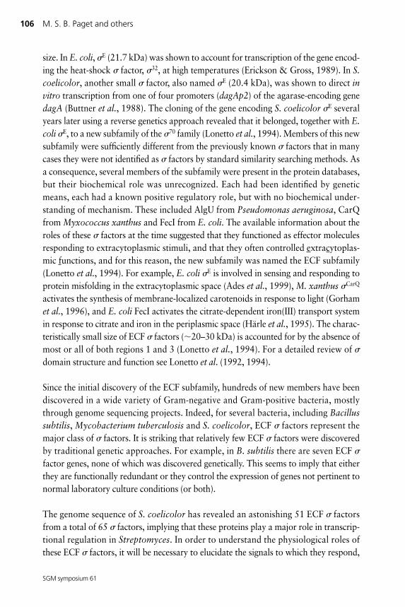
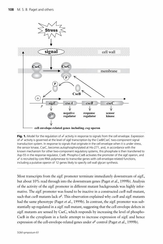
THE �E PATHWAY FOR SENSING AND RESPONDING TO CELLENVELOPE STRESSSince the initial cloning of the sigE gene (Lonetto et al., 1994), extensive analysis sug-gests that E is part of a signal transduction pathway that allows S. coelicolor to senseand respond to changes in the integrity of its cell envelope (Paget et al., 1999a, b). Amodel for the pathway is shown in Fig. 1. The signal transduction system is composedof four proteins, encoded in an operon: E itself; CseA, a negative regulator of unde-fined biochemical function; CseB, a response regulator; and CseC, a sensor histidineprotein kinase with two predicted transmembrane helices; (Csecontrol of sigma E).Expression of E activity is governed at the level of sigE transcription by the CseB/CseCtwo-component signal transduction system. In response to signals that originate in thecell envelope when it is under stress, the sensor kinase, CseC, becomes autophosphor-ylated at His-271, and, in accordance with the known mechanism for other two-component regulatory systems, this phosphate is then transferred to Asp-55 in theresponse regulator, CseB. Phospho-CseB activates the promoter of the sigE operon, andE is recruited by core RNA polymerase to transcribe genes with cell-envelope-relatedfunctions, including a putative operon of 12 genes likely to specify cell wall glycan syn-thesis.
Evidence for the modelsigE null mutants were extremely sensitive to cell wall hydrolytic enzymes, and had analtered cell wall muropeptide profile, suggesting that sigE is required for normal cellwall integrity. Importantly, the sigE mutant was sensitive to both muramidases (forexample, lysozyme) and amidases, which cut the peptidoglycan backbone and thepeptide side chain, respectively, suggesting that the defect in the sigE mutant envelopeallowed hydrolytic enzymes increased access, rather then specifically altering theirtarget sites (Paget et al., 1999a). Mg2� ions are known to have stabilizing effects on cellenvelopes, and sigE null mutants required millimolar levels of Mg2� for normal growthand sporulation, forming crenellated colonies, sporulating poorly, and overproducingthe blue antibiotic actinorhodin in its absence (Paget et al., 1999a).
The ECF sigma factors of S. coelicolor 107
SGM symposium 61
Most transcripts from the sigE promoter terminate immediately downstream of sigE,but about 10% read through into the downstream genes (Paget et al., 1999b). Analysisof the activity of the sigE promoter in different mutant backgrounds was highly infor-mative. The sigE promoter was found to be inactive in a constructed cseB null mutant,such that cseB mutants lack E. This observation explained why cseB and sigE mutantshad the same phenotype (Paget et al., 1999b). In contrast, the sigE promoter was sub-stantially up-regulated in a sigE null mutant, suggesting that the cell envelope defects insigE mutants are sensed by CseC, which responds by increasing the level of phospho-CseB in the cytoplasm in a futile attempt to increase expression of sigE and henceexpression of the cell-envelope-related genes under E control (Paget et al., 1999b).
108 M. S. B. Paget and others
SGM symposium 61
Fig. 1. Model for the regulation of E activity in response to signals from the cell envelope. Expressionof E activity is governed at the level of sigE transcription by the CseB/CseC two-component signaltransduction system. In response to signals that originate in the cell envelope when it is under stress,the sensor kinase, CseC, becomes autophosphorylated at His-271, and, in accordance with theknown mechanism for other two-component regulatory systems, this phosphate is then transferred toAsp-55 in the response regulator, CseB. Phospho-CseB activates the promoter of the sigE operon, andE is recruited by core RNA polymerase to transcribe genes with cell-envelope-related functions,including a putative operon of 12 genes likely to specify cell wall glycan synthesis.
What signal is sensed by CseC?The exact nature of the signal recognized by the sensor kinase is known for relatively fewtwo-component systems. In order to better understand the nature of the signal sensed byCseC, a screening system was developed to test for compounds that induced the sigE pro-moter (H.-J. Hong, M. S. B. Paget, E. Leibovitz & M. J. Buttner, unpublished). The sigEpromoter was placed upstream of a plasmid-borne kanamycin-resistance gene to yield aconstruct that conferred a basal level of kanamycin resistance on the host. A wide selec-tion of antibiotics was then tested to see which increased kanamycin resistance above thebasal level in a plate assay. In agreement with the proposed role for E in controlling cellenvelope integrity, antibiotics that target the cell envelope induce sigE expression. Theseincluded certain �-lactam antibiotics and, most effectively, glycopeptide antibiotics suchas vancomycin and teicoplanin. ‘Negative control’ antibiotics that target the ribosome(e.g. thiostrepton, streptomycin) or DNA gyrase (novobiocin) did not induce sigE expres-sion. In addition to antibiotics, lysozyme was also found to induce sigE expression,making it highly unlikely that CseC senses these inducers directly.
It is important to note that the sigE gene is transcribed under all growth conditionstested, implying that the CseB/CseC signal transduction system may be responding tochanges in cell envelope metabolism that occur during ‘normal’ growth, which areamplified by the effects of antibiotics and enzymes that target the cell envelope.Accordingly, CseC could be activated by the accumulation of an intermediate in pepti-doglycan degradation or biosynthesis, analogous to the control of �-lactam-inducible�-lactamase gene expression in many Gram-negative bacteria (Jacobs et al., 1997).Alternatively, it is conceivable that CseC might be responding to some physical charac-teristic of the cell envelope (e.g. turgor). The KdpD/KdpE sensor kinase/response regu-lator pair of E. coli (Walderhaug et al., 1992; Sugiura et al., 1994) has been proposed tosense and respond to physical changes in the cell envelope.
CseA has a negative role in sigE expressionThe gene immediately downstream from sigE, cseA, appears to play a negative role insigE expression. The basal level of transcription from the sigE promoter was substan-tially higher in a constructed, in-frame cseA deletion mutant, and the maximal level oftranscription from sigEp following induction with vancomycin was also several foldhigher than in the wild-type (H.-J. Hong, M. S. B. Paget, E. Leibovitz & M. J. Buttner,unpublished). Although CseA has no similarity with any other proteins in the data-bases, its first 21 N-terminal amino acids (MAVFVALGVSLAGCGTGGTGA) are pre-dicted to form a single transmembrane domain. Since CseA cannot function as aE-specific anti- factor (E does not direct transcription from the sigE promoter),perhaps it modulates the CseB/CseC signal transduction pathway, for example as aninhibitor of the kinase activity of CseC, or as a CseB-specific phosphatase.
The ECF sigma factors of S. coelicolor 109
SGM symposium 61
�E directs transcription of a putative operon of 12 genes likelyto specify cell wall glycan synthesisAlthough E was discovered by virtue of its ability to direct transcription of dagAp2 invitro, when genetic analysis of sigE began, the activity of this promoter was found to beunaffected in a constructed sigE null mutant (Paget et al., 1999a). Presumably thisreflects relaxed promoter specificity in vitro, and the existence of a closely related ECF that recognizes dagAp2 in vivo. The first bona fide E-dependent promoter identifiedwas hrdDp1 (Paget et al., 1999a; Kang et al., 1997), one of two promoters of the hrdDgene, which itself encodes a factor. However, this discovery was relatively uninforma-tive because the physiological function of HrdD is unknown (hrdD null mutants haveno apparent phenotype; Buttner et al., 1990). To identify further E-dependent promot-ers, computer-searching methods were used to identify sequences in the emerging S.coelicolor genome sequence that closely resemble the hrdDp1 promoter (GCAAC – 17bp – CGTCT). An initial search identified a perfect match lying upstream of 12 genesthat are likely to form an operon (H.-J. Hong, M. S. B. Paget & M. J. Buttner, unpub-lished). The predicted functions of the enzymes encoded by this operon strongly suggestthat the operon specifies the synthesis of a species of cell wall glycan (hence the operonhas been named cwg). High-resolution S1 nuclease mapping showed that the putative�10 and �35 sequences identified by computer searching do indeed correspond to abona fide promoter, and that the cwg promoter is induced by vancomycin in a sigE-dependent manner (H.-J. Hong, M. S. B. Paget & M. J. Buttner, unpublished). Thus aset of genes under E control has been identified that has a clear cell-envelope-relatedfunction, and transcription of these genes has been shown to be induced by vancomycinand, presumably therefore, other cell-wall-targeted antibiotics and enzymes. A con-structed mutant in which the cwg operon was deleted did not show any of the pheno-types associated with sigE mutants, showing that other, as yet unknown, E targetgenes play critical roles in maintaining cell envelope integrity.
THE �R PATHWAY FOR SENSING AND RESPONDING TOOXIDATIVE STRESSR was the second ECF factor to be discovered in S. coelicolor. Like E, it was firstidentified in purified RNA polymerase holoenzyme preparations isolated from liquid-grown cultures (Kang et al., 1997; Paget et al., 1998). The role of R as a key regulatorof the oxidative stress response was discovered after phenotypic analysis of a con-structed sigR null mutant. This mutant was sensitive to oxidizing agents such as thesuperoxide-generating, redox cycling compounds menadione and plumagin, and wasparticularly sensitive to a thiol-specific oxidant called diamide. The cytoplasm of allorganisms is a reducing environment where thiol groups are maintained in theirreduced state. The diamide-sensitive phenotype suggested that sigR mutants may beunable to respond to adverse changes in the thiol–disulphide redox balance, a condition
110 M. S. B. Paget and others
SGM symposium 61
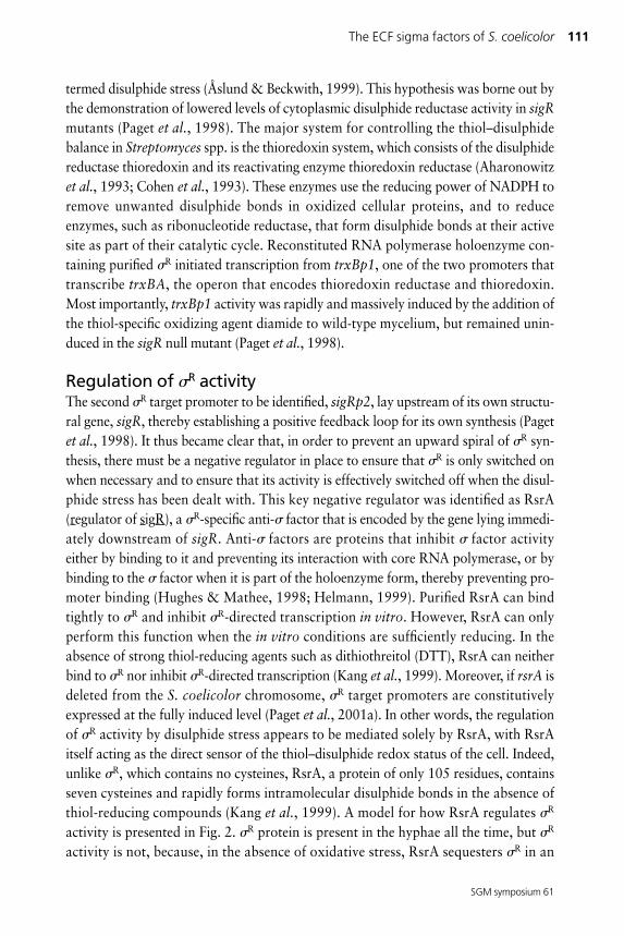
termed disulphide stress (Åslund & Beckwith, 1999). This hypothesis was borne out bythe demonstration of lowered levels of cytoplasmic disulphide reductase activity in sigRmutants (Paget et al., 1998). The major system for controlling the thiol–disulphidebalance in Streptomyces spp. is the thioredoxin system, which consists of the disulphidereductase thioredoxin and its reactivating enzyme thioredoxin reductase (Aharonowitzet al., 1993; Cohen et al., 1993). These enzymes use the reducing power of NADPH toremove unwanted disulphide bonds in oxidized cellular proteins, and to reduceenzymes, such as ribonucleotide reductase, that form disulphide bonds at their activesite as part of their catalytic cycle. Reconstituted RNA polymerase holoenzyme con-taining purified R initiated transcription from trxBp1, one of the two promoters thattranscribe trxBA, the operon that encodes thioredoxin reductase and thioredoxin.Most importantly, trxBp1 activity was rapidly and massively induced by the addition ofthe thiol-specific oxidizing agent diamide to wild-type mycelium, but remained unin-duced in the sigR null mutant (Paget et al., 1998).
Regulation of R activityThe second R target promoter to be identified, sigRp2, lay upstream of its own structu-ral gene, sigR, thereby establishing a positive feedback loop for its own synthesis (Pagetet al., 1998). It thus became clear that, in order to prevent an upward spiral of R syn-thesis, there must be a negative regulator in place to ensure that R is only switched onwhen necessary and to ensure that its activity is effectively switched off when the disul-phide stress has been dealt with. This key negative regulator was identified as RsrA(regulator of sigR), a R-specific anti- factor that is encoded by the gene lying immedi-ately downstream of sigR. Anti- factors are proteins that inhibit factor activityeither by binding to it and preventing its interaction with core RNA polymerase, or bybinding to the factor when it is part of the holoenzyme form, thereby preventing pro-moter binding (Hughes & Mathee, 1998; Helmann, 1999). Purified RsrA can bindtightly to R and inhibit R-directed transcription in vitro. However, RsrA can onlyperform this function when the in vitro conditions are sufficiently reducing. In theabsence of strong thiol-reducing agents such as dithiothreitol (DTT), RsrA can neitherbind to R nor inhibit R-directed transcription (Kang et al., 1999). Moreover, if rsrA isdeleted from the S. coelicolor chromosome, R target promoters are constitutivelyexpressed at the fully induced level (Paget et al., 2001a). In other words, the regulationof R activity by disulphide stress appears to be mediated solely by RsrA, with RsrAitself acting as the direct sensor of the thiol–disulphide redox status of the cell. Indeed,unlike R, which contains no cysteines, RsrA, a protein of only 105 residues, containsseven cysteines and rapidly forms intramolecular disulphide bonds in the absence ofthiol-reducing compounds (Kang et al., 1999). A model for how RsrA regulates R
activity is presented in Fig. 2. R protein is present in the hyphae all the time, but R
activity is not, because, in the absence of oxidative stress, RsrA sequesters R in an
The ECF sigma factors of S. coelicolor 111
SGM symposium 61
RsrA :R complex. R is released during oxidative stress as a direct consequence of theinactivation of RsrA through intramolecular disulphide bond formation. R is then freeto associate with core RNA polymerase and activate transcription of its target genes,including trxBA and other thiol–disulphide oxidoreductase genes (see ‘The R regulon’below). At least in vitro, oxidized RsrA is a direct biochemical substrate for purifiedthioredoxin, the product of the trxA gene (Kang et al., 1999). If the thioredoxin system
112 M. S. B. Paget and others
SGM symposium 61
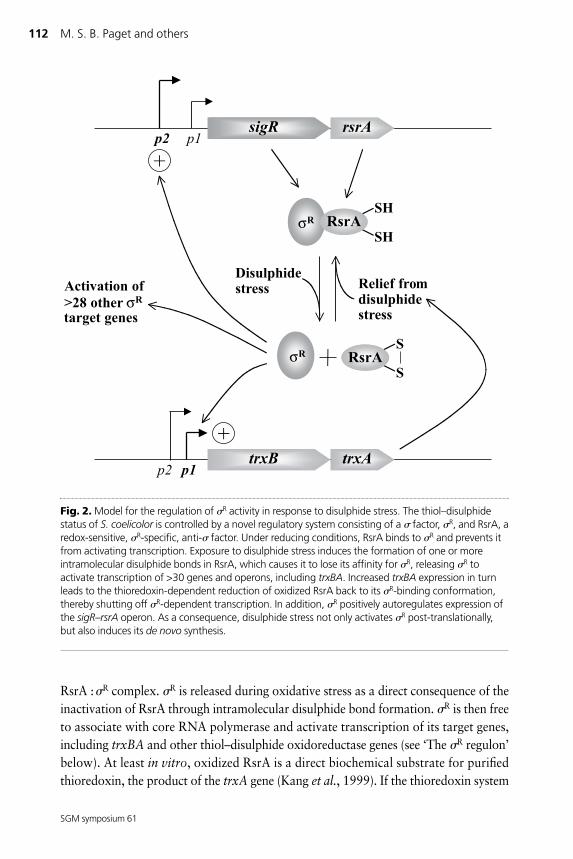
Fig. 2. Model for the regulation of R activity in response to disulphide stress. The thiol–disulphidestatus of S. coelicolor is controlled by a novel regulatory system consisting of a factor, R, and RsrA, aredox-sensitive, R-specific, anti- factor. Under reducing conditions, RsrA binds to R and prevents itfrom activating transcription. Exposure to disulphide stress induces the formation of one or moreintramolecular disulphide bonds in RsrA, which causes it to lose its affinity for R, releasing R toactivate transcription of >30 genes and operons, including trxBA. Increased trxBA expression in turnleads to the thioredoxin-dependent reduction of oxidized RsrA back to its R-binding conformation,thereby shutting off R-dependent transcription. In addition, R positively autoregulates expression ofthe sigR–rsrA operon. As a consequence, disulphide stress not only activates R post-translationally,but also induces its de novo synthesis.
also reduces (reactivates) RsrA in vivo, this would allow it to rebind R and shut downthe response, thereby creating a simple homeostatic feedback loop in which the R
regulon is regulated in response to changes in the thiol–disulphide redox status of thehyphae.
This model raises several important questions, including the exact nature of the redoxevent that inactivates RsrA. Attempts to identify which of the seven cysteines in RsrAform the disulphide bond switch have not been straightforward. In principle, the loss ofa cysteine residue that is involved in inactivating RsrA might be expected to lock RsrAin a constitutively active conformation, causing it to bind R irrespective of the redoxconditions. However, the individual substitution of each of seven RsrA cysteines did notreveal such mutants. Four of the cysteines in RsrA could be substituted, individually orcollectively, still leaving a protein that could both inhibit R activity and release itduring disulphide stress. The remaining three individual cysteine mutants (C11, C41and C44) had no R-binding activity, preventing analysis of their ability to sense redox(Paget et al., 2001a). There is now good evidence to suggest that, in their reduced state,these three cysteines play an important role in the R-binding activity of RsrA by coor-dinating a zinc cofactor (see below).
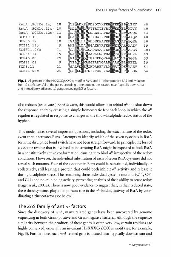
The ZAS family of anti- factorsSince the discovery of rsrA, many related genes have been uncovered by genomesequencing in both Gram-positive and Gram-negative bacteria. Although the sequencesimilarity between the products of these genes is often very low, certain residues arehighly conserved, especially an invariant HisXXXCysXXCys motif (see, for example,Fig. 3). Furthermore, each rsrA-related gene is located near (typically downstream and
The ECF sigma factors of S. coelicolor 113
SGM symposium 61
Fig. 3. Alignment of the HisXXXCysXXCys motif in RsrA and 11 other putative ZAS anti- factorsfrom S. coelicolor. All of the genes encoding these proteins are located near (typically downstreamand immediately adjacent to) genes encoding ECF factors.
immediately adjacent to) an ECF -factor gene, strongly suggesting that the corre-sponding pair of proteins interact. Metal content analysis of RsrA (Paget et al., 2001a)and ChrH (an RsrA-related anti- factor from Rhodobacter sphaeroides; see below)(Newman et al., 2001) revealed that they are zinc metalloproteins. This, together withthe absolute conservation of HisXXXCysXXCys, a potential zinc-binding motif,strongly suggests that all RsrA-related proteins are likely to bind zinc. This new familyof proteins was therefore named the ZAS (zinc-binding anti- factor) family of anti-factors (Paget et al., 2001a).
The redox regulation of RsrA is not a paradigm for all ZASanti- factorsImportantly, although all RsrA-related anti- factors probably bind zinc, it is alreadyclear that their activities are likely to be regulated in diverse ways, so the regulation ofRsrA activity by a reversible thiol–disulphide redox switch is not a paradigm for thewhole family. Thus a gene encoding a ZAS anti- factor lies immediately downstreamof the sigW gene in B. subtilis, but W-dependent gene expression is not induced bydiamide and the known W target genes have no obvious connection to thiol–disul-phide metabolism (Huang et al., 1999; Cao et al., 2001; Wiegert et al., 2001; J.Helmann, pers. comm.). Similarly, the ZAS anti- factor ChrR controls the activity ofE in Rhodobacter sphaeroides, but E directs expression of the cytochrome c2 structu-ral gene (Newman et al., 1999, 2001). Further, deletion of chrR or the E-encodingrpoE does not affect the resistance of R. sphaeroides to diamide, and diamide does notinduce E-dependent gene expression (Newman et al., 2001; T. Donohue & J.Newman, pers. comm.). Eleven of the 51 ECF factors in S. coelicolor are encoded bygenes located near (typically upstream and immediately adjacent to) zas genes, and aretherefore likely to be regulated by a ZAS anti- factor (Fig. 3). Several of these proteinsdiffer from RsrA in having predicted transmembrane helices C-terminal to theHisXXXCysXXCys motif, suggesting that these ZAS proteins may regulate theircognate factor in response to extracytoplasmic signals.
The R regulonSearches for further R target genes were made possible by the generation of a consen-sus target promoter sequence (GGAAT – 18 bp – GTT) using for comparison trxBp1and sigRp2, together with the sequence of hrdDp2, another promoter recognized by R
in vitro. Computer searches showed that this sequence occurred more than 60 times inthe S. coelicolor genome, although only 34 of these were appropriately positioned justupstream from a gene. Each of these 34 sequences was examined experimentally forpromoter activity; including sigRp2, trxBp1 and hrdDp2, 30 were bona fide promotersthat were induced by diamide in a R-dependent manner (Paget et al., 2001b). More
114 M. S. B. Paget and others
SGM symposium 61
than half of the R target genes associated with these promoters have no known biolog-ical function.
Unsurprisingly, several R target genes are likely to play important roles in thiol metab-olism, including a second thioredoxin, trxC, and a glutaredoxin-like gene. Togetherwith the trxBA operon, the induction of these genes by R presumably helps to restorethe thiol–disulphide balance following disulphide stress. Apart from cysteine thiols inproteins, low-molecular-mass thiols are also likely to become oxidized during disul-phide stress, and the induction of the R targets cysM and moeB is likely to act torestore levels of reduced cysteine and the dithiol-containing cofactor molybdopterin,respectively (Paget et al., 2001b). Unlike Gram-negative bacteria and eukaryotes thatuse the cysteine-containing tripeptide glutathione as their major thiol buffer,Streptomyces and mycobacteria use a structurally unrelated, sugar-containing mono-thiol compound called mycothiol (Newton et al., 1996). Although no target genes werefound that were predicted to play a role in mycothiol biosynthesis, the sigR mutant wasfound to have significantly lowered levels of mycothiol (Paget et al., 2001b). The rootcause of diamide sensitivity in sigR mutants could therefore be due to any one of theseR-dependent mechanisms for coping with disulphide stress, or a combination of all ofthem.
At least three R targets encode ribosome-associated products, including relA, ssrA andthe ribosomal protein gene rpmE, suggesting that ribosome composition and functionare modified in response to disulphide stress (Paget et al., 2001b). RelA catalyses theproduction of ppGpp when ribosomes stall due to an uncharged tRNA entering theribosome A-site. This intracellular signalling molecule then elicits the stringent responseby selectively inhibiting transcription of rRNA genes, thereby acting to slow growth(Cashel et al., 1996; Chatterji & Ojha, 2001). In Streptomyces spp., ppGpp also elicitsantibiotic production in response to nutritional stress, and plays a role in differentiation(Chakraburtty & Bibb, 1997). ssrA encodes an unusual small stable tRNA–mRNAhybrid called tmRNA, which also acts when ribosomes stall, either at a rare codon orwhen ribosomes reach the end of a 3� truncated mRNA that lacks a stop codon. tmRNArescues the ribosome by acting as a surrogate mRNA to tag the nascent peptide with ahydrophobic tag that targets the protein for degradation (Keiler et al., 1996; Roche &Sauer, 1999; Karzai et al., 2000). It is tempting to speculate that disulphide stress inhib-its some aspect of the translation process causing ribosomes to stall. A possible riboso-mal target for disulphide stress is the product of the R target gene rpmE, ribosomalprotein L31, which contains a CysXXCys motif. The induction of relA and ssrA maythen provide pathways to rescue stalled ribosomes and to slow ribosome production andgrowth, respectively, thereby focusing available resources on stress survival.
The ECF sigma factors of S. coelicolor 115
SGM symposium 61
Another interesting R target, rbpA, encodes a newly discovered RNA polymerase-binding protein, which may well be a novel low-molecular-mass RNA polymerasesubunit (Paget et al., 2001b). RbpA appears to exist only in the actinomycetes, includ-ing the mycobacteria. Although the role of RbpA is not known, the induction of rbpAtranscription by R suggests that the composition and function of RNA polymerasemay also be modified in response to disulphide stress. Like the ribosome subunit L31,RbpA contains a CysXXCys motif, suggesting that it too may undergo thiol–disulphideredox reactions and may be a target of disulphide stress.
It should be noted that the method used to identify R target promoters means thatthere may be many other, unidentified targets having promoter sequences that differslightly from the consensus sequence used in the computer searches. The total R
regulon may therefore be considerably larger than the current total. Nonetheless, theidentification of 30 genes and operons under R control is a very significant steptowards understanding the cellular response to disulphide stress in S. coelicolor.
Is R a checkpoint in development?A completely unexpected consequence of rsrA inactivation was a block in sporulation,and there is some circumstantial evidence to suggest that S. coelicolor may use R as acheckpoint to inhibit development under conditions of oxidative stress, which maymake sporulation undesirable. S. coelicolor differentiates on solid agar plates byforming aerial hyphae that grow out of the aqueous environment of the substrate myce-lium into the air. These multigenomic aerial hyphae eventually undergo synchronousseptation to produce chains of unigenomic exospores. Developmental mutants that areunable to raise an aerial mycelium have a shiny appearance on agar plates and aretermed ‘bald’ (bld) mutants. Mutants that raise an aerial mycelium in the normal waybut are unable to complete the developmental process by sporulating are termed white(whi) mutants, because the colonies fail to develop the characteristic grey pigment asso-ciated with mature spores.
A constructed rsrA mutant had a classical ‘early’ white phenotype, forming aerial myce-lium, but failing to initiatiate sporulation septation. In contrast, a constructed sigR rsrAdouble mutant sporulated normally, showing that the inability of the rsrA singlemutant to sporulate was a consequence of uncontrolled R activity. One possible expla-nation for these observations is that the high level of free R out-competes a sporula-tion-specific factor, such as WhiG (Chater et al., 1989), for core RNA polymerase(Paget et al., 2001a). However, recent analogous experiments with U and RsuA,another ECF factor :ZAS anti- factor pair in S. coelicolor, provided circumstantialevidence against this model (Gehring et al., 2001). Disruption of rsuA caused a baldphenotype, but a sigU rsuA double mutant developed normally, again showing that the
116 M. S. B. Paget and others
SGM symposium 61
block in differentiation was a consequence of uncontrolled activity. As pointed out byGehring et al. (2001), it seems unlikely that R and U could differentially compete withdifferent factors, one required for aerial mycelium formation and one required forspore formation.
An alternative hypothesis is that the developmental phenotype of the rsrA null mutantis physiologically significant, that R directs transcription of a ‘sporulation inhibitorgene(s) ’, and that S. coelicolor uses this mechanism as a checkpoint to arrest develop-ment under conditions of disulphide stress, which make sporulation undesirable(Gehring et al., 2001; Paget et al., 2001a). If this latter hypothesis is valid, it should bepossible to identify mutations in the proposed ‘sporulation inhibitor gene’ that suppressthe white phenotype of rsrA mutants, provided that there is only one R target gene thatmediates the arrest of development, and that this gene is non-essential. However, thefour rsrA suppressor mutations characterized to date all map to sigR (Paget et al.,2001a).
The �R–RsrA system also exists in pathogenic actinomycetesThe R–RsrA system appears to exist in other actinomycetes. It is certainly present inmycobacteria, where it is named H–RshA (Fernandes et al., 1999; Paget et al., 1998; I.Smith, pers. comm.), and analysis of the near-complete genome sequence ofCorynebacterium diphtheriae (http://www.sanger.ac.uk/Projects/C_diphtheriae/) sug-gests that it also exists in this important actinomycete pathogen (M. S. B. Paget, unpub-lished). Of the 30 S. coelicolor R target genes and operons so far identified, 13 of thehomologues in M. tuberculosis have sequences upstream that resemble the consensusfor R-dependent promoters and may therefore be regulated by H in M. tuberculosis(Paget et al., 2001b). These include homologues of the S. coelicolor genes sigR, trxBA,ssrA, rpmE and rbpA. These observations make it likely that the H–RshA system con-tributes to the well known resistance of M. tuberculosis to oxidative killing by whiteblood cells during human infection.
THE �BldN PATHWAY TO AERIAL MYCELIUM FORMATIONUnlike E and R, which were discovered biochemically, BldN was identified geneticallyin a screen for new genes involved in morphological differentiation (Ryding et al.,1999; Bibb et al., 2000). Two NTG-induced point mutants were isolated in the geneencoding BldN, the two mutants having strikingly different phenotypes. One, R650,had a white colony phenotype, and microscopic examination showed that the colonyproduced aberrant spores that were longer than those of the wild-type. The second,R112, had a more severe phenotype, producing substantially less aerial mycelium thanthe parental strain and only very rare spore chains, sometimes showing highly irregularsporulation septum placement (Ryding et al., 1999). Shotgun complementation of
The ECF sigma factors of S. coelicolor 117
SGM symposium 61
R650 and R112, followed by subcloning and sequencing, showed that this new devel-opmental gene encoded an ECF factor (Bibb et al., 2000). That both these mutantsretained partial BldN activity became clear when a constructed null mutant was foundto have a bald phenotype, devoid of aerial hyphae. Therefore, the gene was namedbldN. Sequence analysis of the two NTG-induced bldN mutant alleles revealed that themore ‘severe’ mutant, R112, carries a mutation in the ribosome-binding site and pre-sumably produces reduced amounts of wild-type BldN, while in the ‘weak’ mutant,R650, the BldN produced carries a glycine to aspartate substitution in region 2.1 (Bibbet al., 2000). In other factors, region 2.1 has been implicated in the interaction of with core RNA polymerase (Burgess & Anthony, 2001), and it is therefore likely thatthe mutant BldN produced by R650 interacts less efficiently with core RNA polymerasethan the wild-type protein.
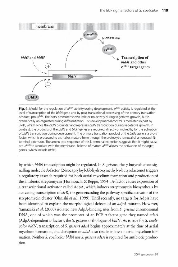
Control of bldN transcriptionThe bldN promoter is temporally regulated, showing little or no activity during vegeta-tive growth, but increasing dramatically during aerial mycelium formation and remain-ing highly active during sporulation (Bibb et al., 2000). Clues as to the mechanism thatcontrols this temporal regulation in S. coelicolor have come from the analysis of bldNtranscription in other bld mutants. No bldN transcripts were detectable in bldG andbldH mutant backgrounds, indicating that bldN expression depends on these twogenes, either directly or indirectly (Fig. 4; Bibb et al., 2000). bldH has not been charac-terized, but bldG encodes a homologue of the SpoIIAA anti-anti- factor from B. subti-lis, implying that the role of bldG is indirect. Anti-anti- factors are proteins thatinhibit the activity of anti- factors, thereby stimulating the activity of its cognate factor. One possibility, therefore, is that bldG mutants have reduced activity of the factor that is required for transcription of the bldN promoter, caused by the uncon-trolled activity of the respective anti- factor.
In contrast to the wild-type, bldN transcripts were readily detectable during vegetativegrowth in a bldD mutant, indicating that bldD acts to repress bldN transcriptionduring vegetative growth (Fig. 4; Elliot et al., 2001). In vitro biochemical experimentsshowed that this effect is direct; BldD is a repressor of the bldN promoter, binding totwo operator sites, one either side of the transcription start site (Elliot et al., 2001).Interestingly, BldD also represses transcription of another key developmental gene,whiG, during vegetative growth (Elliot et al., 2001), and of the development-specificpromoter (p2) of the sigH gene in vegetative hyphae (Kelemen et al., 2001), suggestingthat one of BldD’s roles is to prevent premature expression of developmental genes.
Investigations by Yamazaki et al. (2000), working on the orthologue of BldN inStreptomyces griseus, have raised some intriguing possibilities for another mechanism
118 M. S. B. Paget and others
SGM symposium 61
by which bldN transcription might be regulated. In S. griseus, the �-butyrolactone sig-nalling molecule A-factor (2-isocapryloyl-3R-hydroxymethyl-�-butyrolactone) triggersa regulatory cascade required for both aerial mycelium formation and production ofthe antibiotic streptomycin (Horinouchi & Beppu, 1994). A-factor causes expression ofa transcriptional activator called AdpA, which induces streptomycin biosynthesis byactivating transcription of strR, the gene encoding the pathway-specific activator of thestreptomycin cluster (Ohnishi et al., 1999). Until recently, no targets for AdpA havebeen identified to explain the morphological defects of an adpA mutant. However,Yamazaki et al. (2000) isolated new AdpA-binding sites from S. griseus chromosomalDNA, one of which was the promoter of an ECF factor gene they named adsA(AdpA-dependent factor), the S. griseus orthologue of bldN. As is true for S. coeli-color bldN, transcription of S. griseus adsA begins approximately at the time of aerialmycelium formation, and disruption of adsA also results in loss of aerial mycelium for-mation. Neither S. coelicolor bldN nor S. griseus adsA is required for antibiotic produc-tion.
The ECF sigma factors of S. coelicolor 119
SGM symposium 61
Fig. 4. Model for the regulation of BldN activity during development. BldN activity is regulated at thelevel of transcription of the bldN gene and by post-translational processing of the primary translationproduct, pro-BldN. The bldN promoter shows little or no activity during vegetative growth, but isdramatically up-regulated during differentiation. This developmental control is mediated in part byBldD, which binds the bldN promoter and represses bldN transcription during vegetative growth. Incontrast, the products of the bldG and bldH genes are required, directly or indirectly, for the activationof bldN transcription during development. The primary translation product of the bldN gene is a pro-factor, which is processed to a smaller, mature form through the proteolytic removal of an unusual N-terminal extension. The amino acid sequence of this N-terminal extension suggests that it might causepro-BldN to associate with the membrane. Release of mature BldN allows the activation of its targetgenes, which include bldM.
S. coelicolor does not produce A-factor, but it does produce several closely related �-butyrolactone molecules (Efremenkova et al., 1985; Kawabuchi et al., 1997; Takano etal., 2000). These molecules are involved in a signalling pathway for antibiotic produc-tion, and there is evidence to suggest that some of them may also be involved inmorphological development in S. coelicolor. The predicted AdpA-binding site is notclearly conserved in the promoter region of S. coelicolor bldN, but there is a very closerelative of adpA in the S. coelicolor genome sequence, and it will be interesting to seewhether it has a role in the control of bldN transcription.
Post-translational processing of BldN
Most ECF factors either completely lack conserved region 1 or have only a few resi-dues upstream of region 2.1 (Lonetto et al., 1994). BldN is unusual in having anN-terminal extension of approximately 86 amino acids that is not present in other factors (Bibb et al., 2000). Using a combination of immunoblotting and mutationalanalysis of the N-terminal extension, we have obtained substantial evidence that theprimary translation product of the bldN gene is a pro- factor, which is processed to asmaller, mature form through the proteolytic removal of most of the N-terminal exten-sion (Fig. 4; M. J. Bibb & M. J. Buttner, unpublished). During B. subtilis development,the mother-cell-specific factors E and K are synthesized as inactive pro- factorsthat are subsequently activated by proteolysis of the N-terminal 29 and 20 amino acids,respectively, by membrane-localized proteases (Errington, 1996; Stragier & Losick,1996). In both cases, the activation of this processing event is triggered by signalsderived from the forespore, and this ‘crosstalk’ serves to coordinate the divergent pro-grams of gene expression between the two cellular compartments within the sporan-gium (Errington, 1996; Stragier & Losick, 1996). The pro sequences of both pro-E
and pro-K promote membrane association, whereas the mature forms of these pro-teins are found in the cytoplasm associated with core RNA polymerase (Hofmeister,1998; Ju & Haldenwang, 1999; Ju et al., 1997; Zhang et al., 1998). The putative prosequence of BldN contains a stretch of 20 hydrophobic amino acids (YAVPALAAAAV-PAGPCYALA). It will be interesting in the future to determine if pro-BldN is mem-brane-associated, to identify the pro-BldN protease, and to define the signalsresponsible for triggering the processing event.
The BldN regulonTo date, only one BldN target gene has been identified (Bibb et al., 2000). Given theinvolvement of BldN in the control of aerial mycelium formation, it seemed likely thatother bld genes might be regulated by BldN and would therefore have promotersequences related to the consensus sequences of other ECF factors. Analysis of thepromoter regions of known bld genes revealed a possible ECF consensus-like promoterupstream of bldM. bldM encodes an apparently typical member of the FixJ subfamily
120 M. S. B. Paget and others
SGM symposium 61
of response regulators, although, surprisingly, aspartate-54, the putative site of phos-phorylation, is not required for BldM function (Molle & Buttner, 2000). Transcriptmapping experiments identified two promoters, one of which, bldMp1, correspondedto the putative ECF factor consensus-like sequence. Like the bldN promoter, bldMp1was developmentally regulated, being inactive during vegetative growth, but stronglyup-regulated during aerial mycelium formation and sporulation. Furthermore, bldMp1was inactive in a bldN null mutant and was recognized by reconstituted BldN-contain-ing holoenzyme in vitro, showing that bldM is a direct biochemical target for BldN
holoenzyme (Bibb et al., 2000).
Overlapping promoter specificity between ECF factorsPrior to the discovery of the ECF subfamily, sequence similarity had already been notedbetween the E. coli E target rpoHp3 and the S. coelicolor E target dagAp2 (Erickson& Gross, 1989). Following the characterization of many more promoters under thecontrol of different ECF factors, it became clear that there was a significant degree ofsequence conservation between them. This fact, together with the existence of multipleECF factors in many bacteria, suggested that some promoters might be recognized bymore than one ECF in vivo, and it is now clear that this is indeed the case. Forexample, of the 30 R target promoters known, at least 13 retained some activity in asigR null mutant. Furthermore, this R-independent transcription was constitutive forsome promoters but stimulated by diamide (but with delayed kinetics) for others,implying that it represented more than one additional ECF factor (Paget et al.,2001b). What are the key DNA sequence features that allow some R target promotersto be recognized by additional holoenzymes forms while other promoters are recog-nized uniquely by R? Analysis of the 30 known R target promoters indicates thatmost promoters that are recognized by additional factors contain the �10 sequenceCGTT, whereas those recognized only by R have the �10 sequence TGTT or GGTT.Although the importance of the �10 region of R target promoters in selectivity hasnot been proven, Helmann and colleagues have demonstrated that this region plays acritical role in selectivity between two ECF factors in B. subtilis. Single or doublenucleotide changes in the �10 region of X or W target promoters switched their rec-ognition characteristics such that promoters that were usually recognized by W wererecognized by X, and vice versa (Qiu & Helmann, 2001). Recognition of a single pro-moter by multiple holoenzyme forms provides a very attractive mechanism for integrat-ing different signal transduction pathways at single promoter elements. Overlappingspecificity may be particularly useful in stress responses because different physicalinsults can often lead to the same physiological stress. For example, both oxidativestress and heat shock can induce protein misfolding. Nevertheless, target promotersequence constraints must presumably ensure that, within the total subfamily of 51ECF factors in S. coelicolor, each individual ECF factor has a distinct regulon and a
The ECF sigma factors of S. coelicolor 121
SGM symposium 61
distinct biological role. The future identification of the complete regulons for each ofthese ECF factors using DNA microarrays will begin to address these intriguingissues.
CONCLUSIONSThe ECF subfamily of factors has emerged as a major class of regulatory proteins inStreptomyces spp. Detailed analysis of just three of these proteins – E, R and BldN –has already revealed their involvement in a fascinating range of biological processes andshown that control of their activity can be exerted at several different levels, variouslyinvolving de novo synthesis, pro- processing, and anti- factor-directed regulation.Understanding the role and regulation of each of the remaining 48 ECF factors prom-ises to be an absorbing task.
REFERENCES
Ades, S. E., Connolly, L. E., Alba, B. M. & Gross, C. A. (1999). The Escherichia coli E-dependent extracytoplasmic stress response is controlled by the regulated proteolysisof an anti- factor. Genes Dev 13, 2449–2461.
Aharonowitz, Y., Av-Gay, Y., Schreiber, R. & Cohen, G. (1993). Characterization of abroad-range disulphide reductase from Streptomyces clavuligerus and its possiblerole in �-lactam antibiotic biosynthesis. J Bacteriol 175, 623–629.
Åslund, F. & Beckwith, J. (1999). Bridge over troubled waters: sensing stress by disulfidebond formation. Cell 96, 751–753.
Bibb, M. J., Molle, V. & Buttner, M. J. (2000). BldN, an extracytoplasmic function RNApolymerase sigma factor required for aerial mycelium formation in Streptomycescoelicolor A3(2). J Bacteriol 182, 4606–4616.
Burgess, R. R. & Anthony, L. (2001). How sigma docks to RNA polymerase and what sigmadoes. Curr Opin Microbiol 4, 126–131.
Burgess, R. R., Travers, A. A., Dunn, J. J. & Bautz, E. K. F. (1969). Factor stimulatingtranscription by RNA polymerase. Nature 221, 43–46.
Buttner, M. J., Smith, A. M. & Bibb, M. J. (1988). At least three different RNA polymeraseholoenzymes direct transcription of the agarase gene (dagA) of Streptomyces coeli-color A3(2). Cell 52, 599–607.
Buttner, M. J., Chater, K. F. & Bibb, M. J. (1990). Cloning, disruption and transcriptionalanalysis of three RNA polymerase sigma factor genes of Streptomyces coelicolorA3(2). J Bacteriol 172, 3367–3378.
Cao, M., Bernat, B. A., Wang, Z., Armstrong, R. N. & Helmann, J. D. (2001). FosB, acysteine-dependent fosfomycin resistance protein under the control of W, anextracytoplasmic-function factor in Bacillus subtilis. J Bacteriol 183, 2380–2383.
Cashel, M., Gentry, D. R., Hernandez, V. J. & Vinella, D. (1996). The stringent response.In Escherichia coli and Salmonella: Cellular and Molecular Biology, pp. 1458–1496.Edited by F. C. Neidhardt, R. Curtiss, III, J. L. Ingraham, E. C. C. Lin, K. B. Low, B.Magasanik, W. S. Reznikoff, M. Riley, M. Schaechter & H. E. Umbarger. Washington,DC: American Society for Microbiology.
Chakraburtty, R. & Bibb, M. J. (1997). The ppGpp synthetase (relA) of Streptomyces coeli-
122 M. S. B. Paget and others
SGM symposium 61
color A3(2) plays a conditional role in antibiotic production and morphological differ-entiation. J Bacteriol 179, 5854–5861.
Chater, K. F., Bruton, C. J., Plaskitt, K. A., Buttner, M. J., Mendez, C. & Helmann, J. D.(1989). The developmental fate of Streptomyces coelicolor hyphae depends on agene product homologous with the motility sigma factor of Bacillus subtilis. Cell 59,133–143.
Chatterji, D. & Ojha, A. K. (2001). Revisiting the stringent response, ppGpp and starvationsignalling. Curr Opin Microbiol 4, 160–165.
Cohen, G., Yanko, M., Mislovati, M., Argaman, A., Schreiber, R., Av-Gay, Y. &Aharonowitz, Y. (1993). Thioredoxin-thioredoxin reductase system of Streptomycesclavuligerus: sequences, expression and organization of the genes. J Bacteriol 175,5159–5167.
Efremenkova, O. V., Anisova, L. N. & Bartoshevich, Y. E. (1985). Regulators of differenti-ation in actinomycetes. Antibiot Med Biotekhnol 9, 687–707.
Elliot, M. A., Bibb, M. J., Buttner, M. J. & Leskiw, B. K. (2001). BldD is a direct regulator ofkey developmental genes in Streptomyces coelicolor A3(2). Mol Microbiol 40,257–269.
Erickson, J. W. & Gross, C. A. (1989). Identification of the E subunit of Escherichia coli RNApolymerase: a second alternative factor involved in high-temperature gene expres-sion. Genes Dev 3, 1462–1471.
Errington, J. (1996). Determination of cell fate in Bacillus subtilis. Trends Genet 12, 31–34.Fernandes, N. D., Wu, Q. L., Kong, D., Puyang, X., Garg, S. & Husson, R. N. (1999). A
mycobacterial extracytoplasmic sigma factor involved in survival following heat shockand oxidative stress. J Bacteriol 181, 4266–4274.
Gehring, A. M., Yoo, N. J. & Losick, R. (2001). An RNA polymerase sigma factor thatblocks morphological differentiation by Streptomyces coelicolor A3(2). J Bacteriol183, 5991–5996.
Gorham, H. C., McGowan, S. J., Robson, P. R. H. & Hodgson, D. A. (1996). Light-inducedcarotogenesis in Myxococcus xanthus: light-dependent membrane sequestration ofECF sigma factor CarQ by anti-sigma factor CarR. Mol Microbiol 19, 171–186.
Härle, C., Kim, I., Angerer, A. & Braun, V. (1995). Signal transfer through three compart-ments: transcription initiation of the Escherichia coli ferric citrate transport systemfrom the cell surface. EMBO J 14, 1430–1438.
Helmann, J. D. (1999). Anti-sigma factors. Curr Opin Microbiol 2, 135–141.Hofmeister, A. (1998). Activation of the proprotein transcription factor pro-E is associated
with its progression through three patterns of subcellular localization during sporula-tion in Bacillus subtilis. J Bacteriol 180, 2426–2433.
Horinouchi, S. & Beppu, T. (1994). A-factor as a microbial hormone that controls cellulardifferentiation and secondary metabolism in Streptomyces griseus. Mol Microbiol 12,859–864.
Huang, X., Gaballa, A., Cao, M. & Helmann, J. D. (1999). Identification of target promot-ers for the Bacillus subtilis extracytoplasmic function factor, W. Mol Microbiol 31,361–371.
Hughes, K. T. & Mathee, K. (1998). The anti-sigma factors. Annu Rev Microbiol 52,231–286.
Jacobs, C., Frére, J.-M. & Normark, S. (1997). Cytosolic intermediates for cell wall biosyn-thesis and degradation control inducible �-lactam resistance in Gram-negative bac-teria. Cell 88, 823–832.
Ju, J. & Haldenwang, W. G. (1999). The “pro” sequence of the sporulation-specific
The ECF sigma factors of S. coelicolor 123
SGM symposium 61
transcription factor E directs it to the mother cell side of the sporulation septum. JBacteriol 181, 6171–6175.
Ju, J., Luo, T. & Haldenwang, W. G. (1997). Bacillus subtilis pro-E fusion protein localisesto the forespore septum and fails to be processed when synthesised in the forespore.J Bacteriol 179, 4888–4893.
Kang, J.-G., Hahn, M.-Y., Ishihama, A. & Roe, J.-H. (1997). Identification of sigma factorsfor growth phase-related promoter selectivity of RNA polymerases fromStreptomyces coelicolor A3(2). Nucleic Acids Res 25, 2566–2573.
Kang, J.-G., Paget, M. S. B., Seok, Y.-J., Hahn, M.-Y., Bae, J.-B., Kleanthous, C., Buttner,M. J. & Roe, J.-H. (1999). RsrA, an anti-sigma factor regulated by redox change.EMBO J 18, 4292–4298.
Karzai, A. W., Roche, E. D. & Sauer, R. T. (2000). The SsrA-SmpB system for proteintagging, directed degradation and ribosome rescue. Nat Struct Biol 7, 449–455.
Kawabuchi, M., Hara, Y., Nihira, T. & Yamada, Y. (1997). Production of butyrolactoneautoregulators by Streptomyces coelicolor A3(2). FEMS Microbiol Lett 157, 81–85.
Keiler, K. C., Waller, P. R. & Sauer, R. T. (1996). Role of a peptide tagging system in degra-dation of proteins synthesized from damaged messenger RNA. Science 271,990–993.
Kelemen, G. H., Viollier, P. H., Tenor, J., Marri, L., Buttner, M. J. & Thompson, C. J.(2001). A connection between stress and development in the multicellular pro-karyote Streptomyces coelicolor A3(2). Mol Microbiol 40, 804–814.
Lonetto, M., Gribskov, M. & Gross, C. A. (1992). The sigma 70 family: sequence conserva-tion and evolutionary relationships. J Bacteriol 174, 3843–3849.
Lonetto, M. A., Brown, K. L., Rudd, K. E. & Buttner, M. J. (1994). Analysis of theStreptomyces coelicolor sigE gene reveals the existence of a subfamily of eubacterialRNA polymerase factors involved in the regulation of extracytoplasmic functions.Proc Natl Acad Sci U S A 91, 7573–7577.
Molle, V. & Buttner, M. J. (2000). Different alleles of the response regulator gene bldMarrest Streptomyces coelicolor development at distinct stages. Mol Microbiol 36,1265–1278.
Newman, J. D., Falkowski, M. J., Schilke, B. A., Anthony, L. C. & Donohue, T. J. (1999).The Rhodobacter sphaeroides ECF sigma factor, E, and the target promoters cycAP3and rpoEP1. J Mol Biol 294, 307–320.
Newman, J. D., Anthony, J. R. & Donohue, T. J. (2001). The importance of zinc-binding tothe function of Rhodobacter sphaeroides ChrR as an anti-sigma factor. J Mol Biol313, 485–499.
Newton, G. L., Arnold, K., Price, M. S., Sherill, C., Delcardayre, S. B., Aharonowitz, Y.,Cohen, G., Davies, J., Fahey, R. C. & Davis, C. (1996). Distribution of thiols inmicroorganisms: mycothiol is a major thiol in most actinomycetes. J Bacteriol 178,1990–1995.
Ohnishi, Y., Kameyama, S., Onaka, H. & Horinouchi, S. (1999). The A-factor regulatorycascade leading to streptomycin biosynthesis in Streptomyces griseus: identificationof a target gene for the A-factor receptor. Mol Microbiol 34, 102–111.
Paget, M. S. B., Kang, J.-G., Roe, J.-H. & Buttner, M. J. (1998). R, an RNA polymerasesigma factor that modulates expression of the thioredoxin system in response to oxi-dative stress in Streptomyces coelicolor A3(2). EMBO J 17, 5776–5782.
Paget, M. S. B., Chamberlin, L., Atrih, A., Foster, S. J. & Buttner, M. J. (1999a). Evidencethat the extracytoplasmic function sigma factor, E, is required for normal cell wallstructure in Streptomyces coelicolor A3(2). J Bacteriol 181, 204–211.
124 M. S. B. Paget and others
SGM symposium 61
Paget, M. S. B., Leibovitz, E. & Buttner, M. J. (1999b). A putative two-component signaltransduction system regulates E, a sigma factor required for normal cell wall integ-rity in Streptomyces coelicolor A3(2). Mol Microbiol 33, 97–107.
Paget, M. S. B., Bae, J.-B., Hahn, M.-Y., Li, W., Kleanthous, C., Roe, J.-H. & Buttner, M.J. (2001a). Mutational analysis of RsrA, a zinc-binding anti-sigma factor with a thiol-disulphide redox switch. Mol Microbiol 39, 1036–1047.
Paget, M. S. B., Molle, V., Cohen, G., Aharonowitz, Y. & Buttner, M. J. (2001b).Defining the disulphide stress response in Streptomyces coelicolor A3(2): identifica-tion of the R regulon. Mol Microbiol 42, 1007–1020.
Qiu, J. & Helmann, J. D. (2001). The �10 region is a key promoter specificity determinantfor the Bacillus subtilis extracytoplasmic-function sigma factors X and w. J Bacteriol183, 1921–1927.
Roche, E. D. & Sauer, R. T. (1999). SsrA-mediated peptide tagging caused by rare codonsand tRNA scarcity. EMBO J 18, 4579–4589.
Ryding, N. J., Bibb, M. J., Molle, V., Findlay, K. C., Chater, K. F. & Buttner, M. J. (1999).New sporulation loci in Streptomyces coelicolor A3(2). J Bacteriol 181, 5419–5425.
Stragier, P. & Losick, R. (1996). Molecular genetic analysis of sporulation in Bacillus subtilis.Annu Rev Genet 30, 297–341.
Sugiura, A., Hirokawa, K., Nakashima, K. & Mizuno, T. (1994). Signal-sensing mecha-nisms of the putative osmosensor KdpD in Escherichia coli. Mol Microbiol 14,929–938.
Takano, E., Nihira, T., Hara, Y., Jones, J. J., Gershater, C. J., Yamada, Y. & Bibb, M.(2000). Purification and structural determination of SCB1, a gamma-butyrolactonethat elicits antibiotic production in Streptomyces coelicolor A3(2). J Biol Chem 275,11010–11016.
Walderhaug, M. O., Polarek, J. W., Voelkner, P., Daniel, J. M., Hesse, J. E., Altendorf,K. & Epstein, W. (1992). KdpD and KdpE, proteins that control expression of thekdpABC operon, are members of the two-component sensor-effector class of regula-tors. J Bacteriol 174, 2152–2159.
Wiegert, T., Homuth, G., Versteeg, S. & Schumann, W. (2001). Alkaline shock inducesthe Bacillus subtilis w regulon. Mol Microbiol 41, 59–71.
Yamazaki, H., Ohnishi, Y. & Horinouchi, S. (2000). An A-factor dependent extracytoplas-mic function sigma factor (AdsA) that is essential for morphological development inStreptomyces griseus. J Bacteriol 182, 4596–\4605.
Zhang, B., Hofmeister, A. & Kroos, L. (1998). The prosequence of pro-K promotes mem-brane association and inhibits RNA polymerase core binding. J Bacteriol 180,2434–2441.
The ECF sigma factors of S. coelicolor 125
SGM symposium 61
Related Documents