DEVELOPMENT 957 RESEARCH ARTICLE INTRODUCTION Proteins of the formin family play key roles in the regulation of the cytoskeleton. Although the connection between formins and microtubules is less well understood, formins are implicated in a large number of actin-based processes, including cell polarization, division, movement, stress fiber formation and vesicular trafficking (reviewed by Evangelista et al., 2003; Wallar and Alberts, 2003). Recent work suggested that formins catalyze the assembly of unbranched actin structures and nucleate actin filaments directly (Evangelista et al., 1997; Evangelista et al., 2002; Pruyne et al., 2002; Sagot et al., 2002a; Sagot et al., 2002b). Although all members of this highly conserved family are defined by the presence of the formin homology domain 2 (FH2), which is both necessary and sufficient to nucleate actin in vitro (Pring et al., 2003; Pruyne et al., 2002; Sagot et al., 2002b), formins are multi- domain proteins that contain several other conserved sequences as well. The FH2 domain is usually flanked on the N-terminal side by a proline-rich FH1 domain that can serve as a docking site for the G- actin-binding protein profilin (Chang et al., 1997; Evangelista et al., 1997; Imamura et al., 1997; Watanabe et al., 1997). In addition, the FH1 domain also binds WW domains and SH3 domains, including those of the Src family (Chan et al., 1996; Kamei et al., 1998; Tominaga et al., 2000; Vallen et al., 2000). A distinct formin subfamily, the DRF group (Diaphanous related formins) has the ability to interact with an activated Rho GTPase through an N- terminal GTPase-binding domain (GBD) (Evangelista et al., 1997; Imamura et al., 1997; Kohno et al., 1996; Watanabe et al., 1997). This binding alleviates the autoinhibitory interaction between the GBD and the short C-terminal DAD domain (Diaphanous autoregulatory domain) (Alberts, 2001). Crystallographic analysis of the N-terminal part of mouse Dia1 provided a structural basis of this regulation and identified several other functional domains within this part of the protein, including a dimerization domain (DD), (Fig. 1B) (Otomo et al., 2005a; Rose et al., 2005). In addition, the crystal structure of C-terminal formin domains reveals that the FH2 domain also forms a dimer required for actin nucleation and processive filament capping (Otomo et al., 2005b; Shimada et al., 2004; Xu et al., 2004), strongly suggesting that native formins act in dimeric forms. Recently, a novel formin subtype, DAAM (Dishevelled- associated activator of morphogenesis), has been identified and implicated in planar cell polarity signaling during Xenopus gastrulation (Habas et al., 2001). The polarized orientation of cells within the plane of a tissue, or planar cell polarization (PCP), is an important aspect of cellular differentiation and is often necessary to the formation of functional organs. Genes controlling PCP have been extensively studied in Drosophila, revealing a crucial role for frizzled (fz) signaling through dishevelled (dsh) during the course of PCP establishment. The downstream components of the Fz/PCP pathway include RhoA, Drok, the JNK cascade and several other genes, most of which act tissue specifically (Adler, 2002; Mlodzik, 2002). Recent work has demonstrated that many of the genes involved in PCP signaling are also required for polarized morphogenetic cell movements such as convergent extension during early vertebrate embryogenesis (Fanto and McNeill, 2004; Mlodzik, 2002; Strutt, 2003; Wallingford et al., 2002). Habas et al. (Habas et al., 2001) suggested that the novel FH2 protein Daam1 is required for convergent extension is Xenopus embryos and that Daam1 might function as a bridging factor between Dsh and RhoA. Moreover, Wnt/Fz-mediated activation of RhoA appeared to depend on Dvl (a Dsh homolog) and Daam1 (Habas et al., 2001). However, in contrast to this model, previous work has provided evidence that formins act as Rho effectors downstream of the Rho GTPases (reviewed by Wasserman, 1998). To gain further insights into the function of this novel class of FH2 proteins, we analyzed the single Drosophila member of the DAAM family (DAAM). Phenotypic analysis of DAAM mutants showed that it plays either no role or possibly a redundant role in PCP establishment in Drosophila. However, we found evidence that DAAM is involved in the regulation of the actin cytoskeleton in several different tissues, including the tracheal system. The Drosophila tracheal network is one of the best characterized model The Drosophila formin DAAM regulates the tracheal cuticle pattern through organizing the actin cytoskeleton Tamás Matusek 1 , Alexandre Djiane 2 , Ferenc Jankovics 3 , Damian Brunner 3 , Marek Mlodzik 2, * and József Mihály 1, * Formins are involved in a wide range of cellular processes that require the remodeling of the actin cytoskeleton. Here, we have analyzed a novel Drosophila formin, belonging to the recently described DAAM subfamily. In contrast to previous assumptions, we show that DAAM plays no essential role in planar cell polarity signaling, but it has striking requirements in organizing apical actin cables that define the taenidial fold pattern of the tracheal cuticle. These observations provide evidence the first time that the function of the taenidial organization is to prevent the collapse of the tracheal tubes. Our results indicate that although DAAM is regulated by RhoA, it functions upstream or parallel to the non-receptor tyrosine kinases Src42A and Tec29 to organize the actin cytoskeleton and to determine the cuticle pattern of the Drosophila respiratory system. KEY WORDS: Formin, DAAM, Actin cytoskeleton, Tracheal cuticle, Src kinases, Drosophila Development 133, 957-966 doi:10.1242/dev.02266 1 Institute of Genetics, Biological Research Center, Hungarian Academy of Sciences, H-6726 Szeged, Temesvári krt. 62, Hungary. 2 Brookdale Department of Molecular, Cell and Developmental Biology, Mount Sinai School of Medicine, One Gustave L. Levy Place, New York, NY 10029, USA. 3 European Molecular Biology Laboratory, Meyerhofstrasse 1, 69117, Heidelberg, Germany. *Authors for correspondence (e-mail: [email protected]; [email protected]) Accepted 28 December 2005

Welcome message from author
This document is posted to help you gain knowledge. Please leave a comment to let me know what you think about it! Share it to your friends and learn new things together.
Transcript
DEVELO
PMENT
957RESEARCH ARTICLE
INTRODUCTIONProteins of the formin family play key roles in the regulation of thecytoskeleton. Although the connection between formins andmicrotubules is less well understood, formins are implicated in alarge number of actin-based processes, including cell polarization,division, movement, stress fiber formation and vesicular trafficking(reviewed by Evangelista et al., 2003; Wallar and Alberts, 2003).Recent work suggested that formins catalyze the assembly ofunbranched actin structures and nucleate actin filaments directly(Evangelista et al., 1997; Evangelista et al., 2002; Pruyne et al.,2002; Sagot et al., 2002a; Sagot et al., 2002b).
Although all members of this highly conserved family are definedby the presence of the formin homology domain 2 (FH2), which isboth necessary and sufficient to nucleate actin in vitro (Pring et al.,2003; Pruyne et al., 2002; Sagot et al., 2002b), formins are multi-domain proteins that contain several other conserved sequences aswell. The FH2 domain is usually flanked on the N-terminal side bya proline-rich FH1 domain that can serve as a docking site for the G-actin-binding protein profilin (Chang et al., 1997; Evangelista et al.,1997; Imamura et al., 1997; Watanabe et al., 1997). In addition, theFH1 domain also binds WW domains and SH3 domains, includingthose of the Src family (Chan et al., 1996; Kamei et al., 1998;Tominaga et al., 2000; Vallen et al., 2000). A distinct forminsubfamily, the DRF group (Diaphanous related formins) has theability to interact with an activated Rho GTPase through an N-terminal GTPase-binding domain (GBD) (Evangelista et al., 1997;Imamura et al., 1997; Kohno et al., 1996; Watanabe et al., 1997).This binding alleviates the autoinhibitory interaction between theGBD and the short C-terminal DAD domain (Diaphanousautoregulatory domain) (Alberts, 2001). Crystallographic analysisof the N-terminal part of mouse Dia1 provided a structural basis ofthis regulation and identified several other functional domains within
this part of the protein, including a dimerization domain (DD), (Fig.1B) (Otomo et al., 2005a; Rose et al., 2005). In addition, the crystalstructure of C-terminal formin domains reveals that the FH2 domainalso forms a dimer required for actin nucleation and processivefilament capping (Otomo et al., 2005b; Shimada et al., 2004; Xu etal., 2004), strongly suggesting that native formins act in dimericforms.
Recently, a novel formin subtype, DAAM (Dishevelled-associated activator of morphogenesis), has been identified andimplicated in planar cell polarity signaling during Xenopusgastrulation (Habas et al., 2001). The polarized orientation of cellswithin the plane of a tissue, or planar cell polarization (PCP), is animportant aspect of cellular differentiation and is often necessary tothe formation of functional organs. Genes controlling PCP have beenextensively studied in Drosophila, revealing a crucial role forfrizzled (fz) signaling through dishevelled (dsh) during the course ofPCP establishment. The downstream components of the Fz/PCPpathway include RhoA, Drok, the JNK cascade and several othergenes, most of which act tissue specifically (Adler, 2002; Mlodzik,2002). Recent work has demonstrated that many of the genesinvolved in PCP signaling are also required for polarizedmorphogenetic cell movements such as convergent extension duringearly vertebrate embryogenesis (Fanto and McNeill, 2004; Mlodzik,2002; Strutt, 2003; Wallingford et al., 2002). Habas et al. (Habas etal., 2001) suggested that the novel FH2 protein Daam1 is requiredfor convergent extension is Xenopus embryos and that Daam1 mightfunction as a bridging factor between Dsh and RhoA. Moreover,Wnt/Fz-mediated activation of RhoA appeared to depend on Dvl (aDsh homolog) and Daam1 (Habas et al., 2001). However, in contrastto this model, previous work has provided evidence that formins actas Rho effectors downstream of the Rho GTPases (reviewed byWasserman, 1998).
To gain further insights into the function of this novel class of FH2proteins, we analyzed the single Drosophila member of the DAAMfamily (DAAM). Phenotypic analysis of DAAM mutants showed thatit plays either no role or possibly a redundant role in PCPestablishment in Drosophila. However, we found evidence thatDAAM is involved in the regulation of the actin cytoskeleton inseveral different tissues, including the tracheal system. TheDrosophila tracheal network is one of the best characterized model
The Drosophila formin DAAM regulates the tracheal cuticlepattern through organizing the actin cytoskeletonTamás Matusek1, Alexandre Djiane2, Ferenc Jankovics3, Damian Brunner3, Marek Mlodzik2,* andJózsef Mihály1,*
Formins are involved in a wide range of cellular processes that require the remodeling of the actin cytoskeleton. Here, we haveanalyzed a novel Drosophila formin, belonging to the recently described DAAM subfamily. In contrast to previous assumptions, weshow that DAAM plays no essential role in planar cell polarity signaling, but it has striking requirements in organizing apical actincables that define the taenidial fold pattern of the tracheal cuticle. These observations provide evidence the first time that thefunction of the taenidial organization is to prevent the collapse of the tracheal tubes. Our results indicate that although DAAM isregulated by RhoA, it functions upstream or parallel to the non-receptor tyrosine kinases Src42A and Tec29 to organize the actincytoskeleton and to determine the cuticle pattern of the Drosophila respiratory system.
KEY WORDS: Formin, DAAM, Actin cytoskeleton, Tracheal cuticle, Src kinases, Drosophila
Development 133, 957-966 doi:10.1242/dev.02266
1Institute of Genetics, Biological Research Center, Hungarian Academy of Sciences,H-6726 Szeged, Temesvári krt. 62, Hungary. 2Brookdale Department of Molecular,Cell and Developmental Biology, Mount Sinai School of Medicine, One Gustave L.Levy Place, New York, NY 10029, USA. 3European Molecular Biology Laboratory,Meyerhofstrasse 1, 69117, Heidelberg, Germany.
*Authors for correspondence (e-mail: [email protected]; [email protected])
Accepted 28 December 2005
DEVELO
PMENT
958
systems of branching morphogenesis (Affolter and Shilo, 2000; Uvet al., 2003). During the first phase of tracheal development, theprimordial cells invaginate in each embryonic hemisegment from theepidermis, migrate and undergo cell shape changes to form theprimary branches (Affolter and Shilo, 2000; Manning, 1993; Uv etal., 2003). Subsequently, some tracheal branches fuse with anadjacent branch to build up a continuous tubular network. Thisprocess is mediated by fusion cells located at the branch tips, whichrecognize each other in the adjacent hemisegments and becomedoughnut shaped, forming a lumen that connects the two branches(Samakovlis et al., 1996). Before the end of embryogenesis, trachealcells secrete a cuticle on their apical, luminal surface that protectsthe larvae from dehydration and infection. The tracheal cuticle isdistinguished from the epidermal cuticle by the presence of cuticleridges, often called taenidial folds that are thought to prevent thecollapse of tracheal tubes while allowing them to expand andcontract along their length.
Here, we show that DAAM is required to organize an array ofparallel running actin cables beneath the apical surface of the trachealcells that define the taenidial fold pattern of the cuticle. We found thatthe actin ring pattern corresponds exactly to that of the taenidial foldpattern, and we propose that the actin rings organized by DAAMdefine the position of taenidial fold formation. Our genetic interactionand epistasis data are consistent with a model that DAAM activity isregulated by RhoA. In addition, we show that DAAM works togetherwith the non-receptor tyrosine kinases Src42A and Tec29 to regulatethe actin cytoskeleton of the Drosophila tracheal system.
MATERIALS AND METHODSFly strains and geneticsExcision alleles of DAAM were generated by remobilization of two P-element insertions, EP(1)1336 and EP(1)1542. 300-300 dysgenic maleswere crossed individually to y,w,l(1)/y,w,FM6 females to recover excisions,and establish stocks subsequently. Eye clones were induced by crossingy,w,FRT19A, arm-lacZ/FM6; ey-Flp males to DAAMEx68, w, FRT19A/y,w,FM6 females. In phenotypic rescue experiments, Act-GAL4<UAS-FL-DAAM13.59/CyO,Act-GFP; tub-GAL4<UAS-FL-DAAM13.59/CyO,Act-GFP;or btl-GAL4<UAS-FL-DAAM13.59/CyO,Act-GFP males were crossed tofemales of different DAAM alleles. Rescue was examined in the maleprogeny, DAAMEx68 was rescued to adult viability in 25.8% of theappropriate males, while DAAMEx65 was rescued in 93.6% of the males.
For gain-of-function and epistasis analysis, UAS-C-DAAM/TM3,Sb,Kr-GFP; Src42AE1/CyO,Act-GFP; UAS-C-DAAM/TM3,Ser,Act-GFP;Tec295610<FRT40A/ CyO,Act-GFP; UAS-C-DAAM/TM3, Ser,Act-GFP;RhoA72F/ CyO,Act-GFP; UAS-C-DAAM/TM3,Ser,Act-GFP stocks wereestablished and crossed to btl-GAL4 (on II) homozygous females. In co-expression experiments btl-GAL4<UAS-FL-DAAM13.59/CyO,Act-GFPwas crossed to UAS-C-DAAM/TM3,Sb,Kr-GFP. The appropriategenotypes were selected by the loss of GFP expression. For live imaging,embryos were collected from the progeny of a btl-GAL4 to UAS-actin::GFP/CyO cross.
In addition, we used the following mutant alleles: w1118, Oregon-R,w,Df(1)AD11/FM7c (BL#1098), w,Df(1)AC7/FM7c (BL#1144), Tp(1;3)stasta1:sssta/FM3 (BL#844), and y1,l(1)EFd5/Dp(1;Y)Sz280, y2/FM6, l(1)1111
(BL#6021)
DNA techniques and transgenic constructsBreakpoints of the excisions were mapped by standard Southern blot andPCR analysis. For overexpression and rescue experiments the UAS-FL-DAAM construct has been created by inserting the full-length RE67944cDNA into the pUASp vector, while the UAS-C-DAAM construct carries aPCR amplified C-terminal region of the cDNA in the same pUASp vectorencoding amino acids 518-1153. The polyclonal anti-DAAM antibody wasraised in rabbit injected with a bacterially produced and purified His-taggedC-DAAM protein. After four boosts, the crude serum was used forimmunostaining.
Immunohistochemistry and RNA in situ hybridizationFor RNA in situ hybridization, probe was prepared by the DIG RNALabelling Kit (Roche). Fixation and whole-mount embryo hybridization wascarried out according to standard protocols. For immunofluorescencestaining, the following antibodies were used: rat anti-DCAD2 1:10 (DSHB),rabbit anti-DAAM 1:4000, rat anti-Elav 1:300, mouse anti-�-gal 1:1000(Promega), mouse anti-GFP 1:1000, mouse anti-2A12 1:5 (DSHB) andRhodamine phalloidin at 1:100 (Molecular Probes). Secondary antibodieswere: anti-rabbit-Alexa546, 1:500; anti-rabbit-Cy5, 1:300; anti-mouse-Alexa488, 1:500; anti-rat-Cy5, 1:300; anti-rat-Alexa488, 1:500; anti-mouse-Alexa488, 1:500 (all from Molecular Probes); and anti-mouse-biotinylatedIgM 1:300 (Vector Laboratories). Embryos and larval tracheas weremounted in glycerol/PBS 4:1. Fluorescent and DIC images were collectedusing Zeiss LSM 510 and Olympus FV500 confocal microscopes. Bright-field images were collected using Zeiss Axiocam on Zeiss Axioskop MOT2.Images were assembled in Adobe Photoshop 7.0, GIMP 2.2.6 and ImageJ1.32.
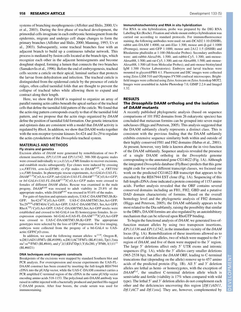
RESULTSThe Drosophila DAAM ortholog and the isolationof DAAM mutantsA recently published phylogenetic analysis (based on sequencecomparisons of 101 FH2 domains from 26 eukaryotic species) hasconcluded that metazoan formins can be grouped into seven majorsubclasses (Higgs and Peterson, 2005). This analysis suggested thatthe DAAM subfamily clearly represents a distinct class. This isconsistent with the previous finding that the DAAM subfamilyexhibits extensive sequence similarity both within and outside oftheir highly conserved FH1 and FH2 domains (Habas et al., 2001).At present, however, very little is known about the in vivo functionof the DAAM subfamily. Sequence analysis revealed the presenceof a single DAAM ortholog in the Drosophila genomecorresponding to the annotated gene CG14622 (Fig. 1A). Althoughthe integrated Drosophila database (FlyBase) predicts that this genemight code for several different transcript classes, we focused ourwork on the predicted CG14622-RB transcript that appears to beencoded by the RE67944 EST clone (Fig. 1A). Sequencing of thisfull-length cDNA clone indicated a protein consisting of 1153 aminoacids. Further analysis revealed that the ORF contains severalconserved domains including an FH1, FH2, GBD and a putativeDAD domain characteristic of formins. Based on the overallhomology level and the phylogenetic analysis of FH2 domains(Higgs and Peterson, 2005), the DAAM subfamily appears to bemost related to the Dia subfamily, raising the possibility that similarto the DRFs, DAAM formins are also regulated by an autoinhibitorymechanism that can be relieved upon RhoGTP binding.
To begin the functional analysis of DAAM, we generated loss-of-function mutant alleles by using two transposon insertions,EP(1)1336 and EP(1)1542, in the immediate vicinity of the DAAMlocus (Fig. 1A). Remobilization of these insertions allowed us toisolate a set of deletion alleles, two of which were mapped to the 5�region of DAAM, and five of them were mapped to the 3� region.The large 5� deletions affect only 5� UTR exons and intronicsequences (Fig. 1A), while the 3� alleles carry smaller deletions(965-2538 bp), but affect the DAAM ORF, leading to C-terminaltruncations that (depending on the allele) remove up to 457 aminoacids of the predicted protein (Fig. 1B). All 5� and 3� deletionalleles are lethal as hemi- or homozygotes, with the exception ofDAAMEx1, the smallest C-terminal deletion allele which issemiviable and fertile (viability is 17% when compared with wildtype). The lethal 5� and 3� deletion alleles do not complement eachother and the deficiencies uncovering this region [Df(1)AD11,Df(1)AC7 and Df(1)sta]. They are, however, complemented by
RESEARCH ARTICLE Development 133 (5)
DEVELO
PMENT
Dp(1;3)sta and Dp(1;Y)Sz280, two transpositional duplications thatcarry the 1F2-3 cytological region, including DAAM. All lethalalleles are viable over DaamEx1, the weakest and homozygousviable allele, with the exception of the two largest C-terminaldeficiency alleles (DaamEx68 and DaamEx36). To demonstrate thatthe lethality associated with these alleles is solely due to the lack ofDAAM, transgenic flies carrying UAS-FL-DAAM (containing thefull length RE67944 cDNA clone) were generated and tested inrescue experiments. When UAS-FL-DAAM was expressed underthe control of Act-Gal4 or tub-Gal4 in a DAAM mutant backgroundlethality was rescued to adult viability, even in case of the largestdeletion allele DaamEx68. Taken together, these results demonstratethat all lethal alleles represent DAAM alleles and these mutationsdo not affect other essential genetic elements.
DAAM is unlikely to be required for PCP signalingin DrosophilaBased on biochemical assays and tissue culture experiments it wassuggested that a human DAAM family member, DAAM1, isrequired for non-canonical Wnt/Fz signaling, and might function asa bridging factor between Dsh and RhoA. Xenopus embryoexperiments supported this view, as the Xenopus ortholog xDaam1has been implicated in PCP signaling during the early steps ofgastrulation (Habas et al., 2001). To test whether the DrosophilaDAAM is involved in PCP signaling, we carried out a loss-of-function analysis of the gene. We have thus induced DAAM mutantclones in the wing and compound eye, two PCP organization modeltissues. Although several alleles were tested, none of them exhibitedPCP defects in the eye or wing (not shown). The same was observedin adult escapers of the DAAMEx1/DAAMEx249 allelic combination.
We have also tested whether the PCP gain-of-function phenotypesinduced by overexpression of PCP proteins can be modified byDAAM mutant alleles. To achieve this, we have compared the effectsof Fz and Dsh overexpression in the wing or eye in wild-type andDAAMEx68 heterozygous mutant backgrounds. No significantdifference between the wild-type and the DAAM mutantbackgrounds was observed (not shown). Thus, the loss-of-functionanalysis of DAAM argues that the Drosophila DAAM ortholog iseither not required for PCP signaling or at best plays a redundant roleduring PCP establishment.
DAAM regulates the formation of taenidial foldsin the cuticle of the tracheaAs the DAAM alleles are lethal, it encodes a gene with essentialfunction(s). To identify these, we examined DAAM mutant larvae,and noticed that these animals display severe trachea defects. Theseinclude the collapse and flattening of the tracheal tubes, in both themain airways and the side branches (Fig. 2B,C). Wild-type trachealcells secrete a cuticle on their apical (luminal) surface, which iscontinuous with the epidermal cuticle. Nevertheless, it is easilydistinguished by the presence of cuticle ridges called taenidial foldsthat project into the lumen (Fig. 2A,B). These ridges form annularrings or run a helical course around the lumen of the tubes. Trachealtubes of DAAM mutant larvae fail to secrete such a highly orderedcuticle. Instead, although some local order is visible, we observedshort and curvy apical ridges that rarely run perpendicular to the tubeaxis (Fig. 2C,D). These striking cuticle pattern defects are detectedthroughout the tracheal system independent of tube types, with theexception of the fusion cells that secrete a different type of cuticlecharacterized by a dotty pattern instead of taenidial folds (see below,Fig. 5C). To confirm that the trachea phenotype is caused by DAAMloss of function, we expressed a UAS-FL-DAAM construct under thecontrol of a trachea specific btl-Gal4 driver. This significantlyrescued the trachea disruptions (Fig. 2E), just as a general driver (e.g.Act-Gal4, not shown).
Next, we examined the expression of DAAM and confirmed thatthe mRNA is expressed in the developing embryonic trachealsystem, starting from stage 13 of embryogenesis onwards (Fig. 3A).Similarly, immunostaining experiments (using a polyclonal anti-DAAM serum raised against the C-terminal half of the protein; seeMaterials and methods for details) revealed the presence of theDAAM protein in the developing embryonic tracheal cells in stage13 and older embryos (Fig. 3B). In DAAMEx68 embryos, the antibodyfailed to detect protein in the trachea (Fig. 3D). Similarly, detectableprotein was absent from mutant clonal tissue in eye-antennalimaginal discs, where DAAM is expressed during later stages ofdevelopment (Fig. 3E-H). This confirmed that the antibody isspecific to DAAM, and that DAAMEx68 is likely to be a protein null.Consistently, the trachea phenotype of DAAMEx68 homo- orhemizygous larvae is very similar to that of DAAMEx68/Df(1)AD11larvae (Fig. 2E).
959RESEARCH ARTICLEDAAM regulates the tracheal cuticle pattern
Fig. 1. The organization of the DAAM locus and theisolation of DAAM mutations. (A) The 1F2-3cytological region includes the predicted gene CG14622that we named DAAM. Position of two P-elementinsertion are shown, EP(1)1336 and EP(1)1542, whichhave been used to generate DAAM loss-of-functionalleles including large 5� deficiencies. The full-lengthcDNA clone RE67944 consists of 12 exons, translationstarts in exon 4. (B) The full-length DAAM cDNA clonecarries a 1165 bp 5� UTR and a 731 bp 3� UTR, andencodes a predicted protein of 1153 amino acids, whichcontains several homology domains, including GBD(GTPase binding domain), DID (Diaphanous inhibitorydomain), DD (dimerization domain), CC (coiled-coilregion), FH1 (formin homology domain 1), FH2 (forminhomology domain 2) and DAD (Diaphanousautoinhibitory domain). The activated form of DAAM (C-DAAM) includes the C-terminal 637 amino acids of theprotein. The position of the 3� deficiency alleles is shownat the bottom.

DEVELO
PMENT
960
Actin organization in wild type and DAAM mutanttracheal tubesAs it is well established that formins regulate the actin cytoskeleton,we investigated actin organization in wild-type and DAAM mutanttracheal cells. This revealed that in wild-type tracheal cells apicallylocalized actin is organized into parallel bundles runningperpendicular to the axis of the tubes (Fig. 4A,C,E), stronglyresembling the organization of teanidial folds in the cuticle.Significantly, the number of actin rings and taenidial folds in a giventracheal region is equal to each other (Fig. 4A-B). To determineprecisely the time when such actin rings or spirals become apparent,we have expressed an Actin-GFP (green fluorescent protein) fusionprotein in the trachea of living embryos (using the btl-Gal4 trachealdriver line). This demonstrated that actin rings are first visible in latestage 15 embryos, just before the onset of cuticle secretion (Fig. 4N).
The embryonic tracheal cells are rather small, and thus it wasdifficult to study the formation of these actin rings at the subcellularlevel. Nevertheless, it is clear that these actin rings are not onlypresent when cuticle secretion begins, but are also detected throughthe third larval instar stage. In contrast to the remarkable actinorganization found in wild-type tracheal cells, in DAAMEx68
mutants this organization is completely abolished. Whereas theabsence of DAAM does not appear to reduce the apical F-actin levelof the tracheal cells, actin cables formed are much shorter andthinner than in wild type (Fig. 4L,M). Additionally, actin bundlesoften appear to be crosslinked to each other instead of runningparallel (Fig. 4L). Taken together, in DAAMEx68 the actin bundles inthe tracheal cells display abnormal morphology and fail to beorganized into parallel running actin rings under the apical surfaceof the cells.
RESEARCH ARTICLE Development 133 (5)
Fig. 3. DAAM expression in the embryonic trachealsystem and the DAAM antibody. RNA in situhybridization of DAAM (A) and immunohistochemicalstaining with a polyclonal anti-DAAM serum (B)revealed a strong expression in the developingembryonic tracheal system. Brown staining in A and redstaining in B show DAAM expression in a stage 16embryo. The anti-DAAM serum barely detects anyDAAM protein in a DAAMEx68 homozygous mutantembryonic trachea (C,D) and in mitotic clones inducedin DAAMEx68, w, FRT19A/ w, arm-lacZ, FRT19A; ey-flp/+larval eye imaginal disc [E-H; homozygous mutantclones (white arrows) lack DAAM staining]. Clones aremarked by absence of �-gal (green). Elav (blue) is aneuronal marker. H is the merge of E-G. Anterior istowards the left, and dorsal is upwards in all panels.
Fig. 2. The cuticle structure of wild-type and DAAMmutant Drosophila tracheal tubes. Schematic drawingof a wild-type main airway (A) shows that tracheal cuticleis laid down on the inner apical surface of tracheal cells.The cuticle is characterized by taenidial folds, runningperpendicular to the tube axis, that are clearly visible on anative tracheal tube dissected out from a third instar larvae(B). The tracheal tubes of a DAAMEx68 homozygousmutant larvae exhibit a strongly impaired cuticle pattern inboth the main airways (C) and the side branches (D), oftenleading to the collapse of the tubes. (E) The tracheaphenotype of DAAMEx68 over Df(1)AD11 (a deficiency thatuncovers DAAM) is as strong as that of the homozygousDAAMEx68 phenotype (compare E with C). (F) btl-Gal4-driven overexpression of the full-length DAAM protein (FL-DAAM13.59) partly rescues the tracheal cuticle defectsinduced by DAAMEx68. Scale bars: 50 �m.

DEVELO
PMENT
Although the global actin organization is severely impaired, localorder can often be detected in small patches within a cell. The cuticlepattern of DAAMEx68 mutants displays a similar phenotype,indicating that actin cables direct the run of taenidial folds, even inthe DAAM mutant situation. Thus, it appears that the major functionof DAAM in Drosophila tracheal cells is to organize an array of actinrings that directs cuticle patterning by specifying the site of taenidialfold formation. Despite the fact that the apical actin level is notsignificantly reduced in DAAM mutants, we cannot exclude thepossibility that in the wild-type situation, DAAM also contributes toactin assembly, a well characterized formin function. Consistentwith this possibility, DAAM largely colocalizes with the apicallyenriched actin bundles in the embryonic trachea cells (Fig. 4E-J).Nevertheless, the potential involvement in actin polymerizationappears be a redundant function. The recently characterized form3is the best candidate to be functionally redundant with DAAM foractin assembly, because it is the only other Drosophila forminknown to be expressed in the tracheal system (Tanaka et al., 2004).
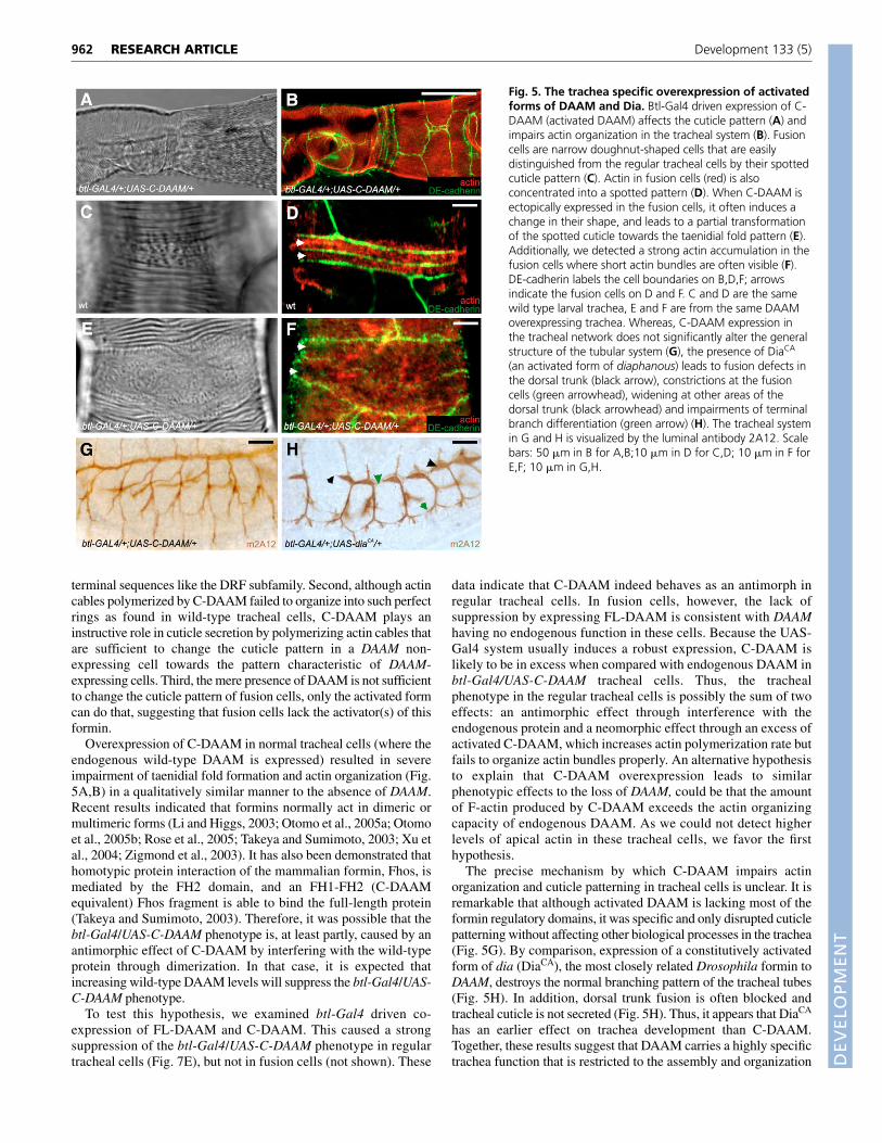
Activated DAAM interferes with cuticlepatterning in tracheal cells and transforms thecuticle pattern of the fusion cellsTruncated formins consisting only of the C-terminal FH1 and FH2domains behave as constitutively active forms in many contexts(Evangelista et al., 1997; Habas et al., 2001; Tominaga et al., 2000;Watanabe et al., 1999). To test whether DAAM activity is regulatedin a similar manner, UAS-C-DAAM (carrying a truncated DAAM
encoding the C-terminal 637 amino acids including the FH1 andFH2 domains; Fig. 1B) transgenic flies were generated. Strikingly,C-DAAM expressed in the trachea (with the btl-Gal4 driver) causeddistinct phenotypic effects in the fusion cells and the regular trachealcells.
After the cuticle is laid down in the tracheal system, fusion cellsare easily distinguished from others because these narrow,doughnut-shaped cells have a characteristic granulated cuticlepattern that is very distinct from the parallel running apical ridges(Fig. 5C). The same difference can be found at the level of actinorganization, as apical actin is also concentrated into a granularpattern (Fig. 5D). Significantly, anti-DAAM staining in wild typerevealed that fusion cells, unlike the normal tracheal cells, do notexpress DAAM (Fig. 4F,G). When C-DAAM is expressed in fusioncells it induces a dramatic change in the actin organization andcuticle pattern, causing strong actin accumulation at the apicalsurface. However, most of the actin is found in largely unorganizedbundles (Fig. 5F), although the typical granular pattern is barelyvisible. In addition, such fusion cells are often wider than their wild-type counterparts, and their granular cuticle pattern is transformedtowards a stripy pattern, partly resembling the taenidial folds ofnormal tracheal cells (Fig. 5E). However, when full-length DAAMwas expressed in the fusion cells (or elsewhere in the trachealsystem) we could not detect any phenotypic effects (not shown).
Several conclusions can be drawn from these data. First, C-DAAM behaves as an activated formin, consistent with its domainstructure and showing that the activity of DAAM is regulated by N-
961RESEARCH ARTICLEDAAM regulates the tracheal cuticle pattern
Fig. 4. DAAM is required toorganize apical actin into parallelrunning bundles in tracheal cells.In a wild-type (wt) tracheal tube,actin is organized into parallelrunning bundles that areperpendicular to the tube axis (A).The number and phasing of theseactin bundles correspond to thetaenidial fold pattern displayed onthe tracheal cuticle (B). Actin bundlesformed in the tracheal cells arelocated at the level of the adherensjunctions (C,D). (C) A confocalprojection of a wild-type trachealtube where actin is visualized in red,while the adherens junctionalmarker, DE-cadherin is shown ingreen. (D) An optical xz sectionalong the white line in C, apical is atthe top. (E-G) The DAAM protein islargely colocalized with actin in theembryonic tracheal cells. Confocalsections have been collected from aone-segment wide region of thedorsal trunk of a stage 16 embryo.The cytoplasm of tracheal cells islabeled with GFP in green, actin isshown in red, DAAM is in blue.Arrow in F indicates the fusion cellslocated at the segmental boundary. DAAM is not expressed in these cells. (H-J) 3D projections of the same confocal sections shown in E-G. Sectionswere rotated within the XZ plane by 90°. There is strong colocalization of actin and DAAM at apical membranes of the tracheal tubes (J). InDAAMEx68 mutant tracheal tubes, not only is the cuticle pattern impaired (K), but actin organization is also severely altered in both the larval (L) andembryonic (M) tracheal cells. Actin cables formed are thinner and shorter than their wild-type counterparts (compare A with L, and E with M), andfail to organize into regularly aligned bundles. The formation of the apical actin bundles in the embryonic tracheal cells (marked by actin::GFP) isfirst detected at approximately 13 hours AEL (N). Scale bars: 20 �m in A for A,B; 50 �m in C for C,D; 50 �m in L for K,L; 10 �m in E-G for E-J;10 �m in M for M.
DEVELO
PMENT
962
terminal sequences like the DRF subfamily. Second, although actincables polymerized by C-DAAM failed to organize into such perfectrings as found in wild-type tracheal cells, C-DAAM plays aninstructive role in cuticle secretion by polymerizing actin cables thatare sufficient to change the cuticle pattern in a DAAM non-expressing cell towards the pattern characteristic of DAAM-expressing cells. Third, the mere presence of DAAM is not sufficientto change the cuticle pattern of fusion cells, only the activated formcan do that, suggesting that fusion cells lack the activator(s) of thisformin.
Overexpression of C-DAAM in normal tracheal cells (where theendogenous wild-type DAAM is expressed) resulted in severeimpairment of taenidial fold formation and actin organization (Fig.5A,B) in a qualitatively similar manner to the absence of DAAM.Recent results indicated that formins normally act in dimeric ormultimeric forms (Li and Higgs, 2003; Otomo et al., 2005a; Otomoet al., 2005b; Rose et al., 2005; Takeya and Sumimoto, 2003; Xu etal., 2004; Zigmond et al., 2003). It has also been demonstrated thathomotypic protein interaction of the mammalian formin, Fhos, ismediated by the FH2 domain, and an FH1-FH2 (C-DAAMequivalent) Fhos fragment is able to bind the full-length protein(Takeya and Sumimoto, 2003). Therefore, it was possible that thebtl-Gal4/UAS-C-DAAM phenotype is, at least partly, caused by anantimorphic effect of C-DAAM by interfering with the wild-typeprotein through dimerization. In that case, it is expected thatincreasing wild-type DAAM levels will suppress the btl-Gal4/UAS-C-DAAM phenotype.
To test this hypothesis, we examined btl-Gal4 driven co-expression of FL-DAAM and C-DAAM. This caused a strongsuppression of the btl-Gal4/UAS-C-DAAM phenotype in regulartracheal cells (Fig. 7E), but not in fusion cells (not shown). These
data indicate that C-DAAM indeed behaves as an antimorph inregular tracheal cells. In fusion cells, however, the lack ofsuppression by expressing FL-DAAM is consistent with DAAMhaving no endogenous function in these cells. Because the UAS-Gal4 system usually induces a robust expression, C-DAAM islikely to be in excess when compared with endogenous DAAM inbtl-Gal4/UAS-C-DAAM tracheal cells. Thus, the trachealphenotype in the regular tracheal cells is possibly the sum of twoeffects: an antimorphic effect through interference with theendogenous protein and a neomorphic effect through an excess ofactivated C-DAAM, which increases actin polymerization rate butfails to organize actin bundles properly. An alternative hypothesisto explain that C-DAAM overexpression leads to similarphenotypic effects to the loss of DAAM, could be that the amountof F-actin produced by C-DAAM exceeds the actin organizingcapacity of endogenous DAAM. As we could not detect higherlevels of apical actin in these tracheal cells, we favor the firsthypothesis.
The precise mechanism by which C-DAAM impairs actinorganization and cuticle patterning in tracheal cells is unclear. It isremarkable that although activated DAAM is lacking most of theformin regulatory domains, it was specific and only disrupted cuticlepatterning without affecting other biological processes in the trachea(Fig. 5G). By comparison, expression of a constitutively activatedform of dia (DiaCA), the most closely related Drosophila formin toDAAM, destroys the normal branching pattern of the tracheal tubes(Fig. 5H). In addition, dorsal trunk fusion is often blocked andtracheal cuticle is not secreted (Fig. 5H). Thus, it appears that DiaCA
has an earlier effect on trachea development than C-DAAM.Together, these results suggest that DAAM carries a highly specifictrachea function that is restricted to the assembly and organization
RESEARCH ARTICLE Development 133 (5)
Fig. 5. The trachea specific overexpression of activatedforms of DAAM and Dia. Btl-Gal4 driven expression of C-DAAM (activated DAAM) affects the cuticle pattern (A) andimpairs actin organization in the tracheal system (B). Fusioncells are narrow doughnut-shaped cells that are easilydistinguished from the regular tracheal cells by their spottedcuticle pattern (C). Actin in fusion cells (red) is alsoconcentrated into a spotted pattern (D). When C-DAAM isectopically expressed in the fusion cells, it often induces achange in their shape, and leads to a partial transformationof the spotted cuticle towards the taenidial fold pattern (E).Additionally, we detected a strong actin accumulation in thefusion cells where short actin bundles are often visible (F).DE-cadherin labels the cell boundaries on B,D,F; arrowsindicate the fusion cells on D and F. C and D are the samewild type larval trachea, E and F are from the same DAAMoverexpressing trachea. Whereas, C-DAAM expression inthe tracheal network does not significantly alter the generalstructure of the tubular system (G), the presence of DiaCA
(an activated form of diaphanous) leads to fusion defects inthe dorsal trunk (black arrow), constrictions at the fusioncells (green arrowhead), widening at other areas of thedorsal trunk (black arrowhead) and impairments of terminalbranch differentiation (green arrow) (H). The tracheal systemin G and H is visualized by the luminal antibody 2A12. Scalebars: 50 �m in B for A,B;10 �m in D for C,D; 10 �m in F forE,F; 10 �m in G,H.
DEVELO
PMENT
of apical actin cables, which direct cuticle patterning, with most ofthe functional specificity coming from the C-terminal FH1-FH2domains.
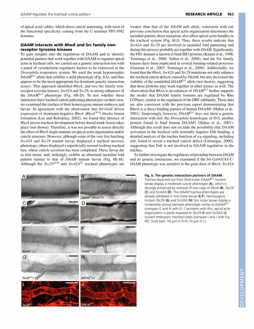
DAAM interacts with RhoA and Src family non-receptor tyrosine kinasesTo gain insights into the regulation of DAAM and to identifypotential partners that work together with DAAM to organize apicalactin in tracheal cells, we carried out a genetic interaction test witha panel of cytoskeleton regulators known to be expressed in theDrosophila respiratory system. We used the weak hypomorphicDAAMEx1 allele that exhibits a mild phenotype (Fig. 6A), and thusappears to be the most appropriate for dominant genetic interactionassays. This approach identified RhoA, and two Src family non-receptor tyrosine kinases, Src42A and Tec29, as strong enhancers ofthe DAAMEx1 phenotype (Fig. 6B-D). To test whether theseinteractors have tracheal cuticle patterning phenotypes on their own,we examined the trachea of their homozygous mutant embryos andlarvae. In agreement with the observation that btl-Gal4 drivenexpression of dominant-negative RhoA (RhoAN19) blocks lumenformation (Lee and Kolodziej, 2002), we found that absence ofRhoA arrests tracheal development before dorsal trunk fusion takesplace (not shown). Therefore, it was not possible to assess directlythe effect of RhoA single mutants on apical actin organization and/orcuticle structure. However, although some of the very few hatchingSrc42A and Tec29 mutant larvae displayed a tracheal necrosisphenotype, others displayed a superficially normal-looking trachealtree, where cuticle secretion has been completed. These larvae dieas first instar, and, strikingly, exhibit an abnormal taenidial foldpattern similar to that of DAAM mutant larvae (Fig. 6E-H).Although the Tec295610 and Src42AE1 tracheal phenotypes are
weaker than that of the DAAM null allele, consistent with ourprevious conclusion that apical actin organization determines thetaenidial pattern, these mutations also affect apical actin bundles inthe tracheal system (Fig. 6I,J). Thus, these results indicate thatSrc42A and Tec29 are involved in taenidial fold patterning andduring this process probably act together with DAAM. Significantly,the FH1 domain is known to bind SH3 proteins (Kamei et al., 1998;Tominaga et al., 2000; Vallen et al., 2000), and the Src familykinases have been implicated in several forming-related processes(Gasman et al., 2003; Tominaga et al., 2000). Additionally, wefound that the RhoA, Src42A and Tec29 mutations not only enhancethe tracheal cuticle defects caused by DAAM, but also decreased theviability of the semilethal DAAMEx1 allele (not shown), suggestingthat these proteins may work together in other tissues as well. Theobservation that RhoA is an enhancer of DAAMEx1 further supportsthe model that DAAM family formins are regulated by RhoGTPases, similar to the regulation of the DRF subfamily. These dataare also consistent with the previous report demonstrating thatRhoA is a direct binding partner of human DAAM1 (Habas et al.,2001). Surprisingly, however, DAAMEx1 does not show a geneticinteraction with dsh, the Drosophila homologue of Dvl, anotherprotein found to bind human DAAM1 (Habas et al., 2001).Although this result does not exclude the possibility that DAAMactivation in the tracheal cells normally requires Dsh binding, adetailed analysis of the trachea function of wg signaling, includingdsh, failed to reveal a tracheal cuticle defect (Llimargas, 2000),suggesting that Dsh is not involved in DAAM regulation in thetrachea.
To further investigate the regulatory relationship between DAAMand its genetic interactors, we examined if the btl-Gal4/UAS-C-DAAM phenotype was sensitive to the gene dose of RhoA, Src42A
963RESEARCH ARTICLEDAAM regulates the tracheal cuticle pattern
Fig. 6. The genetic interaction partners of DAAM.Trachea dissected out from third instar DAAMEx1 mutantlarvae display a moderate cuticle phenotype (A), which isstrongly enhanced by removal of one copy of RhoA (B), Tec29(C) and Src42A (D). The DAAM trachea phenotypes arealready exhibited in first instar larvae (E,F). Homozygousmutant Tec29 (G) and Src42A (H) first instar larvae display amoderately strong taenidial phenotype similar to DAAMEx1
(compare G and H with E). Consistent with this, apical actinorganization is partly impaired in Tec29 (I) and Src42A (J)mutant embryonic tracheal tubes (compare I and J with Fig.4E). Scale bars: 50 �m in A-H; 10 �m in I,J.

DEVELO
PMENT
964
and Tec29. This epistasis analysis indicated that while RhoA did notmodify the effect of C-DAAM expression, Src42A and Tec29dominantly suppressed the respective cuticle defects (Fig. 7A-D).These data are consistent with the model that RhoA is an upstreamregulator of DAAM, whereas the non-receptor tyrosine kinases actdownstream of or in parallel to it. Hence, these results furthersupport the view that the DAAM family formins are regulated byRho GTPases. With respect to non-receptor tyrosine kinases, ourdata support similar conclusions to those of previous reports,demonstrating that mouse Dia1 and mouse Dia2 act upstream of Srcin the regulation of actin dynamics (Tominaga et al., 2000), and thathuman DIA2C is required for Src activation during the process ofendosome regulation (Gasman et al., 2003).
DISCUSSIONActin organization and cuticle structure in theDrosophila tracheal systemThe basic structure of the insect tracheal system is a highlyconserved tubular network in every species. The most importantfunction of this network is to allow oxygen flow to target cells. Thus,tracheal tubes need to be both rigid enough, to ensure continuous airtransport, and flexible enough along the axis of the tubes, to preventthe break down of the tube system when body parts or segmentsmove relative to each other. These requirements are mainly ensuredby the tracheal cuticle, which covers the luminal surface of the tubesand displays cuticle ridges (making the overall structure similar tothe corrugated hose of a vacuum cleaner). Analysis of DAAMmutants provides the first direct evidence that this hypothesis iscorrect. Our data demonstrate that in the absence of DAAM thetaenidial fold pattern is severely disrupted, often leading to thecollapse of the tubes and to discontinuities in the tubular network. Inaddition, our analysis revealed that the remarkably ordered cuticlepattern, displayed in the wild-type trachea tubes, depends onDAAM-mediated apical actin organization. Apical actin isorganized into parallel-running actin cables, much the same wayteanidial folds run in the cuticle. Significantly, the formation of theseactin bundles precedes the onset of cuticle secretion, and the numberand phasing of the actin rings correspond exactly to that of the
taenidial folds in the cuticle. Thus our studies revealed a novel aspectof apical actin organization in the tracheal cells that has not beenappreciated before.
The DAAM gene encodes a novel member of the formin family ofproteins, involved in actin nucleation and polymerization. Consistentwith this, DAAM is colocalized with apical actin in the trachealcells, and the activated form of DAAM is able to induce actinassembly when expressed in tracheal cells (this work) and in othercell types (T.M. and J.M., unpublished). In DAAM mutant trachealcells, apical actin is still detected, albeit at a somewhat lower levelthan in wild type, but the bundles formed in the mutant are muchshorter and thinner than in wild type, and often appear to becrosslinked to each other. Most strikingly, global actin organizationis almost completely lost, although some local order can still bedetected. Remarkably, the cuticle pattern in mutant tracheal cells stillfollows the underlying aberrant actin pattern. Overall, in DAAMmutants, both the tracheal cuticle and the apical actin patternresemble a mosaic of locally ordered patches that failed to becoordinated and aligned with each other and the axis of the trachealtubes. We thus propose that the apical actin bundles play a key rolein patterning the tracheal cuticle by defining the place of taenidialfold formation. Regarding the function of DAAM, our resultssuggest that the major role of this formin in the tracheal cells is toorganize the actin filaments into parallel running actin rings orspirals instead of simply executing the well characterized forminfunction related to actin assembly. However, whether this is a directeffect on actin organization, and thus represents a novel forminfunction, needs to be further elucidated. An alternative model couldbe that DAAM is primarily required for actin polymerization buttightly coupled to an actin ‘organizing’ protein. In such scenario, thepolymerization activity should be a redundant requirement, whereasthe link to the organizing protein would be a DAAM-specificfunction, thereby explaining the presence of unorganized actinbundles in DAAM mutant tracheal cells.
In the case of the main tracheal airways, which are multicellularalong their periphery, it is striking that in wild type the run of theactin bundles is perfectly coordinated across cell boundaries. Inaddition, the run is always perpendicular to that of the tube axis.
RESEARCH ARTICLE Development 133 (5)
Fig. 7. Epistasis experiments with C-DAAM. Cuticledefects induced by C-DAAM expression in tracheal cells (A)are not modified by RhoA (B), but are strongly suppressed bySrc42A (C), Tec29 (D) and co-expression with the full-lengthform of DAAM (FL-DAAM13.59) (E). Scale bars: 50 �m.
DEVELO
PMENT
How does DAAM ensure the coordination of these two aspects ofactin organization? Because the DAAM protein and the apical actincables are both found at the level of the adherens junctions, it ispossible that DAAM regulates the coordination of the actin cablesat the cell boundaries through a direct interaction with junctionalprotein complexes. However, other explanations are also possible,and further experiments will be required to elucidate the molecularmechanism of this regulatory function. The fact that actin cablesnormally run perpendicular to the tube axis seems to suggest thattracheal cells are able to sense a global orientation cue and align theiractin bundles accordingly. The nature and source of this cue isunknown, as is the mechanism by which DAAM is involved in theread-out of this signal. Nevertheless, it is interesting that in DAAMand btl-Gal4/UAS-C-DAAM mutant trachea, the main pattern of thecuticle phenotype is often changing from one segment to the other,suggesting that the effect of the ‘global’ orientation cue is limited tometameric units.
Regulatory interaction between RhoA and DAAMSequence comparisons of FH2 proteins suggest a close phylogeneticrelationship between the DRF, FRL and DAAM subfamilies (Higgsand Peterson, 2005). Members of these three subfamilies have a highlevel of conservation in the FH2 domain, and importantly, also in theregion of the GBD and DAD domains, suggesting that the FRL andDAAM family formins are also regulated by autoinhibition andRhoGTPases, like the DRFs. We present further evidence in supportof this view. First, DAAM and RhoA display a strong geneticinteraction. Second, C-DAAM (an N-terminally truncated form ofDAAM) behaves like an activated form much the same way DRFfamily formins behave. Third, epistasis experiments with C-DAAMand RhoA suggest that RhoA acts upstream of DAAM. Thus, ourdata support the model in which DAAM, at least in the Drosophilatracheal system, is regulated by autoinhibition that can be relievedby RhoGTPases.
This conclusion, however, contradicts the observation thathuman DAAM1 is required for Wnt/Fz/Dvl dependent RhoAactivation in cultured cells and that xDaam1 appears to mediateWnt-11 dependent RhoA activation in Xenopus embryos (Habaset al., 2001). These results suggested that DAAM functionsupstream of RhoA in non-canonical Wnt/Fz-PCP signaling. Anexplanation for these distinct conclusions might be related to thefact that DAAM, in contrast to xDaam1, does not appear to berequired for Fz/Dsh-PCP signaling. Hence, it is possible that theDrosophila ortholog is regulated in the same way as the DRFformins, while the vertebrate family members can be regulated ina different way, once bound by Dsh/Dvl and recruited into PCPsignaling complexes.
DAAM cooperates with Src42A and Tec29 toregulate the tracheal cuticle patternGenetic interactions with the hypomorphic DAAMEx1 alleleidentified two non-receptor tyrosine kinases, Src42A and Tec29, asstrong interacting partners. Although both of these kinases playmultiple roles during embryogenesis, we demonstrate that singlemutants for both affect the tracheal cuticle pattern in a similar wayto DAAM. These results suggest that DAAM and the Src familykinases work together to regulate the actin cytoskeleton and cuticlepattern in tracheal cells. Although it is not known whether DAAMphysically binds Src42A and/or Tec29, it has been established thatthe FH1 region of DRFs and other formins can bind SH3 domains,including those of the Src family kinases (Gasman et al., 2003;Tominaga et al., 2000; Uetz et al., 1996). In agreement with our data
that DAAM acts upstream of Src42A and Tec29 in tracheal cells,cytoskeleton remodeling and SRF activation mediated by mouseDia1 and mouse Dia2 requires Src activity (Tominaga et al., 2000).Moreover, a recent report suggests that RhoD and human DIA2Cregulate endosome dynamics through Src activation, proposing thatSrc activity is stimulated via human DIA2C dependent recruitmentto early endosomes (Gasman et al., 2003). Similarly, the Limbdeformity protein (a formin) interacts with Src on the perinuclearmembranes of primary mouse fibroblasts (Uetz et al., 1996). Basedon these examples, we speculate that in Drosophila tracheal cells theRhoA/DAAM/Src module may not only be required to organizeapical actin bundles, but additionally it might represent a link tosecretory vesicles and to the regulation of exocytosis. Future studieswill be required to test this hypothesis, and to unravel themechanisms whereby DAAM family formins and Src family kinasescontribute to cytoskeletal remodeling in the Drosophila trachealsystem and in other tissues.
We thank M. Krasnow, P. Rorth, M. Affolter, the Developmental StudiesHybridoma Bank, the Bloomington Stock Center and the Szeged Stock Centerfor fly stocks and reagents. We are grateful to H. Gyurkovics, T. Török and J.Reynolds-Kenneally for critical reading and helpful comments on themanuscript. We are particularly grateful to Anna Rehák, Edit Gyányi and EdinaÖrdög for technical assistance. We thank L. Homolya and B. Sarkadi at theNational Medical Center (Budapest) for their patience and generous help withmicroscopy facilities. This research was supported by NIH FIRCA grantTW05893 to M.M. and J.M., and by a NIH NIGMS RO1 grant GM62917 toM.M. J.M. is an EMBO/HHMI Scientist and a Bolyai János Research Scholar.
ReferencesAdler, P. N. (2002). Planar signaling and morphogenesis in Drosophila. Dev. Cell 2,
525-535.Affolter, M. and Shilo, B. Z. (2000). Genetic control of branching morphogenesis
during Drosophila tracheal development. Curr. Opin. Cell Biol. 12, 731-735.Alberts, A. S. (2001). Identification of a carboxyl-terminal diaphanous-related
formin homology protein autoregulatory domain. J. Biol. Chem. 276, 2824-2830.
Chan, D. C., Bedford, M. T. and Leder, P. (1996). Formin binding proteins bearWWP/WW domains that bind proline-rich peptides and functionally resembleSH3 domains. EMBO J. 15, 1045-1054.
Chang, F., Drubin, D. and Nurse, P. (1997). cdc12p, a protein required forcytokinesis in fission yeast, is a component of the cell division ring and interactswith profilin. J. Cell Biol. 137, 169-182.
Evangelista, M., Blundell, K., Longtine, M. S., Chow, C. J., Adames, N.,Pringle, J. R., Peter, M. and Boone, C. (1997). Bni1p, a yeast formin linkingcdc42p and the actin cytoskeleton during polarized morphogenesis. Science276, 118-122.
Evangelista, M., Pruyne, D., Amberg, D. C., Boone, C. and Bretscher, A.(2002). Formins direct Arp2/3-independent actin filament assembly to polarizecell growth in yeast. Nat. Cell Biol. 4, 260-269.
Evangelista, M., Zigmond, S. and Boone, C. (2003). Formins: signaling effectorsfor assembly and polarization of actin filaments. J. Cell Sci. 116, 2603-2611.
Fanto, M. and McNeill, H. (2004). Planar polarity from flies to vertebrates. J. CellSci. 117, 527-533.
Gasman, S., Kalaidzidis, Y. and Zerial, M. (2003). RhoD regulates endosomedynamics through Diaphanous-related Formin and Src tyrosine kinase. Nat. CellBiol. 5, 195-204.
Habas, R., Kato, Y. and He, X. (2001). Wnt/Frizzled activation of Rho regulatesvertebrate gastrulation and requires a novel Formin homology protein Daam1.Cell 107, 843-854.
Higgs, H. N. and Peterson, K. J. (2005). Phylogenetic analysis of the forminhomology 2 domain. Mol. Biol. Cell 16, 1-13.
Imamura, H., Tanaka, K., Hihara, T., Umikawa, M., Kamei, T., Takahashi, K.,Sasaki, T. and Takai, Y. (1997). Bni1p and Bnr1p: downstream targets of theRho family small G-proteins which interact with profilin and regulate actincytoskeleton in Saccharomyces cerevisiae. EMBO J. 16, 2745-2755.
Kamei, T., Tanaka, K., Hihara, T., Umikawa, M., Imamura, H., Kikyo, M.,Ozaki, K. and Takai, Y. (1998). Interaction of Bnr1p with a novel Src homology3 domain-containing Hof1p. Implication in cytokinesis in Saccharomycescerevisiae. J. Biol. Chem. 273, 28341-28345.
Kohno, H., Tanaka, K., Mino, A., Umikawa, M., Imamura, H., Fujiwara, T.,Fujita, Y., Hotta, K., Qadota, H., Watanabe, T. et al. (1996). Bni1p implicatedin cytoskeletal control is a putative target of Rho1p small GTP binding protein inSaccharomyces cerevisiae. EMBO J. 15, 6060-6068.
965RESEARCH ARTICLEDAAM regulates the tracheal cuticle pattern

DEVELO
PMENT
966
Lee, S. and Kolodziej, P. A. (2002). The plakin Short Stop and the RhoA GTPaseare required for E-cadherin-dependent apical surface remodeling during trachealtube fusion. Development 129, 1509-1520.
Li, F. and Higgs, H. N. (2003). The mouse Formin mDia1 is a potent actinnucleation factor regulated by autoinhibition. Curr. Biol. 13, 1335-1340.
Llimargas, M. (2000). Wingless and its signalling pathway have common andseparable functions during tracheal development. Development 127, 4407-4417.
Manning, G. and Krasnow, M. A. (1993). Development of the Drosophilatracheal system. In The Development of Drosophila melanogaster, vol. I (ed.M. Bate and A. Martinez-Arias), pp. 609-686. Plainview (NY): Cold SpringHarbor Laboratory Press.
Mlodzik, M. (2002). Planar cell polarization: do the same mechanisms regulateDrosophila tissue polarity and vertebrate gastrulation? Trends Genet. 18, 564-571.
Otomo, T., Otomo, C., Tomchick, D. R., Machius, M. and Rosen, M. K.(2005a). Structural basis of Rho GTPase-mediated activation of the forminmDia1. Mol. Cell 18, 273-281.
Otomo, T., Tomchick, D. R., Otomo, C., Panchal, S. C., Machius, M. andRosen, M. K. (2005b). Structural basis of actin filament nucleation andprocessive capping by a formin homology 2 domain. Nature 433, 488-494.
Pring, M., Evangelista, M., Boone, C., Yang, C. and Zigmond, S. H. (2003).Mechanism of formin-induced nucleation of actin filaments. Biochemistry 42,486-496.
Pruyne, D., Evangelista, M., Yang, C., Bi, E., Zigmond, S., Bretscher, A. andBoone, C. (2002). Role of formins in actin assembly: nucleation and barbed-endassociation. Science 297, 612-615.
Rose, R., Weyand, M., Lammers, M., Ishizaki, T., Ahmadian, M. R. andWittinghofer, A. (2005). Structural and mechanistic insights into theinteraction between Rho and mammalian Dia. Nature 435, 513-518.
Sagot, I., Klee, S. K. and Pellman, D. (2002a). Yeast formins regulate cell polarityby controlling the assembly of actin cables. Nat. Cell Biol. 4, 42-50.
Sagot, I., Rodal, A. A., Moseley, J., Goode, B. L. and Pellman, D. (2002b). Anactin nucleation mechanism mediated by Bni1 and profilin. Nat. Cell Biol. 4,626-631.
Samakovlis, C., Manning, G., Steneberg, P., Hacohen, N., Cantera, R. andKrasnow, M. A. (1996). Genetic control of epithelial tube fusion duringDrosophila tracheal development. Development 122, 3531-3536.
Shimada, A., Nyitrai, M., Vetter, I. R., Kuhlmann, D., Bugyi, B., Narumiya, S.,Geeves, M. A. and Wittinghofer, A. (2004). The core FH2 domain ofdiaphanous-related formins is an elongated actin binding protein that inhibitspolymerization. Mol. Cell 13, 511-522.
Strutt, D. (2003). Frizzled signalling and cell polarisation in Drosophila andvertebrates. Development 130, 4501-4513.
Takeya, R. and Sumimoto, H. (2003). Fhos, a mammalian formin, directly bindsto F-actin via a region N-terminal to the FH1 domain and forms a homotypiccomplex via the FH2 domain to promote actin fiber formation. J. Cell Sci. 116,4567-4575.
Tanaka, H., Takasu, E., Aigaki, T., Kato, K., Hayashi, S. and Nose, A. (2004).Formin3 is required for assembly of the F-actin structure that mediates trachealfusion in Drosophila. Dev. Biol. 274, 413-425.
Tominaga, T., Sahai, E., Chardin, P., McCormick, F., Courtneidge, S. A. andAlberts, A. S. (2000). Diaphanous-related formins bridge Rho GTPase and Srctyrosine kinase signaling. Mol. Cell 5, 13-25.
Uetz, P., Fumagalli, S., James, D. and Zeller, R. (1996). Molecular interactionbetween limb deformity proteins (formins) and Src family kinases. J. Biol. Chem.271, 33525-33530.
Uv, A., Cantera, R. and Samakovlis, C. (2003). Drosophila trachealmorphogenesis: intricate cellular solutions to basic plumbing problems. TrendsCell Biol. 13, 301-309.
Vallen, E. A., Caviston, J. and Bi, E. (2000). Roles of Hof1p, Bni1p, Bnr1p, andmyo1p in cytokinesis in Saccharomyces cerevisiae. Mol. Biol. Cell 11, 593-611.
Wallar, B. J. and Alberts, A. S. (2003). The formins: active scaffolds that remodelthe cytoskeleton. Trends Cell Biol. 13, 435-446.
Wallingford, J. B., Fraser, S. E. and Harland, R. M. (2002). Convergentextension: the molecular control of polarized cell movement during embryonicdevelopment. Dev. Cell 2, 695-706.
Wasserman, S. (1998). FH proteins as cytoskeletal organizers. Trends Cell Biol. 8,111-115.
Watanabe, N., Madaule, P., Reid, T., Ishizaki, T., Watanabe, G., Kakizuka, A.,Saito, Y., Nakao, K., Jockusch, B. M. and Narumiya, S. (1997). p140mDia, amammalian homolog of Drosophila diaphanous, is a target protein for Rho smallGTPase and is a ligand for profilin. EMBO J. 16, 3044-3056.
Watanabe, N., Kato, T., Fujita, A., Ishizaki, T. and Narumiya, S. (1999).Cooperation between mDia1 and ROCK in Rho-induced actin reorganization.Nat. Cell Biol. 1, 136-143.
Xu, Y., Moseley, J. B., Sagot i Poy, F., Pellman, D., Goode, B. L. and Eck, M. J.(2004). Crystal structures of a Formin Homology-2 domain reveal a tethereddimer architecture. Cell 116, 711-723.
Zigmond, S. H., Evangelista, M., Boone, C., Yang, C., Dar, A. C., Sicheri, F.,Forkey, J. and Pring, M. (2003). Formin leaky cap allows elongation in thepresence of tight capping proteins. Curr. Biol. 13, 1820-1823.
RESEARCH ARTICLE Development 133 (5)
Related Documents