The Double-Stranded DNA Virosphere as a Modular Hierarchical Network of Gene Sharing Jaime Iranzo, a Mart Krupovic, b Eugene V. Koonin a National Center for Biotechnology Information, National Library of Medicine, Bethesda, Maryland, USA a ; Institut Pasteur, Unité Biologie Moléculaire du Gène chez les Extrêmophiles, Paris, France b ABSTRACT Virus genomes are prone to extensive gene loss, gain, and exchange and share no universal genes. Therefore, in a broad-scale study of virus evolution, gene and genome network analyses can complement traditional phylogenetics. We per- formed an exhaustive comparative analysis of the genomes of double-stranded DNA (dsDNA) viruses by using the bipartite net- work approach and found a robust hierarchical modularity in the dsDNA virosphere. Bipartite networks consist of two classes of nodes, with nodes in one class, in this case genomes, being connected via nodes of the second class, in this case genes. Such a net- work can be partitioned into modules that combine nodes from both classes. The bipartite network of dsDNA viruses includes 19 modules that form 5 major and 3 minor supermodules. Of these modules, 11 include tailed bacteriophages, reflecting the di- versity of this largest group of viruses. The module analysis quantitatively validates and refines previously proposed nontrivial evolutionary relationships. An expansive supermodule combines the large and giant viruses of the putative order “Megavirales” with diverse moderate-sized viruses and related mobile elements. All viruses in this supermodule share a distinct morphogenetic tool kit with a double jelly roll major capsid protein. Herpesviruses and tailed bacteriophages comprise another supermodule, held together by a distinct set of morphogenetic proteins centered on the HK97-like major capsid protein. Together, these two supermodules cover the great majority of currently known dsDNA viruses. We formally identify a set of 14 viral hallmark genes that comprise the hubs of the network and account for most of the intermodule connections. IMPORTANCE Viruses and related mobile genetic elements are the dominant biological entities on earth, but their evolution is not sufficiently understood and their classification is not adequately developed. The key reason is the characteristic high rate of virus evolution that involves not only sequence change but also extensive gene loss, gain, and exchange. Therefore, in the study of virus evolution on a large scale, traditional phylogenetic approaches have limited applicability and have to be complemented by gene and genome network analyses. We applied state-of-the art methods of such analysis to reveal robust hierarchical modu- larity in the genomes of double-stranded DNA viruses. Some of the identified modules combine highly diverse viruses infecting bacteria, archaea, and eukaryotes, in support of previous hypotheses on direct evolutionary relationships between viruses from the three domains of cellular life. We formally identify a set of 14 viral hallmark genes that hold together the genomic network. Received 31 May 2016 Accepted 14 June 2016 Published 2 August 2016 Citation Iranzo J, Krupovic M, Koonin EV. 2016. The double-stranded DNA virosphere as a modular hierarchical network of gene sharing. mBio 7(4):e00978-16. doi:10.1128/ mBio.00978-16. Editor Roger Hendrix, University of Pittsburgh Copyright © 2016 Iranzo et al. This is an open-access article distributed under the terms of the Creative Commons Attribution 4.0 International license. Address correspondence to Eugene V. Koonin, [email protected]. This article is a direct contribution from a Fellow of the American Academy of Microbiology. External solicited reviewers: Sergei Maslov, University of Illinois-Urbana-Champagne; Anca Segall, San Diego State University. A major discovery of environmental genomics and viromics over the last decade is that the most common and abundant biological entities on earth are viruses, in particular bacterio- phages (1–5). In marine, soil, and animal-associated environ- ments, virus particles consistently outnumber cells by 1 to 2 orders of magnitude. Viruses are major ecological and even geochemical agents that in large part shape such processes as energy conversion in the biosphere and sediment formation in water bodies by killing off populations of abundant, ecologically important organisms, such as cyanobacteria or eukaryotic algae (3, 5, 6). With the pos- sible exception of some intracellular parasitic bacteria with highly degraded genomes, viruses and/or other selfish elements, such as transposons and plasmids, parasitize all cellular organisms. Com- plementary to their physical dominance in the biosphere, viruses collectively appear to encompass the bulk of the genetic diversity on Earth (7–9). The ubiquity of viruses in the extant biosphere and the results of theoretical modeling indicating that emergence of selfish genetic elements is intrinsic to any evolving system of rep- licators (10–13) jointly imply that virus-host coevolution has been the mode of the evolution of life ever since its origin (14–16). Viruses and related mobile genetic elements (MGE) clearly have not evolved from a single common ancestor: indeed, not a single gene is conserved across the entire “greater virus world” (also known as the virosphere; here, the two terms are used inter- changeably) or even in the majority of selfish elements (17, 18). However, different parts of the virosphere form dense evolution- ary networks in which genomes of various selfish elements are linked through different shared genes (19–21). This type of evo- RESEARCH ARTICLE crossmark July/August 2016 Volume 7 Issue 4 e00978-16 ® mbio.asm.org 1 on December 12, 2019 at University Libraries | Virginia Tech http://mbio.asm.org/ Downloaded from

Welcome message from author
This document is posted to help you gain knowledge. Please leave a comment to let me know what you think about it! Share it to your friends and learn new things together.
Transcript

The Double-Stranded DNA Virosphere as a Modular HierarchicalNetwork of Gene Sharing
Jaime Iranzo,a Mart Krupovic,b Eugene V. Koonina
National Center for Biotechnology Information, National Library of Medicine, Bethesda, Maryland, USAa; Institut Pasteur, Unité Biologie Moléculaire du Gène chez lesExtrêmophiles, Paris, Franceb
ABSTRACT Virus genomes are prone to extensive gene loss, gain, and exchange and share no universal genes. Therefore, in abroad-scale study of virus evolution, gene and genome network analyses can complement traditional phylogenetics. We per-formed an exhaustive comparative analysis of the genomes of double-stranded DNA (dsDNA) viruses by using the bipartite net-work approach and found a robust hierarchical modularity in the dsDNA virosphere. Bipartite networks consist of two classes ofnodes, with nodes in one class, in this case genomes, being connected via nodes of the second class, in this case genes. Such a net-work can be partitioned into modules that combine nodes from both classes. The bipartite network of dsDNA viruses includes19 modules that form 5 major and 3 minor supermodules. Of these modules, 11 include tailed bacteriophages, reflecting the di-versity of this largest group of viruses. The module analysis quantitatively validates and refines previously proposed nontrivialevolutionary relationships. An expansive supermodule combines the large and giant viruses of the putative order “Megavirales”with diverse moderate-sized viruses and related mobile elements. All viruses in this supermodule share a distinct morphogenetictool kit with a double jelly roll major capsid protein. Herpesviruses and tailed bacteriophages comprise another supermodule,held together by a distinct set of morphogenetic proteins centered on the HK97-like major capsid protein. Together, these twosupermodules cover the great majority of currently known dsDNA viruses. We formally identify a set of 14 viral hallmark genesthat comprise the hubs of the network and account for most of the intermodule connections.
IMPORTANCE Viruses and related mobile genetic elements are the dominant biological entities on earth, but their evolution isnot sufficiently understood and their classification is not adequately developed. The key reason is the characteristic high rate ofvirus evolution that involves not only sequence change but also extensive gene loss, gain, and exchange. Therefore, in the studyof virus evolution on a large scale, traditional phylogenetic approaches have limited applicability and have to be complementedby gene and genome network analyses. We applied state-of-the art methods of such analysis to reveal robust hierarchical modu-larity in the genomes of double-stranded DNA viruses. Some of the identified modules combine highly diverse viruses infectingbacteria, archaea, and eukaryotes, in support of previous hypotheses on direct evolutionary relationships between viruses fromthe three domains of cellular life. We formally identify a set of 14 viral hallmark genes that hold together the genomic network.
Received 31 May 2016 Accepted 14 June 2016 Published 2 August 2016
Citation Iranzo J, Krupovic M, Koonin EV. 2016. The double-stranded DNA virosphere as a modular hierarchical network of gene sharing. mBio 7(4):e00978-16. doi:10.1128/mBio.00978-16.
Editor Roger Hendrix, University of Pittsburgh
Copyright © 2016 Iranzo et al. This is an open-access article distributed under the terms of the Creative Commons Attribution 4.0 International license.
Address correspondence to Eugene V. Koonin, [email protected].
This article is a direct contribution from a Fellow of the American Academy of Microbiology. External solicited reviewers: Sergei Maslov, University ofIllinois-Urbana-Champagne; Anca Segall, San Diego State University.
A major discovery of environmental genomics and viromicsover the last decade is that the most common and abundant
biological entities on earth are viruses, in particular bacterio-phages (1–5). In marine, soil, and animal-associated environ-ments, virus particles consistently outnumber cells by 1 to 2 ordersof magnitude. Viruses are major ecological and even geochemicalagents that in large part shape such processes as energy conversionin the biosphere and sediment formation in water bodies by killingoff populations of abundant, ecologically important organisms,such as cyanobacteria or eukaryotic algae (3, 5, 6). With the pos-sible exception of some intracellular parasitic bacteria with highlydegraded genomes, viruses and/or other selfish elements, such astransposons and plasmids, parasitize all cellular organisms. Com-plementary to their physical dominance in the biosphere, viruses
collectively appear to encompass the bulk of the genetic diversityon Earth (7–9). The ubiquity of viruses in the extant biosphere andthe results of theoretical modeling indicating that emergence ofselfish genetic elements is intrinsic to any evolving system of rep-licators (10–13) jointly imply that virus-host coevolution has beenthe mode of the evolution of life ever since its origin (14–16).
Viruses and related mobile genetic elements (MGE) clearlyhave not evolved from a single common ancestor: indeed, not asingle gene is conserved across the entire “greater virus world”(also known as the virosphere; here, the two terms are used inter-changeably) or even in the majority of selfish elements (17, 18).However, different parts of the virosphere form dense evolution-ary networks in which genomes of various selfish elements arelinked through different shared genes (19–21). This type of evo-
RESEARCH ARTICLE
crossmark
July/August 2016 Volume 7 Issue 4 e00978-16 ® mbio.asm.org 1
on Decem
ber 12, 2019 at University Libraries | V
irginia Tech
http://mbio.asm
.org/D
ownloaded from

lutionary relationship results from extensive exchange of genesand gene modules, in some cases between widely different ele-ments, as well as parallel capture of homologous genes from thehosts. Viruses with large genomes possess numerous genes thatwere acquired from the hosts at different stages of evolution; suchgenes are typically restricted in their spread to a narrow group ofviruses. In contrast, the broader connectivity of the evolutionarynetwork in the virus world derives from a small group of genesthat have been termed virus hallmark genes, which encode keyproteins involved in genome replication and virion formation andare shared by overlapping sets of diverse viruses (17–19). Virushallmark genes have no obvious ancestors in cellular life forms,suggesting that virus-like elements evolved at a precellular stage ofthe evolution of life. The hallmark genes comprise only a smallsubset within the set of virus core genes, which for the purpose ofthis work will be defined as the genes that tend to have been re-tained in groups of related genomes along the course of evolution.The concept of core genes, which departs from the intuitive idea ofgenes with universal presence, accounts for the multiple evolu-tionary histories of distinct groups of viruses. The set of core genesalso includes signature genes, i.e., genes that are highly prevalentwithin and specific to one particular group of viruses.
Due to the patchy gene distribution across the diversity of vi-ruses and related MGE, standard methods of phylogenetics andphylogenomics have limited applicability in the study of the evo-lution of the virosphere outside relatively narrow, tight groups.Instead, methods for direct analysis of evolutionary networks arecalled for. These approaches benefit from the vast arsenal of math-ematical concepts and tools that have been developed in differentareas of network research (22–26). Recent application of networkanalysis methods to the comparative analysis of microbial andbacteriophage genomes has been productive, in particular, for theidentification of preferred routes and patterns of horizontal genetransfer (HGT) (27–29).
The viromes and mobilomes (i.e., the supersets of viruses andother selfish elements) of the three domains of cellular life (bacte-ria, archaea, and eukaryotes) are fundamentally different. Al-though several families of double-stranded DNA (dsDNA) virusesare represented in both bacteria and archaea, no viruses are knownto be shared by eukaryotes with any of the other two cellular do-mains, even at the family or order level (30). In bacteria and ar-chaea, the virosphere is heavily dominated by dsDNA viruses, withrelatively limited representation of single-stranded DNA (ssDNA)viruses and only a few narrow groups of RNA viruses. The eukary-otic part of the virosphere is sharply different, with a dominantpresence of RNA viruses; however, dsDNA viruses of eukaryotesare also common and diverse (31). Altogether, the dsDNA virusesoccupy most of the virosphere and include the largest viral ge-nomes, thus presenting ample material for the construction ofgene-sharing networks.
Bipartite networks, also known as 2-mode networks in the so-cial sciences literature (32), are a natural way to represent, in theform of networks, complex systems that consist of two distinctclasses of components (nodes). Straightforward examples fromthe field of biology are metabolic networks, composed of metab-olites and enzymes, or pollination networks, composed of plantsand their pollinators. Similarly, the network of gene sharingamong viruses (due to common ancestry or HGT) calls for a bi-partite network representation, where the two classes of nodes areviral genomes and homologous gene families. Historically, the
lack of analytical tools to deal with bipartite networks has forcedresearchers to turn to monopartite projections (simplified ver-sions of the network involving only one class of nodes), an ap-proach that is not free from some degree of arbitrariness and canintroduce biases (33). However, recent advances in network the-ory, in particular those related to module detection, allow for di-rect analysis of bipartite networks with minimal loss of informa-tion and simultaneous characterization of both classes of nodes(34).
Here we present a bipartite network analysis of all currentlyrecognized families of dsDNA viruses and some related mobilegenetic elements (MGE). Dissection of this network using formalanalytical tools reveals extensive modularity of this part of thevirus world, objectively identifies the set of viral hallmark genes,and quantitatively vindicates previously proposed scenarios forthe origin of diverse groups of viruses.
RESULTSBipartite network of dsDNA viruses. In order to develop a net-work representation of the relationships between all major groupsof dsDNA viruses, we first had to identify the families of homologsthat would become the nodes of the “gene family” class. To thatend, we retrieved all predicted protein-coding sequences frommore than 1,440 viral genomes and related MGE and groupedthem into families by sequence similarity. Because of the largenumber and high diversity of the viral sequences, a multistep ap-proach to the construction of these families was adopted. First, wedeveloped an automated pipeline that combined sequence simi-larity analyses and algorithms for community detection in net-works (see Materials and Methods). A comparison of the genefamilies obtained through this pipeline and the available clustersof orthologous genes for bacteriophages (POGs) (9, 35) and largenucleo-cytoplasmic DNA viruses of eukaryotes (NCVOGs) (36,37) yielded a recall (average fraction of sequences in a POG orNCVOG grouped together in our analysis) of 0.92, and purity (1minus the average fraction of false positives) of 0.89. In a subse-quent step, some of the major families were manually curated toaccount for distant homology relations that, despite beingstrongly supported by previous reports, escaped automatic detec-tion (see Materials and Methods for details).
We represented the web of relations among dsDNA viruses bymeans of a bipartite network in which the nodes belong to twoclasses: genomes and gene families. Edges connect a genome toevery gene family that it contains; conversely, a gene family islinked to all genomes in which it is present. Compared to othernetwork approaches based on genome-genome similarity metrics,the bipartite representation provides for explicit identification notonly of clusters of genomes sharing gene sets but also of the genesthat glue these genomes together.
Due to the high diversity of viral genomes, the full version ofthe network contains a large fraction of ORFans (genes with nodetectable homologs) and rare genes with a patchy distribution,which reduces the signal-to-noise ratio and makes a detailed anal-ysis computationally unfeasible. To overcome these problems, webuilt a reduced version of the network that only contains coregenes, making it computationally tractable and more informativein terms of evolutionary relationships among viruses. In short,core genes were identified based on their low evolutionary lossrates, according to the procedure described in the next section.Table 1 summarizes the basic statistics of the full and core gene
Iranzo et al.
2 ® mbio.asm.org July/August 2016 Volume 7 Issue 4 e00978-16
on Decem
ber 12, 2019 at University Libraries | V
irginia Tech
http://mbio.asm
.org/D
ownloaded from

networks. With the exception of this section, all results presentedhere are based on the core gene version of the bipartite network. Avisual inspection of the network (Fig. 1A) indicates that (i) virusesfrom different taxa generally occupy different regions of the net-
work, and (ii) despite the abundance of taxon-specific genes, thereis a complex pattern of gene sharing involving, to a greater orlesser extent, all included taxa. It is also notable that Polyomaviri-dae and Papillomaviridae (two families of dsDNA viruses with thesmallest genomes) and viruses infecting archaea (primarily hyper-thermophilic Crenarchaeota) form well-defined clusters that areonly weakly connected to the rest of the network.
In the context of networks, the degree of a node (k) is thenumber of edges connected to that node. In the bipartite networkof viruses, the degree of a gene family represents the number ofgenomes in which the gene family is found, and it follows a powerlaw distribution, P(k) � k��, with the exponent � � 2 (Fig. 1B,left). Power law distributions are characterized by having a longtail, which means that the frequency of nodes with high degrees isnot negligible. Thus, the power law distribution of the gene de-grees implies the existence of hubs, i.e., gene families present in alarge fraction of the genomes. Specifically, there are 12 gene fam-ilies present in at least 20% of the analyzed genomes. In contrast,
TABLE 1 Basic properties of the bipartite dsDNA viral network
ElementValue forfull network
Value forcore genesa
Genomes 1,073 1,071Gene families 33,793 1,576Edges 98,343 30,661Edge density 0.003 0.018Modules NAb 19Mean no. of genes per genome 92.1 28.8Mean gene abundance
(mean with ORFan excluded)2.9 (6.7) 19.3
a The core gene version of the full bipartite network is a bipartite subnetwork thatincludes core gene families and genomes with at least one core gene.b NA, not applicable.
FIG 1 The dsDNA virus world as a bipartite network. Nodes corresponding to genomes are depicted as larger circles, and nodes corresponding to core genefamilies are depicted as dots. An edge is drawn whenever a genome harbors a representative of a core gene family. (A) The modular structure of the network ishighlighted by coloring genome nodes according to the module to which they belong (color coding is as described for Fig. 4 to 6). The location of some major viralgroups is indicated for illustrative purposes. (B) The degree distributions of genes (left) and genomes (right). In the case of genes, the best fit to a power lawdistribution is also shown. (C) The scaling of the clustering coefficient, C(k), with respect to the degree k (genes and genomes) suggests a hierarchical modularstructure organized around high-level hallmark genes [large k and small C(k)] and low-level signature genes [small k and large C(k)].
Double-Stranded DNA Virus Network
July/August 2016 Volume 7 Issue 4 e00978-16 ® mbio.asm.org 3
on Decem
ber 12, 2019 at University Libraries | V
irginia Tech
http://mbio.asm
.org/D
ownloaded from

the number of gene families per genome (i.e., the genome degree)follows a roughly uniform distribution with a bulk that encom-passes viruses and MGE encoding from 3 to 100 genes and a tail ofhighly connected nodes that corresponds to giant viruses (Fig. 1B,right). The flat shape of the degree distribution for genomes indi-cates that the genome network, in sharp contrast to the gene net-work, contains few if any prominent hubs: even the largest giantviruses only harbor a small fraction (5% or less) of all gene fami-lies.
Another useful topological measure is the clustering coefficient(C), which quantifies the extent to which the neighbors of a givennode are also connected with each other. For gene families, thebipartite version of the clustering coefficient (38) decays with k asC(k) � k��, where � � 1.2 (Fig. 1C, blue). Such a scaling of theclustering coefficient is often considered an indicator of hierarchi-cal modular organization, in which low-level modules of tightlyconnected nodes join through higher-level nodes to producelarger, less-cohesive supermodules, in an iterative manner (39). Inthe case of the virus network, low-level modules are cemented bysignature genes, whereas hallmark genes, at the top of the hierar-chy, bring modules together. This hierarchical structure is notobserved for genomes (Fig. 1C, red). Accordingly, the bipartitenetwork appears to be held together primarily by hallmark genesrather than chimeric genomes (see also below).
Core genes in viral genomes. Viral gene families show the tri-partite core-shell-cloud structure that has been identified as a gen-eral feature of gene frequency distributions across the diversity oflife forms (40–43). Although there are no universal viral genes, theU-shaped distribution becomes evident when “coreness” is de-fined as the retention probability of a gene (Fig. 2; see Materialsand Methods for details). Figure 2 also shows (in blue) how wide-
spread the genes within each bin are. From this perspective, geneson the left of the distribution are either ORFans or rare genes withpatchy distribution patterns. On the right side, genes with reten-tion propensities close to unity are typically taxon-specific genesrestricted to a small number of closely related genomes. The mostwidespread genes, i.e., the core genes according to the intuitivedefinition, exhibit retention propensities around 0.7 to 0.8. Thatthis value is smaller than unity could be caused by three factors (orany combination thereof): (i) failure to detect all homologs of anancestral gene in a set of distantly related genomes due to highsequence divergence, (ii) loss of ancestral genes in some lineages,or (iii) occasional transfer of a lineage-specific core gene to unre-lated lineages where it does not tend to be retained, thus affectingthe net estimate of its retention probability (although this effect isminimized by setting a similarity threshold to calculate the lossrates; see also Materials and Methods).
We defined core genes as those with a loss rate below unity [i.e.,retention probability greater than exp(�1)]. Application of thiscriterion yielded 1,576 core gene families (see Table S1 in thesupplemental material), which represented about 5% of all thefamilies in the data set. Table 2 lists the top 25 core genes, whichinclude some of the most conspicuous viral hallmark genes thathave been previously identified qualitatively. The abundances ofthese genes were computed by adding the similarity-weightedcontributions of genomes that contain a given gene and normal-ized with respect to the total number of genomes. Remarkably,this list encompasses two major classes for each of several viralhallmark proteins that are responsible for the key functions invirion morphogenesis and genome replication, in particular, ico-sahedral capsid proteins, DNA-packaging ATPases, DNA poly-merases, and primases-helicases. The ranking of the core genesreflects the diversity of the groups of viruses in which these genesare most common. Due to the fact that tailed bacteriophages (or-der Caudovirales) are the most diverse (and most abundant)group of viruses on earth (2, 44, 45), the top four core genes areprimarily represented in these phages. The core set combinesgenes that are limited in their spread but appear to be essential andare never lost in the respective groups of viruses (although in somecases could be missed due to the extreme sequence divergence),such as packaging ATPases or phage portal proteins, and genesthat are represented in highly diverse groups of viruses but are lostrelatively often, such as helicases (Table 2). Extending the lattertrend, there are some noncore genes that are highly abundant andspread over diverse groups of viruses but failed to make it to thecore set due to their high loss rates; the most widespread of these“viral mobilome” genes are listed in Table 3.
A common debate among researchers in virus evolution iswhich genes represent the viral “self” and thus are most importantfor evolutionary reconstruction: those for structural proteins orthose for components of the replication machinery (19, 46–52).The present quantification of gene “coreness” seems to resolve thisdilemma by showing that members of these two functional cate-gories are mixed in the ranked list of core genes (Table 2; see alsoTable S1 in the supplemental material), highlighting the equalimportance of both categories of genes. Below, we return to someof the core genes when discussing connections between differentgroups of viruses.
Modular structure of the viral network. To unveil the internalstructure of the viral network, we applied a module identificationalgorithm to delineate sets of genomes and gene families that are
FIG 2 Core-shell-cloud structure of viral gene families. For each bin, the barindicates the number of gene families with a retention probability in the rangedefined by the x axis. The blue dots indicate the median abundance of suchfamilies in the whole set of genomes (error bars correspond to the 25th and75th percentiles). Family abundances were normalized so that an abundanceequal to 1 means that the given family is present in each genome (the contri-butions of highly similar genomes were downweighted to compensate for sam-pling bias [see Materials and Methods]). The gene families with the highestretention probability (right-most bin) are typically restricted to a small num-ber of genomes (median abundance, approximately 0.06). In contrast, many ofthe “core” genes according to the intuitive definition (i.e., those present in alarge number of genomes) belong to the bin with a retention probability in therange of 0.7 to 0.8. For the purpose of this work, gene families to the right of thedashed, vertical line (i.e., those with a retention probability greater than 1/e)were considered core genes.
Iranzo et al.
4 ® mbio.asm.org July/August 2016 Volume 7 Issue 4 e00978-16
on Decem
ber 12, 2019 at University Libraries | V
irginia Tech
http://mbio.asm
.org/D
ownloaded from

densely connected to each other. The algorithm is aimed at findingthe partition of the network that maximizes Barber’s bipartitemodularity (a standard measure of the quality of the modules).Because modularity optimization is a combinatorial problem, it isgenerally impossible to solve it exactly for large networks. Instead,heuristic or stochastic approaches, such as simulated annealing,have to be used (see Materials and Methods for details), and dif-ferent runs of the same algorithm can yield variable results due tomultiple local maxima. To overcome this difficulty, we ran 100replicates (realizations) of the algorithm which rendered 100 (notnecessarily different) solutions for the module identificationproblem. Of these, the solution with the highest quality (evaluatedas the value of Barber’s bipartite modularity) was taken as theoptimal partition, and the rest of the replicates were used to assessthe robustness of the modules in that optimal partition. Specifi-cally, for each possible pair of genes or genomes assigned to the
same module, we calculated the fraction of replicates in whichboth elements were grouped together. The average fraction overall pairs was taken as the robustness of the module. Similarly, thecross-similarity between two distinct modules is the fraction ofreplicates in which pairs of genomes, with one from each of themodules, appear in the same group.
The network showed a significant modular structure, with anoptimal partitioning that consisted of 19 modules (P � 0.01, com-pared to the Barber’s modularity of a random network with thesame degree distribution) (Table 4; see also Table S3 in the sup-plemental material for the module composition). The modulesgenerally were highly robust, with respect to both genes and ge-nomes, as determined by analysis of the module composition inalternative network partitions (Table 4; Fig. 3). The number ofgenomes in the modules spanned 2 orders of magnitude, from 282in the largest module (module 9) to 3 in the smallest module
TABLE 2 Top 25 core genes sorted by normalized abundance
Family no. Annotationa Retentionb Abundancec Taxon(s) with presence
5 Terminase, large subunit 0.969 0.661 Herpesvirales, Caudovirales13 Major capsid protein, HK97-like 0.960 0.532 Herpesvirales, Caudovirales10 XRE-family HTH domain 0.718 0.327 Caudovirales16 Portal protein 0.740 0.314 Caudovirales8 DEAD-like helicase 0.522 0.298 Megavirales, Caudovirales6 DNA primase/helicase (DnaB) 0.683 0.290 Caudovirales11 DNA polymerase B 0.886 0.255 Adenoviridae, “Megavirales,” Polintons,
some virophages, Baculoviridae andrelated viruses, Herpesvirales,Tectiviridae, mitochondrial andcytoplasmic plasmids, Ampullaviridae,Salterprovirus, some Caudovirales
24 Integrase 0.794 0.254 Caudovirales111 Protease (herpesvirus S21, phage U9/U35) 0.379 0.254 Herpesvirales, Caudovirales22 Bacteriophage HK97-gp10, putative tail component 0.904 0.240 Caudovirales4 D5-like primase-helicase 0.594 0.240 “Megavirales,” Baculoviridae, some
Caudovirales27 HNHc endonuclease 0.600 0.222 Caudovirales20 DNA polymerase A 0.763 0.205 Caudovirales19 Ribonucleotide reductase large subunit 0.571 0.204 “Megavirales,” Herpesvirales, some
Caudovirales (mostly Myoviridae)18 Thioredoxin 0.371 0.174 “Megavirales,” some Caudovirales2 Ribonucleotide reductase small subunit 0.440 0.169 “Megavirales,” Herpesvirales, some
Caudovirales (mostly Myoviridae)23 Phage tail tape measure protein 0.690 0.164 Siphoviridae, some Myoviridae30 UvrD-like helicase 0.886 0.162 “Megavirales,” Herpesvirales, some
Baculoviridae, some Caudovirales35 Portal protein 0.794 0.158 Siphoviridae12 A32-like packaging ATPase (FtsK/HerA) 0.868 0.156 “Megavirales,” Polintons, Lavidaviridae
(virophages), Tectiviridae,Corticoviridae, Turriviridae,Sphaerolipoviridae
26 Phage mu protein F, putative minor head protein 0.612 0.143 Myoviridae, Siphoviridae68 Double jelly roll MCP 0.786 0.136 Adenoviridae, “Megavirales,” Polintons,
Lavidaviridae (virophages), Tectiviridae,Corticoviridae, Turriviridae
44 Baseplate J family protein 0.895 0.133 Myoviridae36 AAA family ATPase 0.843 0.120 Bicaudaviridae, “Megavirales” (no
Poxviridae), someMyoviridae/Siphoviridae
47 RuvC Holliday junction resolvase; poxvirus A22 family 0.600 0.117 “Megavirales,” some Caudoviralesa Bold text is used to denote hallmark genes.b The retention probability of a gene family is equal to exp(�r), where r is the estimated loss rate (see Materials and Methods).c The abundances were normalized to the total number of genomes, such that a family present in every genome would have an abundance equal to 1.
Double-Stranded DNA Virus Network
July/August 2016 Volume 7 Issue 4 e00978-16 ® mbio.asm.org 5
on Decem
ber 12, 2019 at University Libraries | V
irginia Tech
http://mbio.asm
.org/D
ownloaded from

(module 18). Given that highly similar genomes were clusteredprior to network construction, the number of genomes in a mod-ule seems to be a meaningful reflection of its heterogeneity. Eachof the modules contains multiple core genes, with over 300 genesin the largest (in terms of the number of genes), module 3. How-ever, most of these genes are limited to tight groups of viruseswithin a module, whereas only a few hallmark genes hold themodules together.
Of the 19 modules, 11 include tailed bacteriophages, empha-sizing once again that the order Caudovirales is by far the largest,most diverse, extremely heterogeneous group of viruses. Most ofthe phage modules consist of subsets from a particular family (Si-phoviridae: modules 10, 11, 15, and 16; Myoviridae: modules 13,17, and 19; Podoviridae: module 14). However, two modules (9and 12) include representatives of two or three families of tailedphages. It is well known that tailed phages extensively exchange genes,so the existence of robust modularity is remarkable in itself, indicat-ing that despite this genetic fluidity, there are several partly isolatedgene pools in the phage world. Unexpectedly, module 12 also in-cludes, in addition to phages of the family Myoviridae, the onlyknown member of the eukaryotic virus family Malacoherpesviridae.
Apart from the phages, the viruses of hyperthermophilic Cre-narchaeota form a single, highly robust module 1, with the notableexception of the family Turriviridae (see discussion below). Al-though crenarcheal viruses contain many genes with no detectablehomologs, the existence of a network of shared genes among theseviruses has been noticed previously (53). The network analysisdescribed here not only validates this conclusion but also showsthat the crenarcheal virus network is a distinct module isolatedfrom the rest of the virosphere. Other than viruses of hyperther-mophilic Crenarchaeota, archaeal viruses remain poorly sampled,so that the present analysis included only the family Sphaerolipo-viridae, which belongs in module 5 (see details below), and foureuryarchaeal viruses, all of which share the signature genes of Cau-dovirales and were assigned to module 9.
The remaining 7 modules consist, with a single exception, ofviruses infecting eukaryotes. As expected, papillomaviruses andpolyomaviruses, the smallest known dsDNA viruses that appear tohave evolved from ssDNA viruses, replicating via the rolling circlemechanism (31), form a distinct module that is only weakly con-nected to the rest of the network. The proposed order “Megavi-
rales” (also known as NCLDV), which combines several familiesof large and giant dsDNA viruses of eukaryotes that primarilyreplicate in the cytoplasm of the host cell (54–56), is split betweentwo modules that include, respectively, the poxviruses and the restof the NCLDV. This separation is not particularly surprising be-cause poxviruses are known as a highly derived group of theNCLDV (54). As discussed below, these two modules share severallinks and join at a higher level of the network hierarchy (see be-low).
Similarly, the order Herpesvirales is split into two modules thatinclude, respectively, the families Herpesviridae and Alloherpes-viridae, and again, are connected at a higher level. Unexpectedly,the third family of the Herpesvirales, namely, Malacoherpesviridae,has been assigned to a module containing T4-like tailed phages(see below). A separate module consists of several families of vi-ruses that infect arthropods, including Baculoviridae, Nudiviridae,Hytrosaviridae, and Nimaviridae. An evolutionary relationshipbetween these virus families has been suspected from previouscomparative genomic studies, and it has been proposed that theycould be distantly related to the NCLDV (57–59). The networkanalysis suggests a more complicated picture, as discussed below.
The most notable module, module 5, consists of several groupsof viruses and related MGE from all three domains of life, most ofwhich replicate their genomes via the protein-primed replicationmechanism and encode the respective variety of the family B DNApolymerases (pDNAP). Previously, it was hypothesized thatPolintons, the virus-like large transposons that belong to thismodule and have been shown to encode two capsid proteins and,accordingly, predicted to form virions (60), evolved directly frombacterial tectiviruses and then gave rise to several diverse groups ofeukaryotic viruses and MGE (21, 61). Given the wide spread of thePolintons among eukaryotes and their apparent central role in theevolution of the module and beyond (see below), we denote thismodule Polinton-like (PL). The delineation of the PL module inthe present network analysis is compatible with the previouslyproposed evolutionary scenario and also adds to the mix, in addi-tion to the bacteriophage family Tectiviridae, another phage fam-ily, Corticoviridae, and archaeal viruses of the families Turriviridaeand Sphaerolipoviridae. The PL module is held together by thepDNAP and a distinct suite of genes involved in virion morpho-genesis that encode the major double jelly roll (DJR) capsid pro-
TABLE 3 Representative genes from the viral mobilome (low retention propensity, high abundance)
Family no. Annotation Retentiona Abundanceb Taxon(s) with presence
31 HNH endonuclease 0.002 0.134 Mimiviridae/Marseilleviridae, Phycodnaviridae,Caudovirales
32 dUTPase 0.092 0.158 Adenoviridae, “Megavirales,” Herpesvirales,Caudovirales, Baculoviridae
43 HNH endonuclease 0.012 0.103 Mimiviridae/Marseilleviridae, Caudovirales48 BRO protein, phage antirepressor 0.118 0.121 Baculoviridae, Poxviridae, Ascoviridae/Iridoviridae,
Caudovirales56 DUF3310 0.110 0.124 Caudovirales79 DNA methylase N-4/N-6 0.005 0.109 Caudovirales, Phycodnaviridae, Pandoravirus,
Bicaudaviridae80 Peptidoglycan recognition protein 0.049 0.105 Caudovirales91 ssDNA-binding protein, SSB_OBF domain,
COG06290.111 0.122 Caudovirales
30573 Thymidine kinase 0.129 0.178 “Megavirales,” Caudoviralesa The retention probability of a gene family is equal to exp(�r), where r is the estimated loss rate (see Materials and Methods).b The abundances were normalized to the total number of genomes, such that a family present in every genome would have abundance equal to 1.
Iranzo et al.
6 ® mbio.asm.org July/August 2016 Volume 7 Issue 4 e00978-16
on Decem
ber 12, 2019 at University Libraries | V
irginia Tech
http://mbio.asm
.org/D
ownloaded from
tein, the packaging ATPase and, in many viruses, also a minor,single jelly roll capsid protein and a maturation protease (21, 61).The pDNAP is encoded by most viruses in the PL module, with theexception of the archaeal viruses, corticovirus PM2, some vi-
rophages, and Polinton-like viruses (PLV). The cytoplasmic andmitochondrial plasmids also encode pDNAP, which is the onlylink between these small, capsidless genetic elements and the restof the PL module. Similarly, Haloarchaeal viruses of the genus
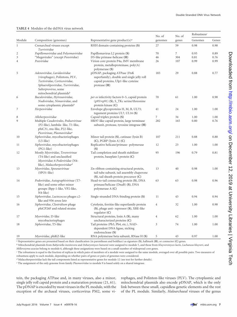
TABLE 4 Modules of the dsDNA virus network
Module Composition (genomes) Representative gene product(s)a
No. ofgenomes
No. ofgenes
Robustnessc
Genomes Genes
1 Crenarcheal viruses exceptTurriviridae
RHH domain-containing proteins (S) 27 59 0.98 0.98
2 Papillomaviridae and Polyomaviridae Papillomavirus L2 protein (S) 70 7 0.93 0.893 “Megavirales” (except Poxviridae) D5-like primase-helicase (H) 46 304 0.81 0.764 Poxviridae Virion core protein P4a, IMV membrane
protein, metalloproteinase, poly(A)polymerase (S)
26 107 0.99 0.99
5 Adenoviridae, Lavidaviridae(virophages), Polintons, PLV,Tectiviridae, Corticoviridae,Sphaerolipoviridae, Turriviridae,Salterprovirus, somemitochondrial plasmidsb
pDNAP, packaging ATPase (FtsKsuperfamily), double and single jelly rollcapsid proteins, Ulp1-like cysteineprotease (H)
183 29 0.88 0.77
6 Baculoviridae, Hytrosaviridae,Nudiviridae, Nimaviridae, andsome cytoplasmic plasmidsb
per os infectivity factors 0–5, capsid protein(p95/vp91) (S), S_TKc serine/threonineprotein kinase (C)
70 61 1.00 0.98
7 Herpesviridae Envelope glycoproteins H, M, B, UL73;tegument proteins UL7, UL16 (S)
41 24 1.00 1.00
8 Alloherpesviridae Capsid triplex protein (S) 7 34 1.00 1.009 Multiple Caudovirales, Peduovirinae
(P2-like), lambda- like, T1-like,phiC31, mu-like, P22-like,Picovirinae, Plasmaviridaee
HK97-like capsid protein, large terminasesubunit, protease, tyrosine integrase (H)
282 165 0.80 0.76
10 Siphoviridae, mycobacteriophages(L5-like)
Minor tail protein (S), cutinase (lysin B)(C), PGRP (lysin A) (C)
107 211 0.88 0.88
11 Siphoviridae, mycobacteriophages(PG1-like)
Replicative helicase/primase- polymerase(S)
12 23 1.00 1.00
12 Mostly Myoviridae, Tevenvirinae(T4-like) and unclassifiedMyoviridae; 6 Podoviridae (N4-like), Malacoherpesviridaed
Tail completion and sheath stabilizerprotein, baseplate J protein (C)
95 196 0.79 0.81
13 Myoviridae, Spounavirinae(SPO1-like)
Zn-ribbon-containing structural protein,tail tube subunit, tail assembly chaperone(S), tail sheath protein precursor (C)
13 40 0.98 1.00
14 Podoviridae, Autographivirinae (T7-like) and some other minorgroups (Bpp-1-like, VP2-like,N4-like)
Head-to-tail connecting protein (S), DNAprimase/helicase (DnaB) (S), DNApolymerase A (C)
63 63 0.98 0.96
15 Siphoviridae, Lactococcus phages c2-like and 936 sensu lato
Single-stranded DNA-binding protein (S) 11 43 0.94 0.94
16 Siphoviridae, Clostridium phagephiCP26F and related strains
Cytolysin, ferritin-like superfamily protein(S), phage anti- repressor (S), XRE-likeregulator (C)
4 32 1.00 0.98
17 Myoviridae, I3-likemycobacteriophages
Structural proteins, lysin A (S), manyuncharacterized proteins (C)
4 62 1.00 1.00
18 Siphoviridae, T5-like Tail proteins (Pb3, Pb4, etc.), NAD-dependent DNA ligase, nickingendonuclease (S)
3 74 1.00 1.00
19 Myoviridae, phiKZ-like RNA polymerase beta subunit, RNase H (S) 5 43 0.97 1.00a Representative genes are presented based on their classification (in parentheses and boldface) as signature (S), hallmark (H), or connector (C) genes.b Mitochondrial plasmids from Babjeviella inositovora and Debaryomyces hansenii were assigned to module 5, and those from Kluyveromyces lactis, Lachancea kluyveri, andMillerozyma acaciae belong to module 6, although these assignations were based on a small number of widespread core genes.c The robustness is equal to the fraction of replicas in which pairs of members of a module were assigned to the same module, averaged over all possible pairs. Two measures ofrobustness apply to each module, depending on whether pairs of genes or pairs of genomes were considered.d Malacoherpesviridae lack the tail components listed as representative genes for module 12 (see text for further details).e The assignment of the only genome from family Plasmaviridae to module 9 is based solely on a shared integrase.
Double-Stranded DNA Virus Network
July/August 2016 Volume 7 Issue 4 e00978-16 ® mbio.asm.org 7
on Decem
ber 12, 2019 at University Libraries | V
irginia Tech
http://mbio.asm
.org/D
ownloaded from

Salterprovirus are included in module 5 based solely on the pres-ence of the pDNAP. The DJR major capsid protein (MCP) and thepackaging ATPase are present in all viruses of the PL module ex-cept for salterproviruses. Thus, the expansive PL module is unifiedby both morphogenetic and replicative genes that are present indifferent members of the module, either separately or together.The finding that all these extremely diverse elements form a singlemodule that is robustly supported at the genome level (althoughless so on the gene level), whereas at the same level of clusteringboth “Megavirales” and Herpesvirales are split, suggests a closerelationship and evolutionary coherence among the PL elements.
Supermodular structure of the dsDNA virus network. Theexistence of substantial cross-similarities between some of themodules (Fig. 3C) implies that there is a higher-order structure inthe virus network, such that some of the modules can be joined togenerate a hierarchy of supermodules. We implemented an itera-tive method to identify such supermodules by building higher-order bipartite networks in which the two classes of nodes are(primary) modules and genes that are shared by these modules.
The first-order module network is depicted in Fig. 4A. Mod-ules representing Caudovirales show a dense web of connections,with numerous shared genes, among which the HK97-like major
FIG 3 Robustness and cross-similarity of modules in the virus bipartite network. (A and B) Heat map representations of the module robustness matrices forgenomes (A) and gene families (B). To generate these matrices, nodes of one class (genomes or gene families) were sorted according to the module they belongto in the optimal partition of the network. For each pair of nodes, the matrix contains the fraction of 100 replicates in which both nodes were placed in the samemodule. Robust modules appear as blocks in the module robustness matrix; deviations from the block structure correspond to modules that are sometimesmerged or nodes without a clear module assignation. The asterisk shows the case of mitochondrial plasmids which belong to module 5 in the best partition butare often assigned to module 14. (C) Quantitative summary of the average robustness of modules at the genome and gene level (elements on the diagonal) andthe cross-similarity between pairs of modules (fraction of replicates in which nodes of both modules appear together; off-diagonal elements). See Table 4 for thelist of the taxa assigned to each module.
Iranzo et al.
8 ® mbio.asm.org July/August 2016 Volume 7 Issue 4 e00978-16
on Decem
ber 12, 2019 at University Libraries | V
irginia Tech
http://mbio.asm
.org/D
ownloaded from
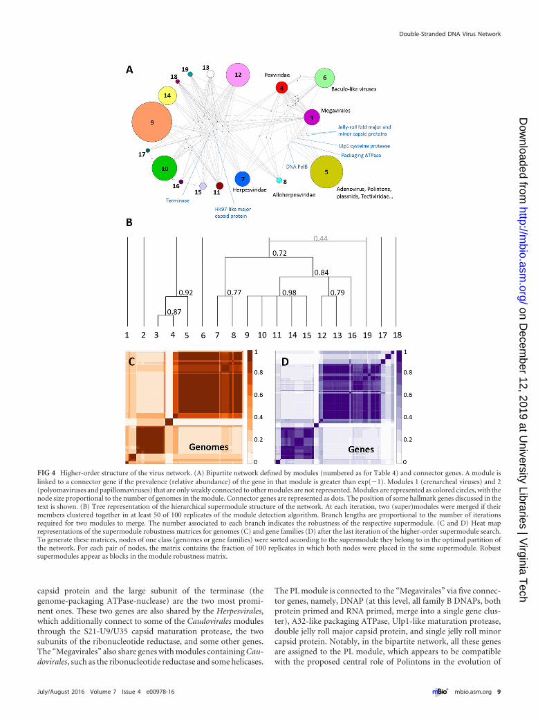
capsid protein and the large subunit of the terminase (thegenome-packaging ATPase-nuclease) are the two most promi-nent ones. These two genes are also shared by the Herpesvirales,which additionally connect to some of the Caudovirales modulesthrough the S21-U9/U35 capsid maturation protease, the twosubunits of the ribonucleotide reductase, and some other genes.The “Megavirales” also share genes with modules containing Cau-dovirales, such as the ribonucleotide reductase and some helicases.
The PL module is connected to the “Megavirales” via five connec-tor genes, namely, DNAP (at this level, all family B DNAPs, bothprotein primed and RNA primed, merge into a single gene clus-ter), A32-like packaging ATPase, Ulp1-like maturation protease,double jelly roll major capsid protein, and single jelly roll minorcapsid protein. Notably, in the bipartite network, all these genesare assigned to the PL module, which appears to be compatiblewith the proposed central role of Polintons in the evolution of
FIG 4 Higher-order structure of the virus network. (A) Bipartite network defined by modules (numbered as for Table 4) and connector genes. A module islinked to a connector gene if the prevalence (relative abundance) of the gene in that module is greater than exp(�1). Modules 1 (crenarcheal viruses) and 2(polyomaviruses and papillomaviruses) that are only weakly connected to other modules are not represented. Modules are represented as colored circles, with thenode size proportional to the number of genomes in the module. Connector genes are represented as dots. The position of some hallmark genes discussed in thetext is shown. (B) Tree representation of the hierarchical supermodule structure of the network. At each iteration, two (super)modules were merged if theirmembers clustered together in at least 50 of 100 replicates of the module detection algorithm. Branch lengths are proportional to the number of iterationsrequired for two modules to merge. The number associated to each branch indicates the robustness of the respective supermodule. (C and D) Heat maprepresentations of the supermodule robustness matrices for genomes (C) and gene families (D) after the last iteration of the higher-order supermodule search.To generate these matrices, nodes of one class (genomes or gene families) were sorted according to the supermodule they belong to in the optimal partition ofthe network. For each pair of nodes, the matrix contains the fraction of 100 replicates in which both nodes were placed in the same supermodule. Robustsupermodules appear as blocks in the module robustness matrix.
Double-Stranded DNA Virus Network
July/August 2016 Volume 7 Issue 4 e00978-16 ® mbio.asm.org 9
on Decem
ber 12, 2019 at University Libraries | V
irginia Tech
http://mbio.asm
.org/D
ownloaded from
dsDNA viruses of eukaryotes (21, 31). The complete list of con-nector genes is provided in Table S2 in the supplemental material.
At the highest order of the network hierarchy, the dsDNA vi-ruses form 5 major supermodules: (i) crenarcheal viruses (exceptTurriviridae), (ii) Polyomaviridae-Papillomaviridae, (iii) PLelements-“Megavirales,” (iv) Baculoviridae and the related fami-lies of arthropod viruses, and (v) Caudovirales-Herpesvirales(Fig. 4B to D). There are also three minor modules encompassingI3-like, T5-like, and phiKZ-like phages that remained isolated (al-though the latter join the main Caudovirales supermodule in 44 ofthe 100 replicates). The most notable result is the unification of thePL elements and the “Megavirales” (including poxviruses) into asingle module with high robustness (0.92), in agreement with theevolutionary scenario that has been proposed primarily on thebasis of the shared morphogenetic genes. In parallel, Herpesviralesmerge with Caudovirales in a moderately supported supermodule(robustness of 0.72).
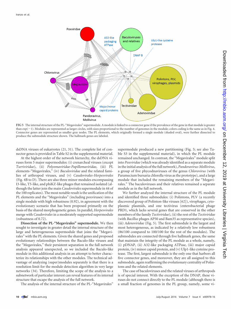
Dissection of the PL-“Megavirales” supermodule. We thensought to investigate in greater detail the internal structure of thelarge and heterogeneous supermodule that joins the “Megavi-rales” with the PL elements. Given the shared genes and proposedevolutionary relationships between the Baculo-like viruses andthe “Megavirales,” their persistent separation in the full networkanalysis appeared unexpected, so we included the Baculo-likemodule in this additional analysis in an attempt to better charac-terize its relationships with the other modules. The technical ad-vantage of analyzing (super)modules separately is that there is aresolution limit for the module detection algorithm in very largenetworks (34). Therefore, limiting the scope of the analysis to asubnetwork of particular interest can reveal features of its internalstructure that escape the analysis of the full network.
The analysis of the internal structure of the PL-“Megavirales”
supermodule produced a new partitioning (Fig. 5; see also Ta-ble S3 in the supplemental material), in which the PL moduleremained unchanged. In contrast, the “Megavirales” module splitinto Poxviridae (which was already identified as a separate modulein the initial analysis of the full network), Pandoravirus-Mollivirus,a group of five phycodnaviruses of the genus Chlorovirus (withParamecium bursaria chlorella virus as the prototype), and a largemodule that included the remaining members of the “Megavi-rales.” The baculoviruses and their relatives remained a separatemodule as in the full network.
We further analyzed the internal structure of the PL moduleand identified three submodules: (i) Polintons, PLV (a recentlydiscovered group of Polinton-like viruses [62]), virophages, cyto-plasmic plasmids, and one tectivirus (enterobacterial phagePRD1, which lacks several genes that are conserved in the othermembers of the family Tectiviridae), (ii) the rest of the Tectiviridae(with Bacillus phages AP50 and Bam35 as representative species),(iii) Adenoviridae (Fig. 5). The first submodule is the largest andmost heterogeneous, as indicated by a relatively low robustness(86/100 compared to 100/100 for the rest of the modules). Thesubmodules are connected through five hallmark genes, the samethat maintain the integrity of the PL module as a whole, namely,(i) pDNAP, (ii) A32-like packaging ATPase, (iii) major capsidprotein, (iv) minor capsid protein, and (v) Ulp1-like cysteine pro-tease. The first, largest submodule is the only one that harbors allfive connector genes, and moreover, they are all assigned to thissubmodule, again reaffirming the evolutionary centrality of Polin-tons and the related elements.
The case of baculoviruses and the related viruses of arthropodsis of special interest. With the exception of the DNAP, these vi-ruses do not connect directly to the PL module (although there isa small fraction of genomes in the PL group, namely, some vi-
FIG 5 The internal structure of the PL-“Megavirales” supermodule. A module is linked to a connector gene if the prevalence of the gene in that module is greaterthan exp(�1). Modules are represented as larger circles, with sizes proportional to the number of genomes in the module; colors coding is the same as in Fig. 4.Connector genes are represented as smaller gray nodes. The PL elements, which originally formed a single module (shaded oval), were further dissected toproduce the submodule structure shown. The hallmark genes are labeled.
Iranzo et al.
10 ® mbio.asm.org July/August 2016 Volume 7 Issue 4 e00978-16
on Decem
ber 12, 2019 at University Libraries | V
irginia Tech
http://mbio.asm
.org/D
ownloaded from

rophages, Polintons and PLV, that possess a D5-like primase-helicase, this gene does not constitute a connector with the wholemodule). Instead, the Baculo-like module shows multiple connec-tions with “Megavirales” and Poxviridae. Most of the shared genesare assigned to the Baculo-like module, apparently because theyare strictly conserved in this small module but not in “Megavi-rales.” These shared genes include the two large subunits of theRNA polymerase, mRNA capping enzyme, transcription factorTFIIS and thiol oxidoreductase. Baculoviruses and their relativesalso harbor genes assigned to other modules and shared by morediverse groups, such as PolB, ribonucleotide reductase (small andlarge subunits), S_TKc serine/threonine protein kinase, and D5-like primase-helicase. Despite the clear relationship with “Mega-virales,” the lack of jelly roll fold capsid proteins, packagingATPase of the FtsK/HerA family and Ulp1 cysteine protease, all ofwhich are the connectors within the PL-“Megavirales” supermod-ule, explains why baculoviruses and their relatives stay as a sepa-rate module even at the highest hierarchy level.
Modularity and interconnections within the order Caudovi-rales. Tailed bacteriophages constitute the most abundant anddiverse group of dsDNA viruses (and all viruses). In order to ob-tain a more precise picture of their module structure and inter-connections, we carried out a detailed analysis of the Caudoviralessubnetwork (Fig. 6; see also Table S3 in the supplemental mate-rial).
This reanalysis rendered a finer-grained network structure inwhich module 9 split into four submodules: 9a, with members ofthe family Myoviridae, such as Mu-like and P2-like phages; 9b,with lambda-like phages and other members of the family Sipho-
viridae (e.g., T1-like and N15-like phages) as well as P22-likephages; 9c, with Phi31-like phages and numerous unclassified si-phoviruses; 9d, containing subfamily Picovirinae and related un-classified members of the family Podoviridae. Two signature genefamilies are associated with submodule 9a: phage tail protein Dand baseplate assembly protein V. In contrast, submodules 9b and9c are more heterogeneous, and their integrity is maintained by anetwork of gene sharing which involves multiple tail componentsand cell lysis proteins. With the exception of the Picovirinae, sub-modules within module 9 are characterized by moderate to highprevalences (relative abundances) of an integrase and a proteinwith a helix-turn-helix DNA-binding domain of the Xre family. Inagreement with the high prevalence of these genes, a survey of thelifestyle of phages in module 9 shows that it is mostly composed oftemperate phages (96 of the 110 based on available data; binomialexact test, P � 10�4).
The large mycobacteriophage module 10 also splits into threesubmodules, each with a distinct set of signature genes (althoughthe large majority of these genes are poorly characterized). Takingthe classification in the Phamerator database (63) (http://phages-db.org) as a reference, submodule 10a contains L5-like and relatedphages (Phamerator cluster A), submodule 10b corresponds toclusters E to G and I to P, and submodule 10c includes membersfrom clusters D, H, and R. The phages in these submodules, as wellas those in module 11 (PG1-like mycobacteriophages, Phamera-tor’s cluster B), share a set of minor tail proteins, a peptidoglycanrecognition protein (lysin A), and a cutinase (lysin B). The last twogenes, required to lyse the mycolic acid-rich outer membrane ofthe Mycobacterium host (64), also appear in the taxonomically
FIG 6 Internal structure of the Caudovirales supermodule. A module is linked to a connector gene if the prevalence of the gene in that module is greater thanexp(�1). Modules are represented as larger circles, with sizes proportional to the number of genomes in the module; color coding is as shown in Fig. 4. Module15 contains Siphoviridae from the Lactococcus phage 936 sensu lato and c2-like groups. Module 16 conatins Clostridium phage phiCP26F and related strains.Connector genes are represented as smaller gray nodes. Hallmark genes are labeled.
Double-Stranded DNA Virus Network
July/August 2016 Volume 7 Issue 4 e00978-16 ® mbio.asm.org 11
on Decem
ber 12, 2019 at University Libraries | V
irginia Tech
http://mbio.asm
.org/D
ownloaded from

unrelated I3-like mycobacteriophages (module 17, Phameratorcluster C). Notably, all phages from submodule 10c, as well asI3-like phages, share the gene coding for the bacterial DNA poly-merase III alpha subunit. Although experimental data on the life-style of mycobacteriophages are scarce, the high prevalence of anintegrase in submodule 10b suggests that most of the phages fromthis subgroup are temperate.
The genomes of I3-like phages (module 17) contain 6 to 10genes (approximately 10% of their total gene content) assigned tosubmodules within the mycobateriophage module 10. Thus, al-though the two modules are not similar enough to merge, thereseems to be a relationship between I3-like phages and other my-cobacteriophages that likely reflects gene exchange, especiallygiven that these phages share related hosts.
Of the three modules (9, 12, 14) for which there is enoughinformation about the lifestyle of their members, modules 12 and14 show a significant predominance of virulent phages (9 of 9 [P �10�4] and 13 of 19 [P � 0.002], respectively). Of these two mod-ules, the former corresponds to T4-like phages, whereas the latterincludes T7-like phages and some other minor groups from thefamily Podoviridae. The reanalysis of the Caudovirales subnetworkresulted in refinement of the T4-like module, with approximately30 unclassified Myoviridae moved to module 9a, and resulting in arobustness of 0.99 (compared to 0.79 in the original module re-ported in Table 4). Signature genes for this refined module includea set of neck, tail, and baseplate proteins, DNAP sliding clamp andclamp loader, and RNAP sigma factor. Module 14, in contrast, isdefined by the central position of the head-to-tail connector pro-tein, which is a signature of T7-like viruses and other phages of thefamily Podoviridae. A single-subunit RNAP is also characteristic ofthis module, although some phages within the module lack it.Finally, two major connector genes, the DNAP A and the DnaBprimase-helicase, are also assigned to module 14.
Of the remaining modules, module 13 consists of SPO1-likephages, module 15 includes Lactococcus phages from the c-like and936 sensu lato groups, and the small modules 16 to 19 correspond,respectively, to Clostridium phage phiCP26F (and related strains),I3-like mycobacteriophages, T5-like phages, and phiKZ-likephages. These modules, with the exception of the Lactococcusphage one, possess large sets of module-specific signature genes(Fig. 7), most of which remain uncharacterized (but see Table 4for a list of some of these genes).
As shown in Fig. 6, global connections across modules occurthrough hallmark genes, such as the HK97-like major capsid pro-tein, portal protein, maturation protease, and DNA primase-helicase of the DnaB family. Additionally, baseplate proteins J andW connect modules within the family Myoviridae. On a local scale,there are two sets of interconnections: (i) those that bring togethermycobacteriophages from submodules 10a, 10b, 10c and, to alesser extent, I3-like phages, as discussed above; (ii) those thatinvolve T4-like and T5-like phages. Hallmark genes excluded,there are 13 connector genes shared by T5-like and T4-like phages,of which many are related to nucleotide metabolism (multiplenucleotide and nucleoside kinases) and DNA repair (DexA exo-nuclease, SbcCD repair exonuclease, RNase H).
Given the multiple connections to other modules, it is clearthat I3-like, T5-like, and phiKZ-like phages are bona fide mem-bers of the Caudovirales network. Therefore, it is surprising thatthey do not join the major bacteriophage supermodule, even at thehighest level of hierarchy. The members of these modules have
large genomes (phiKZ-like, 211 to 317 kb; T5-like, 111 to 122 kb;I3-like, 153 to 165 kb) which are characterized by a significantnumber of conserved genes without clear homologs outside themodule itself (65). Our analysis showed that more than 75% of thegenes that are highly retained in these genomes are module-specific signature genes. In terms of the network, the fact that thesemodules stay separated from the rest reflects this distinctiveness oftheir genome content. In contrast, network analysis places thelarge Vibrio phage KVP40 (245 kb) in the T4-like module, aswidely accepted, and it further merges the T4-like module (alsocharacterized by large genomes but with only 15% of signaturegenes) with the major Caudovirales supermodule.
Orphan modules. A closer inspection of the modules that re-main unmerged showed that crenarcheal viruses do not formstrong connections with the rest of the dsDNA virosphere, al-though several genes are shared with individual families withinother modules. The RHH DNA-binding domain that is typical ofarchaeal viruses also appears with low prevalence in module 5 (PLelements), because the archaeal virus families Turriviridae andSphaerolipoviridae are assigned to that module. Archaeal viruses ofthe family Bicaudaviridae encode an AAA family ATPase that iscommon in “Megavirales” and is also present in T4-like phages.Similarly, archaeal viruses of the family Lipothrixviridae encode aDEAD-like helicase that is shared with members of the “Megavi-rales” as well as some tailed phages. Finally, viruses of the familyAmpullaviridae encode a pDNAP.
Viruses of the families Polyomaviridae and Papillomaviridaeconnect with the other modules solely through the SF3 helicasedomain that is present in the DNA replication proteins of theseviruses (the large T-antigen of polyomaviruses and E1 protein ofpapillomaviruses). However, this protein as a whole (fusion of theN-terminal origin-binding domain and the C-terminal SF3 heli-case domain) has no orthologs among other dsDNA viruses butrather is a derivative of Rep proteins of ssDNA viruses (31). Thecapsid protein of polyomaviruses and papillomaviruses is of thesingle jelly roll variety but shares only extremely distant similarityto the minor capsid proteins of PL elements and “Megavirales.”
Unexpected connections and hybrid viral genomes. The net-work analysis revealed the hybrid nature of several groups of vi-ruses that possess combinations of core genes from different mod-ules as well as unexpected splits of some supposedly well-established groups. The baculoviruses and their relatives areperhaps the prime case in point, as discussed above, but there areseveral other notable examples of virus groups with a “split per-sonality.”
For instance, the archaeal viruses in the family Turriviridaeencode RHH domain proteins that are typical of other archaealviruses but merge into the PL module with the eukaryotic virusesdue to the shared genes for the packaging ATPase and major andminor jelly roll capsid proteins. In a similar vein, viruses of thearchaeal family Ampullaviridae encode a pDNAP but are assignedto the crenarcheal module based on their sharing of a glycosyltransferase with other archaeal viruses.
Mitochondrial plasmids, which in the best solution are as-signed to the PL module, often join module 14 (T7-like, Au-tographivirinae). These elements possess two core genes, namely,pDNAP and a single-subunit phage-type RNAP that, respectively,link them to the PL module and the T7-like module. Because ofthis hybrid composition, module assignment of these plasmids isuncertain.
Iranzo et al.
12 ® mbio.asm.org July/August 2016 Volume 7 Issue 4 e00978-16
on Decem
ber 12, 2019 at University Libraries | V
irginia Tech
http://mbio.asm
.org/D
ownloaded from

Among the cytoplasmic plasmids, three were assigned to theBaculo-like module, whereas the rest were assigned to the PLmodule. Those plasmids that joined the baculoviruses and theirrelatives possess four core genes, namely, pDNAP, DEAD-like he-licase (assigned to Caudovirales but common also among “Mega-virales” and poxviruses), mRNA capping enzyme, and the largest
subunit of RNAP (both assigned to baculoviruses but also nearlyubiquitous in “Megavirales” and represented in all poxviruses).This mix of widespread genes causes the split of the plasmid groupbetween two modules, emphasizing the network character of theevolution of selfish elements.
The order Herpesvirales is split between two compact modules
FIG 7 Characterization of viral hallmark genes and module-specific signature genes. (A) All core gene families sorted by their relative prevalence in the majorsupermodules are shown in gray. Hallmark genes are those that, besides belonging to the set of connector genes, have a relative prevalence greater than 0.35 inat least one of the two major supermodules. (B) Signature genes are those genes with mutual information greater than 0.6 to their best-matching module (x axis)and less than 0.02 to their second match (y axis). The rest of the gene families are represented in gray for comparison. (C) Betweenness-rank distribution for genesin the bipartite network. The nodes with the highest betweenness correspond to hallmark and other connector genes. Signature genes are represented in red. (D)Three-dimensional representation of core genes based on mutual information, relative prevalence, and exclusivity with respect to their assigned module (samecolor coding as in panel C). (E) A histogram with the number of signature, hallmark, connector (nonhallmark), and other (gray) genes per module. Reanalysisof the Caudovirales subnetwork detected 13 signature genes for module 12, which are not shown in the figure. In panels B and D, a large red point indicates theexistence of 205 signature genes whose presence-absence patterns perfectly match their assigned modules.
Double-Stranded DNA Virus Network
July/August 2016 Volume 7 Issue 4 e00978-16 ® mbio.asm.org 13
on Decem
ber 12, 2019 at University Libraries | V
irginia Tech
http://mbio.asm
.org/D
ownloaded from

that duly combine within a supermodule (Fig. 4) and the phagemodule 12 (mostly Tevenvirinae), which includes the only avail-able genome from the family Malacoherpesviridae (66). This her-pesvirus possesses 6 core genes, namely, DNAP (module 5), largesubunit of the terminase (module 9, Caudovirales), large and smallsubunits of the ribonucleotide reductase (assigned to module 12,Tevenvirinae, although also abundant in “Megavirales,” poxvi-ruses, Herpesvirales, and some other phage modules), RNA ligase(assigned to Tevenvirinae but also common in “Megavirales”),and RING finger-containing ubiquitin ligase (assigned to baculo-viruses, also present in poxviruses and some “Megavirales”). Al-though Malacoherpesvirus shares only a few core genes with Her-pesvirales, its clustering with Tevenvirinae is solely based on genesthat are widespread among diverse viruses. Thus, this groupingappears to be an artifact caused by the small number of core genesand the current lack of diversity among the malacoherpesviruses.It seems likely that once more genomes from this group becomeavailable, additional genes shared with Herpesvirales will be iden-tified, likely resulting in a change of the module affinity.
Among the tailed bacteriophages, it is worth dissecting the caseof the N4-like phages that are split between the T7-like and T4-likemodules. Genome analysis shows that N4-like phages contain amix of genes characteristic of each module; they share with T7-likephages the head-to-tail connecting protein, DNAP A, and a single-subunit phage-type RNAP. In contrast, UvrD-like helicase as wellas rIIA and rIIB proteins are shared with T4-like phages. Thischimeric gene composition of N4-like phages makes it difficult toassign them to a single module; indeed, the main difference be-tween those assigned to the T4-like and T7-like modules is thepresence or absence of thioredoxin and ribonucleotide reductase,both formally assigned to the T4-like module, although wide-spread among bacteriophages and eukaryotic viruses.
Members of the Picovirinae (phi29-like bacteriophages) alsoappear to be chimeric entities that encode a genome replicationmachinery shared with the viruses and related MGEs of the PLmodule, whereas virion structure places them among the Caudo-virales. Indeed, phi29-like phages encode bona fide HK97-likemajor capsid proteins (67), a packaging ATPase that is a distanthomolog of the large subunit of the terminase of other tailedphages as well as the portal protein (68). Thus, the phi29-likephages apparently evolved via recombination between a tailedphage and a tectivirus encoding a DJR capsid protein and pDNAP.The tailed phage contributed the genes for the major capsid pro-tein, portal, and some additional proteins involved in virion mor-phogenesis and host recognition, whereas the tectivirus providedthe linear genome scaffold, including the genes for pDNAP andthe terminal protein.
Finally, the family Sphaerolipoviridae is peculiar in that it in-cludes bacterial as well as euryarchaeal viruses (Alphasphaerolipo-virus and Betasphaerolipovirus infect halophilic archaea, whereasGammasphaerolipovirus infects bacteria) (69). All members ofthis family encode an A32-like ATPase and two major capsid pro-teins (typically called the small and large MCP), which correspondto two halves of the double jelly roll capsid protein characteristicof the “Megavirales”-PL supermodule. Thus, in viruses with theDJR capsid proteins, the pseudohexagonal capsomers are formedfrom homotrimers of one capsid protein, whereas in sphaerolipo-viruses similarly shaped capsomers are heterohexamers of thesmall and large major capsid proteins (70, 71). Although theseMCPs were treated as distinct gene families, sphaerolipoviruses
joined the PL module through the packaging ATPase. This assign-ment appears consistent with the previous suggestion that spha-erolipoviruses diverged from the common ancestor shared withother viruses in the PL supermodule prior to the radiation of themajor groups of viruses with the DJR capsid proteins (48). It isworth noting, however, that bacterial sphaerolipoviruses possesstwo hallmark genes from the Caudovirales supermodule, namely,the integrase and an XRE family helix-turn-helix (HTH) domain-containing protein. Thus, this subset of the sphaerolipovirusescomprises another group of viruses with hybrid genomes.
Classification of viral core genes: signature, connector, andhallmark genes, the glue of the virosphere. The viral hallmarkgenes have been previously defined qualitatively as those genesthat are shared by multiple, diverse groups of viruses and have noclose homologs in cellular organisms (17). The present analysisallows us to quantify this concept by cataloguing and classifyingthe genes that connect the nodes (connector genes) in the modulenetwork. Specifically, we identified hallmark genes as the connec-tor genes with a prevalence in any of the two major supermodulesgreater than 0.35 (Fig. 7A; see also Materials and Methods). Thistally of the hallmark genes identified the familiar suspects, such asthe double jelly roll, single jelly roll, and HK97 capsid proteins,terminases and packaging ATPases of the FtsK superfamily (A32-like), DNAP, two primase-helicase families (D5-like and phagereplicative helicase DnaB), and two proteases (Ulp1-like and S21/U9/U35). The list was completed by phage portal and tail proteins,transcriptional regulators of the HTH XRE family, and phage in-tegrases of the tyrosine recombinase superfamily. As suggestedpreviously (17), the number of hallmark genes is small: only 14hallmark genes identified under the above criteria account for57% of the connections in the module network.
From the perspective of network theory, hallmark and connec-tor genes stand out by their high betweenness (72): 44 of the 50genes with the highest betweenness are connector or hallmarkgenes (Fig. 7C). In addition, although they constitute less than 1%of all core gene families, hallmark genes account for 53% of thetotal betweenness centrality of the network. This value increases to89% if all connector genes are considered, although they representless than 4% of the core gene set. Taken together, these resultsvalidate our prevalence-driven approach to identify connectorand hallmark genes and highlight the role of hallmark genes inmaintaining the integrity of the virosphere.
Additionally, we used mutual information to extract signaturegenes, i.e., genes whose presence/absence pattern is diagnostic of amodule (Fig. 7B). As intuitively expected, signature genes showboth high prevalence and high exclusivity among modules(Fig. 7D). As shown in Fig. 7E, the number of signature genes permodule is highly variable: some modules, such as those that in-clude poxviruses, herpesviruses, T5-like phages, and phiKZ-likephages, are well defined by large sets of signature genes; others,such as the PL and the heterogeneous lambda-like module 9, areinstead characterized by a mix of shared hallmark and connectorgenes. In topological terms, signature genes can be viewed asmodule-specific hubs, as opposed to the hallmark genes whichrepresent high-level network hubs. Accordingly, techniques forhub identification and classification would seem suitable for iden-tification of signature genes. However, the small size of somemodules strongly limits the node degree of their signature genes tothe point that they are hardly detectable as hubs. It is for thisreason that we chose an information theoretical approach (mu-
Iranzo et al.
14 ® mbio.asm.org July/August 2016 Volume 7 Issue 4 e00978-16
on Decem
ber 12, 2019 at University Libraries | V
irginia Tech
http://mbio.asm
.org/D
ownloaded from

tual information) instead of hub detection techniques to identifysuch genes.
DISCUSSION
Using bipartite network analysis, we show here that the dsDNAdomain of the virosphere forms an almost fully connected net-work of gene sharing and that this network has a robust, hierar-chical modular architecture. The dsDNA viruses and related MGEcomprise the largest part of the virus world that includes the mostabundant biological entities on earth, the phages of the order Cau-dovirales, as well as some of the most common viruses infectingeukaryotes, such as members of the putative order “Megavirales.”Furthermore, this is the part of the virosphere for which networkanalysis is expected to be most informative, given the large num-ber of genes with highly variable abundances and often complexevolutionary histories.
The existence of a distinct modular architecture of the gene-genome bipartite network is far from trivial. In principle, alterna-tive models of the virosphere can be easily envisaged, includingeither a set of disjointed components, or conversely, a continuousstructure without robust modules. The latter possibility might bedeemed especially plausible given the apparent fluidity of the vi-rosphere, with many documented cases of gene exchange betweendiverse viruses as well as between viruses and hosts, and thegenomic mosaicism that is particularly characteristic of bacterio-phages (63, 73). The modularity of the gene-sharing networks isfairly obvious for some viruses but appears unexpectedly in othercases. For example, given the confident identification of a set ofabout 40 ancestral genes that, some losses notwithstanding, arerepresented in most members of the putative order “Megavirales”(36), it is not surprising that these viruses belong to a compactmodule, with the exception of the Poxviridae, a derived group thatduly joins the module in the next iteration. In contrast, the robust-ness of the PL module, especially the inclusion of the archaealfamilies Turrividiae and Sphaerolipoviridae, was not at all obviousa priori, given the small number of shared genes.
The unification of the PL and “Megavirales” modules into asingle supermodule, while compatible with the previously pro-posed evolutionary scenario (21) is even more striking consider-ing the drastic differences between the genome sizes and generepertoires between the viruses and MGE in the two modules.Equally nontrivial is the unification of the Herpesvirales with theCaudovirales, primarily on the strength of the shared capsid pro-teins, the terminase and the maturation protease.
A major advantage of the bipartite network approach is that itprovides for the relationships among two categories of objects tobe analyzed within the same formal framework (32, 33). This anal-ysis showed that the coherence and robustness of the major mod-ules and supermodules of the network hinge on the uniqueness ofa small subset of the core gene set (less than 1% of the core genesand less than 20% of all connector genes), the 14 hallmark genesthat are responsible for most of the intermodule connections. Pre-viously, the hallmark genes have been identified informally bycomparative analysis of viral genomes (17). Here, we defined thehallmark genes formally and quantitatively as the most prominentconnectors between modules in the viral network. The fact thatthe small set of hallmark genes dramatically differs from the rest ofthe viral genes with respect to their betweenness underlies therobust modularity of the network. The hallmark genes includethose coding for capsid proteins and enzymes involved in virion
morphogenesis, along with genes for replication enzymes, such asDNAPs and primases-helicases. These observations seem to settlethe perceived conflict between the “structural” and “replicative”perspectives on virus evolution (48, 51, 52). Analysis of the nodedegree distributions of the bipartite network for the gene and ge-nome nodes clearly indicates that it is the hallmark genes, and notchimeric viral genomes, that are primarily responsible for the net-work cohesiveness and modularity.
We further defined the signature genes, i.e., those core genesthat showed the highest prevalence within but not between mod-ules and hold together some of the modules (the modules are alsosupported by distinct combinations of the hallmark and otherconnector genes). The hallmark and signature genes correspond,respectively, to the “date” and “party” hubs that have been iden-tified previously in other biological contexts, such as protein-protein interaction networks, and recognized as the basis of thedynamic modularity in these networks (74–77).
The results of the network analysis revealed a surprisingly well-structured dsDNA virosphere by robustly partitioning the dsDNAviruses into two major supermodules, PL-“Megavirales” andCaudovirales-Herpesvirales, which jointly cover most of the ds-DNA virosphere, and two smaller, isolated modules, the crenar-cheal viruses and polyomaviruses-papillomaviruses. The smallpolyomaviruses and papillomaviruses, in a sense, do not rightfullybelong in the dsDNA virus domain of the virosphere, given theirclear evolutionary relationships with ssDNA viruses (31). The fewremaining small modules can be expected to join one of the twosupermodules upon expansion of the membership.
The two supermodules are defined primarily by the two dis-tinct morphogenetic machineries, each including distinct, unre-lated building blocks of icosahedral capsids, the DJR and HK97-like capsid proteins, respectively, and the accompanying genomepackaging ATPases and maturation proteases, which belong tounrelated (proteases) or extremely distantly related (ATPases)protein families. Among the known viruses, the two structuralmodules have not been observed to recombine, i.e., a DJR capsidprotein never combines with a terminase, whereas capsids madeof HK97-like proteins never package DNA with the help of an FtsKsuperfamily (A32-like) ATPase. This strict coupling between thecapsid building blocks and packaging motors is likely to have adistinct mechanistic underpinning that remains to be elucidated.
The split of the supermodules along the line separating themorphogenetic machineries of the respective viruses is compati-ble with the concept of the capsid structure as the “self” of a virus(46). However, the coherence of the supermodules, in particularthe PL-“Megavirales” one, also depends on the replication andtranscription machineries, in particular DNAP and RNAP, whichbring into the module various capsidless elements, such as cyto-plasmic and mitochondrial plasmids and those Polintons that lackcapsid proteins (19).
The PL-“Megavirales” supermodule is by far the largest, mostdiverse group of eukaryotic dsDNA viruses that includes virusesand related MGE from all major eukaryotic taxa (except for landplants, some of which, however, bear imprints of past infectionsby members of the “Megavirales” [78]) as well as two bacterial andtwo archaeal virus families. The viruses and MGE in this super-module differ by more than 3 orders of magnitude in genome sizeand lead vastly different lifestyles, yet they are robustly linked by adistinct set of hallmark and other connector genes as well as mul-tiple signature genes within individual modules. The network
Double-Stranded DNA Virus Network
July/August 2016 Volume 7 Issue 4 e00978-16 ® mbio.asm.org 15
on Decem
ber 12, 2019 at University Libraries | V
irginia Tech
http://mbio.asm
.org/D
ownloaded from

analysis supports the central position of the Polintons and relatedviruses in this heterogeneous supermodule, which conceivably re-flects the evolutionary potential of these elements for combiningfeatures of viruses and transposons. Notably, the PL module is theonly module in the network that brings together viruses infectinghosts from all three domains of cellular life. Among the archaealviruses, the present analysis primarily includes viruses of hyper-thermophilic Crenarchaeota, whereas viruses of mesophilic ar-chaea, in particular Lokiarchaeota, the likely ancestors of eu-karyotes (79, 80), remain poorly characterized. Thus, it iscurrently unclear whether the Polintons, which form the link be-tween the viruses of prokaryotes and those of eukaryotes in thissupermodule, are derived from bacterial viruses (tectiviruses fromthe mitochondrial endosymbiont) or related archaeal viruses thatremain to be identified. Given that the PL module owes its coher-ence to the presence of the JRC (the most common viral structuralprotein, if both single jelly roll and double jelly roll forms areconsidered), the associated small packaging ATPases, and pD-NAP, a simply organized enzyme involved in the replication ofrelatively small DNA genomes, this viral module could be as old ascellular life itself, if not older. Indeed, from the perspective of theprimordial virus world, in which capsids could have played a keyrole in the dissemination of the primitive, virus-like genetic ele-ments (81, 82), the viruses of the PL module appear to be strongcandidates for some of the earliest forms.
Most of the available genomes from the Herpesvirales representthe family Herpesviridae, with a rather homogeneous compositionof the core genes. The rest of the herpesviruses, in the less thor-oughly characterized families Alloherpesviridae and Malacoherpes-viridae, show major differences in the gene repertoires and eitherjoin the Herpesvirales in a second iteration or fail to join at all,remaining in one of the phage modules. Regardless, the unifica-tion of Herpesvirales with Caudovirales is highly robust and isabout as strong as that between different herpesvirus families. Thisresult draws support from structural and biochemical studiesshowing that viruses from both Caudovirales and Herpesviralesexecute strikingly similar programs of virion assembly and matu-ration and employ homologous proteins in the key steps of theseprocesses (83–85). Notably, the Herpesvirales comprise the onlyoffshoot of the Caudovirales (tailed viruses infecting bacteria andarchaea), the most abundant group of viruses altogether, in theeukaryotic world (31). In a sense, herpesviruses that so far havebeen isolated only from metazoa, primarily vertebrates, can beconsidered “animal phages.” It is also of note that the four ar-chaeal members of the order Caudovirales included in this analysisconfidently fall into module 9 together with several groups oftailed bacteriophages (Table 4), possibly testifying to the earlydiversification of the Caudovirales.
Predictably, the majority of the identified modules consist ofphages of the order Caudovirales that join into a supercluster to-gether with Herpesvirales in the second iteration of network anal-ysis. The existence of these robust modules clearly shows that de-spite the well-known fluidity of the phage genomes (63), the poolof phage genes is compartmentalized. The phage modules do notsplit along family lines, although several modules contain largesubsets of phages from the same family. The modules are associ-ated with distinctive sets of signature core genes, such as the head-to-tail connecting protein for the module that harbors most of thePodoviridae, the nicking endonuclease for T5-like viruses, and aset of minor tail proteins for the module that encompasses a large
group of mycobacteriophages of the family Siphoviridae. On alarger scale, modules with taxonomic or ecological similarity arelinked by connector genes, e.g., the baseplate J protein for phagesof the family Myoviridae and the specialized lysins for mycobacte-riophages. The partial lack of family coherence is not especiallysurprising given that phage families have been identified withoutany reference to evolutionary relationships (86).
A network approach in bacteriophage classification has beenreported previously, including a comparison between phage clus-ters and gene modules that were defined independently (not asparts of a bipartite network of the type explored here) (87). De-spite the technical differences, both approaches render similarpictures, with a core of highly interconnected temperate phagesand several peripheral modules which include T4-like phages, T7-like phages, and mycobacteriophages. The two analyses also agreein some specific details, such as the subdivision of the mycobacte-riophages and the isolation of phiKZ-like phages. The differencesseem to stem from the increase in the amount of the genomic dataduring the time that elapsed since the previous analysis, which isparticularly consequential for smaller groups of phages. Thus, inthe study of Lima-Mendez and colleagues (87), T5-like phages,represented at the time by a single genome, appeared as a hybridbetween T4-like phages and SPO1-like phages. In contrast, inclu-sion of two additional members in the present analysis gave rise toa distinct module which, despite the connections to T4-likephages, maintained its identity even at the highest level of hierar-chy.
The only distinct module of dsDNA viruses of eukaryotes (be-sides the polyomavirus-papillomavirus module) that fails to joineither the PL-“Megavirales” or the Herpesvirales-Caudovirales su-permodule includes baculoviruses and related viruses of arthro-pods. This module shares connections with each of the supermod-ules but lacks some of the key hallmark genes, in particular eithertype of the capsid proteins and packaging ATPases, and appar-ently for this reason, it cannot be assigned to either supermodule.Nevertheless, the strongest connection of these viruses is with the“Megavirales,” suggesting that the Baculo-like module is a highlyderived offshoot of the PL-“Megavirales” supermodule that, inparticular, has lost the ancestral morphogenetic machinery. Itshould be noted that the loss of structural and morphogeneticproteins has been observed even among the “Megavirales” them-selves, e.g., in Pandoraviruses (37).
The viruses of archaeal hyperthermophiles include several fami-lies, each with a number of unique features, and with the notableexception of the Turriviridae, form a distinct module in the bipartitenetwork. In sharp contrast with the PL-“Megavirales” andHerpesvirales-Caudovirales supermodules, the archaeal module isheld together not by hallmark genes coding for key proteins ofviral morphogenesis and replication but rather by accessory, reg-ulatory genes, such as those encoding RHH and HTH domain-containing proteins, which are particularly abundant in these vi-ruses (53). These regulatory genes appear to be frequentlytransferred between archaeal viruses as well as between viruses andhosts, which seems to account for the coherence of this module.The extreme diversity of the structures of the crenarcheal virusesand the near lack of identifiable proteins involved in replicationclearly distinguishes them from viruses of bacteria and eukaryotes.The evolutionary processes and pressures that have led to thisdistinct character of the crenarcheal viruses remain elusive (88).Formally, the family Turriviridae links the crenarcheal part of the
Iranzo et al.
16 ® mbio.asm.org July/August 2016 Volume 7 Issue 4 e00978-16
on Decem
ber 12, 2019 at University Libraries | V
irginia Tech
http://mbio.asm
.org/D
ownloaded from
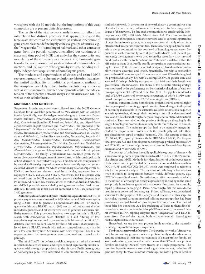
virosphere with the PL module, but the implications of this weakconnection are at present difficult to assess.
The results of the viral network analysis seem to reflect fourinterrelated but distinct processes that apparently shaped thelarge-scale structure of the virosphere: (i) vertical inheritance ofgene ensembles that define highly cohesive groups of viruses, e.g.,the “Megavirales,” (ii) sampling of hallmark and other connectorgenes from the partially compartmentalized but continuous inspace and time pool of MGE that underlies the connectivity andmodularity of the virosphere as a network, (iii) horizontal genetransfer between viruses that yields additional intermodule con-nections, and (iv) capture of host genes by viruses, including mul-tiple, independent acquisitions of homologous genes.
The modules and supermodules of viruses and related MGErepresent groups with coherent evolutionary histories that, giventhe limited applicability of traditional phylogenetic methods tothe virosphere, are likely to help further evolutionary studies aswell as virus taxonomy. Further developments could include ex-tension of the bipartite network analysis to all viruses and MGE aswell as their cellular hosts.
MATERIALS AND METHODSSequences. Protein sequences were collected from the NCBI GenomeDatabase for all available genomes of dsDNA viruses with an assignedfamily. Specifically, we collected genomes belonging to the orders Herpes-virales (families Herpesviridae, Alloherpesviridae, and Malacoherpesviri-dae), Caudovirales (families Siphoviridae, Podoviridae, and Myoviridae),Ligamenvirales (Lipothrixviridae and Rudiviridae), the proposed order“Megavirales” (families Ascoviridae, Asfarviridae, Iridoviridae, Marseille-viridae, Mimiviridae, Phycodnaviridae, and Poxviridae, as well as Pandora-virus and Pithovirus), the families Adenoviridae, Tectiviridae, Ampullaviri-dae, Bicaudaviridae, Corticoviridae, Fuselloviridae, Globuloviridae,Guttaviridae, Sphaerolipoviridae, Turriviridae, Baculoviridae, Nudiviridae,Hytrosaviridae, Nimaviridae, Papillomaviridae, Polyomaviridae, andPlasmaviridae, the genus Salterprovirus, and the available virophages(Lavidaviridae). The family Polydnaviridae was excluded given the ex-treme divergence of the genomes of these viruses, which consist primarilyof host-derived or inactivated viral genes. This data set was complementedby several additional groups of sequences from recently reported yet un-classified viruses or MGE for which evolutionary relationships with ds-DNA viruses have been demonstrated. In particular, sequences from vi-rophages YSLV5, YSLV6, and YSLV7, Mollivirus, and Faustovirus wereretrieved from the NCBI nonredundant protein database. Sequences ofPolintons and Polinto-like viruses, as well as mitochondrial and cytoplas-mic dsDNA plasmids, were added by using previously described customdata sets. In total, the initial data set contained 137,331 sequences from1,442 genomes.
Automatic classification of genes into homologous families. First, allprotein sequences were clustered at 90% identity and 70% coverage byusing CD-HIT (89) to generate a nonredundant data set. For each se-quence in this set, a BLASTp search (90) was carried out against all otherincluded sequences, and hit scores were used to generate a sequence sim-ilarity network. This procedure involved two steps: initially, a BLASTpsearch with composition-based statistics (91) and filtering of low-complexity regions was used to determine valid hits (e-val cutoff of 0.01,database size fixed to 2e7). Scores for those hits were subsequently col-lected from a BLASTp search with neither composition-based statisticsnor a low-complexity filter. Sequences with best (reciprocal) hits to othersequences from the same genome were combined and treated as in-paralogs.
The set of BLAST hits defines a weighted sequence similarity networkin which nodes are sequences and edges connect significantly similar se-quences, with a weight proportional to the hit score. Preliminary groupsof homologous genes were identified as communities in the sequence
similarity network. In the context of network theory, a community is a setof nodes that are densely interconnected compared to the average nodedegree of the network. To find such communities, we employed the Info-map software (92) (100 trials, 2-level hierarchy). The communities ofsequences in the sequence similarity network tend to constitute partitionsof larger homologous groups, with sequences from distantly related taxaoften located in separate communities. Therefore, we applied profile anal-ysis to merge communities that consisted of homologous sequences. Se-quences in each community were aligned with Muscle (93) (default pa-rameters); the alignments were used to predict secondary structure andbuild profiles with the tools “addss” and “hhmake” available within theHH-suite package (94). Profile-profile comparisons were carried out us-ing HHsearch (95). Hits were accepted or rejected based on their proba-bility, relative coverage, and length. Specifically, hits with a probabilitygreater than 0.90 were accepted if they covered at least 50% of the length ofthe profile; additionally, hits with a coverage of 20% or greater were alsoaccepted if their probability was greater than 0.99 and their length wasgreater than 100 amino acids. The choice of this heuristic filtering strategywas motivated by its performance on benchmark collections of viral or-thologous genes (POGs [9] and NCVOGs [37]). This pipeline rendered atotal of 33,980 clusters of homologous sequences, of which 11,950 com-prised multiple sequences and 22,030 were singletons (ORFans).
Manual curation. Some homologous proteins shared among highlydiverse groups of viruses (e.g., capsid proteins) have diverged to the pointof becoming inaccessible to the currently available automatable sequenceanalysis approaches, although their homology can still be demonstratedon a case-by-case basis, through analysis of sequence motifs and structuralsimilarity. Thus, we relied on the previous findings on these highly di-verged homologous proteins to manually curate our collection of homol-ogous sequences. The main groups that had to be manually merged in-cluded the major capsid proteins with the double jelly roll fold, theirassociated minor capsid proteins (pentons), Ulp1-like cysteine proteases(21, 60, 61, 96), capsid proteins with the HK97-like fold (50, 85), herpes-virus protease S21 and homologous bacteriophage prohead proteases U9and U35 (97), and the set of proteins shared among Baculoviridae, Hytro-saviridae, and Nimaviridae (57, 98).
The concept of orthology is readily applicable to groups of viruses withsimilar overall genome organization, such as the NCLDV or the Polinton-like viruses and MGE. Methods for identification of orthologous genesclusters have been implemented in the construction of databases such asPOGs (9, 35) and NCVOGs (36, 37), which were employed as referencesets for the present analysis. The meaning of orthology is less obviouswhen it comes to comparisons between widely different groups, e.g.,NCLDV versus Caudovirales. Nevertheless, an effort was made to adhereto the notion of orthology as closely as possible by including in the samegroup only homologous genes with analogous functions, for example,capsid proteins or packaging ATPases. Accordingly, hits that were due topromiscuous conserved domains, e.g., P-loop ATPases, were consideredspurious for the purpose of this analysis and discarded at this stage. Inparticular, manual curation involved splitting two groups that had beenerroneously merged based on profile-profile comparison. The first ofthose false hits connected A32-like packaging ATPases to thymidine ki-nases (both proteins contain the P-loop NTPase domain); the second falsehit involved mRNA capping enzymes from “Megavirales” and DNA li-gases from Caudovirales (again, both enzymes contain homologousnucleotidyltransferase domains).
Henceforth, we use the term protein family to refer to the manuallycurated groups of homologous sequences.
The bipartite network of viruses. The bipartite network of viruses wasbuilt by connecting genome nodes to protein family nodes whenever agenome contained at least one representative of a given protein family. Toavoid redundancy, genomes that shared more than 90% of their proteinfamilies (including ORFans) were treated as a single pangenome. Theresulting bipartite network contained a giant component, including allgenomes except for two Polintons which together with 5 protein families
Double-Stranded DNA Virus Network
July/August 2016 Volume 7 Issue 4 e00978-16 ® mbio.asm.org 17
on Decem
ber 12, 2019 at University Libraries | V
irginia Tech
http://mbio.asm
.org/D
ownloaded from
constituted their own minor components. For subsequent analyses, onlythe giant component was considered.
In the context of the bipartite network, a singleton is a protein familythat only appears in one genome (or the pan-genome of a set of 90%similar genomes).
Identification of core genes. We adopted an intuitive definition ofcore genes as those genes that tend to be maintained in a genome for longperiods of time, i.e., those with the lowest loss rates in a pure-loss evolu-tionary model. Let us consider a simple model in which, starting from acommon ancestor, the similarity at the family content level diverges uni-
formly in time according to the differential equationdSij
dt� �
Sij
�NiNj
,
where Sij is the number of families shared by genomes i and j and Ni and Nj
are the numbers of families in each genome. The geometric mean in thisexpression aims at biasing the denominator toward the smaller genome,thus avoiding artifacts caused by giant viruses (34); as it has been shownfor the “Megavirales,” similar results can be obtained with min(Ni, Nj) asthe normalization factor (33). This simple evolutionary model generates a
natural distance between any pair of genomes: Dij � � ln�Sij ⁄ �NiNj�.Although a rigorous derivation of this expression from the underlyingmodel requires that Ni and Nj remain constant, the same compositionalmetrics has been also applied to more general cases (e.g., analysis of evo-lutionary relationships within the “Megavirales”). The use of this metricsis additionally supported by the fact that the distance-based tree for the“Megavirales” that it generates shows a good correlation with the treeobtained from the concatenated sequences of the “Megavirales” universalcore genes (34). For a particular protein family, the probability that it ispresent in any pair of genomes conditioned on its presence in the lastcommon ancestor is P11 � e�r Dij⁄Z, where r is the loss rate of the familyrelative to the average divergence rate of genomes. Similarly, the probabil-ity for the family being lost in only one genome of the pair is P10 �2 e�rDij ⁄ 2�1 � e�rDij ⁄ 2� ⁄ Z, where Z � P10 � P11 is a normalization factor.Note that, in a pure loss model, the fact that one of the genomes containsa family implies that the last common ancestor also contained it. How-ever, pairs of genomes that lack any representative of a family have to bediscarded, because there is no guarantee that the family was present intheir common ancestor. We used the expressions for P10, P11, and Dij tocalculate a maximum likelihood estimate of the family-specific loss rate r.The presence of one or a few shared families in otherwise unrelated ge-nomes due to HGT could bias loss rate estimates, thus we only consideredthose pairs of genomes with distances Dij � 1. This condition implies thatboth genomes in the pair have to share more than 35% of their genecontent (relative to the size of the smaller genome), a degree of similaritythat is unlikely to be due to HGT only. Only those gene families with threeor more appearances were considered. Genes with relative loss rate r � 1were assigned to the “core.” Note that different values of this thresholdwould lead to slightly different lists of core genes. However, because thehallmark and signature genes that underlie the multiscale modular struc-ture of the virus network tend to be highly retained, the results are robustto the particular choice of the loss rate threshold. We additionally testedthe consistency of the core gene list by defining viral groups based on thenetwork modules (see below) and recalculating the loss rates separatelyfor each group. The aggregated list of core genes produced in this wayshowed a high agreement with the original one, and in particular it con-tained all hallmark and signature genes. The retention probability of agene family was calculated as exp(�r). This expression provides the prob-ability that a family has not been lost after one time unit, with time mea-sured in the characteristic time scale of genome composition divergence.
Gene family abundances were computed based on genome-weightedcontributions as previously described (99). The purpose of weighting thecontribution of a genome to the abundance of a gene is to compensate forsampling bias by assigning smaller weights to groups of closely relatedgenomes. Distances between pairs of genomes were calculated using thesame compositional metrics as described above. In the case of nonover-lapping genomes, a conservative estimate of the distance between them
was calculated as ln �1 � �NiNj�, which corresponds to the time thatwould take for an ancestor with one more gene to lose all but that onecommon gene. The genome-genome distance matrix was used to build aneighbor-joining tree, and genome weights were extracted from the treefollowing the previously described algorithm (99). Family abundanceswere obtained by adding the weights of all genomes where the family ispresent. Therefore, an abundance equal to 1 means that the family ispresent in all genomes of the data set. The prevalence, or relative abun-dance, of a gene family in a group of genomes was calculated as the sum ofweights of those genomes in the group that harbored the gene familydivided by the sum of weights of all the genomes in the group.
Detection of modules in the bipartite genome: core gene network.To detect sets of related genomes and gene families, we applied Barber’sdefinition of bipartite modularity (100) to the bipartite network consist-ing of genomes and core genes. Simulated annealing heuristics (101) wasused to identify the partition of the network that optimizes the magnitude
Q �1
L�i�G �j�F �aij � pij� ��mi, mj�, where L is the number of links in the
network, G and F represent the sets of genomes and core gene families,respectively, aij denotes the existence of a link between genome i and genefamily j, pij � kikj ⁄ L is the probability that a link exists by chance (with ki
and kj denoting the connectivities of nodes i and j), and � is the Kroneckerdelta function, which takes the value 1 if nodes i and j belong to the samemodule and the value 0 otherwise. The Modular software (102) was usedto carry out the modularity optimization. The significance of the resultingpartition was evaluated by running 100 replicates of a null model of arandomly generated bipartite network with the same size and the samegene- and genome-degree distributions as the original network. All theresults reported here correspond to partitions with a P value of �0.01.Because of the ruggedness of modularity landscapes in large, complexnetworks, the partition found by the module detection algorithm corre-sponds to only one of possibly many local maxima of modularity (34). Toaccount for this limitation, we ran 100 replicates (realizations) of thealgorithm and kept the partition with the highest modularity as the opti-mal partition. The robustness of the modules in the optimal partition wasevaluated by comparing them with the modules obtained in the other 99alternative partitions.
To detect supermodules, we analyzed higher-order bipartite networksconsisting of modules and connector gene families. A gene family was con-sidered a connector between two modules if its relative abundance (preva-lence) in both modules was greater than exp(�1). The choice of this thresholdwas motivated by its correspondence with the expected abundance of a genewith a loss rate equal to 1 after a characteristic unit of time of the gene contentdivergence process. Prevalence thresholds from 0.35 to 0.5 were also tested,with no qualitative differences. Gene families whose abundance exceeded thethreshold in a single module were also kept in the network, although theywere not classified as connectors. Supermodules were identified by applyingthe module detection algorithm to the module-level bipartite network. Thisprocess was iterated in order to delineate a hierarchy of higher-order mod-ules. The iterative search continued until no more mergers occurred or non-significant values of the modularity were obtained. As with primary modules,100 independent replicates were carried out in order to assess the robustnessof the supermodules.
The internal structures of some sets of modules (PL, “Megavirales,”and Baculovirus; PL alone; and modules that included Caudovirales) werefurther analyzed by extracting from the bipartite network a subnetworkconsisting of the genomes assigned to each particular set of modules andthe core genes connected (but not necessarily assigned) to them. Modulesand connector genes in each subnetwork were identified following thesame procedure as in the entire network.
Hallmark and signature genes. The intuitive idea of “hallmark genes”was formalized as follows: (i) a hallmark gene must be a connector be-tween first-order modules and (ii) it must be sufficiently prevalent in atleast one of the two major supermodules. In order to select a suitableprevalence threshold, we searched for gaps in the distribution of relative
Iranzo et al.
18 ® mbio.asm.org July/August 2016 Volume 7 Issue 4 e00978-16
on Decem
ber 12, 2019 at University Libraries | V
irginia Tech
http://mbio.asm
.org/D
ownloaded from

prevalences and found a noticeable discontinuity near 0.35, which ap-proximately agrees with the criterion used to define connector genes.Therefore, we adopted 0.35 as the minimum relative prevalence that aconnector gene must attain in (at least) one of the major supermodules tobe considered hallmark.
Signature genes were defined on the basis of their normalized mutualinformation with respect to their best and second best matching modules.For each gene and each module, their normalized mutual information
(MI) was calculated as MI � a log2
a
�a�b��a�c�� d log2
d
�c�d��b�d�,
where a, b, c, and d are the added relative weights of (a) the genomes fromthe module that contain the gene, (b) the genomes that contain the genebut do not belong to the module, (c) the genomes from the module thatlack the gene, and (d) the genomes that neither belong to the module norcontain the gene. Note that, compared to the traditional formulation ofthe mutual information, we did not take into account the terms associatedwith b and c. In doing so, we excluded the contributions from genes andmodules with complementary patterns. The mutual information was sub-sequently normalized by the joint entropy: H � � �i � �a,b,c,d� i log2i. Foreach gene, we selected the two highest values of the normalized mutualinformation (those that corresponded to the best and second best match-ing modules) and used the graphical representation in Fig. 6B to set the“signature” thresholds (�0.6 for the best match, �0.02 for the second bestmatch).
Bacteriophage lifestyles. Information on the temperate or virulentlifestyle of 171 tailed bacteriophages was collected from (103) (data areavailable at ACLAME website, http://aclame.ulb.ac.be/Classification/Phages/life_style.html). Of these phages, 57 were virulent and 114 weretemperate. The number of phages with available data is much smaller thanthe total number of phages in our network; therefore, we discarded thosemodules for which there were no data for at least 10% of its members. Forthe remaining modules, a binomial exact test was applied to estimate thesignificance of the lifestyle-module association under the null hypothesisthat module composition is random with respect to phage lifestyle.
SUPPLEMENTAL MATERIALSupplemental material for this article may be found at http://mbio.asm.org/lookup/suppl/doi:10.1128/mBio.00978-16/-/DCSupplemental.
Table S1, XLSX file, 0.1 MB.Table S2, XLSX file, 0.05 MB.Table S3, XLSX file, 0.02 MB.
ACKNOWLEDGMENTS
We thank David Kristensen, Sergey Mekhedov, and Natalya Yutin forexpert technical advice and Koonin group members for discussions.
E.V.K. incepted the study; J.I. developed the methods and performedresearch; J.I., M.K., and E.V.K. analyzed the results; J.I. and E.V.K. wrotethe manuscript, which was edited and approved by all authors.
FUNDING INFORMATIONThis work was funded by US Department of Health and Human Services(Intramural funds).
REFERENCES1. Edwards RA, Rohwer F. 2005. Viral metagenomics. Nat Rev Microbiol
3:504 –510. http://dx.doi.org/10.1038/nrmicro1163.2. Rohwer F. 2003. Global phage diversity. Cell 113:141. http://dx.doi.org/
10.1016/S0092-8674(03)00276-9.3. Rohwer F, Thurber RV. 2009. Viruses manipulate the marine environ-
ment. Nature 459:207–212. http://dx.doi.org/10.1038/nature08060.4. Suttle CA. 2005. Viruses in the sea. Nature 437:356 –361. http://
dx.doi.org/10.1038/nature04160.5. Suttle CA. 2007. Marine viruses—major players in the global ecosystem.
Nat Rev Microbiol 5:801– 812. http://dx.doi.org/10.1038/nrmicro1750.6. Fuhrman JA. 1999. Marine viruses and their biogeochemical and eco-
logical effects. Nature 399:541–548. http://dx.doi.org/10.1038/21119.
7. Hendrix RW. 2003. Bacteriophage genomics. Curr Opin Microbiol6:506 –511. http://dx.doi.org/10.1016/j.mib.2003.09.004.
8. Kristensen DM, Mushegian AR, Dolja VV, Koonin EV. 2010. Newdimensions of the virus world discovered through metagenomics.T r e n d s M i c r o b i o l 1 8 : 1 1 – 1 9 . h t t p : / / d x . d o i . o r g / 1 0 . 1 0 1 6 /j.tim.2009.11.003.
9. Kristensen DM, Waller AS, Yamada T, Bork P, Mushegian AR,Koonin EV. 2013. Orthologous gene clusters and taxon signature genesfor viruses of prokaryotes. J Bacteriol 195:941–950. http://dx.doi.org/10.1128/JB.01801-12.
10. Szathmáry E, Demeter L. 1987. Group selection of early replicators andthe origin of life. J Theor Biol 128:463– 486. http://dx.doi.org/10.1016/S0022-5193(87)80191-1.
11. Takeuchi N, Hogeweg P. 2007. The role of complex formation anddeleterious mutations for the stability of RNA-like replicator systems. JMol Evol 65:668 – 686. http://dx.doi.org/10.1007/s00239-007-9044-6.
12. Takeuchi N, Hogeweg P, Koonin EV. 2011. On the origin of DNAgenomes: evolution of the division of labor between template and cata-lyst in model replicator systems. PLoS Comput Biol 7:e1002024. http://dx.doi.org/10.1371/journal.pcbi.1002024.
13. Takeuchi N, Hogeweg P. 2012. Evolutionary dynamics of RNA-likereplicator systems: a bioinformatic approach to the origin of life. PhysLife Rev 9:219 –263. http://dx.doi.org/10.1016/j.plrev.2012.06.001.
14. Forterre P, Prangishvili D. 2009. The great billion-year war betweenribosome- and capsid-encoding organisms (cells and viruses) as the ma-jor source of evolutionary novelties. Ann N Y Acad Sci 1178:65–77.http://dx.doi.org/10.1111/j.1749-6632.2009.04993.x.
15. Forterre P, Prangishvili D. 2013. The major role of viruses in cellularevolution: facts and hypotheses. Curr Opin Virol 3:558 –565. http://dx.doi.org/10.1016/j.coviro.2013.06.013.
16. Koonin EV, Dolja VV. 2013. A virocentric perspective on the evolutionof life. Curr Opin Virol 3:546 –557. http://dx.doi.org/10.1016/j.coviro.2013.06.008.
17. Koonin EV, Senkevich TG, Dolja VV. 2006. The ancient virus world andevolution of cells. Biol Direct 1:29. http://dx.doi.org/10.1186/1745-6150-1-29.
18. Holmes EC. 2011. What does virus evolution tell us about virus origins?J Virol 85:5247–5251. http://dx.doi.org/10.1128/JVI.02203-10.
19. Koonin EV, Dolja VV. 2014. Virus world as an evolutionary network ofviruses and capsidless selfish elements. Microbiol Mol Biol Rev 78:278 –303. http://dx.doi.org/10.1128/MMBR.00049-13.
20. Yutin N, Raoult D, Koonin EV. 2013. Virophages, polintons, andtranspovirons: a complex evolutionary network of diverse selfish geneticelements with different reproduction strategies. Virol J 10:158. http://dx.doi.org/10.1186/1743-422X-10-158.
21. Krupovic M, Koonin EV. 2015. Polintons: a hotbed of eukaryotic virus,transposon and plasmid evolution. Nat Rev Microbiol 13:105–115.http://dx.doi.org/10.1038/nrmicro3389.
22. Dagan T. 2011. Phylogenomic networks. Trends Microbiol 19:483– 491.http://dx.doi.org/10.1016/j.tim.2011.07.001.
23. Alvarez-Ponce D, Lopez P, Bapteste E, McInerney JO. 2013. Genesimilarity networks provide tools for understanding eukaryote originsand evolution. Proc Natl Acad Sci U S A 110:E1594 –E1603. http://dx.doi.org/10.1073/pnas.1211371110.
24. Bapteste E, van Iersel L, Janke A, Kelchner S, Kelk S, McInerney JO,Morrison DA, Nakhleh L, Steel M, Stougie L, Whitfield J. 2013.Networks: expanding evolutionary thinking. Trends Genet 29:439 – 441.http://dx.doi.org/10.1016/j.tig.2013.05.007.
25. Forster D, Bittner L, Karkar S, Dunthorn M, Romac S, Audic S, LopezP, Stoeck T, Bapteste E. 2015. Testing ecological theories with sequencesimilarity networks: marine ciliates exhibit similar geographic dispersalpatterns as multicellular organisms. BMC Biol 13:16. http://dx.doi.org/10.1186/s12915-015-0125-5.
26. Corel E, Lopez P, Méheust R, Bapteste E. 2016. Network-thinking:graphs to analyze microbial complexity and evolution. Trends Microbiol24:224 –237. http://dx.doi.org/10.1016/j.tim.2015.12.003.
27. Popa O, Hazkani-Covo E, Landan G, Martin W, Dagan T. 2011.Directed networks reveal genomic barriers and DNA repair bypasses tolateral gene transfer among prokaryotes. Genome Res 21:599 – 609.http://dx.doi.org/10.1101/gr.115592.110.
28. Popa O, Dagan T. 2011. Trends and barriers to lateral gene transfer inprokaryotes. Curr Opin Microbiol 14:615– 623. http://dx.doi.org/10.1016/j.mib.2011.07.027.
Double-Stranded DNA Virus Network
July/August 2016 Volume 7 Issue 4 e00978-16 ® mbio.asm.org 19
on Decem
ber 12, 2019 at University Libraries | V
irginia Tech
http://mbio.asm
.org/D
ownloaded from

29. Jachiet PA, Colson P, Lopez P, Bapteste E. 2014. Extensive gene re-modeling in the viral world: new evidence for nongradual evolution inthe mobilome network. Genome Biol Evol 6:2195–2205. http://dx.doi.org/10.1093/gbe/evu168.
30. King AMQ, Lefkowitz E, Adams MJ, Carstens B (ed). 2011. Virustaxonomy: ninth report of the International Committee on Taxonomy ofViruses. Elsevier, Amsterdam, Netherlands.
31. Koonin EV, Dolja VV, Krupovic M. 2015. Origins and evolution ofviruses of eukaryotes: the ultimate modularity. Virology 479 – 480:2–25.http://dx.doi.org/10.1016/j.virol.2015.02.039.
32. Borgatti SP, Everett MG. 1997. Network analysis of 2-mode data. Soc Net-works 19:243–269. http://dx.doi.org/10.1016/S0378-8733(96)00301-2.
33. Montañez R, Medina MA, Solé RV, Rodríguez-Caso C. 2010. Whenmetabolism meets topology: reconciling metabolite and reaction net-works . BioEssays 32:246 –256. http : / /dx.doi .org/10.1002/bies.200900145.
34. Fortunato S. 2010. Community detection in graphs. Phys Rep 486:75–174. http://dx.doi.org/10.1016/j.physrep.2009.11.002.
35. Kristensen DM, Cai X, Mushegian A. 2011. Evolutionarily conservedorthologous families in phages are relatively rare in their prokaryotichosts. J Bacteriol 193:1806 –1814. http://dx.doi.org/10.1128/JB.01311-10.
36. Yutin N, Wolf YI, Raoult D, Koonin EV. 2009. Eukaryotic large nucleo-cytoplasmic DNA viruses: clusters of orthologous genes and reconstruc-tion of viral genome evolution. Virol J 6:223. http://dx.doi.org/10.1186/1743-422X-6-223.
37. Yutin N, Wolf YI, Koonin EV. 2014. Origin of giant viruses from smallerDNA viruses not from a fourth domain of cellular life. Virology466 – 467:38 –52. http://dx.doi.org/10.1016/j.virol.2014.06.032.
38. Lind PG, González MC, Herrmann HJ. 2005. Cycles and clustering inbipartite networks. Phys Rev E Stat Nonlin Soft Matter Phys 72(5 Pt2):056127. http://dx.doi.org/10.1103/PhysRevE.72.056127.
39. Barabási AL, Oltvai ZN. 2004. Network biology: understanding the cell’sfunctional organization. Nat Rev Genet 5:101–113. http://dx.doi.org/10.1038/nrg1272.
40. Koonin EV, Wolf YI. 2008. Genomics of bacteria and archaea: theemerging dynamic view of the prokaryotic world. Nucleic Acids Res 36:6688 – 6719. http://dx.doi.org/10.1093/nar/gkn668.
41. Koonin EV. 2011. The logic of chance: the nature and origin of biologicalevolution. FT Press, Upper Saddle River, NJ.
42. Haegeman B, Weitz JS. 2012. A neutral theory of genome evolution andthe frequency distribution of genes. BMC Genomics 13:196. http://dx.doi.org/10.1186/1471-2164-13-196.
43. Lobkovsky AE, Wolf YI, Koonin EV. 2013. Gene frequency distribu-tions reject a neutral model of genome evolution. Genome Biol Evol5:233–242. http://dx.doi.org/10.1093/gbe/evt002.
44. Hendrix RW, Hatfull GF, Smith MC. 2003. Bacteriophages with tails:chasing their origins and evolution. Res Microbiol 154:253–257. http://dx.doi.org/10.1016/S0923-2508(03)00068-8.
45. Krupovic M, Prangishvili D, Hendrix RW, Bamford DH. 2011.Genomics of bacterial and archaeal viruses: dynamics within the pro-karyotic virosphere. Microbiol Mol Biol Rev 75:610 – 635. http://dx.doi.org/10.1128/MMBR.00011-11.
46. Bamford DH. 2003. Do viruses form lineages across different domains oflife? Res Microbiol 154:231–236. http://dx.doi.org/10.1016/S0923-2508(03)00065-2.
47. Bamford DH, Grimes JM, Stuart DI. 2005. What does structure tell usabout virus evolution? Curr Opin Struct Biol 15:655– 663. http://dx.doi.org/10.1016/j.sbi.2005.10.012.
48. Krupovic M, Bamford DH. 2008. Virus evolution: how far does thedouble beta-barrel viral lineage extend? Nat Rev Microbiol 6:941–948.http://dx.doi.org/10.1038/nrmicro2033.
49. Krupovic M, Bamford DH. 2010. Order to the viral universe. J Virol84:12476 –12479. http://dx.doi.org/10.1128/JVI.01489-10.
50. Krupovic M, Bamford DH. 2011. Double-stranded DNA viruses: 20families and only five different architectural principles for virion assem-bly. Curr Opin Virol 1:118 –124. http://dx.doi.org/10.1016/j.coviro.2011.06.001.
51. Koonin EV, Wolf YI, Nagasaki K, Dolja VV. 2009. The complexity ofthe virus world. Nat Rev Microbiol 7:250. http://dx.doi.org/10.1038/nrmicro2030-c2.
52. Krupovic M, Bamford DH. 2009. Does the evolution of viral poly-
merases reflect the origin and evolution of viruses? Nat Rev Microbiol7:250. http://dx.doi.org/10.1038/nrmicro2030-c1.
53. Prangishvili D, Garrett RA, Koonin EV. 2006. Evolutionary genomicsof archaeal viruses: unique viral genomes in the third domain of life.Virus Res 117:52– 67. http://dx.doi.org/10.1016/j.virusres.2006.01.007.
54. Koonin EV, Yutin N. 2010. Origin and evolution of eukaryotic largenucleo-cytoplasmic DNA viruses. Intervirology 53:284 –292. http://dx.doi.org/10.1159/000312913.
55. Colson P, de Lamballerie X, Fournous G, Raoult D. 2012. Reclassifi-cation of giant viruses composing a fourth domain of life in the new orderMegavirales. Intervirology 55:321–332. http://dx.doi.org/10.1159/000336562.
56. Colson P, De Lamballerie X, Yutin N, Asgari S, Bigot Y, Bideshi DK,Cheng XW, Federici BA, Van Etten JL, Koonin EV, La Scola B, RaoultD. 2013. “Megavirales,” a proposed new order for eukaryotic nucleocy-toplasmic large DNA viruses. Arch Virol 158:2517–2521. http://dx.doi.org/10.1007/s00705-013-1768-6.
57. Wang Y, Bininda-Emonds ORP, Jehle JA. 2012. Nudivirus genomicsand phylogeny. In Rijeka GM (ed), Molecular structure, diversity, geneexpression mechanisms and host of virus interactions. InTechOpenhttp://dx.doi.org/10.5772/1346.
58. Wang Y, Jehle JA. 2009. Nudiviruses and other large, double-strandedcircular DNA viruses of invertebrates: new insights on an old topic. JInvertebr Pathol 101:187–193. http://dx.doi .org/10.1016/j.jip.2009.03.013.
59. Jehle JA, Abd-Alla AM, Wang Y. 2013. Phylogeny and evolution ofHytrosaviridae. J Invertebr Pathol 112(Suppl):S62–S67. http://dx.doi.org/10.1016/j.jip.2012.07.015.
60. Krupovic M, Bamford DH, Koonin EV. 2014. Conservation of majorand minor jelly-roll capsid proteins in Polinton (Maverick) transposonssuggests that they are bona fide viruses. Biol Direct 9:6. http://dx.doi.org/10.1186/1745-6150-9-6.
61. Yutin N, Shevchenko S, Kapitonov V, Krupovic M, Koonin EV. 2015.A novel group of diverse Polinton-like viruses discovered by metag-enome analysis. BMC Biol 13:95. http://dx.doi.org/10.1186/s12915-015-0207-4.
62. Yutin N, Kapitonov VV, Koonin EV. 2015. A new family of hybridvirophages from an animal gut metagenome. Biol Direct 10:19. http://dx.doi.org/10.1186/s13062-015-0054-9.
63. Pope WH, Bowman CA, Russell DA, Jacobs-Sera D, Asai DJ, CresawnSG, Jacobs WR, Hendrix RW, Lawrence JG, Hatfull GF, ScienceEducation Alliance Phage Hunters Advancing Genomics and Evolu-tionary Science, Phage Hunters Integrating Research and Education,Mycobacterial Genetics Course. 2015. Whole genome comparison of alarge collection of mycobacteriophages reveals a continuum of phagegenetic diversity. eLife 4:e06416. http://dx.doi.org/10.7554/eLife.06416.
64. Payne K, Sun Q, Sacchettini J, Hatfull GF. 2009. Mycobacteriophagelysin B is a novel mycolylarabinogalactan esterase. Mol Microbiol 73:367–381. http://dx.doi.org/10.1111/j.1365-2958.2009.06775.x.
65. Hendrix RW. 2009. Jumbo bacteriophages. Curr Top Microbiol Immu-nol 328:229 –240. http://dx.doi.org/10.1007/978-3-540-68618-7_7.
66. Davison AJ, Eberle R, Ehlers B, Hayward GS, McGeoch DJ, MinsonAC, Pellett PE, Roizman B, Studdert MJ, Thiry E. 2009. The orderHerpesvirales. Arch Virol 154:171–177. http://dx.doi.org/10.1007/s00705-008-0278-4.
67. Morais MC, Choi KH, Koti JS, Chipman PR, Anderson DL, RossmannMG. 2005. Conservation of the capsid structure in tailed dsDNAbacteriophages: the pseudoatomic structure of phi29. Mol Cell 18:149 –159. http://dx.doi.org/10.1016/j.molcel.2005.03.013.
68. Simpson AA, Tao Y, Leiman PG, Badasso MO, He Y, Jardine PJ, OlsonNH, Morais MC, Grimes S, Anderson DL, Baker TS, Rossmann MG.2000. Structure of the bacteriophage phi29 DNA packaging motor. Na-ture 408:745–750. http://dx.doi.org/10.1038/35047129.
69. Pawlowski A, Rissanen I, Bamford JK, Krupovic M, Jalasvuori M.2014. Gammasphaerolipovirus, a newly proposed bacteriophage genus,unifies viruses of halophilic archaea and thermophilic bacteria within thenovel family Sphaerolipoviridae. Arch Virol 159:1541–1554. http://dx.doi.org/10.1007/s00705-013-1970-6.
70. Rissanen I, Grimes JM, Pawlowski A, Mäntynen S, Harlos K, BamfordJK, Stuart DI. 2013. Bacteriophage P23-77 capsid protein structuresreveal the archetype of an ancient branch from a major virus lineage.Structure 21:718 –726. http://dx.doi.org/10.1016/j.str.2013.02.026.
71. Gil-Carton D, Jaakkola ST, Charro D, Peralta B, Castaño-Díez D,
Iranzo et al.
20 ® mbio.asm.org July/August 2016 Volume 7 Issue 4 e00978-16
on Decem
ber 12, 2019 at University Libraries | V
irginia Tech
http://mbio.asm
.org/D
ownloaded from

Oksanen HM, Bamford DH, Abrescia NG. 2015. Insight into the as-sembly of viruses with vertical single beta-barrel Major capsid proteins.Structure 23:1866 –1877. http://dx.doi.org/10.1016/j.str.2015.07.015.
72. Freeman LC. 1978. Centrality in social networks. I. Conceptual clarifi-cation. Soc Networks 1:215–239. http://dx.doi.org/10.1016/0378-8733(78)90021-7.
73. Hatfull GF. 2010. Mycobacteriophages: genes and genomes. Annu Rev Mi-c r o b i o l 6 4 : 3 3 1 – 3 5 6 . h t t p : / / d x . d o i . o r g / 1 0 . 1 1 4 6 /annurev.micro.112408.134233.
74. Fraser HB, Hirsh AE, Steinmetz LM, Scharfe C, Feldman MW. 2002.Evolutionary rate in the protein interaction network. Science 296:750 –752. http://dx.doi.org/10.1126/science.1068696.
75. Han JD, Bertin N, Hao T, Goldberg DS, Berriz GF, Zhang LV, DupuyD, Walhout AJ, Cusick ME, Roth FP, Vidal M. 2004. Evidence fordynamically organized modularity in the yeast protein-protein interac-tion network. Nature 430:88 –93. http://dx.doi.org/10.1038/nature02555.
76. Chang X, Xu T, Li Y, Wang K. 2013. Dynamic modular architecture ofprotein-protein interaction networks beyond the dichotomy of “date”and “party” hubs. Sci Rep 3:1691. http://dx.doi.org/10.1038/srep01691.
77. Kiran M, Nagarajaram HA. 2013. Global versus local hubs in humanprotein-protein interaction network. J Proteome Res 12:5436 –5446.http://dx.doi.org/10.1021/pr4002788.
78. Maumus F, Epert A, Nogue F, Blanc G. 2014. Plant genomes enclosefootprints of past infections by giant virus relatives. Nat Commun5:4268. http://dx.doi.org/10.1038/ncomms5268.
79. Spang A, Saw JH, Jørgensen SL, Zaremba-Niedzwiedzka K, Martijn J,Lind AE, van Eijk R, Schleper C, Guy L, Ettema TJ. 2015. Complexarchaea that bridge the gap between prokaryotes and eukaryotes. Nature521:173–179. http://dx.doi.org/10.1038/nature14447.
80. Koonin EV. 2015. Archaeal ancestors of eukaryotes: not so elusive anymore. BMC Biol 13:84. http://dx.doi.org/10.1186/s12915-015-0194-5.
81. Koonin EV. 2009. On the origin of cells and viruses: primordial virusworld scenario. Ann N Y Acad Sci 1178:47– 64. http://dx.doi.org/10.1111/j.1749-6632.2009.04992.x.
82. Jalasvuori M, Mattila S, Hoikkala V. 2015. Chasing the origin of viruses:capsid-forming genes as a life-saving preadaptation within a communityof early replicators. PLoS One 10:e0126094. http://dx.doi.org/10.1371/journal.pone.0126094.
83. Baker ML, Jiang W, Rixon FJ, Chiu W. 2005. Common ancestry ofherpesviruses and tailed DNA bacteriophages. J Virol 79:14967–14970.http://dx.doi.org/10.1128/JVI.79.23.14967-14970.2005.
84. Rochat RH, Liu X, Murata K, Nagayama K, Rixon FJ, Chiu W. 2011.Seeing the portal in herpes simplex virus type 1-B capsids. J Virol 85:1871–1874. http://dx.doi.org/10.1128/JVI.01663-10.
85. Rixon FJ, Schmid MF. 2014. Structural similarities in DNA packag-ing and delivery apparatuses in Herpesvirus and dsDNA bacterio-phages. Curr Opin Virol 5:105–110. http://dx.doi.org/10.1016/j.coviro.2014.02.003.
86. Krupovic M, Dutilh BE, Adriaenssens EM, et al. 2016. Taxonomy ofprokaryotic viruses: update from the ICTV bacterial and archaeal virusessubcommittee. Arch Virol 161:1095. http://dx.doi.org/10.1007/s00705-015-2728-0.
87. Lima-Mendez G, Van Helden J, Toussaint A, Leplae R. 2008. Reticulaterepresentation of evolutionary and functional relationships betweenphage genomes. Mol Biol Evol 25:762–777. http://dx.doi.org/10.1093/molbev/msn023.
88. Prangishvili D. 2015. Archaeal viruses: living fossils of the ancient viro-
sphere? Ann N Y Acad Sci 1341:35– 40. http://dx.doi.org/10.1111/nyas.12710.
89. Li W, Godzik A. 2006. Cd-hit: a fast program for clustering and com-paring large sets of protein or nucleotide sequences. BioInformatics 22:1658 –1659. http://dx.doi.org/10.1093/bioinformatics/btl158.
90. Altschul SF, Madden TL, Schäffer AA, Zhang J, Zhang Z, Miller W,Lipman DJ. 1997. Gapped BLAST and psi-BLAST: a new generation ofprotein database search programs. Nucleic Acids Res 25:3389 –3402.http://dx.doi.org/10.1093/nar/25.17.3389.
91. Schäffer AA, Aravind L, Madden TL, Shavirin S, Spouge JL, Wolf YI,Koonin EV, Altschul SF. 2001. Improving the accuracy of PSI-BLASTprotein database searches with composition-based statistics and otherrefinements. Nucleic Acids Res 29:2994 –3005. http://dx.doi.org/10.1093/nar/29.14.2994.
92. Rosvall M, Bergstrom CT. 2008. Maps of random walks on complexnetworks reveal community structure. Proc Natl Acad Sci U S A 105:1118 –1123. http://dx.doi.org/10.1073/pnas.0706851105.
93. Edgar RC. 2004. MUSCLE: multiple sequence alignment with high ac-curacy and high throughput. Nucleic Acids Res 32:1792–1797. http://dx.doi.org/10.1093/nar/gkh340.
94. Meier A, Söding J. 2015. Automatic prediction of protein 3D Structuresby probabilistic Multi-template homology modeling. PLoS Comput Biol11:e1004343. http://dx.doi.org/10.1371/journal.pcbi.1004343.
95. Söding J. 2005. Protein homology detection by HMM-HMM compari-son. Bioinformatics 21:951–960. http://dx.doi.org/10.1093/bioinformatics/bti125.
96. Maaty WS, Ortmann AC, Dlakic M, Schulstad K, Hilmer JK, LiepoldL, Weidenheft B, Khayat R, Douglas T, Young MJ, Bothner B. 2006.Characterization of the archaeal thermophile Sulfolobus turreted icosa-hedral virus validates an evolutionary link among double-stranded DNAviruses from all domains of life. J Virol 80:7625–7635. http://dx.doi.org/10.1128/JVI.00522-06.
97. Cheng H, Shen N, Pei J, Grishin NV. 2004. Double-stranded DNAbacteriophage prohead protease is homologous to herpesvirus protease.Protein Sci 13:2260 –2269. http://dx.doi.org/10.1110/ps.04726004.
98. Wang Y, Bininda-Emonds OR, van Oers MM, Vlak JM, Jehle JA. 2011.The genome of Oryctes rhinoceros nudivirus provides novel insight intothe evolution of nuclear arthropod-specific large circular double-stranded DNA viruses. Virus Genes 42:444 – 456. http://dx.doi.org/10.1007/s11262-011-0589-5.
99. Makarova KS, Wolf YI, Koonin EV. 2015. Archaeal clusters of ortholo-gous genes (arCOGs): an update and application for analysis of sharedfeatures between Thermococcales, Methanococcales, and Methanobac-teriales. Life (Basel) 5:818 – 840. http://dx.doi.org/10.3390/life5010818.
100. Barber MJ. 2007. Modularity and community detection in bipartite net-works. Phys Rev E Stat Nonlin Soft Matter Phys 76(6 Pt 2):066102. http://dx.doi.org/10.1103/PhysRevE.76.066102.
101. Guimerà R, Sales-Pardo M, Amaral LA. 2007. Module identification inbipartite and directed networks. Phys Rev E Stat Nonlin Soft Matter Phys76(3 Pt 2):036102. http://dx.doi.org/10.1103/PhysRevE.76.036102.
102. Marquitti FMD, Guimarães PR, Pires MM, Bittencourt LF. 2014.Modular: software for the autonomous computation of modularity inlarge network sets. Ecography 37:221–224. http://dx.doi.org/10.1111/j.1600-0587.2013.00506.x.
103. Lima-Mendez G, Toussaint A, Leplae R. 2007. Analysis of the phagesequence space: the benefit of structured information. Virology 365:241–249. http://dx.doi.org/10.1016/j.virol.2007.03.047.
Double-Stranded DNA Virus Network
July/August 2016 Volume 7 Issue 4 e00978-16 ® mbio.asm.org 21
on Decem
ber 12, 2019 at University Libraries | V
irginia Tech
http://mbio.asm
.org/D
ownloaded from
Related Documents